Embed Size (px)
Citation preview
Label-free imaging of Schwann cell myelination bythird harmonic generation microscopyHyungsik Lima,1, Denis Sharoukhova, Imran Kassimb, Yanqing Zhangc, James L. Salzerc,and Carmen V. Melendez-Vasquezb
Departments of aPhysics and bBiology, Hunter College of the City University of New York, New York, NY 10065; and cDepartment of Cell Biology andNeurology, New York University School of Medicine, NY 11203
Edited by Ben A. Barres, Stanford University School of Medicine, Stanford, CA, and approved November 11, 2014 (received for review September 15, 2014)
Understanding the dynamic axon–glial cell interaction underlyingmyelination is hampered by the lack of suitable imaging techni-ques. Here we demonstrate third harmonic generation microscopy(THGM) for label-free imaging of myelinating Schwann cells in liveculture and ex vivo and in vivo tissue. A 3D structure was acquiredfor a variety of compact and noncompact myelin domains, includingjuxtaparanodes, Schmidt–Lanterman incisures, and Cajal bands. Othersubcellular features of Schwann cells that escape traditional opticalmicroscopies were also visualized. We tested THGM for morphometryof compact myelin. Unlike current methods based on electron micros-copy, g-ratio could be determined along an extended length of my-elinated fiber in the physiological condition. The precision of THGM-based g-ratio estimation was corroborated in mouse models of hypo-myelination. Finally, we demonstrated the feasibility of THGM tomonitor morphological changes of myelin during postnatal develop-ment and degeneration. The outstanding capabilities of THGM maybe useful for elucidation of the mechanism of myelin formationand pathogenesis.
myelin | Schwann cell | multiphoton microscopy | label-free imaging |morphometry
Myelin is a multiple-layered membrane sheath surroundingthe axon. In myelinated nerves, the conduction of action
potentials is much faster and the speed depends strongly on thestructure of myelin. The structural integrity must be thereforetightly regulated for proper conduction of neuronal impulses, butthe underlying axon–glial cell interaction is not well understood.Since the days of Ramón y Cajal, light microscopy has beenwidely used to visualize myelin morphology (1). A variety offluorescent probes specifically binding to myelin componentshave allowed studies of the interaction between molecules (2–4).However, most such labeling methods are not suitable forunraveling in vivo dynamics of myelination because cell mem-branes are compromised during staining (especially immuno-histochemistry) and/or the procedures are prohibitively time-consuming and invasive. It is thus of great interest to developlabel-free methods. Recently, spectral confocal reflectance mi-croscopy has been demonstrated for high-resolution in vivo im-aging of myelin (5). There are also techniques of nonlinearoptical microscopy, which are generally known to be more ad-vantageous for imaging deep live tissue. Coherence anti-StokesRaman scattering (CARS) microscopy, which requires two syn-chronized short pulse lasers for excitation, has been used forimaging in vivo myelin and detecting pathology (6, 7). Thirdharmonic generation (THG) microscopy is based on anothernonlinear optical process of light emission, yielding distinguish-able images from CARS. Though it has been demonstrated forimaging the white matter in the brain (8), so far few studies haveapplied THG microscopy (THGM) for elucidating the mechanismof myelin formation. Moreover, the omission of the peripheralnervous systems (PNS) in the previous studies is not trivial con-sidering the significant departure from the CNS in terms of themyelin-forming glia and molecular subdomains: Schwann cellswrap individual internodes in the PNS, and oligodendrocytes form
multiple myelin sheaths in the CNS. The basal lamina and nodalmicrovilli are unique to Schwann cells, and Schmidt–Lantermanincisures are more pronounced in the PNS (9). Here we demon-strate, to our knowledge for the first time, the utility of THGM asa method for morphological analysis of myelinating Schwann cellsin live culture and ex vivo and in vivo tissue.
ResultsMyelin Is the Primary Source of THG Emission. THG emission arisesfrom the myelin sheath due to the discontinuous refractive indexat the interface of aqueous media and membrane lipids. Becausediscontinuity of the same kind exists in the axonal membranes aswell, we asked how specific the THG emission is to myelinatedvs. unmyelinated axons. To this end, we established myelinatingcocultures of dorsal root ganglion (DRG) and Schwann cells.Live-cell THGM imaging was performed at 14 d in vitro (DIV);at this time, cultures contain a mix of myelinated and un-myelinated axons (Fig. 1). The emission showed the opticalproperties characteristic of THG. The intensity of signal variedwhen the polarization of excitation beam was rotated, yieldingnear extinction for orthogonal polarizations relative to the ori-entation of fibers. THG emission was highly directional, propa-gating predominantly into the forward direction (or transmission)rather than backward. Sparse elongated segments were observedwith lengths between 60 and 160 μm, indicative of myelinatingSchwann cells (Fig. 1A). The intensity of signal from the mye-linated segments was ∼5× higher than the background of un-myelinated DRG neurites.
Significance
Myelin, a specialized membrane wrapping around the axon, iscrucial for proper neuronal information processing. We de-scribe a label-free imaging method, i.e., third harmonic gen-eration microscopy (THGM), for visualization of Schwann cellmyelination in live culture and ex vivo and in vivo tissue. THGMis capable of imaging compact myelin as well as non–compactsubcellular domains, including nodal segments, Schmidt–Lanterman incisures, and Cajal bands, thus attractive forelucidating their functional roles; it also facilitates image-basedmorphometry, allowing the g-ratio of myelinated fibers tobe measured in physiological conditions. The demonstratedcapabilities of THGM are valuable for investigating the in-tricate interaction between axon and myelin-forming glial cellunderlying formation and maintenance of myelin in the pe-ripheral nervous system.
Author contributions: H.L. and C.V.M.-V. designed research; H.L., D.S., I.K., and C.V.M.-V.performed research; Y.Z. and J.L.S. contributed new reagents/analytic tools; H.L., D.S., andC.V.M.-V. analyzed data; and H.L., J.L.S., and C.V.M.-V. wrote the paper.
The authors declare no conflict of interest.
This article is a PNAS Direct Submission.1To whom correspondence should be addressed. Email: [email protected].
This article contains supporting information online at www.pnas.org/lookup/suppl/doi:10.1073/pnas.1417820111/-/DCSupplemental.
www.pnas.org/cgi/doi/10.1073/pnas.1417820111 PNAS | December 16, 2014 | vol. 111 | no. 50 | 18025–18030
NEU
ROSC
IENCE
ENGINEE
RING
To confirm that the THG signal indeed originated frommyelinated fibers, we prepared two groups of DRG neuronscultured for the same period (2 wk), with or without Schwanncells. The elongated segments emitting strong THG were ob-served only in the myelinating coculture (Fig. 1B), verifying thatTHG arises from the ensheathed axons and also that the sensi-tivity of THGM was sufficiently high to detect single myelinatingaxons. It was also possible to find the locations of heminodes andnodes at the junction of neighboring internodes (Fig. 1C). Theability to localize the nascent nodes of Ranvier could be usefulfor studying the time course of nodal development during myelinformation.
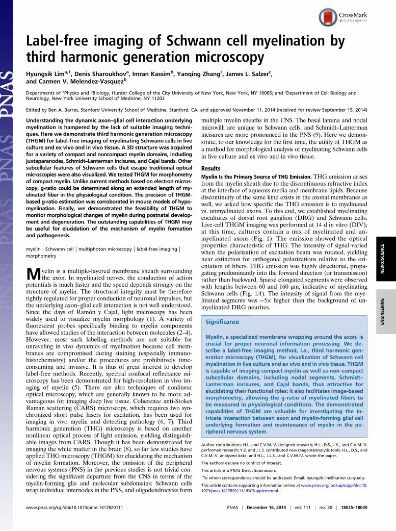
Myelin Subdomains Are Visualized by THGM. Production of THGsignal is confined within a tightly focused laser beam, facilitatingvisualization of the 3D structure of myelinated axons. In ourexperiments, the theoretical FWHM diameter of the excitationlaser focus was 0.4 μm and 1.4 μm in the lateral and axial dimen-sions, respectively. The high resolution of THGM suggested that itmight enable imaging the subcellular structure of myelinated axons.We therefore sought to identify specific subdomains of myelin-ated peripheral nerves, including the paranodes and Schmidt–Lanterman incisures. We used transgenic mice whose myelindomains were labeled with fluorescent proteins expressed underspecific promoters; coregistration of simultaneously acquired two-photon excited fluorescence (TPEF) and THG permitted definitiveidentification of the THG features. To make the identification moreevident, we used isolated myelinated axons obtained from teasingfresh sciatic nerves. All data presented in this report were acquiredwithout fixation. The first transgenic mouse used was a Rosa26-mT/mG (10), a reporter line that expresses tandem dimer Tomato(tdTomato) at axonal and glial cell membranes. Substantial overlapof tdTomato and THG signals was found along the continuousmembranes, verifying the membranes as the origin of THG (Fig. 2 Aand B). The lateral resolution of THGM was sufficiently high todiscriminate the adaxonal (inner) and abaxonal (outer) membranesof compact myelin. Schmidt–Lanterman incisures were observable inboth tdTomato and THG channels (Fig. 2A, arrowheads), whereasthe nodal axolemma was detected only as an interrupted THG lining(Fig. 2B, arrow). To validate the localization of the nodes with re-spect to the THG signal, we used a transgenic mouse expressingneurofascin 186 (NF186), a nodal cell-adhesion molecule, taggedwith EGFP and expressed under the control of Thy-1.2 promoter(11). The myelinated axons of the NF186 animal showed that EGFPsignal was diffuse and weak around Schmidt–Lanterman incisureswhere NF186 do not cluster (Fig. 2C), but strong EGFP fluores-cence arising from the nodal axolemma coincided with the gap inTHGM, confirming that the node of Ranvier is located at theinterrupted THG membranes (Fig. 2D). These results show thatTHGM is capable of distinguishing major domains such as thenodes and incisures.The spatial resolution of THGM was further tested for visu-
alizing the detailed morphology of subdomains using freshly
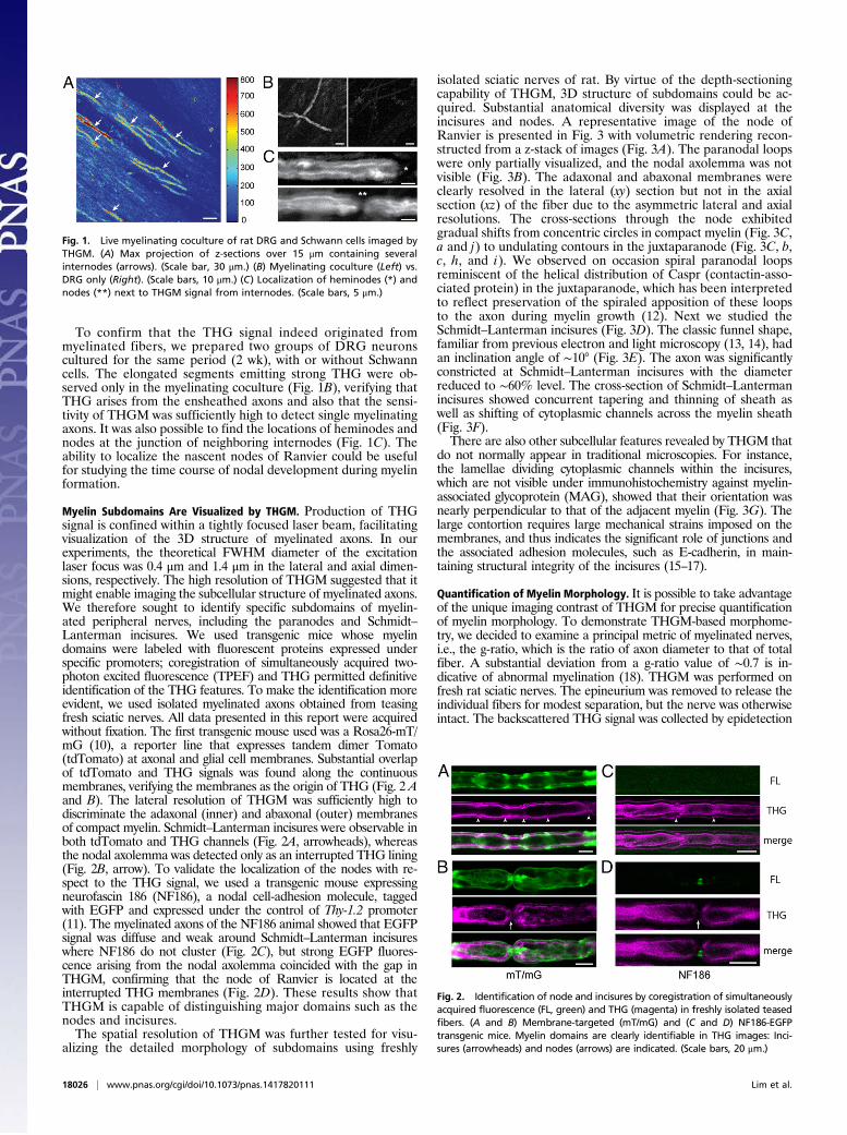
isolated sciatic nerves of rat. By virtue of the depth-sectioningcapability of THGM, 3D structure of subdomains could be ac-quired. Substantial anatomical diversity was displayed at theincisures and nodes. A representative image of the node ofRanvier is presented in Fig. 3 with volumetric rendering recon-structed from a z-stack of images (Fig. 3A). The paranodal loopswere only partially visualized, and the nodal axolemma was notvisible (Fig. 3B). The adaxonal and abaxonal membranes wereclearly resolved in the lateral (xy) section but not in the axialsection (xz) of the fiber due to the asymmetric lateral and axialresolutions. The cross-sections through the node exhibitedgradual shifts from concentric circles in compact myelin (Fig. 3C,a and j) to undulating contours in the juxtaparanode (Fig. 3C, b,c, h, and i). We observed on occasion spiral paranodal loopsreminiscent of the helical distribution of Caspr (contactin-asso-ciated protein) in the juxtaparanode, which has been interpretedto reflect preservation of the spiraled apposition of these loopsto the axon during myelin growth (12). Next we studied theSchmidt–Lanterman incisures (Fig. 3D). The classic funnel shape,familiar from previous electron and light microscopy (13, 14), hadan inclination angle of ∼10° (Fig. 3E). The axon was significantlyconstricted at Schmidt–Lanterman incisures with the diameterreduced to ∼60% level. The cross-section of Schmidt–Lantermanincisures showed concurrent tapering and thinning of sheath aswell as shifting of cytoplasmic channels across the myelin sheath(Fig. 3F).There are also other subcellular features revealed by THGM that
do not normally appear in traditional microscopies. For instance,the lamellae dividing cytoplasmic channels within the incisures,which are not visible under immunohistochemistry against myelin-associated glycoprotein (MAG), showed that their orientation wasnearly perpendicular to that of the adjacent myelin (Fig. 3G). Thelarge contortion requires large mechanical strains imposed on themembranes, and thus indicates the significant role of junctions andthe associated adhesion molecules, such as E-cadherin, in main-taining structural integrity of the incisures (15–17).
Quantification of Myelin Morphology. It is possible to take advantageof the unique imaging contrast of THGM for precise quantificationof myelin morphology. To demonstrate THGM-based morphome-try, we decided to examine a principal metric of myelinated nerves,i.e., the g-ratio, which is the ratio of axon diameter to that of totalfiber. A substantial deviation from a g-ratio value of ∼0.7 is in-dicative of abnormal myelination (18). THGM was performed onfresh rat sciatic nerves. The epineurium was removed to release theindividual fibers for modest separation, but the nerve was otherwiseintact. The backscattered THG signal was collected by epidetection
Fig. 1. Live myelinating coculture of rat DRG and Schwann cells imaged byTHGM. (A) Max projection of z-sections over 15 μm containing severalinternodes (arrows). (Scale bar, 30 μm.) (B) Myelinating coculture (Left) vs.DRG only (Right). (Scale bars, 10 μm.) (C) Localization of heminodes (*) andnodes (**) next to THGM signal from internodes. (Scale bars, 5 μm.)
Fig. 2. Identification of node and incisures by coregistration of simultaneouslyacquired fluorescence (FL, green) and THG (magenta) in freshly isolated teasedfibers. (A and B) Membrane-targeted (mT/mG) and (C and D) NF186-EGFPtransgenic mice. Myelin domains are clearly identifiable in THG images: Inci-sures (arrowheads) and nodes (arrows) are indicated. (Scale bars, 20 μm.)
18026 | www.pnas.org/cgi/doi/10.1073/pnas.1417820111 Lim et al.
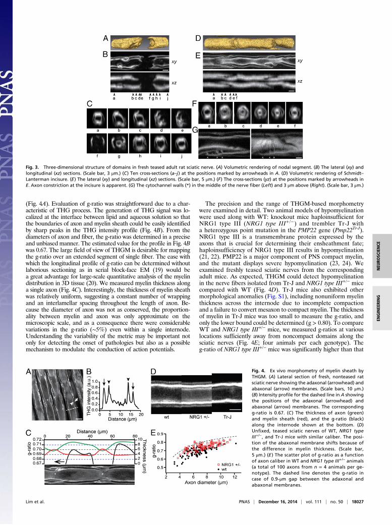
(Fig. 4A). Evaluation of g-ratio was straightforward due to a char-acteristic of THG process. The generation of THG signal was lo-calized at the interface between lipid and aqueous solution so thatthe boundaries of axon and myelin sheath could be easily identifiedby sharp peaks in the THG intensity profile (Fig. 4B). From thediameters of axon and fiber, the g-ratio was determined in a preciseand unbiased manner. The estimated value for the profile in Fig. 4Bwas 0.67. The large field of view of THGM is desirable for mappingthe g-ratio over an extended segment of single fiber. The ease withwhich the longitudinal profile of g-ratio can be determined withoutlaborious sectioning as in serial block-face EM (19) would bea great advantage for large-scale quantitative analysis of the myelindistribution in 3D tissue (20). We measured myelin thickness alonga single axon (Fig. 4C). Interestingly, the thickness of myelin sheathwas relatively uniform, suggesting a constant number of wrappingand an interlamellar spacing throughout the length of axon. Be-cause the diameter of axon was not as conserved, the proportion-ality between myelin and axon was only approximate on themicroscopic scale, and as a consequence there were considerablevariations in the g-ratio (∼5%) even within a single internode.Understanding the variability of the metric may be important notonly for detecting the onset of pathologies but also as a possiblemechanism to modulate the conduction of action potentials.
The precision and the range of THGM-based morphometrywere examined in detail. Two animal models of hypomyelinationwere used along with WT: knockout mice haploinsufficient forNRG1 type III (NRG1 type III+ /−) and trembler Tr-J witha heterozygous point mutation in the PMP22 gene (Pmp22Tr-J).NRG1 type III is a transmembrane protein expressed by theaxons that is crucial for determining their ensheathment fate;haploinsufficiency of NRG1 type III results in hypomyelination(21, 22). PMP22 is a major component of PNS compact myelin,and the mutant displays severe hypomyelination (23, 24). Weexamined freshly teased sciatic nerves from the correspondingadult mice. As expected, THGM could detect hypomyelinationin the nerve fibers isolated from Tr-J and NRG1 type III+/− micecompared with WT (Fig. 4D). Tr-J mice also exhibited othermorphological anomalies (Fig. S1), including nonuniform myelinthickness across the internode due to incomplete compactionand a failure to convert mesaxon to compact myelin. The thicknessof myelin in Tr-J mice was too small to measure the g-ratio, andonly the lower bound could be determined (g > 0.80). To compareWT and NRG1 type III+/− mice, we measured g-ratios at variouslocations sufficiently away from noncompact domains along thesciatic nerves (Fig. 4E; four animals per each genotype). Theg-ratio of NRG1 type III+/− mice was significantly higher than that
Fig. 3. Three-dimensional structure of domains in fresh teased adult rat sciatic nerve. (A) Volumetric rendering of nodal segment. (B) The lateral (xy) andlongitudinal (xz) sections. (Scale bar, 3 μm.) (C) Ten cross-sections (a–j) at the positions marked by arrowheads in A. (D) Volumetric rendering of Schmidt–Lanterman incisure. (E) The lateral (xy) and longitudinal (xz) sections. (Scale bar, 5 μm.) (F) The cross-sections (yz) at the positions marked by arrowheads inE. Axon constriction at the incisure is apparent. (G) The cytochannel walls (*) in the middle of the nerve fiber (Left) and 3 μm above (Right). (Scale bar, 3 μm.)
Fig. 4. Ex vivo morphometry of myelin sheath byTHGM. (A) Lateral section of fresh, nonteased ratsciatic nerve showing the adaxonal (arrowhead) andabaxonal (arrow) membranes. (Scale bars, 10 μm.)(B) Intensity profile for the dashed line in A showingthe positions of the adaxonal (arrowhead) andabaxonal (arrow) membranes. The correspondingg-ratio is 0.67. (C ) The thickness of axon (green)and myelin sheath (red), and the g-ratio (black)along the internode shown at the bottom. (D)Unfixed, teased sciatic nerves of WT, NRG1 typeIII+/−, and Tr-J mice with similar caliber. The posi-tion of the abaxonal membrane shifts because ofthe difference in myelin thickness. (Scale bar,5 μm.) (E ) The scatter plot of g-ratio as a functionof axon caliber in WT and NRG1 type III+/− animals(a total of 100 axons from n = 4 animals per ge-notype). The dashed line denotes the g-ratio incase of 0.9-μm gap between the adaxonal andabaxonal membranes.
Lim et al. PNAS | December 16, 2014 | vol. 111 | no. 50 | 18027
NEU
ROSC
IENCE
ENGINEE
RING
of WT (0.75 ± 0.005 vs. 0.69 ± 0.01, mean ± SEM; P = 0.0016,t test), which is consistent with the previous EM measurements(21). The result verifies the capability of THGM-based mor-phometry to detect modest hypomyelination. The measuredg-ratio was lower for axons of smaller calibers, which could be dueto a limit in the measurement imposed by the resolution ofTHGM. The g-ratio could not be evaluated when the spacingbetween the adaxonal and abaxonal boundaries is much smallerthan the optical resolution, resulting in underestimation of theparameter more significant for thinner axons. The upper boundfor the measured g-ratio values corresponded to ∼0.9-μm spacing(dashed line in Fig. 4E). The apparent limit in the measurablemyelin thickness was a little larger than the FWHM diameter ofthe diffraction-limited focus, presumably because of degradationin the resolution due to specimen-induced optical aberrations.
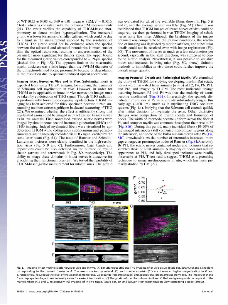
Imaging Intact Nerves ex Vivo and in Vivo. Substantial merit isexpected from using THGM imaging for studying the dynamicsof Schwann cell myelination in vivo. However, in order forTHGM to be applicable to intact in vivo nerves, the images mustbe taken by epidetection of THG signal. Though THG radiationis predominantly forward-propagating, epidetection THGM im-aging has been achieved for thick specimen because turbid sur-rounding medium causes significant backward-scattering of THG(25). We examined whether the effect is sufficiently strong thatmyelinated axons could be imaged in intact excised tissues as wellas in live animals. First, nonteased excised sciatic nerves wereimaged by simultaneous second harmonic generation (SHG) andTHG imaging. Indeed myelinated fibers were visualized by epi-detection THGM while collagenous endoneurium and perineu-rium were simultaneously recorded via SHG signal excited by thesame laser beam (Fig. 5A). The node of Ranvier and Schmidt–Lanterman incisures were clearly identified in the high-resolu-tion views (Fig. 5 B and C). Furthermore, Cajal bands andappositions could be also detected on the surface of myelinsheath (arrows and arrowheads in Fig. 5D, respectively). Theability to image these domains in intact nerves is attractive forelucidating their functional roles (26). We tested the feasibility ofTHGM-based g-ratio measurement for intact tissues. The g-ratio
was evaluated for all of the available fibers shown in Fig. 5 Band C, and the average g-ratio was 0.62 (Fig. 5F). Once it wasestablished that THGM images of intact nerves ex vivo could beacquired, we then performed in vivo THGM imaging of sciaticnerve using live mice. Although the brightness of the imagesobtained was comparable to the ex vivo conditions, the resolu-tion of imaging was degraded by motion artifacts, and subcellulardetails could not be resolved even with image registration (Fig.5G). The movement of nerves as much as a few micrometers persecond, especially in the axial direction, was sufficient to con-found g-ratio analysis. Nevertheless, it was possible to visualizenodes and incisures in living mice (Fig. 5G, arrow). Suitablemethods to immobilize in vivo tissue could further improve theoverall image quality.
Imaging Postnatal Growth and Pathological Myelin. We examinedthe utility of THGM for studying developing myelin. Rat sciaticnerves were harvested from animals (n = 3) at P2, P4, P6, P11,and P18, and imaged by THGM. The most noticeable changeoccurring between P2 and P4 was that the majority of axonsbecome myelinated (Fig. S1A). Interestingly, the sparsely dis-tributed internodes at P2 were already substantially long at thisearly age (∼100 μm), much as in myelinating DRG coculturesystems (Fig. 1A), implying that the Schwann cell extends quicklyafter initial decision to myelinate the axon. Other distinctivechanges were compaction of myelin sheath and formation ofnodes. The width of internode became uniform across the fiber atP4, and compact myelin was common throughout the nerve at P6(Fig. S1B). During this period, many individual fibers (10–20% ofthe imaged internodes) still contained noncompact regions alongthe internode, and some of the bulbs remained even after P6 (Fig.S1C, arrowheads). As the number of internodes increased, moregaps emerged as presumptive nodes of Ranvier (Fig. S1D, arrows).By P11, the sciatic nerves contained nodes and incisures that re-sembled those of adult animals. A majority of nodes had matureappearance at P11, and fully developed incisures were readilyobservable at P18. These results suggest THGM as a promisingtechnique to image myelinogenesis in situ, which has been pri-marily studied by EM (27).
Fig. 5. Imaging-intact murine sciatic nerves ex vivo and in vivo. (A) Simultaneous SHG and THG imaging of ex vivo tissue. (Scale bar, 50 μm.) (B and C) Regionscorresponding to the colored frames in A. The axons marked by asterisk (*) and double asterisks (**) are shown at higher magnification in D andE, respectively, focused at the level of the abaxonal membrane. Cajal bands (red arrowheads) and appositions (green arrows) are visible. The images of D andE are displayed on logarithmic intensity scales for easier identification. (F) The g-ratio of the fibers shown in B and C. Red and green points correspond to themarked fibers in B and C, respectively. (G) Imaging of in vivo tissue. (Scale bar, 30 μm.) (Lower) High-magnification view containing a node (arrow).
18028 | www.pnas.org/cgi/doi/10.1073/pnas.1417820111 Lim et al.
We also studied whether THGM can detect demyelination. Tomodel demyelination in vitro, we treated myelinating coculturesat 14 d after ascorbate addition with glial growth factor (GGF;200 ng/mL), an isoform of neuregulin 1 known to induce de-myelination (28). As a control, a group of cells was cultured forthe same periods without GGF treatment. Cells imaged withTHGM after 3 and 7 d of GGF treatment are depicted in Fig. S2D and E, respectively. Various patterns of deformation werevisible in the cells affected by GGF, which confirms that theorigin of THG is the myelin sheath. Disrupted myelin sheathswere prominent after 3 d, and extensive demyelination wasapparent after 7 d.
DiscussionElectron and light microscopy have long been the methods ofchoice for visualizing myelin morphology (9). Myelin structurespertaining to physiology, from the interlamellar spacing to theinternode length, cannot be captured by a single technique be-cause the dimension spans over seven orders of magnitude.Moreover, even a single nerve bundle contains hundreds ofaxons confounding the determination of a microscopic biometric(e.g., the g-ratio) for a population of peripheral nerves. Thesubmicrometer resolution and wide field of view (∼1 mm2) ofTHGM facilitates rapid surveillance of compact and noncompactmyelin subdomains over many internodes, and also any gradientfrom proximal to distal nerves relative to the spinal cord. Ex-amination of peripheral nerves could be greatly simplified bymeans of label-free THGM avoiding elaborate procedures forfixation, labeling, or embedding. The removal of fixation anddisruptive staining, as well as the freedom from photobleaching,is crucial for studying the native dynamics of myelin formationand impairment in live cells.We have shown that THG signal is specific to myelin sheath
and provides a previously unidentified imaging contrast for vi-sualizing diverse myelin domains in living animals, includingSchmidt–Lanterman incisures, node of Ranvier, and Cajal bands.The demonstrated capability is highly beneficial for investigatingthe in vivo dynamics of domains. We have also shown the utilityof THG for estimation of g-ratio. The current method of g-ratiomeasurement is based on EM sections lacking 3D spatial re-lationship, so the parameter is evaluated only at a single pointalong the axon. To overcome the limitation and also to make g-ratio estimation more convenient, alternative methods usinglight microscopy have been developed but with limited success.The use of Luxol fast blue stain in conjunction with wide-fieldmicroscopy does not yield accurate results. CARS microscopywas demonstrated for evaluation of g-ratio (6). However, theidentification of axon and fiber boundaries is ambiguous, re-quiring an arbitrary definition of myelin thickness because CARSsignal arises from the bulk of the myelin sheath. Recently de-veloped spectral confocal reflectance microscopy is inadequatefor precise determination of g-ratio because the reflection occursprimarily at the top and bottom surfaces of myelin sheath (5). Bycontrast, THGM images the entire axial sections (Fig. 3) and thesensitivity to the interfaces enables unambiguous and bias-freeevaluation of the g-ratio. The demonstrated g-ratio measure-ment relies on peak detection, a computationally cheap androbust process, so it is amenable to automated morphometricanalysis. Furthermore, the longitudinal map of g-ratio can beacquired without having to prepare thin sections. Using THGMmorphometry, we have found significant variations in g-ratiowithin a single internode.Although pseudocrystalline compact myelin is conducive to
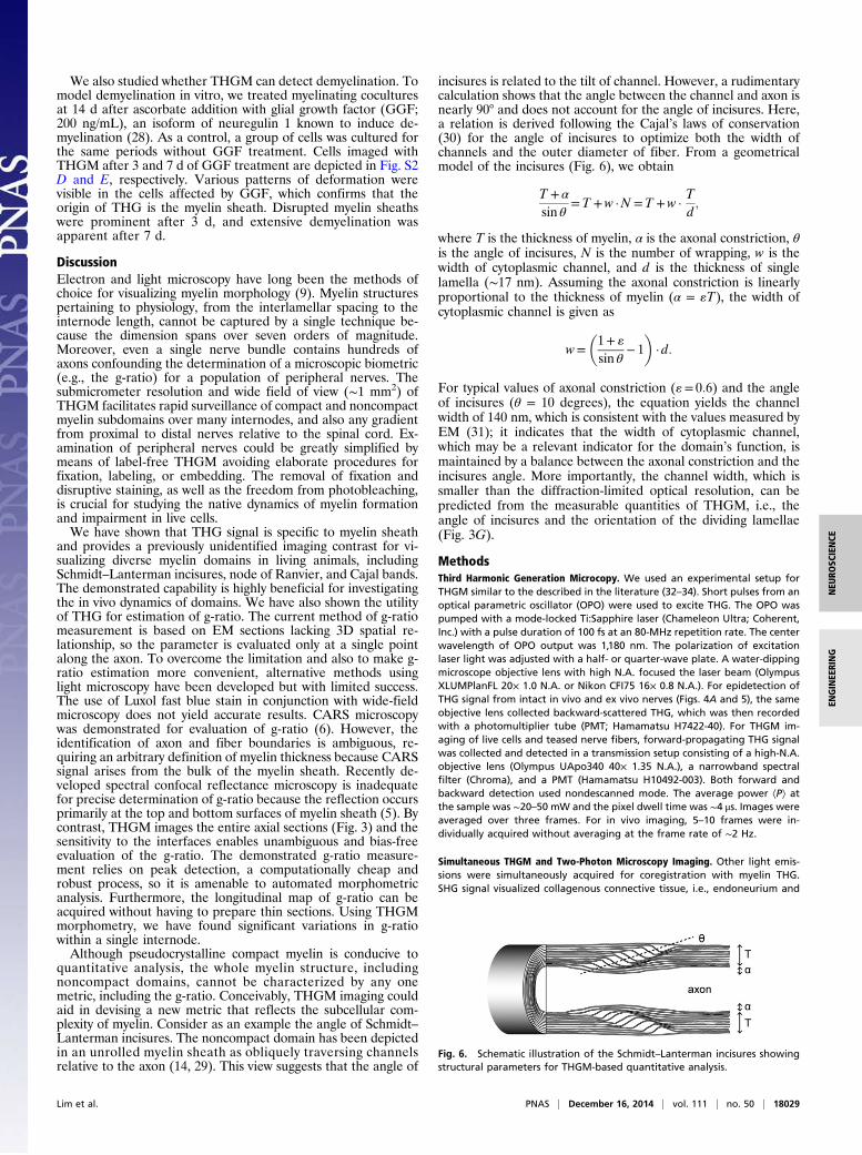
quantitative analysis, the whole myelin structure, includingnoncompact domains, cannot be characterized by any onemetric, including the g-ratio. Conceivably, THGM imaging couldaid in devising a new metric that reflects the subcellular com-plexity of myelin. Consider as an example the angle of Schmidt–Lanterman incisures. The noncompact domain has been depictedin an unrolled myelin sheath as obliquely traversing channelsrelative to the axon (14, 29). This view suggests that the angle of
incisures is related to the tilt of channel. However, a rudimentarycalculation shows that the angle between the channel and axon isnearly 90° and does not account for the angle of incisures. Here,a relation is derived following the Cajal’s laws of conservation(30) for the angle of incisures to optimize both the width ofchannels and the outer diameter of fiber. From a geometricalmodel of the incisures (Fig. 6), we obtain
T + α
sin θ=T +w ·N =T +w ·
Td;
where T is the thickness of myelin, α is the axonal constriction, θis the angle of incisures, N is the number of wrapping, w is thewidth of cytoplasmic channel, and d is the thickness of singlelamella (∼17 nm). Assuming the axonal constriction is linearlyproportional to the thickness of myelin (α = «T), the width ofcytoplasmic channel is given as
w=�1+ «
sin θ− 1
�· d:
For typical values of axonal constriction («= 0:6) and the angleof incisures (θ = 10 degrees), the equation yields the channelwidth of 140 nm, which is consistent with the values measured byEM (31); it indicates that the width of cytoplasmic channel,which may be a relevant indicator for the domain’s function, ismaintained by a balance between the axonal constriction and theincisures angle. More importantly, the channel width, which issmaller than the diffraction-limited optical resolution, can bepredicted from the measurable quantities of THGM, i.e., theangle of incisures and the orientation of the dividing lamellae(Fig. 3G).
MethodsThird Harmonic Generation Microcopy. We used an experimental setup forTHGM similar to the described in the literature (32–34). Short pulses from anoptical parametric oscillator (OPO) were used to excite THG. The OPO waspumped with a mode-locked Ti:Sapphire laser (Chameleon Ultra; Coherent,Inc.) with a pulse duration of 100 fs at an 80-MHz repetition rate. The centerwavelength of OPO output was 1,180 nm. The polarization of excitationlaser light was adjusted with a half- or quarter-wave plate. A water-dippingmicroscope objective lens with high N.A. focused the laser beam (OlympusXLUMPlanFL 20× 1.0 N.A. or Nikon CFI75 16× 0.8 N.A.). For epidetection ofTHG signal from intact in vivo and ex vivo nerves (Figs. 4A and 5), the sameobjective lens collected backward-scattered THG, which was then recordedwith a photomultiplier tube (PMT; Hamamatsu H7422-40). For THGM im-aging of live cells and teased nerve fibers, forward-propagating THG signalwas collected and detected in a transmission setup consisting of a high-N.A.objective lens (Olympus UApo340 40× 1.35 N.A.), a narrowband spectralfilter (Chroma), and a PMT (Hamamatsu H10492-003). Both forward andbackward detection used nondescanned mode. The average power ÆPæ atthe sample was ∼20–50 mW and the pixel dwell time was ∼4 μs. Images wereaveraged over three frames. For in vivo imaging, 5–10 frames were in-dividually acquired without averaging at the frame rate of ∼2 Hz.
Simultaneous THGM and Two-Photon Microscopy Imaging. Other light emis-sions were simultaneously acquired for coregistration with myelin THG.SHG signal visualized collagenous connective tissue, i.e., endoneurium and
Fig. 6. Schematic illustration of the Schmidt–Lanterman incisures showingstructural parameters for THGM-based quantitative analysis.
Lim et al. PNAS | December 16, 2014 | vol. 111 | no. 50 | 18029
NEU
ROSC
IENCE
ENGINEE
RING
perineurium, whereas TPEF from EGFP and tdTomato revealed subcellularfeatures. SHG was excited by the same OPO output beam as THG anddetected after a narrowband spectral filter at half the excitationwavelength (λ =590 nm). For TPEF, a separate mode-locked Ti:Sapphire laser (Tsunami; Spectra-Physics, Inc.) was used with the excitation wavelength at 850 nm and 880 nm forEGFP and tdTomato, respectively. For simultaneous THGM and TPEF imaging,the laser beam was combined into the optical path using a dichroic filter(Chroma 950DC). The SHG and TPEF signals were acquired by epidetection.
Image Processing and Quantification of Myelin. Myelin morphology wasquantified via image processing using ImageJ (National Institutes of Health)andMATLAB (MathWorks, Inc.). Volumetric reconstruction was created usingAmira (VSG). For estimation of g-ratio, automatic peak detection was per-formed on THG intensity profile across myelinated axon to objectively deter-mine the boundaries of the myelin sheath. G-ratio was calculated from theobtained diameters of fiber and axon. Statistical analyses of the measuredg-ratiowere performed using Prism (GraphPad, Inc.). Density ofmyelin duringpostnatal growth was quantified. At each time point, a total of six nerveregions of interest (ROIs) with an area of 114 × 114 μm2 were selected fromthree animals. Each ROI was converted into a binary image by automaticthresholding, which distinguished myelin from nonmyelin. The density ofmyelin was obtained by counting the number of nonzero pixels and thenaveraging over six ROIs.
Animals. Sprague–Dawley rats were obtained from Charles River Laborato-ries. Transgenic mT/mG [Gt(ROSA)26Sortm4(ACTB-tdTomato,-EGFP)] and myelinmutant trembler (Pmp22Tr-J) mice were obtained from Jackson Laboratories.
Myelinating Schwann Cell Culture for Live Cell Imaging. Myelinating coculturesystem was prepared as described (35). DRG neurons were isolated from E16rat spinal cords and maintained in serum-free neurobasal (NB) medium (2%B27 supplement, 2 mM L-glutamine, 0.4% glucose, and 50 ng/mL 2.5S NGF).Nonneuronal cells were removed by feeding the cultures with NB mediumcontaining 5-fluorodeoxyuridine and uridine. Schwann cells were isolated
from postnatal day 2 sciatic and expanded for ∼3 wk in D media (10% FBS,and 2 mM L-glutamine) supplemented with 4 μM forskolin and 5 ng/mL ofthe EGF domain of rhNRG-1-β1 (R&D Systems). Myelinating cocultures wereestablished by seeding purified DRG neuron cultures with 100,000 Schwanncells in C media (10% FBS, 2 mM L-glutamine, 0.4% glucose, and 50 ng/mL2.5S NGF). After 3 d, cocultures were changed to media supplemented with50 μg/mL ascorbic acid to initiate basal lamina formation and myelination.For demyelination experiment mature myelinating cocultures (14–21 d)were treated with GGF (200 ng/mL) for 2–7 d, a treatment that is known tocause demyelination in vitro (28).
Preparation of Teased Nerve Fiber. Sciatic nerves were harvested from miceand rats of specified ages. Fresh (unfixed) sciatic nerves were teased using fineneedles in ice-cold Dulbecco’s PBS. The teased nerve fibers were then trans-ferred to a glass-bottom dish (MatTek) and held down with a slice anchor(Warner Instruments) for imaging.
Animal Surgery for in Vivo Imaging. Animal surgery for in vivo imaging ofsciatic nerves was performed as previously described (5, 36, 37). The pro-cedure was approved by the Hunter College Institutional Animal Care andUse Committee. Animals were anesthetized by isoflurane inhalation andplaced on a temperature-controlled heating pad. The hind leg was shavedand sterilized with betadine. A small incision was made on the skin. Thevastus lateralis and biceps femoris muscles were separated using retractors.The sciatic nerve was exposed and gently lifted with a thin spatula in-troduced underneath the nerve. The position of spatula was adjusted usinga micromanipulator (Narishige) to orient the sciatic nerve relative to theexcitation laser beam and to mitigate breathing artifacts. After imaging, theanimal was euthanized.
ACKNOWLEDGMENTS. We thank Tomasz Rusielewicz and Matt Urbanski forhelp with preparation of the sciatic nerve samples. This work was funded byNational Institutes of Health Grants GM096884 and MD007599 (to H.L.),NS26001 (to J.L.S.), and NS000001 (to C.M.-V.).
1. Ramón y Cajal S (1991) Degeneration and Regeneration of the Nervous System(Oxford Univ Press, London).
2. Condie AG, Gerson SL, Miller RH, Wang Y (2012) Two-photon fluorescent imagingof myelination in the spinal cord. ChemMedChem 7(12):2194–2203.
3. Romanelli E, et al. (2013) Cellular, subcellular and functional in vivo labeling of thespinal cord using vital dyes. Nat Protoc 8(3):481–490.
4. Reynolds RJ, Little GJ, Lin M, Heath JW (1994) Imaging myelinated nerve fibres byconfocal fluorescence microscopy: Individual fibres in whole nerve trunks tracedthrough multiple consecutive internodes. J Neurocytol 23(9):555–564.
5. Schain AJ, Hill RA, Grutzendler J (2014) Label-free in vivo imaging of myelinatedaxons in health and disease with spectral confocal reflectance microscopy. Nat Med20(4):443–449.
6. Wang H, Fu Y, Zickmund P, Shi R, Cheng JX (2005) Coherent anti-stokes Ramanscattering imaging of axonal myelin in live spinal tissues. Biophys J 89(1):581–591.
7. Bélanger E, et al. (2012) Live animal myelin histomorphometry of the spinal cord withvideo-rate multimodal nonlinear microendoscopy. J Biomed Opt 17(2):021107.
8. Farrar MJ, Wise FW, Fetcho JR, Schaffer CB (2011) In vivo imaging of myelin in thevertebrate central nervous system using third harmonic generation microscopy. Bio-phys J 100(5):1362–1371.
9. Peters A, Palay SL, Webster HD (1991) Fine Structure of the Nervous System: Neuronsand Their Supporting Cells (Oxford Univ Press, London).
10. Muzumdar MD, Tasic B, Miyamichi K, Li L, Luo L (2007) A global double-fluorescentCre reporter mouse. Genesis 45(9):593–605.
11. Zhang Y, et al. (2012) Assembly and maintenance of nodes of ranvier rely on distinctsources of proteins and targeting mechanisms. Neuron 73(1):92–107.
12. Pedraza L, Huang JK, Colman D (2009) Disposition of axonal caspr with respect to glialcell membranes: Implications for the process of myelination. J Neurosci Res 87(15):3480–3491.
13. Bunge MB, Bunge RP, Peterson ER, Murray MR (1967) A light and electron microscopestudy of long-term organized cultures of rat dorsal root ganglia. J Cell Biol 32(2):439–466.
14. Ghabriel MN, Allt G (1981) Incisures of Schmidt–Lanterman. Prog Neurobiol 17(1-2):25–58.
15. Fannon AM, et al. (1995) Novel E-cadherin-mediated adhesion in peripheral nerve:Schwann cell architecture is stabilized by autotypic adherens junctions. J Cell Biol129(1):189–202.
16. Poliak S, Matlis S, Ullmer C, Scherer SS, Peles E (2002) Distinct claudins and associatedPDZ proteins form different autotypic tight junctions in myelinating Schwann cells.J Cell Biol 159(2):361–372.
17. Tricaud N, Perrin-Tricaud C, Brusés JL, Rutishauser U (2005) Adherens junctions inmyelinating Schwann cells stabilize Schmidt–Lanterman incisures via recruitment ofp120 catenin to E-cadherin. J Neurosci 25(13):3259–3269.
18. Lazzarini RA (2004) Myelin Biology and Disorders (Elsevier, San Diego).19. Denk W, Horstmann H (2004) Serial block-face scanning electron microscopy to recon-
struct three-dimensional tissue nanostructure. PLoS Biol 2(11):e329.20. Tomassy GS, et al. (2014) Distinct profiles of myelin distribution along single axons of
pyramidal neurons in the neocortex. Science 344(6181):319–324.21. Taveggia C, et al. (2005) Neuregulin-1 type III determines the ensheathment fate of
axons. Neuron 47(5):681–694.22. Michailov GV, et al. (2004) Axonal neuregulin-1 regulates myelin sheath thickness.
Science 304(5671):700–703.23. Suter U, et al. (1992) Trembler mouse carries a point mutation in a myelin gene.
Nature 356(6366):241–244.24. Notterpek L, Shooter EM, Snipes GJ (1997) Upregulation of the endosomal-lysosomal
pathway in the trembler-J neuropathy. J Neurosci 17(11):4190–4200.25. Débarre D, Olivier N, Beaurepaire E (2007) Signal epidetection in third-harmonic
generation microscopy of turbid media. Opt Express 15(14):8913–8924.26. Court FA, et al. (2004) Restricted growth of Schwann cells lacking Cajal bands slows
conduction in myelinated nerves. Nature 431(7005):191–195.27. Webster HD (1971) The geometry of peripheral myelin sheaths during their formation
and growth in rat sciatic nerves. J Cell Biol 48(2):348–367.28. Zanazzi G, et al. (2001) Glial growth factor/neuregulin inhibits Schwann cell myeli-
nation and induces demyelination. J Cell Biol 152(6):1289–1299.29. Arroyo EJ, Scherer SS (2000) On the molecular architecture of myelinated fibers.
Histochem Cell Biol 113(1):1–18.30. Ramón y Cajal S (1995) Histology of the Nervous System of Man and Vertebrates
(Oxford Univ Press, London).31. Hall SM, Williams PL (1970) Studies on the “incisures” of Schmidt and Lanterman.
J Cell Sci 6(3):767–791.32. Yelin D, Silberberg Y (1999) Laser scanning third-harmonic-generation microscopy in
biology. Opt Express 5(8):169–175.33. Squier J, Muller M, Brakenhoff G, Wilson KR (1998) Third harmonic generation mi-
croscopy. Opt Express 3(9):315–324.34. Débarre D, et al. (2006) Imaging lipid bodies in cells and tissues using third-harmonic
generation microscopy. Nat Methods 3(1):47–53.35. Einheber S, et al. (1997) The axonal membrane protein Caspr, a homologue of neu-
rexin IV, is a component of the septate-like paranodal junctions that assemble duringmyelination. J Cell Biol 139(6):1495–1506.
36. Williams PL, Hall SM (1970) In vivo observations on mature myelinated nerve fibres ofthe mouse. J Anat 107(Pt 1):31–38.
37. Gonzalez S, Fernando RN, Perrin-Tricaud C, Tricaud N (2014) In vivo introduction oftransgenes into mouse sciatic nerve cells in situ using viral vectors. Nat Protoc 9(5):1160–1169.
18030 | www.pnas.org/cgi/doi/10.1073/pnas.1417820111 Lim et al.


![Oligodendroglial myelination requires astrocyte … 5...Accordingly, genetic impairment of endogenous lipid synthesis in Schwann cells (SC) interferes with the acute phase of PNS myelination[5]](https://img.pdfslide.net/doc/110x75/5ca0fba988c9932f098b64ec/oligodendroglial-myelination-requires-astrocyte-5accordingly-genetic-impairment.jpg)