Embed Size (px)
Citation preview

J. clin. Path. (1968), 21, 656-660
Labial salivary gland biopsy in Sjogren's diseaseD. M. CHISHOLM AND D. K. MASON
From the Department of Oral Medicine, University of Glasgow Dental Hospital and School, Glasgow
syNoPsis A labial biopsy technique is described and was used to study 40 patients with connectivetissue disease and 60 postmortem subjects. More than one focus of lymphocytes per 4 sq mm ofminor salivary tissue was found to be a consistent finding in patients with Sjogren's disease. Thelabial biopsy is shown to be a further valuable investigative procedure in such patients.
Sjogren's disease, first described in 1933 (Sjogren,1933), consists of chronic inflammation of thelacrimal and salivary glands leading to dryness ofthe eye (keratoconjunctivitis sicca) and dryness ofthe mouth (xerostomia), and in a proportion ofcases, lacrimal and/or salivary gland enlargementmay also be present. In 50 to 60% of patients thedisease may be associated with a connective tissuedisorder, usually rheumatoid arthritis but occasion-ally also with polymyositis, polyarteritis nodosa,progressive systemic sclerosis (scleroderma) andsystemic lupus erythematosus. The term 'siccacomplex' is applied to those cases of Sjogren'sdisease not associated with rheumatoid arthritis orother connective tissue disease. In Sjogren's diseasea preponderance of female patients, especially inthe older age group, is consistently noted.The histopathological characters of the major
salivary glands in Sjogren's disease include par-enchymal and ductal alterations. There is a decreaseor disappearance of acini, lymphocytic infiltrationand hyperplasia of the lining cells of the intra-glandular ducts. The formation of epimyoepithelialcell islands has also been described (Morgan andCastleman, 1953) and appears to be a late featureof the disease process.Though it would be of considerable value to
obtain a major salivary gland biopsy in patientswith Sj6gren's disease, in the majority of cases thiscannot be justified because of the inconvenience topatients and the possible complication of salivaryfistula. In addition, because of their small size andtheir relationship to important neurovascularanatomical structures, needle biopsy is unsatisfactory.
Recently, however, in studies on children withfibrocystic disease, Warwick, Bernard, and Meskin(1964) have reported involvement of the labialmucous salivary glands. Furthermore, Calman andReifman (1966) have reported the involvement ofReceived for publication 26 February 1968.
the minor buccal glands in one patient with Sj6gren'ssyndrome, and Cifarelli, Bennett, and Zaino (1966),Cahn (1967), and Bertram (1967) have described apatient with this condition in which the minorpalatal salivary glands showed the characteristicSjogren's histopathology.The aims of the present study were:1 To investigate the histopathological appear-
ances of the minor salivary glands in Sjogren'sdisease, rheumatoid arthritis, and various otherconnective tissue diseases.
2 To determine the postmortem appearances ofthe minor salivary glands, suitable exclusions havingbeen made (Waterhouse and Doniach, 1966).
3 To assess the value of the minor salivary glandbiopsy to other methods of investigating salivarygland involvement in Sjogren's disease. Theseinclude sialography, measurement of salivary flowrate, and salivary duct antibody findings.
MATERIALS AND METHODS
PATIENTS Forty patients were studied. The clinicaldiagnosis, sex distribution, and mean age are shownin Table II.The diagnosis of Sjogren's disease was based on the
criteria described by Bloch, Buchanan, Wohl, and Bunim(1965) and patients were required to have at least twoof the three major components of the syndrome. Thediagnosis of rheumatoid arthritis was based on thecriteria of the American Rheumatism Association(Ropes, Bennett, Cobb, Jacox, and Jessar, 1958). Theophthalmological examination was performed by themethod described by Williamson, Cant, Mason, Greig,and Boyle (1967).
ORAL EXAMINATION Each patient was carefully ques-tioned regarding a history of xerostomia and of associatedoral and pharyngeal symptoms of Sjogren's disease. Inthe 10 patients with Sjogren's disease, salivary flowstudies were performed using a modified Carlson-Crittenden cup with an outer chamber diameter of20 mm and an inner chamber diameter of 10 mm.
656
copyright. on 17 O
ctober 2018 by guest. Protected by
http://jcp.bmj.com
/J C
lin Pathol: first published as 10.1136/jcp.21.5.656 on 1 S
eptember 1968. D
ownloaded from
Labial salivary gland biopsy in Sjogren's disease
Parotid saliva was collected from each patient underresting conditions and after stimulation with fruit gumsand lemon juice. In addition, sialography was performedon these patients using the hydrostatic techniquedescribed by Park and Mason (1966).
SALIVARY DUCT ANTIBODY The method described byMacSween, Goudie, Anderson, Armstrong, Murray,Mason, Jasani, Boyle, Buchanan, and Williamson (1967)was used to detect the presence or absence of salivaryduct antibody in the sera of all 40 patients studied.
MINOR SALIVARY GLAND BiOPSY The minor salivaryglands of the lower lip were chosen for biopsy since theyare easily accessible, they lie above the muscle layer,separated from the mucous membrane by a thin layer offibrous connective tissue and the chance of excessivebleeding is minimal, for the arterial supply to the lip isdeep to the gland (Meskin, Bernard, and Warwick, 1964).With the patient seated in the dental chair and after
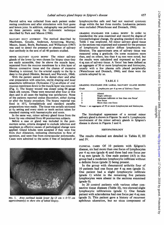
oral preparation with cetavlon, sterile draping and localanaesthetic infiltration (Xylocaine 2 %) an ellipse of oralmucous membrane down to the muscle layer was removed(Fig. 1). The biopsy wound was closed using 04 gaugeblack silk sutures. These were removed after four or fivedays and in all cases the healing was satisfactory. Noneof the patients reported undue discomfort, either duringor after the biopsy procedure. The biopsy material wasfixed in 10% formaldehyde and standard paraffinpreparations were made. The tissue was serially sectionedat 6,u setting and every ninth or tenth section taken forstandard staining with haematoxylin and eosin.
In the same way, minor salivary gland tissue from thelower lip was obtained from 60 postmortem subjects.
Before a case or gland was included in the post-mortem series, criteria designed to exclude infection andneoplasm as effective causes of pathological change wereapplied. Gland lobules were accepted if they were freefrom duct dilatation, indicating obstruction to flow ofsecretion, and were free from extravascular polymorphs.Cases were admitted to the series if free of neoplasm of
FIG. 1. Area outlined inside lower lip (3 cm x 075 cm
approximately) to show site of labial biopsy.
lymphocyte-like cells and had not received cytotoxicdrugs within the last three months. Leukaemia patientswere excluded (Waterhouse and Doniach, 1966).
GRADING STANDARD FOR LABIAL BIOPSY In order tostandardize the area examined and record the degree ofhistopathological change, the grading standard shown inTable I was employed. All minor salivary gland tissuein the sections was examined and scanned for the presenceof lymphocytic foci and/or diffuse lymphocytic in-filtration. The approximate area of salivary tissue wasmeasured using a graticule. On some occasions morethan, and on others, less than 4 sq mm were noted, butthe results were calculated and expressed as foci per4 sq mm of salivary tissue. A 'focus' has been defined asan aggregate of 50 or more lymphocytes and histiocytes,usually with a few plasma cells placed peripherally(Waterhouse and Doniach, 1966), and these were thecriteria adopted by us.
TABLE IGRADING STANDARD FOR LABIAL SALIVARY GLAND BIOPSY
Grade Lymphocytes per 4 sq mm of Salivary Tissue
0
234
AbsentSlight infiltrateModerate infiltrate or less than one focus'One focusMore than one focus
'Focus = an aggregate of 50 or more lymphocytes and histiocytes.
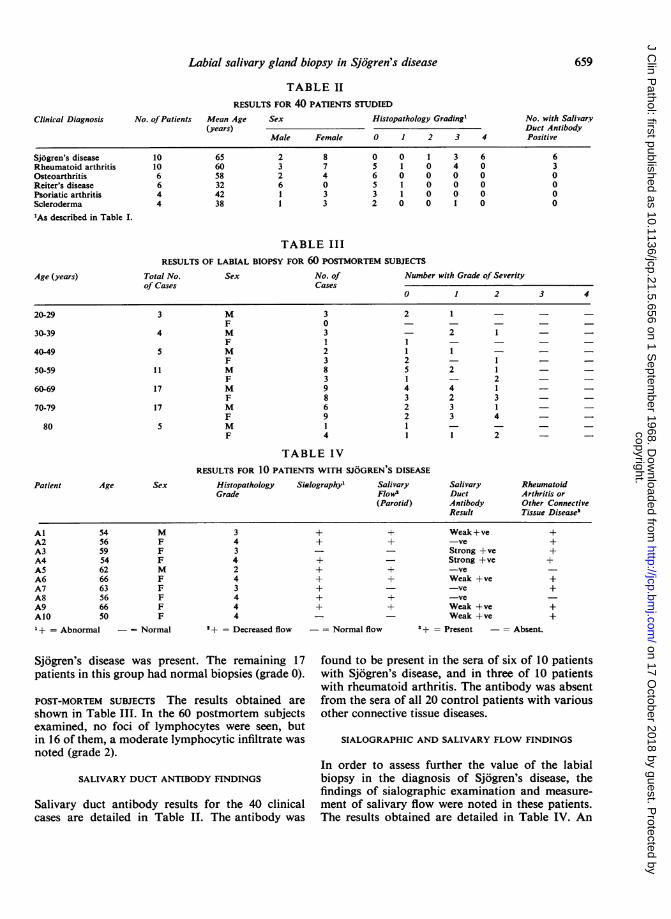
The histological appearance of a normal minorsalivary gland is shown in Figures 2a and b. Lymphocyticinvolvement of the minor salivary glands in Sjogren'sdisease is shown in Figures 3 and 4.
HISTOPATHOLOGY
The results obtained are detailed in Tables II, HIand IV.
CLINICAL CASES Of 10 patients with Sjogren'sdisease, six had more than one focus of lymphocytesper 4 sq mm (grade 4) and three had one focus per4 sq mm (grade 3). One male patient (AS) in thisgroup had a moderate lymphocytic infiltrate withouta definite focus (grade 2) being present.
In the group with rheumatoid arthritis four of10 patients had one focus per 4 sq mm (grade 3).One patient had a slight lymphocytic infiltrate(grade 1) whilst in the remaining five patientslymphocytes were absent in the sections examined(grade 0).
In 20 control patients with various other con-nective tissue diseases (Table II), two showed slightlymphocytic infiltrations (grade 1). One femalepatient with scleroderma had one focus per 4 sq mm(grade 3). This patient gave a history of recurrentaphthous ulceration, but no sicca component of
657
copyright. on 17 O
ctober 2018 by guest. Protected by
http://jcp.bmj.com
/J C
lin Pathol: first published as 10.1136/jcp.21.5.656 on 1 S
eptember 1968. D
ownloaded from

4r:
/ t C .
FIG. 2a. Photomicrograph showing proximity of labial FIG. 2b. Photomicrograph showing normal appearancesmucous glands to mucosal surface. Stained H. & E. x 54. of labial mucous glands. x 144.
I..- , ,, 4#o>t'W:,>P
S.a.-C.j;;^ 2 :"
FIG. 3. FIG. 4.
FIG. 3. Focal involvement of labial mucous glands: a periductal focus of lymphocytes. Note also the associateddiffuse lymphocytic infiltrate. x 54.FIG. 4. Focal lymphocytic involvement of minor salivary gland in Sjogren's disease. x 144.
copyright. on 17 O
ctober 2018 by guest. Protected by
http://jcp.bmj.com
/J C
lin Pathol: first published as 10.1136/jcp.21.5.656 on 1 S
eptember 1968. D
ownloaded from
659Labial salivary gland biopsy in Sjdgren's disease
TABLE II
Clinical Diagnosis
RESULTS FOR 40 PATIENTS STUDIED
No. ofPatients Mean Age Sex Histop(years)
pathology Grading' No. with SalivaryDuct Antibody
Male Female 0 1 2 3 4 Positive
Sjogren's disease 10 65 2 8 0 0 1 3 6 6Rheumatoid arthritis 10 60 3 7 5 1 0 4 0 3Osteoarthritis 6 58 2 4 6 0 0 0 0 0Reiter's disease 6 32 6 0 5 1 0 0 0 0Psoriatic arthritis 4 42 1 3 3 1 0 0 0 0Scleroderma 4 38 1 3 2 0 0 1 0 0
'As described in Table I.
TABLE IIIRESULTS OF LABIAL BIOPSY FOR 60 POSTMORTEM SUBJECTS
Age (years) Total No. Sex No. of Number with Grade of Severityof Cases Cases
0 1 2 3 4
20-29 3 M 3 2 1 - - -F 0 - - - -
30-39 4 M 3 - 2 1 - -F 1 1 - - - -
40-49 5 M 2 1 1 - - -F 3 2 - 1 - -
50-59 11 M 8 5 2 1 - -F 3 1 - 2 - -
60-69 17 M 9 4 4 1 - -F 8 3 2 3 - -
70-79 17 M 6 2 3 1F 9 2 3 4 - -
80 5 M I 1 - - - -F 4 1 1 2 - -
TABLE IVRESULTS FOR 10 PATIENTS WITH SJOGREN'S DISEASE
Patient Age Sex Histopathology Sialographyl Salivary Salivary RheumatoidGrade Flow2 Duct Arthritis or
(Parotid) Antibody Other ConnectiveResult Tissue Disease3
AlA2A3A4A5A6A7A8A9A10+ = Abnormal
54565954626663566650
MFFFMFFFFF
3434243444
+
+
-= Normal I+ = Decreased flow -= Normal flow
Sjogren's disease was present. The remaining 17patients in this group had normal biopsies (grade 0).
POST-MORTEM SUBJECTS The results obtained areshown in Table III. In the 60 postmortem subjectsexamined, no foci of lymphocytes were seen, butin 16 of them, a moderate lymphocytic infiltrate wasnoted (grade 2).
SALIVARY DUCT ANTIBODY FINDINGS
Salivary duct antibody results for the 40 clinicalcases are detailed in Table II. The antibody was
+ Weak + ve+ -ve- Strong +ve- Strong +ve+ -ve+ Weak +ve
-ve+ -ve+ Weak +ve- Weak +ve
3 + = Present -= Absent.
++++
++
++
found to be present in the sera of six of 10 patientswith Sjogren's disease, and in three of 10 patientswith rheumatoid arthritis. The antibody was absentfrom the sera of all 20 control patients with variousother connective tissue diseases.
SIALOGRAPHIC AND SALIVARY FLOW FINDINGS
In order to assess further the value of the labialbiopsy in the diagnosis of Sjogren's disease, thefindings of sialographic examination and measure-ment of salivary flow were noted in these patients.The results obtained are detailed in Table IV. An
copyright. on 17 O
ctober 2018 by guest. Protected by
http://jcp.bmj.com
/J C
lin Pathol: first published as 10.1136/jcp.21.5.656 on 1 S
eptember 1968. D
ownloaded from

D. M. Chisholm and D. K. Mason
abnormal sialogram was found in eight of 10 patientswith Sjogren's disease. In this group six of 10patients had a decreased parotid salivary flow.Patient A3 had a normal parotid salivary flow anda normal sialogram reading but the labial biopsyshowed one focus per 4 sq mm (grade 3).
DISCUSSION
Although only a moderate series of patients hasbeen investigated, the results show a definite as-sociation between foci of lymphocytes involvinglabial minor salivary glands in both Sjogren'sdisease and rheumatoid arthritis. Evidence that theassociation is with both is provided by the positiveresults in two patients (A5 and A8) (Table IV) whohad 'sicca complex' alone. In these patients, therheumatoid arthritis component of Sjogren's triadwas absent.The degree of involvement was considerably more
severe in the Sjdgren's group. The typical appear-ances of Sj6gren's syndrome with ductal aberrationsand the formation of epimyoepithelial cell islandswere not found in any of the sections examined.This lack of ductal change was a feature of thehistopathological findings. Whether this was due tothe lack of severity of the cases examined, thesmallness of the sample, or reflected a later involve-ment of these minor glands in the disease processis not known. It might also be a feature of thecondition when mucous glands alone are involved.
It was interesting to find that four of 10 patientswith rheumatoid arthritis alone had one focusper 4 sq mm (grade 3). These results are in keepingwith the focal adenitis of the major salivary glandsnoted in postmortem subjects with rheumatoidarthritis by Waterhouse and Doniach (1966). Theysuggest that this finding indicates a subclinical formof Sjogren's disease in the rheumatoid patient andfurther suggest that the lesion may not be pro-gressive since Sjogren's disease appears to be afairly rare clinical condition compared to rheumatoidarthritis alone.The results, therefore, indicate the need to follow
up those patients with a positive labial biopsy forany manifestations of the 'sicca complex' componentof Sjogren's disease.
In 17 of 20 control patients with various otherconnective tissue diseases, the minor salivary glands
were unremarkable histologically. In none of the60 postmortem subjects was focal adenitis of theminor salivary glands observed. These resultsindicate that foci of lymphocytes do not normallyoccur in the minor oral salivary glands. However,it was of interest that of 16 subjects with a grade 2biopsy, 12 were female. Waterhouse and Doniach(1966) demonstrated a moderate adenitis in themajor salivary glands at necropsy in 23% of femalesand only 9% of males. Furthermore, they suggestedthat the submandibular salivary gland is the mostsensitive indicator of salivary gland histopathologyin Sjogren's disease. It would be most useful,therefore, to compare the submandibular and labialsalivary glands in a series of postmortem cases.
All six patients with Sjogren's disease who hadthe salivary duct antibody present also had apositive biopsy (grades 3 or 4). In the rheumatoidarthritis group, one of three patients with thesalivary duct antibody present had a positive biopsy(grade 3). It will be necessary, however, to study alarger group of patients before a correlation betweenthese findings can be stated.The results of the labial biopsy would suggest that
the technique is a useful investigative procedure inpatients with Sj6gren's disease. The biopsy is assensitive a diagnostic test of salivary gland involve-ment in Sj6gren's disease as sialography andmeasurement of parotid salivary flow rate in thepatients studied with this condition.
REFERENCES
Bloch, K. J., Buchanan, W. W., Wohl, M. J., and Bunim, J. J. (1965).Medicine (Baltimore), 44, 187.
Bertram, U. (1967). Acta odent. scand., 25, suppl. 49.Cahn, L. (1967). Brit. dent. J. 122, 387.Calman, H. I., and Reifman, S. (1966). Oral Surg., 21, 158.Cifarelli, P. S., Bennett, M. J., and Zaino, E. C. (1966). Arch. intern.
Med., 117, 429.MacSween, R. N. M., Goudie, R. B., Anderson, J. R., Armstrong, E.,
Murray, M. A., Mason, D. K., Jasani, M. K., Boyle, J. A.,Buchanan, W. W., and Williamson, J. (1967). Ann. rheum.Dis., 26, 402.
Meskin, L. H., Bernard, B., and Warwick, W. J. (1964). J. Amer.med. Ass., 188, 82.
Morgan, W. S., and Castleman, B. (1953). Amer. J. Path., 29, 471.Park, W. M., and Mason, D. K. (1966). Radiology, 86, 116.Ropes, M. W., Bennett, G. A., Cobb, S., Jacox, R., and Jessar, R. A.
(1958). Bull. rheum. Dis., 9, 175.Sjogren, H. (1933). Acta opthal. (Kbh.), 11, 1.Warwick, W. J., Bernard, B., and Meskin, L. H. (1964). Pediatrics,
34, 621.Waterhouse, J. P., and Doniach, I. (1966). J. Path. Bact., 91, 53.Williamson, J., Cant, J. S., Mason, D. K., Greig, W. R., and Boyle,
J. A. (1967). Brit. J. Ophthal., 51, 721.
660
copyright. on 17 O
ctober 2018 by guest. Protected by
http://jcp.bmj.com
/J C
lin Pathol: first published as 10.1136/jcp.21.5.656 on 1 S
eptember 1968. D
ownloaded from