Embed Size (px)
Citation preview
1
LABORATORY MANUAL
FOR
MEDICAL NEUROANATOMY BMS 647
(also ANAT414 Physical Therapy and ANAT/NEUS 403 Graduate)
DEPARTMENT OF ANATOMY & CELL BIOLOGY
UNIVERSITY OF ILLINOIS AT CHICAGO
Course Directors: Conwell H. Anderson, PhD James R. Unnerstall, PhD
Spring Semester 2014
11/7/13

2
ATTEND NEUROANATOMY LABS UNLESS YOU ARE DISSECTOR IN GROSS These laboratory sessions provide you with an opportunity to view and study the gross human brain as well as the organization of spinal cord and brainstem. The laboratories will greatly aid in your mastery of the vocabulary of neuroanatomy and in visualizing the three dimensional relationships of structures in the central nervous system. A midterm exam will be 40% of the M-1 neuroanatomy grade, and will include introductory lectures, and sensory systems as-well-as the first four lab sessions. There will also be a final LABORATORY PRACTICAL EXAMINATION equal to 20% of the M-1 grade. The final lab exam will use wet specimens (sections and half brains), photos of the University of Illinois brain stem images, and MRI/CT scans. The web site http://tigger.uic.edu/classes/anat/anat403/ includes introductory material for each laboratory session. Short videos will aid your studies. Please note: For dissection of the brains, the M-1’s have been formed into groups of six students. M-1 group # will correspond to a brain bucket #. DPT go to rooms 521-518. Each brain will be in formalin in a plastic bag which was then floated in a bucket filled with saline. 1. DPT/Grad/Post put your names on the label supplied with the bucket. 2. Open the plastic bag and dump the brain and formalin into the bucket, 3. Is bucket too heavy? Then excess fluid may be poured into the sink. Save the plastic bag for lab sessions #4 and #5. The dilute formalin will preserve the brain for your studies. By gently rinsing the brain in tap water, you will remove some of the powerful formalin fumes experienced when viewing the brain. You may rinse again during the lab session. Do not throw pieces of human tissue into the sink or waste paper basket. Any blood vessels, meninges or pieces of brain removed from the brain should be returned to your bucket, or into the waste bucket provided at the side of the sink. The web site includes an instructional program titled “UIC Brain Stem” by Drs. Anderson, McAuley and Unnerstall. You should start using the instructional program in conjunction with the spinal cord in the 1st laboratory. See pg 28+ this lab manual. References in this manual are given to the atlas by D.E. Haines , 8th ed. and the text by D. Purves, 5th ed, (or J. Nolte, 6th ed.)
3
LABORATORY I - Meninges, Vessels and Nerves
SEE IT FIRST AT: http://tigger.uic.edu/classes/anat/anat403/ This laboratory period will be devoted to an examination of the meninges, blood vessels, and cranial nerves on the surface of the brain. Rinse brain gently with tap water. With the aid of your lecture notes, and books, identify and examine the structures outlined below. Please bring your lab manual and atlas to the lab. References to atlas, Haines 8th ed, and text=Purves 5th ed (Nolte 6th ed.) I. Meninges (atlas 8th pg 56; Purves pg742, Nolte Chap 4) The meninges support and protect the brain, but they can also give rise to tumors, i.e. meningiomas (atlas 8th, pg 64). 1. Dura mater (for some specimens this will have been left in the skull). The
dura, firmly attached to cranial periosteum, also has folds that partially separate parts of the brain, find the falx cerebri and tentorium cerebelli.
Superior sagittal sinus, and nearby lacunae. Epidural “space” is between the bone and dura. Subdural “space” is between the dura and arachnoid. 2. Arachnoid mater Thin trabeculae connect it to the pia mater. Arachnoid granulations (near superior sagittal sinus) Subarachnoid space (circulating CSF, and where space is enlarged called cisterns; The arteries and veins on the surface of the brain are in this space. Subarachnoid bleeding, e.g. from aneurisms, causes vascular spasms and severe Headache!) 3. Pia mater pia-glial membrane will not be removed in dissection. Arterioles and venules penetrate pia to reach brain. Endothelium of brain capillaries forms the BBB(Purves pg741). Study Questions 1. What is an epidural hematoma? Occurs when middle meningeal a. is torn. 2. Where is the superior sagittal sinus?
3 Describe the source and direction of blood flow through the confluence of sinuses. 4. What is a subdural hematoma? Occurs when bridge veins to sup. sag. sinus are torn. 5. What is a subarachnoid cistern? 6. Where is cerebrospinal fluid located in relation to the meninges? 7. Where is the pia membrane found?
8. What are arachnoid villi and where are they located? 9. What is the fold of dura separating middle cranial fossa from the posterior cranial fossa?

4
II. The arterial supply of the brain and the Circle of Willis (atlas 8th pg 25 and 299-305 and 68 for hemorrhage. Purves pg 736+. Nolte Pg 125-8)
a. The system of vertebral arteries (atlas 8th ed pg 300) anterior and posterior
spinal arteries (check models and your text) Posterior inferior cerebellar artery (PICA) Basilar artery Anterior inferior cerebellar artery (AICA) Labyrinthine artery (check models and your text) Pontine arteries (or paramedian a’s.) Superior cerebellar artery Posterior cerebral artery Posterior communicating artery Posterior choroidal arteries (difficult to reach, do not break brain)
b. The systems of the internal carotid arteries (atlas 8th ed 294) Ophthalmic artery (may be found in the cranium in gross anat)
Anterior choroidal artery (has anastomosis with posterior choroidal a.) Anterior cerebral artery and its Anterior communicating artery Posterior communicating artery Middle cerebral artery (the main branch of internal carotid)
c. Ganglionic and lenticulostriate arteries (gently, lift the circle of Willis and proximal part of the middle cerebral arteries to reveal these small branches.) (atlas 8th ed pg 25 and 296)
Study Questions:
1. Which artery supplies blood to the medial surface of the cerebral cortex? 2. Which major vessels supply the small arteries penetrating the anterior perforated substance? 3. Which cerebral artery supplies blood to the posterior (occipital) part of the cerebral hemisphere? 4. What is the main mechanism to control cerebral blood flow? 5. Identify an anastomosis between the vertebral and internal carotid circulations. 6. What are the “arteries of cerebral hemorrhage"?
III. The drainage of blood from the brain a. The veins of the cerebral hemispheres (atlas, pp. 17, 19, 21, 29, and 269, Purves
pg 736. Nolte pg 144) You will be most successful finding veins as they approach the superior sagittal sinus.

5
b. The sinuses which you may find if your specimen has a dural cap. the superior-posterior group Superior sagittal sinus Inferior sagittal sinus Straight sinus Occipital sinus
the anterior-inferior group remain in the skull, and will be discussed and dissected in gross human anatomy.
c. The absorption of the CSF is through arachnoid granulations, which are
most easily seen near the superior sagittal sinus. Study Questions:
1. Which large vein drains the blood from the cranial cavity? 2. What dural sinus is at the ventral edge of the falx cerebri? 3. What area of the brain drains venous blood into the (Great) Vein of Galen(not yet seen)? 4. Which dural sinus receives venous blood from the superior parts of the cerebral hemisphere? 5. Which cranial nerves could be compromised by a tumor or carotid artery aneurism in the cavernous
sinus? A gross anatomy question! IV. The cranial nerves (atlas 8th ed 24, 42-50; Purves pg 723; Nolte pg 66)
Find the attachment of eleven of the twelve cranial nerves to the basal and ventral surface of the brain (The fourth nerve will be seen later). If the 10th, 11th and 12th nerves are missing, check the rubber models for the approximate positions, or view the specimen in use by the group next to you. Please identify the cranial nerves using your books as reference.
A. Cranial Nerves Connected to Forebrain Area of Attachment of Nerves to Brain
1. Olfactory Nerve............... Olfactory Bulb. The olfactory bulb connects to the hemisphere through the olfactory
tract. 2. Optic "Nerve"................... Diencephalon, lat. geniculate nucleus of
thalamus. B. Cranial Nerves Connected to Midbrain Area of Attachment of Nerves to Brain 3. Oculomotor...................... Interpeduncular fossa of midbrain 4. Trochlear..(see it later)......... Immediately behind the inferior colliculi in the
dorsal midbrain (only nerve dorsally attached).

6
C. Cranial Nerves connected to Pons or Medulla Area of Attachment of Nerves 5. Trigeminal (motor & sensory roots)....................... Lateral pons through the middle cerebellar
peduncle. 6. Abducens........................ junction of pons - medulla near mid-line. 7. Facial and Intermedius.......... Pons-medulla junction laterally at the 8. Vestibulocochlear Pontocerebellar angle. 9, 10. Glossopharyngeal and Vagus......... By numerous rootless along the upper half of the medulla in the
postero-lateral sulcus. 11. Spinal Accessory................ By numerous rootless along the inferior portion of
the medulla in the postero-lateral sulcus. (Most specimens will not have this nerve intact)
12. Hypoglossal..................... By numerous rootlets along the antero-lateral sulcus
of the medulla, between the inferior olive and the pyramid.
Study Questions:
1. Which cranial nerve could be damaged in trauma to the cribriform plate of the anterior cranial fossa? 2. Which cranial nerve originates from the dorsal surface of the brain stem? 3. Which cranial nerve is supplied by the ophthalmic artery? 4. Which cranial nerves would be damaged by a tumor in the ponto-cerebellar angle? 5. Which cranial nerve can be seen exiting from the brainstem between the inferior olive and the pyramid? What
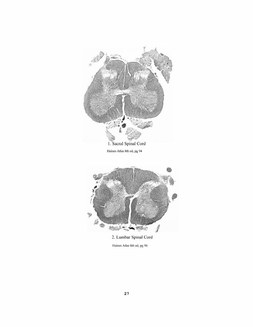
other nerves innervate the tongue (gross anatomy question.)? V. Slides 1-4 from the U of I Spinal cord and Brainstem series will be discussed. Identify structures listed below: (atlas, pgs 87-95; Purves pg 722 & 758-9; Nolte pg 236) and page 27 - 28 of this manual. 1. Sacral spinal cord Substantia Gelatinosa (pain processing) Fasciculus Gracilis (fine touch, position sense for leg) Lower motor neurons (send axons to muscles) Lissauer's tract Nucleus proprius (Body of dorsal horn) (sensory processing) Dorsal and ventral spinal arteries Cauda equina

7
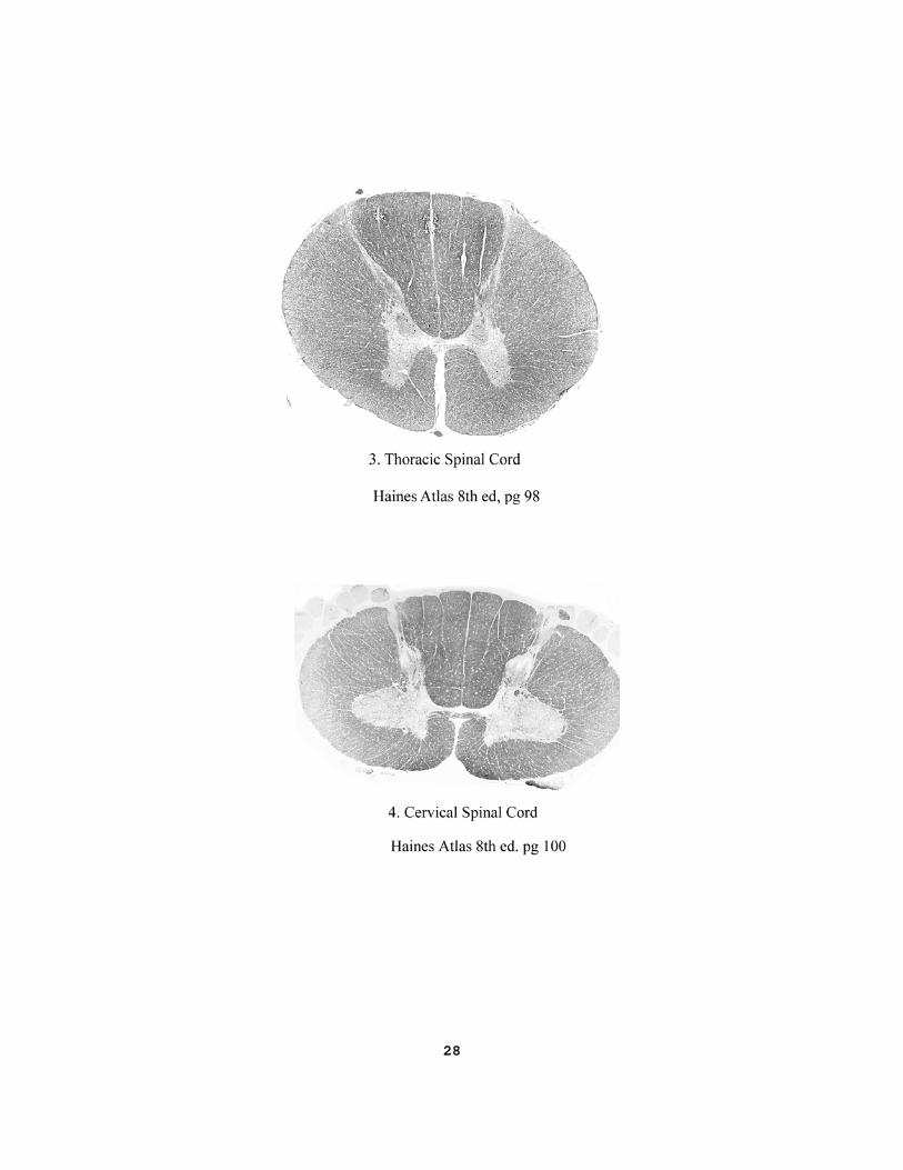
2. Lumbar spinal cord. Substantia Gelatinosa Fasciculus Gracilis Dorsal root (and medial division in dorsal horn) Lower motor neurons (send axons to muscles) Lateral funiculus Ventral funiculus 3. Thoracic spinal cord. Substantia Gelatinosa Fasciculus Gracilis Ventral horn Nucleus dorsalis (of Clarke) (spinocerebellar projections) Intermediolateral cell column (sympathetic preganglionic)
4. Cervical spinal cord.
1. What relationships do the major vessels of the spinal cord have to the fissures and funiculi? (atlas 8th ed pg 105; Purves pg 737; Nolte pg 262) 2. What peripheral nervous system cells are derived from neural crest? (Purves pg 480; Nolte, pg 39)
8
LABORATORY II - Identify External Features SEE IT FIRST AT: http://tigger.uic.edu/classes/anat/anat403/ also, the video “Lateral Surface of Brain”. For now, you may leave the large vessels intact on the medial surface of cortex and on the brain stem. If you have completed your identification of the meninges, vessels, and cranial nerves, proceed to thoroughly remove the blood vessels and the arachnoid from gyri and sulci of the cerebral hemisphere. You will be able to see and define the cerebral lobes and their gyri as well as some gross features of the brainstem and the ventral surface of the brain. (atlas 8th eds, pgs. 13-25; Purves pg 746; Nolte pg 59-63). You may see these structures in situ in Visible Man at http://www.anatlab.net I. Locate the frontal lobe, its gyri and its sulci. The somatic motor areas are at the back of this
lobe. The prefrontal cortex, which is critical to “human” behaviors, is found in what cranial fossa? What are the arterial suppliers? Which tract is close to the gyrus rectus and orbital gyri? On the inferior frontal gyrus, identify the triangular and opercular gyri (Broca’s area in the language dominant hemisphere).
II. Locate the parietal lobe, gyri and sulci. The primary somatosensory area is on the postcentral
gyrus. What are the arterial suppliers? In the inferior parietal lobule, find the end of the lateral fissure surrounded by the supramarginal gyrus, and the angular gyrus immediately behind the supramarginal. On your specimen, which lateral fissure goes further back? (Post to Blackboard) The superior parietal lobule is above the intraparietal sulcus.
III. Locate the temporal lobe, its gyri and fissures on both lateral and inferior surfaces. Find the
transverse gyri in the floor of the lateral fissure (primary auditory cortex, atlas, 8th eds pg 38). Identify the Parahippocampal gyrus and uncus. What cranial fossa contains this lobe? What are the arterial suppliers?
IV. Locate the insula (Island of Reil)(Receives a variety of visceral sensory connections. (8th ed pg
38; Purves pg 728; Nolte pg 60). V. Locate the occipital lobe. Note:(The gyri on the medial surface will be seen after the brain is cut
in half, and those on the lateral surface will not be individually identified in this course). This lobe is notable for processing visual information. What is its arterial supply?
VI. Locate and identify the following structures on the base of the brain and brainstem: (atlas, pg.
24, also note cranial nerves in MRI, pgs 42-48) 1. Anterior perforated substance, (atlas pg 26) (olfactory cortex) 2. Optic chiasm (vision) 3. Chiasmatic cistern (atlas 8th pg 62) 4. Infundibulum (control of pituitary) 5. Mammillary bodies (memory, orientation in environment) 6. Midbrain is “hidden” in the mesencephalic flexure. 7. Pons 8. Cerebellum – Flocculus (atlas pg 22) (motor synergy)
9
9. Cisterna magna (or Cerebellomedullary cistern) (atlas 8th pg 62) 10. Choroid plexus at the lateral recess of the fourth ventricle, and looks like fine
grain sand. (What nerves are close to it?) 11. Inferior olive of the medulla (motor pattern learning) 12. Pyramids of the medulla (voluntary motor pathway) VIII. Slides from the UIC Brainstem series will be discussed. Identify structures labeled on the
UIC Brain Stem and Spinal Cord: (atlas, pgs 87-95; Purves pg 757; Nolte pg 236) and page 29-30 of this manual.
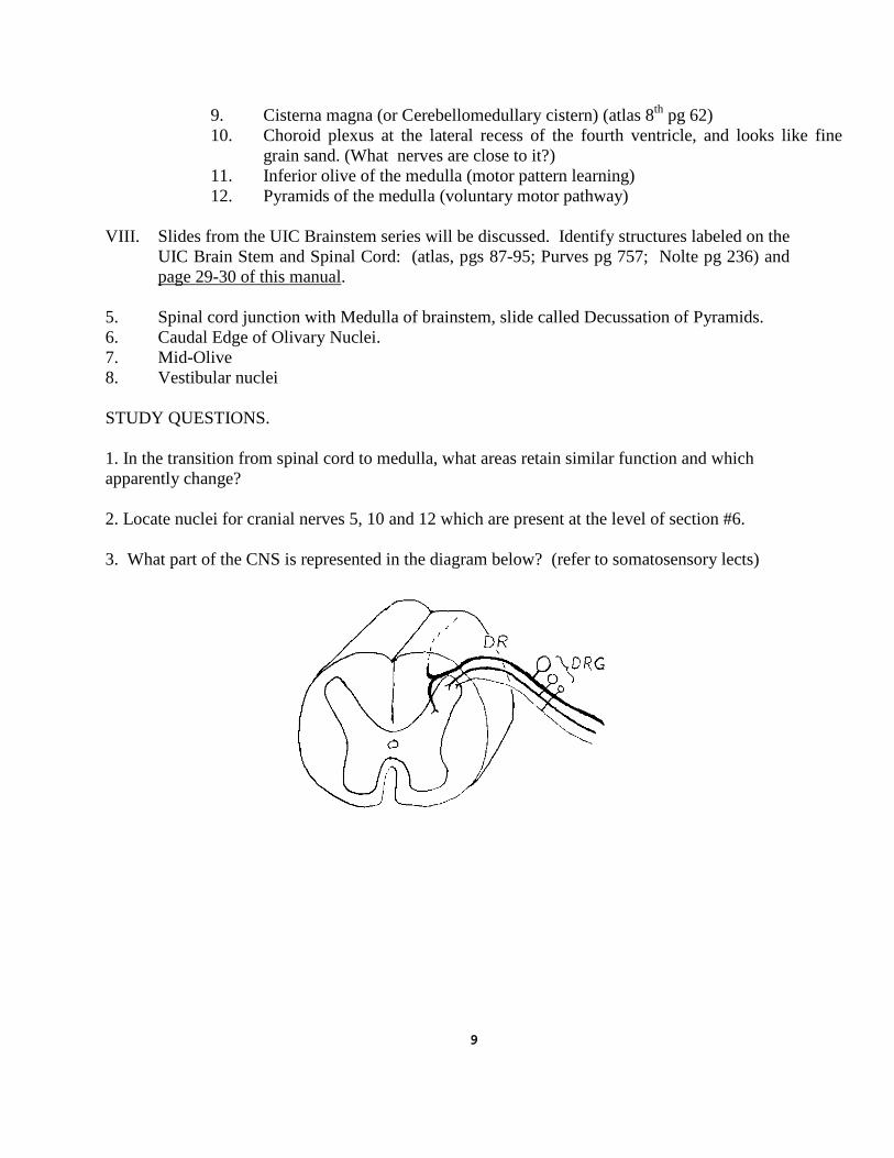
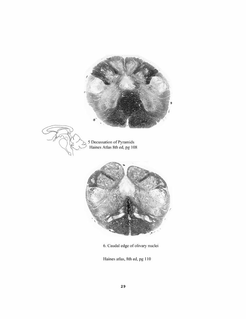
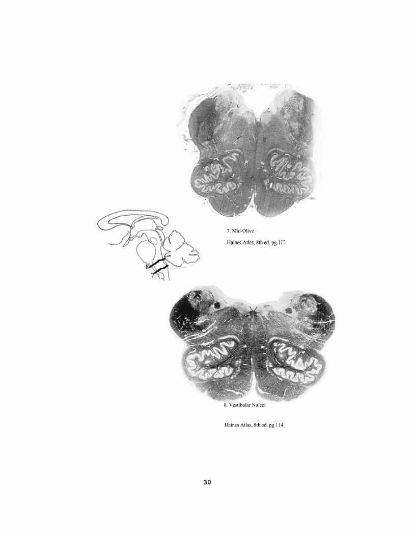
5. Spinal cord junction with Medulla of brainstem, slide called Decussation of Pyramids. 6. Caudal Edge of Olivary Nuclei. 7. Mid-Olive 8. Vestibular nuclei STUDY QUESTIONS. 1. In the transition from spinal cord to medulla, what areas retain similar function and which apparently change? 2. Locate nuclei for cranial nerves 5, 10 and 12 which are present at the level of section #6. 3. What part of the CNS is represented in the diagram below? (refer to somatosensory lects)
10
LABORATORY III - Dorsal Brainstem and Medial Hemisphere. SEE IT FIRST AT http://tigger.uic.edu/classes/anat/anat403/ and video on midsagittal plane. I. Remove cerebellum: Remove the cerebellum by sectioning the three cerebellar peduncles bilaterally. A probe, or your finger, in the medial foramen of 4th ventricle (Magendie) will help guide the cut toward the floor of the 4th ventricle. With a scalpel, cut dorsal to the fifth cranial nerve through the middle and inferior peduncles, on both sides. Separating the cerebellum and br stem, continue the cut superiorly into the roof of the ventricle to sever the superior peduncles. A horizontal section of cerebellum about 1/3rd of the distance up from the inferior edge at the “tonsil” reveals the dentate nucleus. (atlas 8th pg 120; Purves pg 419) II. In the floor of the fourth ventricle identify:(atlas 8th pg 32-4; Nolte pg 270) 1. Rhomboid shape of the IVth ventricle. 2. Stria medullaris (Not present in all brains) 3. Facial colliculus (7th Nv axons that innervate face muscles) 4. Vestibular area (nuclei for balance and sense of gravity) 5. Hypoglossal trigone (motor neurons for tongue) 6. Vagal trigone (parasympathetic neurons for heart and GI) 7. Obex (posterior end of IVth ventricle) 8. Sulcus limitans (separates motor from sensory nuclei) 9. Use information from your textbook to help identify the location of the
locus coeruleus (norepinephrine) and area postrema (emetic center). III. Cut the brain in half. The following directions help you to cut the brain and brain stem in the midsagittal plane. Looking at the dorsal, superior, surface of the brain, bring the knife down between the hemispheres to section the corpus callosum with a shallow cut. Next, displace the vessels away from the midline of the brain stem, particularly the basilar, and anterior communicating arteries. Have a colleague hold the brainstem straight, if necessary, then firmly draw the clean knife through the midline of the brainstem and optic chiasm, thus cutting the brain in half. Avoid the use of a back and forth sawing motion. Identify on the mid-sagittal surface:(atlas, pg 31; Purves pg 730&743; Nolte pg 56 & 64) 1. Corpus callosum (axons connecting the two hemispheres)
2. Anterior commissure (axons connecting right and left amygdala) 3. Interventricular Foramen (of Monro) 4. Septum pellucidum 5. Fornix (axons from hippocampus) 6. Lateral ventricles 7. Third ventricle, including its recesses 8. Cerebral aqueduct (= Iter, Aqueduct of Sylvius) 9. Fourth ventricle 10. Choroid plexus of the third, fourth and lateral ventricles
11
11. Internal cerebral vein (atlas pg 28) 12. Caudate nucleus (forming the floor of the lateral ventricle) 13. Stria terminalis (tract from amygdala to hypothalamus) 14. Cerebral gyri and sulci including Parieto-occipital sulcus Calcarine fissure (bordered by primary visual cortex) Cingulate gyrus (part of limbic system)
Paracentral lobule (motor and sensory contralateral leg) Parahippocampal gyrus (gateway to limbic system) Collateral sulcus Uncus (olfactory cortex) 15. Thalamus Massa intermedia (thalamic adhesion) Stria medullaris thalami Anterior thalamic tubercle (limbic system) Hypothalamic sulcus Infundibulum (hypothalamic connections to pituitary) Lamina terminalis (rostral limit of neural tube) 16. Epithalamus including Pineal gland (nocturnal secretion, melatonin) Posterior commissure Stria medullaris thalami Habenula 17. Mesencephalon Tectum (= corpora quadrigemina) Superior colliculi (visual reflexes) Inferior colliculi (auditory relay)
Quadrigeminal, interpeduncular and ambient cisterns (atlas pg 62) Tegmentum
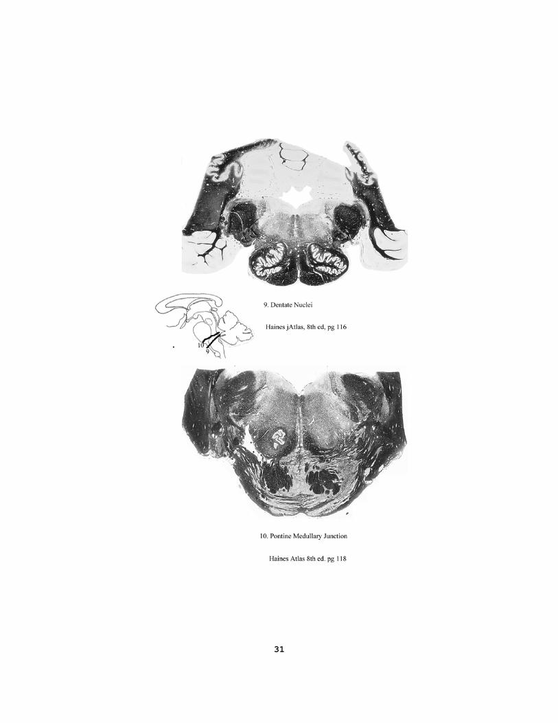
Cerebral peduncles (contains axons from cerebral cortex) Trochlear nerve (to Sup. Oblique m.,turns eye down and out) Brachium of the inferior colliculus (auditory tract) Brachium of the superior colliculus (vision tract) V. U of I brain stem: See pages 31 to 32 in this manual. 9. dentate nuclei 10. pontine-medullary junction 11. abducens nuclei Study Questions: 1. In development of spinal cord, to what do the roof and floor plates contribute? (atlas 8th ed pg 184; Purves pg 480; Nolte pg 42)
2. Which cranial nerve nuclei are near the surface of the floor of the 4th ventricle? 3. What functions are associated with: calcarine (striate) cortex; inferior colliculus; superior colliculus; 4. Trace the flow of cerebrospinal fluid, and its production and absorption. Identify loci where CSF flow
might be restricted, causing hydrocephalus.(Purves pg 743)

12
Questions related to the eye: 1. Is the retina part of CNS or PNS? 2. What layers are separated in retinal detachment? Relate this to the development of eye. 3. What is the numerical relationship between cones and ganglion cells vs between rods and ganglion cells? What is the advantage of convergence for the information from rods? 4. Which hemi-retina has the blind spot? Which visual field contains the blind spot? 5. Which action requires more muscle activity, looking at a distant or a close object? 6. What are the sources of innervation to the iris to produce a small pupil, or a large pupil? 7. If the medial rectus muscle contracts to turn the eye toward the nose, then which cranial nerve must act to reduce muscle tone in the lateral rectus muscle? 8. In which compartment(s) of the eye is vitreous humor found? 9. Why does a major portion of the refraction of light rays take place at the air to cornea transition?
13
LABORATORY IV – HORIZONTAL BRAIN SECTIONS SEE IT FIRST AT: http://tigger.uic.edu/classes/anat/anat403/ and the video on horizontal sections. In this laboratory the half brain with the vessels and brain stem present will be cut into horizontal sections. Before proceeding to cut the brains, you must remove all the blood vessels from the surface and sulci of the cortex. Attempting to make the sections without first removing the vessels will result in very rough cut surfaces which are useless for your study. Make each cut with a single stroke of the blade, avoiding a sawing back and forth movement. I. Horizontal Sections: (atlas 8th ed pgs 84-87) You may leave the brainstem attached to the diencephalon. Only three sections are needed, but you may cut more sections if you wish. 1. The first section should pass through the gyrus cinguli. Identify: Width of sulci and gyri Central white matter of hemisphere Cingulum (long association bundle) Short U-shaped bundles connecting adjacent gyri What large artery supplies this medial side of the hemisphere? 2. The second section is below the body of corpus callosum. (atlas 8th pg 85) Identify: Roof of lateral ventricle Caudate nucleus (head, body and tail, motor planning) Stria Terminalis (limbic system axons) Vena Terminalis (thalamostriate vein) Choroid plexus (CSF secretion) Anterior thalamic tubercle (Nucleus of limbic system) Fornix (connections of hippocampus) What relationships does the caudate nucleus have to the lat. ventricle? 3. The third section should pass through the massa intermedia of the
thalamus (or mid-thalamus if no massa intermedia is present). (atlas 8th ed pgs 86 and 172; Purves pg 752(E); Nolte pg 397)
Identify: Internal capsule (anterior and posterior limbs and genu)
Corpus striatum (caudate N, Putamen N, Globus Pallidus) Extreme capsule Thalamus (Pulvinar, Medial geniculate body) Brachium of the inferior colliculus Tail of caudate Hippocampus (a gyrus of limbic system) and fornix What arteries supply blood to the hippocampus? What two nuclei are connected by cellular bridges across ant limb int cap?
14
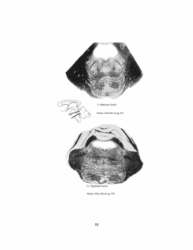
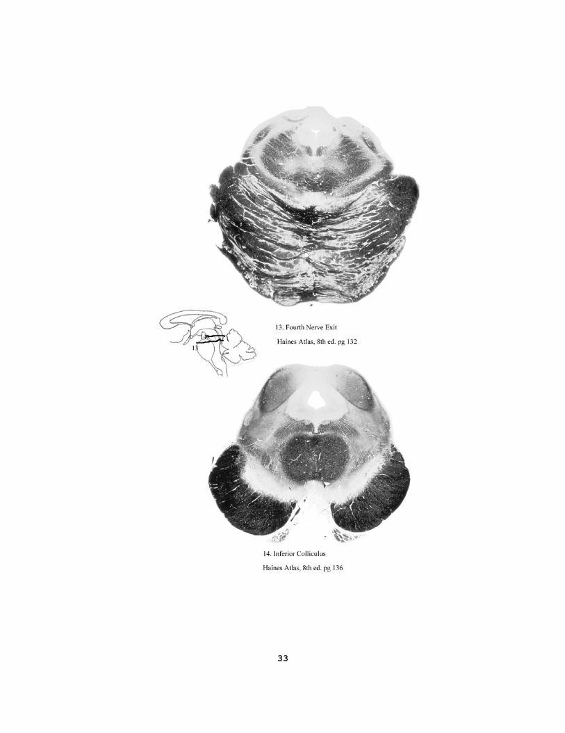
II. Slides of the U of I brain stem series will be discussed. Slides 12-14. Pgs 32-33 this manual. 12. trigeminal nuclei 13. fourth nerve exit 14. inferior colliculus 1. Hemisection of spinal cord results in what sensory deficits? Primarily section of the dorsal column and the anterolateral system (ALS). 2. Do any of the structures you have observed have neurogenesis in the adult?
On the following page draw on the images the sensory origin, nerve and CNS pathways, and location of synapses in the dorsal column-medial lemniscus system and in the anterolateral system.
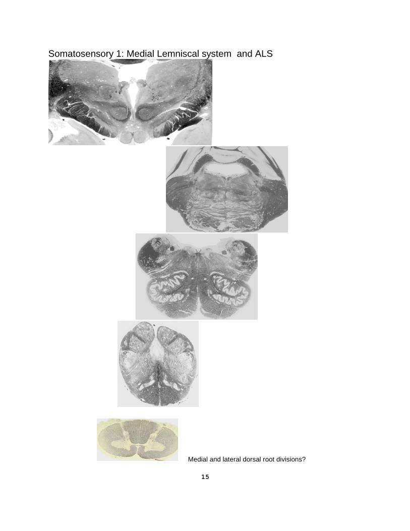
15
Somatosensory 1: Medial Lemniscal system and ALS
Medial and lateral dorsal root divisions?
16
LABORATORY V – CORONAL BRAIN SECTIONS (and Histology of eye) SEE IT FIRST AT: http://tigger.uic.edu/classes/anat/anat403/ and the video on coronal sections. In this laboratory session a half brain will be cut into coronal sections. Before proceeding to cut
the brain, you must remove all the blood vessels from the surface and sulci of the cortex. Make each cut with a single stroke of the clean blade, avoiding a sawing, back and forth movement.
II. Frontral Sections: (atlas 8th ed pgs 74-82).
Remove the brainstem with one smooth cut through the superior colliculus level of the midbrain. (atlas 8th, pgs 20, 134, and pg 140. Purves pg 756. Nolte pg 389)
Identify the following structures in the plane of this section:
Cerebral peduncle (crus cerebri, axons from motor cortex) Substantia nigra (degenerates in Parkinson’s Disease) Red nucleus (motor control) Tegmentum (below cerebral aqueduct) Tectum (above cerebral aqueduct) We suggest frontal sections vertically through the following specific locations. (atlas 8th pgs 74-82. Purves pg 748-9. Nolte pg 639-56) 1. Behind the genu of the corpus callosum:(atlas 8th pg 74. Nolte pg 640) Identify: The anterior limit of the lateral ventricle. The head of the
caudate and its relation to the lateral ventricle and septum pellucidum. Cerebral cortex and the white matter of the hemisphere
2. Through anterior commissure: (atlas 8th ed pg 75; Purves pg 748B; Nolte pg 642) Identify: Anterior commissure (connects amygdaloid nuclei) Head of caudate (degenerates in Huntington’s Disease) Putamen (motor planning) Globus Pallidus (motor planning) Genu of internal capsule Columns of the fornix Interventricular foramen (of Monro) Optic chiasm Amygdaloid nuclei and the Uncus External capsule (cortico-striate axons) Claustrum Extreme capsule (white matter of insula) Insular cortex (visceral sensations)
17
3. Through the massa intermedia: (atlas 8th pg 78; Purves pg 400; Nolte pg 644)
Identify: Thalamus: midline, medial and lateral nuclei Anterior thalamic tubercle (limbic functions) Posterior limb of internal capsule (sensory radiations) Globus pallidus (motor control) Putamen (motor control) Body of caudate Optic tract (axons after the optic chiasm) Subthalamus (motor control) 4. Through the posterior commissure: (atlas 8th pg 80. Purves pg 749; Nolte pg652) Identify: Pulvinar (supports visual association cortex) Medial and lateral geniculate bodies (auditory and vision) Inferior horn of lateral ventricle Hippocampal gyrus (episodic memory) Fornix (and fimbria) (axons from hippocampus) Choroid fissure (medial limit of the lateral ventricle) Dentate gyrus (part of hippocampal formation) 5. Behind the thalamus: (atlas 8th ed pg 82. Nolte pg 656) You should now be able to identify the turn of the ventricle from its superior
portion toward its temporal horn. Probe the depths (highly variable) of the occipital horn of lateral ventricle into the occipital lobe.
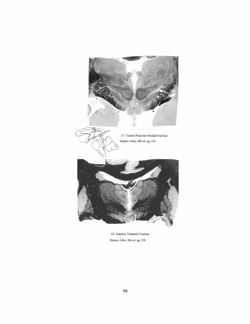
III. Slides of the U of I brain stem series will be discussed. Slides 15-17. Pg 34 this manual. 15. superior colliculus 16. coronal section through thalamus and brain stem 17. ventral posterior medial nucleus Study Questions:
1. What structures are seen in a frontal section at the level of the genu of internal capsule? 2. What relationships does the caudate nucleus have to the lateral ventricle? 3. What arteries supply the arterial blood to the: internal capsule; hippocampus; occipital lobe? 4. What is medial to the posterior limb of internal capsule? 5. The thalamic somato-sensory radiations are found in what part of internal capsule?
18
LABORATORY VI - IMAGING THE BRAIN
You must now relate the wet specimens of previous labs to 2-D images.
I. Pay particular attention to the Mid-sagittal planes among the parasagittal images. In the horizontal and in the coronal images take particular note of the structures in the planes of the basal ganglia and internal capsule.
II. You have identified many structures and landmarks on the wet specimens of the
brain. Now, you must translate that knowledge to the two dimensional images that you will experience throughout your clinical career. Examples of the results obtained with CT scans and MRI will be reviewed in this session. Scans in which a contrast was injected into the vascular system are designated "+c", and will give a white line (clear) indication of the midsagittal fissure as well as segments of the arteries. (atlas 8th pg 28-31, 74-82. Purves pg 748-752; Nolte pg 113-6)
III. Plastic embedded thick sections of the brain will be available for review in the
remaining laboratory sessions. Great care should be taken not to drop the blocks or otherwise mar the surfaces. Examination of both the front and back of these sections will be a useful exercise in furthering your three dimensional understanding of the brain.
CT and MRI Scans
On a mid-sagittal plane identify: cingulate gyrus, superior frontal gyrus, lingual gyrus and associated sulci. Corpus callosum, septum, fornix, thalamus, optic chiasm and mammillary body. Superior and inferior colliculi, pineal, tegmentum, basilar pons, 4th ventricle.
On coronal sections identify: Insular cortex Superior temporal gyrus and sulcus Hippocampus Parahippocampal gyrus Internal Capsule Basal Ganglia Thalamus Sella turcica and pituitary gland
In general identify: Sulcus width
Cerebral white matter Ventricular system (and choroid plexus) Quadrigeminal (superior) cistern (plus the ambient cistern) Interpeduncular cistern Cerebellomedullary cistern (cisterna magna)
19
Optic tracts On the CT scans with contrast medium ‘+c’, bright signal, and MRI as dark (negative) images: Basilar artery Internal carotid artery Proximal part of post. cerebral A Proximal part of middle cerebral A Straight sinus Choroid plexus of lateral ventricle Inferior sagittal sinus Anterior cerebral A MRI from a clinical case, including Diffusion Tensor Imaging (DTI), determine:
1. Which lobe and side had a tumor? 2. Was the white matter involved? (Purves pg 516; Nolte pg 410-1) 3. What complaints may have been happening (Purves 599-601; Nolte pg 567-8)? At the
Web site see additional ‘case studies’. Slides of the U of I brain stem series will be discussed. Slides 18-20. Pgs 35-36 this manual. 18. anterior thalamic nucleus 19. mammillothalamic fasciculus 20. ventral anterior thalamic nucleus
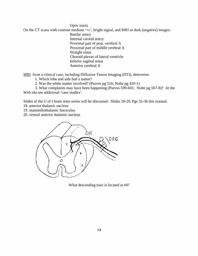
What descending tract is located at #4?
20
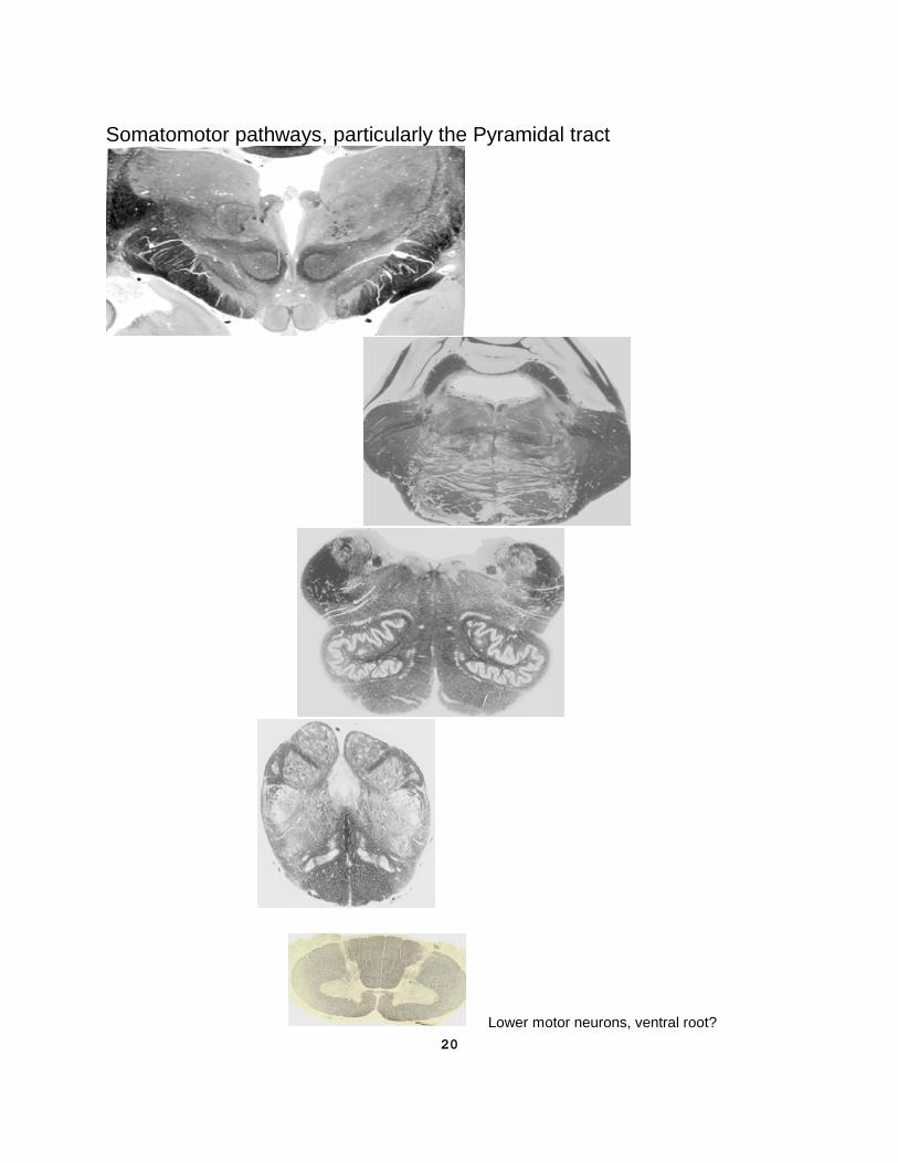
Somatomotor pathways, particularly the Pyramidal tract
Lower motor neurons, ventral root?
21
LABORATORY VII - Special View of the CNS SEE IT FIRST AT: http://tigger.uic.edu/classes/anat/anat403/ and the video of pro-section. In this laboratory analyze a pro-section of the brain which will be provided by instructors. These pro-sections will be available in the laboratories through the end of the course, and may appear on the laboratory examination. You should be actively reviewing the UIC brain stem set with the aid of the computer program (UIC Brainstem) prepared by Drs. Anderson, McAuley and Unnerstall.
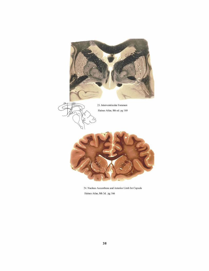
Insert the somatosensory and motor pathways on attached drawing. What is the blood supply to the tracts in cerebral cortex, internal capsule, medulla and spinal cord? Slides of the U of I brain stem series will be discussed. Slides 21-24. Pgs 36-38 this manual. 21. optic chiasm 22. anterior commissure 23. interventricular foramen 24. nucleus accumbens and anterior limb internal capsule STUDY QUESTIONS
1. What relationships do each of the cerebellar peduncles have to the IVth ventricle?
2. Where is the reticular formation of the brainstem located?
3. Internal arcuate fibers of the medulla arise from dorsal column nuclei and project to which thalamic nucleus?
4. At what cross sectional level of brainstem is the red nucleus found? 5. Find the nuclei for cranial nerves 6, 8, 10 and 12 in the floor of the fourth ventricle.
6. What cranial nerve approaches brain stem dorsal to the inferior Cerebellar Peduncle?
7. Review the blood supply to the brainstem, cerebellum and internal capsule. (atlas, 8th pg. 105, 121,
135, and 147, 169)
8. What two motor cranial nerves exit from the medulla near the pyramid?
9. What cranial nerves contain axons from the cells in the nucleus ambiguous?
10. What cranial nerve nucleus lies deep to the genu of the VII cranial nerve?
11. What cranial nerve nucleus sends axons into the interpeduncular fossa?
12. Locate the cell bodies of the cranial nerve which exits from the dorsum of the brainstem (look at the inferior colliculus level).
22
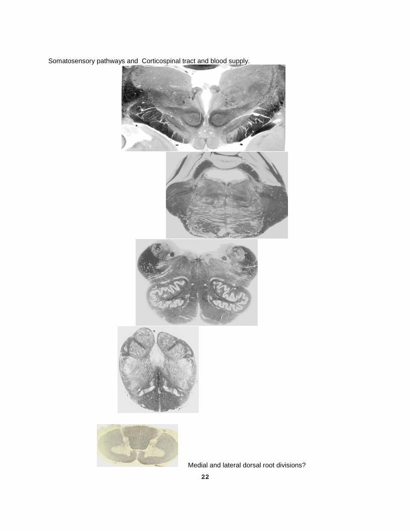
Somatosensory pathways and Corticospinal tract and blood supply.
Medial and lateral dorsal root divisions?
23
LABORATORY VIII - CASE STUDIES IN NEUROANATOMY SEE MORE AT: http://tigger.uic.edu/classes/anat/anat403/ "LESIONS LAB" In this exercise, we will utilize a case-oriented problem solving approach to review and integrate the neuroanatomical facts and concepts covered in previous labs and lectures. On the following pages, two examples of "idealized" case studies of patients exhibiting a variety of neurological symptoms are presented. Following each case, you will find an image taken from your brainstem slide collection with specific regions darkened. These darkened regions represent areas of damage following stroke or injury. Your task is to match the symptoms to the lesion. Identify the nuclei, nerves or tracts involved in the lesions that would produce the symptoms that were described. If the symptoms are sudden in onset, assume a vascular accident and identify the arterial branches that could be involved. When the left side of the image is shadowed, assume that the damage is on the left side of the brain and vice versa. Since these images are taken from our slide collection, you may not see every salient feature on the image that corresponds to the symptoms. However, keep in mind the relative level (three dimensional) represented by the image, hence, the structures you would expect to find at that level. These examples are given to whet your appetite. We will review these cases as a group in class, as well as present additional cases. To prepare for these cases, study the questions presented at the end of this section. This laboratory exercise provides an excellent opportunity to "pull together" what you have learned in neuroanatomy during the past few weeks while reviewing the localization of major systems in the CNS and their blood supply. Take this exercise seriously and think about the material before the lecture. Many of these cases will come up on exams - either the one at the end of the course or the STEP-1 later on in your training and career. The 8th ed of Haines Atlas has a number of lesion and symptom pages which will help you think about organization of the CNS. E.g. pgs 52, 104, 121, 135, 147, 168-9, 188-229, 263. All slides of the U of I brain stem series have been discussed in earlier labs and in lectures.

24
Case 1. A 24 year old male was brought into the emergency room following a motorcycle accident. He suffered an apparent fracture of the spine in the lower back region. Neurological examination revealed the following: (atlas 8th pg 104) 1. spastic paralysis of the lower left limb; 2. increased deep tendon reflexes of the lower left limb; 3. Babinski sign on the left; 4. loss of position sense, touch and pressure sensations of the lower left limb; 5. complete anesthesia of the L2 dermatome on the left side; 6. loss of pain sensations and thermal anesthesia of the L3-S4 dermatomes on the
right side.
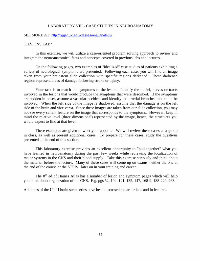
25
Case 2. A 60 year old dock worker was brought to the emergency room unconscious after collapsing while loading a truck. After regaining consciousness, an examination was performed which revealed the following: (atlas 8th pg 120-1) 1. paralysis of both right limbs; 2. hypertonicity of both right limbs; 3. increased deep tendon reflexes on the right side; 4. dysarthria (speech disturbance); 5. deviation of the tongue to the left when protruded; 6. loss of position sense, pressure and two-point discriminative touch on the right
side of the body.

26
Study Questions 1. Define the terms "contralateral" and "ipsilateral". In Case 1, if you could not tell which
side of the body the lesion was on, how would you describe where spastic paralysis would be seen with respect to the lesion, and loss of pain and temperature sensation in the lower lumbar and sacral dermatomes with respect to the lesion?
2. What connections are associated with the flocculo-nodular lobe of the cerebellum? 3. What are the names of, and connections of the basal nuclei of the cerebellum? 4. The brachium of inferior colliculus has axons that connect to which thalamic nucleus? 5. The brachium of the superior colliculus has axons that connect which cell groups? 6. What distinguishes a lower motor neuron deficit from an upper motor neuron deficit?
Identify four sources of upper motor neurons. 7. At what points on the brainstem can you obtain an alternating (i.e. partly ipsilateral,
partly contralateral) constellation of signs of paralysis? (Hint: Cranial Nerves 3, 6 and 12, and the cortico-spinal tract)
8. What arteries provide the blood supply to the ventral medial medulla? Or to the dorsal
lateral medulla? 9. How do the following lesions result in different clinical signs of Facial nerve function?
(1) Section of the Facial nerve; (2) vascular lesion of the motor nucleus of VII; (3) cortical lesion in the face area of the precentral gyrus.
10. Pupillary dilation and lateral strabismus (turning of the eye) would be signs of damage to
which cranial nerve? Which cranial nerve would be damaged if medial strabismus was noted upon neurological examination?
11. Which artery(ies) supply blood to the hippocampal gyrus? What would be the expected
neurological effect of bilateral damage to the medial temporal lobe? 12. Parkinson's disease is the result of the degeneration of what group of neurons? Where do
these neurons normally project? What is the neurotransmitter utilized by the degenerating neurons?
27
28
29
30
31
32
33

34
35

36

37
38


![COMMISSION ON LABORATORY …pathology.jhu.edu/department/MAS_GEN_09272007[1].pdfLaboratory General OUTLINE SUMMARY OF CHANGES 3 INSPECTION TECHNIQUES – KEY POINTS](https://img.pdfslide.net/doc/110x75/5b87a1cd7f8b9a3d028b590a/commission-on-laboratory-1pdflaboratory-general-outline-summary-of-changes-3-inspection.jpg)