Embed Size (px)
Citation preview
CH A P T E R
31
Laparoscopic Hiatal Herniorrhaphy
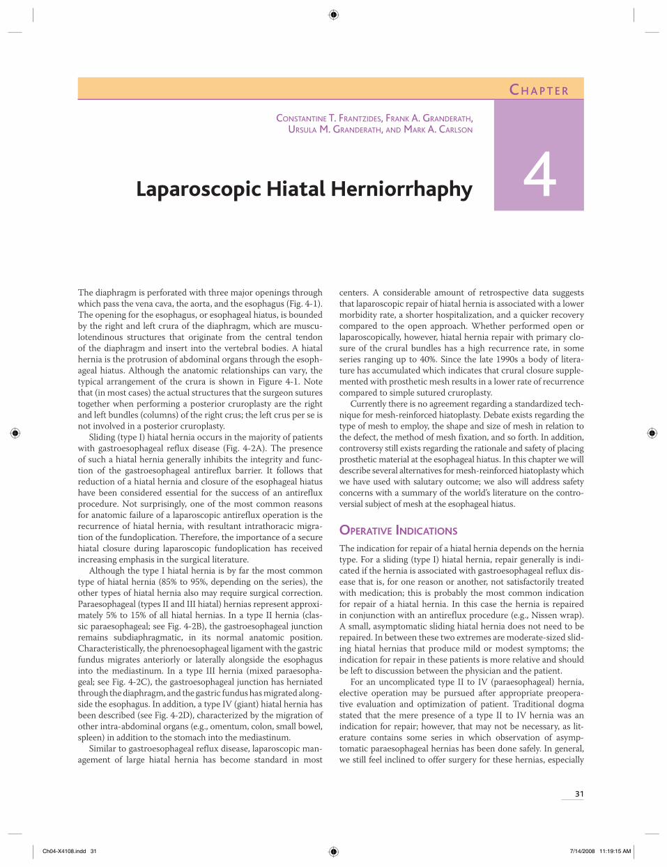
The diaphragm is perforated with three major openings through which pass the vena cava, the aorta, and the esophagus (Fig. 4-1). The opening for the esophagus, or esophageal hiatus, is bounded by the right and left crura of the diaphragm, which are muscu-lotendinous structures that originate from the central tendon of the diaphragm and insert into the vertebral bodies. A hiatal hernia is the protrusion of abdominal organs through the esoph-ageal hiatus. Although the anatomic relationships can vary, the typical arrangement of the crura is shown in Figure 4-1. Note that (in most cases) the actual structures that the surgeon sutures together when performing a posterior cruroplasty are the right and left bundles (columns) of the right crus; the left crus per se is not involved in a posterior cruroplasty.
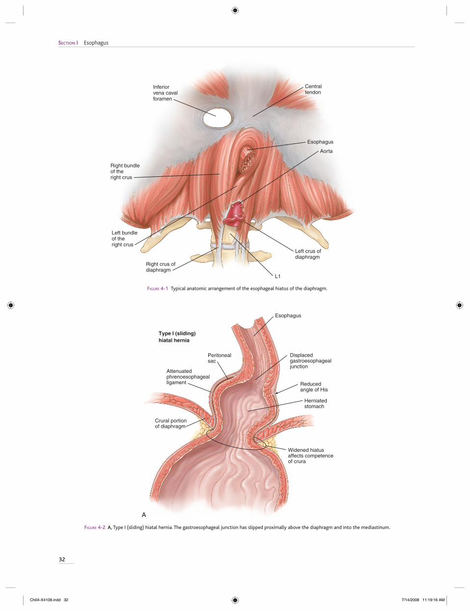
Sliding (type I) hiatal hernia occurs in the majority of patients with gastroesophageal reflux disease (Fig. 4-2A). The presence of such a hiatal hernia generally inhibits the integrity and func-tion of the gastroesophageal antireflux barrier. It follows that reduction of a hiatal hernia and closure of the esophageal hiatus have been considered essential for the success of an antireflux procedure. Not surprisingly, one of the most common reasons for anatomic failure of a laparoscopic antireflux operation is the recurrence of hiatal hernia, with resultant intrathoracic migra-tion of the fundoplication. Therefore, the importance of a secure hiatal closure during laparoscopic fundoplication has received increasing emphasis in the surgical literature.
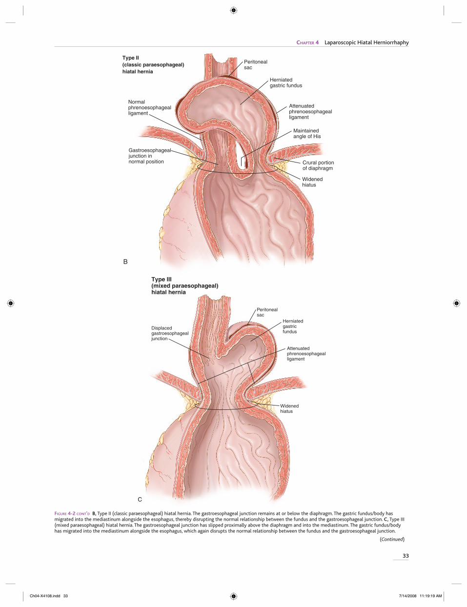
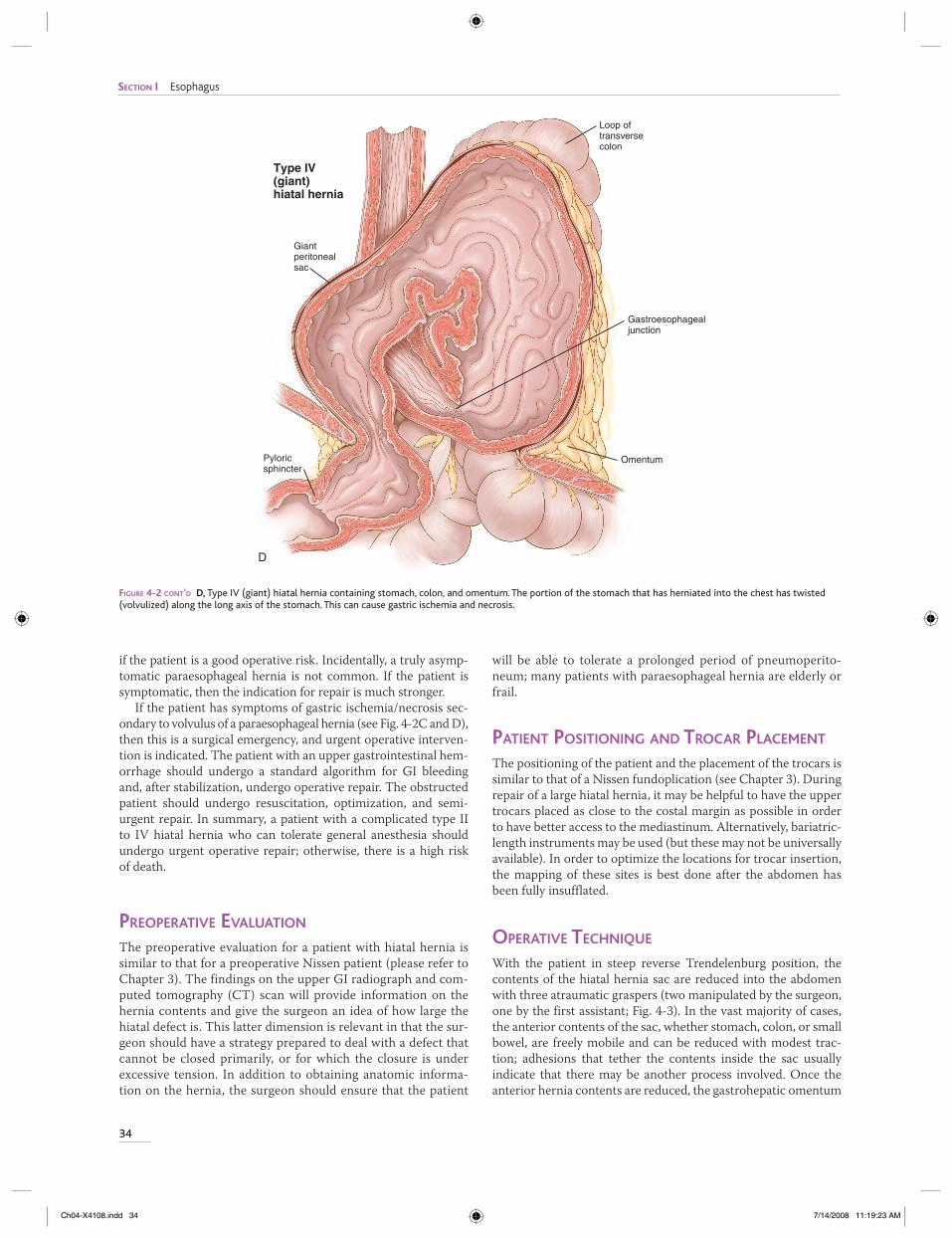
Although the type I hiatal hernia is by far the most common type of hiatal hernia (85% to 95%, depending on the series), the other types of hiatal hernia also may require surgical correction. Paraesophageal (types II and III hiatal) hernias represent approxi-mately 5% to 15% of all hiatal hernias. In a type II hernia (clas-sic paraesophageal; see Fig. 4-2B), the gastroesophageal junction remains subdiaphragmatic, in its normal anatomic position. Characteristically, the phrenoesophageal ligament with the gastric fundus migrates anteriorly or laterally alongside the esophagus into the mediastinum. In a type III hernia (mixed paraesopha-geal; see Fig. 4-2C), the gastroesophageal junction has herniated through the diaphragm, and the gastric fundus has migrated along-side the esophagus. In addition, a type IV (giant) hiatal hernia has been described (see Fig. 4-2D), characterized by the migration of other intra-abdominal organs (e.g., omentum, colon, small bowel, spleen) in addition to the stomach into the mediastinum.
Similar to gastroesophageal reflux disease, laparoscopic man-agement of large hiatal hernia has become standard in most
centers. A considerable amount of retrospective data suggests that laparoscopic repair of hiatal hernia is associated with a lower morbidity rate, a shorter hospitalization, and a quicker recovery compared to the open approach. Whether performed open or laparoscopically, however, hiatal hernia repair with primary clo-sure of the crural bundles has a high recurrence rate, in some series ranging up to 40%. Since the late 1990s a body of litera-ture has accumulated which indicates that crural closure supple-mented with prosthetic mesh results in a lower rate of recurrence compared to simple sutured cruroplasty.
Currently there is no agreement regarding a standardized tech-nique for mesh-reinforced hiatoplasty. Debate exists regarding the type of mesh to employ, the shape and size of mesh in relation to the defect, the method of mesh fixation, and so forth. In addition, controversy still exists regarding the rationale and safety of placing prosthetic material at the esophageal hiatus. In this chapter we will describe several alternatives for mesh-reinforced hiatoplasty which we have used with salutary outcome; we also will address safety concerns with a summary of the world’s literature on the contro-versial subject of mesh at the esophageal hiatus.
OPERATIVE INDICATIONS
The indication for repair of a hiatal hernia depends on the hernia type. For a sliding (type I) hiatal hernia, repair generally is indi-cated if the hernia is associated with gastroesophageal reflux dis-ease that is, for one reason or another, not satisfactorily treated with medication; this is probably the most common indication for repair of a hiatal hernia. In this case the hernia is repaired in conjunction with an antireflux procedure (e.g., Nissen wrap). A small, asymptomatic sliding hiatal hernia does not need to be repaired. In between these two extremes are moderate-sized slid-ing hiatal hernias that produce mild or modest symptoms; the indication for repair in these patients is more relative and should be left to discussion between the physician and the patient.
For an uncomplicated type II to IV (paraesophageal) hernia, elective operation may be pursued after appropriate preopera-tive evaluation and optimization of patient. Traditional dogma stated that the mere presence of a type II to IV hernia was an indication for repair; however, that may not be necessary, as lit-erature contains some series in which observation of asymp-tomatic paraesophageal hernias has been done safely. In general, we still feel inclined to offer surgery for these hernias, especially
CONSTANTINE T. FRANTZIDES, FRANK A. GRANDERATH, URSULA M. GRANDERATH, AND MARK A. CARLSON
4
Ch04-X4108.indd 31Ch04-X4108.indd 31 7/14/2008 11:19:15 AM7/14/2008 11:19:15 AM
SECTION I Esophagus
32
FIGURE 4-1 Typical anatomic arrangement of the esophageal hiatus of the diaphragm.
L1
Inferiorvena cavalforamen
Right crus ofdiaphragm
Right bundleof the right crus
Left bundle of theright crus
Left crus ofdiaphragm
Aorta
Esophagus
Centraltendon
Herniatedstomach
Peritonealsac
Attenuatedphrenoesophagealligament
Crural portionof diaphragm
Esophagus
Displacedgastroesophagealjunction
Reduced angle of His
Widened hiatusaffects competenceof crura
Type I (sliding)hiatal hernia
A
FIGURE 4-2 A, Type I (sliding) hiatal hernia. The gastroesophageal junction has slipped proximally above the diaphragm and into the mediastinum.
Ch04-X4108.indd 32Ch04-X4108.indd 32 7/14/2008 11:19:16 AM7/14/2008 11:19:16 AM
33
CHAPTER 4 Laparoscopic Hiatal Herniorrhaphy
Crural portionof diaphragm
Normalphrenoesophagealligament
Gastroesophagealjunction innormal position
Maintainedangle of His
Peritonealsac
Herniatedgastric fundus
Attenuatedphrenoesophagealligament
Widenedhiatus
Type II (classic paraesophageal)hiatal hernia
B
(Continued)
FIGURE 4-2 CONT’D B, Type II (classic paraesophageal) hiatal hernia. The gastroesophageal junction remains at or below the diaphragm. The gastric fundus/body has migrated into the mediastinum alongside the esophagus, thereby disrupting the normal relationship between the fundus and the gastroesophageal junction. C, Type III (mixed paraesophageal) hiatal hernia. The gastroesophageal junction has slipped proximally above the diaphragm and into the mediastinum. The gastric fundus/body has migrated into the mediastinum alongside the esophagus, which again disrupts the normal relationship between the fundus and the gastroesophageal junction.
Peritonealsac
Displacedgastroesophagealjunction
Attenuatedphrenoesophagealligament
Type III (mixed paraesophageal)hiatal hernia
C
Herniatedgastricfundus
Widenedhiatus
Ch04-X4108.indd 33Ch04-X4108.indd 33 7/14/2008 11:19:19 AM7/14/2008 11:19:19 AM
SECTION I Esophagus
34
Loop oftransversecolon
Giantperitonealsac
Pyloricsphincter
Type IV(giant)hiatal hernia
D
Omentum
Gastroesophagealjunction
FIGURE 4-2 CONT’D D, Type IV (giant) hiatal hernia containing stomach, colon, and omentum. The portion of the stomach that has herniated into the chest has twisted (volvulized) along the long axis of the stomach. This can cause gastric ischemia and necrosis.
if the patient is a good operative risk. Incidentally, a truly asymp-tomatic paraesophageal hernia is not common. If the patient is symptomatic, then the indication for repair is much stronger.
If the patient has symptoms of gastric ischemia/necrosis sec-ondary to volvulus of a paraesophageal hernia (see Fig. 4-2C and D), then this is a surgical emergency, and urgent operative interven-tion is indicated. The patient with an upper gastrointestinal hem-orrhage should undergo a standard algorithm for GI bleeding and, after stabilization, undergo operative repair. The obstructed patient should undergo resuscitation, optimization, and semi-urgent repair. In summary, a patient with a complicated type II to IV hiatal hernia who can tolerate general anesthesia should undergo urgent operative repair; otherwise, there is a high risk of death.
PREOPERATIVE EVALUATION
The preoperative evaluation for a patient with hiatal hernia is similar to that for a preoperative Nissen patient (please refer to Chapter 3). The findings on the upper GI radiograph and com-puted tomography (CT) scan will provide information on the hernia contents and give the surgeon an idea of how large the hiatal defect is. This latter dimension is relevant in that the sur-geon should have a strategy prepared to deal with a defect that cannot be closed primarily, or for which the closure is under excessive tension. In addition to obtaining anatomic informa-tion on the hernia, the surgeon should ensure that the patient
will be able to tolerate a prolonged period of pneumoperito-neum; many patients with paraesophageal hernia are elderly or frail.
PATIENT POSITIONING AND TROCAR PLACEMENT
The positioning of the patient and the placement of the trocars is similar to that of a Nissen fundoplication (see Chapter 3). During repair of a large hiatal hernia, it may be helpful to have the upper trocars placed as close to the costal margin as possible in order to have better access to the mediastinum. Alternatively, bariatric-length instruments may be used (but these may not be universally available). In order to optimize the locations for trocar insertion, the mapping of these sites is best done after the abdomen has been fully insufflated.
OPERATIVE TECHNIQUE
With the patient in steep reverse Trendelenburg position, the contents of the hiatal hernia sac are reduced into the abdomen with three atraumatic graspers (two manipulated by the surgeon, one by the first assistant; Fig. 4-3). In the vast majority of cases, the anterior contents of the sac, whether stomach, colon, or small bowel, are freely mobile and can be reduced with modest trac-tion; adhesions that tether the contents inside the sac usually indicate that there may be another process involved. Once the anterior hernia contents are reduced, the gastrohepatic omentum
Ch04-X4108.indd 34Ch04-X4108.indd 34 7/14/2008 11:19:23 AM7/14/2008 11:19:23 AM