Embed Size (px)
Citation preview
Inventor: Carlson, Mark A. Laparoscopic Mesh Stapler
April 2, 2007 Page 1 of 5
Laparoscopic Mesh Stapler
Narrative
ABSTRACT Minimally invasive (laparoscopic) ventral hernia repair is performed by covering the hernia
defect with a sheet of prosthetic mesh, which requires fixation with a combination of staples and anchoring sutures. There are two major issues with this technique: (1) sometimes some of the staples need to be removed in order to adjust the mesh position prior to final fixation; and (2) anchoring sutures are a source of chronic abdominal wall pain. The proposed device addresses these two issues by employing two types of staples (denoted here as spikes), temporary or permanent; the former spike can be removed easily so that the mesh position can be adjusted, and the latter spike has an anchoring ability which should make anchoring sutures unnecessary.
BACKGROUND
A hernia of the abdominal wall is defined as a defect in the musculoaponeurotic layers through
which there is protrusion of intraabdominal contents (Figure 1). The most common type of abdominal wall hernia after groin (inguinal) hernia is the incisional hernia. An incisional hernia occurs in a previous abdominal incision which has lost integrity (broken down) over time. An incisional hernia can cause pain and GI distress, and also can be a cosmetic issue. In rare cases, bowel can become incarcerated in an incisional hernia, causing bowel strangulation and necrosis, and even patient death. If a patient has a symptomatic incisional hernia and the patient can tolerate a general anesthetic, then incisional hernia repair is indicated.
More than 300,000 incisional and ventral hernia repairs are performed in the United States each year (National Center for Health Statistics; http://www.cdc.gov/nchs/). More than 50% of these repairs are accomplished with a minimally invasive (laparoscopic) approach, and that fraction is increasing. Although the trend in General Surgery is to operate with minimally invasive techniques, the need for open surgery will persist for the foreseeable future. As long as open surgical incisions are utilized, there will continue to be a defined incidence of incisional hernia (about 20-30% with the use of long midline incisions; see example in Figure 1A). It follows that the need for incisional hernia repair also will continue.
The standard technique of incisional hernia repair (whether performed open or laparoscopically) is to cover the defect with a sheet of prosthetic mesh. Controlled data in the surgical literature indicates that the risk of hernia recurrence is decreased in patients who have a mesh herniorrhaphy compared to patients who have herniorrhaphy without mesh. The technical details the mesh implantation are somewhat controversial; in the laparoscopic approach, the consensus of most abdominal wall surgeons is to place a sheet of mesh intraabdominally which overlaps the hernial defect by 3-4 cm on all sides, and to secure the mesh to the interior of the abdominal wall with sutures and/or staples (Figure 1B).
STATEMENT OF NEED
The fixation of prosthetic mesh during minimally invasive ventral hernia repair is complicated by
a number of problems. One issue is the difficulty in centering the mesh over the defect so that the mesh overlap is approximately equal on all sides of the mesh (Figure 1A). There have been a number of techniques proposed to overcome this difficulty, but in the end the surgeon has to fire a stapler or tacker to begin securing the mesh to the anterior abdominal wall. Often times the surgeon discovers that the placement of the initial staples or tacks was not to his/her liking; at this point, the surgeon either has to
Inventor: Carlson, Mark A. Laparoscopic Mesh Stapler
April 2, 2007 Page 2 of 5
make do with this placement, or s/he has to start over again by first removing the errant staples or tacks before firing some more. This latter option is not easy, because none of the currently available staples or tacks can be readily removed once they have been shot into the abdominal wall.
Another problem that most surgeons perceive with mesh fixation is that the currently available staples or tacks do not penetrate deep enough into the abdominal wall to provide adequate anchorage. The typical penetration depth for the hernia tackers/staplers on the market is in the range of 3 mm. In order to compensate for this perceived inadequacy of anchorage, a common practice is to place transabdominal fixation sutures (Figure 1B) in addition to the intraabdominal staples or tacks. This combination of staples/tacks + sutures is believed to provide optimal mesh anchorage. Unfortunately, none of these issues has yet to be addressed in a controlled trial. The problem with placing transabdominal fixation sutures is that they increase the invasiveness of the operation, and can cause long-term abdominal wall pain.
The proposed device is a laparoscopic mesh stapler for use during minimally invasive ventral hernia repair; this device will address both of the problems listed above. That is, the device will allow for easy placement and removal of temporary anchorage spikes. In addition to the temporary spikes, the device also will fire permanent spikes of such configuration that will eliminate the need for transabdominal fixation sutures.
DEVICE DESCRIPTION: OVERVIEW
The laparoscopic mesh stapler has the ability to fire two different kinds of staples (herein referred
to as spikes): temporary and permanent. The temporary spikes would have a relatively modest anchoring ability; they are meant to be placed during the initial stages of the mesh fixation, and simply hold the mesh in place much in the same way a tailor would pin up a hem during the fitting of a suit. Once the surgeon was satisfied that the mesh was properly placed to cover the hernia defect, the surgeon then could fire permanent spikes with the same device. The permanent spikes would have up to 10 mm of tissue penetration, with an expanded configuration that would result in a high pull-out force. After placement of the permanent spikes, transabdominal fixation sutures would not be necessary. Stainless steel would be the material used for both the temporary and permanent spikes.
DETAILED DEVICE DESCRIPTION
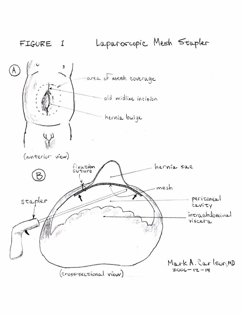
Figure 1. Anatomy of an incisional hernia and a mesh repair.
A. Anterior view of a patient with an incisional (ventral) hernia. This typical patient had a
previous midline incision which subsequently developed a hernia. The dotted oval around the defect depicts the approximate extent that a prosthetic mesh should overlap the defect after a hernia repair has been performed.
B. Cross-sectional view of a minimally invasive ventral hernia repair with a conventional
laparoscopic tacker. During a laparoscopic intraabdominal procedure of any kind, the peritoneal cavity typically is insufflated with carbon dioxide gas. This expands the peritoneal cavity into a much larger space than what it is normally; this expansion allows for insertion of instruments etc. to perform the minimally invasive surgical procedure. In this Figure the hernia defect (sac) also has been expanded by the CO2. A piece of prosthetic mesh is shown in cross-section, with the conventional laparoscopic tacker shown in position to fire a tack. This piece of mesh is depicted nicely applied to the abdominal wall in an ideal position to cover the defect; not depicted are the often cumbersome maneuvers necessary to get this floppy sheet of mesh up in its final position. A useful analogy to this situation is trying to staple a rug to the ceiling of a room by only using two hands. Also shown in this Figure is a transabdominal fixation suture; it is not uncommon for these sutures to cause long-term abdominal wall pain directly over the suture. This complication presumably is secondary to inadvertent abdominal wall nerve entrapment by the suture. The arrows in this Figure represent the natural tendency of the intraabdominal viscera to press
Inventor: Carlson, Mark A. Laparoscopic Mesh Stapler
April 2, 2007 Page 3 of 5
against the prosthetic mesh. If this mesh is not adequately secured, then there is a high likelihood of mesh migration secondary to the pressure of the intraabdominal viscera.
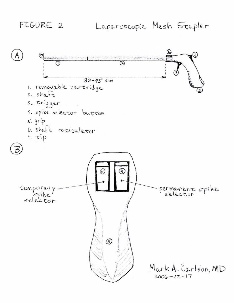
Figure 2. Side and handle-end views of the laparoscopic mesh stapler.
A. Side view. The device has the appearance of a conventional laparoscopic stapler. The shaft (2)
can be 30-45 cm long to accommodate regular and extra-large sized patients. The firing end or tip (7) of the stapler has a removable cartridge (1) which houses the spikes. This removable component allows for multiple uses of the same device, or for employment of spikes with differing capabilities. The handle or grip (5) of the device house the firing trigger (3) and spike selector buttons (4) to choose between temporary or permanent spikes. The shaft (2) also incorporates a roticulator (6), which permits the surgeon to rotate the device along the long axis of the shaft.
B. Handle-end view. This view reveals the two spike selector buttons (4), one for temporary and
the other for permanent spikes. Actuation of either button results in firing of the respective spike upon pulling the device’s trigger.
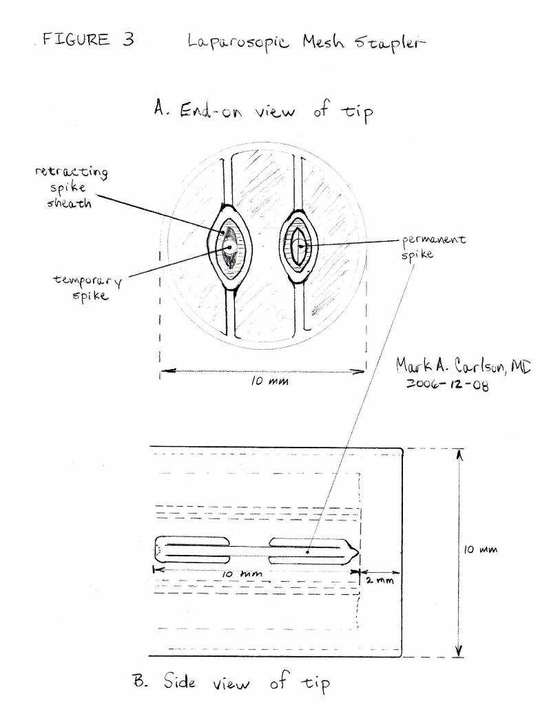
Figure 3. End-on and side views of the tip of the laparoscopic mesh stapler.
A. End-view of the tip. The initial configuration of the device utilizes a 10 mm shaft diameter;
some currently available devices employ a 5 mm shaft diameter. The larger diameter was chosen for the initial configuration of the device primarily for ease of product development. It is foreseeable that, if successful, the device eventually could be engineered down to a 5 mm shaft diameter. Inside the tip are shown end-on views of the sheaths which house both the temporary and permanent spikes. In the configuration shown, the sheaths are retractable upon firing of the instrument (see Figure 7).
B. Side view of the tip. In this phantom view a permanent spike is shown contained within a
retractable sheath at the end (tip) of the device. The configuration shown uses a retractable sheath (see Figure 7) to house the spikes.
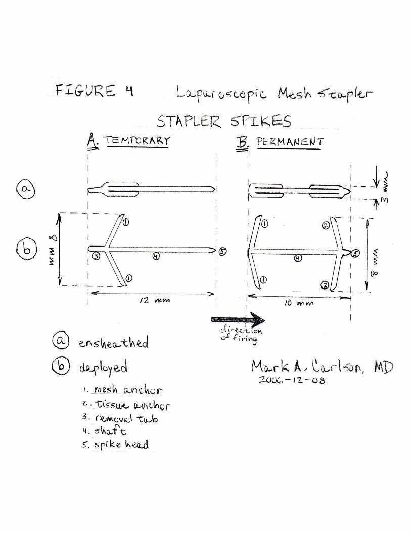
Figure 4. Close-up views of the spikes (staples) of the laparoscopic mesh stapler.
A. Temporary spike. This spike is shown in the ensheathed position (a) and also in the deployed
position (b). The direction of firing (deployment) is shown by the large, solid arrow. The temporary spike consists of a shaft (4) which is 12 mm in length. At the head (5) of the shaft there is a pointed tip; at the tail of the spike there is a tab (3) which can be grasped by a laparoscopic forceps in order to remove the spike. Also near the tail of the spike there are two mesh anchors (1) which deploy in the direction of firing (i.e., forward-deploying) in order to catch the mesh surface. The breadth of the spike with the mesh anchors deployed is 8 mm; the temporary spike otherwise is 3 mm in breath in the nondeployed (i.e., ensheathed) position.
B. Permanent spike. This spike also is shown in the ensheathed position (a) and also in the
deployed position (b). A principle differences between the permanent vs. the temporary spike is that the former has two tissue anchors (2) which deploy in the opposite direction of firing (i.e., rearward-deploying). These anchors catch tissue and resist pull-out force. The permanent spike also does not have the removal tab (3) which the temporary spike has. The permanent spike is somewhat shorter than the temporary spike (10 vs. 12 mm), but the breadth in the ensheathed (3 mm) and deployed (8 mm) positions are the same between the two spike types. In an alternative configuration (not shown), the permanent spike could have an array of four, six, or eight tissue anchors spaced at 90˚, 60˚, or 45˚ intervals around the spike tip instead of two anchors (as shown) spaced with a 180˚ interval. An increased number of tissue anchors would increase the pull-out force (i.e., tissue anchoring ability) of the spike.
Inventor: Carlson, Mark A. Laparoscopic Mesh Stapler
April 2, 2007 Page 4 of 5
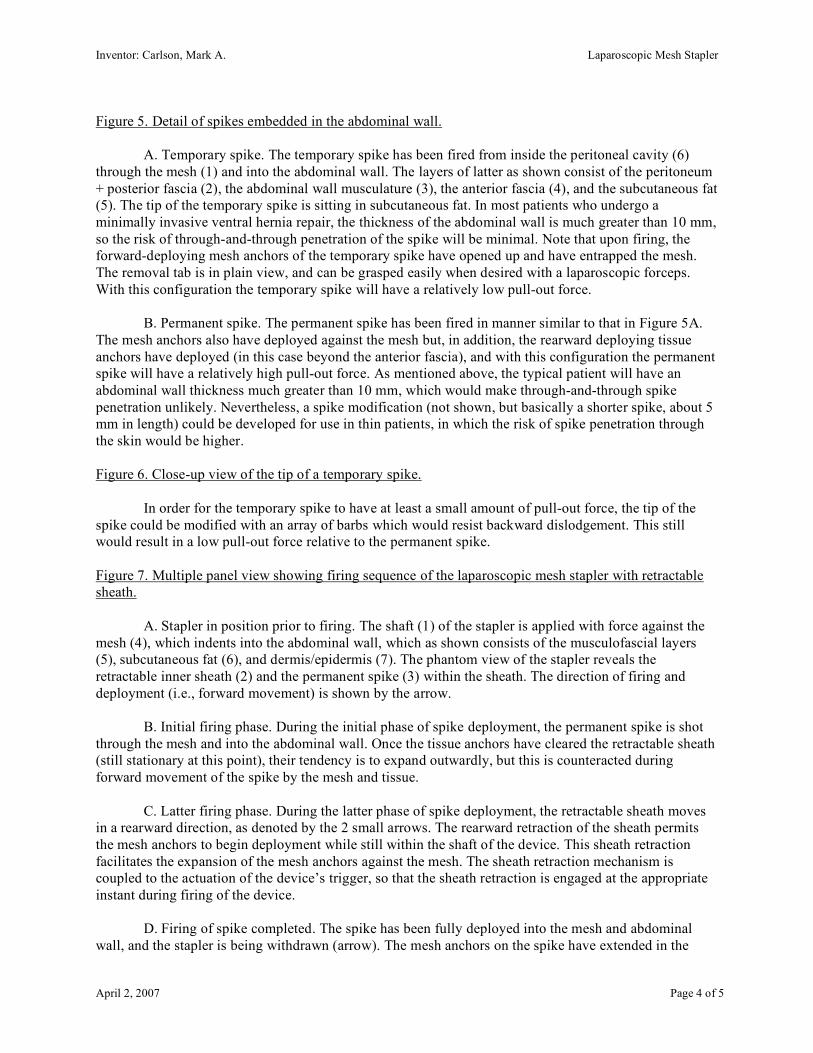
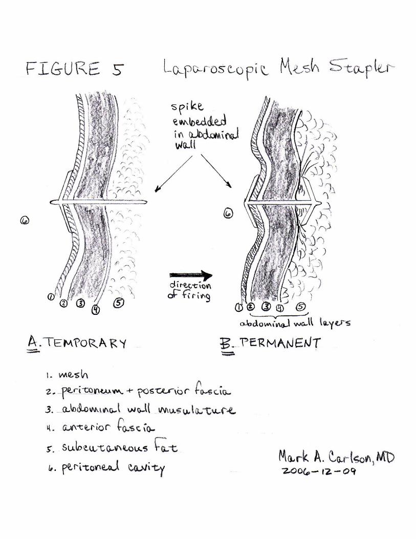
Figure 5. Detail of spikes embedded in the abdominal wall.
A. Temporary spike. The temporary spike has been fired from inside the peritoneal cavity (6)
through the mesh (1) and into the abdominal wall. The layers of latter as shown consist of the peritoneum + posterior fascia (2), the abdominal wall musculature (3), the anterior fascia (4), and the subcutaneous fat (5). The tip of the temporary spike is sitting in subcutaneous fat. In most patients who undergo a minimally invasive ventral hernia repair, the thickness of the abdominal wall is much greater than 10 mm, so the risk of through-and-through penetration of the spike will be minimal. Note that upon firing, the forward-deploying mesh anchors of the temporary spike have opened up and have entrapped the mesh. The removal tab is in plain view, and can be grasped easily when desired with a laparoscopic forceps. With this configuration the temporary spike will have a relatively low pull-out force.
B. Permanent spike. The permanent spike has been fired in manner similar to that in Figure 5A.
The mesh anchors also have deployed against the mesh but, in addition, the rearward deploying tissue anchors have deployed (in this case beyond the anterior fascia), and with this configuration the permanent spike will have a relatively high pull-out force. As mentioned above, the typical patient will have an abdominal wall thickness much greater than 10 mm, which would make through-and-through spike penetration unlikely. Nevertheless, a spike modification (not shown, but basically a shorter spike, about 5 mm in length) could be developed for use in thin patients, in which the risk of spike penetration through the skin would be higher.
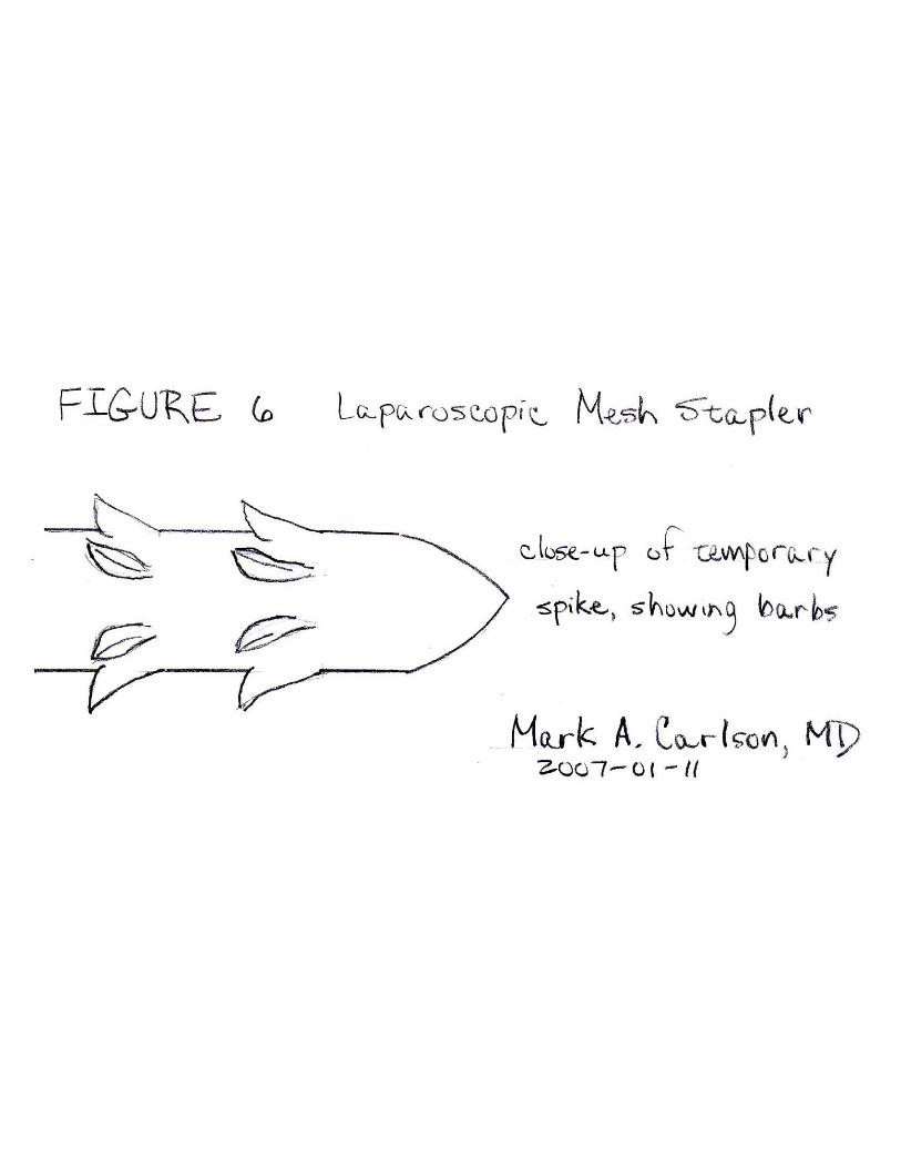
Figure 6. Close-up view of the tip of a temporary spike.
In order for the temporary spike to have at least a small amount of pull-out force, the tip of the
spike could be modified with an array of barbs which would resist backward dislodgement. This still would result in a low pull-out force relative to the permanent spike.
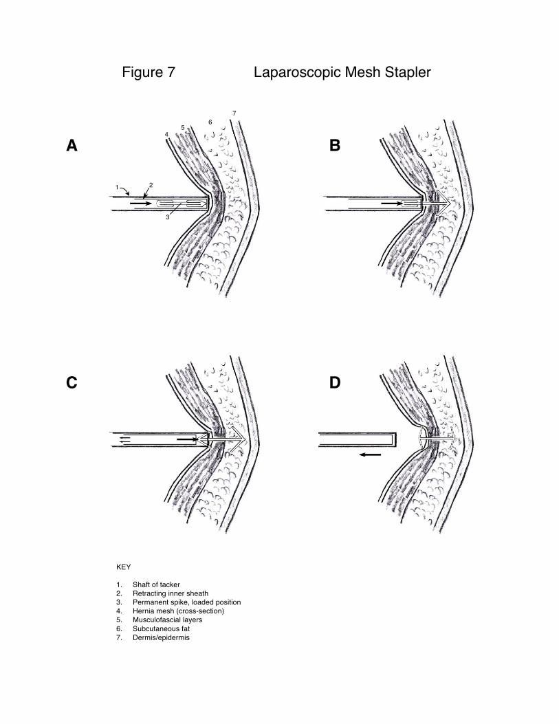
Figure 7. Multiple panel view showing firing sequence of the laparoscopic mesh stapler with retractable sheath.
A. Stapler in position prior to firing. The shaft (1) of the stapler is applied with force against the
mesh (4), which indents into the abdominal wall, which as shown consists of the musculofascial layers (5), subcutaneous fat (6), and dermis/epidermis (7). The phantom view of the stapler reveals the retractable inner sheath (2) and the permanent spike (3) within the sheath. The direction of firing and deployment (i.e., forward movement) is shown by the arrow.
B. Initial firing phase. During the initial phase of spike deployment, the permanent spike is shot
through the mesh and into the abdominal wall. Once the tissue anchors have cleared the retractable sheath (still stationary at this point), their tendency is to expand outwardly, but this is counteracted during forward movement of the spike by the mesh and tissue.
C. Latter firing phase. During the latter phase of spike deployment, the retractable sheath moves
in a rearward direction, as denoted by the 2 small arrows. The rearward retraction of the sheath permits the mesh anchors to begin deployment while still within the shaft of the device. This sheath retraction facilitates the expansion of the mesh anchors against the mesh. The sheath retraction mechanism is coupled to the actuation of the device’s trigger, so that the sheath retraction is engaged at the appropriate instant during firing of the device.
D. Firing of spike completed. The spike has been fully deployed into the mesh and abdominal
wall, and the stapler is being withdrawn (arrow). The mesh anchors on the spike have extended in the
Inventor: Carlson, Mark A. Laparoscopic Mesh Stapler
April 2, 2007 Page 5 of 5
forward direction, and have caught the surface of the mesh. The tissue anchors of the spike also have completed there extension in the rearward direction; any force tending to pull the spike out (i.e., in the rearward direction) will be countered by the deployed tissue anchors.
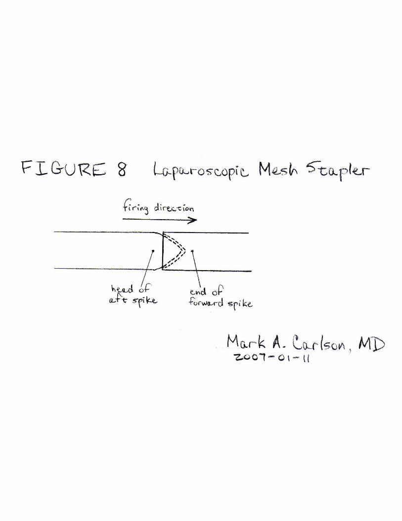
Figure 8. Schematic showing interlocking mechanism of spike head and tail.
The device is intended to be a consecutive multifire instrument, with a magazine of about 12-15
spikes of both the temporary and permanent type (i.e., up to 30 spikes total). The spikes will be lined up in single file within the sheath of the device. At the end of each spike there will be a small “hollow” into which the tip of the preceding spike will fit with an interlocking fashion, as shown. A firing pin (not shown) placed proximally in the device will fit into the hollow of the most proximal (i.e., closest to the handle of the device) spike. When the device trigger is actuated, the firing pin will push the entire line of spikes forward, and deploy one spike per firing cycle out of the tip of the device.
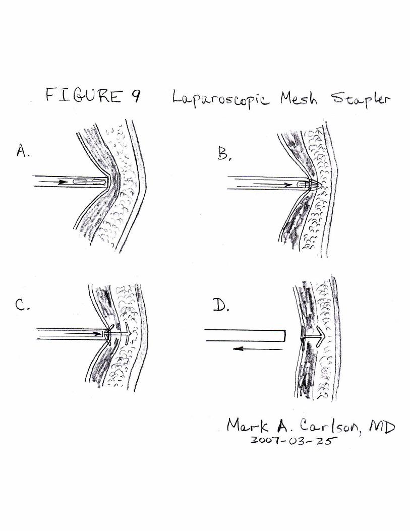
Figure 9. Multiple panel view showing alternative firing sequence of the laparoscopic mesh stapler with a nonretractable sheath.
A. Stapler in position prior to firing. In this alternative device configuration, the sheath which
houses the spikes within the shaft of the device is a nonretractable unit. This device configuration will have less moving parts in its interior. Similar to Figure 7, the shaft of the device is applied with force against the mesh, which indents into the abdominal wall, which as shown consists of the musculofascial layers, subcutaneous fat, and dermis/epidermis (refer to Figure 7A). The phantom view of the stapler reveals the nonretractable inner sheath and a permanent spike within the sheath (refer to Figure 7A). The direction of firing and deployment (i.e., forward movement) is shown by the arrow.
B. Initial firing phase. This phase is analogous to that shown in Figure 7B. During the initial
phase of spike deployment, the permanent spike is shot through the mesh and into the abdominal wall. Once the tissue anchors have cleared the retractable sheath (still stationary at this point), their tendency is to expand outwardly, but this is counteracted during forward movement of the spike by the mesh and tissue.
C. Latter firing phase. This phase differs from that shown in Figure 7C. The nonretractable sheath
in the device configuration shown in Figure 9C is offset further back from the tip of the shaft compared to the retractable sheath of the device shown in Figure 7C. This allows for earlier expansion of the mesh anchors within the shaft of the device, which should allow this mesh anchors to catch an adequate bite of the mesh as the spike is shot into the mesh and abdominal wall.
D. Firing of spike completed. This phase is analogous to that shown in Figure 7D. The spike has
been fully deployed into the mesh and abdominal wall, and the stapler is being withdrawn (arrow). The mesh anchors on the spike have extended in the forward direction, and have caught the surface of the mesh. The tissue anchors of the spike also have completed there extension in the rearward direction; any force tending to pull the spike out (i.e., in the rearward direction) will be countered by the deployed tissue anchors.
A B
C D
2
3
45
67
1
KEY
1. Shaft of tacker2. Retracting inner sheath3. Permanent spike, loaded position4. Hernia mesh (cross-section)5. Musculofascial layers6. Subcutaneous fat7. Dermis/epidermis
Figure 7 Laparoscopic Mesh Stapler