Embed Size (px)
Citation preview

Nephrourol Mon. 2017 May; 9(3):e45086.
Published online 2017 March 7.
doi: 10.5812/numonthly.45086.
Case Report
Large Cell Neuroendocrine Carcinoma of Prostate: A Rare Interesting
Case and Literature Review
Reza Mahdavi Zafarghandi,1 Mahmood Reza Kalantari,2 Alireza Akhavan Rezayat,3,* and Amir Abbas
Asadpour3
1Professor of Urology, Mashhad University of Medical Sciences, Mashhad, IR Iran2Associated Professor of Pathology, Mashhad University of Medical Sciences, Mashhad, IR Iran3Assistant professor of Urology, Mashhad University of Medical Sciences, Mashhad, IR Iran
*Corresponding author: Alireza Akhavan Rezayat, Ghaem hospital, Ahmad Abad Ave., Mashhad, IR Iran. Tel: +98-9153148223, Fax: +98-5138012857, E-mail:[email protected]
Received 2016 December 29; Accepted 2017 February 12.
Abstract
Prostate cancer (PC) is one of the most common forms of malignancies and the second cause of cancer death all around the worldand the eight causes in Iran. The main portion of PC is adenocarcinoma, in some cases neuroendocrine differentiation occurs. Neu-roendocrine prostate cancers (NePCs) incidence varies from 0.5% to 2% of all prostate cancers. Large cell neuroendocrine differen-tiation is very rare. In this study we presented a 71-year-old man with large cell neuroendocrine carcinoma of the prostate.
Keywords: Neuroendocrine, Prostate Cancer, Adenocarcinoma
1. Introduction
The majority of prostate cancers are adenocarcinoma,but NE differentiation can occur in 10% of cases in part de-pending on the number of slides studied and the num-ber of antibodies used (1). Neuroendocrine cells are smallsubjects in the normal prostate tissue. They are derivedfrom putative stem cells and are androgen receptor neg-ative. In recent studies, it was proposed that they regu-late prostate epithelium secretary and proliferative activ-ity. NE cells also can be found in prostate cancers (2, 3). Themain portion of PC is adenocarcinoma, in some cases neu-roendocrine differentiation occurs (4). Neuroendocrineprostate cancers (NePCs) incidence varies from 0.5% to 2%of all PCs (3). Neuroendocrine prostate adenocarcinomasare androgen receptor negative (5).
NE tumors of prostate are three groups in regard toworld health organization (WHO) histologic classification:1- focal NE differentiation in conventional prostate cancer(PC); 2- carcinoid tumor; 3- small cell NE carcinoma (1).However prostate adenocarcinomas with NE differentia-tion are divided into Paneth cell, carcinoid tumor, smallcell carcinoma, large cell carcinoma (LCNEC), and mixedform in regard to 2013 prostate cancer foundation clas-sification system (6). Prostate cancer manifestations varyfrom silent diseases to high grade aggressive forms (3).
Most of neuroendocrine carcinomas in prostate occurin the content of typical adenocarcinoma. Pure neuroen-docrine prostate cancers (NePCs) are very rare. The most
frequent type of NePCs is small cell carcinoma whereaslarge cell carcinoma (LCNEC) and carcinoid tumor are ex-tremely rare (4, 5). To date, most published LCNECs repre-sented progression from prior typical adenocarcinoma inthe content of long standing androgen deprivation ther-apy (ADT) (6). NE cells and NePCs lack AR and are clinicallyconsidered hormone refractory. Overall, NePCs are aggres-sive and are associated with rapid disorientation and poorprognosis (1, 2).
De Novo LCNEC is exceptionally rare particularly inpure forms. Until now, only four series of LCNEC have beenpublished (7). The largest series by Evans et al. described7 cases with only one pure de novo case. The last casewas published by Acosta Gonzalez et al. (2). In this study,we present a 71 year old man with pure de novo LCNEC ofprostate.
2. Case Presentation
The patient’s informed consent has been obtained forthe publication of the case reports. A 71-year-old male re-ferred to our clinic with increased dysuria, frequently andurgently for 3 months. He had no systemic signs and symp-toms such as fever, night sweats, anorexia, and weightloss. On the digital rectal examination, prostate was en-larged. Prostate specific antigen (PSA) was in the normalrange (0.09 ng/mL). Ultrasonography indicated moderateenlargement of prostate and Benign prostate hyperpla-sia (BPH). So, tamsulosin was administered for him in re-
Copyright © 2017, Nephrology and Urology Research Center. This is an open-access article distributed under the terms of the Creative CommonsAttribution-NonCommercial 4.0 International License (http://creativecommons.org/licenses/by-nc/4.0/) which permits copy and redistribute the material just innoncommercial usages, provided the original work is properly cited.
Mahdavi Zafarghandi R et al.
gard to BPH diagnosis. He referred again without any re-lief after 3 months. His international prostate symptomsscore (IPSS) was 22 before treatment and had remained un-changed after three months. He underwent transurethralresection of the prostate (TURP) because of the severityof the symptoms and the patient’s desire. Histopathol-ogy examination and urine cytology was conducted afterTURP. First pathologist reported poorly differentiated car-cinoma, so colonoscopy and whole body scan were per-formed for him and both were negative. Second patholo-gist proposed LCNEC. The patient refused treatment in thisstep and presented again after three months with uremia,confusion, and poor general condition. He complained ofsever pelvic pain. In the first step, abdomino-pelvic ultra-sonography was performed for him and it showed bilateralhydronephrosis. He underwent hemodialysis due to in-creased creatinine (8 mg/dL) and ultrasound guided bilat-eral nephrostomy for obstructive uropathy. A subsequentnon-contrast CT confirmed a large pelvic mass with pelviclymphadenopathy (Figure 1). Tumor was unresectable andhe was referred to the oncology department for palliativeradiotherapy.
2.1. Pathological Findings
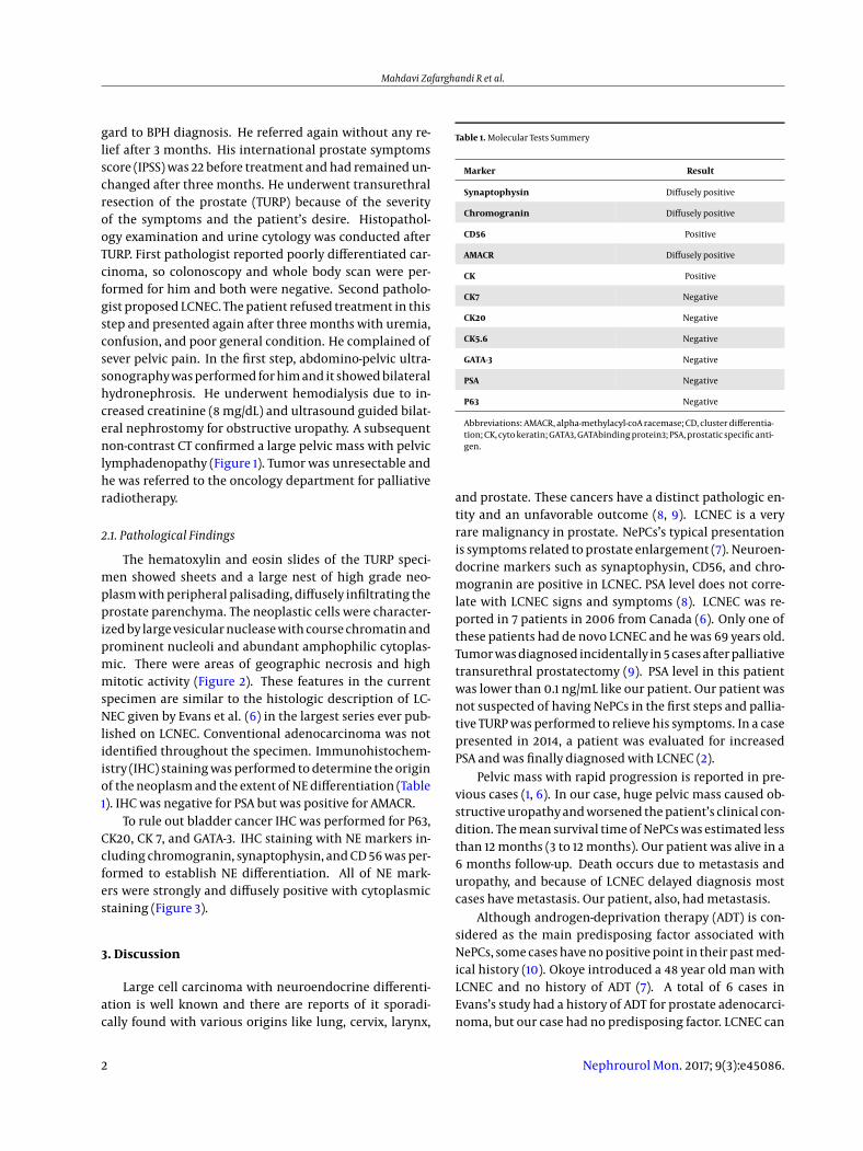
The hematoxylin and eosin slides of the TURP speci-men showed sheets and a large nest of high grade neo-plasm with peripheral palisading, diffusely infiltrating theprostate parenchyma. The neoplastic cells were character-ized by large vesicular nuclease with course chromatin andprominent nucleoli and abundant amphophilic cytoplas-mic. There were areas of geographic necrosis and highmitotic activity (Figure 2). These features in the currentspecimen are similar to the histologic description of LC-NEC given by Evans et al. (6) in the largest series ever pub-lished on LCNEC. Conventional adenocarcinoma was notidentified throughout the specimen. Immunohistochem-istry (IHC) staining was performed to determine the originof the neoplasm and the extent of NE differentiation (Table1). IHC was negative for PSA but was positive for AMACR.
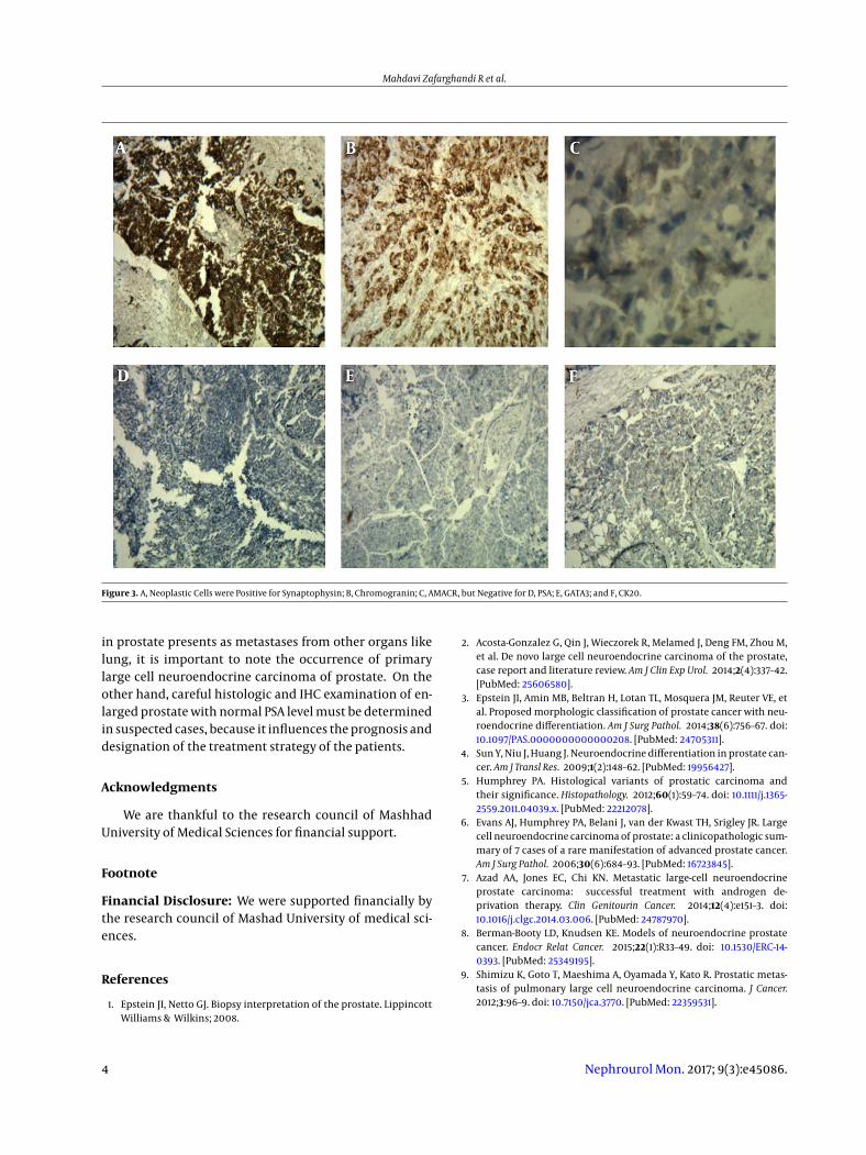
To rule out bladder cancer IHC was performed for P63,CK20, CK 7, and GATA-3. IHC staining with NE markers in-cluding chromogranin, synaptophysin, and CD 56 was per-formed to establish NE differentiation. All of NE mark-ers were strongly and diffusely positive with cytoplasmicstaining (Figure 3).
3. Discussion
Large cell carcinoma with neuroendocrine differenti-ation is well known and there are reports of it sporadi-cally found with various origins like lung, cervix, larynx,
Table 1. Molecular Tests Summery
Marker Result
Synaptophysin Diffusely positive
Chromogranin Diffusely positive
CD56 Positive
AMACR Diffusely positive
CK Positive
CK7 Negative
CK20 Negative
CK5.6 Negative
GATA-3 Negative
PSA Negative
P63 Negative
Abbreviations: AMACR, alpha-methylacyl-coA racemase; CD, cluster differentia-tion; CK, cyto keratin; GATA3, GATAbinding protein3; PSA, prostatic specific anti-gen.
and prostate. These cancers have a distinct pathologic en-tity and an unfavorable outcome (8, 9). LCNEC is a veryrare malignancy in prostate. NePCs’s typical presentationis symptoms related to prostate enlargement (7). Neuroen-docrine markers such as synaptophysin, CD56, and chro-mogranin are positive in LCNEC. PSA level does not corre-late with LCNEC signs and symptoms (8). LCNEC was re-ported in 7 patients in 2006 from Canada (6). Only one ofthese patients had de novo LCNEC and he was 69 years old.Tumor was diagnosed incidentally in 5 cases after palliativetransurethral prostatectomy (9). PSA level in this patientwas lower than 0.1 ng/mL like our patient. Our patient wasnot suspected of having NePCs in the first steps and pallia-tive TURP was performed to relieve his symptoms. In a casepresented in 2014, a patient was evaluated for increasedPSA and was finally diagnosed with LCNEC (2).
Pelvic mass with rapid progression is reported in pre-vious cases (1, 6). In our case, huge pelvic mass caused ob-structive uropathy and worsened the patient’s clinical con-dition. The mean survival time of NePCs was estimated lessthan 12 months (3 to 12 months). Our patient was alive in a6 months follow-up. Death occurs due to metastasis anduropathy, and because of LCNEC delayed diagnosis mostcases have metastasis. Our patient, also, had metastasis.
Although androgen-deprivation therapy (ADT) is con-sidered as the main predisposing factor associated withNePCs, some cases have no positive point in their past med-ical history (10). Okoye introduced a 48 year old man withLCNEC and no history of ADT (7). A total of 6 cases inEvans’s study had a history of ADT for prostate adenocarci-noma, but our case had no predisposing factor. LCNEC can
2 Nephrourol Mon. 2017; 9(3):e45086.

Mahdavi Zafarghandi R et al.
Figure 1. Massive Pelvic Mass with Neural Invasion and Nephrostomy
Figure 2. A, Sheet of Neoplastic Cells; B, Infiltrating Prostate Parenchyma; C, with Geographic Necrosis; D, The Neoplastic Cells had Prominent Nucleoli.
present in young males and might have a genetic base (7,11). Animal model studies showed that prostate neuroen-docrine cells could show a malignant transformation, aswell (12). Some de novo LCNEC cases express androgen re-ceptor (AR) and might be androgen dependent but othercases are AR negative (2). The main and exact mechanismand underlying causes of LCNEC are unknown.
In our case, CD56 was positive and Evans showed thatthis immunohistochemical marker was positive in all LC-NEC patients (6). It was reported in previous studies thatLCNEC cells expressed CD56, chromogranin, and synapto-physin (10). LCNEC can be diagnosed if one of the markersbecome positive. The first report of LCNEC Immunohisto-chemistry findings was published by Wynn et al. in 2000(12). And this marker should be evaluated in suspectedcases.
Travis et al. described LCNEC by specific immunohis-tochemistry (IHC) and electron microscopy (EM) features.He studied 5 cases of LCNEC and showed that these patientsprognosis varies between atypical carcinoid and small cellcarcinoma (12). The overall survival of patients with NePCsis estimated 9 to 12 months (6). All patients in Evans’s study
died soon after diagnosis. It seems that increased neuroen-docrine differentiation is correlated with more aggressiveforms of diseases and a poor prognosis (6). Our patient wasdischarged from the hospital and 6 months after his dis-charge he was still alive.
Our patient had sever pelvic pain which might have oc-curred due to neural invasion, or the compression effect oflarge pelvic lymph nodes and huge tumor. In other casereports, large mass led to urinary retention despite of pain(13). In most cases, LCNEC has been diagnosed by delay, andtumor was not resectable. Pelvic lymph node infiltrationand metastases are common in patients with LCNEC likeour patient.
LCNEC responded poorly to standard NePCschemotherapy protocols. There are some recommen-dations for using novel and additional treatment suchas somatostatin analogues in these cases, but more casesshould be evaluated to develop the exact therapeuticstrategy for LCNEC (14-16).
In conclusion, considering LCNEC as a differential di-agnosis in patients with prostate cancer is important. Al-though sometimes large cell neuroendocrine carcinoma
Nephrourol Mon. 2017; 9(3):e45086. 3
Mahdavi Zafarghandi R et al.
Figure 3. A, Neoplastic Cells were Positive for Synaptophysin; B, Chromogranin; C, AMACR, but Negative for D, PSA; E, GATA3; and F, CK20.
in prostate presents as metastases from other organs likelung, it is important to note the occurrence of primarylarge cell neuroendocrine carcinoma of prostate. On theother hand, careful histologic and IHC examination of en-larged prostate with normal PSA level must be determinedin suspected cases, because it influences the prognosis anddesignation of the treatment strategy of the patients.
Acknowledgments
We are thankful to the research council of MashhadUniversity of Medical Sciences for financial support.
Footnote
Financial Disclosure: We were supported financially bythe research council of Mashad University of medical sci-ences.
References
1. Epstein JI, Netto GJ. Biopsy interpretation of the prostate. LippincottWilliams & Wilkins; 2008.
2. Acosta-Gonzalez G, Qin J, Wieczorek R, Melamed J, Deng FM, Zhou M,et al. De novo large cell neuroendocrine carcinoma of the prostate,case report and literature review. Am J Clin Exp Urol. 2014;2(4):337–42.[PubMed: 25606580].
3. Epstein JI, Amin MB, Beltran H, Lotan TL, Mosquera JM, Reuter VE, etal. Proposed morphologic classification of prostate cancer with neu-roendocrine differentiation. Am J Surg Pathol. 2014;38(6):756–67. doi:10.1097/PAS.0000000000000208. [PubMed: 24705311].
4. Sun Y, Niu J, Huang J. Neuroendocrine differentiation in prostate can-cer. Am J Transl Res. 2009;1(2):148–62. [PubMed: 19956427].
5. Humphrey PA. Histological variants of prostatic carcinoma andtheir significance. Histopathology. 2012;60(1):59–74. doi: 10.1111/j.1365-2559.2011.04039.x. [PubMed: 22212078].
6. Evans AJ, Humphrey PA, Belani J, van der Kwast TH, Srigley JR. Largecell neuroendocrine carcinoma of prostate: a clinicopathologic sum-mary of 7 cases of a rare manifestation of advanced prostate cancer.Am J Surg Pathol. 2006;30(6):684–93. [PubMed: 16723845].
7. Azad AA, Jones EC, Chi KN. Metastatic large-cell neuroendocrineprostate carcinoma: successful treatment with androgen de-privation therapy. Clin Genitourin Cancer. 2014;12(4):e151–3. doi:10.1016/j.clgc.2014.03.006. [PubMed: 24787970].
8. Berman-Booty LD, Knudsen KE. Models of neuroendocrine prostatecancer. Endocr Relat Cancer. 2015;22(1):R33–49. doi: 10.1530/ERC-14-0393. [PubMed: 25349195].
9. Shimizu K, Goto T, Maeshima A, Oyamada Y, Kato R. Prostatic metas-tasis of pulmonary large cell neuroendocrine carcinoma. J Cancer.2012;3:96–9. doi: 10.7150/jca.3770. [PubMed: 22359531].
4 Nephrourol Mon. 2017; 9(3):e45086.

Mahdavi Zafarghandi R et al.
10. Terry S, Beltran H. The many faces of neuroendocrine differenti-ation in prostate cancer progression. Front Oncol. 2014;4:60. doi:10.3389/fonc.2014.00060. [PubMed: 24724054].
11. Hoof P, Tsai-Nguyen G, Paulson S, Syed A, Mora A Jr. Neuroen-docrine carcinoma of the prostate gland. Proc (Bayl Univ Med Cent).2016;29(1):68–9. [PubMed: 26722176].
12. Wynn SS, Nagabundi S, Koo J, Chin NW. Recurrent prostate car-cinoma presenting as omental large cell carcinoma with neu-roendocrine differentiation and resulting in bowel obstruction.Arch Pathol Lab Med. 2000;124(7):1074–6. doi: 10.1043/0003-9985(2000)124<1074:RPCPAO>2.0.CO;2. [PubMed: 10888786].
13. Okoye E, Choi EK, Divatia M, Miles BJ, Ayala AG, Ro JY. De novo large cellneuroendocrine carcinoma of the prostate gland with pelvic lymphnode metastasis: a case report with review of literature. Int J Clin Exp
Pathol. 2014;7(12):9061–6.14. Priemer DS, Montironi R, Wang L, Williamson SR, Lopez-Beltran A,
Cheng L. Neuroendocrine Tumors of the Prostate: Emerging In-sights from Molecular Data and Updates to the 2016 World HealthOrganization Classification. Endocr Pathol. 2016;27(2):123–35. doi:10.1007/s12022-016-9421-z. [PubMed: 26885643].
15. Lin D, Tan AJ, De Sousa AF, Singh-Rai R. A rare case of large cell neu-roendocrine carcinoma.BMJCaseRep. 2014;2014doi: 10.1136/bcr-2014-206403. [PubMed: 25331150].
16. Travis WD, Linnoila RI, Tsokos MG, Hitchcock CL, Cutler GB Jr, NiemanL, et al. Neuroendocrine tumors of the lung with proposed criteria forlarge-cell neuroendocrine carcinoma. An ultrastructural, immuno-histochemical, and flow cytometric study of 35 cases.Am J Surg Pathol.1991;15(6):529–53. [PubMed: 1709558].
Nephrourol Mon. 2017; 9(3):e45086. 5