Embed Size (px)
Citation preview
Lasers in dermatology
Magdolna Gaál M.D., PhD
Lasers work by emitting a powerful beam of light,
which causes a certain effect on a specific target.
Laser surgery
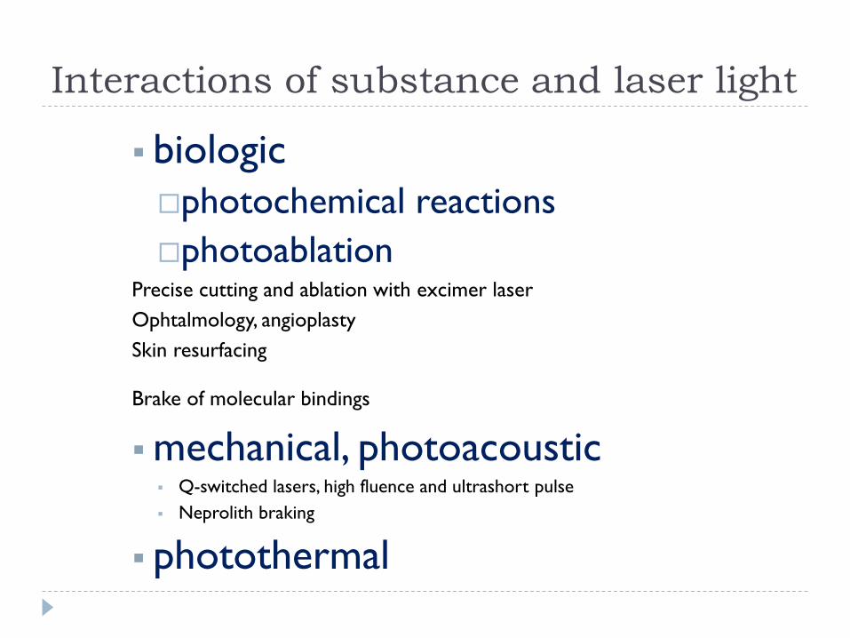
Interactions of substance and laser light
▪ biologic
photochemical reactions
photoablationPrecise cutting and ablation with excimer laser
Ophtalmology, angioplasty
Skin resurfacing
Brake of molecular bindings
▪mechanical, photoacoustic▪ Q-switched lasers, high fluence and ultrashort pulse
▪ Neprolith braking
▪ photothermal
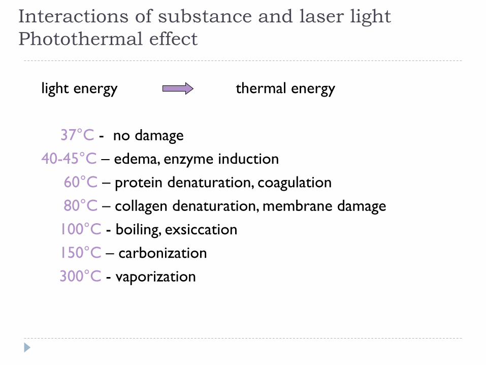
Interactions of substance and laser light
Photothermal effect
light energy thermal energy
37°C - no damage
40-45°C – edema, enzyme induction
60°C – protein denaturation, coagulation
80°C – collagen denaturation, membrane damage
100°C - boiling, exsiccation
150°C – carbonization
300°C - vaporization
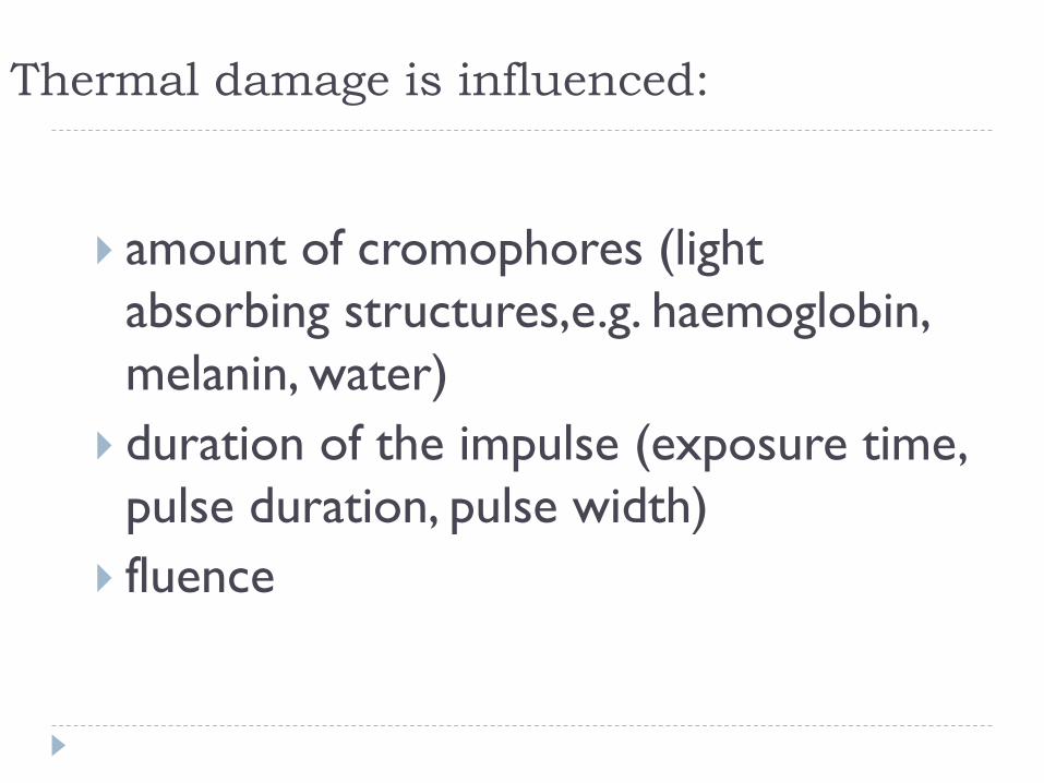
Thermal damage is influenced:
amount of cromophores (light
absorbing structures,e.g. haemoglobin,
melanin, water)
duration of the impulse (exposure time,
pulse duration, pulse width)
fluence
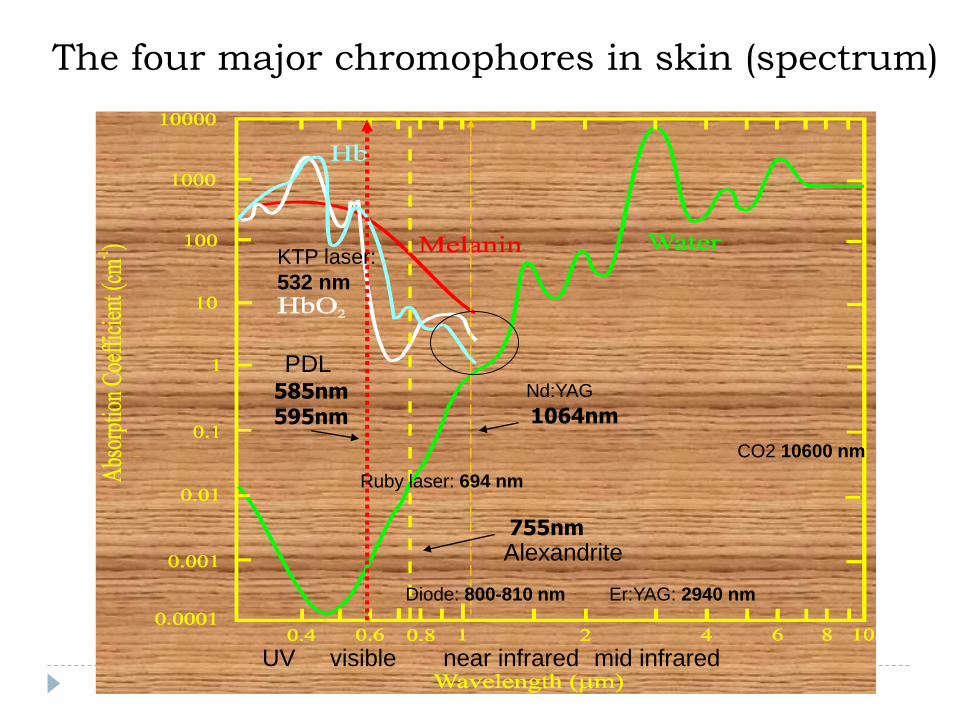
The four major chromophores in skin (spectrum)
585nm595nm
755nm
1064nm
CO2 10600 nm
Nd:YAG
Ruby laser: 694 nm
PDL
Alexandrite
Diode: 800-810 nm
KTP laser:
532 nm
visibleUV near infrared mid infrared
Er:YAG: 2940 nm
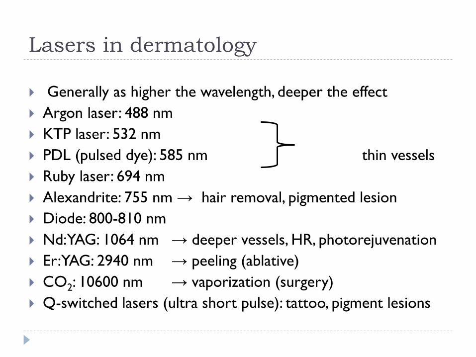
Lasers in dermatology
Generally as higher the wavelength, deeper the effect
Argon laser: 488 nm
KTP laser: 532 nm
PDL (pulsed dye): 585 nm thin vessels
Ruby laser: 694 nm
Alexandrite: 755 nm → hair removal, pigmented lesion
Diode: 800-810 nm
Nd:YAG: 1064 nm → deeper vessels, HR, photorejuvenation
Er:YAG: 2940 nm → peeling (ablative)
CO2: 10600 nm → vaporization (surgery)
Q-switched lasers (ultra short pulse): tattoo, pigment lesions
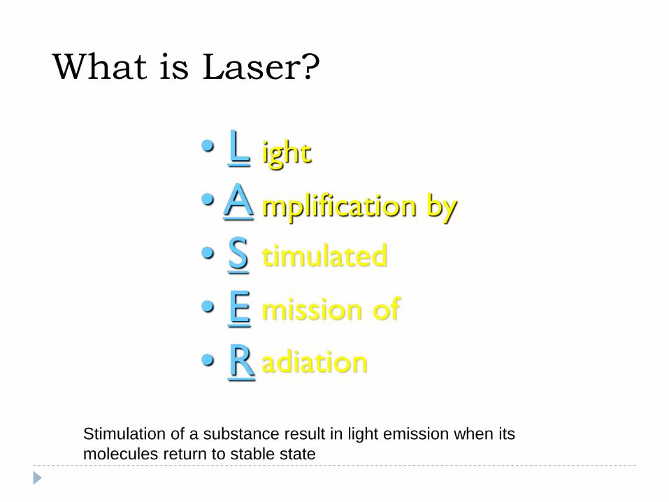
What is Laser?
ight
mplification by
adiation
timulated
• L
• A
• S
• E
• R
mission of
Stimulation of a substance result in light emission when its
molecules return to stable state
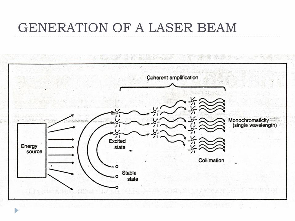
GENERATION OF A LASER BEAM
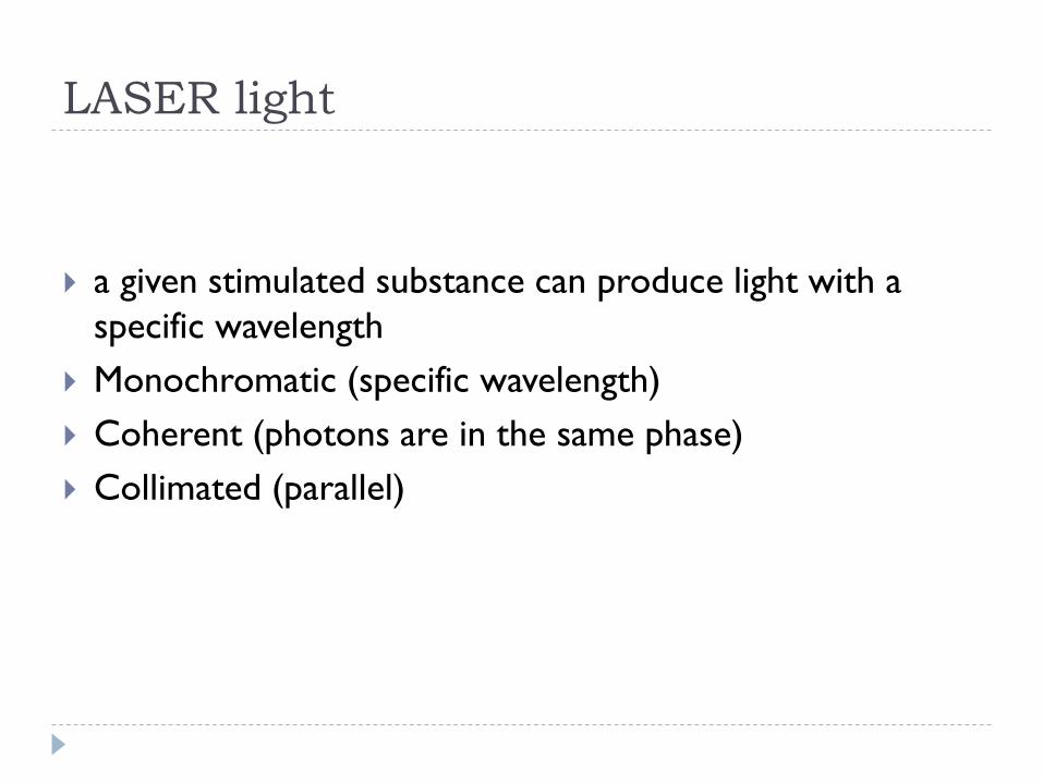
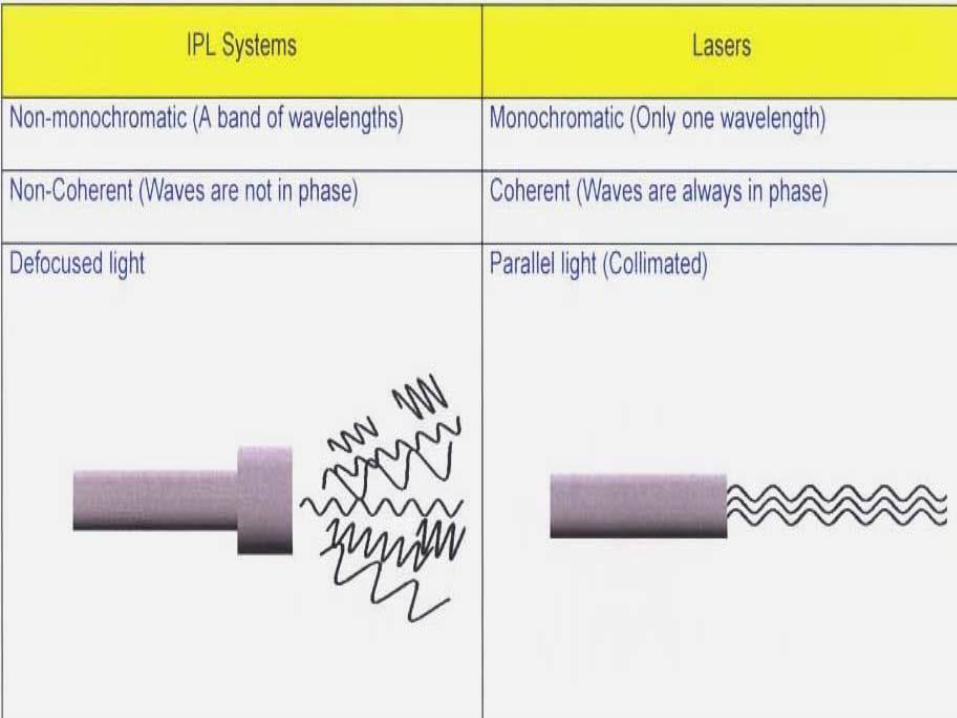
LASER light
a given stimulated substance can produce light with a
specific wavelength
Monochromatic (specific wavelength)
Coherent (photons are in the same phase)
Collimated (parallel)
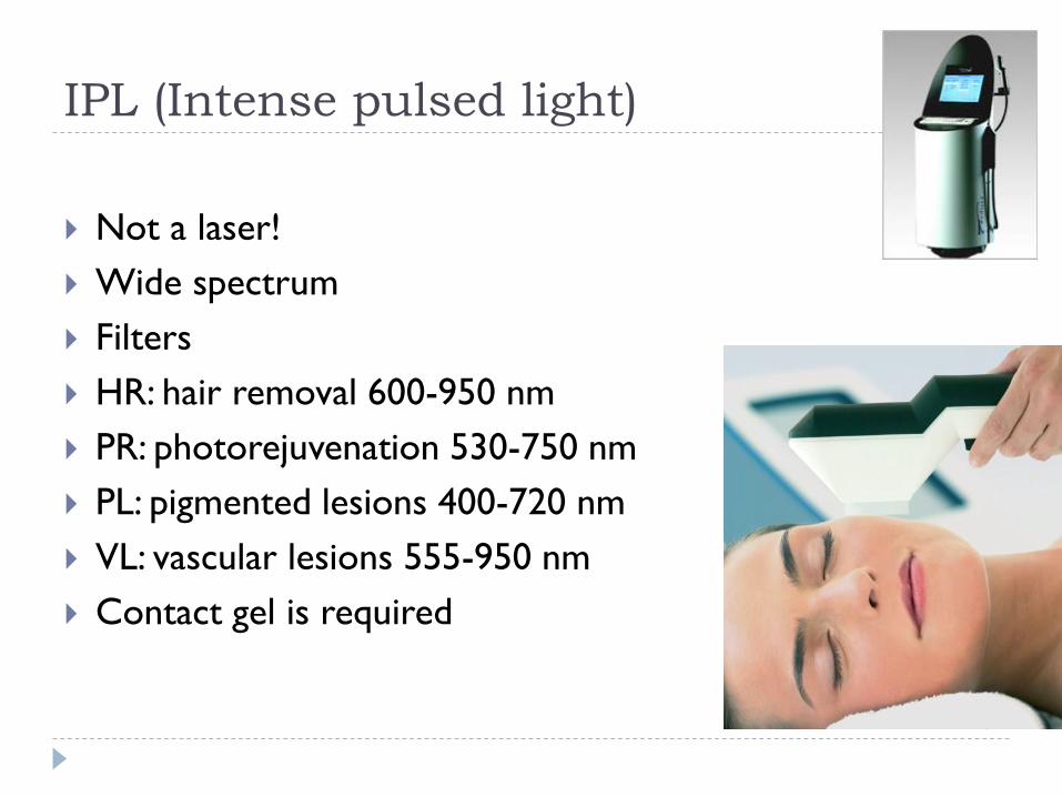
IPL (Intense pulsed light)
Not a laser!
Wide spectrum
Filters
HR: hair removal 600-950 nm
PR: photorejuvenation 530-750 nm
PL: pigmented lesions 400-720 nm
VL: vascular lesions 555-950 nm
Contact gel is required
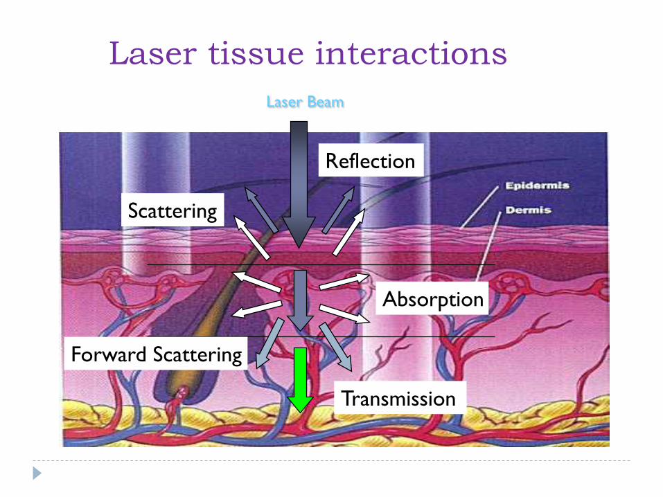
Laser Beam
Reflection
Scattering
Absorption
Forward Scattering
Transmission
Laser tissue interactions
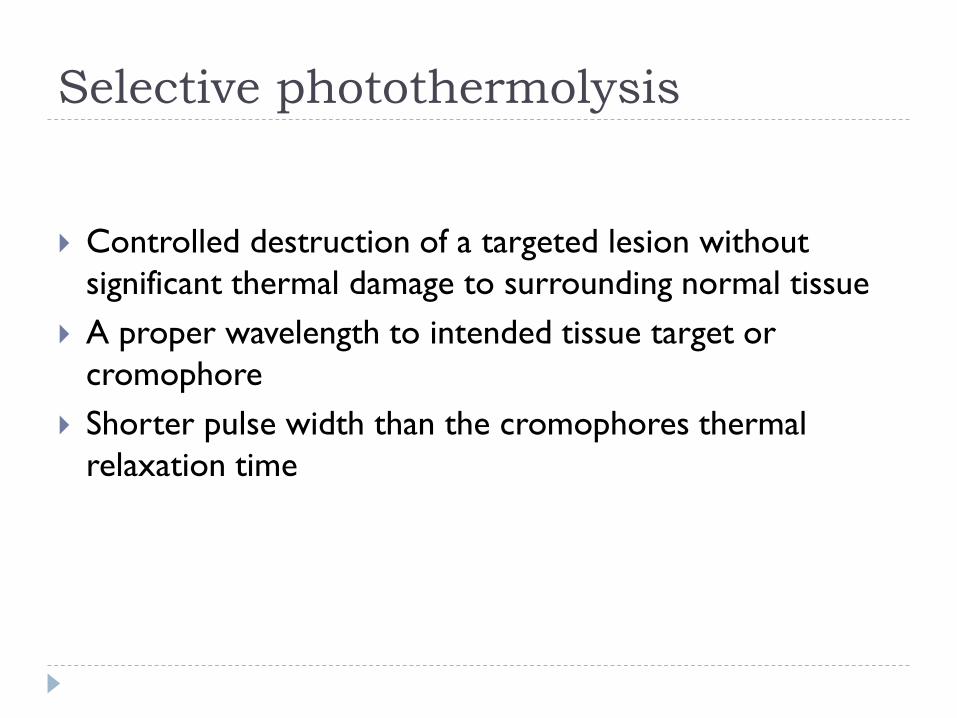
Selective photothermolysis
Controlled destruction of a targeted lesion without
significant thermal damage to surrounding normal tissue
A proper wavelength to intended tissue target or
cromophore
Shorter pulse width than the cromophores thermal
relaxation time
Thermal Relaxation Time
The time necessary for the target to cool down50%, through the transfer of its heat to surrounding tissue via thermal diffusion.
➢Laser hair removal
Hair removal
➢ alexandrite, Nd:YAG, IPL
➢ target: the hair bulb and a stem cell area (near adhesion of m. arrector pili)
➢ result depends: type of laser, color and thickness of the hair shaft, skin pigmentation
➢ ideal patient: light skin, dark hair
➢ hair shaft in anagen phasis, monthly, 5-7 treatment sessions
➢ maintaining sessions (1-2/year )
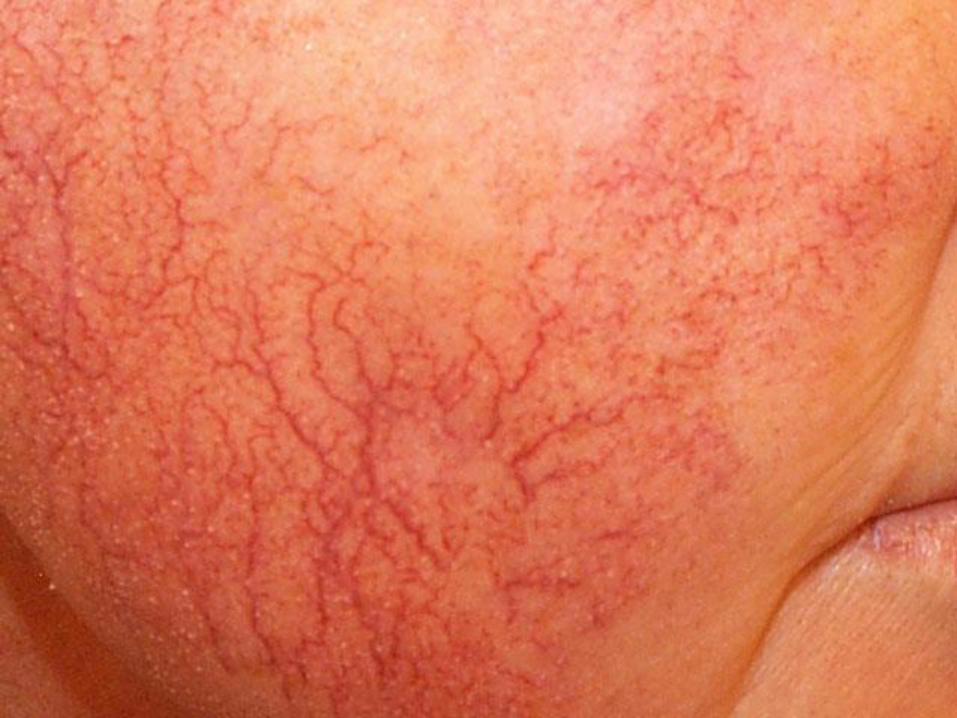
➢Vascular Lesions
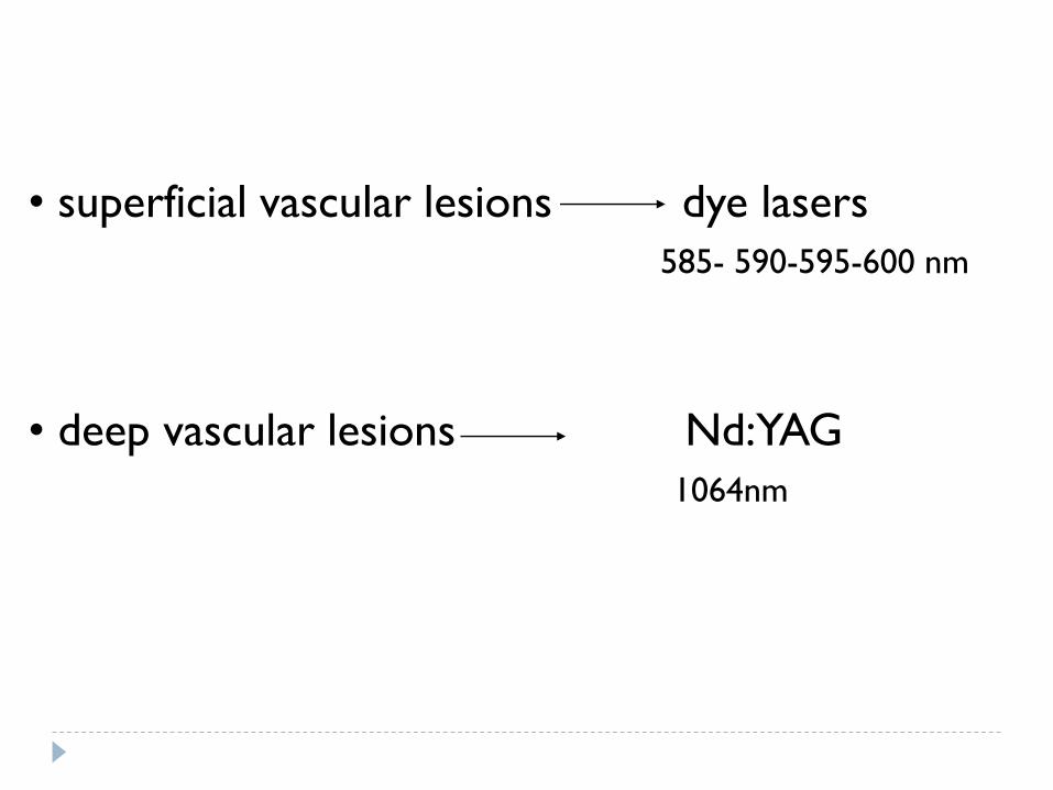
• superficial vascular lesions dye lasers
585- 590-595-600 nm
• deep vascular lesions Nd:YAG
1064nm
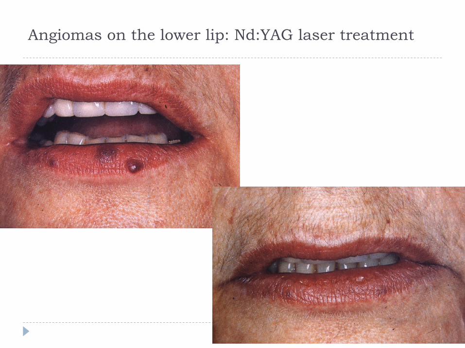
Angiomas on the lower lip: Nd:YAG laser treatment
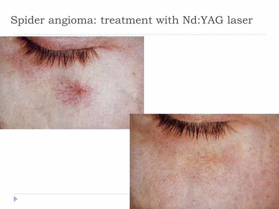
Spider angioma: treatment with Nd:YAG laser
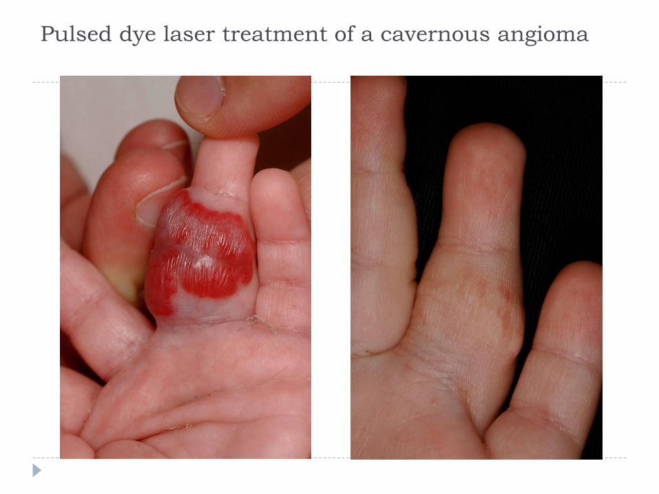
Pulsed dye laser treatment of a cavernous angioma
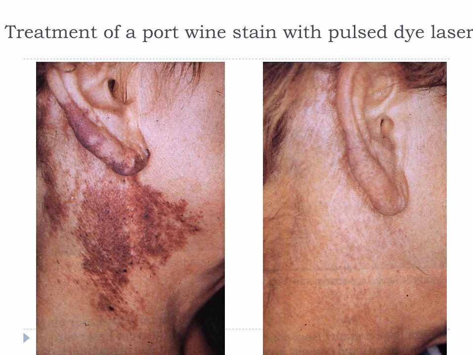
Treatment of a port wine stain with pulsed dye laser
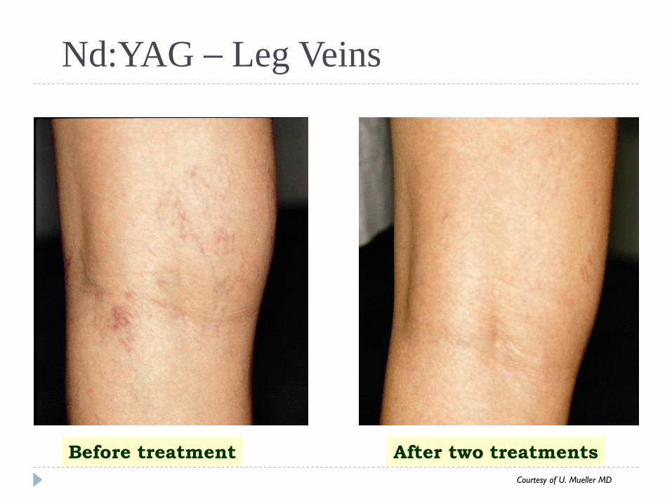
Courtesy of U. Mueller MD
Before treatment After two treatments
Nd:YAG – Leg Veins
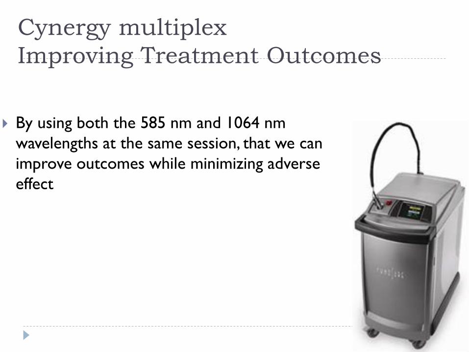
Cynergy multiplex
Improving Treatment Outcomes
By using both the 585 nm and 1064 nm
wavelengths at the same session, that we can
improve outcomes while minimizing adverse
effect
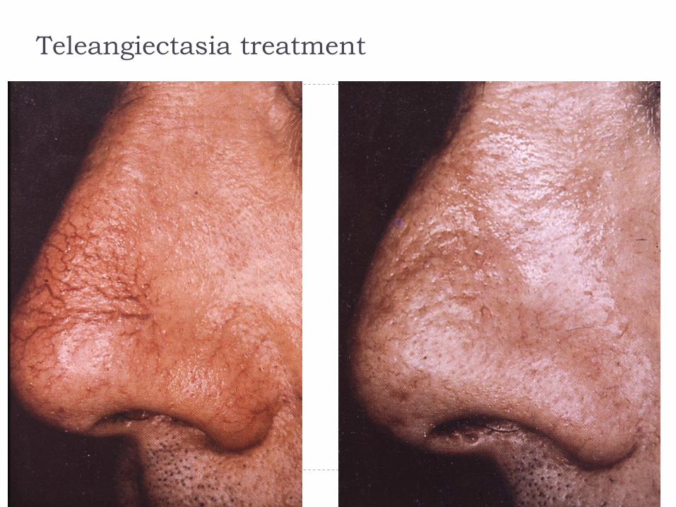
Teleangiectasia treatment
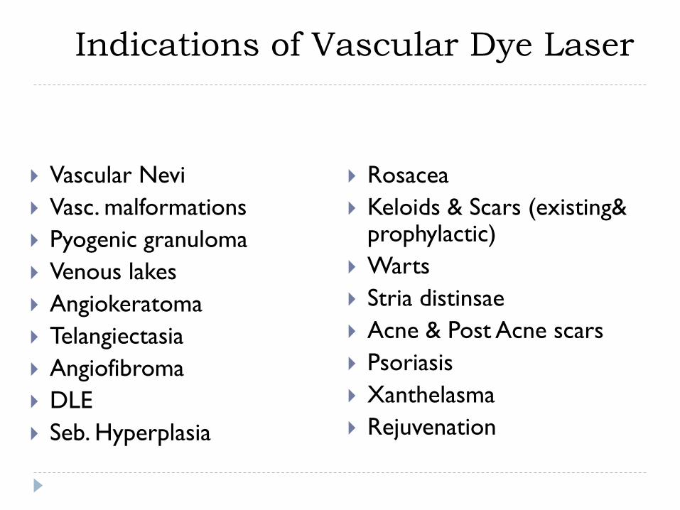
Indications of Vascular Dye Laser
Vascular Nevi
Vasc. malformations
Pyogenic granuloma
Venous lakes
Angiokeratoma
Telangiectasia
Angiofibroma
DLE
Seb. Hyperplasia
Rosacea
Keloids & Scars (existing& prophylactic)
Warts
Stria distinsae
Acne & Post Acne scars
Psoriasis
Xanthelasma
Rejuvenation
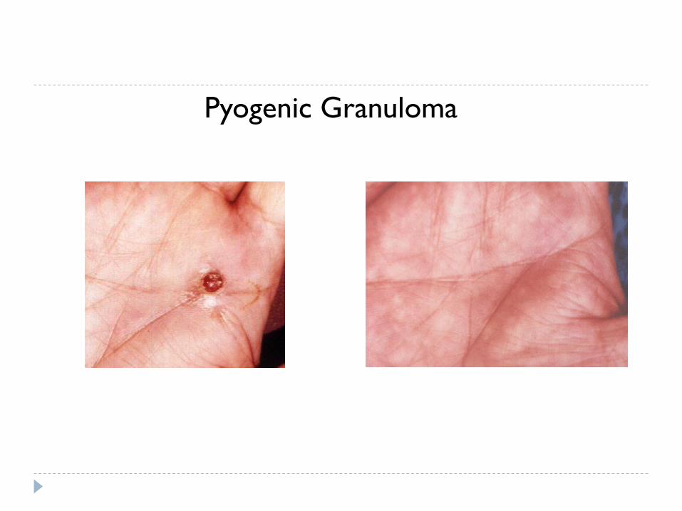
Pyogenic Granuloma
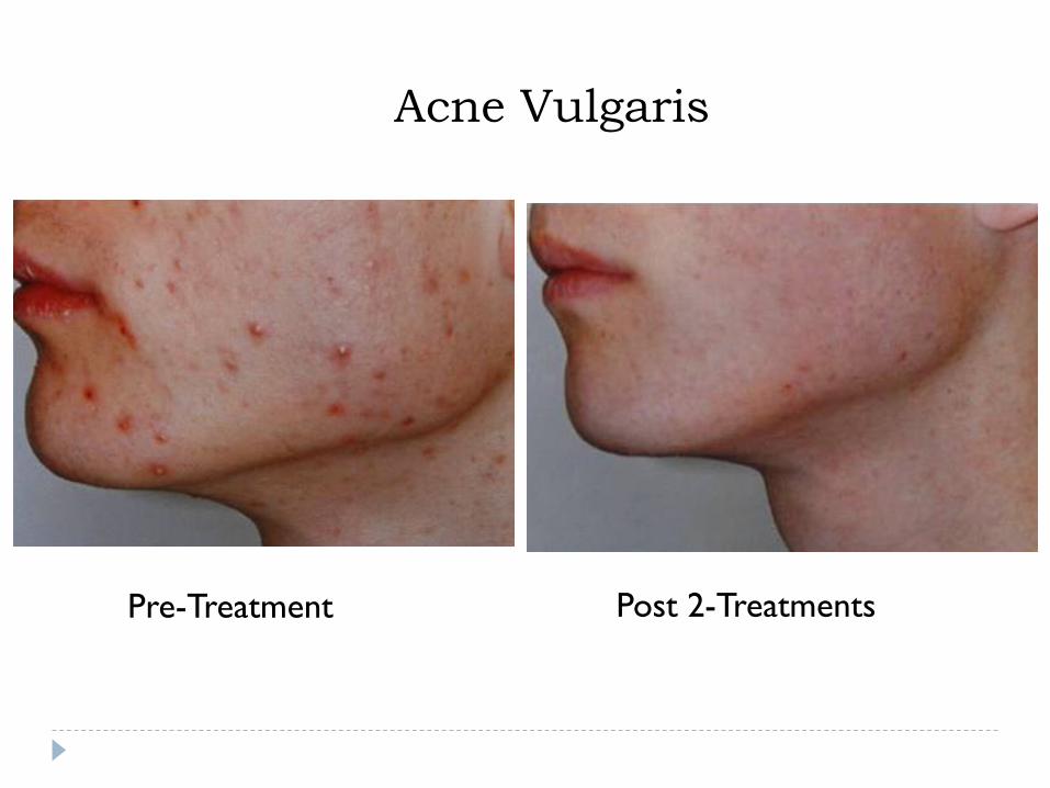
Pre-Treatment Post 2-Treatments
Acne Vulgaris
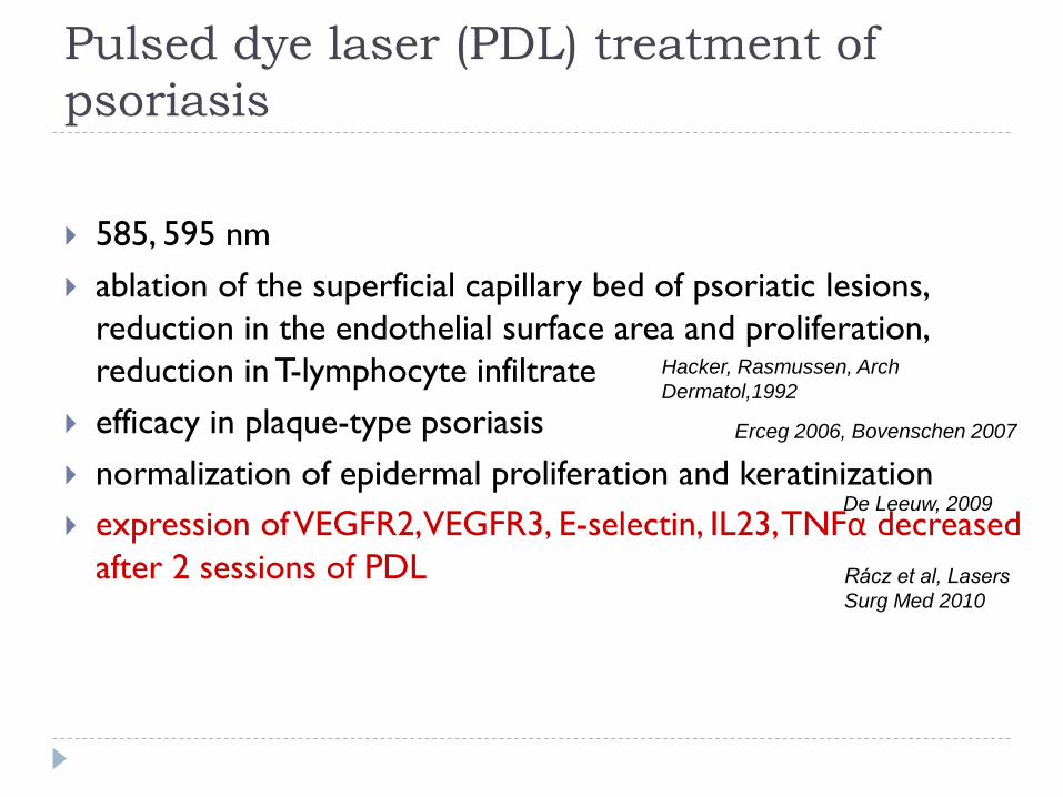
Pulsed dye laser (PDL) treatment of
psoriasis
585, 595 nm
ablation of the superficial capillary bed of psoriatic lesions,
reduction in the endothelial surface area and proliferation,
reduction in T-lymphocyte infiltrate
efficacy in plaque-type psoriasis
normalization of epidermal proliferation and keratinization
expression of VEGFR2, VEGFR3, E-selectin, IL23, TNFα decreased
after 2 sessions of PDL
Hacker, Rasmussen, Arch
Dermatol,1992
Erceg 2006, Bovenschen 2007
De Leeuw, 2009
Rácz et al, Lasers
Surg Med 2010
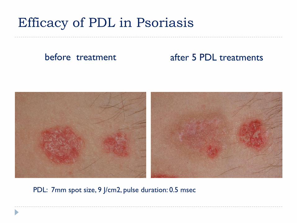
Efficacy of PDL in Psoriasis
before treatment after 5 PDL treatments
PDL: 7mm spot size, 9 J/cm2, pulse duration: 0.5 msec
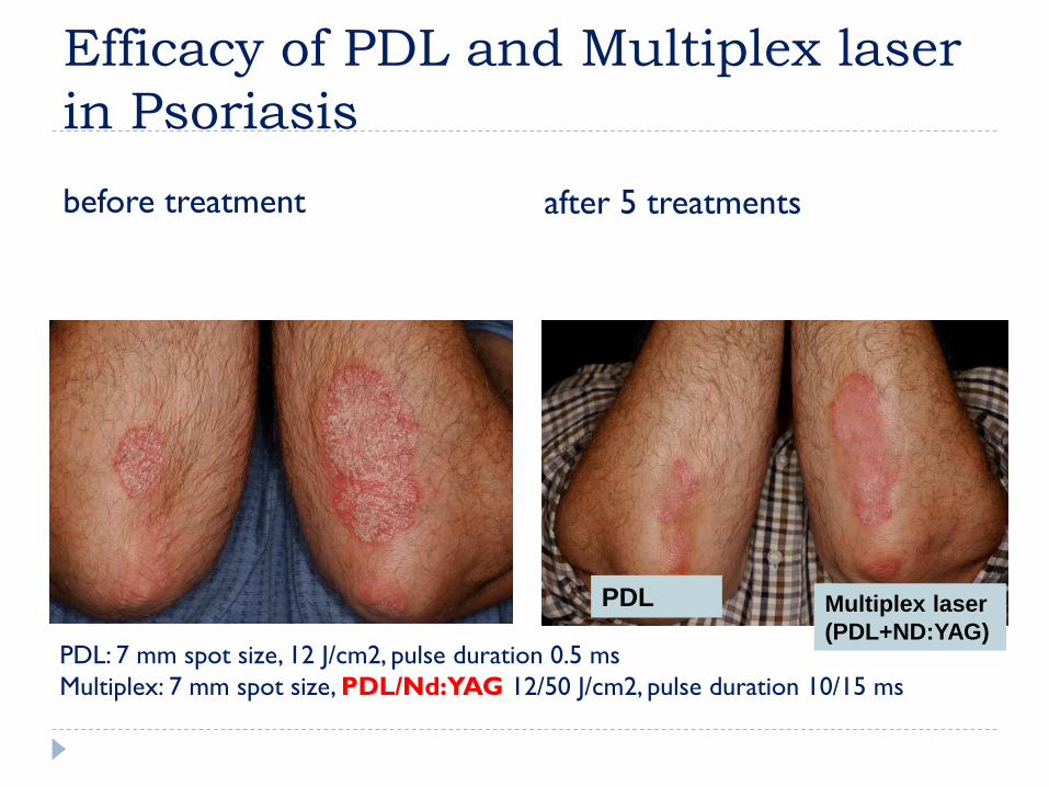
Efficacy of PDL and Multiplex laser
in Psoriasis
before treatment after 5 treatments
PDL Multiplex laser
(PDL+ND:YAG)PDL: 7 mm spot size, 12 J/cm2, pulse duration 0.5 ms
Multiplex: 7 mm spot size, PDL/Nd:YAG 12/50 J/cm2, pulse duration 10/15 ms
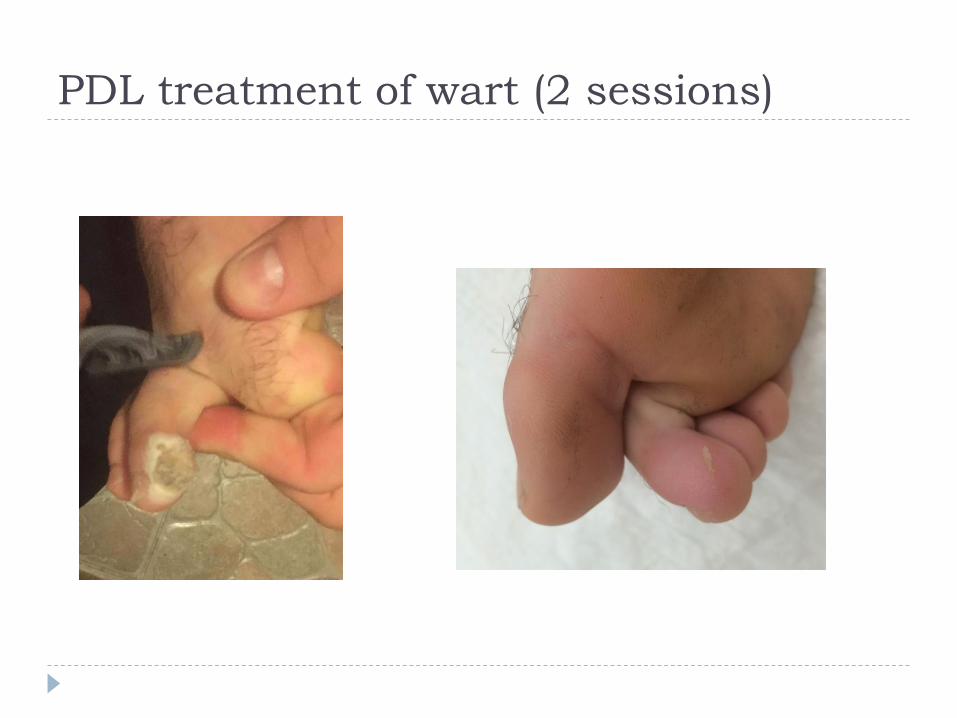
PDL treatment of wart (2 sessions)
d
b
c
a
treated control
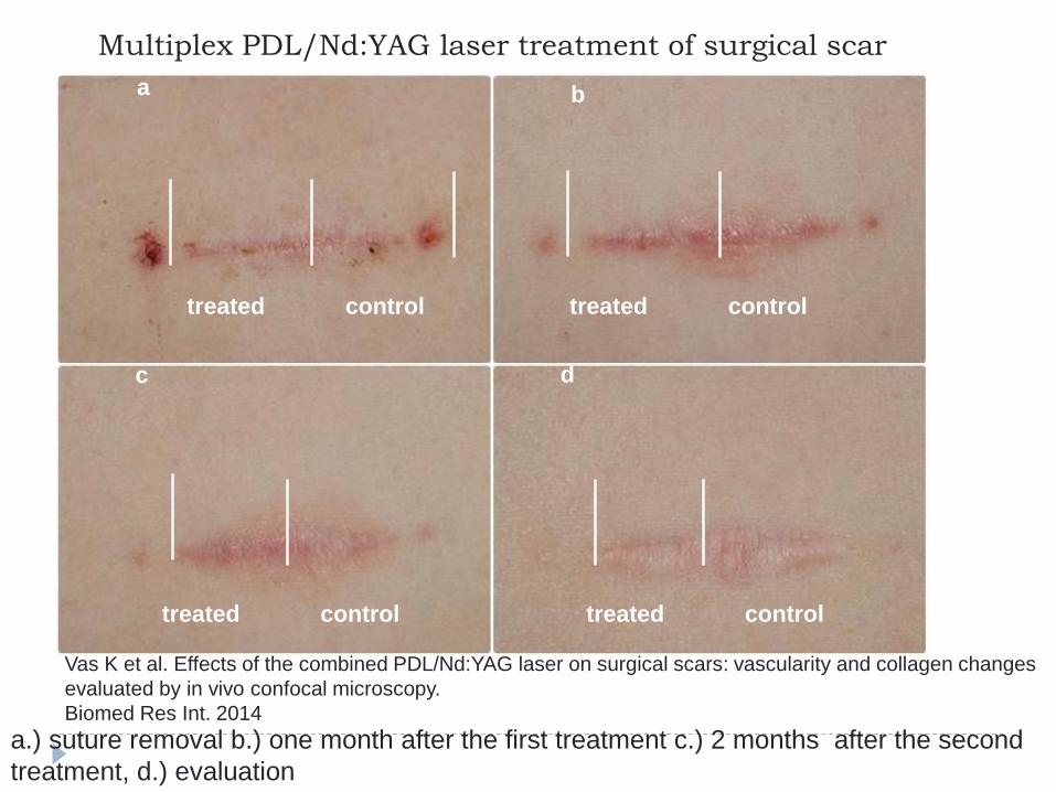
Multiplex PDL/Nd:YAG laser treatment of surgical scar
a.) suture removal b.) one month after the first treatment c.) 2 months after the second
treatment, d.) evaluation
Vas K et al. Effects of the combined PDL/Nd:YAG laser on surgical scars: vascularity and collagen changes
evaluated by in vivo confocal microscopy.
Biomed Res Int. 2014
treated control
treated control
treated control
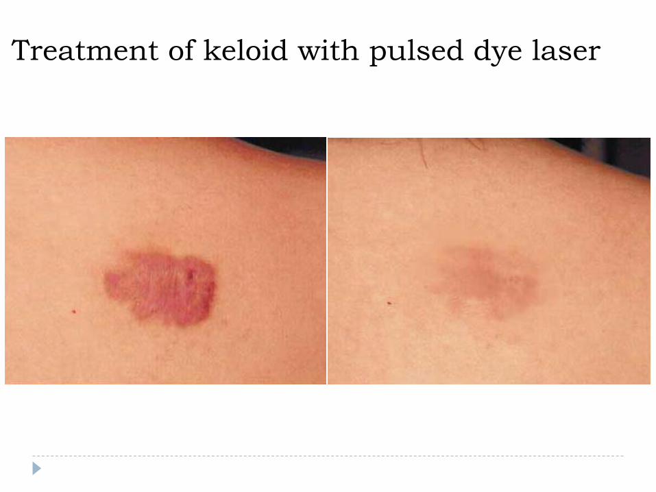
Treatment of keloid with pulsed dye laser
Laser treatment of tattoo and pigmented
lesions
Q-Switch (ns) vs. Long Pulse (ms) lasers
Melanosome
approximately 1m across
TRT 100’s of ns
Q-switched Lasers
Most likely rupture melanosomes, leading to cell damage
Long Pulse Light Sources
Most likely damage cells with heat
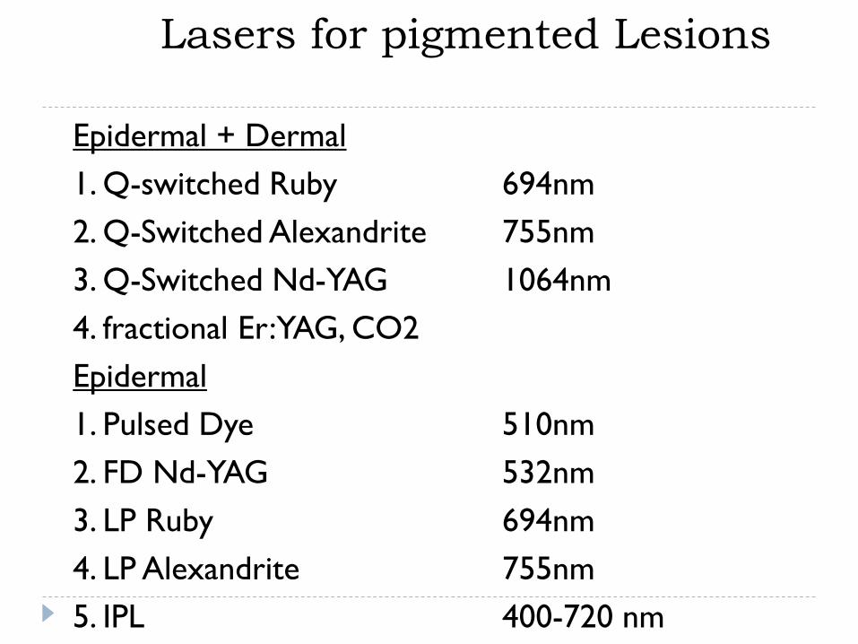
Epidermal + Dermal
1. Q-switched Ruby 694nm
2. Q-Switched Alexandrite 755nm
3. Q-Switched Nd-YAG 1064nm
4. fractional Er:YAG, CO2
Epidermal
1. Pulsed Dye 510nm
2. FD Nd-YAG 532nm
3. LP Ruby 694nm
4. LP Alexandrite 755nm
5. IPL 400-720 nm
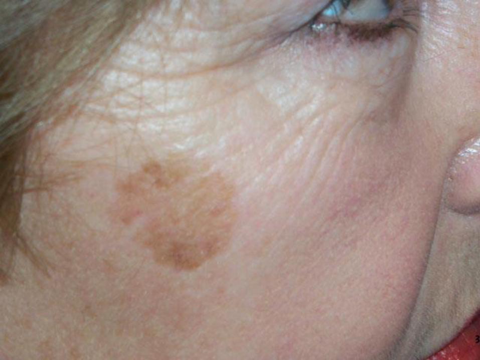
Lasers for pigmented Lesions
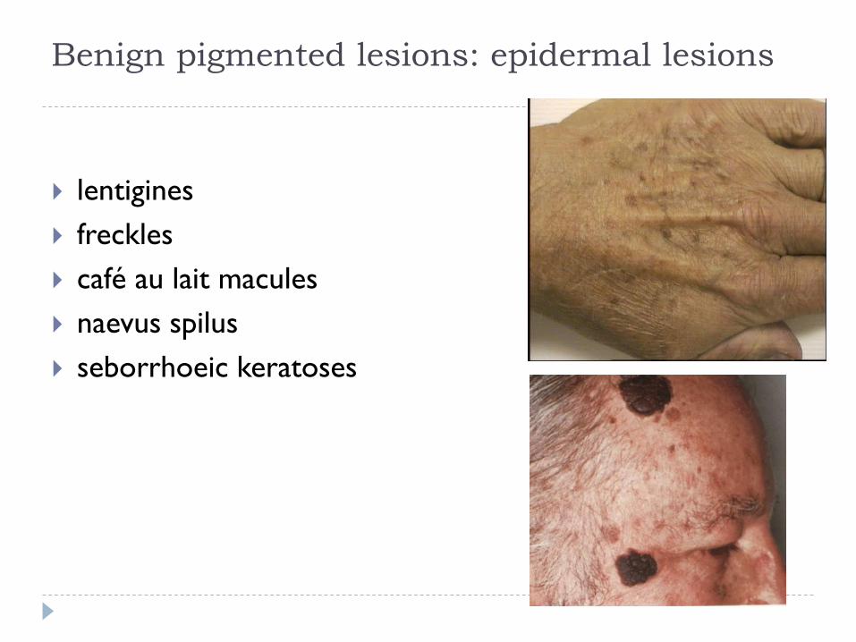
Benign pigmented lesions: epidermal lesions
lentigines
freckles
café au lait macules
naevus spilus
seborrhoeic keratoses
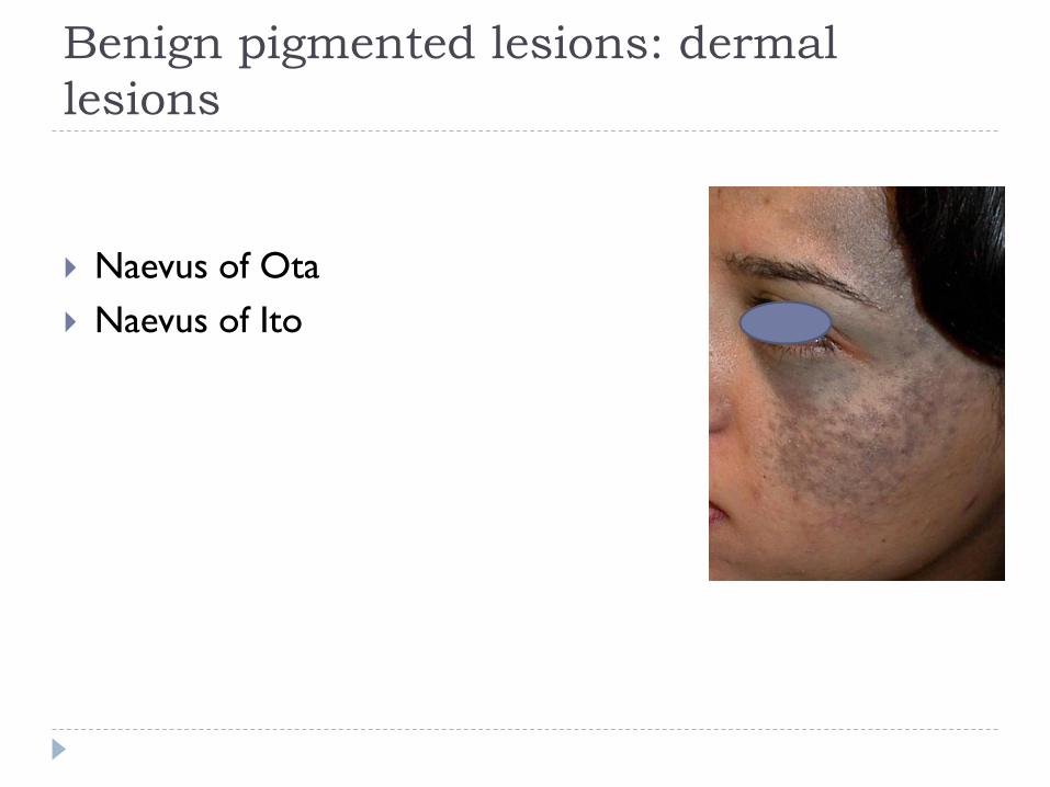
Benign pigmented lesions: dermal
lesions
Naevus of Ota
Naevus of Ito
Benign pigmented lesions: dermal-
epidermal lesions
melasma
post-inflammatory hyperpigmentation
Becker’s naevus
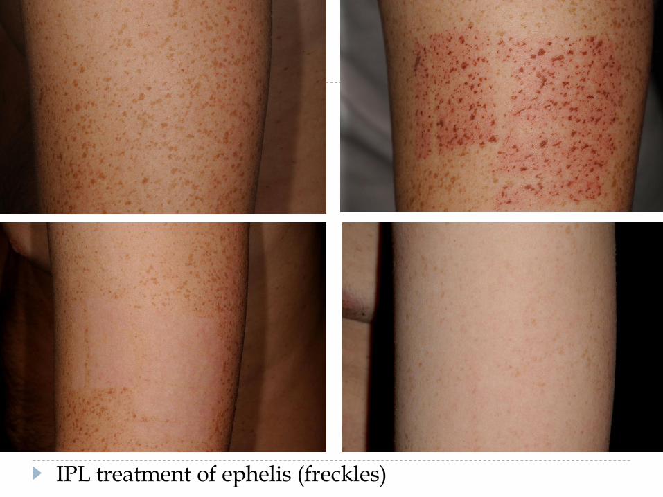
IPL treatment of ephelis (freckles)
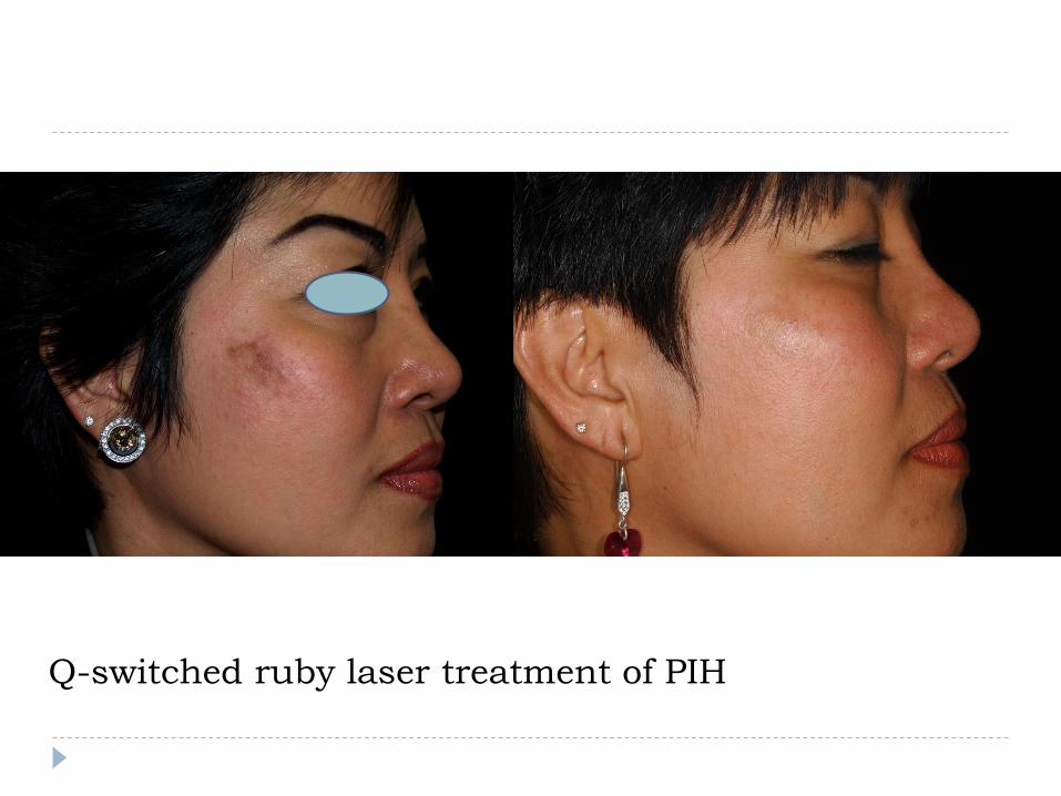
Q-switched ruby laser treatment of PIH
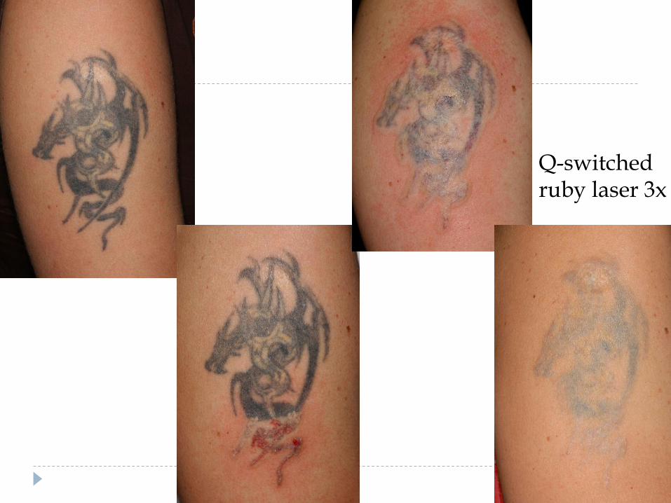
Q-switched ruby laser 3x
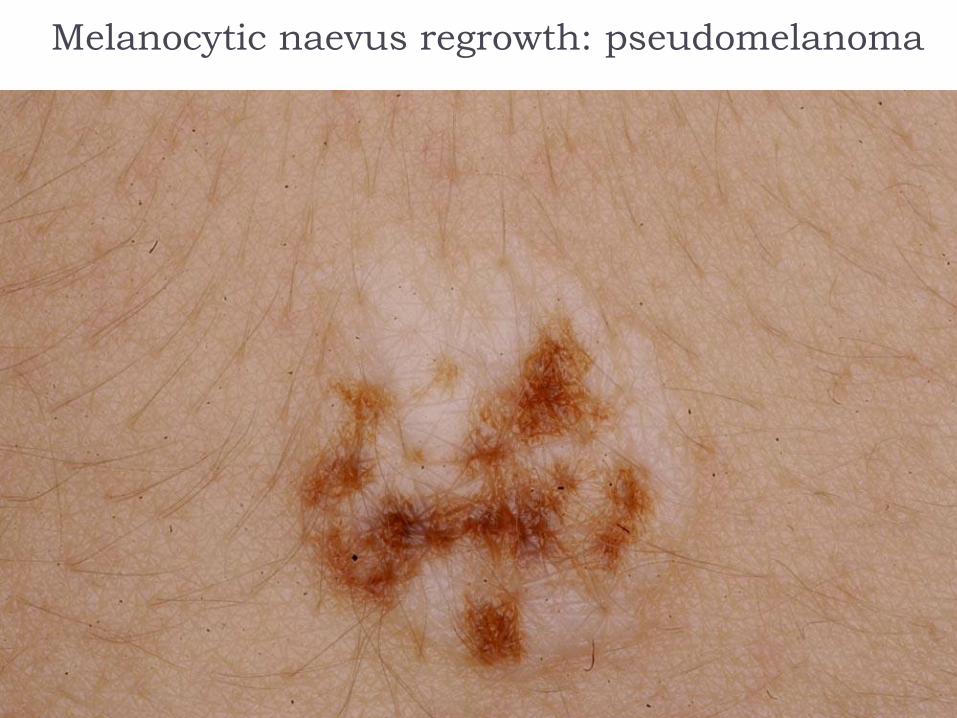
Melanocytic naevus regrowth: pseudomelanoma
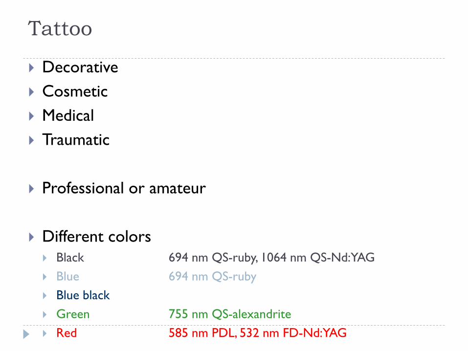
Tattoo
Decorative
Cosmetic
Medical
Traumatic
Professional or amateur
Different colors
Black 694 nm QS-ruby, 1064 nm QS-Nd:YAG
Blue 694 nm QS-ruby
Blue black
Green 755 nm QS-alexandrite
Red 585 nm PDL, 532 nm FD-Nd:YAG
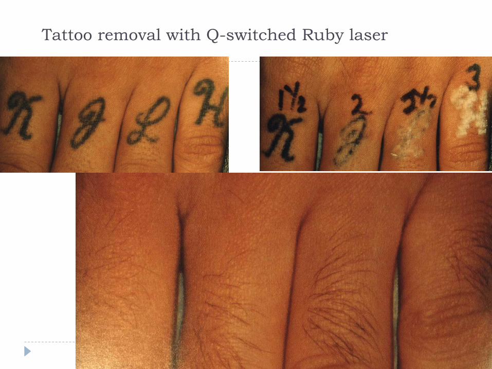
Tattoo removal with Q-switched Ruby laser
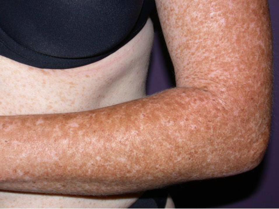
➢Photorejuvenation
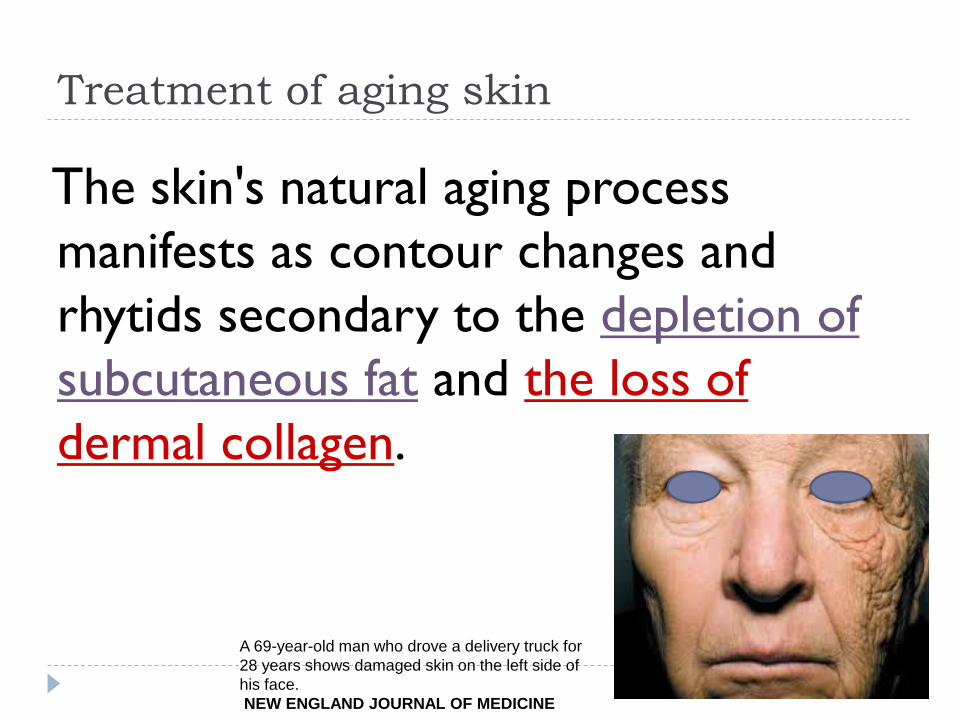
Treatment of aging skin
The skin's natural aging process
manifests as contour changes and
rhytids secondary to the depletion of
subcutaneous fat and the loss of
dermal collagen.
A 69-year-old man who drove a delivery truck for
28 years shows damaged skin on the left side of
his face.
NEW ENGLAND JOURNAL OF MEDICINE
Resurfacing, Photorejuvenation
Non-ablative photorejuvenation:
• IPL
• Nd:YAG
• Diode laser 1450 nm
• Er:glass 1540 nm
• Ablative laser resurfacing
2940 nm Er:YAG
fractional Er:YAG
fractional 10600 nm CO2
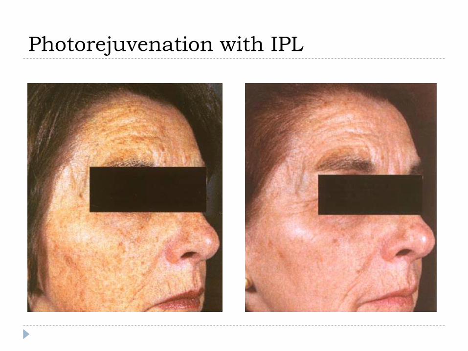
Photorejuvenation with IPL
Ablative lasers
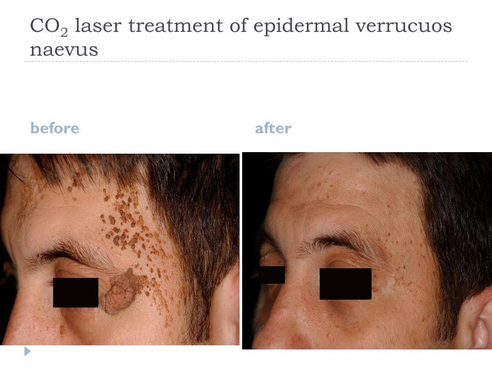
CO2 laser treatment of epidermal verrucuos
naevus
before after
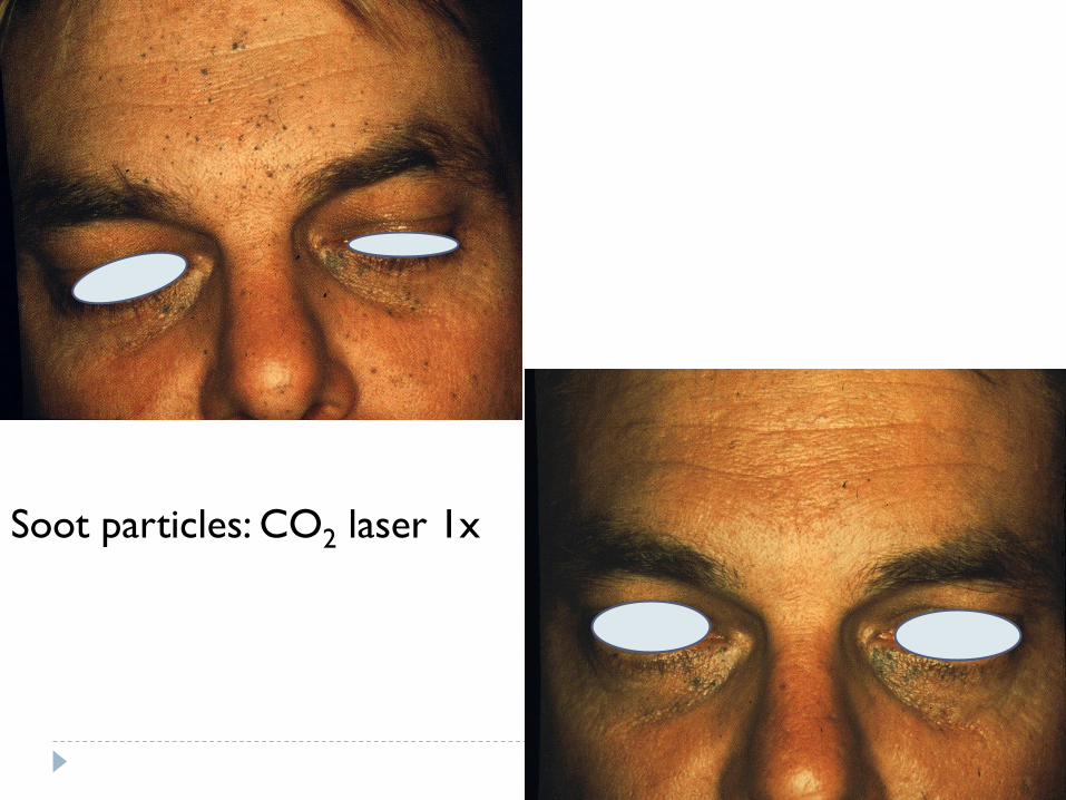
Soot particles: CO2 laser 1x
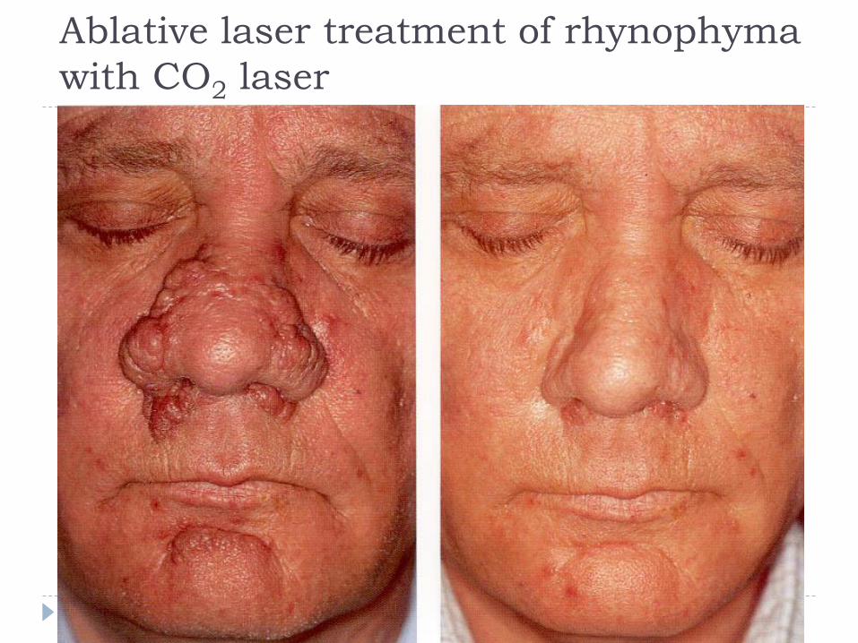
Ablative laser treatment of rhynophyma
with CO2 laser
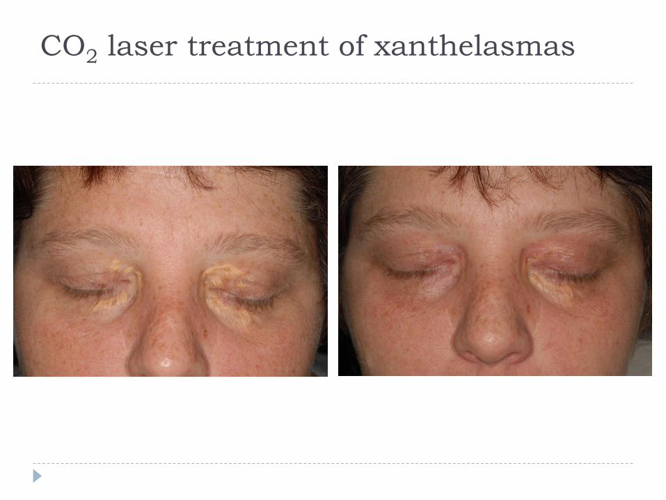
CO2 laser treatment of xanthelasmas
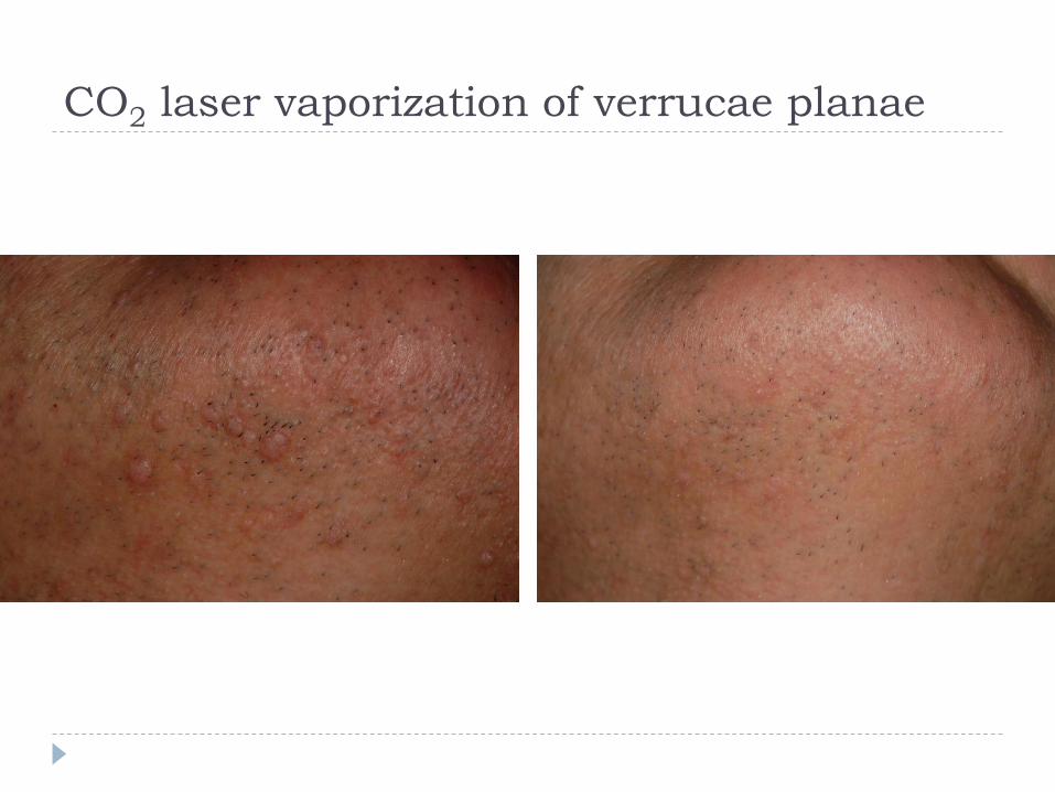
CO2 laser vaporization of verrucae planae
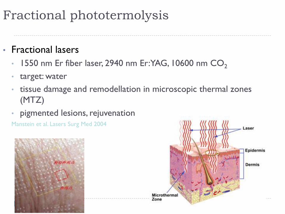
Fractional phototermolysis
• Fractional lasers
• 1550 nm Er fiber laser, 2940 nm Er:YAG, 10600 nm CO2
• target: water
• tissue damage and remodellation in microscopic thermal zones
(MTZ)
• pigmented lesions, rejuvenationManstein et al. Lasers Surg Med 2004
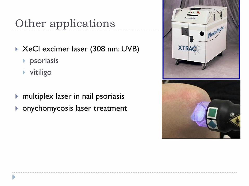
Other applications
XeCl excimer laser (308 nm: UVB)
psoriasis
vitiligo
multiplex laser in nail psoriasis
onychomycosis laser treatment
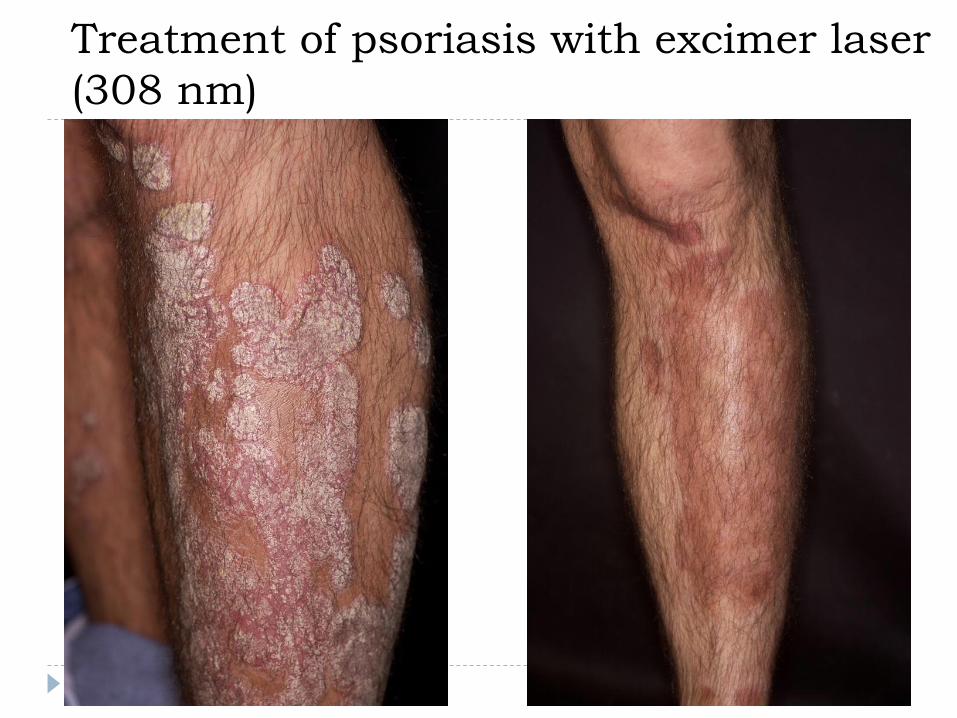
Treatment of psoriasis with excimer laser
(308 nm)
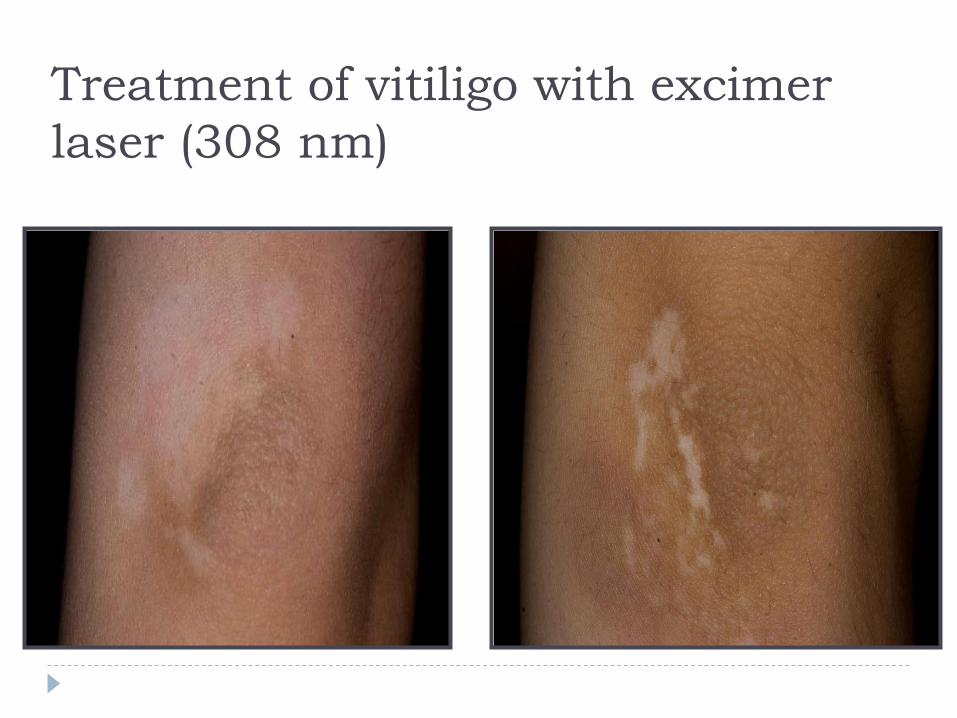
Treatment of vitiligo with excimer
laser (308 nm)
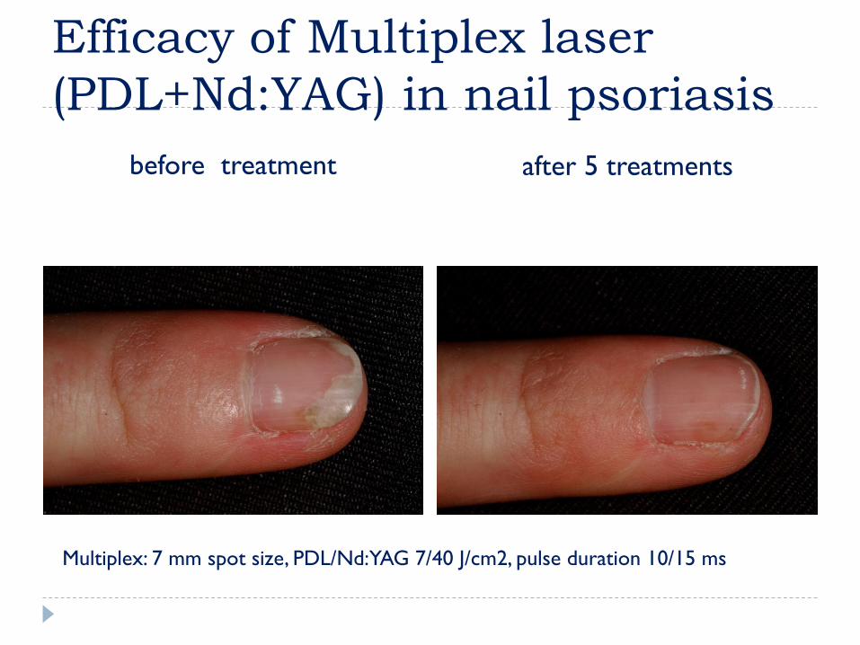
Efficacy of Multiplex laser
(PDL+Nd:YAG) in nail psoriasis
before treatment after 5 treatments
Multiplex: 7 mm spot size, PDL/Nd:YAG 7/40 J/cm2, pulse duration 10/15 ms
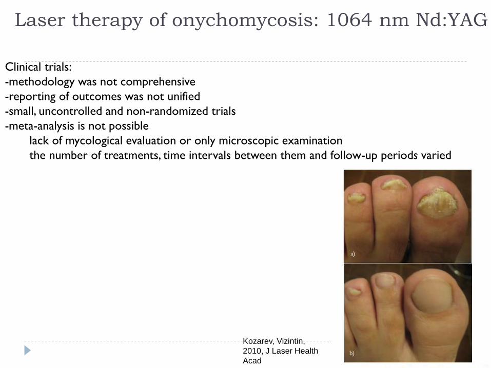
Laser therapy of onychomycosis: 1064 nm Nd:YAG
Clinical trials:
-methodology was not comprehensive
-reporting of outcomes was not unified
-small, uncontrolled and non-randomized trials
-meta-analysis is not possible
lack of mycological evaluation or only microscopic examination
the number of treatments, time intervals between them and follow-up periods varied
Kozarev, Vizintin,
2010, J Laser Health
Acad
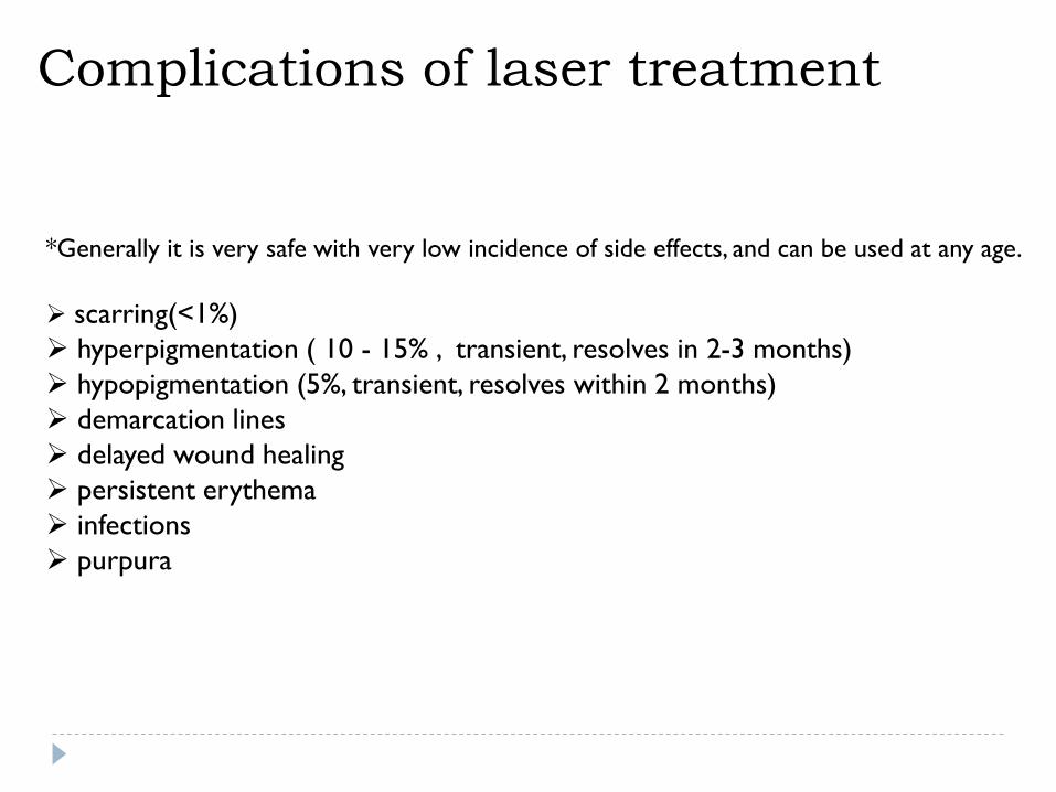
Complications of laser treatment
*Generally it is very safe with very low incidence of side effects, and can be used at any age.
➢ scarring(<1%)
➢ hyperpigmentation ( 10 - 15% , transient, resolves in 2-3 months)
➢ hypopigmentation (5%, transient, resolves within 2 months)
➢ demarcation lines
➢ delayed wound healing
➢ persistent erythema
➢ infections
➢ purpura
„Although facial teleangiectasia do improve after a single purpura-free
treatment with PDL, they improve more after purpura is induced.”
Murad Alam, Jeffrey Dover, Kenneth Arndt;
Derm Surg, July 2003
Complications of laser treatment:
purpura
Purpura and odema occuring
after the treatment were
transient.
Pretreatment measures
written consent
pretreatment photograph
anesthesia (usually topical)
safety measures (precautions)
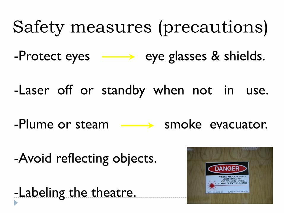
-Protect eyes eye glasses & shields.
-Laser off or standby when not in use.
-Plume or steam smoke evacuator.
-Avoid reflecting objects.
-Labeling the theatre.
Safety measures (precautions)
Post treatment measures
sunscreen for 3 months after the end of last session.
topical antibiotic ointment twice/day until
disappearance of purpura / crust.
bleaching agent whenever there is history of PIH.