Embed Size (px)
Citation preview

Journal of Pharmacy and Pharmacology 6 (2018) 647-658 doi: 10.17265/2328-2150/2018.07.002
Lasertherapy as A Strategy for Treatment Healing under
Caloric Restriction – Study in Rats
Leandro C. Moscardi, Talita P. Espíndola, Amanda A. Ferreira, Naiara Alves, Maria Esméria C. Amaral, Andréa
A. Aro, Rodrigo A. Dalia, Marcelo A. M. Esquisatto, Fernanda A. S. Mendonça, Gláucia M. T. Santos and Thiago
A. M. Andrade
Graduate Program in Biomedical Sciences of the Hermínio Ometto University Center, UNIARARAS, Araras, São Paulo 13607-339,
Brazil
Abstract: Complications in the healing process are challenging, especially in clinical situations of caloric restriction (CR). The lasertherapy becomes an important strategy that aids the repair, especially in CR. Thus, it is important to investigate the InGaAlP-660 nm laser as an strategy to repair cutaneous wounds in rats submitted to 30% of CR and to understand the tissue repair in clinical situations of CR. Thirty-six male Wistar rats were used, of which half were fed with 30% less ration, and half with ad libitum diet, for 21 days. Then, punch lesions of 1.5 cm in diameter were made on the animals backs, which were divided into: NR (no-restricted), R (restricted)—both before lesion; C (control), RC (restricted-control), L (laser), RL (restricted-laser)—after lesion. Samples of the skin/lesion/scar were collected on the 2nd, 7th and 14th days post-injury for histological, biochemical and molecular analyses. The R group showed reduction of body mass, epidermal/dermal thickness, inflammation, angiogenesis, fibroplasia and collagenesis. The RL group showed control of inflammation, oxidative damage and increase of antioxidants than RC, which probably favored angiogenesis, collagenesis and reepithelialization, similar to C and L. Thus, 30% of CR impaired the skin (before lesion). In the lesion, lasertherapy has shown to be effective in tissue repair mainly in CR status, being thus, the laser clinically important strategy to tissue repair in critical situations of caloric restriction. Key words: Wound healing, skin, caloric restriction, wound treatment, therapeutic strategy, lasertherapy.
1. Introduction
Wound healing involves celular events, cytokines
and growth factors production on wound, aiming the
complete reepithelialization. In inflammatory phase,
mediators promote chemotaxis, mainly of neutrophils
and macrophages, which are involved in tissue
debridement and development of oxidative stress [1].
This is characterized by imbalance between oxidants
and antioxidants, due to the excessive production of
ROS (reactive oxygen) and RNS (nitrogen) species
and the reduced neutralization of these by the
antioxidants. Then, oxidative damage is aggravated,
mainly when associated with caloric restriction (CR),
Corresponding author: Thiago Antônio Moretti de Andrade, Ph.D., research fields: immunophysiopathology of healing using new therapeutics in differents experimental models.
causing difficulties on tissue healing [2]. Moreover,
angiogenesis improves nutritional and oxygen supply
in the wound bed. The collagen and extracellular
matrix production by fibroblasts recompose the
damaged tissue, promoting the reepithelialization [3].
In this view, it is noted the healing process requires
adequate nutritional status, since it consumes a good
part of the body’s reserve. In CR, the level of nutrients
provided is comparable to ad libitum consumption,
but with a reduction in calories consumed, i.e.,
undernutrition without malnutrition [4].
Moreover, there are impacts of CR on the immune
system [5] and, consequently, on the wound healing,
because the immune system is expensive in terms of
demands for energy [6] being it the first systems
whose sacrifices might be made at the beginning of
CR [5]. In these ways, CR was found to have adverse
D DAVID PUBLISHING

Lasertherapy as a Strategy for Treatment Healing under Caloric Restriction—Study in Rats
648
effects on wound healing [7] and resulted in decreased
of fibroblasts function and collagen accumulation [8],
limiting the granulation tissue.
The sirtuin (SIRT) regulates activity of many
proteins that are related to energy metabolism, cell
survival and longevity [9]. CR increases the
expression of SIRT-1 in multiple tissues and it is
increased in response to nutrient deprivation in
cultured cells [10]. SIRT-1 controls the
gluconeogenic/glycolytic pathways in the liver in
response to fasting or food restriction [11]. Moreover,
SIRT activation by CR has been implicated in
changes in the immune system, because the immune
activation is energetically expensive [12]. It has been
shown that activation of the immune system
significantly increases mortality in nutrient-limited
settings [13]. Thus, the inhibitory effect of SIRT-1 on
NF-κB activity is consistent with its role in regulating
the available nutrients during times of energy
crisis, diverting calories away from the immune
system to spare them for essential survival processes
[12], which probably can cause deficit in the healing
process.
In this context, there is an intense difficulty in the
treatment of some types of cutaneous wounds,
especially when associated with nutritional scarcity.
Low-intensity lasertherapy has been noted for its
important healing activity [14]. Moreover, there is
evidence that laser has been shown important healing
effects on tissues due to its physiological and
therapeutic effects depending on the absorption of
photons by chromophores in the target tissue. Studies
have shown that the photons emitted by the laser are
transformed in ATP and used by the cell to power
needed metabolic activities, such as cell proliferation,
and collagen synthesis. All of them, accelerate the
tissue repair in chronic wounds [15, 16], especially
when associated with oxidative stress status, like
nutritional deficit and clinical situations of CR, for
example. In this view, it was hypothesized that
wounds associated with 30% of CR are healing slowly
or not at all, may benefit from lasertherapy.
Cardiovascular diseases, diabetes, cancer,
inflammation, chronic wounds and neurodegeneration
disease are examples of human pathologies, directly or
indirectly affected by amount of nutrients [17].
Furthermore, patients bedridden by these and/or other
pathologies, when presenting cutaneous lesions, either
from post-surgical or pressure wounds, will be
difficult to heal due to the nutritional scarcity caused
by these pathologies and/or poor nutrition. And this,
probably could be the particular clinical situation that
lasertherapy is more effective. Thus, it is important to
investigate the laser (InGaAlP-660 nm) as an strategy
to repair cutaneous wounds in rats submitted to 30%
of CR and to understand the tissue repair in clinical
situations of CR.
2. Material and Methods
2.1 Animals
All surgical and experimental procedures used in
this study were conducted according to the
experimental requirements and biodiversity rights of
the National Institutes of Health for the Care and Use
of Laboratory Animals ((NIH Publication 80-23,
reviewed in 1996). Studies were performed according
to the norms established by the Arouca Law, approved
by the ethical principles in animal research adopted by
COBEA (Brazilian College of Animal Experimen-
tation) and approved by the Ethics Commission on
Animal Use (CEUA) of the Herminio Ometto
University Center (UNIARARAS) protocol nº
021/2015.
Thirty-six male Wistar rats (Rattus norvegicus
albinus) within 90 days of age, obtained from the
Center of Animal Experimentation “Prof. Dr. Luiz
Edmundo de Magalhães” from the UNIARARAS
(Herminio Ometto University Center) were used. The
animals were housed in individual polycarbonate
cages under constant temperature conditions (23 ±
2 °C), humidity (55%) and light/dark 12:12 hours

Lasertherapy as a Strategy for Treatment Healing under Caloric Restriction—Study in Rats
649
cycle with free access to commercial standard ration
and water for the entire experiment period.
2.2 Experiment Design
2.2.1 Experimental Model of 30% of Caloric
Restriction.
The ration was daily weighed for five days to
evaluate the animals intake-average on ad libitum diet.
For CR establishment, it was offered to 18 animals (R
group—before lesion = RC, and RL groups, after
lesion) 30% less ration from the intake-average,
during 21 days. The others 18 rats were the control
groups (NR groups—before lesion = C, and L groups,
after lesion), which remained on ad libitum diet during
35 days of experiment.
On the 21st day of CR, wounds were performed on
the all animals dorsum, which have been followed-up
by 2nd, 7th and 14th days (n = 3 rats/group/time of
follow-up) [18].
2.2.2 Caloric Restriction Confirmation
The blood samples were collected directly from the
heart-left ventricle of last euthanasia on 14th day
post-injury (35th day of CR). Serums were
subsequently stored at -20 °C for analysis of glucose,
cholesterol, triglycerides, protein and albumin
according to the manufacturer’s standard instructions
(Laborlab, Guarulhos-SP-Brazil). In addition, the
animals were weighed on 1st, 7th, 14th, 21st and 35th
days of CR (35th day of CR was similar to 14th day
post-injury) and also analyzed for SIRT-1 expression
by Western blotting [18].
2.2.3 Surgical Procedure and Experimental Groups
The rats were anesthetized by the combination of
ketamine (3.0 mL/kg) with xylazine (1.0 mL/kg) for
skin injuries performance. Then, trichotomy was
performed on the skin of the dorsum of all animals.
For a greater use (refinement) and a reduction on the
animal’s amount, two excisions were performed on
the dorsal region of each rat using a histological punch
of 1.5 cm diameter, reaching the dermo-epidermal
region until the exposure of the muscular fascia [16,
19-21]. These both skin samples obtained were used
to investigate the skin changes caused by 30% of CR
in the skin before lesion (without treatment), obtaining
the groups (n = 6 animals/group): NR
(no-restricted—on ad libitum diet) and R
(restricted—under 30% of CR).
After performing the two wounds, all animals were
divided into four distinct experimental groups (each
group with n = 3 animals/group/time of follow-up): C
(control: wounds treated topically with laser switched
off and ad libitum diet); RC (restricted-control: treated
with laser switched off and 30% of CR); L (laser:
wounds with topically lasertherapy and ad libitum
diet); RL (restricted-laser: lasertherapy and 30% of
CR).
Then, both lesions from each rat received the same
specific treatment and no occlusive dressing was used.
The animals were placed in individual cages and
received 10 drops of analgesic (dipyrone) diluted in
250 mL of water for 72 h.
2.2.4 Lasertherapy
It was used Photon Lase III (DMC®-São Carlos-SP-Brazil)
InGaAlP (indium-gallium-aluminum-phosphor): 660
nm wavelength (visible-red), 3.63 W/cm² of irradiance,
100 mW of power, 134.54 J/cm² of energy density, 3.7
kJ of energy/point, 14.8 J of total energy dose, 0.0275
cm² of beam covering area application, 37 seconds
under continuous mode in 4 points at the wound
extremities, ±2.0 mm of distance from each wounds
by 90° of angle between wound and applicator probe.
The treatments were started soon after the
experimental wound and performed 3×/week (at the
same time).
2.2.5 Sample Harvesting
After 2nd, 7th and 14th days post-wound, the
animals were euthanized with anesthetic deepening
(ketamine: 3.0 mL/Kg and xylazine: 1.0 mL/Kg, 300
μL/Kg animal body weight) and cervical dislocation.
Then, both skin/wound/scar were harvested and
divided in parts for histologicals, biochemicals and
moleculars studies (n = 3 part of wound/scar of
Lasertherapy as a Strategy for Treatment Healing under Caloric Restriction—Study in Rats
650
rats/group/time of follow-up).
2.2.6 Evaluation of Wounds Reepithelialization
On the 0, 2nd, 7th and 14th days, both wounds of
each animal were photographed in basic mode,
without flash, without zoom and using the same
camera. To standardize the distance from the camera
to the wound, the camera was attached to a support 30
cm apart and perpendicular to the rat wound. A ruler,
arranged beside the animal and along the wounds, was
used to standardize the unit area of the wounds in 2
cm and also to serve as a known measure in the
calibration of the ImageJ software when calculating
the area of the wound.
ImageJ software was used to determinate the wound
area to calculate the Wound Healing Rate: WHR
(wound healing rate) = (initial area - final area) /
initial area. The initial area corresponds to the day of
the surgical procedure (0 day) and the final area
corresponds to the day of euthanasia (2nd, 7th or 14th
days, post-injury). WHR values greater than zero
represent a decrease in wounded area
(reepithelialization) and smaller than zero represent an
increase in wounded area [16, 20].
2.2.7 Histomorphometry analysis
The skin/wound/scar (n = 3/group/time of
follow-up) biopsies were conditioned for 48 hours in
10% buffered formaldehyde solution and followed by
paraffin-histological processing (Hematoxylin-Eosin)
for simultaneous quantification of inflammatory
infiltrate, fibroblasts and blood vessels using the “Cell
Counter” Plugin of ImageJ software. It was used 5
images (magnification: 400×) and the quantification
of inflammatory infiltrate, fibroblasts and blood
vessels were double-blind. Results were expressed as
number of cells’ average [21].
For measuring epidermal and dermal thickness,
histological images (400× of magnification) were
taken in sequence from the epidermis to the
subcutaneous cellular tissue. These images were
automatically grouped using the Photomerge function
of the Adobe Photoshop CC 2018. By the ruler on
Photoshop, 3 lines/image was traced on epidermis and
dermis and then, Photoshop supplied the thickness
(cm) [16].
2.2.8 Myeloperoxidase (MPO)
The MPO assay was performed for the
determination of the neutrophilic infiltrate by the
macerate supernatant samples of the skin/wound/scar
of the rats during the follow-up and treatments.
Samples were homogenized by Polytron®
homogenizer (Kinematica, USA). After centrifugation,
5.0 µL of supernatant was diluted in 45 µL 0.08 M
NaPO4. MPO activity was detected using 25 µL 1.6
mM tetramethylbenzidine (Sigma-Aldrich®, USA) and
read at 450 nm (SpectraMax®, USA) [21].
2.2.9 N-Acetylglicosaminidase (NAG)
NAG assay was performed to macrophagic
infiltration. It was placed 3.0 μL of supernatant
from the macerated samples and it was added
30 μL p-nitrophenyl-2-acetamide-β-D-glucopyranoside
(Sigma-Aldrich), diluted in 50 μL of 50 mM citrate
buffer. Finally, 50 μL of 0.2 M glycine was added and
read at 405 nm [22].
2.2.10 Hydroxyproline (HO-Pro)
HO-Pro assay was performed to total collagen
determiantion. After hydrolysis in 6 N HCl at 110 °C,
the samples were treated with 1.41% chloramine T
and 15% p-dimethylaminobenzaldehyde, followed by
incubation at 60 °C and read at 550 nm [23].
2.2.11 Western blotting
Samples were homogenized, treated with Laemmli
buffer and coursing on SDS-PAGE (10% to 12%
Tris-acrylamide) in minigel apparatus (Miniprotean).
The proteins were transferred to PVDF
(polyvinylidene difluoride) membrane (Immun-Blot®
Bio-Rad), which were incubated overnight with: 1:200
anti-TGF-β1, anti-CAT, anti-SOD1, anti-SOD2,
anti-VEGF, β-actin (Santa Cruz Biotechnology-USA);
1:500 anti-collagen I (SIGMA) and, 1:1,000
anti-SIRT-1 (CellSinaling, USA) and 1:2,000
anti-collagen III (Santa Cruz Biotechnology-USA).
The membrane was incubated with

Lasertherapy as a Strategy for Treatment Healing under Caloric Restriction—Study in Rats
651
chemiluminescence antibodies kit and revealed in
Syngene G:BOX-photodocumentator. The bands’
intensity was evaluated by densitometry using ImageJ
[18].
2.2.12 Oxidative stress and antioxidants
The lipoperoxidation products were analyzed by
measuring the formation of thiobarbituric acid reactive
species (TBARS) [24]. Then 100 µL tissue
homogenates was deproteinized with 20%
trichloroacetic acid. The supernatant was exposed to
0.7% thiobarbituric acid, heated to 95 °C and read at
535 nm.
The -SH groups (antioxidant) were quantified by
the reaction of the sulfhydryl group with 5,5' dithiobis
(2-nitrobenzoic acid) (DTNB) and read at 412 nm
[25].
2.3 Statistical Analysis
Data were expressed as mean ± standard error. All
data were passed for Kolmogorov-Smirnov normality
test. For the data fitting the normality curve, the
statistical test chosen was t-Student test for before
lesion groups (comparing NR/R groups) and ANOVA
Two-Way (Multiple-comparisons) and Tukey
post-test to groups after lesion (comparing
C/RC/L/LR groups). For the data not fitting in the
normality curve (glucose/cholesterol/protein/albumin)
the statistical test was Mann-Whitney (for
triglycerides was t-Student test), all were performed in
Statistic 7.0 and GraphPad Prism 6.0 softwares. The
pre-established level of significance was p < 0.05.
3. Results and Discussion
3.1 Caloric Restriction Status and Implications on
Healing
Nutritional deficiency remains one of the most
common causes of morbidity and mortality among
children, also prevalent among elderly people [26] and
hospitalized patients specially in underdeveloped
countries. Even in industrialized countries, people are
vulnerable to protein malnutrition, which can
accompany weight control, lifestyle-related disease
and aging. Diets in these populations are frequently
deficient in macronutrients, micronutrients, or both
[27-29]. The nutritional deficit is usually found in
these at-risk populations and is a critical factor in
susceptibility to infection [30]. Numerous nutrients
are known to be important for optimal wound healing.
CR is associated with immunological changes, making
wound healing inadequate due to inflammatory phase
prolongation and fibroplasia impairment, reduction in
collagen and extracellular matrix synthesis,
angiogenesis and wound remodeling [31, 32].
As a treatment of these immunocompromised
wounds, lasertherapy has been an important
alternative because the phototherapy depends on the
absorption of photons by chromophores in the target
tissue, accelerating the tissue repair [15] mainly as in
pathological conditions such as in CR status. Then,
wounds associated with nutritional scarcity heal
slowly or not at all, and the lasertherapy could benefit
the treatment in a 30% of CR animal model. In this
way, it becomes relevant to investigate the effects of
lasertherapy as a strategy in the repair of cutaneous
lesions in rats submitted to 30% of caloric restriction.
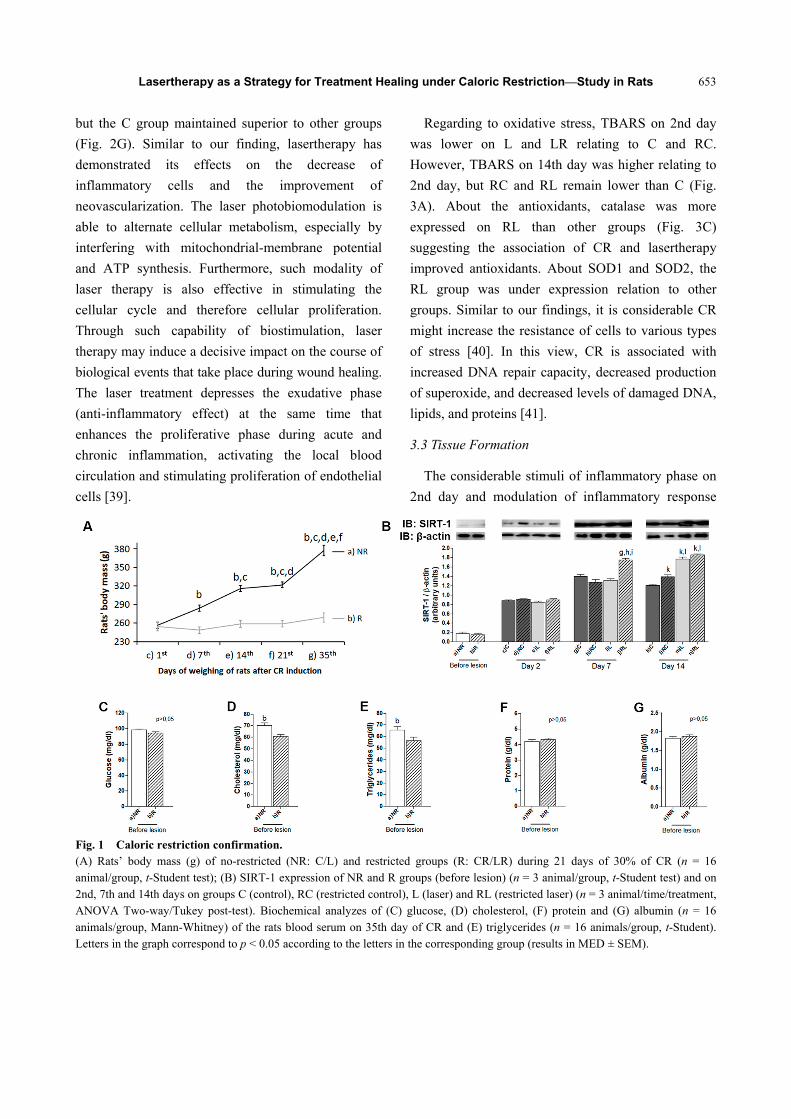
To confirm CR-status, R group showed important
reduction of body weight relation to NR from 7th to
35th days of CR. Only NR presented important
increase of body mass during the follow-up of CR
relation to previous days (Fig. 1A). This finding
corroborates with Hunt et al. [4] on 40% of CR.
About the biochemical analyses, there was no
difference between NR and R for glucose, protein and
albumin (Fig. 1C, 1F and 1G) on the 35th day of CR.
However, about cholesterol and triglycerides (Fig. 1D
and 1E) the R group was lower than NR,
corroborating to Amaral et al. [18]. Low body weight,
hypocholesterolemia and hypotriglyceridemia
contents are features usually found in animals
submitted to CR, indicating the adequacy of the
animal model used in this work [18]. The
Caloric-Restriction-Society/CRS consists of a group

Lasertherapy as a Strategy for Treatment Healing under Caloric Restriction—Study in Rats
652
of humans individuals who practice CR with optimum
nutrition (CRON). The subjects of the study were 18
volunteers (the CRONies) that have been practicing
the restricted-diet for 3-15 years. Classical signs of
CR adaptations were evident in the CRONies,
showing significantly lower blood glucose levels,
corroborating to our results, insulin levels, and blood
pressure. Lipid profiles of the CRONies were
significantly healthier than the height-matched
controls. Specifically, total cholesterol, LDL, and
triglycerides were all reduced, corroborating to our
results, and HDL was elevated compared to controls
[33]. Regarding to our similar results of serum protein
and albumin between both groups, it is possibly due to
the slow and late reduction in CR [34].
CR induces the expression of SIRT-1 that
participates as signaling of nutritional scarcity [35].
Serum was collected from humans after CR increased
SIRT-1 expression. Among the changes related to
SIRT-1 are increased hepatic gluconeogenesis,
decreased glycolysis and increased hypothalamic
signaling of hunger [36]. In our results SIRT-1 was
increased, mainly on 7th and 14th days and in LR its
expression was superior to the other groups (Fig. 1B).
In addition, the increase of SIRT-1 in the L and LR
compared to C and CR on 14th day, might suggest
laser associated with CR can increase SIRT-1
expression on tissues. Besides SIRT-1 being a CR
marker, it has shown SIRT-1 is required for efficient
wound healing when it was used on mice with
epidermis-specific SIRT-1 deletion. SIRT-1
deficiency in this model inhibits the repair of
epidermis and dermis, altering the production of many
cytokines, inhibited the recruitment of macrophages,
neutrophils and mast cells, increased oxidative stress
and reduced the angiogenesis in granulation tissue,
similar to our results (Figs. 2 and 3). SIRT-1 when
inhibited, decreased wound healing, while it
moderately increased proliferation, suggesting that
SIRT-1’s role in wound healing is a complex
well-coordinated process and proliferation alone is not
sufficient for wound repair. Possibly other functions
of epidermal SIRT-1 including regulating of cell
migration, inflammation and stress response, are the
more important determinants for SIRT1’s role in
wound healing [37].
3.2 Inflammation, Oxidative Stress, Antioxidants and
Angiogenesis Profiles
The question that emerges from these observations
is why natural selection would favor a system whose
inflammation is regulated by nutrient availability. In
the case of CR, the explanation for this is that immune
activation is energetically expensive and increases
energy consumption even in nutrient-limited settings
[13]. Thus, the inhibitory effect of SIRT-1 on NF-κB
activity is consistent with its role in regulating of
available nutrients during times of energy crisis,
diverting calories away from the immune system to
spare them for essential survival processes [12]. We
showed that R group reduced inflammatory infiltrate,
TGF-β1, NAG, blood-vessels and VEGF (Fig. 1B, 1C,
1E, 1F and 1G) relation to NR, This corroborates with
the findings of Yeung et al. [38] that CR inhibits
NF-κB expression by reducing inflammation and
consequently, the impairment of angiogenesis. On the
2nd day, RC and RL showed lower recruitment of
inflammatory infiltrate and TGF-β1 than C. On the 7th
day, L remained the level of inflammatory infiltrate
similar to 2nd day and higher relation to the other
groups, while C group remained superior TGF-β1
relating to other groups and similar to 2nd day (Fig.
2B and 2C) suggesting the reduction on inflammation
in CR and more evidenced when associated with laser.
Regarding to MPO, RC was higher than other
groups on the 2nd day (Fig. 2D). About NAG on the
2nd day, RC and L were lower than C, while these
were lower than C and RL on the 14th day (Fig. 2E).
The blood-vessel was higher on RL than other groups
for all times of follow-up (Fig. 2F). VEGF was more
pronunciated on L and RL on 2nd day than C and RC.
On the 14th day, this expression reduced considerably,
Lasertherapy as a Strategy for Treatment Healing under Caloric Restriction—Study in Rats
653
but the C group maintained superior to other groups
(Fig. 2G). Similar to our finding, lasertherapy has
demonstrated its effects on the decrease of
inflammatory cells and the improvement of
neovascularization. The laser photobiomodulation is
able to alternate cellular metabolism, especially by
interfering with mitochondrial-membrane potential
and ATP synthesis. Furthermore, such modality of
laser therapy is also effective in stimulating the
cellular cycle and therefore cellular proliferation.
Through such capability of biostimulation, laser
therapy may induce a decisive impact on the course of
biological events that take place during wound healing.
The laser treatment depresses the exudative phase
(anti-inflammatory effect) at the same time that
enhances the proliferative phase during acute and
chronic inflammation, activating the local blood
circulation and stimulating proliferation of endothelial
cells [39].
Regarding to oxidative stress, TBARS on 2nd day
was lower on L and LR relating to C and RC.
However, TBARS on 14th day was higher relating to
2nd day, but RC and RL remain lower than C (Fig.
3A). About the antioxidants, catalase was more
expressed on RL than other groups (Fig. 3C)
suggesting the association of CR and lasertherapy
improved antioxidants. About SOD1 and SOD2, the
RL group was under expression relation to other
groups. Similar to our findings, it is considerable CR
might increase the resistance of cells to various types
of stress [40]. In this view, CR is associated with
increased DNA repair capacity, decreased production
of superoxide, and decreased levels of damaged DNA,
lipids, and proteins [41].
3.3 Tissue Formation
The considerable stimuli of inflammatory phase on
2nd day and modulation of inflammatory response
Fig. 1 Caloric restriction confirmation. (A) Rats’ body mass (g) of no-restricted (NR: C/L) and restricted groups (R: CR/LR) during 21 days of 30% of CR (n = 16 animal/group, t-Student test); (B) SIRT-1 expression of NR and R groups (before lesion) (n = 3 animal/group, t-Student test) and on 2nd, 7th and 14th days on groups C (control), RC (restricted control), L (laser) and RL (restricted laser) (n = 3 animal/time/treatment, ANOVA Two-way/Tukey post-test). Biochemical analyzes of (C) glucose, (D) cholesterol, (F) protein and (G) albumin (n = 16 animals/group, Mann-Whitney) of the rats blood serum on 35th day of CR and (E) triglycerides (n = 16 animals/group, t-Student). Letters in the graph correspond to p < 0.05 according to the letters in the corresponding group (results in MED ± SEM).

Lasertherapy as a Strategy for Treatment Healing under Caloric Restriction—Study in Rats
654
Fig. 2 Inflammation and angiogenic profile. (A) Representative photomicrograph (HE-400 × magnification) highlighting the inflammatory infiltrate (orange arrow) and fibroblastos (yellow arrow); (B) Inflammatory cells average; (C) TGF-β1 expression; (D) MPO: myeloperoxidase (neutrophils); (E) NAG: N-Acetylglicosaminidase (macrophages); (F) blood-vessels’ average and VEGF expression of NR and L (before lesion) (n = 3 animals/treatment, t-Student test) and on 2nd, 7th and 14th days on groups C (control), RC (restricted control), L (laser) and RL (restricted laser) (n = 3 animals/time/treatment, ANOVA Two-way/Tukey post-test). Letters in the graph correspond to p < 0.05 according to the letters in the corresponding group (results in MED ± SEM).

Lasertherapy as a Strategy for Treatment Healing under Caloric Restriction—Study in Rats
655
Fig. 3 Oxidative stress and antioxidants. (A) TBARS; (B) –SH groups, expression of (C) catalase; (D) SOD1; and (E) SOD2 of NR and L (before lesion) (n = 3 animals/treatment, t-Student test) and on 2nd, 7th and 14th days on groups C (control), RC (restricted control), L (laser) and RL (restricted laser) (n = 3 animals/time/treatment, ANOVA Two-way/Tukey post-test). Letters in the graph correspond to p < 0.05 according to the letters in the corresponding group (results in MED ± SEM).
during the follow-up, associated to oxidative damage
control by higher antioxidants production and
pronounced angiogenic stimuli, suggest the
improvement on reepithelialization on LR superior to
RC and similar to L and C (Fig. 4A). The nutritional
deficit on RC could have become difficult for the
wound healing, but when associated with lasertherapy
RL was effective on repair. The CR seems to have also
negatively influenced fibroplasia, mainly on RL on
7th and 14th days (Fig. 4B). However, this seems not
to influence the collagenesis: RL showed superior
HO-Pro than RC/L on 7th day and than L on 14th day
(Fig. 4C) and superior collagen III/I than other groups
(Fig. 4D and 4E).
The scarcity of nutrients on R was capable to make
changes on fibroplasia, HO-Pro and collagen I relation
to NR (Fig. 4B-4E). Furthermore, the CR was capable
to become the skin (epidermis/dermis) thinner relating
to NR (Fig. 4F-4H), suggesting the considerable
protein catabolism due to nutrient scarcity [11]. In
these ways, CR has shown adverse effects on wound
healing and resulted in decreased collagen
accumulation and crosslinks [8]. Impaired wound
healing was observed in CR in mice but a short period
of ad libitum feeding enhanced their capacity to
undergo wound repair. In our study, certainly this
wound healing capacity was enhanced by lasertherapy.
The cells from CR animals preserved proliferative,

Lasertherapy as a Strategy for Treatment Healing under Caloric Restriction—Study in Rats
656
Fig. 4 Tissue formation. (A) wound healing rate [WHR=(initial area-final area)/initial area]; (B) fibroblasts average; (C) HO-Pro: hydroxyproline, expression of (D) collagen III and (E) collagen I; (F-H) Epidermal and (G-H) dermal thickness of NR and L (before lesion) (n = 3 animals/treatment, t-Student test) and on 2nd, 7th and 14th days on groups C (control), RC (restricted control), L (laser) and RL (restricted laser) (n = 3 animals/time/treatment, ANOVA Two-way/Tukey post-test). Letters in the graph correspond to p < 0.05 according to the letters in the corresponding group (results in MED ± SEM).
biosynthetic, and contractile capacities, which was
only evident once an adequate source of nutrients
became available [42]. Therefore, cell function may
be limited by the scarcity of nutrients and limit the
growth of granulation tissue.
The use of laser associated with CR of 30% was
effective in healing, reducing inflammation and
controlling oxidative damage by increasing
antioxidants. This favored reepithelialization by
stimulating the formation of granulation tissue,
fibroplasia and collagenesis.
4. Conclusions
The 30% of CR was able to impair the skin (before
lesion). Laser therapy has shown to be effective in
tissue repair mainly in CR status, being thus the laser
Lasertherapy as a Strategy for Treatment Healing under Caloric Restriction—Study in Rats
657
clinically important strategy to tissue repair in critical
situations of CR. Future studies about others clinical
situations are important to investigate the action of
laser therapy on tissue repair.
Acknowledgements
To financial support of the Hermínio Ometto
Foundation (FHO | UNIARARAS), to undergraduate
students Daniela C. Rosa, Danielle S. M. S. Oliveira,
Eloísa A. S. Costa, Nathalia M. Lázaro, Marina Viel,
Juliana A. Maia, Daniella N. R. Neodini and Leonardo
Bagne.
References
[1] Zeng, R., Lin, C., Lin, Z., Chen, H., Lu, W., Lin, C., et al. 2018. “Approaches to Cutaneous Wound Healing: Basics and Future Directions.” Cell Tissue Res. 10: 1-16.
[2] Marreiro, D. D., Cruz, K. J., Morais, J. B., Beserra, J. B., Severo, J. S., and de Oliveira, A. R. 2017. “Zinc and Oxidative Stress: Current Mechanisms.” Antioxidants 6 (2): 24.
[3] Gallego-Muã±Oz, P., Ibares-Frã-As, L., Valsero-Blanco, M. C., Cantalapiedra-Rodriguez, R., Merayo-Lloves, J., and Martã-Nez-Garcã-A, M. C. 2017. “Effects of TGFβ1, PDGF-BB, and bFGF, on Human Corneal Fibroblasts Proliferation and Differentiation during Stromal Repair.” Cytokine. 96: 94-101.
[4] Hunt, N. D., Li, G. D., Zhu, M., Miller, M., Levette, A., Chachich, M. E., et al. 2012. “Effect of Calorie Restriction and Refeeding on Skin Wound Healing in the Rat.” Age 36: 1453-8.
[5] Speakman, J. R., and Mitchell, S. E. 2011. “Caloric Restrictionm.” Mol Aspects Med. 32: 159-221.
[6] Demas, G. E. 2004. “The Energetics of Immunity: A Neuroendocrine Link between Energy Balance and Immune Function.” Horm Behav. 45: 173-80.
[7] Ingram, D. K., and de Cabo, R. 2017. “Calorie Restriction in Rodents: Caveats to Consider.” Ageing Res. Rev. 39: 15-28.
[8] Reiser, K., Mcgee, C., Rucker, R., and Mcdonald, R. 1995. “Effects of Aging and Caloric Restriction on Extracellular Matrix Biosynthesis in a Model of Injury Repair in Rats.” J. Gerontol. A. Biol. Sci. Med. Sci. 50a: B40-7.
[9] Michan, S., and Sinclair, D. 2007. “Sirtuins in Mammals: Insights into Their Biological Function.” Biochem J. 404: 1-13.
[10] Kanfi, Y., Shalman, R., Peshti, V., Pilosof, S. N., Gozlan, Y. M., Pearson, K. J., et al. 2008. “Regulation of SIRT1
Protein Levels by Nutrient Availability.” FEBS Lett. 582: 2417-23.
[11] Gerhart-Hines, Z., Rodgers, J. T., Bare, O., Lerin, C., Kim, S., Mostoslavsky, R., et al. 2007. “Metabolic Control of Muscle Mitochondrial Function and Fatty Acid Oxidation through SIRT1/PGC-1alpha.” EMBO J. 26: 1913-23.
[12] Gillum, M. P., Erion, D. M., and Shulman, G. I. 2011. “Sirtuin-1 Regulation of Mammalian Metabolism.” Trends Mol Med. 17: 8-13.
[13] Moret, Y., and Schmid-Hempel, P. 2000. “Survival for Immunity: The Price of Immune System Activation for Bumblebee Workers.” Science 290: 1166-8.
[14] Ustaoglu, G., Ercan, E., and Tunali, M. 2017. “Low-Level Laser Therapy in Enhancing Wound Healing and Preserving Tissue Thickness at Free Gingival Graft Donor Sites: A Randomized, Controlled Clinical Study.” Photomed Laser Surg. 35: 223-30.
[15] Karu, T. I. 2010. “Mitochondrial Mechanisms of Photobiomodulation in Context of New Data about Multiple Roles of ATP.” Photomed Laser Surg. 28: 159-60.
[16] Leite, S. N., Massonmeyers, D. D. S., Leite, M. N., Enwemeka, C. S., and Frade, M. A. C. 2014. “Phototherapy Promotes Healing of Cutaneous Wounds in Undernourished Rats.” An. Bras. Dermatol. 89: 899-904.
[17] Taormina, G., and Mirisola, M. G. 2014. “Calorie Restriction in Mammals and Simple Model Organisms.” Biomed. Res. Int. 2014: 1-10.
[18] Do Amaral, M. E., Ueno, M., Oliveira, C. A., Borsonello, N. C., Vanzela, E. C., Ribeiro, R. A., et al. 2011. “Reduced Expression of SIRT1 Is Associated with Diminished Glucose-Induced Insulin Secretion in Islets from Calorie-Restricted Rats.” J. Nutr. Biochem. 22: 554-9.
[19] Leite, S. N., Jordão Júnior, A. A., Andrade, T. A., Masson, D. S., and Frade, M. A. 2011. “Experimental Models of Malnutrition and Its Effect on Skin Trophism.” An. Bras. Dermatol. 86: 681-8.
[20] Caetano, G. F., Frade, M. A., Andrade, T. A., Leite, M. N., Bueno, C. Z., Moraes, A. M., et al. 2015. “Chitosan-Alginate Membranes Accelerate Wound Healing.” J. Biomed. Mater. Res. B Appl. Biomater. 103: 1013-22.
[21] Andrade, T. A. M., Masson-Meyers, D. S., Caetano, G. F., Terra, V. A., Ovidio, P. P., Jordão-Júnior, A. A., et al. 2017. “Skin Changes in Streptozotocin-Induced Diabetic Rats.” Biochem. Biophys. Res. Commun. 2: 1154-61.
[22] Costa, R., Valente, I., Duarte, D., Rodrigues, J. A., Gomes, P., and Soares, R. 2013. “Xanthohumol Modulates Inflammation, Oxidative Stress, and Angiogenesis in Type 1 Diabetic Rat Skin Wound

Lasertherapy as a Strategy for Treatment Healing under Caloric Restriction—Study in Rats
658
Healing.” J. Nat. Prod. 22: 2047-53. [23] De Aro, A. A., Ferrucci, D. L., Borges, F. P.,
Stach-Machado, D. R., Macedo, D. V., and Pimentel, E. R. 2014. “Exhaustive Exercise with Different Rest Periods Changes the Collagen Content and MMP-2 Activation on the Calcaneal Tendon.” Anat Rec. 297: 281-8.
[24] Buege, J. A., and Aust, S. D. 1978. “Microsomal Lipid Peroxidation.” Methods Enzymol. 52: 302-10.
[25] Sedlak, J., and Lindsay, R. H. 1968. “Estimation of Total, Protein-Bound, and Nonprotein Sulfhydryl Groups in Tissue with Ellman’s Reagent.” Anal Biochem. 25: 192-205.
[26] Sullivan, D. H., Sun, S., and Walls, R. C. 1999. “Protein-Energy Undernutrition among Elderly Hospitalized Patients: A Prospective Study.” JAMA 281: 2013-9.
[27] Waitzberg, D. L., Caiaffa, W. T., and Correia, M. I. 2001. “Hospital Malnutrition: The Brazilian National Survey (IBRANUTRI): A Study of 4000 Patients.” Nutrition 17: 573-80.
[28] Cowan, D. T., Roberts, J. D., Fitzpatrick, J. M., While, A. E., and Baldwin, J. 2004. “Nutritional Status of Older People in Long Term Care Settings: Current Status and Future Directions.” Int. J. Nurs. Stud. 41 (3): 225-37.
[29] Müller, O., and Krawinkel, M. 2005. “Malnutrition and Health in Developing Countries.” CMAJ 173: 279-86.
[30] Schaible, U. E., and Kaufmann, S. H. 2007. “Malnutrition and Infection: Complex Mechanisms and Global Impacts.” PLoS Med. 4: e115.
[31] Boelsma, E., Lp, V. D. V., Goldbohm, R. A., Klöpping-Ketelaars, I. A., Hendriks, H. F., and Roza, L. 2003. “Human Skin Condition and Its Associations with Nutrient Concentrations in Serum and Diet.” Am. J. Clin. Nutr. 77 (2): 348-55.
[32] Shanley, D. P., Aw, D., Manley, N. R., and Palmer, D. B. 2009. “An Evolutionary Perspective on the Mechanisms
of Immunosenescence.” Trends Immunol. 30 (7): 374-81. [33] Dirks, A. J., and Leeuwenburgh, C. 2006. “Caloric
Restriction in Humans: Potential Pitfalls and Health Concerns.” Mech Ageing Dev. 127: 1-7.
[34] Mechanick, J. I. 2004. “Practical Aspects of Nutritional Support for Wound-Healing Patients.” Am. J. Surg. 188: 52-56.
[35] Chalkiadaki, A., and Guarente, L. 2012. “Sirtuins Mediate Mammalian Metabolic Responses to Nutrient Availability.” Nat. Rev. Endocrinol. 17: 287-96.
[36] Kelly, G .S. 2010. “A Review of the Sirtuin System, Its Clinical Implications, and the Potential Role of Dietary Activators like Resveratrol: Part 2.” Altern. Med. Rev. 15: 313-28.
[37] Lei, Q., Sample, A., Han, L., Wu, X., and He, Y. Y. 2017. “Epidermal SIRT1 Regulates Inflammation, Cell Migration, and Wound Healing.” Sci. Rep. 7 (1): 14110.
[38] Yeung, F., Hoberg, J. E., Ramsey, C. S., Keller, M. D., Jones, D. R., Frye, R. A., et al. 2004. “Modulation of NF-kappaB-Dependent Transcription and Cell Survival by the SIRT1 Deacetylase.” EMBO J. 23: 2369-80.
[39] Medrado, A. P., Soares, A. P., Santos, E. T., Reis, S. R., and Andrade, Z. A. 2008. “Influence of Laser Photobiomodulation Upon Connective Tissue Remodeling during Wound Healing.” J. Photochem Photobiol B 92 (3): 144-52.
[40] Mattson, M. P. 2008. “Dietary Factors, Hormesis and Health.” Ageing Res Rev. 7: 43-8.
[41] Valko, M., Leibfritz, D., Moncol, J., Cronin, M. T., Mazur, M., and Telser, J. 2007. “Free Radicals and Antioxidants in Normal Physiological Functions and Human Disease.” Int. J. Biochem Cell Biol. 39: 44-84.
[42] Reed, M. J., Penn, P. E., Li, Y., Birnbaum, R., Vernon, R. B., Johnson, T. S., et al. 1996. “Enhanced Cell Proliferation and Biosynthesis Mediate Improved Wound Repair in Refed, Caloric-Restricted Mice.” Mech Ageing Dev. 89 (1): 21-43.