Embed Size (px)
Citation preview

INTRODUCTION
Madagascar’s extant fauna is one of themost unique and endemic on the planet(Goodman and Benstead, 2005), and ex-ploring the circumstances leading to themodern distribution of its animals has beenthe focus of decades of scientific research.However, in spite of these efforts, the evo-lutionary history of the island’s bats, per-haps the least-studied of Madagascar’s ex-tant mammals, has remained poorly known.
Present knowledge regarding the evolu-tionary history of modern Malagasy mam-mals is based on subfossils (referring totheir geologically shallow age), with ourfirst glimpse at a mere ≈ 26,000 years BP
(Simons et al., 1995). A diverse subfossilfauna has been described from Holocenedeposits, including crocodyliforms, turtles,giant lemurs, bats, carnivorans, pigs, ro-dents, pygmy hippos, the aardvark-likePlesiorycteropus, and birds (e.g., Godfreyet al., 1990; Burney et al., 1997; Gommeryet al., 2003; Goodman et al., 2004). Morethan 30 subfossil localities are presentlyrecognized on Madagascar, and are largelydistributed in the western and southern por-tions of the island, with a few in the northand on the Central High Plateau (Godfrey etal., 1999; Burney et al., 2004). During theend of the 19th and first third of the 20thcentury, an emphasis was placed on the col-lection of larger fossils such as primates,
Acta Chiropterologica, 9(1): 39–65, 2007PL ISSN 1508-1109 © Museum and Institute of Zoology PAS
Late Pleistocene bat fossils from Anjohibe Cave, northwestern Madagascar
KAREN E. SAMONDS
Redpath Museum, McGill University, 859 Sherbrooke St. W., Montréal, Québec H3A 2K6, CanadaE-mail: [email protected]
In spite of decades of research on Madagascar’s unique and endemic modern fauna, the evolutionary history ofthe island’s bat fauna remains largely unknown. Their origin and evolution is largely unknown because of thenature of the fossil record; the deepest well-dated glimpse of Madagascar’s mammal groups comes from only26,000 years ago. Bat remains have frequently been recovered from paleontological sites, but have been rarelyidentified or described. It therefore remains unknown whether bats underwent a reduction in species diversitysimilar to that seen in many of Madagascar’s vertebrate clades. Herein I describe a collection of newlydiscovered subfossil bats from Anjohibe Cave, northwestern Madagascar, some estimated to have beendeposited about 80,000 years ago. Five bat genera are represented as subfossil (Rousettus, Eidolon,Hipposideros, Triaenops, and Myotis) with four of these genera present in Anjohibe Cave today. The subfossilmaterial has yielded two new species, indicating that Malagasy bats experienced recent species turnover,paralleling what is seen in much of the island’s terrestrial vertebrate fauna.
Key words: Madagascar, subfossil, Chiroptera, Anjohibe Cave, Late Pleistocene

hippos, carnivorans, and elephant birds;while bat remains have been recovered fromsubfossil sites, they have seldom been iden-tified or described (Sabatier and Legendre,1985; Ravoavy, 1986; Burney et al., 1997).
As of May 2007, 37 species of extantbats are presently recognized in Madagas-car, ≈ 60% of which are endemic (Eger and Mitchell, 2003; Goodman and Cardiff,2004; Goodman et al., 2005a, 2005b,2006a, 2006b, In press; Bates et al., 2006).As more concentrated collecting efforts aremade, new species of Malagasy bats are stillbeing discovered at a high rate (e.g., Good-man and Cardiff, 2004; Goodman et al.,2005a, 2006a, 2006b, In press).
Madagascar’s present assemblage ofvertebrate species, while exceptionallyunique, represents only a subset of the di-versity known from the island’s recent geo-logical past. Multiple extinction events dur-ing the Holocene exterminated nearly all ofMadagascar’s large-bodied native animals(MacPhee, 1986; MacPhee and Marx, 1997;Burney, 1999). Radiocarbon dates suggest
that these extinctions, occurring approxi-mately 2,000 to 500 years BP, likely post-dated the arrival of humans (Burney, 1999,2003; Burney et al., 1997, 2003, 2004), es-timated at approximately 2,300 years BP(Burney et al., 2004; Perez et al., 2005).
Herein I report on a diverse assemblageof newly discovered bat fossils from An-johibe Cave, northwestern Madagascar;some of this material is estimated to havebeen deposited approximately 80,000 yearsago. This is more than three times the age ofthe oldest described Cenozoic vertebratesubfossils, providing a deeper glimpse intothis gap and the evolutionary history ofMalagasy bats.
MATERIALS AND METHODS
Study Site
Anjohibe Cave, also known in the literature asTsinjomitondraka or Grottes d’Andranoboka, is locat-ed in the southern part of the current dry savannah ofthe Mahavo plains in northwestern Madagascar,northeast of Mahajanga (Decary, 1934; Burney et al.,
40 K. E. Samonds
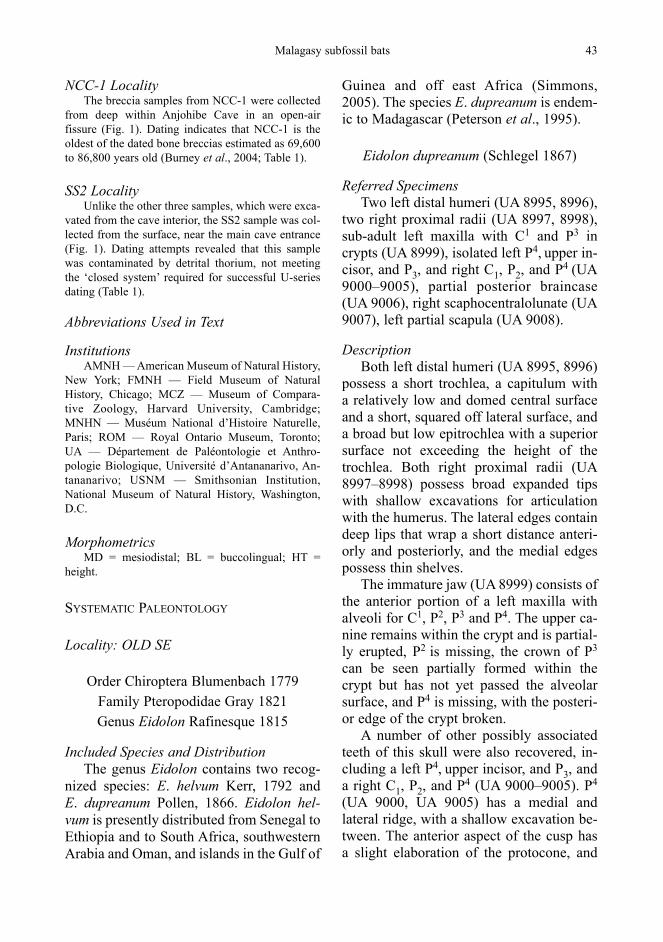
FIG. 1. Anjohibe Cave, northwestern Madagascar. Breccia localities are shown in small black stars on the Anjohibe Cave detail map. Adapted from de Saint-Ours and Paulian (1953) and Burney et al. (1997). Map scale in meters

1997 — Fig. 1). The caves at Anjohibe are part of a karst cave system formed within the Eocene lime-stone plateau (Besairie, 1956). Anjohibe (‘big cave’in Malagasy) consists of a large cave system with an underground river running in the southeastern por-tion (de Saint-Ours and Paulian, 1953; Burney et al.,1997), and several extant animals inhabiting the cave,including bats. The region has undergone extensivehuman modification, and subfossil representation ofarboreally adapted groups (e.g., sloth lemurs) sug-gests that this region was considerably more forestedin the past.
Fossils frequently occur in accumulations be-neath dolines (large sinkholes) or skylights (openingsin the ceiling), as animals fall through these from thesurface. Within the cave interior, some fossils are alsoconcentrated into bone breccias, collections of bonefragments and sediment cemented together with min-eral deposits. Breccias demonstrate a large range ofdepositional processes; some fossils appear to havebeen slowly trapped within forming speleothems,while others appear to have been transported largedistances by subterranean streams, and subsequentlydeposited and consolidated. The breccia samples usedin this study were collected from four localities inAnjohibe Cave (Fig. 1 and Table 1). In some cases,these samples contained high concentrations of batbones (Fig. 2).
Uranium-Series Dating
Breccia samples were dated at Florida StateUniversity using Uranium-series (230Th/234U) tech-niques on the flowstone intercalated with the bonebreccia layers. Speleothems were sampled and ana-lyzed according to standard dating techniques (e.g.,Brook et al., 1990; Burney et al., 1994, 2004).
Fossil Preparation
The breccias containing these fossils were col-lected in 1996 by the team of Dr. David Burney, andaccessed into the collections of the Département dePaléontologie et Anthropologie Biologique, Univer-sité d’Antananarivo, Madagascar (UA). Fossils wereextracted from breccia matrix using standard aceticacid preparation techniques (Toombs and Rixon,1959; Grant, 1989; Leiggi and May, 1994; Lindsay,1995).
Species Identification
The osteological terminology and measurementsused herein follow Hershkovitz (1971), Sigé et al.
Malagasy subfossil bats 41
TAB
LE1.
Age
det
erm
inat
ions
bas
ed o
n U
-ser
ies
anal
ysis
for b
recc
ia s
ampl
es c
olle
cted
from
Anj
ohib
e C
ave
Prov
enan
ceM
ater
ial
Dat
ing
Age
est
imat
eLa
t/Lon
gC
omm
ents
Sour
cem
etho
d/E
leva
tion
NC
C-1
#3
Spel
eoth
emU
-ser
ies
69.6
± 7
.3 k
yr B
P15
°32.
55’S
Dire
ctly
bel
ow b
one
brec
cia
in c
olla
psed
-B
urne
y et
al.
(200
4)46
°53.
17’E
cave
dep
osit
100
mN
CC
-1 #
4Sp
eleo
them
U-s
erie
s86
.8 ±
7.8
kyr
BP
15°3
2.55
’SLa
min
ated
sta
lagm
ite o
n flo
or o
f B
urne
y et
al.
(200
4)46
°53.
17’E
colla
psed
-cav
e fe
atur
e10
0 m
OLD
SE
Spel
eoth
emU
-ser
ies1
≤ 10
,000
kyr
BP
–C
olla
psed
cav
e re
gion
D. A
. Bur
ney,
per
s. co
mm
., 20
01TW
-10
Spel
eoth
emU
-ser
ies1
≤ 10
,000
kyr
BP
–C
olla
psed
cav
e re
gion
D. A
. Bur
ney,
per
s. co
mm
., 20
01SS
2Su
rfac
e bl
ock
U-s
erie
s2>
? ky
r15
°32.
39’S
Col
lect
ed n
ear c
ave
entra
nce
D. A
. Bur
ney,
per
s. co
mm
., 20
0146
°53.
09’E
222
m

(1982), Hand (1993, 1998), Freeman (1998), and Sa-monds (2006). A large comparative sample of pho-tographs, epoxy dental casts, and measurements ofmodern Malagasy bats was collected to facilitateidentification, focusing when possible on specimenscollected from Anjohibe Cave (Samonds, 2006). Inaddition, collections of non-Malagasy bats were pho-tographed and measured to aid in interpreting materi-al that did not match modern taxa known from the is-land. Measurements were made with 500-172 Mitu-toyo digital calipers to 0.01 mm, or with an MA285Meihi Techno optical reticule X, 1/100 (0.01 mm).
Quantitative Methods
When sample sizes were adequate, discriminantfunction analysis was used to examine potential groupdifferences and degrees of group overlap. This analy-sis (SPSS 11.0) includes a multivariate analysis ofvariance to test for significant differences among des-ignated groups (e.g., Wilks’ lambda for the overallanalysis as well as subsets of the discriminant axes).The percentage variance explained by each axis is re-ported, and classification results pinpoint any individ-ual ‘misclassified’ to another group. When sample
sizes were judged too small, including cases wherethere was only one specimen in a group, one-samplet-tests were enlisted to evaluate ‘group’ differences,or whether or not the sample value belonged to thelarger, given ‘population’.
Anjohibe Cave Breccia Localities
OLD SE LocalityThe OLD SE samples were collected 13 August
1996 from a collapsed area that blocked a long, dead-end passage extending southeastward from one of thecave’s largest caverns (Fig. 1). Dating indicates thatthe four samples collected were approximately 10,000years old or younger (D. A. Burney, pers. comm.,2003; see Table 1).
TW-10 LocalityBreccia samples were recovered from the TW-10
locality, approximately 60 m north of Entrance V1 (deSaint-Ours and Paulian, 1953), and located on thesoutheast side of the subterranean river (Fig. 1).Dating indicates that these samples were approxi-mately 10,000 years old or younger (D. A. Burney,pers. comm., 2003; see Table 1).
42 K. E. Samonds
FIG. 2. Anjohibe Cave breccia sample TW-10, illustrating density of bones and teeth
NCC-1 LocalityThe breccia samples from NCC-1 were collected
from deep within Anjohibe Cave in an open-air fissure (Fig. 1). Dating indicates that NCC-1 is theoldest of the dated bone breccias estimated as 69,600to 86,800 years old (Burney et al., 2004; Table 1).
SS2 LocalityUnlike the other three samples, which were exca-
vated from the cave interior, the SS2 sample was col-lected from the surface, near the main cave entrance(Fig. 1). Dating attempts revealed that this samplewas contaminated by detrital thorium, not meetingthe ‘closed system’ required for successful U-seriesdating (Table 1).
Abbreviations Used in Text
InstitutionsAMNH — American Museum of Natural History,
New York; FMNH — Field Museum of Natural History, Chicago; MCZ — Museum of Compara-tive Zoology, Harvard University, Cambridge;MNHN — Muséum National d’Histoire Naturelle,Paris; ROM — Royal Ontario Museum, Toronto; UA — Département de Paléontologie et Anthro-pologie Biologique, Université d’Antananarivo, An-tananarivo; USNM — Smithsonian Institution,National Museum of Natural History, Washington,D.C.
MorphometricsMD = mesiodistal; BL = buccolingual; HT =
height.
SYSTEMATIC PALEONTOLOGY
Locality: OLD SE
Order Chiroptera Blumenbach 1779Family Pteropodidae Gray 1821Genus Eidolon Rafinesque 1815
Included Species and DistributionThe genus Eidolon contains two recog-
nized species: E. helvum Kerr, 1792 and E. dupreanum Pollen, 1866. Eidolon hel-vum is presently distributed from Senegal toEthiopia and to South Africa, southwesternArabia and Oman, and islands in the Gulf of
Malagasy subfossil bats 43
Guinea and off east Africa (Simmons,2005). The species E. dupreanum is endem-ic to Madagascar (Peterson et al., 1995).
Eidolon dupreanum (Schlegel 1867)
Referred SpecimensTwo left distal humeri (UA 8995, 8996),
two right proximal radii (UA 8997, 8998),sub-adult left maxilla with C1 and P3 incrypts (UA 8999), isolated left P4, upper in-cisor, and P3, and right C1, P2, and P4 (UA9000–9005), partial posterior braincase(UA 9006), right scaphocentralolunate (UA9007), left partial scapula (UA 9008).
DescriptionBoth left distal humeri (UA 8995, 8996)
possess a short trochlea, a capitulum with a relatively low and domed central surfaceand a short, squared off lateral surface, anda broad but low epitrochlea with a superiorsurface not exceeding the height of thetrochlea. Both right proximal radii (UA8997–8998) possess broad expanded tipswith shallow excavations for articulationwith the humerus. The lateral edges containdeep lips that wrap a short distance anteri-orly and posteriorly, and the medial edgespossess thin shelves.
The immature jaw (UA 8999) consists ofthe anterior portion of a left maxilla withalveoli for C1, P2, P3 and P4. The upper ca-nine remains within the crypt and is partial-ly erupted, P2 is missing, the crown of P3
can be seen partially formed within thecrypt but has not yet passed the alveolarsurface, and P4 is missing, with the posteri-or edge of the crypt broken.
A number of other possibly associatedteeth of this skull were also recovered, in-cluding a left P4, upper incisor, and P3, anda right C1, P2, and P4 (UA 9000–9005). P4
(UA 9000, UA 9005) has a medial and lateral ridge, with a shallow excavation be-tween. The anterior aspect of the cusp has a slight elaboration of the protocone, and
a lower but well-formed paracone. There isa thin ridge connecting the two cusps medi-olaterally, and the posterior aspect of thecrown is open between the medial and lat-eral ridges. The left upper incisor (UA9001) contains a single cusp terminating ata blunt point. P3 (UA 9004) has a tall, bluntcusp anteriorly, with a small posterior shelf,and a strong excavation on the posterior as-pect of the cusp. C1 (UA 9002) is tall with a wide base, and possesses a thin groove on its anterior surface. P2 (UA 9003) has a short, wide crown with a single cusp onthe anterolateral aspect. Only UA 9001 and9002 have any root formation; the remain-ing teeth possess crowns with no roots.
The partial posterior braincase (UA9006) consists of the parietal and occipitalregions. UA 9006 has a poorly definedsagittal crest and relatively well-developedlambdoidal crest with posteriorly deflectedand thin paraoccipital processes. The brain-case is superior-inferiorly compressed, andthe foramen magnum is ovoid with elongat-ed and well-developed occipital condyles.
The right scaphocentralolunate (UA9007) is large, with a broad and flat proxi-mal articular surface for its articulation withthe radius. The lateral distal surface con-tains a large fossa for the trapezium, and the medial aspect tapers to a narrow point.The partial left scapula (UA 9008) con-tains a narrow, shallow glenoid fossa, a short, robust acromial process, and a rela-tively longer and more slender coracoidprocess.
ComparisonThe three pteropodid bats presently in-
habiting Madagascar are of dramaticallydifferent body size; from largest to smallestthese are: Pteropus rufus (500–750 g),E. dupreanum (235–346 g), and Rousettusmadagascariensis (35–84 g) (Peterson etal., 1995; Russ et al., 2001). The distalhumeral widths of UA 8995 and 8996
44 K. E. Samonds
(13.70 and 13.47 mm, respectively) placethem near but outside the observed val-ues of the modern Malagasy E. dupreanum (n = 4, 0 = 12.73 mm, SD = 0.36). One-sample t-tests suggest that both of thesemeasurements are significantly larger thanin modern populations (P = 0.012 and0.025, respectively). However, extant maleE. dupreanum are about 10% larger than females (MacKinnon et al., 2003) and, giv-en that none of the measured museum spec-imens were of known sex determination,the possibility exists that this discrepancy insize between fossil and modern specimensreflects differences in size dimorphism.Based on this information, and the mor-phology of these elements, both are referredto E. dupreanum.
The proximal radii UA 8997 and 8998are also referable to the family Pteropo-didae based on their large size. The maxi-mum proximal radius width of UA 8997and 8998 (7.73 mm and 7.80 mm, respec-tively) places them within observed valuesof modern Malagasy E. dupreanum (n = 4,0 = 7.86 mm, SD = 0.378). One-sample t-tests suggest that neither of these speci-mens are significantly different from mod-ern populations (P = 0.54 and 0.77, respec-tively).
UA 8999 is referred to Pteropodidaebased on the simple morphology and size ofthe teeth, as pteropodid teeth are highlymodified for frugivory and nectarivory(Bergmans, 1990), and frequently peg-shaped with little or no elaboration of the crown. As the isolated teeth (UA 9000–9005) are unworn, their size and morphol-ogy can be closely matched to those of modern Malagasy E. dupreanum. The presence of this subadult in the depositscould indicate that the cave was used as a nursery colony for this taxon. Detailedcomparisons of maximum neurocranialbreadth and height are impossible due to the fragmentary nature of specimen UA
9006, but the broad shape and relative-ly large size of the partial braincase allowsfor identification of UA 9006 as E. duprea-num.
While the morphology and articular rela-tionships of the scaphocentralolunate aresimilar in all bats (Stafford and Thorington,1998), UA 9007 is identified as pteropodidbased on its extremely large size. Detailedstatistical comparisons were not possibledue to the lack of comparative museumspecimens, as bat postcrania (specifical-ly isolated wrist and ankle bones) are ex-tremely rare. However, the length of this el-ement (10.45 mm) appears to fall most ap-propriately within the size range of themedium-sized Malagasy fruitbat E. dupre-anum, because P. rufus has a substantiallylarger scaphocentralolunate (14.25 mm, n =2) and R. madagascariensis has a muchsmaller body size.
The relatively simple morphology andlarge size of the scapula UA 9008, in ad-dition to the glenoid size (height = 7.29 mm,breadth 4.40 mm) places it within the ob-served values of E. dupreanum (meanheight = 7.11 mm, SD = 0.36; mean breadth= 4.25 mm, SD = 0.32, in both cases n = 4).One-sample t-tests suggest that both ofthese measurements are not significant frommodern populations (P = 0.40 and 0.43,respectively).
Family Hipposideridae Lydekker 1891
Genus Hipposideros Gray 1831
Included Species and DistributionHipposideros is a widely distributed
genus that occurs throughout the Old Worldtropics. The species H. commersoni is en-demic to Madagascar (Simmons, 2005).Subfossil H. commersoni have previouslybeen reported from the younger deposits ofAnjohibe Cave (Burney et al., 1997) andfrom the Lake Tsimanampetsotsa region in
the southwest (Sabatier and Legendre, 1985;MacPhee, 1986).
Hipposideros sp. cf. H. commersoni
Referred SpecimensLeft P4.
DescriptionUA 9009 consists of a left P4 possessing
one main cusp anteriorly and a smaller pos-terior shelf. There is no anterolingual cusppresent, as in the case of several species ofHipposideros. The hypoconal basin is shal-low. There is a low but distinct cingular heeland a basal cingulum surrounds the major-ity of the crown. The anterior aspect of themain cusp is convex anteriorly.
ComparisonThe morphology of UA 9009 most
closely resembles that of members of thefamily Hipposideridae, which possess a P4
that is generally tall with a well-developedprincipal cusp, well-formed posterior shelf,and shallow hypoconal basin. Within thetwo genera of Hipposideridae presently onMadagascar, this tooth most closely match-es those of Hipposideros; representatives ofthe other genus, Triaenops, are significantlysmaller. Further support for this designationis the fact that H. commersoni is the onlyextant Malagasy non-pteropodid bat speciescomparable in size. The only other bat onMadagascar approaching the large size ofH. commersoni is Mops midas (45 g — Fen-ton and Rautenbach, 1986), but this molos-sid species has a P4 with a more flattenedanterior face, stronger lingual cingulum,and taller main anterior cusp.
Although similar in overall tooth mor-phology, a one-sample t-test suggests thatUA 9009 is significantly different frommodern H. commersoni, exhibiting larg-er dimensions for mesiodistal length, buccolingual breadth, and height, and
Malagasy subfossil bats 45

subfossil H. besaoka sp. nov. (Table 2; see Locality: TW-10, below). Its P4 alsolacks an anterolingual cusp (present in both extant H. commersoni and H. besaoka).Based on the data presented here, UA 9009is referred to Hipposideros sp. cf. H. com-mersoni, and considered distinct from bothtaxa.
Genus Triaenops Dobson 1871
Included Species and DistributionSpecies of Triaenops are widespread
throughout eastern Africa, Madagascar, thecoast of the Arabian Peninsula, and AldabraIsland. This genus is presently divided intofive species, with three of them endemic toMadagascar (T. auritus, T. furculus, andT. rufus), one endemic to the Aldabra Island(Triaenops nov. sp.), and one known onlyfrom Africa and parts of the Middle East (T. persicus — Simmons, 2005; Ranivo andGoodman, 2006; Goodman and Ranivo, Inpress).
Triaenops goodmani sp. nov.(Fig. 3A–D)
EtymologySpecific name for Dr Steven M. Good-
man, in recognition of his significant contri-butions to the field of modern Malagasy batresearch.
Type Specimen Partial left dentary with P4M1–2 (UA
9010; Fig. 3A–B).
46 K. E. Samonds
Character UA 9009 vs. H. commersoni vs. H. besaokan 0 SD t-test n 0 SD t-test
MD length of P4 2.12 40 1.99 0.145 -5.31*** 31 2.13 0.104 0.57BL length of P4 2.40 40 2.14 0.132 -12.33*** 31 2.52 0.168 3.97***HT P4 2.35 37 2.15 0.137 -8.79*** 27 2.18 0.152 -5.73***
TABLE 2. One-sample t-test results (2-tailed) for single Hipposideros P4 from OLD SE (UA 9009), compared toextant H. commersoni and subfossil H. besaoka from Anjohibe Cave (TW-10). Probability level: *** — P < 0.001
Referred SpecimensPartial left dentary with M2–3 (UA 9011;
Fig. 3C); partial right dentary with M2–3(UA 9012; Fig. 3D).
DiagnosisLarger than any known species of
Triaenops. Molar crowns narrow, with pro-toconid and hypoconid more rounded labi-ally than seen in modern Triaenops species.M2 molar dimensions significantly longerthan in T. auritus, T. rufus, and T. furculus(P < 0.001). Protoconid only slightly tallerthan hypoconid; in T. auritus, T. rufus, andT. furculus, the protoconid is noticeablytaller than the hypoconid. Relative height ofparaconid, entoconid, and metaconid allshorter than in T. auritus, but similar tocondition in T. rufus and T. furculus.Molars lacking well developed preen-tocristid between metaconid and entoconid.Talonid noticeably wider than talonid.Molars containing small, labially expandedanterior and posterior cingula, and M2 con-taining small lingually expanded shelf be-tween protoconid and hypoconid.
DescriptionUA 9010 (Fig. 3A, B) is a partial left
dentary containing P4M1–2. It is relativelycomplete anteriorly, with alveoli for P2 andthe lateral incisor, but is broken posteriorly.P4 is approximately the height of the tallestcusp (paracone) of M1, or only slightlyhigher. The morphology is caniniform withone very tall median cusp lacking a mesialand buccal shelf. M1 is unworn, and has

a tall trigonid and only slightly lower ta-lonid. The protoconid is the tallest cusp, andthere is a well-developed but only slightlyshorter hypoconid. The paraconid and ento-conid are shorter than the hypoconid, butapproximate each other in height, while themetaconid is slightly lower. The hypo-conulid is the lowest cusp; it is noticeablebut small. There is a distinct gap betweenthe entoconid and hypoconulid, and no dis-tinct ridge uniting the metaconid and ento-conid. M1 possesses a talonid wider than thetrigonid. M2 is comparable in morphologyand size to M1, but contains a talonid onlyslightly wider than the trigonid.
UA 9011 is a partial left dentary (Fig.3C) broken anterior to M2, but containingboth M2 and M3, both of which are relative-ly worn. UA 9011 has a complete ascendingramus and a relatively deep mandibular cor-pus. The morphology of M2 is the same asthat seen in UA 9010. M3 also mirrors theoverall morphology of the other molars, butis slightly smaller, and has no space be-tween the entoconid and hypoconulid.There is a weak ridge between the meta-conid and entoconid, which is deflectedslightly labially; in both M1 and M2 this an-gle is more parallel to the toothrow. M3 pos-sesses a trigonid and talonid of equal width.
Malagasy subfossil bats 47
FIG. 3. Specimens of T. goodmani sp. nov. from OLD SE. A — UA 9010, labial view of type specimen ofT. goodmani sp. nov. partial left dentary with P4M1–2; B — UA 9010, occlusal view; C — UA 9011, lingualview of T. goodmani sp. nov. partial left dentary with M2,3; D — UA 9012, lingual view of T. goodmani sp. nov.
partial right dentary with M2,3
1 mm
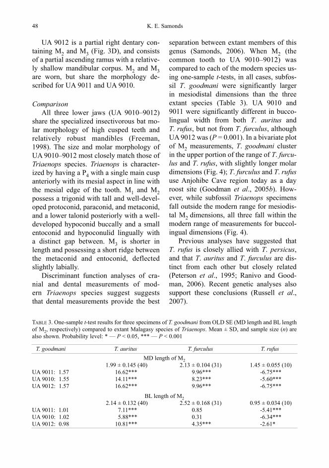
UA 9012 is a partial right dentary con-taining M2 and M3 (Fig. 3D), and consistsof a partial ascending ramus with a relative-ly shallow mandibular corpus. M2 and M3are worn, but share the morphology de-scribed for UA 9011 and UA 9010.
ComparisonAll three lower jaws (UA 9010–9012)
share the specialized insectivorous bat mo-lar morphology of high cusped teeth andrelatively robust mandibles (Freeman,1998). The size and molar morphology ofUA 9010–9012 most closely match those ofTriaenops species. Triaenops is character-ized by having a P4 with a single main cuspanteriorly with its mesial aspect in line withthe mesial edge of the tooth. M1 and M2possess a trigonid with tall and well-devel-oped protoconid, paraconid, and metaconid,and a lower talonid posteriorly with a well-developed hypoconid buccally and a smallentoconid and hypoconulid lingually with a distinct gap between. M3 is shorter inlength and possessing a short ridge betweenthe metaconid and entoconid, deflectedslightly labially.
Discriminant function analyses of cra-nial and dental measurements of mod-ern Triaenops species suggest suggests that dental measurements provide the best
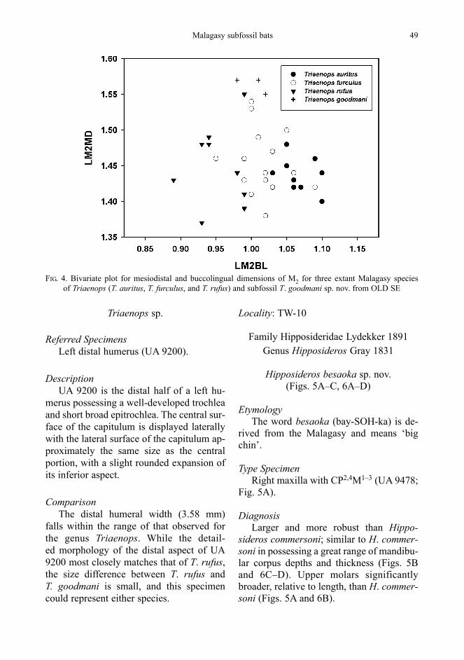
separation between extant members of thisgenus (Samonds, 2006). When M2 (thecommon tooth to UA 9010–9012) was compared to each of the modern species us-ing one-sample t-tests, in all cases, subfos-sil T. goodmani were significantly larger in mesiodistal dimensions than the three extant species (Table 3). UA 9010 and 9011 were significantly different in bucco-lingual width from both T. auritus and T. rufus, but not from T. furculus, althoughUA 9012 was (P = 0.001). In a bivariate plotof M2 measurements, T. goodmani clusterin the upper portion of the range of T. furcu-lus and T. rufus, with slightly longer molardimensions (Fig. 4); T. furculus and T. rufususe Anjohibe Cave region today as a dayroost site (Goodman et al., 2005b). How-ever, while subfossil Triaenops specimensfall outside the modern range for mesiodis-tal M2 dimensions, all three fall within themodern range of measurements for buccol-ingual dimensions (Fig. 4).
Previous analyses have suggested that T. rufus is closely allied with T. persicus,and that T. auritus and T. furculus are dis-tinct from each other but closely related(Peterson et al., 1995; Ranivo and Good-man, 2006). Recent genetic analyses alsosupport these conclusions (Russell et al.,2007).
48 K. E. Samonds
T. goodmani T. auritus T. furculus T. rufusMD length of M2
1.99 ± 0.145 (40) 2.13 ± 0.104 (31) 1.45 ± 0.055 (10)UA 9011: 1.57 16.62*** 9.96*** -6.75***UA 9010: 1.55 14.11*** 8.23*** -5.60***UA 9012: 1.57 16.62*** 9.96*** -6.75***
BL length of M22.14 ± 0.132 (40) 2.52 ± 0.168 (31) 0.95 ± 0.034 (10)
UA 9011: 1.01 7.11*** 0.85 -5.41***UA 9010: 1.02 5.88*** 0.31 -6.34***UA 9012: 0.98 10.81*** 4.35*** -2.61*
TABLE 3. One-sample t-test results for three specimens of T. goodmani from OLD SE (MD length and BL lengthof M2, respectively) compared to extant Malagasy species of Triaenops. Mean ± SD, and sample size (n) arealso shown. Probability level: * — P < 0.05, *** — P < 0.001
Malagasy subfossil bats 49
FIG. 4. Bivariate plot for mesiodistal and buccolingual dimensions of M2 for three extant Malagasy species of Triaenops (T. auritus, T. furculus, and T. rufus) and subfossil T. goodmani sp. nov. from OLD SE
Triaenops sp.
Referred SpecimensLeft distal humerus (UA 9200).
DescriptionUA 9200 is the distal half of a left hu-
merus possessing a well-developed trochleaand short broad epitrochlea. The central sur-face of the capitulum is displayed laterallywith the lateral surface of the capitulum ap-proximately the same size as the centralportion, with a slight rounded expansion ofits inferior aspect.
ComparisonThe distal humeral width (3.58 mm)
falls within the range of that observed forthe genus Triaenops. While the detail-ed morphology of the distal aspect of UA9200 most closely matches that of T. rufus,the size difference between T. rufus and T. goodmani is small, and this specimencould represent either species.
Locality: TW-10
Family Hipposideridae Lydekker 1891Genus Hipposideros Gray 1831
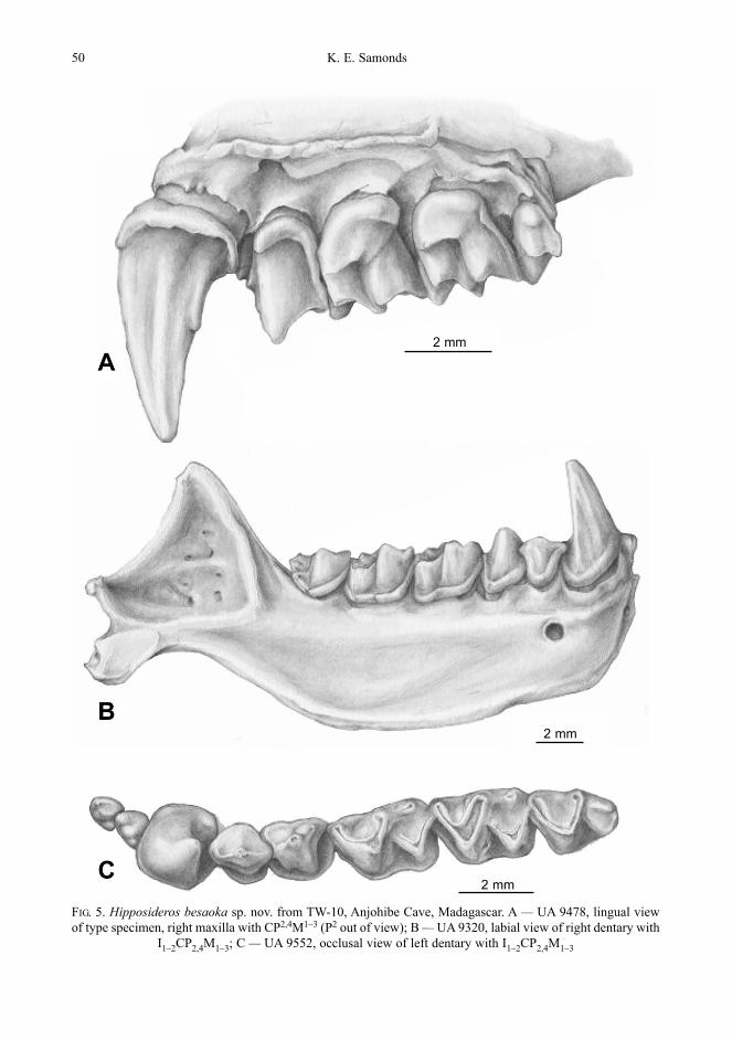
Hipposideros besaoka sp. nov.(Figs. 5A–C, 6A–D)
EtymologyThe word besaoka (bay-SOH-ka) is de-
rived from the Malagasy and means ‘bigchin’.
Type SpecimenRight maxilla with CP2,4M1–3 (UA 9478;
Fig. 5A).
DiagnosisLarger and more robust than Hippo-
sideros commersoni; similar to H. commer-soni in possessing a great range of mandibu-lar corpus depths and thickness (Figs. 5Band 6C–D). Upper molars significantlybroader, relative to length, than H. commer-soni (Figs. 5A and 6B).
50 K. E. Samonds
FIG. 5. Hipposideros besaoka sp. nov. from TW-10, Anjohibe Cave, Madagascar. A — UA 9478, lingual view of type specimen, right maxilla with CP2,4M1–3 (P2 out of view); B — UA 9320, labial view of right dentary with
I1–2CP2,4M1–3; C — UA 9552, occlusal view of left dentary with I1–2CP2,4M1–3
2 mm
2 mm
2 mm

Malagasy subfossil bats 51
Referred Specimens Twenty-six upper right maxillae (UA
9284–9301, 9478, 9504–9511; Fig. 5A), 27upper left maxillae (UA 9269–9283,9512–9523; Fig. 6A–B), 78 left dentaries(UA 9232–9268, 9302–9319, 9330–9345,9497–9503, 9552; Fig. 6C–D), 40 right dentaries (UA 9218–9231, 9320–9329,9481– 9496; Fig. 5B), 42 left and right pre-maxillae (UA 9346–9387), 26 isolated up-per incisors (UA 9525, 9480 [lot of 25]),118 isolated lower incisors (UA 9526, 9479 [lot of 117]), 41 upper left canines(UA 9398–9404, 9437–9470), 43 upperright canines (UA 9527, 9388–9397,9405–9436), 26 lower left canines (UA9528 [lot of 26]), 25 lower right canines(UA 9530, 9529 [lot of 24]), 22 left P2(UA 9531, 9472 [lot of 17], 9550 [lot of 4]),26 right P2 (UA 9551, 9471 [lot of 25]), 2 right P4 (UA 9532, 9548), 4 left P4
(UA 9537, 9473, 9547 [lot of 2]), 1 right P4(UA 9539), 3 left P4 (UA 9538, 9549 [lot of2]), 7 right M1 (UA 9543 [lot of 7]), 7 leftM1 (UA 9474, 9545 [lot of 6]), 8 right M2 (UA 9476, 9477, 9544 [lot of 6]), 7 leftM2 (UA 9475, 9546 [lot of 6]), 1 right M3
(UA 9533), 3 left M3 (UA 9534 [lot of 3]),18 right M1–2 (UA 9541 [lot of 18]); 13 leftM1–2 (UA 9536 [lot of 4], 9542 [lot of 9]), 8 right M3 (UA 9540 [lot of 8]), 6 left M3(UA 9535 [lot of 6]).
Description The premaxillae rami were frequently
fused at the midline; these elements werenever found in association with skulls. The posterior margin of the premaxilla is narrowed to a thin point and flares gently anteriorly. The anterior margin ta-pers to a V-shape posteriorly between theincisors, which are located at the anterior-most edge near the tip of each anterolateralprojection. The ventral surface is concavelongitudinally. Each premaxilla has a largeanterior palatal foramen. The line of contact
between premaxillae is thin with no crest onthe dorsal surface. Each premaxilla houses a single incisor, which is directed an-teroventrally. Upper incisors are reducedand weakly bilobed, with small flat crownsand thin roots that project minimally abovethe alveolar line.
The maxillae (Figs. 5A and 6A–B) arefrequently well preserved, although manycontain worn teeth. Maxillae possess uppercanines with one long principal cusp and a small posterior shelf approximately half-way up the posterior aspect of the tooth, ex-tremely reduced anterior upper premolar(P2) wedged between the upper canine andP4, and a wide P4 with a tall anterior cusp,lower posterior shelf, and a well-defined an-terolingual cusp. M1 has a closed protofossawith weak dihedral crest and a posteriorlydeflected but low cingular heel. M2 is com-parable to M1 but is squarer and slightlysmaller, with a less developed and posteri-orly deflected heel. M3 has an extremely re-duced W-pattern, where the premetacrista isless than 1/3 the length of the preparacrista,and is significantly smaller (about 1/2) thanin both M1 and M2.
Lower jaws (Figs. 5B and 6C–D) show a large range of corpus depth and thickness,with some extremely thin and shallow (UA9244; Fig. 6C), and others robust and deep (UA 9338; Fig. 6D). The dentary con-tains two small, anteroposteriorly com-pressed lower tricuspid incisors of approxi-mately the same size. The lower canine has a single tall and narrow cusp. P2 is large and moderately wide with a large maincusp connected by steep crests to the ante-rior and posterior edges of the crown, and a smaller cusp posterior to the main cuspwithin the posterior crest. P4 has two dis-tinct roots, one tall median cusp, a tiny anterolingual cusp, and a small but well-developed posterolingual cusp. The post-erior ridge descending from the main cuspconsists of two parts; the first descending

52 K. E. Samonds
FIG. 6. Specimens of H. besaoka sp. nov. from TW-10, Anjohibe Cave, Madagascar. A — UA 9272, labial viewof left maxilla with CP2,4M1–3; B — UA 9272, lingual view of left maxilla with CP2,4M1–3; C — UA 9244, labialview of left dentary with CP2,4M1–2 and partial M3; D — UA 9338, labial view of left dentary with CP2,4M1–3
illustrating depth of mandibular corpus
weakly and the second turning more sharplyinferiorly towards the posterolingual cusp.
M1 is large, with a high trigonid and relatively low talonid basin (Fig. 5C). The
protoconid is very tall and well-developed,the metaconid is slightly lower, and theparaconid approximates the height of themetaconid or surpasses it slightly. The
1 mm

hypoconid is quite low. Both the anteriorand posterior cingula are weak, and the pro-toconid and hypoconid are at approximatelythe same distance from the midline of thetooth. The morphology of M2 mirrors thatof M1, and the size is comparable, or onlyslightly smaller. The angle made by the pre-and postprotocristid is conspicuously nar-rower in M2 and M3 than in M1. M3 is muchshorter than M1 and M2 in mesiodistallength, and has a greatly reduced talonid.The ridge between the metaconid and ento-conid is well developed but its posteriormargin is flexed laterally; the hypocristid iscompletely absent.
ComparisonFor complete dentitions, the specimens
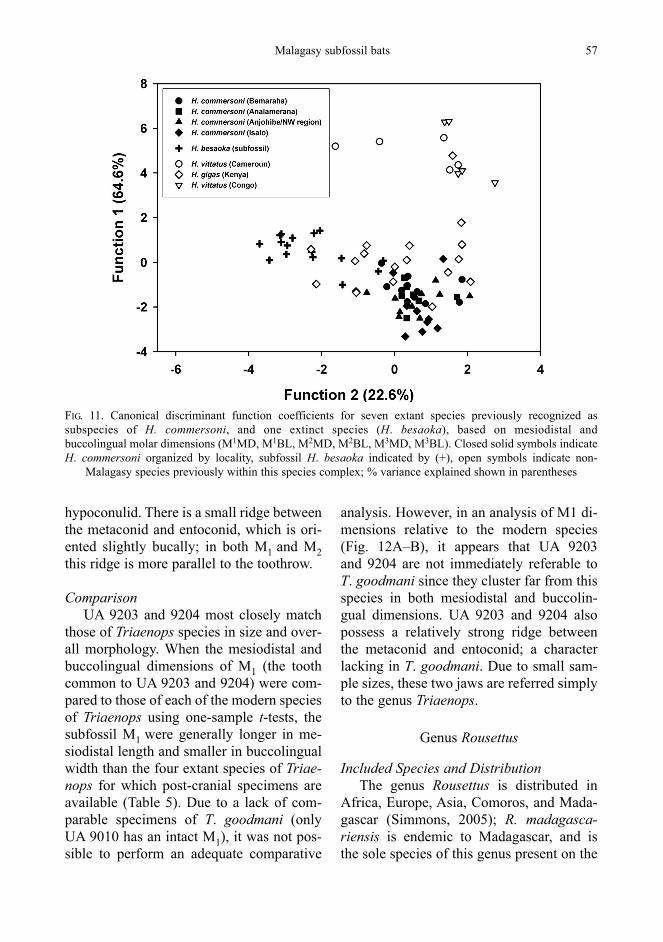
considered here are immediately referableto Hipposideros based in part on the dentalformula 1/2 1/1 2/2 3/3, the small upper premolar shifted laterally relative to the restof the toothrow (such that the canine andsecond upper premolar are in contact ornearly so), and the distinctive morphologyof P2, being large and possessing a steeplysloped and broad buccal surface (Hand andKirsch, 1998, 2003). A Discriminant Func-tion Analysis of the Hipposideros fossilsfrom TW-10 indicates that the subfossilsample is significantly different than mod-ern H. commersoni (Fig. 7); H. besaoka up-per molars are significantly broader (e.g.,buccolingual dimensions) than seen in mod-ern H. commersoni (Fig. 8).
Subfossil Hipposideros mandibles alsoshow a large range of mandibular corpusdepth and thickness sizes significantly different from modern samples (Fig. 9).Overall, subfossil Hipposideros mandibleshave a thicker and more robust mandib-ular corpus for their depth than modern H. commersoni, a morphology that, in mo-lossid bats, has been correlated with spe-cializations for hard-object feeding (Free-man, 1981).
To compare subfossil Hipposiderosspecimens from Anjohibe to modern pop-ulations on the island, it was important to address the issue of latitudinal clines.Hipposideros commersoni is broadly dis-tributed throughout Madagascar, encom-passing a wide range of habitats and tem-perature gradients. If modern groups dis-play predictable relationships between size and latitude, the larger size of subfos-sil Hipposideros might, at least in part, beattributable to changes in climate, as hasbeen previously demonstrated for bats else-where in the Old World tropics (Storz et al.,2001).
Modern populations of H. commersonifrom four distant regions of the island werecompared to each other in an attempt to address this issue. These four regions weredivided into operational taxonomic units(OTU’s) based on geography includingAnalamerana, the northwest of the island(including Anjohibe Cave, Mampikony, and Ankarafantsika), Bemaraha, and Isalo(Fig. 10). Subpopulations of H. commer-soni do demonstrate significant differencesbetween sites, with the most northern(Analamerana) and the most southern(Isalo) showing the closest relationship, andthe other two sites clustering discretely(Fig. 10).
Nevertheless, when six upper molarmeasurements available for the subfossilsample are examined, and other extralim-ital species of Hipposideros that were for-merly considered subspecies of H. commer-soni are included, H. commersoni spec-imens group closely together, with the subfossils and extralimital species group-ing farther away (Fig. 11). This suggeststhat the morphological differences be-tween the fossil specimens and extant Ma-lagasy H. commersoni exceed the differ-ences observed among the various extant H. commersoni populations within Mada-gascar.
Malagasy subfossil bats 53

54 K. E. Samonds
Locality: NCC-1
Hipposideros commersoni E. Geoffroy 1813
Referred Specimens Isolated lower incisor (UA 9201), right
M3 (UA 9202).
Description UA 9201 has a well-defined crown that
is trilobed and anteroposteriorly com-pressed. The central lobe is the largest, with
the mesial lobe slightly smaller and the dis-tal lobe smallest. It contains a single rootwith a slight flare at its tip. UA 9202 is a M3consisting of a complete crown with noroots preserved. The protoconid is tall andwell-developed, while the paraconid andmetaconid are relatively low. There is awell-developed crest (preentocristid) link-ing the metaconid to the entoconid, which isroughly parallel to the protocristid. Thetrigonid is relatively wide. The talonid is ex-tremely reduced to a small shelf containinga small hypoconid.
FIG. 7. Canonical discriminant function coefficients for Hipposideros from TW-10 and H. commersoni. A —upper molar measurements (M1MD, M1BL, M2MD, M2BL, M3MD, M3BL), Wilks’ lamba = 0.18, Chi-square= 88.25, P < 0.001; B — lower molar measurements (M1MD, M1BL, M2MD, M2BL, M3MD, M3BL); Wilks’
lamba = 0.64, Chi-square = 54.07, P < 0.001. Line represents median, all outliers included
A
B
n
n

Malagasy subfossil bats 55
FIG. 8. Bivariate plot for mesiodistal and buccolingual M1 molar dimensions for Hipposideros commersonimales and females, and subfossil H. besaoka from TW-10
ComparisonThe morphology and size of UA 9201
matches that seen in the modern form ofHipposideros commersoni. UA 9202 showsthe distinctive M3 morphology associatedwith typical insectivorous bats, and is within the general size range of both theMalagasy Hipposideros and Mops midasmiarensis. However, M. m. miarensis has a slightly different M3 morphology, with a more anteroposteriorly compressed trigo-nid, and a preentocristid that does not par-allel the protocristid, but rather forms a greater angle at its junction with the meta-conid.
An analysis of UA 9202 indicates that it is not statistically distinct from modern H. commersoni, but is different from the referred material of H. besaoka describ-ed from locality TW-10 (Table 4). UA 9202is therefore referred here to H. commer-soni.
Triaenops sp.
Referred SpecimensTwo left dentaries, one with P4M1 (UA
9203), and one with M1–2 and part of a bro-ken M3 (UA 9204).
Description UA 9203 is a well-preserved left dentary
with P4M1 intact. There is a small alveolusfor P2, but the jaw is broken anteriorly. Theposterior portion of the jaw is broken, andthe mandibular corpus depth is relativelyshallow. A relatively large mental foramenis present in line with the anterior aspect ofP4. P4 has a single tall median cusp, and isapproximately the same height as the tallestcusp of M1 (paracone). M1 is unworn, andpossesses a relatively tall trigonid. Theparaconid, entoconid, and metaconid are ap-proximately the same height. The hypo-conulid is small, and there is a distinct gap

56 K. E. Samonds
FIG. 9. Canonical discriminant function coefficients for Hipposideros from TW-10, measurements includecorpus depth, corpus thickness, and ascending ramus height; Wilks’ lamba = 0.62, Chi-square = 74.77,
P < 0.001. Line represents median, all outliers included
FIG. 10. Canonical discriminant function coefficients based on dental and mandibular measurements for fourpopulations of Malagasy H. commersoni by site; % variance explained presented in parentheses. Measurementsinclude mandible length, corpus depth, corpus thickness, ascending ramus height, P4MD, P4BL, M1MD, M1BL,
M2MD, M2BL, M3MD, M3BL, P4MD, P4BL, M1MD, M1BL, M2MD, M2BL, M3MD, M3BL, and M3HT
between the entoconid and hypoconulid. M1also has a ridge between the metaconid and entoconid. UA 9204 is a left dentarywith M1–2, a broken M3, and a shallow thinmandibular corpus. The M1 morphology is
identical to that described for UA 9203, andthe morphology of M2 mirrors that of M1.M3 is similar in morphology to the othermolars, but is slightly smaller, and has no space between the entoconid and
n
hypoconulid. There is a small ridge betweenthe metaconid and entoconid, which is ori-ented slightly bucally; in both M1 and M2this ridge is more parallel to the toothrow.
ComparisonUA 9203 and 9204 most closely match
those of Triaenops species in size and over-all morphology. When the mesiodistal andbuccolingual dimensions of M1 (the toothcommon to UA 9203 and 9204) were com-pared to those of each of the modern speciesof Triaenops using one-sample t-tests, thesubfossil M1 were generally longer in me-siodistal length and smaller in buccolingualwidth than the four extant species of Triae-nops for which post-cranial specimens areavailable (Table 5). Due to a lack of com-parable specimens of T. goodmani (onlyUA 9010 has an intact M1), it was not pos-sible to perform an adequate comparative
analysis. However, in an analysis of M1 di-mensions relative to the modern species(Fig. 12A–B), it appears that UA 9203 and 9204 are not immediately referable toT. goodmani since they cluster far from thisspecies in both mesiodistal and buccolin-gual dimensions. UA 9203 and 9204 alsopossess a relatively strong ridge betweenthe metaconid and entoconid; a characterlacking in T. goodmani. Due to small sam-ple sizes, these two jaws are referred simplyto the genus Triaenops.
Genus Rousettus
Included Species and DistributionThe genus Rousettus is distributed in
Africa, Europe, Asia, Comoros, and Mada-gascar (Simmons, 2005); R. madagasca-riensis is endemic to Madagascar, and is the sole species of this genus present on the
Malagasy subfossil bats 57
FIG. 11. Canonical discriminant function coefficients for seven extant species previously recognized assubspecies of H. commersoni, and one extinct species (H. besaoka), based on mesiodistal and buccolingual molar dimensions (M1MD, M1BL, M2MD, M2BL, M3MD, M3BL). Closed solid symbols indicate H. commersoni organized by locality, subfossil H. besaoka indicated by (+), open symbols indicate non-
Malagasy species previously within this species complex; % variance explained shown in parentheses

58 K. E. Samonds
TABLE 4. One-sample t-test (2-tailed) results for Hipposideros sp. from NCC-1 compared to extant H. commersoni and subfossil H. besaoka from Anjohibe Cave (TW-10). Mean, SD, and sample size (n) are alsoshown. Probability level: *** — P < 0.001
Character UA 9015vs. H. commersoni vs. H. besaoka
n 0 SD t-test n 0 SD t-test MD length of M3 2.23 40 2.22 0.127 -0.55 100 2.37 0.098 14.06***BL length of M3 1.65 40 1.69 0.132 1.82 100 1.76 0.087 13.19***
TABLE 5. One-sample t-test results for MD and BL lengths of M1 for Triaenops sp. from NCC-1. Malagasyspecies include T. auritus, T. furculus and T. rufus; T. persicus is distributed in Africa and the Middle East. Mean ± SD, and sample size (n) are also shown. Probability level: * — P < 0.05, ** — P < 0.01, *** — P < 0.001
Triaenops sp. T. auritus T. furculus T. rufus T. persicusMD length of M1
1.48 ± 0.046 (9) 1.46 ± 0.041 (15) 1.59 ± 0.057 (10) 1.56 ± 0.066 (6)UA 9016: 1.69 -13.64*** -22.00*** -5.33*** -4.80**UA 9017: 1.55 -4.50** -8.65*** 2.44* 0.44
BL length of M11.02 ± 0.039 (9) 0.99 ± 0.054 (15) 0.90 ± 0.048 (10) 0.94 ± 0.056 (6)
UA 9016: 0.82 15.49*** 12.11*** 5.46*** 5.13**UA 9017: 0.85 13.21*** 9.95*** 3.49** 3.81*
island. It is presently distributed throughoutthe eastern rainforest, the deciduous forestsof the west, southwest and north, and theCentral High Plateau (MacKinnon et al.,2005).
Rousettus sp. cf. R. madagascariensis
Referred SpecimensRight M3 (UA 9206).
DescriptionUA 9206 consists of a partial tooth with
part of its crown missing. The two roots arepartially intact. The base of the crown isvery shallow and relatively broad. The an-terobuccal aspect of the crown is complete,and preserves a distinct ridge.
ComparisonBased on the small size and overall
shape of the crown base and roots, UA 9206is assigned to the genus Rousettus, speciesof which possess teeth that are typically
simple and ‘rousettine’, but are also consid-erably narrow (Bergmans, 1977, 1994). UA9206 appears to best match the morphologyof M3, as this tooth lacks well-developedcusps, has a narrow width, and is short inlength.
Locality: SS2
Hipposideros sp. cf. H. commersoni
Referred SpecimensTwo isolated left M3 (UA 9207, 9208),
isolated left M1 (UA 9209), partial rightmaxilla (UA 9210), right dentary with P4(UA 9211), left dentary with isolated M1(UA 9214), right P4 (UA 9213).
DescriptionUA 9207 and 9208 are both left M3 con-
sisting of complete crowns with partialroots intact. In both, the trigonid is relative-ly wide and the protoconid and metaconidare similar in height. The protoconid is

well-developed, and the talonid basin is ex-tremely reduced with a small hypoconid.The preentocristid is more or less parallel tothe protocristid. UA 9209 consists of a leftM1 crown with partial roots, but the hypo-conal basin is broken. The protofossa is
closed, and the large cingular heel is de-flected posteriorly.
The partial right maxilla, UA 9210, con-tains P2,4M1–3. A large infraorbital foramenlies superior to M1. P2 is vestigial and extremely reduced. It is displaced laterally
Malagasy subfossil bats 59
FIG. 12. Boxplots of mesiodistal and buccolingual dimensions of M1 for three extant Malagasy species ofTriaenops (T. auritus, T. furculus, and T. rufus), subfossil Trianeops from NCC-1, and subfossil T. goodmani sp.
nov. from OLD SE; A — mesiodistal dimensions; B — buccolingual
A
B
n

such that it is situated lateral to the anter-obuccal corner of P4. P4 has a tall, well-developed principal cusp, a relatively tallposterior cusp, a tiny cingular cusp, and a shallow hypoconal basin. The lingual as-pect of the crown has a relatively well-developed cingulum. M1 has a well-devel-oped but low cingular heel. The paraconeand metacone are the tallest cusps, followed by the parastyle, mesostyle, and metastylerespectively; the protocone is low. Theparacrista and premetacrista are approxi-mately equal in length and parallel to each other, as are the postparacrista andmetacrista. The hypoconal basin is well-developed and the heel is posteriorly de-flected. M2 is similar to M1, although it has a smaller hypoconal basin and less posteri-orly deflected heel. M3 is substantially re-duced in size, and has complete loss of thetalonid, a medially displaced metaconid,and only the remnant of a heel.
UA 9211 is a right dentary with P4,which has a tall median cusp and a shorterposterior cusp. The corpus is relatively deepand robust, and there are alveoli present for P2, C and the lateral lower incisor. UA 9214 consists of a left dentary with M1. The ramus is broken and is relative-ly deep. M1 has a tall protoconid, slightlyshorter hypoconid, and lower but approxi-mately equal paraconid and entoconid; thelowest cusp is the hypoconulid. The trigo-nid is high while the talonid is low but com-plete.
UA 9213 is a right P4 with a large maincusp, a small posterior shelf, and a vestigialanterolingual cusp. There is a low cingularheel and the crown is surrounded by a basalcingulum. The hypoconal basin is shallow.
ComparisonAll of these specimens are referable
to Hipposideros. However, the fact that UA 9207 and 9208 appear significantly different from both H. commersoni and
60 K. E. Samonds
H. besaoka (Table 6), and combined withthe small sample size, this makes assign-ment to species uncertain.
Rousettus madagascariensis
Referred SpecimenDistal right humerus (UA 9216).
DescriptionUA 9216 is the distal end of a relatively
large right humerus, with a relatively well-developed trochlea, and a flat broad capitu-lum. The shaft is extremely thin and theepitrochlea is flat and broad. The trochlea islow and steeply angled towards the junctionwith the epitrochlea, and the lateral surfaceof the capitulum has a round excavation onits lateral surface.
ComparisonPteropodid humeri are typically relative-
ly simple and primitive, with less elabora-tion of the distal aspect than seen in otherbats. Based on the size and specific mor-phology of this specimen, UA 9216 is re-ferred to R. madagascariensis.
Family Vespertilionidae Gray 1821Subfamily Vespertilioninae Gray 1821
Genus Myotis Kaup 1829
Included Species and DistributionMyotis is a very speciose and wide-
spread genus, with 100 species presentlyrecognized (Simmons, 2005). Two subspe-cies of M. goudoti were previously recog-nized: M. g. anjouanensis Dorst, 1960,restricted to Anjouan Island in the Como-ros and M. g. goudoti Dorst, 1960, endem-ic to Madagascar; both of these are now recognized as distinct species (Simmons,2005). Myotis goudoti inhabits most habitattypes in Madagascar (Eger and Mitchell,2003).

Myotis goudoti
Referred SpecimenLower right canine (UA 9217).
DescriptionUA 9217 is a very small isolated right
canine with a single tall pointed main cusp.The lingual surface is spade-shaped andslightly concave.
ComparisonUA 9217 matches the morphology and
size of M. goudoti. This species has uniquesmall pointed canines with a single maincusp, and the unique lingual spade-shapedsurface, which is broad inferiorly. This isolated canine (1.47 mm) fits within the modern size range seen in this species(0 = 1.33, n = 3, SD = 0.076, one-sample t-test, P = 0.083).
DISCUSSION
Subfossil versus Modern Assemblages
Of the subfossil bat specimens describedin these breccia samples, all are attributableto genera represented on the island today.Nine species of extant bats are known to use the Anjohibe Cave system as a dayroost based on a recent census (Pteropus
Malagasy subfossil bats 61
TABLE 6. One-sample t-test results for Hipposiderosfrom SS2 (M3) compared to extant H. commersoniand subfossil H. besaoka from Anjohibe Cave (TW-10). Mean ± SD, and sample size (n) are also shown.Probability level: * — P < 0.05, *** — P < 0.001
Hipposideros H. commersoni H. besaokasp. (TW-10)
MD length of M32.22 ± 0.127 (40) 2.37 ± 0.098 (100)
UA 9020: 2.32 -5.03*** 4.87***UA 9021: 2.27 -2.54* 9.97***
BL length of M3UA 9020: 1.63 2.77** 15.49***UA 9021: 1.70 -0.57 7.44***
rufus, R. madagascariensis, H. commer-soni, T. furculus, T. rufus, Miniopterus ma-navi, M. gleni, Myotis goudoti, and Oto-mops madagascariensis — Goodman et al.,2005; S. M. Goodman, pers. comm.). Thesubfossils recovered from Anjohibe Caveindicate the presence of five genera, one ofwhich (Eidolon) no longer inhabits thiscave system. Of the subfossil taxa identi-fied and described here, only four extantspecies (E. dupreanum, R. madagascarien-sis, H. commersoni, and Triaenops sp. cf.T. furculus) were previously recorded fromthe younger sediments of Anjohibe Cave(Burney et al., 1997); one is new for the site (M. goudoti). Furthermore, two of thetaxa identified are new and described here(T. goodmani and H. besaoka).
The large sample of subfossils referableto Hipposideros, its presence in all brecciasamples, and the fact that species are alsocommonly found in the younger sedimentsof the cave indicates that the genus waswell represented in the overall cave fauna inthe recent geological past. The only otherrecord of Hipposideros from a subfossil de-posit on the island is from Lake Tsimanam-petsotsa, southwestern Madagascar (Saba-tier and Legendre, 1985).
The evidence of a distinctly larger spe-cies of Hipposideros (H. besaoka) fromTW-10 is especially significant, because theextant species H. commersoni is the largestinsectivorous bat known from the island(Russ et al., 2001). This new species hassignificantly wider molars than seen inmodern populations of H. commersoni,with a more robust and deeper mandibularcorpus.
Although common as a breccia subfos-sil, and extremely numerous within themore recent Holocene deposits, Eidolon du-preanum is currently locally extinct in theAnjohibe Cave system. This species hasbeen recorded from many regions of Mad-agascar, but may be susceptible to human

pressures (Racey et al., 2002; MacKinnonet al., 2003). Roost sites appear to be espe-cially at risk; recent ecological studies sug-gest that nearly half of the Eidolon roostssurveyed were deserted at least in part dueto human hunting (Racey et al., 2002). Inmany regions Eidolon is able to persist indeforested areas (MacKinnon et al., 2003).
The extinction of Eidolon at Anjohibeappears to have occurred in the past fewdecades. Interviews with local villagerssuggest that older adults recognize thebones of Eidolon as a ‘fanihy’ (fruit bat)that no longer occurs in the cave, but onethat lived previously in the cave system during their childhood (S. M. Goodman,pers. comm.). The large quantity of Eidolonbones on the surface of the cave floor alsosupports a recent extinction event.
It is interesting to note that not all spe-cies presently inhabiting the cave were de-tected as subfossils (e.g., Otomops, Minio-pterus). This may indicate that these generacolonized the cave more recently. However,it is equally possible that they were not rep-resented in this sample due to chance ortaphonomic factors.
CONCLUSIONS
Caves are among the most delicate andvulnerable ecosystems (Watson et al., 1997;van Beynen and Townsend, 2005), and areparticularly susceptible to human pressures.Previous research has demonstrated a majorloss of biodiversity in many of Madagas-car’s species during the Holocene (Godfreyet al., 1990; Goodman, 1994; Burney et al.,1997; Gommery et al., 2003), yet whetherMalagasy bats experienced similar extinc-tions has never been addressed. This papermarks the first systematic study of subfossilMalagasy bats.
On the basis of subfossil remains re-covered within Anjohibe Cave, it is clear that bats on the island experienced local
extirpation (as seen in Eidolon) and island-wide extinction as seen by T. goodmani andthe largest known insectivorous bat on theisland, H. besaoka. It is also interesting tonote that the subfossil forms tend to be larg-er than their extant relatives, although thedifference is less than in other groups (e.g.,Cryptoprocta — Goodman et al., 2004; var-ious lemur groups including Daubentonia— Godfrey and Jungers, 2002).
ACKNOWLEDGMENTS
I wish to thank the Government of Madagascarfor permission to conduct research in Madagascar,which was in collaboration with the Département dePaléontologie et Anthropologie Biologique, Univer-sité d’Antananarivo, Antananarivo. I also wish tothank Drs. David Krause, William Jungers, MaureenO’Leary, Steven Goodman, Nancy Simmons, andMitchell Irwin.
Special thanks to financial support from the CaveConservancy Foundation, Cave Research Foundation,and the Field Museum of Natural History. I am grate-ful to the museums and curators that helped to facili-tate this research, including Bill Stanley (FMNH),Judith Chupasko (MCZ), Nancy Simmons, Bob Randall, and Jean Spence (AMNH), Linda Gordonand Helen James (USNM), Judith Eger (ROM),Ronan Kirsch, Christiane Denys, and Marc Godinot(MNHN), and Armand Rasoamiaramanana andGisPle Francine Noro Randria (UA).
Thanks to the research team that collected anddated the Anjohibe Cave breccia, including DavidBurney, Jim Cowart, Fred Grady, Helen James, andWilliam Jungers. I am grateful to Fred Grady(USNM) for his instruction in acetic acid preparationtechniques, as well as the efforts of volunteer fossilpreparators Kenneth Lee, Olivia van Nieuwenhuizen,Jennie Yu, and Jessica DeLalio, and to Colleen Lodgefor her drawings of H. besaoka. Mitchell Irwin,Steven Goodman, and one anonymous reviewer pro-vided helpful comments on an earlier version of thispaper.
LITERATURE CITED
BERGMANS, W. 1977. Notes on new material of Rousettus madagascariensis Grandidier, 1929(Mammalia, Megachiroptera). Mammalia, 41:67–74.
BERGMANS, W. 1990. Taxonomy and biogeography of
62 K. E. Samonds

African fruit bats (Mammalia, Megachiroptera).3: The genera Scotonycteris Matschie, 1894, Ca-sinycteris Thomas, 1910, Pteropus Brisson, 1762,and Eidolon Rafinesque, 1815. Beaufortia, 40:111–177.
BERGMANS, W. 1994. Taxonomy and biogeography ofAfrican fruit bats (Mammalia, Megachiroptera).4: The genus Rousettus Gray, 1821. Beaufortia,44: 79–126.
BESAIRIE, H. 1956. Carte géologique (Majunga, N°2):Service Géologique de Madagascar, map scale1:500,000, 1 sheet.
BROOK, G. A., D. A. BURNEY, and J. B. COWART. 1990.Desert paleoenvironmental data from cavespeleothems with examples from the Chihuahuan,Somali-Chalbi, and Kalahari deserts. Paleogeo-graphy, Paleoclimatology, Palaeoecology, 76:311–329.
BURNEY, D. A. 1999. Rates, patterns and processes oflandscape transformation and extinction in Mad-agascar. Pp. 145–164, in Extinctions in near time(R. D. MACPHEE, ed.), Kluwer Academic Press,New York, xvi + 394 pp.
BURNEY, D. A. 2003. Madagascar’s prehistoric eco-systems. Pp. 47–51, in The natural history ofMadagascar (S. M. GOODMAN and J. P. BENSTEAD,eds.). The University of Chicago Press, Chicago,xxi + 1709 pp.
BURNEY, D. A., G. A. BROOK, and J. B. COWART. 1994.A Holocene pollen record for the Kalahari Desertof Botswana from a U-series dated speleothem.Holocene, 4: 225–232.
BURNEY, D. A., H. F. JAMES, F. V. GRADY, J.-G. RAFA-MANTANANTSOA, RAMILISONINA, H. T. WRIGHT,and J. B. COWART. 1997. Environmental change,extinction and human activity: evidence fromcaves in NW Madagascar. Journal of Biogeo-graphy, 24: 755–767.
BURNEY, D. A., G. S. ROBINSON, and L. P. BURNEY.2003. Sporormiella and the late Holocene extinc-tions in Madagascar. Proceedings of the NationalAcademy of Sciences, USA, 100: 10800–10805.
BURNEY, D. A., L. P. BURNEY, L. R. GODFREY, W. L.JUNGERS, S. M. GOODMAN, H. T. WRIGHT, and A.J. T. JULL. 2004. A chronology for late prehistoricMadagascar. Journal of Human Evolution, 47:25–63.
DECARY, R. 1934. Les grottes d’Anjohibe. La Revuede Madagascar, 8: 81–85.
DE SAINT-OURS, J., and R. PAULIAN. 1953. Les grottesd’Andranoboka. L’Institut de Recherche Scienti-fique, Antananarivo.
EGER, J., and L. MITCHELL. 2003. Chiroptera, bats. Pp. 1287–1298, in The natural history of Mada-gascar (S. M. GOODMAN and J. P. BENSTEAD,
eds.). The University of Chicago Press, Chicago,xxi + 1709 pp.
FENTON, M. B., and I. L. RAUTENBACH. 1986. A comparison of the roosting and foraging behav-iour of three species of African insectivorous bats.Canadian Journal of Zoology, 64: 2860–2867.
FREEMAN, P. W. 1981. A multivariate study of thefamily Molossidae (Mammalia: Chiroptera): mor-phology, ecology, evolution. Fieldiana: Zoology(N.S.), 7: 1–173.
FREEMAN, P. W. 1998. Form, function, and evolutionin skulls and teeth of bats. Pp. 140–156, in Bat biology and conservation (T. H. KUNZ and P. A.RACEY, eds.). Smithsonian Institution Press,Washington, D.C., xiv + 365 pp.
GODFREY, L. R., and W. L. JUNGERS. 2002. Quatern-ary fossil lemurs. Pp. 97–121, in The primate fos-sil record (W. HARTWIG, ed.). Cambridge Univer-sity Press, New York, xiii + 544 pp.
GODFREY, L. R., E. L. SIMONS, P. J. CHATRATH, and B. RAKOTOSAMIMANANA. 1990. A new fossil le-mur (Babakotia, Primates) from northern Mada-gascar. Les Comptes Rendus de l’Académie desSciences, Paris, 310: 81–87.
GODFREY, L. R., W. L. JUNGERS, E. L. SIMONS, P. S.CHATRATH, and B. RAKOTOSAMIMANANA. 1999.Past and present distributions of lemurs in Mada-gascar. Pp. 19–53, in New directions in lemurstudies (B. RAKOTOSAMIMANANA, H. RASAMIMA-NANA, J. U. GANZHORN, and S. M. GOODMAN,eds.). Plenum Publishers, New York, x + 295 pp.
GOMMERY, D., F. SENEGAS, P. MEIN, S. TOMBOMIA-DANA, B. RAMANIVOSOA, J. CAUVIN, and C. CAU-VIN. 2003. Résultats préliminaires des sites sub-fossiles d’Antsingiavo (Madagascar). ComptesRendus Palevol, 2: 639–648.
GOODMAN, S. M. 1994. The enigma of antipredatorbehavior in lemurs: evidence of a large extinct ea-gle in Madagascar. International Journal of Pri-matology, 15: 129–134.
GOODMAN, S. M., and J. P. BENSTEAD. 2005. Updatedestimates of biotic diversity and endemism forMadagascar. Oryx, 39: 73–77.
GOODMAN, S. M., and S. C. CARDIFF. 2004. A newspecies of Chaerephon (Molossidae) from Mada-gascar with notes on other members of the fami-ly. Acta Chiropterologica, 6: 227–248.
GOODMAN, S. M., and J. RANIVO. 2004. The taxonom-ic status of Neoromicia somalicus malagasyensis.Mammalian Biology, 69: 434–438.
GOODMAN, S. M., and J. RANIVO. In press. A new spe-cies of Triaenops (Mammalia, Chiroptera, Hippo-sideridae) from Aldabra Atoll, Picard Island (Sey-chelles). Zoosystema.
GOODMAN, S. M., R. M. RASOLOARISON, and J. U.
Malagasy subfossil bats 63

GANZHORN. 2004. On the specific identification of subfossil Cryptoprocta (Mammalia, Carnivora)from Madagascar. Zoosystema, 26: 129–143.
GOODMAN, S. M. G., R. K. B. JENKINS, and F. H.RATRIMOMANARIVO. 2005a. A review of the genusScotophilus (Chiroptera: Vespertilionidae) onMadagascar, with the description of a new spe-cies. Zoosystema 27: 867–882.
GOODMAN, S. M., D. ANDRIAFIDISON, R. ANDRIANA-IVOARIVELO, S. G. CARDIFF, E. IFTICENE, R. K. B.JENKINS, A. KOFOKY, T. MBOHOAHY, D. RAKO-TONDRAVONY, J. RANIVO, F. RATRIMOMANARIVO, J.RAZAFIMANAHAKA, and P. A. RACEY. 2005b. Thedistribution and conservation of bats in the dry re-gions of Madagascar. Animal Conservation, 8:1–13.
GOODMAN, S. M., F. H. RATRIMOMANARIVO, and F. H.RANDRIANANDRIANINA. 2006a. A new species ofScotophilus (Chiroptera: Vespertilionidae) fromwestern Madagascar. Acta Chiropterologica, 8:21–37.
GOODMAN S. M., S. G. CARDIFF, J. RANIVO, and A. L.RUSSELL. 2006b. A new species of Emballonura(Chiroptera: Emballonuridae) from the dry re-gions of Madagascar. American Museum Novi-tates, 3538: 1–24.
GOODMAN, S. M., F. RAKOTONDRAPARANY, and A. KO-FOKY. 2007. The description of a new species ofMyzopoda (Myzopodidae: Chiroptera) from west-ern Madagascar. Mammalian Biology, 72: 65–81.
GOODMAN, S. M. G., S. G. CARDIFF, and F. H. RATRI-MOMANARIVO. In press. First record of Coleu-ra (Chiroptera: Emballonuridae) on Madagascar and identification and diagnosis of members ofthe genus. Systematics and Biodiversity.
GRANT, R. E. 1989. Extraction of fossils from carbon-ates by acid. Pp. 237–243, in Paleotechniques (R. M. FELDMANN, R. E. CHAPMAN, and J. T. HAN-NIBAL, eds.). The Paleontological Society SpecialPublication, 4.
HAND, S. J. 1993. First skull of a species of Hip-posideros (Brachipposideros) (Microchiroptera:Hipposideridae), from Australian Miocene sedi-ments. Memoirs of the Queensland Museum, 31:179–192.
HAND, S. J. 1998. Xenorhinos, a new genus of OldWorld leaf-nosed bats (Microchiroptera: Hippo-sideridae) from the Australian Miocene. Journalof Vertebrate Paleontology, 18: 430–439.
HAND, S. J., and J. A. W. KIRSCH 1998. A southern ori-gin for the Hipposideridae (Microchiroptera)?:Evidence from the Australian fossil record. Pp.72–90, in Bat biology and conservation (T. H.KUNZ and P. A. RACEY, eds.). Smithsonian Insti-tution Press, Washington, D.C., xiv + 365 pp.
HAND, S. J., and J. A. W. KIRSCH. 2003. Archerops, a new annectent hipposiderid genus (Mammalia:Microchiroptera) from the Australian Miocene.Journal of Paleontology, 77: 1139–1151.
HERSHKOVITZ, P. 1971. Basic crown patterns and cusphomologies of mammalian teeth. Pp. 95–150, inDental morphology and evolution (A. A. DAHL-BERG, ed.). The Chicago University Press, Chica-go, x + 350 pp.
LEIGGI, P., and P. MAY. 1994. Organic acid extractionof fossils from carbonate matrices: direct immer-sion or modified transfer methods. Pp. 156–179,in Vertebrate paleontological techniques, Vol. 1(P. LEIGGI and P. MAY, eds.). Cambridge Univer-sity Press, Cambridge, 366 pp.
LINDSAY, W. 1995. A review of the acid technique. Pp. 95–101, in The care and conservation of pa-laeontological materials (C. COLLINS, ed.). Butter-worth-Heinemann Ltd., Oxford, 139 pp.
MACKINNON, J. L., C. E. HAWKINS, and P. A. RACEY.2003. Pteropodidae, fruit bats. Pp. 1299–1302, inThe natural history of Madagascar (S. M. GOOD-MAN and J. P. BENSTEAD, eds.). The University ofChicago Press, Chicago, xxi + 1709 pp.
MACPHEE, R. D. E. 1986. Environment, extinction,and Holocene vertebrate localities in southernMadagascar. National Geographic Research, 2:441–455.
MACPHEE, R. D. E., and P. A. MARX. 1997. The40,000-year plague. Pp. 169–217, in Naturalchange and human impact in Madagascar (S. M.GOODMAN and B. D. PATTERSON, eds.). Smith-sonian Institution Press, Washington, D.C., xiii +432 pp.
PEREZ, V. R., L. R. GODFREY, M. NOWAK-KEMP, D. A.BURNEY, J. RATSIMBAZAFY, and N. VASEY. 2005.Evidence of early butchery of giant lemurs inMadagascar. Journal of Human Evolution, 49:722–742.
PETERSON, R., J. EGER, and L. MITCHELL. 1995. Chi-roptPres. Faune de Madagascar, Vol 84. MuséumNational d’Histoire Naturelle, Paris, 204 pp.
RACEY, P. A., C. HAWKINS, E. LONG, D. ANDRIA-FIDISON, R. ANDRIANAIVOARIVELO, S. FIDIA-RISOAVONINARINO, C. NIRINA, M. RAHERIRIA-RISENA, S. RAKAMIARISON, G. RAKOTONDRAT-SIMBA, R. RALISOAMALA, J. RANIVO, F. RATRI-MOMANARIVO, N. RAZADINDRAKOTO, V. RAZAKA-RIVONY, and J. MACKINNON. 2002. The role offruit bats in maintaining biodiversity in Malagasyforests. Abstract of the 16th Annual SCB Meet-ing, University of Kent at Canterbury, UnitedKingdom.
RANIVO, J., and S. M. GOODMAN. 2006. Révision tax-inomique des Triaenops malgaches (Mammalia,
64 K. E. Samonds

Malagasy subfossil bats 65
Chiroptera, Hipposideridae). Zoosystema, 28:963–985.
RAVOAVY, F. 1986. Identification et mise en cataloguedes vertPbres non-primates subfossiles et actuelsdes grottes d’Anjohibe (Majunga). Fouille 1986.Mémoire DEA. Université d’Antanananarivo,Madagascar, ix + 54 pp.
RUSS, J., D. BENNETT, K. ROSS, and A. KOFOKY. 2001.The bats of Madagascar: a field guide with de-scriptions of echolocation calls. Viper Press,Glossup, United Kingdom, 100 pp.
RUSSELL, A. L., J. RANIVO, E. P. PALKOVACS, S. M.GOODMAN, and A. D. YODER. 2007. Working at the interface of phylogenetics and population genetics: a biogeographical analysis of Triaenopsspp. (Chiroptera: Hipposideridae) in Madagascar.Molecular Ecology, 16: 839–851.
SABATIER, M., and S. LEGENDRE. 1985. Une faune B rongeurs et chiroptPres Plio-PléistocPnes de Ma-dagascar. Actes du 100e CongrPs National des So-ciétés Savantes, Montpellier, Sciences, 6: 21–28.
SAMONDS (IRWIN), K. E. 2006. The origin and evolu-tion of Malagasy bats: Implications of new LatePleistocene fossils and cladistic analyses for re-constructing biogeographic history. Ph.D. Disser-tation, Department of Anatomical Sciences, StonyBrook University, Stony Brook, xx + 403 pp.
SIGÉ, B., S. HAND, and M. ARCHER. 1982. An Aus-tralian Miocene Brachipposideros (Mammalia,Chiroptera) related to Miocene representativesfrom France. Paleovertebrata, 12: 149–172.
SIMMONS, N. B. 2005. Order Chiroptera. Pp. 312–529,in Mammal species of the World: a taxonomic andgeographic reference, 3rd edition (D. E. WILSONand D. M. REEDER, eds.). Johns Hopkins Uni-versity Press, Baltimore, 2142 pp.
SIMONS, E. L., D. A. BURNEY, P. S. CHATRATH, L. R.GODFREY, W. L. JUNGERS, and B. RAKOTOSAMI-MANANA. 1995. AMS 14C dates for extinct lemursfrom caves in the Ankarana Massif, northernMadagascar. Quaternary Research, 43: 249–254.
STAFFORD, B. J., and R. W. THORINGTON, JR. 1998.Carpal development and morphology in archon-tan mammals. Journal of Morphology, 235:135–155.
STORZ, J. F., J. BALASINGH, H. R. BHAT, P. T. NATHAN,D. P. S. DOSS, A. A. PRAKASH, and T. H. KUNZ.2001. Clinal variation in body size and sexual di-morphism in an Indian fruit bat, Cynopterussphinx (Chiroptera: Pteropodidae). BiologicalJournal of the Linnean Society, 72: 17–31.
TOOMBS, H. A., and A. E. RIXON. 1959. The use ofacids in the preparation of vertebrate fossils.Curator, 11: 304–312.
VAN BEYNEN, P., and K. TOWNSEND. 2005. A distur-bance index for karst environments. Environ-mental Management, 36: 101–116.
WATSON, J., E. HAMILTON-SMITH, E. GILLIESON, andK. KIERNAN. 1997. Guidelines for cave and karstprotection. International Union for Conserva-tion of Nature and Natural Resources, Cambridge,53 pp.
Received 15 September 2006, accepted 12 December 2006


![New discoveries and interpretations of hominid fossils and … et al[1]..pdf · 2015-01-20 · New discoveries and interpretations of hominid fossils and artifacts from Vindija Cave,](https://img.pdfslide.net/doc/110x75/5f74fde678915a438b381442/new-discoveries-and-interpretations-of-hominid-fossils-and-et-al1pdf-2015-01-20.jpg)