Embed Size (px)
Citation preview
James Jelinek 1·2
James G. Smirniotopoulos2·3
Joseph E. Parisi4
Michael Kanzer4
Received August 3, 1989; revision requested September 28, 1989; revision received November 16, 1989; accepted November 17, 1989.
The opinions and assertions contained herein are the private views of the authors and are not to be construed as oHicial or as reflecting the views of the Department of the Army or the Department of Defense.
' Department of Radiology, Walter Reed Army Medical Center, Washington, DC 20307-5001.
2 Department of Radiology and Nuclear Medicine, Uniformed Services University of the Health Sciences, Bethesda, MD 20814-5001 .
3 Department of Radiologic Pathology, Armed Forces Insti tute of Pathology, Washington, DC 20306-6000. Address reprint requests to J. G. Smirniotopoulos.
• Department of Neuropathology, Armed Forces Institute of Pathology, Washington, DC 20306-6000.
0195- 6108/90/1103-0567
Lateral Ventricular Neoplasms of the Brain: Differential Diagnosis Based on Clinical, CT, and MR Findings
567
The differential diagnosis of lateral ventricular tumors was explored by retrospective analysis of 47 pathologically proved cases identified by CT and{or MR imaging. Third ventricular tumors adjacent to the foramen of Monro (e.g., colloid cysts) were excluded. Forty-six patients underwent CT, and eight had MR imaging. The most common neoplasms were choroid plexus papilloma (10 cases) and meningioma (nine cases). Other common neoplasms included subependymoma (six cases), subependymal giant cell astrocytoma (five cases), and metastasis/lymphoma (four cases). Important features for differential diagnosis included age of the patient, the tumor's location within the lateral ventricle, and density on CT before IV administration of contrast material. Fifty percent of the tumors were in the ventricular atrium. All intraventricular tumor types (except subependymoma) showed contrast enhancement. MR was most useful in evaluating tumor location, size, and extent, but it did not help in eliminating alternative diagnoses.
On the basis of patients' ages, specific tumor location, and the appearance on CT scans or MR images, an algorithm for differential diagnosis of lateral ventricular tumors was developed.
AJNR 11:567-574, May{June 1990; AJR 155: August 1990
Lateral ventricular neoplasms are easily detected with CT and MR. Previous studies of the radiologic appearance of intraventricular tumors have been based on histologic type alone (1-3] . This study of 47 lateral ventricular neoplasms by CT andjor MR correlates imaging characteristics of these lesions with patients' ages and the specific location of the tumor within the lateral ventricle. We think that this approach improves the accuracy of the preoperative diagnosis.
Materials and Methods
Forty-seven cases of lateral ventricular neoplasms with cross-sectional imaging studies were found in the 1978-1988 neuroradiology archives. The radiologic, histologic, and clinical data of these cases were reviewed retrospectively . Tissue was derived by needle biopsy (three cases), open biopsy (40 cases), or autupsy (four cases). All pathologic cases were reviewed at the Armed Forces Institute of Pathology (AFiP), and all these diagnoses were confirmed. Tumors were considered to be within the lateral ventricle only if radiologic studies showed that the lesion was predominantly intraventricular with little extraventricular extent or that the lesion arose from within the lateral ventricle. Primary or metastatic neoplasms that appeared largely parenchymal and had some lateral ventricular extension were not included in this study.
CT scans, both unenhanced (39 of 46 cases) and enhanced (42 of 46 cases) , were examined to determine the location and size of each tumor. Additionally , the neoplasm's morphologic characteristics were evaluated, including unenhanced CT appearance, degree of enhancement, and the presence of calcification or cystic change.
MR studies were available in 1 0 patients. These studies were evaluated for the signal characteristics on T1-weighted , T2-weighted , and either proton-density-weighted or inter-
568 JELINEK ET AL. AJNR:11 , MayfJune 1990
mediate images. Contour, appearance, and specific tissue-signal characteristics , such as fat or hemorrhage , were evaluated. MR was compared with CT in nine cases to assess its usefulness in predicting tumor histology and for any potential advantage of MR in showing more clearly the anatomic extent of disease.
The data were tabulated for radiologic features, specific location of the tumor within the lateral ventricle, and patients ' ages. Lateral ventricu lar masses were localized to three geographic regions: the trigone, the foramen of Monro, and the body of the lateral ventricle. An algorithm for the differential diagnosis was developed on the basis of the patient 's age at presentation and the location of the tumor within the lateral ventricle. Age categories included young children (0-5 years) , older children and young adults (6-30 years) , and older adults (> 30 years).
Results
The numbers of males and females were approximately equal in all tumor categories, including meningioma (Table 1 ). The natural age grouping of the tumors is shown in Table 2 and Fig . 1.
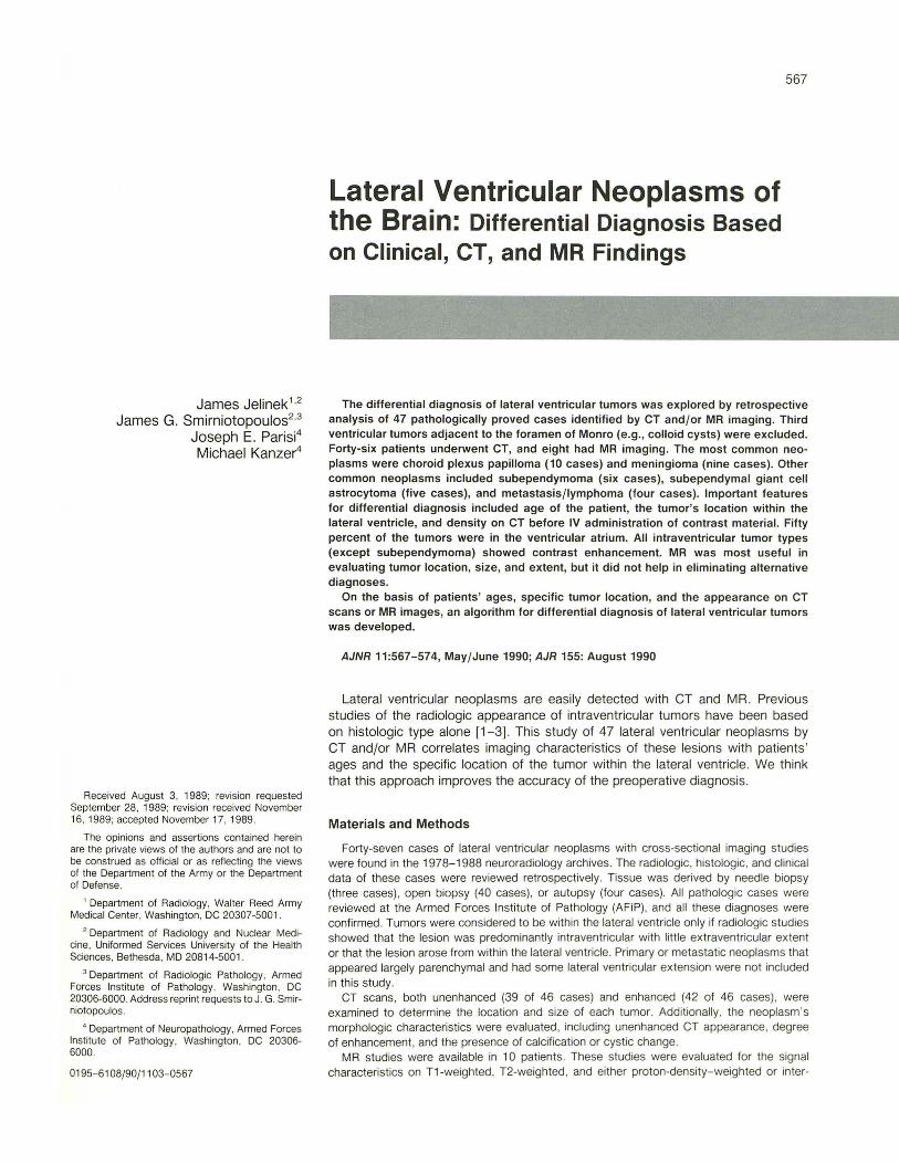
TABLE 1: Histologic Types: Summary of 47 Patients with Lateral Ventricular Neoplasm
Sex Age (years) Tumor Number
Male Female 0-5 6-30 >30
Meningioma 9 5 4 0 1 8 Choroid plexis papilloma 10 6 4 9 1 0 Subependymoma 6 3 3 0 0 6 SGCA 5 3 2 0 5 0 Lymphoma, metastasis 4 2 2 0 0 4 PNET 2 1 1 2 0 0 Teratoma 1 1 0 1 0 0 Pilocytic astrocytoma 3 0 3 0 3 0 Oligodendrogl ioma 2 1 1 0 2 0 Ependymoma 2 1 1 0 2 0 Mixed glioma 1 1 0 0 1 0 Glioblastoma 2 2 0 0 0 2
Note.-SGCA = subependymal giant cell astrocytoma; PNET = primitive neuroectodermal tumor.
Young Children (0- 5 Years)
Location.-ln young children , there were 12 tumors: nine choroid plexus papillomas (CPPs), two primitive neuroectodermal tumors (PNETs), and one teratoma (Figs. 2 and 3). All but one of all CPPs occurred in this age group, and PNET and teratoma occurred exclusively in children less than 1 year old. All masses in the trigone were CPPs, and all the CPPs occurred in the trigone except two (one arose in an older child in the foramen of Monro, and one in the younger group occurred in the body of the lateral ventricle) . No lesions occurred in the foramen of Monro in this age group. The PNETs and teratoma diffusely involved the body of the lateral ventricle.
CT appearance.-AII but one of the lesions studied by CT were hyperdense on noncontrast CT. The CPPs tended to enhance in a more uniform and intense pattern (eight of nine) than did the teratoma or the PNETs. Calcification was present in the teratoma, the PNETs, and five of the eight CPPs studied. Cystic changes were present in the teratoma, the PNETs, and four of the eight CPPs studied.
Older Children and Young Adults (6-30 Years)
Location.- There were 15 tumors in this age group, two of which usually occur in either the older or younger age range. One CPP (of the foramen of Monro) occurred in this age group, and one intraventricular meningioma occurred in the foramen of Monro of a 22-year-o/d woman with multiple meningiomas. Of the remaining 13 tumors, all were /ow-grade gliomas, including five subependymal giant cell astrocytomas (SGCAs), three pilocytic astrocytomas, two oligodendrogliomas, two ependymomas, and one mixed glioma (Figs. 4 and 5). The SGCAs occurred in only one location (the foramen of Monro), and the mixed glioma occurred in the body of the lateral ventricle. The pilocytic astrocytomas, oligodendrogliomas, and ependymomas occurred at two different sites each .
CT appearance.- The degree of enhancement, calcification , and presence of cystic changes were generally variable
TABLE 2: Location and Age of Lateral Ventricular Neoplasm
Age (years)
0-5
6-30
> 30
Foramen of Monro
0
5 SGCAs 2 Pilocytic astrocytomas 1 CPP 1 Meningioma 1 Oligodendroglioma 2 Metastases
Trigone/Atrium
8 CPPs
1 Ependymoma 1 Oligodendroglioma
8 Meningiomas
Body of Lateral Ventricle
2 PNETs 1 Teratoma 1 CPP 1 Mixed glioma 1 Ependymoma 1 Pi locytic astrocytoma
2 Glioblastomas 1 Lymphoma 1 Metastasis 6 Subependymomas
Note.-CPP = choroid plexus papilloma; PNET = primitive neuroectodermal tumor; SGCA = subependymal giant cell astrocytoma.
AJNR:1 1, MayfJune 1990 LATERAL VENTRICULAR NEOPLASMS
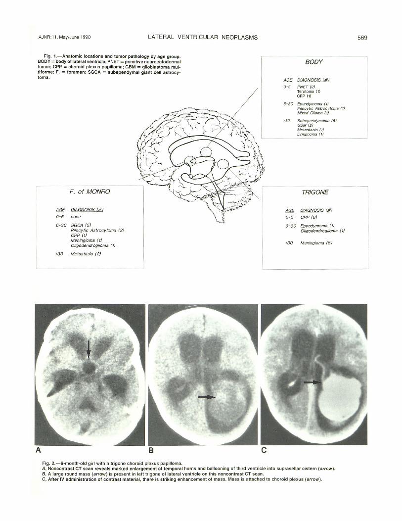
Fig. 1.-Anatomic locations and tumor pathology by age group. BODY= body of lateral ventricle; PNET = primitive neuroectodermal tumor; CPP = choroid plexus papilloma; GBM = glioblastoma multiforme; F. = foramen ; SGCA = subependymal giant cell astrocytoma.
F. of MONRO
AGE DIAGNOSIS(#)
0-5 none
6 -30 SGCA (5) Pilocytic Astrocytoma (2) CPP (1) Meningioma (1) Oligodendroglioma (1)
>30 Metastasis (2)
Fig. 2.-9-month-old girl with a trigone choroid plexus papilloma.
BODY
AGE DIAGNOSIS (#)
0-5 PNET (2) Teratoma ( 1) CPP (1)
6-30 Ependymoma (1) Pilocytic Astrocytoma (1) Mixed Glioma (1)
>30 Subependymoma (6) GBM (2) Metastasis (1) Lymphoma (1)
TRIGONE
DIAGNOSIS(#)
CPP (B)
>30 Meningioma (8)
A, Noncontrast CT scan reveals marked enlargement of temporal horns and ballooning of third ventricle into suprasellar cistern (arrow). 8 , A large round mass (arrow) is present in left trigone of lateral ventricle on this noncontrast CT scan. C, After IV administration of contrast material , there is striking enhancement of mass. Mass is attached to choroid plexus (arrow).
569
570 JELINEK ET AL. AJNR :11 , MayfJune 1990
A B
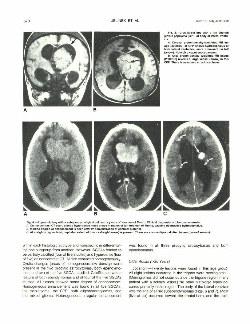
Fig. 3.-3-week-old boy with a left choroid plexus papilloma (CPP) of body of lateral ventricle.
A, Coronal proton-density-weighted MR image (2000/20) of CPP shows hydrocephalus of both lateral ventricles, more prominent on left (arrow). Note also caput succedaneum.
8, Axial proton-density-weighted MR image (2000/30) reveals a large vessel (arrow) in this CPP. There is asymmetric hydrocephalus.
Fig. 4.-6-year-old boy with a subependymal giant cell astrocytoma of foramen of Monro. Clinical diagnosis is tuberous sclerosis. A, On noncontrast CT scan, a large hyperdense mass arises in region of left foramen of Monro, causing obstructive hydrocephalus. B, Marked degree of enhancement is seen after IV administration of contrast material. C, At a slightly higher level, cephalad extent of tumor (straight arrow) is present. There are also multiple calcified tubers (curved arrows).
within each histologic subtype and nonspecific in differentiating one subgroup from another. However, SGCAs tended to be partially calcified (four of five studied) and hyperdense (four of five) on noncontrast CT. All five enhanced homogeneously. Cystic changes (areas of homogeneous low density) were present in the two pilocytic astrocytomas , both ependymomas, and two of the five SGCAs studied . Calcification was a feature of both ependymomas and of four of the five SGCAs studied. All tumors showed some degree of enhancement. Homogeneous enhancement was found in all five SGCAs, the meningioma, the CPP, both oligodendrogliomas, and the mixed glioma. Heterogeneous irregular enhancement
was found in all three pilocytic astrocytomas and both ependymomas.
Older Adults (> 30 Years)
Location.- Twenty lesions were found in this age group. All eight lesions occurring in the trigone were meningiomas. (Meningiomas did not occur outside the trigone region in any patient with a solitary lesion.) No other histologic types occurred primarily in this region . The body of the lateral ventricle was the site of all six subependymomas (Figs. 6 and 7). Most (five of six) occurred toward the frontal horn, and the sixth
AJNR:11 , MayfJune 1990 LATERAL VENTRICULAR NEOPLASMS 571
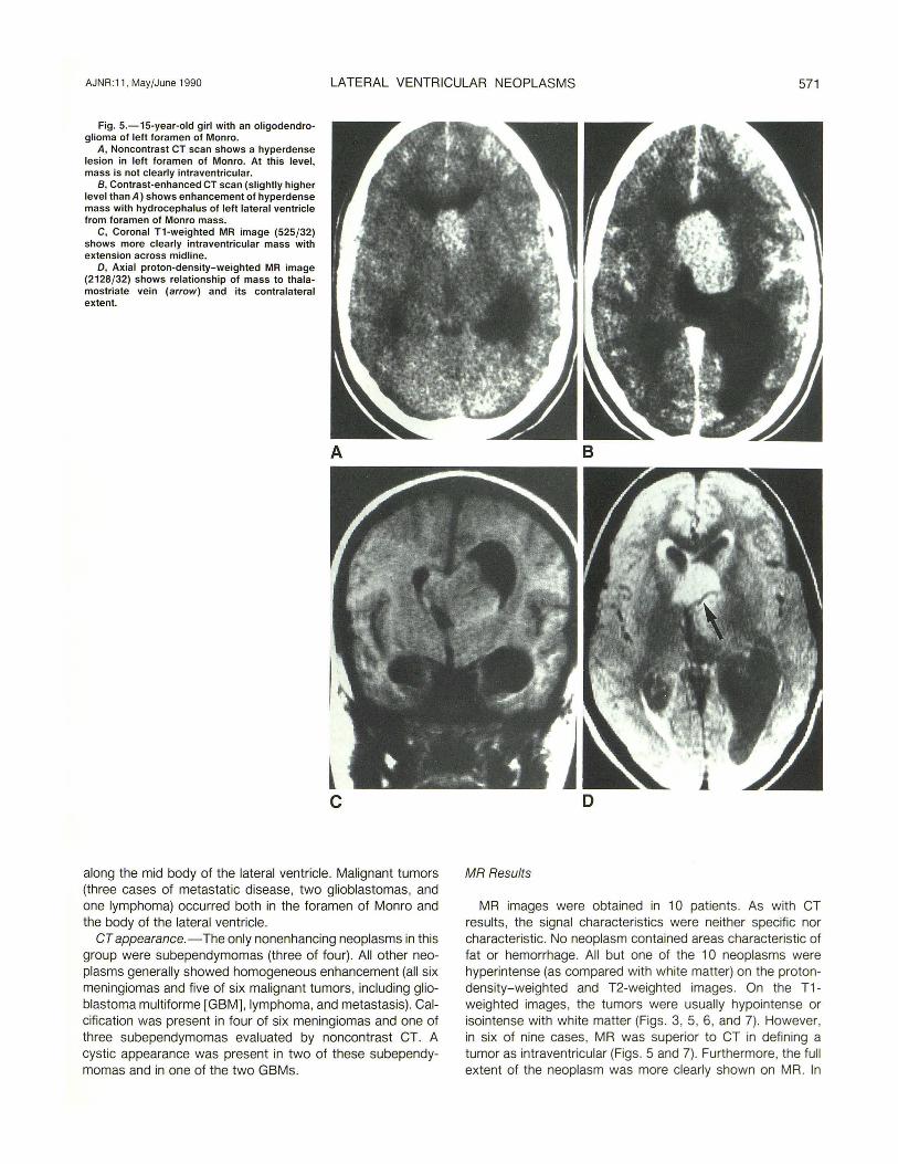
Fig. 5.-15-year-old girl with an oligodendroglioma of left foramen of Monro.
A, Noncontrast CT scan shows a hyperdense lesion in left foramen of Monro. At this level, mass is not clearly intraventricular.
B, Contrast-enhanced CT scan (slightly higher level than A) shows enhancement of hyperdense mass with hydrocephalus of left lateral ventricle from foramen of Monro mass.
C, Coronal T1-weighted MR image (525/32) shows more clearly intraventricular mass with extension across midline.
D, Axial proton-density-weighted MR image (2128/32) shows relationship of mass to thalamostriate vein (arrow) and its contralateral extent.
A
c
along the mid body of the lateral ventricle. Malignant tumors (three cases of metastatic disease, two glioblastomas, and one lymphoma) occurred both in the foramen of Monro and the body of the lateral ventricle.
CT appearance.-The only nonenhancing neoplasms in this group were subependymomas (three of four). All other neoplasms generally showed homogeneous enhancement (all six meningiomas and five of six malignant tumors , including glioblastoma multiforme [GBM] , lymphoma, and metastasis). Calcification was present in four of six meningiomas and one of three subependymomas evaluated by noncontrast CT. A cystic appearance was present in two of these subependymomas and in one of the two GBMs.
B
D
MR Results
MR images were obtained in 10 patients. As with CT results, the signal characteristics were neither specific nor characteristic. No neoplasm contained areas characteristic of fat or hemorrhage. All but one of the 10 neoplasms were hyperintense (as compared with white matter) on the protondensity-weighted and T2-weighted images. On the T1-weighted images, the tumors were usually hypointense or isointense with white matter (Figs. 3, 5, 6, and 7). However, in six of nine cases, MR was superior to CT in defining a tumor as intraventricular (Figs. 5 and 7). Furthermore, the full extent of the neoplasm was more clearly shown on MR. In

572 JELINEK ET AL. AJNR :11, May/June 1990
A B
A B Fig. 7.-69-year-old man with frontal horn subependymoma.
c
Fig. 6.-62-year-old man with subependymoma of body of lateral ventricle.
A, Sagittal T1-weighted MR image (450/ 20) shows a large mass arising in body of lateral ventricle. Extent of mass, both anterior and posterior, is well defined.
B, Axial T2-weighted MR image (2450/ 100) shows heterogeneity (arrows) of neoplasm, but overall dimensions are obscured on T2-weighted image.
A, Axial CT scan after administration of IV contrast material shows a mass arising in region of left caudate nucleus. Appearance is that of a parenchymal mass (arrow) extending into frontal horn and foramen of Monro of left lateral ventricle.
B, Sagittal T1 -weighted MR image (450 / 20) reveals a primarily intraventricular mass in frontal horn of lateral ventricle. Multiple, discrete, hypointense areas are present within mass, compatible with cystic changes (arrow).
C, On a coronal T2-weighted MR image (2000 / 60), a left frontal horn mass is shown by hyperintense CSF (arrow) . T2-weighted images frequently obscured intraventricular neoplasms.
no case was CT better than MR at localizing or defining the extent or spread of tumor.
Discussion
Intraventricular neoplasms are uncommon CNS masses, representing only 10% of all neoplasms [4). Previous studies of intraventricular neoplasms categorized by histologic types have shown consistent trends in tumor location and in the age of the patient at presentation [1-3). Within the lateral ventricle, specific regions can be identified as sites of predilection for each particular tumor. As we tabulated the data, we found that these tumors tended to occur in natural agerelated groupings. The CT density and morphology characteristics were nonspecific in terms of the patients ' ages or tumor location. MR signal intensity patterns were equally
unhelpful in predicting tumor histology, although MR was better than CT in defining tumor location and extent of disease. From our data, we suggest the algorithm shown in Figure 8 and described in detail in the following sections.
Young Children (0-5 Years)
In children less than 12 years old , many CNS neoplasms are infratentorial [5 , 6). However, in the first year of life and possibly in the first 3 years, supratentorial lesions are more common [6, 7]. Many of these arise within the lateral ventricles and in the third ventricle [1 , 7] . Several articles have discussed the differential diagnosis and CT findings of fourth ventricular masses in children [8-12). Lateral ventricular neoplasms in children less than 5 years old are usually choroid plexus papillomas (CPP) (Figs. 2 and 3) or choroid plexus carcinoma,
AJNR :11 , May/June 1990 LATERAL VENTRICULAR NEOPLASMS 573
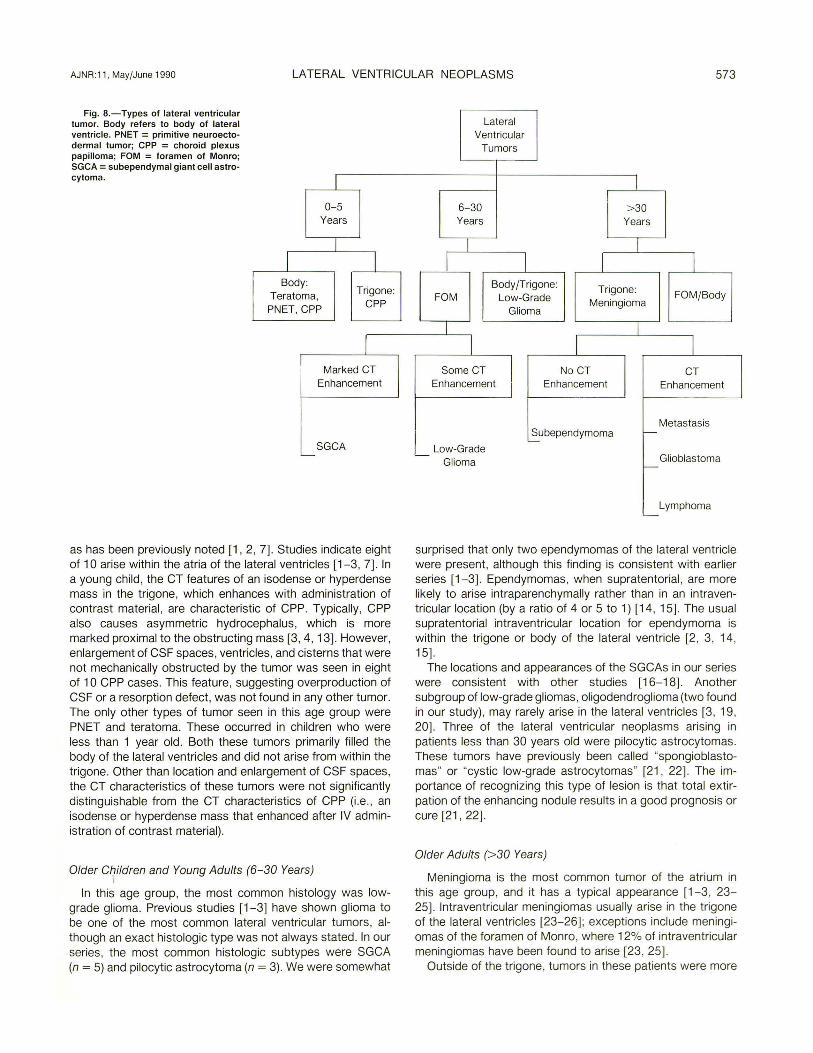
Fig. 8.- Types of lateral ventricular tumor. Body refers to body of lateral ventricle. PNET = primitive neuroectodermal tumor; CPP = choroid plexus papilloma; FOM = foramen of Monro; SGCA = subependymal giant cell astrocytoma.
I Body:
Teratoma,
0-5 Years
I Trigone:
PNET, CPP CPP
I Marked CT
Enhancement
SGCA '---
as has been previously noted [1 , 2, 7] . Studies indicate eight of 10 arise within the atria of the lateral ventricles [1-3 , 7]. In a young child , the CT features of an isodense or hyperdense mass in the trigone, which enhances with administration of contrast material , are characteristic of CPP. Typically , CPP also causes asymmetric hydrocephalus , which is more marked proximal to the obstructing mass [3, 4, 13]. However, enlargement of CSF spaces , ventricles, and cisterns that were not mechanically obstructed by the tumor was seen in eight of 1 0 CPP cases. This feature, suggesting overproduction of CSF or a resorption defect, was not found in any other tumor. The only other types of tumor seen in this age group were PNET and teratoma. These occurred in children who were less than 1 year old . Both these tumors primarily filled the body of the lateral ventricles and did not arise from within the trigone. Other than location and enlargement of CSF spaces, the CT characteristics of these tumors were not significantly distinguishable from the CT characteristics of CPP (i .e. , an isodense or hyperdense mass that enhanced after IV administration of contrast material).
Older Children and Young Adults (6-30 Years) I
In this age group, the most common histology was lowgrade glioma. Previous studies [1-3] have shown glioma to be one of the most common lateral ventricular tumors , although an exact histologic type was not always stated. In our series, the most common histologic subtypes were SGCA (n = 5) and pilocytic astrocytoma (n = 3). We were somewhat
Lateral Ventricular
Tumors
I 6-30 >30 Years Years
I I I I I
Body /Trigone: Trigone: FOM Low-Grade FOMfBody
Glioma Meningioma
I I
I Some CT No CT CT
Enhancement Enhancement Enhancement
Metastasis Subependymoma f-
'--Low-Grade
- Glioma Glioblastoma f-
Lymphoma L-
surprised that only two ependymomas of the lateral ventricle were present, although this finding is consistent with earlier series [1-3]. Ependymomas, when supratentorial, are more likely to arise intraparenchymally rather than in an intraventricular location (by a rat io of 4 or 5 to 1) [14, 15]. The usual supratentorial intraventricular location for ependymoma is within the trigone or body of the lateral ventricle [2 , 3, 14, 15].
The locations and appearances of the SGCAs in our series were consistent with other studies [16-18] . Another subgroup of low-grade gliomas, oligodendroglioma (two found in our study), may rarely arise in the lateral ventricles [3, 19, 20] . Three of the lateral ventricular neoplasms arising in patients less than 30 years old were pilocytic astrocytomas. These tumors have previously been called "spongioblastomas" or "cystic low-grade astrocytomas" [21, 22] . The importance of recognizing this type of lesion is that total extirpation of the enhancing nodule results in a good prognosis or cure [21, 22].
Older Adults (>30 Years)
Meningioma is the most common tumor of the atrium in this age group, and it has a typical appearance [1-3, 23-25] . Intraventricu lar meningiomas usually arise in the trigone of the lateral ventricles [23-26]; exceptions include meningiomas of the foramen of Monro, where 12% of intraventricular meningiomas have been found to arise [23 , 25] .
Outside of the trigone, tumors in these patients were more
574 JELINEK ET AL. AJNR :11 , May/June 1990
likely to be malignant (either primary or metastatic), especially if the lesion showed contrast enhancement [2, 3]. The only consistently nonenhancing tumors in the body or the foramen of Monro were subependymomas. Supratentorial subependymomas were found in this group; most subependymomas occur in the fourth ventricle, and less frequently they arise in the third ventricle, the septum pellucidum, or the walls of the lateral ventricle [27 -30]. The average age of subependymoma patients depends on whether they are symptomatic (average age, 39) or asymptomatic (average age, 59) at the time of diagnosis [27]. Although some studies confirm these neoplasms in the older age group [1 , 27, 28], as did our study, others have indicated their occurrence in a slightly younger age group [29, 30]. Other confusing features of subependymomas are their morphologic characteristics on noncontrast and contrast CT. Our cases were generally hypodense or isodense with minimal to no enhancement, which is consistent with some other studies [1 , 27, 28]. However, other reports suggest that subependymoma is an enhancing lesion [29 , 30]. Foci of cystic changes in subependymoma are common [27-30] (Fig. 6).
Conclusions
In summary, lateral ventricular neoplasms are uncommon. The focus of the differential diagnosis can be narrowed considerably by knowing the patient's age and the exact location of the tumor within the ventricles. Some neoplasms have characteristic CT findings that may suggest the diagnosis, given the appropriate tumor location and age group. MR was more useful in showing the location and extent of disease but did not add to the specificity of the differential diagnosis. The differential diagnosis of lateral ventricular neoplasms, like that of the third and fourth ventricle tumors, is best determined by consideration of the patient's age, exact intraventricular location of the tumor, and CT appearance with and without contrast material.
ACKNOWLEDGMENTS
We thank Bonnie Yelverton and Wendy Baker for their help in manuscript preparation.
REFERENCES
1. Morrison G, Sobel DF, Kelley WM , Norman D. Intraventricular mass lesions. Radiology 1984;153:435-442
2. Kendall B. Reider-Grosswater I, Valentine A. Diagnosis of masses presenting within the ventricle on computed tomography. Neuroradiology 1983;25 : 11 -22
3. Silver AJ, Ganti SR , Hilal SK. Computed tomography of tumors involving the atria of the lateral ventricles. Radiology 1982;145 :71-78
4. Rosenberg RN , ed. The clinical neurosciences , vol. 4. Neuroradiology. New York: Churchill Livingstone, 1984:424-441
5. Tadmor R, Harwood-Nash DC, Slotti G, et al. Intracranial neoplasms in children: the effect of computed tomography on age distribution. Radiology 1982;145:371-373
6. Naidich TP, Zimmerman RA. Primary brain tumors in children . Semin Roentgeno/1984;19 :100-114
7. Radkowski MA, Naidich TP, Tomita T, Byrd SE , Melone DG. Neonatal brain tumors: CT and MR findings . J Comput Assist Tomogr 1988;12: 10-20
8. Naidich TP, Lin JP, Leeas NE, Pudlowski RM , Naidich JB. Primary tumors and other masses of the cerebellum and fourth ventricle : differential diagnosis by computed tomography. Neuroradiology 1977; 14 : 1 53-17 4
9. Zee ES, Segall HD, Miller C, et al. Less common CT features of medulloblastoma. Radiology 1982;144:97-102
1 0. Zimmerman RA, Bilaniuk L T, Pahlajani H. Spectrum of medulloblastomas demonstrated by computed tomography. Radiology 1978;126:137-141
11 . Berger PE, Kirks DR, Gilda! DL. Computed tomography in infants and children: intracranial neoplasms. AJR 1976;127 : 129-137
12. Bauer HL, Houser OW. Computed tomography in the diagnosis of posterior fossa lesions. Radio/ Clin North Am 1976;14: 129-147
13. Eisenberg HM, McComb JG, Lorenzo AV. Cerebrospinal fluid overproduction and hydrocephalus associated with choroid plexus papilloma. J Neurosurg 1974;40 :381-385
14. Swartz JD, Zimmerman RA, Bilaniuk LT. Computed tomography of intracranial ependymomas. Radiology 1982;143:97-101
15. Armington WG , Osborn AG, Cubberly DA, et al. Supratentorial ependymoma: CT appearance. Radiology 1985;157 :367-372
16. Moran V, O'Keeffe F. Giant cell astrocytoma in tuberous sclerosis: computed tomographic findings. Clin Radio/1986 ;37:543-545
17. Sima AF, Robertson DM. Subependymal giant cell astrocytoma. J Neurosurg 1979;50:240-245
18. Winter J. Computed tomography in diagnosis of intracranial tumours versus tubers in tuberous sclerosis. Acta Radio/1982;23:337-343
19. Kikuchi K, Kowada M, Mineura K, Vemura K. Primary oligodendroglioma of the lateral ventricle with computed tomographic and positron emission tomographic evaluation. Surg Neuro/1985;23 :483-488
20. Maiuri F, Giamundo A, DiPrisco B. Primary intraventricular oligodendroglioma. Surg Neuro/1982;18:364-366
21 . Palma L, Guidetti B. Cystic pilocytic astrocytomas of the cerebral hemispheres. J Neurosurg 1985;62 :811-815
22. Gol A. The relatively benign astrocytomas of the cerebrum. J Neurosurg 1961 ;18:501-506
23. Kobayashi S, Okazaki H, MacCarty CS. Intraventricular meningioma. Mayo Clin Proc 1971 ;46:735-741
24 . Mani RL, Hedgcock MW, Mass Sl, Gilmor RL, Enzmann DR, Eisenberg RL. Radiographic diagnosis of meningioma of the lateral ventricle . J Neurosurg 1978;49:249-255
25 . Strenger SW, Huang YP, Sachdev VP. Malignant meningioma within the third ventricle : a case report . Neurosurgery 1987;20:465-468
26. Ladenheim JC. Choroid plexus meningiomas of the lateral ventricle . Springfield , IL: Charles C Thomas, 1963
27 . Gandolfi A, Brizzi RE , Tedesche F, Paini P, Bassi P. Symptomatic subependymoma of the fourth ventricle. J Neurosurg 1981 ;55 :841-844
28. Vaquero J, Cabezudo JM, Nombela L. CT scan in subependymomas. Br J Radio/1983;56 :425-426
29. Stevens JM, Kendall BE , LoveS. Radiologic features of subependymoma with emphasis on computed tomography. Neuroradiology 1984;26 : 223- 228
30. Lobato RD, Cabello A, Carmena JJ, de Ia Fuente M, Munoz MJ . Subependymoma of the lateral ventricle . Surg Neural 1981 ; 15 : 144-14 7