Embed Size (px)
Citation preview
Contents lists available at ScienceDirect
Oral Oncology
journal homepage: www.elsevier.com/locate/oraloncology
Leading edge or tumor core: Intratumor cancer stem cell niches in oralcavity squamous cell carcinoma and their association with stem cell function
Farshad N. Chowdhurya, Julie Reisingerb, Karina E. Gomezb, Tugs-Saikhan Chimedb,Carissa M. Thomasc, Phuong N. Leb, Bettina Millerb, John J. Mortonb, Cera M. Nietob,Hilary L. Somersetd, Xiao-Jing Wangd, Stephen B. Keysarb,⁎, Antonio Jimenoa,b,⁎
a Department of Otolaryngology, University of Colorado Denver School of Medicine (UCDSOM), Aurora, CO 80045, USAbDivision of Medical Oncology, Department of Medicine, UCDSOM, Aurora, CO 80045, USAc Department of Otolaryngology – Head and Neck Surgery, University of Toronto, University Health Network/Princess Margaret Cancer Centre, Toronto, ON M4Y 2X5,CanadadDepartment of Pathology, UCDSOM, Aurora, CO 80045, USA
A R T I C L E I N F O
Keywords:Cancer stem cellsStem cell nicheHead and neck neoplasmsOral cancerSurgical marginsLocal neoplasm recurrenceTumor microenvironmentNeoplasm invasion
A B S T R A C T
Objectives: To describe differences in cancer stem cell (CSC) presence and behavior associated with their in-tratumor compartment of origin using a patient-derived xenograft (PDX) model of oral cavity squamous cellcarcinoma (OCSCC).Materials and methods: Four HPV-negative OCSCC PDX cases were selected (CUHN004, CUHN013, CUHN096,CUHN111) and the percentage of CSCs (ALDH+CD44high) was measured in the tumor Leading Edge (LE) andCore compartments of each PDX tumor case via fluorescence activated cell sorting (FACS). The fraction of cells inthe proliferative phase was measured by Ki-67 labelling index of paraffin embedded tissue. The proliferation andinvasion of LE versus Core CSCs were compared using sphere and Matrigel invasion assays, respectively.Results: Both CUHN111 and CUHN004 demonstrate CSC enrichment in their LE compartments while CUHN013and CUHN096 show no intratumor difference. Cases with LE CSC enrichment demonstrate greater Ki-67 la-belling at the LE. CSC proliferative potential, assessed by sphere formation, reveals greater sphere formation inCUHN111 LE CSCs, but no difference between CUHN013 LE and Core CSCs. CUHN111 CSCs do not demonstratean intratumor difference in invasiveness while CUHN013 LE CSCs are more invasive than Core CSCs.Conclusion: A discrete intratumor CSC niche is present in a subset of OCSCC PDX tumors. The CSC functionalphenotype with regard to proliferation and invasion is associated with the intratumor compartment of origin ofthe CSC: LE or Core. These individual functional characteristics appear to be modulated independently of oneanother and independently of the presence of an intratumor CSC niche.
Introduction
The treatment of head and neck squamous cell carcinoma (HNSCC)remains a clinical challenge given its rising overall incidence andstagnant five-year survival rate over the last decade [1–3]. Adequatesurgical resection is defined by negative histologic margins, however,local recurrence occurs in up to 30% of cases in which surgical marginswere negative at the time of resection [4,5]. This reality has driven aninterest in molecular assessments of margin adequacy and has high-lighted the importance of decoding tumor behavior at the surgicalmargin [6,7].
The cancer stem cell (CSC) has been proposed as one possible driver
of both local recurrence and regional spread in HNSCC given the abilityof a single CSC to asymmetrically divide and repopulate the tumor oforigin while also resisting chemotherapy, radiotherapy, and immunesurveillance [8–10]. CSCs are typically identified by a combination ofcell surface protein expression and intracellular enzymatic activity,with HNSCC CSCs commonly defined by the co-expression of high le-vels of cell surface protein CD44 and high aldehyde dehydrogenase(ALDH) activity [11,12]. Notably, these ALDH+CD44high cells alsodemonstrate upregulation of genes that are both associated withstemness and are putative prognostic markers of the molecular margin[13–16].
Prior studies have investigated the prognostic implications of the
https://doi.org/10.1016/j.oraloncology.2019.09.011Received 24 May 2019; Received in revised form 2 September 2019; Accepted 10 September 2019
⁎ Corresponding authors at: Division of Medical Oncology, Department of Medicine, UCDSOM, Aurora, CO 80045, USA (A. Jimeno).E-mail addresses: [email protected] (S.B. Keysar), [email protected] (A. Jimeno).
Oral Oncology 98 (2019) 118–124
Available online 03 October 20191368-8375/ © 2019 Elsevier Ltd. All rights reserved.
T
presence of these markers in the margin and margin-adjacent tissues viaimmunohistochemistry (IHC) with often contradictory results [17–23].Here, we seek to further define the behavior of CSCs at the tumorleading edge (LE) by utilizing a patient-derived xenograft (PDX) modelof HNSCC to identify the CSC niche and associated CSC functionalphenotypes.
Methods
PDX case generation
The protocol for studies involving human subjects was approved bythe Colorado Multiple Institutional Review Board (COMIRB #08-0552),and informed written consent was obtained from all patients whosetissues were used for this study. The University of Colorado InstitutionalAnimal Care and Use Committee approved all experiments involvingmice.
PDX case selection
Tissue was selected from existing PDX cases. PDX generation andcharacterization was previously reported [24]. Available PDX caseswere narrowed to those whose originating patient case was an HPV-negative oral cavity squamous cell carcinoma (OCSCC) primary. Caseswith available PDX tissue were selected: CUHN004, CUHN013,CUHN096, CUHN111.
PDX tumor digestion and FACS analysis
PDX tumors were individually sectioned using a #10 scalpel andsurgical loupe magnification to produce a 2–3mm leading edge (LE)rim of tissue and its associated tumor core of tissue. In subsequent steps,tissue was not comingled. PDX tumor tissue was finely minced with ascalpel and dissociated in DMEM containing 1mg/ml collagenase IV(Worthington, Lakewood, NJ) at 37 °C for 1 h. Cells were filtered(40 µm) and red blood cells were lysed in Ammonimum-Chloride-Potassium (ACK) lysing buffer (Life Technologies, Carlsbad, CA).Staining with Aldefluor (Stem Cell Technologies, Vancouver, Canada)was performed following the manufacturer’s instructions. Briefly, cellswere suspended in Aldefluor staining buffer containing Aldefluor re-agent (5 µl/ml) and incubated at 37 °C for 30min. DEAB (N,N-diethy-laminobenzaldehyde) was used as a negative control for setting gates.Following incubation with Aldefluor, cells were stained with the fol-lowing antibodies: 1:100 anti-mouse H-2Kd (BioLegend, San Diego,CA), 1:10 anti-human CD44 (BD Biosciences, San Jose, CA) with speciesspecificity validated by the manufacturer. Cell sorting was performedusing a MoFlo XDP (Beckman Coulter, Fort Collins, CO) and flow cy-tometric analysis was completed on a Gallios 561 (Beckman Coulter). Inorder to select only live, human, CSCs for subsequent functional assayswe gated anti-mouse H2kd–/DAPI– to exclude mouse cells and deadcells prior to gating on our ALDH+/CD44high cells of interest(Supplemental Fig. 1).
Supplementary data associated with this article can be found, in theonline version, at https://doi.org/10.1016/j.oraloncology.2019.09.011.
Sphere assay
Tumors were harvested, sectioned into LE and Core compartments,and processed to single cell suspension for FACS as described above.ALDH+CD44high CSCs were sorted and collected. Cells were plated at5000 cells per well in 96-well ultra-low attachment plates in 150 µl ofserum-free media (DMEM F12 [Gibco], 20 ng/mL recombinant humanepithelial growth factor, 20 ng/mL recombinant human fibroblastgrowth factor, and 1x B27 supplement) for 12 days. Spheres were im-aged, measured, and quantified using Zeiss Axio Observer Z1 inverted
microscope (Zeiss, Oberkochen, Germany) (Zeiss software Rel. 4.8). 3Dstructures larger than 30 µm were scored as spheres. Relative SphereFormation was calculated by normalizing the sphere formation countsof each condition to the average Core sphere formation count.
Immunohistochemistry (IHC)
Slides were de-paraffinized and re-hydrated in graded concentra-tions of alcohol by standard techniques before antigen retrieval in ci-trate buffer pH 6.0 (#S1699 Dako) at 121 °F for 10min. Next, the slideswere cooled for 20min before washing in wash buffer (#K8007 Dako).All staining was done in a Dako Autostainer. Slides were incubated indual endogenous enzyme block (#S2003 Dako) for 10min, and inprotein free blocking solution (#X0909 Dako) for 20min, followed byprimary antibody (1:100 #RM-9106-S Thermoscientific Ki-67) and in-cubated for 60min at room temperature. Staining was developed usingthe following conditions: EnVision+Dual Link System HRP (#K4061Dako) for 30min and substrate-chromogen (DAB+) Solution (#K3468Dako) for 5min. Slides were counterstained with hematoxylin (#S3301Dako) for 10min.
Ki-67 labeling index
To calculate the Ki-67 labeling index, a PDX tumor from each of thexenograft cases of interest was stained for Ki-67 as above. Three 5xhigh-powered fields (HPFs) were selected from the strongest stainingareas of the LE and Core areas in each case. The number of nucleatedand Ki-67 positive cells was then scored automatically using a thresh-olding analysis in Image-J (Fiji distribution) [25] and a percentage ofKi-67 positive cells was calculated and reported in triplicate as the la-belling index.
In vitro Matrigel-coated invasion assay
CUHN111 and CUHN013 PDX tumors were harvested, sectionedinto Core and LE compartments, and processed to separate single cellsuspensions for the LE and Core for FACS as described above. LE andCore ALDH+CD44high CSCs were sorted and collected. Corning BioCoatMatrigel-coated 8 μm pore PET membrane 24 well inserts (Corning,Corning, NY) were brought to room temperature and hydrated in serumDMEM media for 2 h at 37 °C in a humidified tissue culture incubator,5% CO2 atmosphere. 2x10⁵ LE and Core CSCs were separately added tothe inserts in DMEM containing 0.5% FBS while DMEM containing 10%FBS was used as a chemoattractant. Plates were incubated for 72 hbefore fixing with 10% formalin (15min) and staining with 0.5%crystal violet (15min). Invasion was quantified as the total number ofinvaded cells over the entirety of the membrane at 5X magnification.Relative Invasion was calculated by normalizing the absolute number ofinvaded cells in each condition to the average number of invaded cellsin the Core condition for that given experiment.
Statistical analysis
Differences in CSC composition of PDX tumor compartments wereanalyzed using two-tailed ratio paired t-tests. Sphere size differenceswere analyzed using two-tailed unpaired t-tests, and Relative SphereFormation was analyzed using two-tailed ratio paired t-tests. Ki-67 la-beling indices and normalized relative invasion values were comparedusing two-tailed unpaired t-tests. All alpha was set at 0.05. Columnstatistics, computations, and analyses were performed in GraphpadPrism v8.0.0.
F.N. Chowdhury, et al. Oral Oncology 98 (2019) 118–124
119

Results
PDX case demographics
Fig. 1A describes four patient-derived xenografts (PDXs) generated
from HPV-negative oral cavity squamous cell cancer (OCSCC) patients.Male and female gender was represented equally, and the mean age ofall patients was 64.5 years (SEM ± 4.03). The primary tumor was inthe floor of mouth in three of four cases (75%), and the engrafted tissuewas resected from a first primary lesion in two of the four cases (50%).
Fig. 1. CSC isolation schematic and overview of selected cases. (1A) Profiles of included PDX cases. (1B) Schematic of PDX tumor processing wherein whole PDXtumors are grossly sectioned into leading edge and core components and then digested to a single-cell suspension before incubating with CD44 and Aldefluour assayprior to FACS.
Fig. 2. ALDH+/CD44HIGH enrichment by tumor compartment. Each pair of connected points represents the LE and Core compartments belonging to the sameindividual PDX tumor. (2A-1 and 2B-1) Cases CUHN111 and CUHN004 both demonstrate ALDH+CD44HIGH enrichment of the LE compartment (geometric meanCUHN111 LE:Core Ratio 3.74 ± 0.124, p=0.0006; geometric mean CUHN004 LE:Core Ratio 2.80 ± 0.457, p= 0.0129). (2C-1 and 2D-1) In contrast, CUHN013and CUHN096 do not demonstrate a preferential localization of ALDH+CD44HIGH cells to a given compartment (geometric mean CUHN013 LE:Core Ratio0.980 ± 0.165, p= 0.958; geometric mean CUHN096 LE:Core ratio 1.23 ± 0.073, p= 0.247). (2A-2–2D-2) There is no intratumor difference in the percent livecells at the time of sorting (geometric mean CUHN111 LE:Core Ratio 1.10 ± 0.033, p= 0.265; geometric mean CUHN004 LE:Core Ratio 1.07 ± 0.037, p= 0.467;geometric mean CUHN013 LE:Core Ratio 1.08, p= 0.677; geometric mean CUHN096 LE:Core Ratio 0.951 ± 0.0304, p=0.484).
F.N. Chowdhury, et al. Oral Oncology 98 (2019) 118–124
120

All cases were staged T2 or greater, all cases demonstrated nodal dis-ease (either clinical or pathological based on neck dissection speci-mens), and none demonstrated distant metastasis at the time of resec-tion.
HNSCC CSCs demonstrate different intratumor niches
FACS was used to determine the distribution of ALDH+CD44high
double-positive tumor cells by comparing the cellular composition ofthe leading edge (LE, outer 2–3mm) and Core tissue compartments ofindividual xenograft tumors (Fig. 1B). The percent ALDH+CD44high
cells out of the total live, human cell population was calculated for eachcompartment on a tumor-by-tumor basis (Supplemental Fig. 1). Ratiopaired t-tests were utilized to compare compartment composition whileaccounting for baseline differences in the overall ALDH+CD44high po-pulation across individual xenograft tumors of a given parent case(Fig. 2). Two cases, CUHN111 and CUHN004, demonstratedALDH+CD44high enrichment of the respective LE compartments(CUHN111 LE:Core Ratio 3.74 ± 0.12, p < 0.001; CUHN004 LE:CoreRatio 2.80 ± 0.46, p=0.013, Fig. 2A-1 and 2B-1). Neither CUHN111nor CUHN004 demonstrated significant differences in the percent livecells of the overall sorted population (Fig. 2A-2 and 2B-2). Conversely,neither CUHN013 nor CUHN096 demonstrate ALDH+CD44high en-richment (CUHN013 LE:Core Ratio 0.94 ± 0.12, p=0.822; CUHN096LE:Core ratio 1.23 ± 0.07, p=0.247, Fig. 2C-1 and 2D-1). Again,there was no intratumor difference in the percent live cells in the sortedpopulations in the two compartments (Fig. 2C-2 and 2D-2). These datasuggest that CSCs are enriched within a tumor LE niche in a subset ofOCSCC cases.
Tumor proliferation measured by Ki-67 labeling index varies by tumorcompartment
Ki-67 labeling index was used to investigate differences in theproliferating fraction of the bulk tumor cells within the LE and Corecompartments of PDX tumors. The degree of tumor proliferation asmeasured by Ki-67 labeling index is greater in the LE of CUHN111 (LE0.587 ± 0.038, vs Core 0.236 ± 0.037, p=0.001, Fig. 3A) andCUHN004 (LE 0.457 ± 0.027, vs Core 0.174 ± 0.035, p=0.003,Fig. 3B). There is no difference in Ki-67 labelling index between com-partments of CUHN013 (Fig. 3C) and CUHN096 (Fig. 3D). Taken with
the CSC localization data (Fig. 2), those cases with CSC enrichmentwithin a LE intratumor niche demonstrate a greater fraction of cells inthe proliferative phase at the tumor LE.
Relative sphere formation of ALDH+CD44high cells is associated withintratumor location
CUHN111 and CUHN013 PDX cases were selected for further ana-lysis based on the availability of adequate PDX tissue and due to theiropposing CSC localization patterns (Fig. 2). Sorted ALDH+CD44high
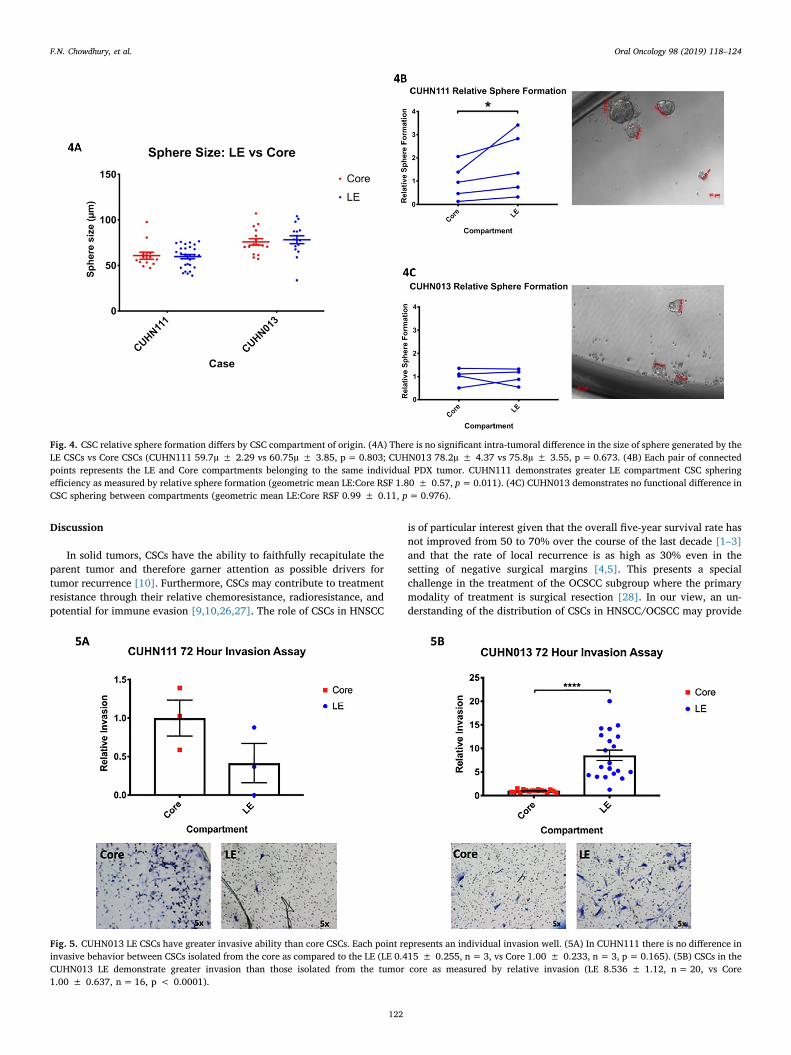
cells were seeded in a sphere formation assay to asses for functionaldifferences in proliferation based on the intratumor location of theALDH+CD44high cells. The size of the sphere produced did not differbased on whether the ALDH+CD44high cells were isolated from the LEor Core of the PDX tumor (Fig. 4A). When comparing relative sphereformation (RSF), the CUHN111 LE ALDH+CD44high cells demonstrategreater sphere-forming ability than those in the Core (geometric meanLE:Core RSF 1.80 ± 0.57, p=0.011, Fig. 4B). In contrast, there was nodifference in the RSF of ALDH+CD44high cells between the LE and Corecompartments of CUHN013 (Fig. 4C). Together, these results suggestthat CSCs within a LE CSC niche demonstrate greater proliferative ac-tivity than do their counterpart CSCs in the tumor core. CSCs in caseswith no discrete intratumor CSC niche show no difference in pro-liferative activity.
Invasive potential varies by tumor compartment
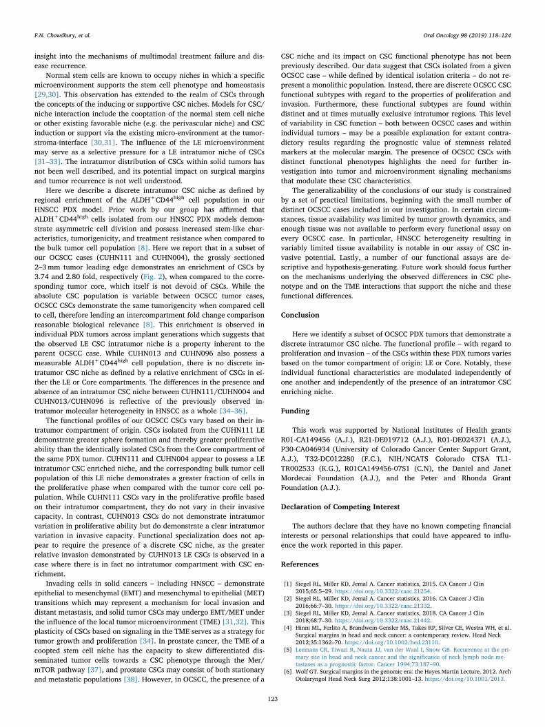
Sorted ALDH+CD44high cells from both the LE and Core wereseeded in Matrigel coated invasion chambers to assess for differences ininvasiveness. The CUHN111 LE CSCs demonstrate no greater relativeinvasion than those of the CUHN111 Core (Fig. 5A). In contrast,CUHN013 LE CSCs clearly demonstrate greater relative invasion thanthose of the CUHN013 Core (LE 8.536 ± 1.12, vs Core 1.00 ± 0.637,p < 0.0001, Fig. 5B). While the CUHN111 LE CSCs occupy an enrichedCSC niche with an increased cellular growth fraction and greater CSCproliferation, these LE CSCs do not show greater invasiveness thanthose in the CUHN111 Core. Though the LE CSCs of CUHN013 neitheroccupy an enriched CSC niche nor display enhanced proliferativeability, these LE CSCs do demonstrate greater relative invasion thantheir Core counterparts.
Fig. 3. KI-67 labeling index by tumor compartment. In both CUHN111 (3A) and CUHN004 (3B), the leading edge compartment demonstrates a higher KI-67 labelingindex than the respective core compartment (CUHN111 LE 0.587 ± 0.038, vs Core 0.236 ± 0.037, p=0.001; CUHN004 LE 0.457 ± 0.027, vs Core0.174 ± 0.035, p=0.003). In contrast, there is no difference in the KI-67 labelling index between the core and leading edge compartments of CUHN013 (LE0.415 ± 0.105, vs Core 0.410 ± 0.056, p=0.967, 3C) and CUHN096 (LE 0.713 ± 0.078, vs Core 0.613 ± 0.015, p=0.283, 3D). Representative micrographs of10x HPF for each case are shown. Three 10x HPF of the greatest staining area within each compartment were scored for analysis.
F.N. Chowdhury, et al. Oral Oncology 98 (2019) 118–124
121
Discussion
In solid tumors, CSCs have the ability to faithfully recapitulate theparent tumor and therefore garner attention as possible drivers fortumor recurrence [10]. Furthermore, CSCs may contribute to treatmentresistance through their relative chemoresistance, radioresistance, andpotential for immune evasion [9,10,26,27]. The role of CSCs in HNSCC
is of particular interest given that the overall five-year survival rate hasnot improved from 50 to 70% over the course of the last decade [1–3]and that the rate of local recurrence is as high as 30% even in thesetting of negative surgical margins [4,5]. This presents a specialchallenge in the treatment of the OCSCC subgroup where the primarymodality of treatment is surgical resection [28]. In our view, an un-derstanding of the distribution of CSCs in HNSCC/OCSCC may provide
Fig. 4. CSC relative sphere formation differs by CSC compartment of origin. (4A) There is no significant intra-tumoral difference in the size of sphere generated by theLE CSCs vs Core CSCs (CUHN111 59.7µ ± 2.29 vs 60.75µ ± 3.85, p= 0.803; CUHN013 78.2µ ± 4.37 vs 75.8µ ± 3.55, p= 0.673. (4B) Each pair of connectedpoints represents the LE and Core compartments belonging to the same individual PDX tumor. CUHN111 demonstrates greater LE compartment CSC spheringefficiency as measured by relative sphere formation (geometric mean LE:Core RSF 1.80 ± 0.57, p=0.011). (4C) CUHN013 demonstrates no functional difference inCSC sphering between compartments (geometric mean LE:Core RSF 0.99 ± 0.11, p=0.976).
Fig. 5. CUHN013 LE CSCs have greater invasive ability than core CSCs. Each point represents an individual invasion well. (5A) In CUHN111 there is no difference ininvasive behavior between CSCs isolated from the core as compared to the LE (LE 0.415 ± 0.255, n=3, vs Core 1.00 ± 0.233, n= 3, p= 0.165). (5B) CSCs in theCUHN013 LE demonstrate greater invasion than those isolated from the tumor core as measured by relative invasion (LE 8.536 ± 1.12, n= 20, vs Core1.00 ± 0.637, n=16, p < 0.0001).
F.N. Chowdhury, et al. Oral Oncology 98 (2019) 118–124
122
insight into the mechanisms of multimodal treatment failure and dis-ease recurrence.
Normal stem cells are known to occupy niches in which a specificmicroenvironment supports the stem cell phenotype and homeostasis[29,30]. This observation has extended to the realm of CSCs throughthe concepts of the inducing or supportive CSC niches. Models for CSC/niche interaction include the cooptation of the normal stem cell nicheor other existing favorable niche (e.g. the perivascular niche) and CSCinduction or support via the existing micro-environment at the tumor-stroma-interface [30,31]. The influence of the LE microenvironmentmay serve as a selective pressure for a LE intratumor niche of CSCs[31–33]. The intratumor distribution of CSCs within solid tumors hasnot been well described, and its potential impact on surgical marginsand tumor recurrence is not well understood.
Here we describe a discrete intratumor CSC niche as defined byregional enrichment of the ALDH+CD44high cell population in ourHNSCC PDX model. Prior work by our group has affirmed thatALDH+CD44high cells isolated from our HNSCC PDX models demon-strate asymmetric cell division and possess increased stem-like char-acteristics, tumorigenicity, and treatment resistance when compared tothe bulk tumor cell population [8]. Here we report that in a subset ofour OCSCC cases (CUHN111 and CUHN004), the grossly sectioned2–3mm tumor leading edge demonstrates an enrichment of CSCs by3.74 and 2.80 fold, respectively (Fig. 2), when compared to the corre-sponding tumor core, which itself is not devoid of CSCs. While theabsolute CSC population is variable between OCSCC tumor cases,OCSCC CSCs demonstrate the same tumorigencity when compared cellto cell, therefore lending an intercompartment fold change comparisonreasonable biological relevance [8]. This enrichment is observed inindividual PDX tumors across implant generations which suggests thatthe observed LE CSC intratumor niche is a property inherent to theparent OCSCC case. While CUHN013 and CUHN096 also possess ameasurable ALDH+CD44high cell population, there is no discrete in-tratumor CSC niche as defined by a relative enrichment of CSCs in ei-ther the LE or Core compartments. The differences in the presence andabsence of an intratumor CSC niche between CUHN111/CUHN004 andCUHN013/CUHN096 is reflective of the previously observed in-tratumor molecular heterogeneity in HNSCC as a whole [34–36].
The functional profiles of our OCSCC CSCs vary based on their in-tratumor compartment of origin. CSCs isolated from the CUHN111 LEdemonstrate greater sphere formation and thereby greater proliferativeability than the identically isolated CSCs from the Core compartment ofthe same PDX tumor. CUHN111 and CUHN004 appear to possess a LEintratumor CSC enriched niche, and the corresponding bulk tumor cellpopulation of this LE niche demonstrates a greater fraction of cells inthe proliferative phase when compared with the tumor core cell po-pulation. While CUHN111 CSCs vary in the proliferative profile basedon their intratumor compartment, they do not vary in their invasivecapacity. In contrast, CUHN013 CSCs do not demonstrate intratumorvariation in proliferative ability but do demonstrate a clear intratumorvariation in invasive capacity. Functional specialization does not ap-pear to require the presence of a discrete CSC niche, as the greaterrelative invasion demonstrated by CUHN013 LE CSCs is observed in acase where there is in fact no intratumor compartment with CSC en-richment.
Invading cells in solid cancers – including HNSCC – demonstrateepithelial to mesenchymal (EMT) and mesenchymal to epithelial (MET)transitions which may represent a mechanism for local invasion anddistant metastasis, and solid tumor CSCs may undergo EMT/MET underthe influence of the local tumor microenvironment (TME) [31,32]. Thisplasticity of CSCs based on signaling in the TME serves as a strategy fortumor growth and proliferation [34]. In prostate cancer, the TME of acoopted stem cell niche has the capacity to skew differentiated dis-seminated tumor cells towards a CSC phenotype through the Mer/mTOR pathway [37], and prostate CSCs may consist of both stationaryand metastatic populations [38]. However, in OCSCC, the presence of a
CSC niche and its impact on CSC functional phenotype has not beenpreviously described. Our data suggest that CSCs isolated from a givenOCSCC case – while defined by identical isolation criteria – do not re-present a monolithic population. Instead, there are discrete OCSCC CSCfunctional subtypes with regard to the properties of proliferation andinvasion. Furthermore, these functional subtypes are found withindistinct and at times mutually exclusive intratumor regions. This levelof variability in CSC function – both between OCSCC cases and withinindividual tumors – may be a possible explanation for extant contra-dictory results regarding the prognostic value of stemness relatedmarkers at the molecular margin. The presence of OCSCC CSCs withdistinct functional phenotypes highlights the need for further in-vestigation into tumor and microenvironment signaling mechanismsthat modulate these CSC characteristics.
The generalizability of the conclusions of our study is constrainedby a set of practical limitations, beginning with the small number ofdistinct OCSCC cases included in our investigation. In certain circum-stances, tissue availability was limited by tumor growth dynamics, andenough tissue was not available to perform every functional assay onevery OCSCC case. In particular, HNSCC heterogeneity resulting invariably limited tissue availability is notable in our assay of CSC in-vasive potential. Lastly, a number of our functional assays are de-scriptive and hypothesis-generating. Future work should focus furtheron the mechanisms underlying the observed differences in CSC phe-notype and on the TME interactions that support the niche and thesefunctional differences.
Conclusion
Here we identify a subset of OCSCC PDX tumors that demonstrate adiscrete intratumor CSC niche. The functional profile – with regard toproliferation and invasion – of the CSCs within these PDX tumors variesbased on the tumor compartment of origin: LE or Core. Notably, theseindividual functional characteristics are modulated independently ofone another and independently of the presence of an intratumor CSCenriching niche.
Funding
This work was supported by National Institutes of Health grantsR01-CA149456 (A.J.), R21-DE019712 (A.J.), R01-DE024371 (A.J.),P30-CA046934 (University of Colorado Cancer Center Support Grant,A.J.), T32-DC012280 (F.C.), NIH/NCATS Colorado CTSA TL1-TR002533 (K.G.), R01CA149456-07S1 (C.N), the Daniel and JanetMordecai Foundation (A.J.), and the Peter and Rhonda GrantFoundation (A.J.).
Declaration of Competing Interest
The authors declare that they have no known competing financialinterests or personal relationships that could have appeared to influ-ence the work reported in this paper.
References
[1] Siegel RL, Miller KD, Jemal A. Cancer statistics, 2015. CA Cancer J Clin2015;65:5–29. https://doi.org/10.3322/caac.21254.
[2] Siegel RL, Miller KD, Jemal A. Cancer statistics, 2016. CA Cancer J Clin2016;66:7–30. https://doi.org/10.3322/caac.21332.
[3] Siegel RL, Miller KD, Jemal A. Cancer statistics, 2018. CA Cancer J Clin2018;68:7–30. https://doi.org/10.3322/caac.21442.
[4] Hinni ML, Ferlito A, Brandwein-Gensler MS, Takes RP, Silver CE, Westra WH, et al.Surgical margins in head and neck cancer: a contemporary review. Head Neck2012;35:1362–70. https://doi.org/10.1002/hed.23110.
[5] Leemans CR, Tiwari R, Nauta JJ, van der Waal I, Snow GB. Recurrence at the pri-mary site in head and neck cancer and the significance of neck lymph node me-tastases as a prognostic factor. Cancer 1994;73:187–90.
[6] Wolf GT. Surgical margins in the genomic era: the Hayes Martin Lecture, 2012. ArchOtolaryngol Head Neck Surg 2012;138:1001–13. https://doi.org/10.1001/2013.
F.N. Chowdhury, et al. Oral Oncology 98 (2019) 118–124
123
jamaoto.82.[7] Mao L, Clark D. Molecular margin of surgical resections-where do we go from here?
Cancer 2015;121:1914–6. https://doi.org/10.1002/cncr.29299.[8] Keysar SB, Le PN, Miller B, Jackson BC, Eagles JR, Nieto C, et al. Regulation of head
and neck squamous cancer stem cells by PI3K and SOX2. JNCI J Natl Cancer Inst2017;109:djw189. https://doi.org/10.1093/jnci/djw189.
[9] Islam F, Gopalan V, Smith RA, Lam AKY. Translational potential of cancer stemcells: a review of the detection of cancer stem cells and their roles in cancer re-currence and cancer treatment. Exp Cell Res 2015;335:135–47. https://doi.org/10.1016/j.yexcr.2015.04.018.
[10] Mannelli G, Gallo O. Cancer stem cells hypothesis and stem cells in head and neckcancers. Cancer Treat Rev 2012;38:515–39. https://doi.org/10.1016/j.ctrv.2011.11.007.
[11] Keysar SB, Jimeno A. More than markers: biological significance of cancer stem cell-defining molecules. Mol Cancer Ther 2010;9:2450–7. https://doi.org/10.1158/1535-7163.MCT-10-0530.
[12] Prince ME, Sivanandan R, Kaczorowski A, Wolf GT, Kaplan MJ, Dalerba P, et al.Identification of a subpopulation of cells with cancer stem cell properties in headand neck squamous cell carcinoma. Proc Natl Acad Sci 2007;104:973–8. https://doi.org/10.1073/pnas.0610117104.
[13] van Houten VMM, Leemans CR, Kummer JA, Dijkstra J, Kuik DJ, van den BrekelMWM, et al. Molecular diagnosis of surgical margins and local recurrence in headand neck cancer patients: a prospective study. Clin Cancer Res 2004;10:3614–20.https://doi.org/10.1158/1078-0432.CCR-03-0631.
[14] Sterz CM, Kulle C, Dakic B, Makarova G, Böttcher MC, Bette M, et al. A basal-cell-like compartment in head and neck squamous cell carcinomas represents the in-vasive front of the tumor and is expressing MMP-9. Oral Oncol 2010;46:116–22.https://doi.org/10.1016/j.oraloncology.2009.11.011.
[15] Fraga CAde C, Oliveira MVMde, Domingos PLB, Botelho ACde C, Guimarães ALS,Teixeira-Carvalho A, et al. Infiltrating CD57+ inflammatory cells in head and necksquamous cell carcinoma. Appl Immunohistochem Mole Morphol 2012;20:285–90.https://doi.org/10.1097/PAI.0b013e318228357b.
[16] Major AG, Pitty LP, Farah CS. Cancer stem cell markers in head and neck squamouscell carcinoma. Stem Cells Int 2013;2013:1–13. https://doi.org/10.1007/s11912-010-0087-2.
[17] Lee SH, Oh S-Y, Do SI, Lee HJ, Kang HJ, Rho YS, et al. SOX2 regulates self-renewaland tumorigenicity of stem-like cells of head and neck squamous cell carcinoma. BrJ Cancer 2014;111:2122–30. https://doi.org/10.1038/bjc.2014.528.
[18] Li W, Li B, Wang R, Huang D, Jin W, Yang S. SOX2 as prognostic factor in head andneck cancer: a systematic review and meta-analysis. Acta Otolaryngol2014;134:1101–8. https://doi.org/10.3109/00016489.2014.913311.
[19] Tang X-B, Shen X-H, Li L, Zhang Y-F, Chen G-Q. SOX2 overexpression correlateswith poor prognosis in laryngeal squamous cell carcinoma. Auris Nasus Larynx2013;40:481–6. https://doi.org/10.1016/j.anl.2013.01.003.
[20] Bochen F, Adisurya H, Wemmert S, Lerner C, Greiner M, Zimmermann R, et al.Effect of 3q oncogenes SEC62 and SOX2 on lymphatic metastasis and clinical out-come of head and neck squamous cell carcinomas. Oncotarget 2017;8:4922–34.https://doi.org/10.18632/oncotarget.13986.
[21] Bayo P, Jou A, Stenzinger A, Shao C, Gross M, Jensen A, et al. Loss of SOX2 ex-pression induces cell motility via vimentin up-regulation and is an unfavorable risk
factor for survival of head and neck squamous cell carcinoma. Mol Oncol2015;9:1704–19. https://doi.org/10.1016/j.molonc.2015.05.006.
[22] Avincsal MO, Jimbo N, Fujikura K, Shinomiya H, Otsuki N, Morimoto K, et al.Epigenetic down-regulation of SOX2 is an independent poor prognostic factor forhypopharyngeal cancers. Histopathology 2017. https://doi.org/10.1111/his.13436.
[23] Chung JH, Jung HR, Jung AR, Lee YC, Kong M, Lee J-S, et al. SOX2 activationpredicts prognosis in patients with head and neck squamous cell carcinoma. Sci Rep2018:1–11. https://doi.org/10.1038/s41598-018-20086-w.
[24] Keysar SB, Astling DP, Anderson RT, Vogler BW, Bowles DW, Morton JJ, et al. Apatient tumor transplant model of squamous cell cancer identifies PI3K inhibitors ascandidate therapeutics in defined molecular bins. Mol Oncol 2013;7:776–90.https://doi.org/10.1016/j.molonc.2013.03.004.
[25] Schindelin J, Arganda-Carreras I, Frise E, Kaynig V, Longair M, Pietzsch T, et al. Fiji:an open-source platform for biological-image analysis. Nat Methods2012;9:676–82. https://doi.org/10.1038/nmeth.2019.
[26] Gan GN, Eagles J, Keysar SB, Wang G, Glogowska MJ, Altunbas C, et al. Hedgehogsignaling drives radioresistance and stroma-driven tumor repopulation in head andneck squamous cancers. Cancer Res 2014;74:7024–36. https://doi.org/10.1158/0008-5472.CAN-14-1346.
[27] Gan GN, Jimeno A. Emerging from their burrow: hedgehog pathway inhibitors forcancer. Expert Opin Investig Drugs 2016. https://doi.org/10.1080/13543784.2016.1216973.
[28] Pfister DG, Spencer S. NCCN clinical practice guidelines: head and neck cancers.Oral Dis 2017.
[29] Moore KA, Lemischka IR. Stem cells and their niches. Science 2006;311:1880–5.https://doi.org/10.1126/science.1110542.
[30] Sneddon JB, Werb Z. Location, location, Location: the cancer stem cell niche. CellStem Cell 2007;1:607–11. https://doi.org/10.1016/j.stem.2007.11.009.
[31] Oskarsson T, Batlle E, Massagué J. Metastatic stem cells: sources, niches, and vitalpathways. Cell Stem Cell 2014;14:306–21. https://doi.org/10.1016/j.stem.2014.02.002.
[32] Plaks V, Kong N, Werb Z. The cancer stem cell niche: how essential is the niche inregulating stemness of tumor cells? Cell Stem Cell 2015;16:225–38. https://doi.org/10.1016/j.stem.2015.02.015.
[33] Kise K, Kinugasa-Katayama Y, Takakura N. Tumor microenvironment for cancerstem cells. Adv Drug Deliv Rev 2016;99:197–205. https://doi.org/10.1016/j.addr.2015.08.005.
[34] Prasetyanti PR, Medema JP. Intra-tumor heterogeneity from a cancer stem cellperspective. Mol Cancer 2017;16:41. https://doi.org/10.1186/s12943-017-0600-4.
[35] Jou A, Hess J. Epidemiology and molecular biology of head and neck cancer. OncolRes Treat 2017;40:328–32. https://doi.org/10.1159/000477127.
[36] Leemans CR, Braakhuis BJM, Brakenhoff RH. The molecular biology of head andneck cancer. Nat Rev Cancer 2010;11:9–22. https://doi.org/10.1038/nrc2982.
[37] Shiozawa Y, Berry JE, Eber MR, Jung Y, Yumoto K, Cackowski FC, et al. Themarrow niche controls the cancer stem cell phenotype of disseminated prostatecancer. Oncotarget 2016;7:41217–32. https://doi.org/10.18632/oncotarget.9251.
[38] Hermann PC, Bhaskar S, Cioffi M, Heeschen C. Cancer stem cells in solid tumors.Semin Cancer Biol 2010;20:77–84. https://doi.org/10.1016/j.semcancer.2010.03.004.
F.N. Chowdhury, et al. Oral Oncology 98 (2019) 118–124
124