Embed Size (px)
Citation preview
ORIGINAL ARTICLE
The acidic tumor microenvironment drives a stem-like phenotypein melanoma cells
Elena Andreucci1 & Silvia Peppicelli1 & Jessica Ruzzolini1 & Francesca Bianchini1 & Alessio Biagioni1 &
Laura Papucci1 & Lucia Magnelli1 & Benedetta Mazzanti2 & Barbara Stecca3 & Lido Calorini1,4
Received: 25 November 2019 /Revised: 14 July 2020 /Accepted: 5 August 2020# The Author(s) 2020
AbstractAcidosis characterizes the microenvironment ofmost solid tumors and is considered a new hallmark of cancer. It is mainly causedby both “aerobic” and “anaerobic” glycolysis of differently adapted cancer cells, with the final product lactic acid beingresponsible of the extracellular acidification. Many evidences underline the role of extracellular acidosis in tumor progression.Among the different findings, we demonstrated that acidosis-exposed cancer cells are characterized by an epithelial-to-mesenchymal transition phenotype with high invasive ability, high resistance to apoptosis, anchorage-independent growth,and drug therapy. Acidic melanoma cells over-express SOX2, which is crucial for the maintenance of their oxidative metabolism,and carbonic anhydrase IX, that correlates with poor prognosis of cancer patients. Considering these evidences, we realized thatthe profile outlined for acid cancer cells inevitably remind us the stemness profile. Therefore, we wondered whether extracellularacidosis might induce in cancer cells the acquisition of stem-like properties and contribute to the expansion of the cancer stem cellsub-population. We found that a chronic adaptation to acidosis stimulates in cancer cells the expression of stem-related markers,also providing a high in vitro/in vivo clonogenic and trans-differentiating ability. Moreover, we observed that the acidosis-induced stem-like phenotype of melanoma cells was reversible and related to the EMT induction. These findings help tocharacterize a further aspect of stem cell niche, contributing to the sustainment and expansion of cancer stem cell subpopulation.Thus, the usage of agents controlling tumor extracellular acidosis might acquire great importance in the clinic for the treatment ofaggressive solid tumor.
Key messages• Extracellular acidosis up-regulates EMT and stem-related markers in melanoma cells• Acidic medium up-regulates in vitro self-renewal capacity of melanoma cells• Chronic acidosis adaptation induces trans-differentiation ability in melanoma cells• Melanoma cells adapted to acidosis show higher tumor-initiating potential than control cells• Extracellular acidosis promotes a stem-like phenotype in prostate and colorectal carcinoma cells
Keywords Acidosis . Tumor microenvironment .Melanoma . Cancer stem cells
Elena Andreucci and Silvia Peppicelli contributed equally to this work.
* Elena [email protected]
* Silvia [email protected]
1 Department of Experimental and Clinical Biomedical Sciences“Mario Serio”, Section of Experimental Pathology and Oncology,University of Florence, Florence, Italy
2 Haematology Unit, Department of Experimental and ClinicalMedicine, University of Florence, Florence, Italy
3 Tumor Cell Biology Unit - Core Research Laboratory, Institute forCancer Research, Prevention and Clinical Network (ISPRO),Florence, Italy
4 Center of Excellence for Research, Transfer and High EducationDenoTHE, University of Florence, Florence, Italy
https://doi.org/10.1007/s00109-020-01959-y
/ Published online: 15 August 2020
Journal of Molecular Medicine (2020) 98:1431–1446
AbbreviationsCAIX Carbonic anhydrase IXCSC Cancer stem cellsELDA Extreme limiting dilution analysisEMT Epithelial-to-mesenchymal transitionTIC Tumor-initiating cells
Introduction
Several evidences suggest that cancer stem cells (CSC) are theultimate responsible of not only tumor initiation but also met-astatic disease. CSC represent indeed the only cell populationwithin the tumor bulk with tumor-initiating and self-renewalabilities, so that they are likely the unique cancer sub-population able to generate organ metastases [1].
Among several solid tumors, various reports indicate thatmelanomas are particularly enriched in CSC [2] and indeedconsidered one of the most aggressive cancers [3]. Melanomacells display high phenotypic heterogeneity, expression of de-velopmental genes, differentiation plasticity, and high tumor-igenicity supporting the presence of a high number of CSC[4]. Melanoma cells often display some characteristics of neu-ral cells [4] and are capable to organize vessel-like structuresmimicking the endothelial cells, undergoing the so-calledvasculogenic mimicry [5]. In specific conditions, melanomacells may also acquire fibroblastic or adipogenic differentia-tion markers [6]. Quintana and colleagues reported that theincidence of tumorigenic cells in melanoma is greater overthat observed for any other human cancer type [7].Interestingly, by deeply investigating the tumorigenic andnon-tumorigenic subpopulations, they did not find any signif-icant phenotypic differences that could account for the diversetumorigenic ability of these two populations [7]. They thusunderlined the importance to investigate the role of epigenetic,genetic, and also environmental factors in inducing tumorige-nicity. This represents an intriguing starting point to investi-gate how tumor microenvironment could influence cancerprogression toward malignancy. An embryonic microenviron-ment has been shown to revert melanoma cells toward a morebenign phenotype [8], while many evidences suggested thatenvironmental factors such hypoxia and extracellular acidosisare critical for melanocyte transformation and progression to-ward malignancy [9–13].
Acidosis of tumor microenvironment is almost an obliga-tory step associated with the high proliferative rate of cancercells exploiting glycolysis not only when oxygen tension re-duces (anaerobic glycolysis) but even in the presence of suf-ficient oxygen to sustain oxidative phosphorylation (i.e., nearblood vessels) (aerobic glycolysis or “Warburg effect”), with asubsequent lactic acid release [14]. Moreover, also the mito-chondrial respiration, through the spontaneous or enzymaticCO2 hydration to carbonic acid, contributes to the
extracellular medium acidification [15]. As a direct conse-quence, almost all solid tumors experience extracellular aci-dosis (ranging a pH from 6.4 to 7.0) [16], which is also causedby reduced lymphatic circulation and high interstitial pressure[17]. Many evidences highlighted the role of extracellular ac-idosis in the acquisition of tumor aggressive features, so that itis now recognized as a hallmark of cancer [9, 18, 19]. In thelast years, we contributed to disclose most of the promotingeffects of extracellular acidosis towards tumor malignancy,and the profile we delineated for the acid-adapted tumor cellsinevitably reminds us the one proposed for CSC. Indeed, ex-tracellular acidosis stimulates in various types of cancer sev-eral aspects related to stemness. Besides disclosing that acid-adapted melanoma cells overexpress well-known stem-relatedmarkers (i.e., SOX2—that sustains the oxidative metabolismexploited under acidosis [20]—and CAIX [21]—that, pro-posed as a marker of the pre-metastatic niche, always associ-ates with resistant phenotype and correlates with very poorprognosis of tumor patients), we and other research groupsdemonstrated that extracellular acidosis induces theepithelial-to-mesenchymal transition (EMT) program[22–25], associated with a reduced proliferation rate, a highmetalloprotease-dependent invasive ability [25, 26], and highapoptotic resistance in tumor cells [25, 27, 28]. Importantly, aswe already reported [25], extracellular acidosis increases mel-anoma cell metastatic potential by triggering an “incomplete”or “partial” EMT program, that allows tumor cells to co-express mesenchymal and epithelial traits at the same time,and to be prompt to undergo the reverse mesenchymal-to-epithelial transition (MET) program at convenience duringthe metastatic cascade, a distinct sign of the high-plastic tumorphenotype induced by the acidic microenvironment. Anotherimportant trait of acidic cancer cell defining a high-resistanttumor phenotype is the anoikis resistance that we found sig-nificantly increased upon acidic exposure of melanoma cells[11]. Anoikis resistance is a crucial aspect for aggressive tu-mor cell subpopulation that, once detached from the primarymass, must survive into blood stream before their secondarylodgment. In addition, as a further indication of acidosis-induced resistant phenotype, acidic BRAF-mutatedmelanomacells were found to not respond to targeted and standard che-motherapy, confirming the great impact of acidic tumor mi-croenvironment in cancer progression [12].
Thus, we have investigated if an acidic tumor micro-environment could actually promote the expansion of theCSC subpopulation by inducing the acquisition of criticalstem-like features in tumor cells. To investigate the influ-ence of extracellular acidosis on stemness, cancer cellswere chronically exposed to pH 6.7 ± 0.1 (which is thepH generally found in solid tumors, including melanoma)and considered “acid-adapted” when they recovered aproliferation rate similar to control cells maintained atstandard pH [27, 29].
1432 J Mol Med (2020) 98:1431–1446
Methods
Cell cultures
Melanoma cell lines A375M6 [20] and M21 (kindly providedby Dr. Antony Montgomery, The Scripps Research Institute,La Jolla, CA), prostate cell line PC3 (purchased from theAmerican Type Culture Collection, ATCC, Rockville, MD)and colon cancer cell line HCT116 (a kind gift of Dr. MatteoLulli, Department of Clinical and Experimental BiomedicalSciences “Mario Serio”, University of Florence, Italy) [30],STR profiled and mycoplasma tested, were cultured inDMEM 4.5 g/l glucose, 2 mM L-glutamine, and 10% FBS(Euroclone, Milan Italy). The chronic extracellular acidic con-dition was mimicked in vitro as previously reported [11].Briefly, chemical acidified medium was obtained by addingHCl 1 N in complete culture medium to reach pH 6.7 ± 0.1.pH value was monitored by using Orion pH meter 520A-1 atregular intervals during the first hour after the acidification tocheck the maintenance of correct pH of the medium. To obtainchronic acidic cultures, cancer cells were constantly subjectedto a pH 6.7 medium for at least 3 months, until they recovereda similar growth rate as parent cells maintained at standard pH7.4 condition. During the long-lasting acidic treatment, nosignificant death of cells was found.
MTT assay
Cell viability was assessed using MTT (3-(4,5-dimethyl-thiazol-2-yl)-2,5-diphenyltetrazolium bromide) tetrazoliumreduction assay (Sigma Aldrich, Milan, Italy) as previouslydescribed. Control or acid-adapted cancer cells were platedinto 96-multiwell plates in complete mediumwithout red phe-nol. After 24, 48, or 72 h the MTT reagent was added to themedium and plates were incubated at 37 °C. After 3 h, MTTwas removed and the blue MTT–formazan product was solu-bilized with dimethyl sulfoxide (DMSO) (Sigma Aldrich).The absorbance of the formazan solution was read at595 nm using the microplate reader (Bio-Rad, Milan, Italy).
Flow cytometry
Cells were harvested by using Accutase (Euroclone, Milan,Italy), collected in flow cytometer tubes (2 × 10⁵ cells/tube),and stained 1 h at 4 °C with anti-CD133 (AffimetrixeBioscience, part of Thermo Fisher Scientific, Monza, Italy),anti-CD243 (Affimetrix eBioscience), anti-CD34 (BDPharMingen, San Diego, CA), anti-CD105 (Ancell, Bayport,MN), anti-CD73 (BD PharMingen), and anti-CD90 (BDPharMingen) antibodies. For intracellular antigen detection,cells were permeabilized for 15 min with 0.25% Tryton X-100 PBS, and then incubated 1 h at 4 °C with anti-ALDH1A1(Abcam, Milan, Italy), anti-Nanog (GeneTex, CA, USA),
anti-KLF4 (GeneTex), anti-OCT4 (GeneTex), and anti-SOX2 (GeneTex) antibodies. Cells were washed in PBS andincubated 1 h in the dark at 4 °C with secondary antibodiesconjugated with FITC (Merk Millipore, Milan, Italy), AlexaFluor 488 (Thermo Fisher Scientific, Monza Italy), PE(Immunotools, Germany), or APC (Immunotools). Sampleswere washed in PBS and analyzed at BD FACSCanto (BDBiosciences, Milan, Italy). The flow cytometer was calibratedusing cells incubated with secondary antibody only. For eachsample, 1 × 10⁴ events were analyzed.
Tumorsphere formation assay
Control or acid-adapted cancer cells were plated at 150 cells/cm2 on a low-attachment 100 mm plate in DMEM/F12 sup-plemented with N2, 5 g/ml insulin, 20 ng/ml FGF-2, and 20ng/ml EGF (all fromThermo Fisher Scientific) at standard pH.Cells were allowed to form tumorspheres by 10–15 days. Forserial passaging, 10-day-old tumorspheres were dissociated insingle cells with Accutase (Euroclone), and plated at 150 cells/cm2 on a low-attachment 100 mm plate for 10 additional days.
In vitro limiting dilution assay
Control or acid-adapted cancer cells were seeded into ultra-low attachment 96-well plate at different cell doses, with amaximum of 100 cells per well and a minimum of one cellper well, and incubated in spheroid-forming conditions for 10days at 37 °C, at standard pH. Colony formation was assessedby visual inspection. For each dilution series, we countedwells that showed sphere formation on day 11. Data wereanalyzed and displayed using the Extreme limiting dilutionassay (ELDA) software available at http://bioinf.wehi.edu.au/software/elda/ [31].
Western blotting analysis
Cells were washed with ice cold PBS containing 1 mMNa4VO3, and lysed in 100 μl of cell RIPA lysis buffer(Merk Millipore) containing PMSF (Sigma-Aldrich), sodiumorthovanadate (Sigma-Aldrich), and protease inhibitor cock-tail (MerkMillipore) as previously described [11]. Aliquots ofsupernatants containing equal amounts of protein (50–100μg)in Bolt LDS Sample Buffer (Thermo Fisher Scientific) wereseparated on Bolt® Bis-Tris Plus gels 4–12% precast poly-acrylamide gels (Thermo Fisher Scientific). Fractionated pro-teins were transferred from the gel to a PVDF nitrocellulosemembrane using an electroblotting apparatus (Bio-Rad,Segrate, MI, Italy). Blots were blocked for 1 h, at room tem-perature, with Odyssey blocking buffer (Dasit Science,Cornaredo, MI, Italy), and the membrane was probed at 4°C overnight with primary antibodies diluited in a solutionof 1:1 Odyssey blocking buffer/T-PBS buffer. The primary
1433J Mol Med (2020) 98:1431–1446
antibodies were mouse anti-Nanog, mouse anti-Oct4, mouseanti-NFkB p65 (1:500, GeneTex), mouse anti-Vimentin, rab-bit anti-Zeb1 (1:1000, Santa Cruz Biotechnology, Santa Cruz,California), rabbit anti-Slug (1:500, Cell signalingTechnology, Danvers, MA, US), rabbit anti-Snai1 (1:500,Biorbyt, Cambridge, United Kingdom), and mouse anti N-Cadherin (1:1000, DAKO Agilent, Milan, Italy). The mem-brane was washed in T-PBS buffer, incubated for 1 h at roomtemperature with goat anti-rabbit IgG Alexa Flour 750 anti-body or with goat anti-mouse IgG Alexa Fluor 680 antibody(Invitrogen, Monza, Italy), and then visualized by an OdysseyInfrared Imaging System (LI-COR® Bioscience). Mouseanti-β-tubulin monoclonal antibody (Sigma, Saint Louis,MO, USA) was used to assess equal amount of protein loadedin each lane.
siRNA transfection
For transfections, control siRNA (siCTRL, Thermo FisherScientific) and siRNA for RelA (NF-kB coding gene)(siRELA, Thermo Fisher Scientific) were diluted inOptimem medium (Thermo Fisher Scientific) to a final con-centration of 5 nM and 50 nM for A375M6 and M21, respec-tively. Transfections were performed using Lipofectamine3000 reagent (Thermo Fisher Scientific), following the man-ufacturer's instructions.
Mouse xenograft assays
Six-week-old female CD-1 nude mice (Charles River, Calco,Lecco, Italy) were injected subcutaneously in lateral flankswith 102 and 103 A375M6 and M21 melanoma cellssuspended in Matrigel (BD Biosciences)/DMEM (1:1)(DMEM at standard pH 7.4). Six animals for each cell lineand dilution were used for the experiments, in order to get astatistically significant data and at the same time to accom-plish to the 3R principle (Replacement, Reduction, andRefinement). Animals were monitored daily; subcutaneoustumor size was measured every 2–3 days by a caliper, andtumor volumes calculated using the formula V = 4/3π × W/2× (L/2)2, where W and L are, respectively, tumor width (per-pendicular tumor diameter) and length (largest tumor diame-ter). Mice were sacrificed before showing evident signals ofdiscomfort with an overdose of isoflurane. Statistical analysisof tumor take was performed using the ELDA software [31].Experiments with animals were conducted in accordance withnational guidelines and were approved by the ethics commit-tee of the Animal Welfare Office of the Italian Ministry ofHealth (n°401/2015/PR) and conformed to the legal mandatesand Italian guidelines for the care and maintenance of labora-tory animals.
Adipocyte and osteocyte melanoma celldifferentiation
Sub-confluent melanoma cells seeded in 6-well plate weretreated with pro-adipogenic differentiating medium—DMEM 1 g/L glucose supplemented with 10% FBS(Euroclone), 0.5 mM isobutyl methylxanthine, 1 μM dexa-methasone, 10 μg/ml of insulin, and 70 μM indomethacin(Sigma Aldrich)—or pro-osteogenic differentiatingmedium—DMEM 1 g/L glucose supplemented with 10%FBS (Euroclone), 10 nM dexamethasone, 100 μg/ml ascorbicacid, and 10 mM β-glycerophosphate (Sigma Aldrich)—for 3weeks. Pro-adipogenic and pro-osteogenic differentiating me-dia were prepared and administered at standard pH. Mediawere replaced every other day. Lipid drops of adipogenic-differentiated melanoma cells were stained by Oil Red O(Sigma Aldrich): briefly, cells were fixed for 30 min at roomtemperature in 4% formaldehyde, then the stock solution(30 mg Oil Red O powder/10 ml isopropanol) was diluted3:2 (V:V) in deionized H2O and fixed cells stained for 5min. Calcium deposits of osteoblast-like-differentiated mela-noma cells were stained with Alizarin Red (Sigma Aldrich):briefly, cells were fixed in 70% ethanol for 1 h at 4 °C andstained for 10 min with 40 mM Alizarin Red solution in de-ionized H2O at pH 4.2.
To quantify adipocyte and osteoblast-like differentiation ofmelanoma cells, qPCR analysis was performed for pro-adipogenic (LPL, CEBPα and PPARγ) and pro-osteogenic(ALPL, COL1A1, DMP1, and SOST) differentiation genes.
Quantitative real-time PCR
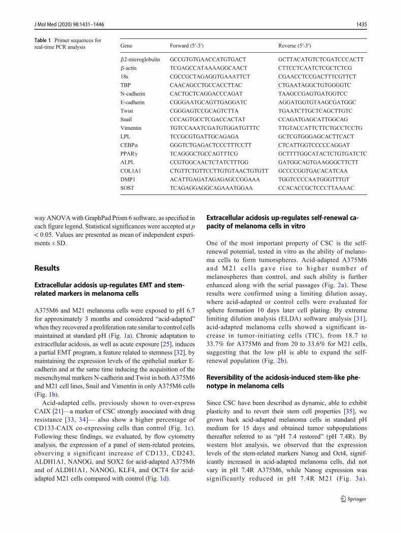
Total RNA was prepared using Tri Reagent (Sigma-Aldrich),agarose gel checked for integrity, and reverse transcribed withiScript cDNA Synthesis Kit (Bio-Rad) according to the man-ufacturer’s instructions. Selected genes were evaluated by areal-time qPCR with 7500 Fast Real-Time PCR System(Applied Biosystems, Monza, Italy). Fold change was deter-mined by the comparative Ct method calculating the averageof β-actin, β2-microglobulin, TATA-Box Binding Protein(TBP), and 18s used as reference genes. Amplification wasperformed with the PCR setting: 40 cycles of 95 °C for 15 sand of 60 °C for 60 s using PowerUp SYBR Green MasterMix (Thermo Fisher Scientific). Primer sequences (IDT,Tema Ricerca, Bologna, Italy) are listed in Table 1 (β-actin,β2-microglobulin, TBP, and 18s used as reference genes).
Statistical analysis
The experiments were performed at least five times for a reli-able application of statistics. All samples used were includedin the statistical analysis. Statistical analysis was performed byT test, One-way analysis of variance (ANOVA), and Two-
1434 J Mol Med (2020) 98:1431–1446
wayANOVAwith GraphPad Prism 6 software, as specified ineach figure legend. Statistical significances were accepted at p< 0.05. Values are presented as mean of independent experi-ments ± SD.
Results
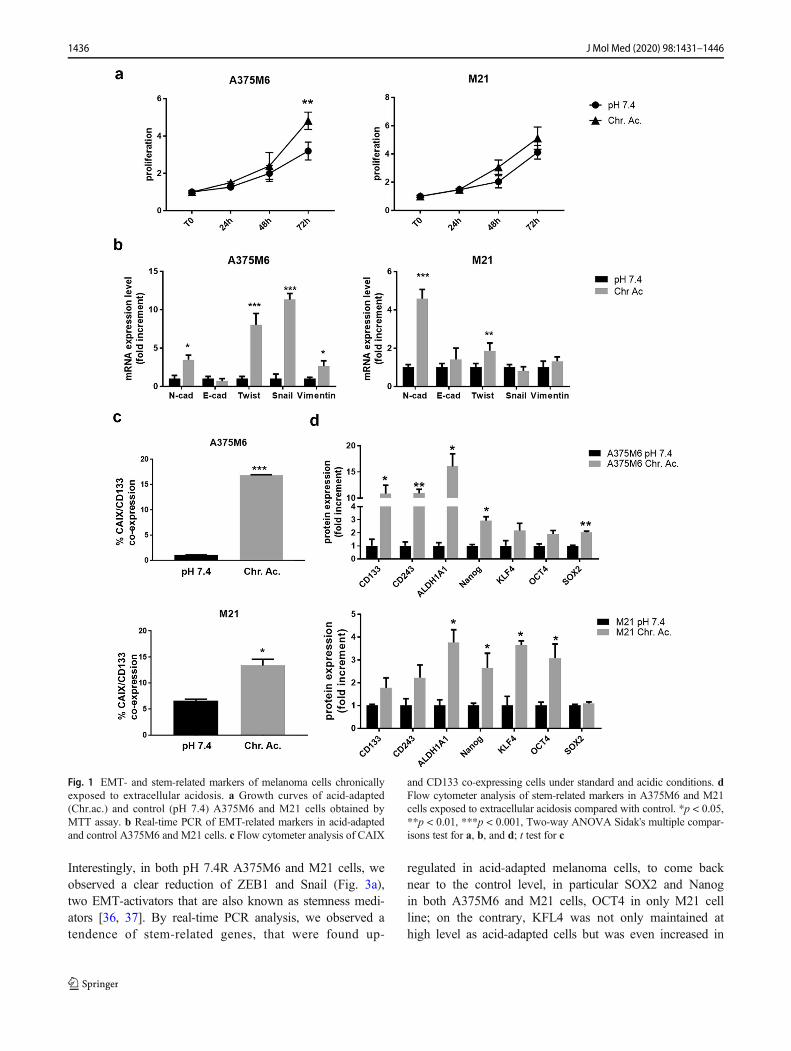
Extracellular acidosis up-regulates EMT and stem-related markers in melanoma cells
A375M6 and M21 melanoma cells were exposed to pH 6.7for approximately 3 months and considered “acid-adapted”when they recovered a proliferation rate similar to control cellsmaintained at standard pH (Fig. 1a). Chronic adaptation toextracellular acidosis, as well as acute exposure [25], inducesa partial EMT program, a feature related to stemness [32], bymaintaining the expression levels of the epithelial marker E-cadherin and at the same time inducing the acquisition of themesenchymal markers N-cadherin and Twist in both A375M6and M21 cell lines, Snail and Vimentin in only A375M6 cells(Fig. 1b).
Acid-adapted cells, previously shown to over-expressCAIX [21]—a marker of CSC strongly associated with drugresistance [33, 34]— also show a higher percentage ofCD133-CAIX co-expressing cells than control (Fig. 1c).Following these findings, we evaluated, by flow cytometryanalysis, the expression of a panel of stem-related proteins,observing a significant increase of CD133, CD243,ALDH1A1, NANOG, and SOX2 for acid-adapted A375M6and of ALDH1A1, NANOG, KLF4, and OCT4 for acid-adapted M21 cells compared with control (Fig. 1d).
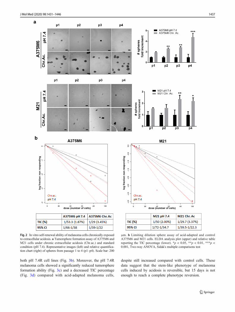
Extracellular acidosis up-regulates self-renewal ca-pacity of melanoma cells in vitro
One of the most important property of CSC is the self-renewal potential, tested in vitro as the ability of melano-ma cells to form tumorspheres. Acid-adapted A375M6and M21 ce l l s gave r i se to h igher number ofmelanospheres than control, and such ability is furtherenhanced along with the serial passages (Fig. 2a). Theseresults were confirmed using a limiting dilution assay,where acid-adapted or control cells were evaluated forsphere formation 10 days later cell plating. By extremelimiting dilution analysis (ELDA) software analysis [31],acid-adapted melanoma cells showed a significant in-crease in tumor-initiating cells (TIC), from 18.7 to33.7% for A375M6 and from 20 to 33.6% for M21 cells,suggesting that the low pH is able to expand the self-renewal population (Fig. 2b).
Reversibility of the acidosis-induced stem-like phe-notype in melanoma cells
Since CSC have been described as dynamic, able to exhibitplasticity and to revert their stem cell properties [35], wegrown back acid-adapted melanoma cells in standard pHmedium for 15 days and obtained tumor subpopulationsthereafter referred to as “pH 7.4 restored” (pH 7.4R). Bywestern blot analysis, we observed that the expressionlevels of the stem-related markers Nanog and Oct4, signif-icantly increased in acid-adapted melanoma cells, did notvary in pH 7.4R A375M6, while Nanog expression wassignificantly reduced in pH 7.4R M21 (Fig. 3a).
Table 1 Primer sequences forreal-time PCR analysis Gene Forward (5′-3′) Reverse (5′-3′)
β2-microglobulin GCCGTGTGAACCATGTGACT GCTTACATGTCTCGATCCCACTT
β-actin TCGAGCCATAAAAGGCAACT CTTCCTCAATCTCGCTCTCG
18s CGCCGCTAGAGGTGAAATTCT CGAACCTCCGACTTTCGTTCT
ΤΒP CAACAGCCTGCCACCTTAC CTGAATAGGCTGTGGGGTC
N-cadherin CACTGCTCAGGACCCAGAT TAAGCCGAGTGATGGTCC
E-cadherin CGGGAATGCAGTTGAGGATC AGGATGGTGTAAGCGATGGC
Twist CGGGAGTCCGCAGTCTTA TGAATCTTGCTCAGCTTGTC
Snail CCCAGTGCCTCGACCACTAT CCAGATGAGCATTGGCAG
Vimentin TGTCCAAATCGATGTGGATGTTTC TTGTACCATTCTTCTGCCTCCTG
LPL TCCGCGTGATTGCAGAGA GCTCGTGGGAGCACTTCACT
CEBPα GGGTCTGAGACTCCCTTTCCTT CTCATTGGTCCCCCAGGAT
PPARγ TCAGGGCTGCCAGTTTCG GCTTTTGGCATACTCTGTGATCTC
ALPL CCGTGGCAACTCTATCTTTGG GATGGCAGTGAAGGGCTTCTT
COL1A1 CTGTTCTGTTCCTTGTGTAACTGTGTT GCCCCGGTGACACATCAA
DMP1 ACATTGAGATAGAGAGCCGGAAA TGGTCCCCAATGGGTTTGT
SOST TCAGAGGAGGCAGAAATGGAA CCACACCGCTCCCTTAAAAC
1435J Mol Med (2020) 98:1431–1446
Interestingly, in both pH 7.4R A375M6 and M21 cells, weobserved a clear reduction of ZEB1 and Snail (Fig. 3a),two EMT-activators that are also known as stemness medi-ators [36, 37]. By real-time PCR analysis, we observed atendence of stem-related genes, that were found up-
regulated in acid-adapted melanoma cells, to come backnear to the control level, in particular SOX2 and Nanogin both A375M6 and M21 cells, OCT4 in only M21 cellline; on the contrary, KFL4 was not only maintained athigh level as acid-adapted cells but was even increased in
Fig. 1 EMT- and stem-related markers of melanoma cells chronicallyexposed to extracellular acidosis. a Growth curves of acid-adapted(Chr.ac.) and control (pH 7.4) A375M6 and M21 cells obtained byMTT assay. b Real-time PCR of EMT-related markers in acid-adaptedand control A375M6 and M21 cells. c Flow cytometer analysis of CAIX
and CD133 co-expressing cells under standard and acidic conditions. dFlow cytometer analysis of stem-related markers in A375M6 and M21cells exposed to extracellular acidosis compared with control. *p < 0.05,**p < 0.01, ***p < 0.001, Two-way ANOVA Sidak's multiple compar-isons test for a, b, and d; t test for c
1436 J Mol Med (2020) 98:1431–1446
both pH 7.4R cell lines (Fig. 3b). Moreover, the pH 7.4Rmelanoma cells showed a significantly reduced tumorsphereformation ability (Fig. 3c) and a decreased TIC percentage(Fig. 3d) compared with acid-adapted melanoma cells,
despite still increased compared with control cells. Thesedata suggest that the stem-like phenotype of melanomacells induced by acidosis is reversible, but 15 days is notenough to reach a complete phenotype reversion.
Fig. 2 In vitro self-renewal ability ofmelanoma cells chronically exposedto extracellular acidosis. a Tumorsphere formation assay of A375M6 andM21 cells under chronic extracellular acidosis (Chr.ac.) and standardcondition (pH 7.4). Representative images (left) and relative quantifica-tion chart (right) of spheres from passage 1 to 4 (p1–p4). Scale bar: 200
μm. b Limiting dilution sphere assay of acid-adapted and controlA375M6 and M21 cells. ELDA analysis plot (upper) and relative tablereporting the TIC percentage (lower). *p < 0.05, **p < 0.01, ***p <0.001, Two-way ANOVA, Sidak's multiple comparisons test
1437J Mol Med (2020) 98:1431–1446
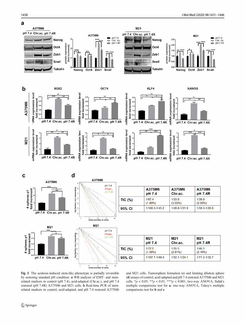
Fig. 3 The acidosis-induced stem-like phenotype is partially reversibleby restoring standard pH condition. a WB analysis of EMT- and stem-related markers in control (pH 7.4), acid-adapted (Chr.ac.), and pH 7.4restored (pH 7.4R) A375M6 and M21 cells. b Real-time PCR of stem-related markers in control, acid-adapted, and pH 7.4 restored A375M6
and M21 cells. Tumorsphere formation (c) and limiting dilution sphere(d) assays of control, acid-adapted and pH 7.4 restoredA375M6 andM21cells. *p < 0.05, **p < 0.01, ***p < 0.001; two-way ANOVA, Sidak'smultiple comparisons test for a; one-way ANOVA, Tukey's multiplecomparisons test for b and c
1438 J Mol Med (2020) 98:1431–1446
EMT is required for the acquisition of the acidosis-induced stem-like phenotype in melanoma cells
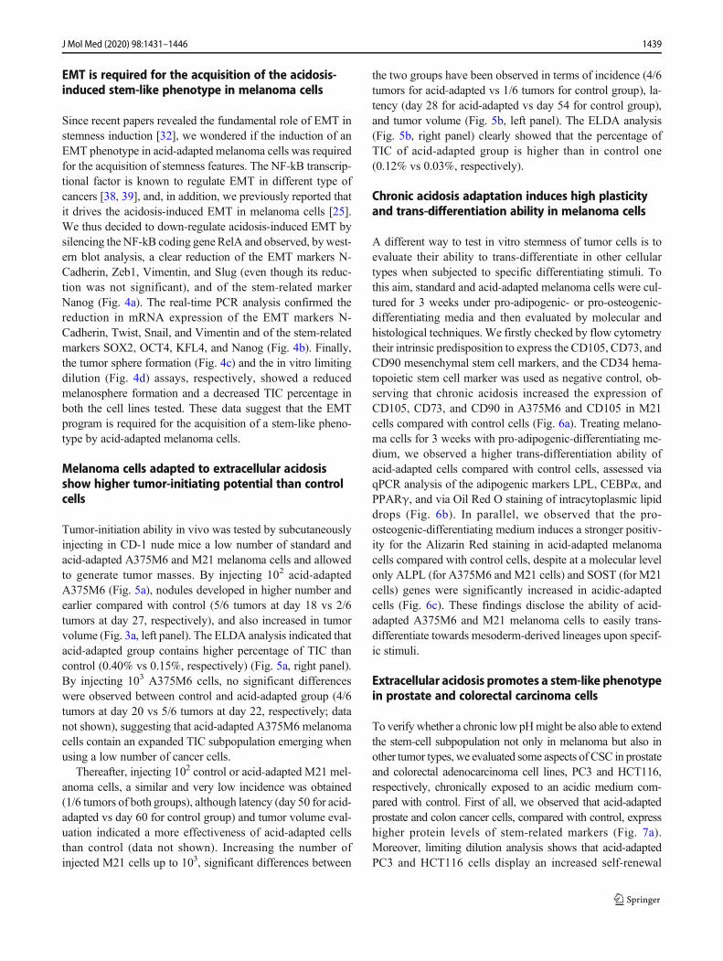
Since recent papers revealed the fundamental role of EMT instemness induction [32], we wondered if the induction of anEMT phenotype in acid-adapted melanoma cells was requiredfor the acquisition of stemness features. The NF-kB transcrip-tional factor is known to regulate EMT in different type ofcancers [38, 39], and, in addition, we previously reported thatit drives the acidosis-induced EMT in melanoma cells [25].We thus decided to down-regulate acidosis-induced EMT bysilencing the NF-kB coding gene RelA and observed, bywest-ern blot analysis, a clear reduction of the EMT markers N-Cadherin, Zeb1, Vimentin, and Slug (even though its reduc-tion was not significant), and of the stem-related markerNanog (Fig. 4a). The real-time PCR analysis confirmed thereduction in mRNA expression of the EMT markers N-Cadherin, Twist, Snail, and Vimentin and of the stem-relatedmarkers SOX2, OCT4, KFL4, and Nanog (Fig. 4b). Finally,the tumor sphere formation (Fig. 4c) and the in vitro limitingdilution (Fig. 4d) assays, respectively, showed a reducedmelanosphere formation and a decreased TIC percentage inboth the cell lines tested. These data suggest that the EMTprogram is required for the acquisition of a stem-like pheno-type by acid-adapted melanoma cells.
Melanoma cells adapted to extracellular acidosisshow higher tumor-initiating potential than controlcells
Tumor-initiation ability in vivo was tested by subcutaneouslyinjecting in CD-1 nude mice a low number of standard andacid-adapted A375M6 and M21 melanoma cells and allowedto generate tumor masses. By injecting 102 acid-adaptedA375M6 (Fig. 5a), nodules developed in higher number andearlier compared with control (5/6 tumors at day 18 vs 2/6tumors at day 27, respectively), and also increased in tumorvolume (Fig. 3a, left panel). The ELDA analysis indicated thatacid-adapted group contains higher percentage of TIC thancontrol (0.40% vs 0.15%, respectively) (Fig. 5a, right panel).By injecting 103 A375M6 cells, no significant differenceswere observed between control and acid-adapted group (4/6tumors at day 20 vs 5/6 tumors at day 22, respectively; datanot shown), suggesting that acid-adapted A375M6 melanomacells contain an expanded TIC subpopulation emerging whenusing a low number of cancer cells.
Thereafter, injecting 102 control or acid-adapted M21 mel-anoma cells, a similar and very low incidence was obtained(1/6 tumors of both groups), although latency (day 50 for acid-adapted vs day 60 for control group) and tumor volume eval-uation indicated a more effectiveness of acid-adapted cellsthan control (data not shown). Increasing the number ofinjected M21 cells up to 103, significant differences between
the two groups have been observed in terms of incidence (4/6tumors for acid-adapted vs 1/6 tumors for control group), la-tency (day 28 for acid-adapted vs day 54 for control group),and tumor volume (Fig. 5b, left panel). The ELDA analysis(Fig. 5b, right panel) clearly showed that the percentage ofTIC of acid-adapted group is higher than in control one(0.12% vs 0.03%, respectively).
Chronic acidosis adaptation induces high plasticityand trans-differentiation ability in melanoma cells
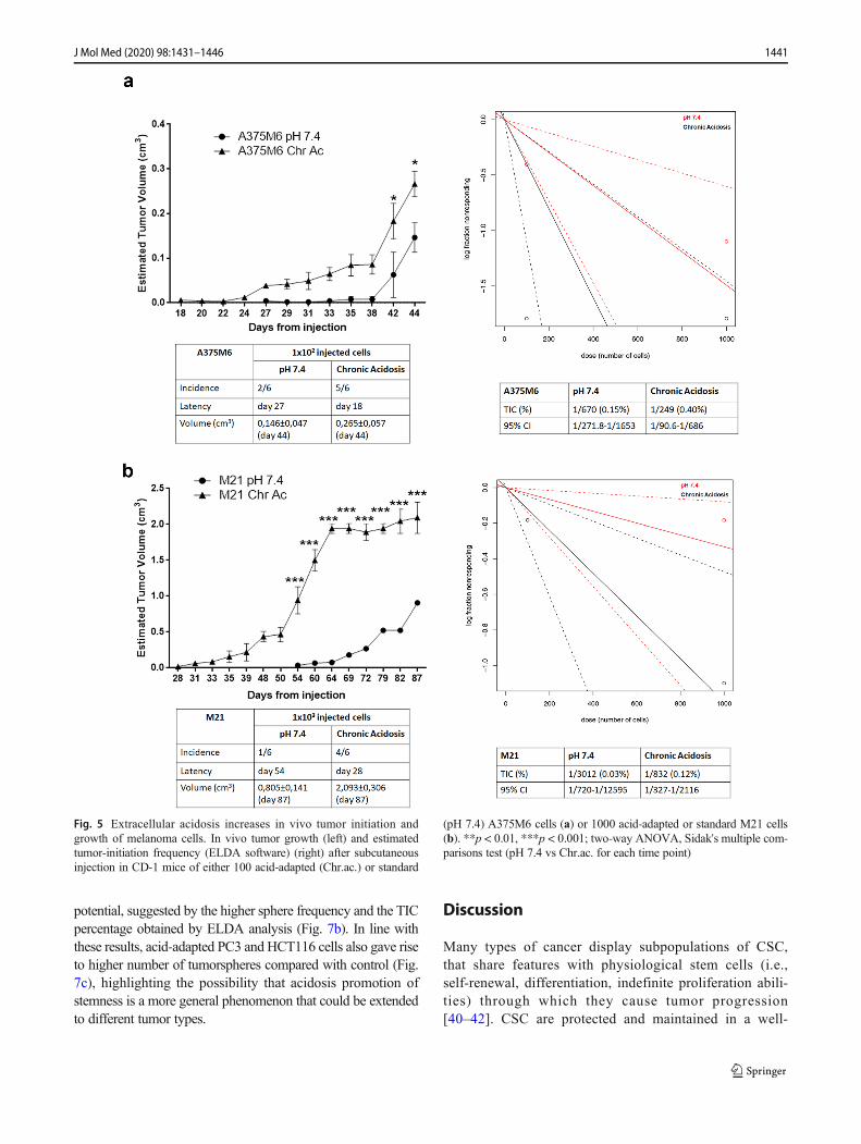
A different way to test in vitro stemness of tumor cells is toevaluate their ability to trans-differentiate in other cellulartypes when subjected to specific differentiating stimuli. Tothis aim, standard and acid-adapted melanoma cells were cul-tured for 3 weeks under pro-adipogenic- or pro-osteogenic-differentiating media and then evaluated by molecular andhistological techniques. We firstly checked by flow cytometrytheir intrinsic predisposition to express the CD105, CD73, andCD90 mesenchymal stem cell markers, and the CD34 hema-topoietic stem cell marker was used as negative control, ob-serving that chronic acidosis increased the expression ofCD105, CD73, and CD90 in A375M6 and CD105 in M21cells compared with control cells (Fig. 6a). Treating melano-ma cells for 3 weeks with pro-adipogenic-differentiating me-dium, we observed a higher trans-differentiation ability ofacid-adapted cells compared with control cells, assessed viaqPCR analysis of the adipogenic markers LPL, CEBPα, andPPARγ, and via Oil Red O staining of intracytoplasmic lipiddrops (Fig. 6b). In parallel, we observed that the pro-osteogenic-differentiating medium induces a stronger positiv-ity for the Alizarin Red staining in acid-adapted melanomacells compared with control cells, despite at a molecular levelonly ALPL (for A375M6 and M21 cells) and SOST (for M21cells) genes were significantly increased in acidic-adaptedcells (Fig. 6c). These findings disclose the ability of acid-adapted A375M6 and M21 melanoma cells to easily trans-differentiate towards mesoderm-derived lineages upon specif-ic stimuli.
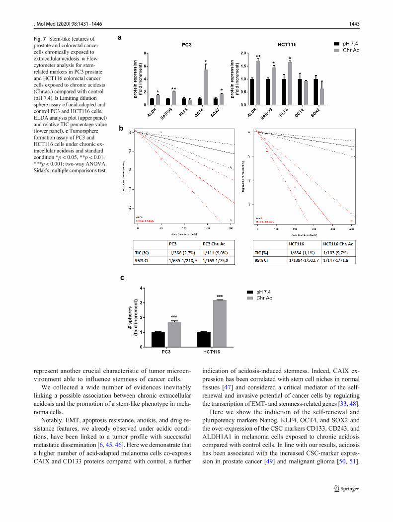
Extracellular acidosis promotes a stem-like phenotypein prostate and colorectal carcinoma cells
To verify whether a chronic low pHmight be also able to extendthe stem-cell subpopulation not only in melanoma but also inother tumor types, we evaluated some aspects of CSC in prostateand colorectal adenocarcinoma cell lines, PC3 and HCT116,respectively, chronically exposed to an acidic medium com-pared with control. First of all, we observed that acid-adaptedprostate and colon cancer cells, compared with control, expresshigher protein levels of stem-related markers (Fig. 7a).Moreover, limiting dilution analysis shows that acid-adaptedPC3 and HCT116 cells display an increased self-renewal
1439J Mol Med (2020) 98:1431–1446
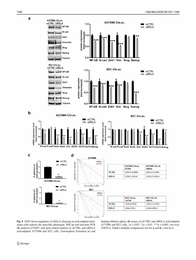
Fig. 4 EMT down-regulation via RELA silencing in acid-adapted mela-noma cells reduces the stem-like phenotype. WB (a) and real-time PCR(b) analysis of EMT- and stem-related markers in siCTRL and siRELAacid-adapted A375M6 and M21 cells. Tumorsphere formation (c) and
limiting dilution sphere (d) assays of siCTRL and siRELA acid-adaptedA375M6 and M21 cells. *p < 0.05, **p < 0.01, ***p < 0.001; two-wayANOVA, Sidak's multiple comparisons test for a and b; t test for c
1440 J Mol Med (2020) 98:1431–1446
potential, suggested by the higher sphere frequency and the TICpercentage obtained by ELDA analysis (Fig. 7b). In line withthese results, acid-adapted PC3 and HCT116 cells also gave riseto higher number of tumorspheres compared with control (Fig.7c), highlighting the possibility that acidosis promotion ofstemness is a more general phenomenon that could be extendedto different tumor types.
Discussion
Many types of cancer display subpopulations of CSC,that share features with physiological stem cells (i.e.,self-renewal, differentiation, indefinite proliferation abili-ties) through which they cause tumor progression[40–42]. CSC are protected and maintained in a well-
Fig. 5 Extracellular acidosis increases in vivo tumor initiation andgrowth of melanoma cells. In vivo tumor growth (left) and estimatedtumor-initiation frequency (ELDA software) (right) after subcutaneousinjection in CD-1 mice of either 100 acid-adapted (Chr.ac.) or standard
(pH 7.4) A375M6 cells (a) or 1000 acid-adapted or standard M21 cells(b). **p < 0.01, ***p < 0.001; two-way ANOVA, Sidak's multiple com-parisons test (pH 7.4 vs Chr.ac. for each time point)
1441J Mol Med (2020) 98:1431–1446
defined microenvironment providing a permissive nicheable to enforce their survival ability and aggressive
properties [43]. Alongside hypoxia, that plays an impor-tant role in stemness [44], extracellular acidosis might
Fig. 6 Chronic acidosis adaptation induces high plasticity and trans-differentiation ability in melanoma cells. a Flow cytometer analysis forthe expression of the mesenchymal markers CD105, CD73, and CD90 inacid-adapted (Chr.ac.) A375M6 andM21 compared with relative control.CD34 used as negative control. b Real-time PCR analysis for adipogenicdifferentiation markers LPL, CEBPα, and PPARγ (left) and representa-tive pictures of Oil Red O lipid drop staining (right) in acid-adapted
A375M6 andM21 compared with control cells. cReal-time PCR analysisfor osteogenic differentiation markers ALPL, COL1A1, DMP1, andSOST (left) and representative pictures of Alizarin Red calcium phos-phate deposit staining (right) in acid-adapted A375M6 and M21 com-pared with control cells. Scale bar: 50 μm. *p < 0.05, **p < 0.01, ***p< 0.001; two-way ANOVA, Sidak's multiple comparisons test
1442 J Mol Med (2020) 98:1431–1446
represent another crucial characteristic of tumor microen-vironment able to influence stemness of cancer cells.
We collected a wide number of evidences inevitablylinking a possible association between chronic extracellularacidosis and the promotion of a stem-like phenotype in mela-noma cells.
Notably, EMT, apoptosis resistance, anoikis, and drug re-sistance features, we already observed under acidic condi-tions, have been linked to a tumor profile with successfulmetastatic dissemination [6, 45, 46]. Here we demonstrate thata higher number of acid-adapted melanoma cells co-expressCAIX and CD133 proteins compared with control, a further
indication of acidosis-induced stemness. Indeed, CAIX ex-pression has been correlated with stem cell niches in normaltissues [47] and considered a critical mediator of the self-renewal and invasive potential of cancer cells by regulatingthe transcription of EMT- and stemness-related genes [33, 48].
Here we show the induction of the self-renewal andpluripotency markers Nanog, KLF4, OCT4, and SOX2 andthe over-expression of the CSC markers CD133, CD243, andALDH1A1 in melanoma cells exposed to chronic acidosiscompared with control cells. In line with our results, acidosishas been associated with the increased CSC-marker expres-sion in prostate cancer [49] and malignant glioma [50, 51],
Fig. 7 Stem-like features ofprostate and colorectal cancercells chronically exposed toextracellular acidosis. a Flowcytometer analysis for stem-related markers in PC3 prostateand HCT116 colorectal cancercells exposed to chronic acidosis(Chr.ac.) compared with control(pH 7.4). b Limiting dilutionsphere assay of acid-adapted andcontrol PC3 and HCT116 cells.ELDA analysis plot (upper panel)and relative TIC percentage value(lower panel). c Tumorsphereformation assay of PC3 andHCT116 cells under chronic ex-tracellular acidosis and standardcondition *p < 0.05, **p < 0.01,***p < 0.001; two-way ANOVA,Sidak's multiple comparisons test.
1443J Mol Med (2020) 98:1431–1446
osteosarcoma [52], and the maintenance of stemness in mes-enchymal cells [53, 54]. Also, OCT-4 expression in murinefibroblasts increases as pH shifts from 7.4 to 6.5 [55].
We confirmed that extracellular acidosis induces the self-renewal capability of cancer cells by the in vitro tumorsphereformation and limiting dilution assays. We found that acid-adapted cells give a higher number of melanospheres withbigger size than control cells and preserve their stem-like phe-notype for at least four passages, whereas control cells gradu-ally lose their ability to form melanospheres. This effect is notlimited to melanoma, but rather proved in PC3 prostate andHCT116 colon cancer cells, that indeed also showed highexpression of stem-related proteins.
We also showed that the stem-like phenotype induced inmelanoma cells by acidosis is a reversible phenomenon, assuggested by the reduced expression of stem-related markersand by the lower self-renewal capability of acid-adapted cellsgrown back for 15 days at standard pH medium, comparedwith acid-adapted cells. The phenotype we observed was notcompletely reverted at control level, but the phenomenon wasclear enough to confirm that the acidosis-induced stem-likephenotype is dynamic and subjected to variations dependingon environmental conditions, an evidence that contributes todefine the increased plasticity and aggressiveness of acid-adapted tumor cells [35].
Several studies have demonstrated the link between EMTand stemness in a variety of human carcinoma [32, 45], andrecently, EMT with its mediators, such as Zeb1 [36], Snail[37], Slug [56], and NF-kB [57, 58], has been described as akey mechanism by which cells are conferred with stem-cellproperties. Here, we demonstrated that the stem-like pheno-type induced in acid-adapted melanoma cells is EMT-depen-dent, since by inhibiting the well-known EMT mediator NF-kB, the stem-like phenotype acquired by acid-adapted mela-noma cells has clearly been reduced.
CSC are also denominated tumor-initiating cells (TIC) be-ing responsible for the initiation, recurrence, and metastases ofa tumor. For that, increased in vivo tumorigenicity is one ofthe hallmarks of CSC. Interestingly, melanoma TIC popula-tion has been demonstrated to depend on SOX2 expression[59], that we found up-regulated under acidic conditions [20].The in vivo evaluation of the tumor-initiation ability of acid-adapted melanoma cells compared with control showed thatchronic acidosis induces the earlier development of a highernumber of tumors compared with the standard pH condition,and the ELDA statistical analysis revealed a higher percentageof TIC in acid-adapted group than control. Some evidencesshowed that increasing the extracellular pH reduces tumorgrowth, prevents cancer invasion [26, 60], and restores drugsensitivity [61]. Recently acidosis has been proposed as po-tential therapeutic target to eradicate CSC [62].
Another hallmark of CSC is the multi-lineage differentia-tion plasticity that influences tumor progression and drug
resistance [63]. Based on the origin of melanoma, we thoughtthat the melanoma CSC should potentially differentiate intomesenchymal cell types, such as adipocytes and osteoblast-like cells. By treating melanoma cells with appropriate differ-entiating media, we observed that acid-adapted melanomacells, compared with control, show higher ability to trans-differentiate in adipocyte and osteoblast-like cells. A recentstudy showed that a continuous exposure to low pHe for 21days of bone marrow stem cells and dental pulp stem cellsimpairs the osteogenic differentiation of both cell types [53],suggesting that extracellular acidosis is crucial for the mainte-nance of a de-differentiated state.
To conclude, our results provide evidences that acidityleads to a reprogramming of a very plastic tumor phenotypetoward the expression of stem-related markers, highclonogenic, and trans-differentiating ability. This could clarifythe reasons why extracellular acidosis is able to promote ag-gressive traits in cancer, dramatically contributing to tumorprogression and metastatic disease. Thus, acidosis needs tobe considered a new and specific cue of stem cell niche, ableto offer an innovative target for cancer therapy.
Acknowledgements Open access funding provided by Università degliStudi di Firenze within the CRUI-CARE Agreement.
Author contributions Silvia Peppicelli: Conceptualization, Investigation,Formal analysis, Writing-Original Draft, Visualization, Projectadministration.
Elena Andreucci: Conceptualization, Investigation, Formal analysis,Writing-Original Draft, Visualization, Project administration.
Jessica Ruzzolini: Validation, Formal analysis, Visualization.Francesca Bianchini: Investigation, Visualization, Writing-Review
and editing.Alessio Biagioni: Validation, Formal analysis, Visualization.Laura Papucci: Investigation, Formal analysis.Lucia Magnelli: Investigation, Formal analysis.Benedetta Mazzanti: Methodology, Resources.Barbara Stecca: Methodology, Investigation, Writing-Review and
editing.Lido Calorini: Resources, Writing-Original Draft, Supervision,
Funding acquisition.
Funding information This study was financially supported by IstitutoToscano Tumori. Elena Andreucci and Alessio Biagioni were supportedby a AIRC fellowship for Italy.
Compliance with ethical standards
Conflicts of interest The authors declare that they have no conflict ofinterest.
Open Access This article is licensed under a Creative CommonsAttribution 4.0 International License, which permits use, sharing, adap-tation, distribution and reproduction in any medium or format, as long asyou give appropriate credit to the original author(s) and the source, pro-vide a link to the Creative Commons licence, and indicate if changes weremade. The images or other third party material in this article are includedin the article's Creative Commons licence, unless indicated otherwise in acredit line to the material. If material is not included in the article's
1444 J Mol Med (2020) 98:1431–1446
Creative Commons licence and your intended use is not permitted bystatutory regulation or exceeds the permitted use, you will need to obtainpermission directly from the copyright holder. To view a copy of thislicence, visit http://creativecommons.org/licenses/by/4.0/.
References
1. Peitzsch C, Tyutyunnykova A, Pantel K, Dubrovska A (2017)Cancer stem cells: the root of tumor recurrence and metastases.Semin Cancer Biol 44:10–24
2. Seftor EA, Margaryan NV, Seftor REB, Hendrix MJC (2019)Heterogeneity of Melanoma with Stem Cell Properties. Adv ExpMed Biol 1139:105–114
3. Siegel R, Ma J, Zou Z, Jemal A (2014) Cancer statistics, 2014. CACancer J Clin. 64:9–29
4. Fang D, Nguyen TK, Leishear K, Finko R, Kulp AN, Hotz S, vanBelle PA, Xu X, Elder DE, Herlyn M (2005) A tumorigenic sub-population with stem cell properties in melanomas. Cancer Res 65:9328–9337
5. Anurag C, Singh N, Vipin Kumar G, Verma M (2019)Vasculogenic Mimicry and Its Role in Cancer. Am J Pharmacol2:1013
6. Schatton T, Frank MH (2008) Cancer stem cells and human malig-nant melanoma. Pigment Cell Melanoma Res 21:39–55
7. Quintana E, Shackleton M, Sabel MS, Fullen DR, Johnson TM,Morrison SJ (2008) Efficient tumour formation by single humanmelanoma cells. Nature. 456:593–598
8. Postovit L-M, Seftor EA, Seftor REB, Hendrix MJC (2006)Influence of the microenvironment on melanoma cell fate determi-nation and phenotype. Cancer Res 66:7833–7836
9. Webb BA, Chimenti M, Jacobson MP, Barber DL (2011)Dysregulated pH: a perfect storm for cancer progression. Nat RevCancer 11:671–677
10. Peppicelli S, Andreucci E, Ruzzolini J, Laurenzana A, Margheri F,Fibbi G, del Rosso M, Bianchini F, Calorini L (2017) The acidicmicroenvironment as a possible niche of dormant tumor cells. CellMol Life Sci. 74:2761–2771
11. Peppicelli S, Ruzzolini J, Bianchini F, Andreucci E, Nediani C,Laurenzana A et al (2019) Anoikis resistance as a further trait ofacidic-adapted melanoma cells. J Oncol. 2019:8340926
12. Ruzzolini J, Peppicelli S, Andreucci E, Bianchini F, Margheri F,LaurenzanaA, Fibbi G, Pimpinelli N, Calorini L (2017) Everolimusselectively targets vemurafenib resistant BRAFV600E melanomacells adapted to low pH. Cancer Lett 408:43–54
13. Muz B, de la Puente P, Azab F, Azab AK (2015) The role ofhypoxia in cancer progression, angiogenesis, metastasis, and resis-tance to therapy. Hypoxia (Auckl) 3:83–92
14. Vander Heiden MG, Cantley LC, Thompson CB (2009)Understanding the Warburg effect: the metabolic requirements ofcell proliferation. Science. 324:1029–1033
15. Mookerjee SA, Goncalves RLS, Gerencser AA, Nicholls DG,Brand MD (2015) The contributions of respiration and glycolysisto extracellular acid production. Biochim Biophys Acta 1847:171–181
16. Reshetnyak YK, Yao L, Zheng S, Kuznetsov S, Engelman DM,Andreev OA (2011) Measuring tumor aggressiveness and targetingmetastatic lesions with fluorescent pHLIP. Mol Imaging Biol 13:1146–1156
17. Vaupel P (2004) Tumor microenvironmental physiology and itsimplications for radiation oncology. Semin Radiat Oncol 14:198–206
18. Damaghi M, Gillies R (2017) Phenotypic changes of acid-adaptedcancer cells push them toward aggressiveness in their evolution inthe tumor microenvironment. Cell Cycle 16:1739–1743
19. Pillai SR, Damaghi M, Marunaka Y, Spugnini EP, Fais S, GilliesRJ (2019) Causes, consequences, and therapy of tumors acidosis.Cancer Metastasis Rev 38:205–222
20. Andreucci E, Pietrobono S, Peppicelli S, Ruzzolini J, Bianchini F,Biagioni A, Stecca B, Calorini L (2018) SOX2 as a novel contrib-utor of oxidative metabolism in melanoma cells. Cell CommunSignal 16:87
21. Andreucci E, Peppicelli S, Carta F, Brisotto G, Biscontin E,Ruzzolini J, Bianchini F, Biagioni A, Supuran CT, Calorini L(2017) Carbonic anhydrase IX inhibition affects viability of cancercells adapted to extracellular acidosis. J Mol Med 95:1341–1353
22. Riemann A, Rauschner M, Gießelmann M, Reime S, Haupt V,Thews O (2019) Extracellular acidosis modulates the expressionof epithelial-mesenchymal transition (EMT) markers and adhesionof epithelial and tumor cells. Neoplasia 21:450–458
23. Suzuki A, Maeda T, Baba Y, Shimamura K, Kato Y (2014) Acidicextracellular pH promotes epithelial mesenchymal transition inLewis lung carcinoma model. Cancer Cell Int 14:129
24. Zhu S, Zhou HY, Deng SC, Deng SJ, He C, Li X, Chen JY, Jin Y,Hu ZL, Wang F, Wang CY, Zhao G (2017) ASIC1 and ASIC3contribute to acidity-induced EMT of pancreatic cancer throughactivating Ca2+/RhoA pathway. Cell Death Dis 8:e2806
25. Peppicelli S, Bianchini F, Torre E, Calorini L (2014) Contributionof acidic melanoma cells undergoing epithelial-to-mesenchymaltransition to aggressiveness of non-acidic melanoma cells. ClinExp Metastasis 31:423–433
26. Estrella V, Chen T, LloydM,Wojtkowiak J, Cornnell HH, Ibrahim-Hashim A, Bailey K, Balagurunathan Y, Rothberg JM, Sloane BF,Johnson J, Gatenby RA, Gillies RJ (2013) Acidity generated by thetumor microenvironment drives local invasion. Cancer Res 73:1524–1535
27. Persi E, Duran-Frigola M, Damaghi M, Roush WR, Aloy P,Cleveland JL, Gillies RJ, Ruppin E (2018) Systems analysis ofintracellular pH vulnerabilities for cancer therapy. Nat Commun9:2997
28. Ryder C, McColl K, Zhong F, Distelhorst CW (2012) Acidosispromotes Bcl-2 family-mediated evasion of apoptosis: involvementof acid-sensing G protein-coupled receptor Gpr65 signaling toMek/Erk. J Biol Chem 287:27863–27875
29. Wojtkowiak JW, Rothberg JM, Kumar V, Schramm KJ, Haller E,Proemsey JB, Lloyd MC, Sloane BF, Gillies RJ (2012) Chronicautophagy is a cellular adaptation to tumor acidic pHmicroenviron-ments. Cancer Res 72:3938–3947
30. Andreucci E, Ruzzolini J, Peppicelli S, Bianchini F, Laurenzana A,Carta F, Supuran CT, Calorini L (2019) The carbonic anhydrase IXinhibitor SLC-0111 sensitises cancer cells to conventional chemo-therapy. J Enzyme Inhib Med Chem 34:117–123
31. Hu Y, Smyth GK (2009) ELDA: extreme limiting dilution analysisfor comparing depleted and enriched populations in stem cell andother assays. Journal of Immunological Methods 347:70–78
32. Fabregat I, Malfettone A, Soukupova J (2016) New insights into thecrossroads between EMT and stemness in the context of cancer. JClin Med. 5:37
33. Ledaki I, McIntyre A, Wigfield S, Buffa F, McGowan S, Baban D,Li JL, Harris AL (2015) Carbonic anhydrase IX induction defines aheterogeneous cancer cell response to hypoxia and mediates stemcell-like properties and sensitivity to HDAC inhibition. Oncotarget6:19413–19427
34. Supuran CT, Alterio V, Di Fiore A, D’Ambrosio K, Carta F, MontiSM et al (2018) Inhibition of carbonic anhydrase IX targets primarytumors, metastases, and cancer stem cells: three for the price of one.Med Res Rev 38:1799–1836
1445J Mol Med (2020) 98:1431–1446
35. Sugihara E, Saya H (2013) Complexity of cancer stem cells. Int JCancer 132:1249–1259
36. Preca BT, Bajdak K,Mock K, Sundararajan V, Pfannstiel J, MaurerJ, Wellner U, Hopt UT, Brummer T, Brabletz S, Brabletz T,Stemmler MP (2015) A self-enforcing CD44s/ZEB1 feedback loopmaintains EMT and stemness properties in cancer cells. Int J Cancer137:2566–2577
37. Hojo N, Huisken AL, Wang H, Chirshev E, Kim NS, Nguyen SM,Campos H, Glackin CA, Ioffe YJ, Unternaehrer JJ (2018) Snailknockdown reverses stemness and inhibits tumour growth in ovar-ian cancer. Sci Rep 8:8704
38. Huber MA, Azoitei N, Baumann B, Grünert S, Sommer A,Pehamberger H, Kraut N, Beug H, Wirth T (2004) NF-kappaB isessential for epithelial-mesenchymal transition and metastasis in amodel of breast cancer progression. J Clin Invest 114:569–581
39. Nomura A, Majumder K, Giri B, Dauer P, Dudeja V, Roy S,Banerjee S, Saluja AK (2016) Inhibition of NF-kappa B pathwayleads to deregulation of epithelial-mesenchymal transition and neu-ral invasion in pancreatic cancer. Lab Invest 96:1268–1278
40. Dean M, Fojo T, Bates S (2005) Tumour stem cells and drug resis-tance. Nat Rev Cancer 5:275–284
41. Korkaya H, Wicha MS (2010) Cancer stem cells: nature versusnurture. Nat Cell Biol 12:419–421
42. Reya T, Morrison SJ, Clarke MF, Weissman IL (2001) Stem cells,cancer, and cancer stem cells. Nature 414:105–111
43. Plaks V, Kong N, Werb Z (2015) The cancer stem cell niche: howessential is the niche in regulating stemness of tumor cells? CellStem Cell 16:225–238
44. Yun Z, Lin Q (2014) Hypoxia and regulation of cancer cellstemness. Adv Exp Med Biol 772:41–53
45. Shibue T, Weinberg RA (2017) EMT, CSCs, and drug resistance:the mechanistic link and clinical implications. Nat Rev Clin Oncol14:611–629
46. Mani SA, GuoW, Liao M-J, Eaton EN, Ayyanan A, Zhou AY et al(2008) The epithelial-mesenchymal transition generates cells withproperties of stem cells. Cell 133:704–715
47. Liao S-Y, Lerman MI, Stanbridge EJ (2009) Expression of trans-membrane carbonic anhydrases, CAIX and CAXII, in human de-velopment. BMC Dev Biol 9:22
48. Lock FE, McDonald PC, Lou Y, Serrano I, Chafe SC, Ostlund Cet al (2013) Targeting carbonic anhydrase IX depletes breast cancerstem cells within the hypoxic niche. Oncogene 32:5210–5219
49. Huang S, Tang Y, Peng X, Cai X, Wa Q, Ren D, Li Q, Luo J, Li L,Zou X, Huang S (2016) Acidic extracellular pH promotes prostatecancer bone metastasis by enhancing PC-3 stem cell characteristics,cell invasiveness and VEGF-induced vasculogenesis of BM-EPCs.Oncol Rep 36:2025–2032
50. Filatova A, Seidel S, Böğürcü N, Gräf S, Garvalov BK, Acker T(2016) Acidosis acts through HSP90 in a PHD/VHL-independentmanner to promote HIF function and stem cell maintenance inglioma. Cancer Res 76:5845–5856
51. Hu P, Li S, Tian N, Wu F, Hu Y, Li D, Qi Y, Wei Z, Wei Q, Li Y,Yin B, Jiang T, Yuan J, Qiang B, Han W, Peng X (2019) Acidosisenhances the self-renewal and mitochondrial respiration of stemcell-like glioma cells through CYP24A1-mediated reduction of vi-tamin D. Cell Death Dis 10:25
52. Avnet S, Di Pompo G, Chano T, Errani C, Ibrahim-Hashim A,Gillies RJ et al (2017) Cancer-associated mesenchymal stroma fos-ters the stemness of osteosarcoma cells in response to intratumoralacidosis via NF-κB activation. Int J Cancer. 140:1331–1345
53. Massa A, Perut F, Chano T, Woloszyk A, Mitsiadis TA, Avnet Set al (2017) The effect of extracellular acidosis on the behaviour ofmesenchymal stem cells in vitro. Eur Cell Mater 33:252–267
54. Hazehara-KunitomoY,Hara ES, OnoM,AungKT,Komi K, PhamHT, Akiyama K, Okada M, Oohashi T, Matsumoto T, Kuboki T(2019) Acidic pre-conditioning enhances the stem cell phenotype ofhuman bone marrow stem/progenitor cells. Int J Mol Sci 20.
55. SomA, Bloch S, Ippolito JE, Achilefu S (2016) Acidic extracellularpH of tumors induces octamer-binding transcription factor 4 ex-pression in murine fibroblasts in vitro and in vivo. Sci Rep 6:27803
56. GuoW,Keckesova Z, Donaher JL, Shibue T, Tischler V, ReinhardtF, Itzkovitz S, Noske A, Zürrer-Härdi U, Bell G, Tam WL, ManiSA, van Oudenaarden A, Weinberg RA (2012) Slug and Sox9cooperatively determine the mammary stem cell state. Cell 148:1015–1028
57. Zakaria N, Mohd Yusoff N, Zakaria Z, Widera D, Yahaya BH(2018) Inhibition of NF-κB signaling reduces the stemness charac-teristics of lung cancer stem cells. Front Oncol 8:166
58. Xiang T, Long H, He L, Han X, Lin K, Liang Z, Zhuo W, Xie R,Zhu B (2015) Interleukin-17 produced by tumor microenvironmentpromotes self-renewal of CD133+ cancer stem-like cells in ovariancancer. Oncogene 34:165–176
59. Santini R, Vinci MC, Pandolfi S, Penachioni JY, Montagnani V,Olivito B, Gattai R, Pimpinelli N, Gerlini G, Borgognoni L, SteccaB (2012) Hedgehog-GLI signaling drives self-renewal and tumor-igenicity of human melanoma-initiating cells. Stem Cells 30:1808–1818
60. Silva AS, Yunes JA, Gillies RJ, Gatenby RA (2009) The potentialrole of systemic buffers in reducing intratumoral extracellular pHand acid-mediated invasion. Cancer Res 69:2677–2684
61. Wojtkowiak JW, Verduzco D, Schramm KJ, Gillies RJ (2011)Drug resistance and cellular adaptation to tumor acidic pH micro-environment. Mol Pharm 8:2032–2038
62. Vander Linden C, Corbet C (2019) Therapeutic targeting of cancerstem cells: integrating and exploiting the acidic niche. Front Oncol9:159
63. Kemper K, de Goeje PL, Peeper DS, van Amerongen R (2014)Phenotype switching: tumor cell plasticity as a resistance mecha-nism and target for therapy. Cancer Res 74:5937–5941
Publisher’s note Springer Nature remains neutral with regard to jurisdic-tional claims in published maps and institutional affiliations.
1446 J Mol Med (2020) 98:1431–1446


![Tumor Microenvironment Hijacking the Immune System [Read-Only]](https://img.pdfslide.net/doc/110x75/61bf372f43ec6023e9684384/tumor-microenvironment-hijacking-the-immune-system-read-only.jpg)