Embed Size (px)
Citation preview

Leading Edge
Review
Modeling Development and Disease with Organoids
Hans Clevers1,*1Hubrecht Institute/Royal Netherlands Academy of Arts and Sciences, Princess Maxima Centre and University Medical Centre Utrecht,
3584CT Utrecht, The Netherlands
*Correspondence: [email protected]://dx.doi.org/10.1016/j.cell.2016.05.082
Recent advances in 3D culture technology allow embryonic and adult mammalian stem cells toexhibit their remarkable self-organizing properties, and the resulting organoids reflect key struc-tural and functional properties of organs such as kidney, lung, gut, brain and retina. Organoid tech-nology can therefore be used to model human organ development and various human pathologies‘in a dish.’’ Additionally, patient-derived organoids hold promise to predict drug response in apersonalized fashion. Organoids open up new avenues for regenerative medicine and, in combina-tion with editing technology, for gene therapy. The many potential applications of this technologyare only beginning to be explored.
In 1975, James Rheinwald and Howard Green described the first
long-term culture of normal human cells (Rheinwald and Green,
1975). For this, they combined freshly isolated keratinocytes
with irradiated mouse 3T3 fibroblasts, established in the same
lab years earlier. As in stratified skin, cell division was confined
to the basal layer of the growing clones, while superficial
layers consisted of terminally differentiating keratinocytes that
gradually developed a cornified cell envelope. Successive
improvements in the methodology allowed the cultivation of
large confluent sheets of epidermis grown from relatively small
numbers of primary proliferative keratinocytes (for which the
term ‘‘stem cell’’ was not applied). Green and co-workers per-
formed the first successful treatment of two third-degree burn
patients with cultured autologous keratinocyte sheets at the
Peter Bent Brigham Hospital in 1980 (O’connor et al., 1981). In
a particularly dramatic demonstration of the potential of the
method, they showed, in the summer of 1983, that this approach
was life-saving for the 5-year-old Jamie Selby and his 6-year-old
brother Glen, who had both sustained burns over >95% of their
body surface (Gallico et al., 1984).
In his own lab, Rheinwald built on this work to establish a
comparable method for culturing another stratified squamous
epithelium, the cornea (Lindberg et al., 1993). De Luca and Pel-
legrini applied this technology for the treatment of corneal blind-
ness with a high rate of success, as reported upon long-term
follow-up of 112 patients. Their procedure was straightforward:
a 1–2 mm biopsy from the limbal region of the healthy eye was
grown in culture on 3T3 feeder cells, and the resulting sheet
was grafted onto the injured eye (Pellegrini et al., 1997; Rama
et al., 2010). While the term ‘‘organoid’’ was not used in these
pioneering studies, Rheinwald and Green were the first to recon-
stitute 3D tissue structure from cultured human stem cells.
Organoids revealed their first popularity in the years 1965–
1985, shown by an increase in the PubMed search term ‘‘orga-
noids’’ (Figure 1), mostly in classic developmental biology
experiments that sought to describe organogenesis by cell
dissociation and reaggregation experiments (for an overview,
see Lancaster and Knoblich, 2014). The past 7–8 years have
1586 Cell 165, June 16, 2016 ª 2016 Elsevier Inc.
witnessed a revival of the organoid, yet in a somewhat different
guise: an organoid is now defined as a 3D structure grown
from stem cells and consisting of organ-specific cell types that
self-organizes through cell sorting and spatially restricted line-
age commitment (after Eiraku and Sasai, 2012; Lancaster and
Knoblich, 2014).
Organoids can be initiated from the two main types of stem
cells: (1) pluripotent embryonic stem (ES) cells and their synthetic
induced pluripotent stem (iPS) cell counterparts and (2) organ-
restricted adult stem cells (aSCs). Both approaches exploit the
seemingly infinite expansion potential of normal stem cells in
culture. For ES and iPS cells, here collectively termed pluripotent
stem cells or PSCs, this potential has been an essential prereq-
uisite for their discovery. By contrast, aSCs—with the exception
of Green’s skin cells—were long believed to be incapable of sig-
nificant proliferation outside of the body. Yet, recent years have
witnessed the rapid development of growth factor cocktails that
mimic the various organ stem cell niches. When PSCs and aSCs
are allowed to differentiate in culture, they display an uncanny
capacity to self-organize into structures that reflect crucial as-
pects of the tissues to which they are fated.
Organoids Derived from Pluripotent Stem CellsEver since pluripotent ES and iPS cell lines were established,
scientists have applied insights from developmental biology to
derive differentiated cell types from these stem cells (Chen
et al., 2014; Cherry and Daley, 2012) (Figure 2). Yoshiki Sasai
and his colleagues were the first to take this one step further
by asking whether such an in vitro system could recapitulate
some of the robust regulatory systems of organogenesis—in
terms of not only cell differentiation, but also spatial patterning
and morphogenesis. In a remarkable tour de force, they devel-
oped methods to generate brain structures, retina, and pituitary
‘in a dish’ (Eiraku and Sasai, 2012).
Brain Organoids
The central nervous system derives from the neural ectoderm.
Set up first as the neural plate, it is then shaped into the neural
tube through folding and fusion. Morphogen gradients in this
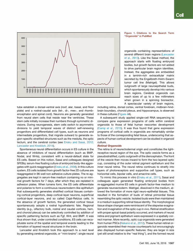
Figure 1. Citations to the Search Term
‘‘Organoids’’ in PubMed
tube establish a dorsal-ventral axis (roof, alar, basal, and floor
plate) and a rostral-caudal axis (tel-, di-, mes-, and rhomb-
encephalon and spinal cord). Neurons are generally generated
from neural stem cells that reside near the ventricles. These
stem cells initially increase their numbers through symmetric di-
visions. During neurogenesis, stem cells switch to asymmetric
divisions to yield temporal waves of distinct self-renewing
progenitors and differentiated cell types, such as neurons and
intermediate progenitors, that migrate outward to generate re-
gion-specific stratified structures such as the medulla, the optic
tectum, and the cerebral cortex (see Eiraku and Sasai, 2012;
Lancaster and Knoblich, 2014).
Spontaneous neural differentiation occurs in ES culture in the
absence of inhibitors of neural differentiation (such as BMP,
Nodal, and Wnts), consistent with a neural-default state for
ES cells. Based on this notion, Sasai and colleagues designed
SFEBq: serum-free floating culture of embryoid body-like aggre-
gateswith quick reaggregation (Eiraku et al., 2008). In this culture
system, ES cells isolated from growth factor-free 2D cultures are
reaggregated in 96-well non-adhesive culture plates. The re-ag-
gregates are kept in serum-free medium containing no or mini-
mal growth factors for 7 days, after which they are replated in
adhesion plates. When lumens form, the ES cells differentiate
and polarize to form a continuous neurectoderm-like epithelium
that subsequently generates stratified cortical tissues contain-
ing cortical progenitors, deep cortical-layer neurons, superficial
cortical-layer neurons, and so called Cajal-Retzius cells. In
the absence of growth factors, the generated cortical tissue
spontaneously adopts a rostral hypothalamic fate. Regional
identity (e.g., olfactory bulb, rostral and caudal cortices, hem,
and choroid plexus) can be selectively controlled by addition of
specific patterning factors such as Fgf, Wnt, and BMP. It was
thus shown that, under controlled conditions, ES cells can reca-
pitulate some of the spatial and temporal events leading to the
formation of layered neural structures in the brain.
Lancaster and Knoblich took this approach to a next level
by generating cerebral organoids, or ‘‘mini-brains’’: single neural
organoids containing representations of
several different brain regions (Lancaster
et al., 2013). Like the Sasai method, the
approach starts with floating embryoid
bodies, but growth factors are not added
to drive particular brain region identities.
Instead, the aggregates are embedded
in a laminin-rich extracellular matrix
secreted by the Engelbreth-Holm-Swarm
tumor cell line (Matrigel). This allows
outgrowth of large neuroepithelial buds,
which spontaneously develop into various
brain regions. Cerebral organoids can
reach sizes of up to a few millimeters
when grown in a spinning bioreactor.
A spectacular variety of brain regions,
including retina, dorsal cortex, ventral forebrain, midbrain-hind-
brain boundary, choroid plexus, and hippocampus, is observed
in these cultures (Figure 4A).
A subsequent study applied single-cell RNA sequencing to
compare gene expression programs of cells within cerebral
organoids to those of fetal human neocortex development
(Camp et al., 2015). It was thus found that gene expression
programs of cortical cells in organoids are remarkably similar
to those of the corresponding fetal tissue, underscoring that as-
pects of human cortical development can be studied in organoid
culture.
Retinal Organoids
The retina is of neurectodermal origin and constitutes the light-
receptive neural region of the eye. The optic vesicle forms as a
pseudostratified, cystic outgrowth of the diencephalon. The front
of the vesicle then moves inward to form the two-layered optic
cup, consisting of the outer retinal pigment epithelium and the
inner neural retina. The neural retina continues to stratify into
layers of photoreceptors and supportive cell types, such as
horizontal cells, bipolar cells, and amacrine cells.
To mimic this process in vitro (Eiraku et al., 2011), Sasai and
colleagues again generated floating embryoid bodies from
re-aggregated murine ES cells in growth factor-free medium to
generate neuroectoderm. Matrigel, dissolved in the medium, al-
lowed the formation of more rigid neuro-epithelial tissues. This
resulted in the formation of buds of retinal primordial tissue
resembling the optic vesicle. Isolated budswere thenmaintained
in amedium supporting retinal tissue identity. Themorphological
tissue shape changes were reminiscent of the stepwise evagina-
tion and invagination of the optic cup in vivo. Retinal stratification
with proper apical-basal polarity occurred, andmarkers of neural
retina and pigment epithelium were expressed in a spatially cor-
rect manner. More recently, optic cup organoids were generated
from human PSCs (Nakano et al., 2012). These human retinal or-
ganoids resembled their mouse counterparts but encouragingly
also displayed human-specific features: they are larger in size
(yet still small relative to the ‘‘real thing’’), and the formed neural
Cell 165, June 16, 2016 1587

Figure 2. Schematic of the Various Organoids that Can Be Grown from PSCs and the Developmental Signals that Are EmployedAdapted from Lancaster and Knoblich, 2014.
retina grows into a thick multi-layered tissue containing both
rods and cones, whereas cones were rarely observed in mouse
organoid cultures.
Adenohypophysis Organoids
The adenohypophysis secretes multiple systemic hormones.
During early mammalian development, its anlage originates as
a placode in the non-neural head ectoderm near the anterior
neural plate. The thickened placode invaginates and detaches
from the oral ectoderm, forming a hollowed epithelial vesicle,
Rathke’s pouch. This process depends on poorly defined
cross-signaling between ectoderm and developing neural tube.
Sasai’s group sought to recapitulate the inductive microenviron-
ment of this morphogenetic field in order to promote the simulta-
neous generation of both tissues within the same aggregate
of SFEBq-cultured ES cells. Three-fold larger cell aggregates
were required, compared to the above protocols. Hedhehog
and Notch antagonists were added to block neural fate in the
outer layers and to allow the subsequent development of all
major hormone-producing linages, respectively. Under these
conditions, ES cells differentiated into head ectoderm and hypo-
thalamic neuroectoderm in adjacent layers within the aggregate.
Rathke’s-pouch-like structures arose at the interface of these
two epithelia, and the various endocrine cell types were subse-
quently formed. Upon transplantation under the kidney capsule
of hypophysectomized mice, the aggregates partially rescued
systemic glucocorticoid level and prolonged survival of themice.
Cerebellar Organoids
The initial phase of cerebellar development depends on the
function of the isthmic organizer located at the midbrain-hind-
brain boundary. Sasai and colleagues focused on the induction
of isthmic development in an attempt to create functional Pur-
kinje cells, the beautiful key output cells of the cerebellum. Again,
they started from a mouse SFEBq culture. In order to produce
1588 Cell 165, June 16, 2016
caudal brain structures, Fgf2 was added soon after initiation
of the culture. To dorsalize the caudalized brain organoids, a
Hedgehog inhibitor was added during the second week. These
conditions recapitulated early cerebellar plate development,
eventually leading to the formation of mature Purkinje cells (Mu-
guruma et al., 2010). In a subsequent study, the investigators
reported that the addition of Fgf19 and SDF1 to this protocol
allows human ES cells to generate a polarized structure reminis-
cent of the first trimester cerebellum (Muguruma et al., 2015).
Hippocampus
The hippocampus develops from the dorsomedial telencephalon
through a precursor structure termed the medial pallium. A
final protocol developed by Sasai and coworkers involved the
in vitro generation of a reliable source of hippocampal tissue
from human ES cells (Sakaguchi et al., 2015). SFEBq served
once again as the starting material. Stimulation by BMP and
Wnt induced choroid plexus, the dorsomedial-most part of
the telencephalon. Careful titration of BMP and Wnt exposure
allowed the self-organization of tissue resembling the medial
pallium, located adjacent to choroid plexus in the developing
brain. Following long-term dissociation culture, granule neurons
and pyramidal neurons were formed, both of which were electri-
cally functional within connected networks.
In addition to these CNS organoids, protocols have also been
developed to grow various endodermal organoids from PSCs.
Formation of the endoderm germ layer during gastrulation re-
quires Nodal signaling. Definitive endoderm presents as a 2D
sheet of cells, which is subsequently patterned along the ante-
rior-posterior axis and folded into a primitive gut tube, from
which all endodermal organs arise. The foregut forms the ante-
rior section of this tube and generates, e.g., the thyroid, lungs,
stomach, liver, and pancreas. The mid- and hindgut develop
into small intestine, colon, and rectum. Insights into the signals

that control these fate decisions in vivo can be exploited in vitro.
Exposure to Nodal or its mimetic Activin directs differentiation
of PSCs into definitive endoderm and serves as a common start-
ing point of these protocols. Exposure to subsequent inductive
signals can then induce the various endodermal organ identities
(reviewed in Sinagoga and Wells, 2015).
Stomach Organoids
The stomach develops from the posterior foregut. Wells and col-
leagues used activin treatment of humanPSCs to generate defin-
itive endoderm (McCracken et al., 2014). Subsequent addition of
BMP inhibitors and of FGF andWnt activators instructed the cells
toward a foregut fate. When retinoic acid was applied, the orga-
noids were specified toward a posterior foregut fate. Finally, high
concentrations of EGF then converted these into human gastric
organoids, progressing through molecular and morphogenetic
stages that resembled those of the developing antrum of the
mouse stomach. Organoids contained primitive gastric gland-
and pit-like domains, proliferative zones with Lgr5+ stem cells,
mucous cells, and a host of gastric endocrine cells.
Lung and Thyroid Organoids
The lung and the thyroid arise from Nkx2-1+ progenitors in the
developing ventral foregut endoderm. An initial study demon-
strated the directed differentiation of primordial lung and thyroid
progenitors from ESCs. The protocol involves activin-induced
definitive endoderm and treatment with TGFb /BMP inhibitors,
followed by BMP/FGF stimulation, and results in a relatively
pure population of progenitors that recapitulate early develop-
mental milestones of lung/thyroid development (Longmire
et al., 2012). This study has been the stepping stone for subse-
quent attempts to create organoids representing mature ver-
sions of the two organs.
A first description of the generation of lung organoids from iPS
cells was reported by Rossant and colleagues and involved at its
last stage air-liquid interphase culture. The protocol was applied
to CFTR mutant iPS cells as a proof of concept for modeling
cystic fibrosis (Wong et al., 2012). Snoeck and colleagues de-
signed an improved four-stage, 50-day protocol (Huang et al.,
2014). First, definitive endoderm was induced by Activin A. Sub-
sequently, anterior foregut endodermwas induced by sequential
inhibition of BMP, TGF-b, and Wnt signaling. The cells were then
ventralized by Wnt, BMP, FGF, and RA to obtain lung and airway
progenitors. Finally, epithelial cell types (basal, goblet, Clara,
ciliated, type I and type II alveolar epithelial cells) were matured
using Wnt, FGF, c-AMP, and glucocorticoids. Spence and col-
leagues (Dye et al., 2015) similarly started from Activin-treated
human PSCs but then followed a slightly different trajectory.
Subsequent addition of TGFb/BMP inhibitors, FGF4, and Wnt
activators instructed the cells toward an anterior foregut fate.
When the Hedgehog pathway was simultaneously activated,
organoids were ventrally specified toward a lung fate. Upon
embedding inMatrigel and prolonged exposure to Fgf10, mature
lung organoids arose. The cultures could be maintained for
several months and resembled proximal airways, containing
basal cells, ciliated cells, and Clara cells. The endodermal airway
tissues were found to be often surrounded by smooth muscle
actin (SMA)-positive mesenchymal cells. Early markers of the
distal (alveolar) airways were expressed early in culture but
were lost later.
Initial attempts to create thyroid organoids involved forced
expression of the lineage-specific transcription factors NKX2.1
and PAX8 and encouragingly resulted in the formation of mouse
and human thyroid follicles in vitro and upon transplantation (An-
tonica et al., 2012; Ma et al., 2015). Kotton and colleagues
applied an improved version of their ‘‘all soluble factor’’ protocol
(Longmire et al., 2012), followed by sequential treatment by
BMP4/FGF2 and induced maturation by 3D plating in the pres-
ence of thyroid-stimulating hormone in Matrigel. The resulting
fully mature murine thyroid follicular organoids secreted thyroid
hormones in vivo upon transplantation and rescued hypothyroid
mice. The same protocol allowed the derivation of human thyroid
progenitors from iPS cells (Kurmann et al., 2015).
Small Intestinal Organoids
Wnt and FGF signals are known to specify definitive endoderm
toward mid-/hindgut fates (‘‘posteriorization’’). To generate in-
testinal organoids (McCracken et al., 2014; Spence et al.,
2011), Activin-treated human PSCs were cultured with FGF4
and WNT3a. Mid/hindgut spheroids budded off from the 2D
monolayer epithelium and were further cultured in Matrigel along
with a pro-intestinal growth factor cocktail, defined previously for
expansion of adult crypt cultures (Sato et al., 2009; see below).
The organoids expanded over 1–3 months to give rise to a polar-
ized intestinal epithelium patterned into villus-like structures and
crypt-like proliferative zones and containing all major epithelial
cell types. Intestinal mesenchyme (presumably derived from
mesodermal remnants after endoderm induction) surrounded
the epithelial structures and consisted of myofibroblasts and
smooth muscle cells (McCracken et al., 2014; Spence et al.,
2011). Transplantation of these organoids into immunodeficient
mice yielded human epithelium and laminated human mesen-
chyme, supported by mouse vasculature. The transplanted
tissue was functional, as shown by permeability and peptide
uptake tests (Watson et al., 2014).
Liver Organoids
During early hepatogenesis, progenitor cells delaminate from the
foregut endoderm to form a condensed tissue mass termed the
liver bud, which is vascularized soon thereafter. Taniguchi and
co-workers exploited cross-signaling between endodermal
epithelial, mesenchymal, and endothelial progenitors in an effort
to generate tissues reminiscent of the human liver bud. Human
PSCs were induced into hepatic endodermal cells in 2D culture
(a protocol involving activin treatment, followed by bFGF/
BMP4). The human PSC-derived hepatic cells were mixed with
mesenchymal stem cells and endothelial cells. Plated at high
density on a layer of Matrigel, 3D aggregates spontaneously
formed (Takebe et al., 2013). These liver bud-like aggregates
contained blood vessels that, upon transplantation into mice,
connected to the host vessels within 48 hr. Liver-specific func-
tions such as protein production and human-specific drugmeta-
bolism became evident over time. Furthermore, mesenteric liver
bud transplantation rescued recipient mice from drug-induced
lethal liver failure.
The Mesodermal KidneyThe kidney, with its more than 20 specialized cell types, exhibits
the highest architectural complexity of all organs outside of the
CNS. The adult kidney, or metanephric kidney, arises from the
Cell 165, June 16, 2016 1589

Figure 3. Schematic of the Various Regions of the Body that Can Be Cultured as aSC-Derived Organoids
posterior end of the embryonic intermediate mesoderm, which in
turn derives from the primitive streak (presomitic mesoderm).
The intermediate mesoderm generates the two key kidney
progenitor populations: the ureteric epithelium and the meta-
nephric mesenchyme. Through reciprocal interactions, these
form the collecting ducts and nephrons (i.e., the epithelia of
glomeruli and proximal and distal renal tubules), respectively.
Until recently, the complex spatial and temporal control of organ-
ogenesis has stood in the way of a detailed molecular under-
standing of specification of individual cell types. Despite this,
rapid progress has been made in establishing protocols for
differentiation of human PSCs into virtually complete ‘‘mini-kid-
neys.’’
First, in 2013, it was shown how to induce intermediate meso-
derm from PSCs under defined media conditions (Mae et al.,
2013). One of the renal precursor tissues that derives from the in-
termediate mesoderm, the ureteric epithelium, can be generated
from human PSCs in 2D via a similar mesodermal specification
step (Xia et al., 2013). Upon aggregation with dissociated mouse
embryonic kidney, these progenitors self-organize into 3D
ureteric bud structures. The second renal precursor tissue, the
metanephric mesenchyme, can be created from human and
mouse embryoid bodies through sequential exposure to defined
soluble factors. Coculturing of the resulting metanephric mesen-
chyme with spinal cord tissue, a nephric inducer, produces well-
organized nephric tubules and nascent glomeruli (Taguchi et al.,
2014).
Little and colleagues managed to balance the two divergent
commitment paths to produce both principal lineages of the kid-
ney simultaneously (Takasato et al., 2014). Their original protocol
involves the application of Activin A and Bmp4 to human PSCs
cultured in 2D to generate primitive streak identity. Fgf9 drives
these cells toward an intermediate mesoderm identity, after
which they spontaneously develop further into ureteric bud and
metanephric mesenchyme. The cells display 3D morphologies
when grown at low density in 2D or when cocultured with mouse
kidney reaggregates. In both cases, structures resembling
ureteric epithelium and proximal tubules appear. In a spectac-
ular follow-up study, the protocol was further refined and simpli-
fied: human PSCs are cultured in 2D in the presence of Wnt
signals for 4 days followed by 3-day exposure to Fgf9. After
1590 Cell 165, June 16, 2016
this, the cells are pelleted and cultured as 3D organoids for up
to an additional 3 weeks. Numbers of nephrons are strongly
increased upon a brief (1 hr) exposure to a Wnt agonist at the
start of organoid culture. A complex multicellular kidney orga-
noid results that contains fully segmented nephrons and is sur-
rounded by endothelia and renal interstitium (Figure 4B). Kidney
organoids may contain >500 nephrons with defined glomeruli
comprising a Bowman’s capsule with podocytes and connected
to proximal tubules. Occasionally, glomeruli show evidence of
endothelial invasion.
While remarkably complete, further improvements of the pro-
tocol will focus on tubular functional maturation, more extensive
glomerular vascularization, and the formation of a contiguous
collecting ductal tree ‘‘with a single exit path for urine’’ (Takasato
et al., 2015).
Organoids Derived from Adult Stem CellsWhile PSC-based organoids exploit developmental processes
for their establishment, aSCs can be coerced to form organoids
by creating conditions that mimic the stem cell niche environ-
ment during physiological tissue self-renewal or during damage
repair (Figure 3). As first described for gut stem cells (Korinek
et al., 1998), the Wnt pathway has emerged as the major driver
of epithelial aSCs (Clevers et al., 2014). Lgr5 (a receptor for the
secreted Wnt-amplifying R-spondins and itself encoded by a
Wnt target gene) marks active aSCs in many, if not all, epithelia.
It is not surprising thatWnt activators (Wnt3A, R-spondins, or the
small molecule GSK3 inhibitor CHIR) are key components of
most aSC culture protocols and that Lgr5+ stem cells invariably
appear in such cultures. Below, I discuss the establishment of
feeder layer/serum-free, fully defined 3D culture conditions for
a rapidly growing list of epithelial tissues.
Small Intestine and Colon
The small intestinal epithelium displays an extremely short turn-
over time of �5 days. Actively proliferating Lgr5+ intestinal stem
cells reside at the crypt base (Barker et al., 2007). Their rapidly
dividing, transit-amplifying (TA) daughter cells occupy the
remainder of the crypts and, upon differentiation, move onto
the flanks of the villi to eventually die at the villus tips. Differ-
entiated cell types include absorptive enterocytes, multiple
secretory cell types (Paneth cells, goblet cells, enteroendocrine

cells, and tuft cells), and the M cells that cover Peyer’s patches
(Clevers, 2013).
Crypt stem cells are tightly controlled by four signaling path-
ways. Wnt constitutes the key pathway to maintain stem cell
fate and drive proliferation of stem and TA cells. Notch helps to
maintain the undifferentiated state of proliferative stem and
TA cells: when Notch signaling is blocked, the cells instantly
differentiate into goblet cells. Epidermal growth factor (EGF) sig-
nals exert strong mitogenic effects on stem and TA cells. And
finally, BMP signals are active in the villus compartment, and
their inhibition is crucial to create a crypt-permissive environ-
ment (Clevers, 2013).
Encouraged by the observation that Lgr5 crypt stem cells can
go through thousands of cell divisions in vivo, we established
a culture system that allows growth of epithelial organoids
(‘‘mini-guts’’) from a single Lgr5 stem cell (Sato et al., 2009).
Whole crypts or single Lgr5 stem cells are suspended in Matrigel
and are cultured in serum-free medium supplemented with three
recombinant proteins: R-spondin-1 (a Wnt signal amplifier and
ligand of Lgr5), EGF, and the BMP inhibitor Noggin. For colon
crypt culture, Wnt3a is additionally required because colon
epithelium itself makes little, if any, Wnt. The organoids strictly
consist of a simple highly polarized epithelium, tightly closing
off a central lumen. Crypt-like structures project outward. The
basal side of the cells is oriented toward the surrounding Matri-
gel. Enterocyte brush borders form the luminal surface, while
secretion by Paneth and goblet cells occurs toward the lumen.
All cell types of the epithelium are represented at normal ratios
(Grun et al., 2015; Sato et al., 2009). The organoids can be
passaged weekly at a 1:5 ratio for years and are remarkably
stable, both genetically and phenotypically (Sato and Clevers,
2013).
Addition of Wnt3A to the combination of growth factors al-
lowed seemingly indefinite growth of mouse colon organoids.
Addition of nicotinamide, along with a small molecule inhibitor
of Alk and an inhibitor of p38, was required for long-term culture
of human small intestine and colon organoids (Jung et al., 2011;
Sato et al., 2011). Given that Lgr5 protein expression is vanish-
ingly low, other stem cell markers have been explored to initiate
intestinal organoid cultures, including CD24 (von Furstenberg
et al., 2011), EphB2 (Jung et al., 2011), and CD166+/GRP78�(Wang et al., 2013). As proof of stability upon culture, a large
batch of organoids was grown from a single Lgr5 colon stem
cell and transplanted per anum into multiple mice with experi-
mental colitis. The organoids readily integrated as functional
epithelial patches that were indiscernible from the surrounding
host epithelium (Yui et al., 2012).
In a different approach, fragments of neonatal mouse intestine
containing epithelial and mesenchymal elements were grown in
serum-containing medium (without specific growth factors) in
collagen with air-liquid interface. The expanding cystic struc-
tures consisted of a simple epithelium in which all cell types
were discernible. The structures were surrounded by myofibro-
blasts and were responsive to R-spondin and to Notch inhibition
(see above) (Ootani et al., 2009).
The mini-gut culture system has since been adapted for the
generation of organoids representing the epithelial compart-
ments of a series of mouse and human tissues of ecto-, meso-
and endodermal origin. The essential components appear to
be: (1) a potent source of Wnt, (2) a potent activator of tyrosine
kinase receptor signaling like EGF, (3) inhibition of BMP/Tgfb sig-
nals, and (4) Matrigel. It is not essential to start from purified
Lgr5+ aSCs. Small fragments of primary tissue serve well as
starting material, possibly due to the fact that the culture condi-
tions mimic a damage response, which in many tissues can re-
cruit committed cells back to a stem cell state (Clevers, 2015)
Stomach
Rapidly proliferating Lgr5 stem cells are located at the base of
pyloric glands of the adult mouse stomach. With slight modifica-
tions to the mini-gut culture system, single Lgr5 cells efficiently
generated long-term, continuously expanding organoids closely
resembling mature pyloric epithelium (Barker et al., 2010). At the
base of glands of the gastric corpus, Troy marks specialized
Chief cells. In a remarkable example of cellular plasticity, these
Chief cells spontaneously dedifferentiate to act as multipotent
epithelial stem cells in vivo, particularly upon damage. Single
Troy+ chief cells can be cultured to generate long-lived gastric
organoids, containing the various cell types of corpus glands
(Stange et al., 2013). Very similar conditions have allowed
long-term culturing of human stomach organoids that maintain
many characteristics of the original tissue (Bartfeld et al., 2015).
Liver and Pancreas
The two cell types of the liver proper (the hepatocyte and the bile
duct cell) turn over at a slow, pedestrian rate. In agreement, Lgr5
is not expressed at appreciable levels in the healthy adult mouse
liver. Yet, we observed that small Lgr5+ cells appear near bile
ducts upon toxic damage, coinciding with robust Wnt pathway
activation. These damage-induced Lgr5+ cells generated hepa-
tocytes and bile ducts in vivo. When cultured in a modified
version of the mini-gut medium, single Lgr5+ cells could be
clonally expanded as organoids, consisting largely of progen-
itor cells expressing early bile duct and hepatocyte markers.
Removal of mitotic stimuli and simultaneous inhibition of Notch
signals led to hepatocyte lineage differentiation. Upon transplan-
tation, these organoids matured into functional hepatocytes
(Huch et al., 2013b). In a follow-up study, we defined conditions
for long-term expansion of adult bipotent progenitor cells from
human liver. Somewhat surprisingly, one-third of all mature bile
duct cells could initiate clonal liver organoid growth. Deep
sequencing of clonal organoids derived at different intervals of
culture revealed a highly stable genome at the structural level,
while single base changes occurred at very low rates. Again,
the cells could be converted into functional hepatocytes
in vitro and upon transplantation into mice (Huch et al., 2015).
The same protocols allowed long-term expansion of canine liver
progenitor cells that could be differentiated toward functional
hepatocytes (Nantasanti et al., 2015).
Like their liver counterparts, the exocrine/acinar, ductal, and
endocrine cell types of the adult pancreas turn over very slowly.
Wnt signaling is inactive and Lgr5 is not expressed under phys-
iological conditions, yet the Wnt pathway is robustly activated
upon injury, concomitant with induced Lgr5 expression in regen-
erating pancreatic ducts. Under modified mini-gut conditions,
single isolated duct cells could be cultured long-term as pancre-
atic progenitor organoids. Clonal pancreas organoids differ-
entiated along ductal and endocrine lineages when grafted
Cell 165, June 16, 2016 1591

in vivo in a developing pancreas, indicative of bipotentiality
(Huch et al., 2013a). Similar observations were made for human
pancreatic organoids (Boj et al., 2015). Grompe and colleagues
addressed the identity of the organoid-initiating epithelial cell
from mouse pancreas and liver using a set of cell surface
markers and found that the transcriptomes of the two popula-
tions overlapped extensively. Pancreatic organoid cells had the
unexpected capacity to generate hepatocyte-like cells upon
transplantation in a mouse liver damage model, indicative of
the close kinship of these two progenitor populations (Dorrell
et al., 2014).
Prostate
The pseudostratified prostate epithelium consists of basal and
luminal cells.Wedevelopedamini-gut-based3Dcultureprotocol
that supports long-term expansion of primary mouse and human
prostate organoids, composed of fully differentiated basal and
luminal cells. Single human luminal as well as basal cells gave
rise to organoids, yet luminal-cell-derived organoids more
closely resembled prostate glands. Stimulation with R-spondin/
Wnt was not essential for continued growth of the organoids
but strongly induced luminal cells, leading to a prostate-like
pseudostratified structure of the organoids. Long-term cultured
organoids were genetically stable and reconstitute prostate
glands in recombination assays (Karthaus et al., 2014). Indepen-
dently, Shen and colleagues developed a Matrigel/EGF-based
culture system supplemented with androgens and reported
very similar observations (Chua et al., 2014).
Mammary Gland
This pseudostratified epithelium consists of two major cell
lineages. The inner (luminal) cells secrete milk, while the con-
tractile outer layer of myoepithelial (basal) cells ejects the milk.
No long-term organoid protocol has been reported yet.
However, encouragingly, freshly isolated human mammary
epithelial cells have been cultured for two to three passages
in floating collagen gels in the presence of a Rho-associated
kinase inhibitor to form branching ducts with alveoli at their
tips. Basal and luminal markers were expressed at correct po-
sitions, and the ducts displayed contractility. Thus, the orga-
noids resembled terminal ductal-lobular units, the functional
units of the mammary gland (Linnemann et al., 2015). Since
Wnt signals and Lgr5 have been implied in mammary stem
cell biology (Plaks et al., 2013; Rios et al., 2014), it will be of
interest to test the effects of the addition of Wnt/R-spondin
to these cultures.
Fallopian Tube
The fallopian tube of the uterus is lined by a simple columnar
epithelium in which secretory cells produce tubular fluid, while
ciliated cells facilitate transport of gametes. Since the epithelium
is exposed to cyclical hormonal changes, self-renewal mecha-
nisms are of critical importance for its integrity. Notably, recent
evidence has indicated that the fallopian tube epithelium is the
tissue of origin for ovarian cancer. Based on the mini-gut-proto-
col, long-term, stable 3D organoid cultures were established
from human fallopian tubes. Single epithelial stem cells gave
rise to clonal organoids containing both ciliated and secretory
cells, thus establishing an experimental system for the study of
the human fallopian tube epithelium in health and disease (Kess-
ler et al., 2015).
1592 Cell 165, June 16, 2016
Taste Buds
Previous studies had shown that Lgr5 marks adult stem cells in
the rapidly self-renewing taste buds of the tongue. Using the
original mini-gut culture protocol, single isolated stem cells
from taste tissue generated continuously expanding 3D organo-
ids, which phenotypically contained mature taste receptor cells
(Ren et al., 2014). To assay functionality of these cells, cultured
organoids were reseeded in 2D onto laminin-coated coverslips
in the same culture medium. By calcium imaging assays, dose-
dependent responses to tastants were readily documented,
demonstrating that functional taste cells can be generated
ex vivo from single Lgr5+ taste bud stem cells. Moreover, it could
be concluded that single stem cells generate all taste cell types
and that the formation of taste cells does not require innervation.
Lung
Hogan and colleagues reported an early bronchiolar lung orga-
noid culture protocol, involving Matrigel supplemented with
EGF, e.g. Single basal cells isolated from the trachea grew into
‘‘tracheospheres’’ consisting of a pseudostratified epithelium
with basal cells and ciliated luminal cells. These organoids could
be passaged at least twice. No mature Clara-, neuroendocrine-
or mucus-producing cells were observed (Rock et al., 2009). In
a later study, this clonal 3D organoid assay was used to screen
for factors controlling generation of ciliated versus secretory
cells from basal cells. It was thus found that IL-6 treatment
resulted in the formation of multiciliated cells at the expense of
secretory and basal cells (Tadokoro et al., 2014). Figure 4C de-
picts a human airway organoid.
Organoids representing the distal airways (‘‘alveolospheres’’)
have been more recently established. The alveoli consist of
gas-exchanging type I and surfactant-secreting type II cells.
While both cell types originally derive from a common progenitor,
it appears that, later in life, a rare self-renewing type II cell acts
as the stem cell to regenerate the alveolar epithelium. Indeed,
sorted type II cells remained proliferative in short-term culture
and could generate type I cells (Desai et al., 2014; Treutlein
et al., 2014). Alternative culture conditions allowed establish-
ment of mouse and human alveolospheres from single type I
as well as type 2 alveolar cells, containing both cell types in
the same organoid. Having said this, these alveolosphere culture
conditions are as yet not fully defined, requiring co-culture with
non-epithelial cells (e.g., mouse lung fibroblasts) (Barkauskas
et al., 2013; Jain et al., 2015).
Salivary Gland
Coppes and colleagues have exploited organoid culture to
expand single salivary gland cells in vitro into distinct lobular or
ductal/lobular organoids, containing some salivary gland line-
ages. The original short-term culture technology depended on
FGF, EGF, and Matrigel. The cultured cells were able to effi-
ciently restore radiation-damaged salivary gland function in
transplanted mice (Nanduri et al., 2014). In a follow-up study,
robust Wnt pathway activation through the addition of Wnt3A
and R-spondin allowed long-term expansion of the organoids,
containing all differentiated salivary gland cell types. Transplan-
tation of these cells into submandibular glands of irradiated mice
robustly restored saliva secretion and increased the number of
functional acini in vivo (Maimets et al., 2016). Since post-radia-
tion hyposalivation often leads to irreversible and untreatable

Figure 4. A ‘‘Mini-Brain’’ Generated from PSCs(A) A complex morphology with heterogeneous regions containing neural progenitors (SOX2, red) and neurons (TUJ1, green) is apparent (Lancaster et al., 2013).Courtesy of Madeline Lancaster.(B) Immunofluorescent image of an entire kidney organoid grown from PSCs with patterned nephrons. Podocytes of the forming glomeruli (NPHS1, yellow), earlyproximal tubules (lotus tetragonolobus lectin, pink), and distal tubules/collecting ducts (E-Cadherin, green). Courtesy of Melissa Little.(C) 3D reconstruction of the midsection of a human aSC-derived lung organoid stained for intermediate filaments of basal cells (green), the actin cytoskeleton(red), and nuclei (blue) and imaged by confocal microscopy (N. Sachs and H.C., unpublished data).
xerostomia, this condition may present an early opportunity for
the development of organoid technology-based cell therapy.
Esophagus
All examples above represent simple or two-layered epithelia.
Lagasse and colleagues showed that the keratinizing stratified
epithelium of the esophagus can also be cultured as organoids
in ‘‘mini-gut’’ medium (DeWard et al., 2014). Basal cells in the
mouse esophagus represent a heterogeneous population of
proliferative cells. When plated as single cells, these give rise
to organoids that were morphologically similar to normal esoph-
ageal tissue, with small basal-like cells in contact with the extra-
cellular matrix, large flat suprabasal-like cells in the interior, and
hardened keratinized material in the center. Expression of spe-
cific markers for each of these cell types confirmed the correct
layering of the organoid walls. It will be of interest to determine
whether basal cells from other squamous epithelia (epidermis,
vagina) will also be amenable to organoid culture.
Applications of Organoid TechnologyBoth PCS- and aSC-based organoids can be initiated from sin-
gle cells and cultured long-term and are amenable to essentially
all cell-biological and molecular analyses that have been devel-
oped for ‘‘traditional’’ cell lines. As such, they provide a new
window—between cell lines and in vivo studies—to studying
basic gene functions and cellular processes. In addition to this,
organoid technology also holds great promise for translational
research. Below, I give some examples of its translational appli-
cations.
Infectious Disease
Since organoids—unlike cell lines—ideally represent all cellular
components of a given organ, they are theoretically well suited
for infectious disease studies, particularly of pathogens that
are restricted to man and are dependent on specialized cell
types. In an illustrative application, iPS-derived lung organoids
were generated from an otherwise healthy child who suffered
life-threatening influenza and carried null alleles in the interferon
regulatory factor 7 gene. These organoids produced less type I
interferon and displayed increased influenza virus replication
(Ciancanelli et al., 2015). In another example, human stomach
organoids, grown from PSCs or aSCs, can be productively in-
fected by Helicobacter pylori (Bartfeld et al., 2015; McCracken
et al., 2014).
As a striking example, Qian et al. developed a miniaturized
spinning bioreactor to generate forebrain-specific organoids
from human iPSCs, following the Lancaster/Knoblich protocol.
These organoids recapitulate many features of cortical develop-
ment, including the formation of a distinct human-specific outer
radial glia cell layer. Infection of these developing forebrain orga-
noidswith Zika virus (ZIKV) resulted in the preferential infection of
neural progenitors, resulting in cell death, decreased prolifera-
tion, and a reduced neuronal cell-layer volume, thus modeling
ZIKV-associated microcephaly. The authors propose this as a
versatile experimental for mechanistic studies as well as for
testing of potential ZIKV antiviral drugs (Qian et al., 2016).
Hereditary DiseaseOrganoids can be used to study and model organ-specific
monogenic hereditary diseases. Knoblich and colleagues identi-
fied a patient with a mutation in the CDK5RAP2 and severe
microcephaly. The corresponding iPS cells made significant
smaller ‘‘mini-brains,’’ containing only occasional neuroepithelial
regionswith signs of remature neural differentiation, a phenotype
that could be rescued by reintroducing the CDK5RAP2 protein
(Lancaster et al., 2013).
Cystic fibrosis (CF) is caused by a spectrum ofmutations in the
cystic fibrosis transmembrane conductance regulator (CFTR)
chloride channel that is normally expressed in epithelial cells of
many organs. Mirroring the in vivo situation, surface expression
of CFTR was absent in iPS-derived lung organoids from CF
patients but could be restored by treatment with a (then still
experimental) small molecule that corrects some of the common
CF-processing mutations (Wong et al., 2012). Dekkers and
Cell 165, June 16, 2016 1593
colleagues derived intestinal organoids from rectal biopsies of a
series of CF patients. Forskolin induces a robust swelling of wild-
type organoids due to fluid transport to the organoid lumen. This
swelling response is absent in CF organoids yet can be restored
for the common, temperature-sensitive CFTR-F508del mutant
by culturing at 27�C and also by the addition of experimental
CFTR corrector compounds (Dekkers et al., 2013).
Independently, the Verma lab generated iPS cells from CF
patients and corrected the mutation by CRISPR/Cas9. The cor-
rected iPS cells were subsequently converted to mature airway
epithelial cells demonstrating recovery of normal CFTR function
(Firth et al., 2015).
Liver organoids from alpha 1-antitrypsin deficiency patients
reproduced the deleterious effects of the mutant protein pre-
cipitates in hepatocytes, while the absence of mature biliary cells
in liver organoids from an Alagille syndrome patient mirrored the
in vivo biliary tree abnormalities (Huch et al., 2015). Liver organo-
ids from dogs deficient in the copper-transporter COMMD1
mimicked the disease by accumulating toxic levels of copper,
which couldbe salvagedby re-expression ofwild-typeCOMMD1
protein (Nantasanti et al., 2015).
ToxicologyThe possibility to grow human organoids representative of the
main targets for drug-related toxicity (gut, liver, kidney) opens
up theoretical avenues to complement animal-based toxicology
with assays performed directly on these vulnerable human tis-
sues. In one such example, Little and colleagues have utilized
human kidney organoids to illustrate that cisplatin acts as a
nephrotoxicant (Takasato et al., 2015).
CancerOnce culturing protocols for human aSC-based organoids were
established, we have shown the feasibility of growing organoids
from primary colon, prostate, and pancreatic cancers (Boj et al.,
2015; Gao et al., 2014; Sato et al., 2011; van de Wetering et al.,
2015). These cancer organoids provide the unique opportunity
for functional testing (e.g., for drug sensitivity) and for correlating
such data with the genetic make-up of individual tumors.
Cancer can also be modeled in organoids derived from
wild-type stem cells. Kuo and colleagues probed the metastatic
potential of TGFBR2 loss in murine stomach organoids by its
shRNA knockdown within Cdh1�/�;Tp53�/� organoids. This
resulted in invasive phenotypes in vitro and in robust metas-
tasis in vivo (Nadauld et al., 2014). Using similar shRNA-based
strategies, the same authors created combinatorial Apc, p53,
KrasG12D, and Smad4 mutations in wild-type murine colon or-
ganoids and observed progressive transformation to an invasive
adenocarcinoma-like histology in vivo, recapitulating the multi-
hit model of colorectal cancer (CRC) (Li et al., 2014). In a different
approach, Huang et al. established a three-step culture method
using human PSCs to generate 3D structures closely resembling
human fetal exocrine pancreas (Huang et al., 2015). Expression
of mutant KRAS and/or TP53 in these early pancreas organoids
induced abnormal ductal architecture and nuclear morphology
consistent with neoplastic transformation in culture and in vivo.
17 of 20 primary human pancreas cancers could be propagated
under the ‘‘third-step’’ conditions (i.e., EGF and FGF). Thus,
1594 Cell 165, June 16, 2016
more than one culture condition can be applied to efficiently
grow human pancreas cancer organoids (Boj et al., 2015; Huang
et al., 2015).
In a marriage between organoid and CRISPR/CAS9 technolo-
gies, two independent studies havemodeled the ‘‘adenoma-car-
cinoma sequence’’ by introducing four sequential mutations into
human colon organoid stem cells. Xenotransplantation revealed
the progressive transformation of the wild-type stem cells into
adenocarcinomas (Drost et al., 2015; Matano et al., 2015). These
approaches may allow rapid modeling of novel (combinations of)
gene mutations, as found in the ongoing genome- and world-
wide sequencing efforts on large solid tumor panels.
Personalized MedicineIn principle, the aSC-based organoid technology allows rapid
ex vivo testing of drug responses on the affected tissue of indi-
vidual patients. As a first example, the colon organoid-based
CF test (Dekkers et al., 2013) can be read out in weeks after
biopsy. The approach has already been applied for identification
and successful treatment of patients with very rare CFTR muta-
tions, who otherwise have no access to the recently introduced
CF drugs (Dekkers, 2016). The feasibility of culturing various
solid tumors directly from the patient in the form of tumor orga-
noids (see above) holds a similar promise, yet the applicability
of such an approach is less clear than in the case of the ‘‘single
genetic lesion’’ CF organoids. Tumor organoids grow with
unpredictable and often slower kinetics when compared to
wild-type organoids, and—like the original tumors—display a
heterogeneous genetic make-up. Ongoing trials will reveal the
validity and applicability of tumor organoids in the assessment
of drug response at the level of the individual patient.
Regenerative Medicine and Gene Therapy
Proof-of-concept studies have demonstrated the feasibility
of expanding organoids from (single) aSCs followed by safe
transplantation into animals. This was first done for murine small
intestine (Fordham et al., 2013; Yui et al., 2012). When small in-
testinal organoids are transplanted to colon, they retain original
small intestinal features like villus formation and the presence
of Paneth cells, indicative of the phenotypic stability of cultured
aSC organoids (Fukuda et al., 2014). Of note, it may be advanta-
geous to modify the culture conditions to selectively expand
stem cells at the cost of differentiated cells (Wang et al.,
2015; Yin et al., 2014) prior to transplantation. We have used
CRISPR/Cas9 genome editing to correct the CFTR locus by
homologous recombination in cultured intestinal stem cells of
CF patients. The corrected allele was fully functional, as demon-
strated in clonally expanded organoids (Schwank et al., 2013).
This approach can presumably be used for gene correction in
any clonally expandable cell population derived frommonogenic
hereditary disease patients.
ConclusionsIn this Review, I have attempted to describe the state of the art of
the explosively developing field of PSC- and aSC-based organo-
ids. The current versions of organoids have clear limitations,
e.g., innervation, blood vessels, and immune cells are absent,
and as a consequence, disease processes are only partially
recapitulated. Yet it is anticipated that the potent self-organizing

properties of organoids may extend beyond their current bound-
aries and allow the proper incorporation of additional cellular
(or microbial) elements. From a basic science perspective,
PSC-based organoids will by their very nature play a key role
in understanding the developmental biology of organs and will
thus complement the long tradition of in vivo studies in this field.
From the same perspective, aSC-based organoids provide basic
insights into the processes that allow aSCs to maintain and
repair established tissues. Yet, because of the ease of produc-
tion and the close resemblance to human organs in health and
disease, organoids hold great appeal for translational research
and invite an almost immediate application into the clinic.
ACKNOWLEDGMENTS
Thanks for comments to the text to Melissa Little, Hans Snoeck, Juergen
Knoblich, and Esther Verheyen and to Janny van Eldik for secretarial help.
Apologies to those scientists whose work could not be cited due to space re-
strictions. Written as visiting professor at Memorial Sloan Kettering Cancer
Center, New York, NY. H.C. is named as inventor on several patents related
to Lgr5 stem-cell-based organoid technology.
REFERENCES
Antonica, F., Kasprzyk, D.F., Opitz, R., Iacovino, M., Liao, X.H., Dumitrescu,
A.M., Refetoff, S., Peremans, K., Manto, M., Kyba, M., and Costagliola, S.
(2012). Generation of functional thyroid from embryonic stem cells. Nature
491, 66–71.
Barkauskas, C.E., Cronce, M.J., Rackley, C.R., Bowie, E.J., Keene, D.R.,
Stripp, B.R., Randell, S.H., Noble, P.W., and Hogan, B.L. (2013). Type 2
alveolar cells are stem cells in adult lung. J. Clin. Invest. 123, 3025–3036.
Barker, N., van Es, J.H., Kuipers, J., Kujala, P., van den Born, M., Cozijnsen,
M., Haegebarth, A., Korving, J., Begthel, H., Peters, P.J., and Clevers, H.
(2007). Identification of stem cells in small intestine and colon by marker
gene Lgr5. Nature 449, 1003–1007.
Barker, N., Huch, M., Kujala, P., van de Wetering, M., Snippert, H.J., van Es,
J.H., Sato, T., Stange, D.E., Begthel, H., van den Born, M., et al. (2010).
Lgr5(+ve) stem cells drive self-renewal in the stomach and build long-lived
gastric units in vitro. Cell Stem Cell 6, 25–36.
Bartfeld, S., Bayram, T., van deWetering, M., Huch, M., Begthel, H., Kujala, P.,
Vries, R., Peters, P.J., and Clevers, H. (2015). In vitro expansion of human
gastric epithelial stem cells and their responses to bacterial infection. Gastro-
enterology 148, 126–136.e6.
Boj, S.F., Hwang, C.I., Baker, L.A., Chio, I.I., Engle, D.D., Corbo, V., Jager, M.,
Ponz-Sarvise, M., Tiriac, H., Spector, M.S., et al. (2015). Organoid models of
human and mouse ductal pancreatic cancer. Cell 160, 324–338.
Camp, J.G., Badsha, F., Florio, M., Kanton, S., Gerber, T., Wilsch-Brauninger,
M., Lewitus, E., Sykes, A., Hevers, W., Lancaster, M., et al. (2015). Human ce-
rebral organoids recapitulate gene expression programs of fetal neocortex
development. Proc. Natl. Acad. Sci. USA 112, 15672–15677.
Chen, K.G., Mallon, B.S., McKay, R.D., and Robey, P.G. (2014). Human plurip-
otent stem cell culture: considerations for maintenance, expansion, and
therapeutics. Cell Stem Cell 14, 13–26.
Cherry, A.B., and Daley, G.Q. (2012). Reprogramming cellular identity for
regenerative medicine. Cell 148, 1110–1122.
Chua, C.W., Shibata, M., Lei, M., Toivanen, R., Barlow, L.J., Bergren, S.K.,
Badani, K.K., McKiernan, J.M., Benson, M.C., Hibshoosh, H., et al. (2014).
Single luminal epithelial progenitors can generate prostate organoids in cul-
ture. Nat. Cell Biol. 16, 951–961.
Ciancanelli, M.J., Huang, S.X., Luthra, P., Garner, H., Itan, Y., Volpi, S., Lafaille,
F.G., Trouillet, C., Schmolke, M., Albrecht, R.A., et al. (2015). Infectious dis-
ease. Life-threatening influenza and impaired interferon amplification in human
IRF7 deficiency. Science 348, 448–453.
Clevers, H. (2013). The intestinal crypt, a prototype stem cell compartment.
Cell 154, 274–284.
Clevers, H. (2015). STEM CELLS. What is an adult stem cell? Science 350,
1319–1320.
Clevers, H., Loh, K.M., and Nusse, R. (2014). Stem cell signaling. An integral
program for tissue renewal and regeneration: Wnt signaling and stem cell
control. Science 346, 1248012.
Dekkers, F.e.a. (2016). Identifying potential clinical responders to CFTR-
modulating drugs using rectal cystic fibrosis organoids. Sci. Transl. Med.,
in press.
Dekkers, J.F., Wiegerinck, C.L., de Jonge, H.R., Bronsveld, I., Janssens, H.M.,
de Winter-de Groot, K.M., Brandsma, A.M., de Jong, N.W., Bijvelds, M.J.,
Scholte, B.J., et al. (2013). A functional CFTR assay using primary cystic
fibrosis intestinal organoids. Nat. Med. 19, 939–945.
Desai, T.J., Brownfield, D.G., and Krasnow, M.A. (2014). Alveolar progenitor
and stem cells in lung development, renewal and cancer. Nature 507, 190–194.
DeWard, A.D., Cramer, J., and Lagasse, E. (2014). Cellular heterogeneity in the
mouse esophagus implicates the presence of a nonquiescent epithelial stem
cell population. Cell Rep. 9, 701–711.
Dorrell, C., Tarlow, B., Wang, Y., Canaday, P.S., Haft, A., Schug, J., Streeter,
P.R., Finegold, M.J., Shenje, L.T., Kaestner, K.H., and Grompe, M. (2014). The
organoid-initiating cells in mouse pancreas and liver are phenotypically and
functionally similar. Stem Cell Res. (Amst.) 13, 275–283.
Drost, J., van Jaarsveld, R.H., Ponsioen, B., Zimberlin, C., vanBoxtel, R., Buijs,
A., Sachs, N., Overmeer, R.M., Offerhaus, G.J., Begthel, H., et al. (2015).
Sequential cancer mutations in cultured human intestinal stem cells. Nature
521, 43–47.
Dye, B.R., Hill, D.R., Ferguson, M.A., Tsai, Y.H., Nagy, M.S., Dyal, R., Wells,
J.M., Mayhew, C.N., Nattiv, R., Klein, O.D., et al. (2015). In vitro generation
of human pluripotent stem cell derived lung organoids. eLife 4, 4.
Eiraku, M., and Sasai, Y. (2012). Self-formation of layered neural structures in
three-dimensional culture of ES cells. Curr. Opin. Neurobiol. 22, 768–777.
Eiraku, M., Watanabe, K., Matsuo-Takasaki, M., Kawada, M., Yonemura, S.,
Matsumura, M., Wataya, T., Nishiyama, A., Muguruma, K., and Sasai, Y.
(2008). Self-organized formation of polarized cortical tissues from ESCs and
its active manipulation by extrinsic signals. Cell Stem Cell 3, 519–532.
Eiraku, M., Takata, N., Ishibashi, H., Kawada, M., Sakakura, E., Okuda, S.,
Sekiguchi, K., Adachi, T., and Sasai, Y. (2011). Self-organizing optic-cup
morphogenesis in three-dimensional culture. Nature 472, 51–56.
Firth, A.L., Menon, T., Parker, G.S., Qualls, S.J., Lewis, B.M., Ke, E., Dargitz,
C.T., Wright, R., Khanna, A., Gage, F.H., and Verma, I.M. (2015). Functional
Gene Correction for Cystic Fibrosis in Lung Epithelial Cells Generated from
Patient iPSCs. Cell Rep. 12, 1385–1390.
Fordham, R.P., Yui, S., Hannan, N.R., Soendergaard, C., Madgwick, A.,
Schweiger, P.J., Nielsen, O.H., Vallier, L., Pedersen, R.A., Nakamura, T.,
et al. (2013). Transplantation of expanded fetal intestinal progenitors contrib-
utes to colon regeneration after injury. Cell Stem Cell 13, 734–744.
Fukuda, M., Mizutani, T., Mochizuki, W., Matsumoto, T., Nozaki, K., Sakamaki,
Y., Ichinose, S., Okada, Y., Tanaka, T., Watanabe, M., and Nakamura, T.
(2014). Small intestinal stem cell identity is maintained with functional Paneth
cells in heterotopically grafted epithelium onto the colon. Genes Dev. 28,
1752–1757.
Gallico, G.G., 3rd, O’Connor, N.E., Compton, C.C., Kehinde, O., and Green, H.
(1984). Permanent coverage of large burn wounds with autologous cultured
human epithelium. N. Engl. J. Med. 311, 448–451.
Gao, D., Vela, I., Sboner, A., Iaquinta, P.J., Karthaus, W.R., Gopalan, A., Dow-
ling, C., Wanjala, J.N., Undvall, E.A., Arora, V.K., et al. (2014). Organoid cul-
tures derived from patients with advanced prostate cancer. Cell 159, 176–187.
Grun, D., Lyubimova, A., Kester, L., Wiebrands, K., Basak, O., Sasaki, N.,
Clevers, H., and van Oudenaarden, A. (2015). Single-cell messenger RNA
sequencing reveals rare intestinal cell types. Nature 525, 251–255.
Cell 165, June 16, 2016 1595

Huang, S.X., Islam, M.N., O’Neill, J., Hu, Z., Yang, Y.G., Chen, Y.W., Mumau,
M., Green, M.D., Vunjak-Novakovic, G., Bhattacharya, J., and Snoeck, H.W.
(2014). Efficient generation of lung and airway epithelial cells from human
pluripotent stem cells. Nat. Biotechnol. 32, 84–91.
Huang, L., Holtzinger, A., Jagan, I., BeGora, M., Lohse, I., Ngai, N., Nostro, C.,
Wang, R., Muthuswamy, L.B., Crawford, H.C., et al. (2015). Ductal pancreatic
cancer modeling and drug screening using human pluripotent stem cell- and
patient-derived tumor organoids. Nat. Med. 21, 1364–1371.
Huch, M., Bonfanti, P., Boj, S.F., Sato, T., Loomans, C.J., van deWetering, M.,
Sojoodi, M., Li, V.S., Schuijers, J., Gracanin, A., et al. (2013a). Unlimited in vitro
expansion of adult bi-potent pancreas progenitors through the Lgr5/R-spon-
din axis. EMBO J. 32, 2708–2721.
Huch,M., Dorrell, C., Boj, S.F., van Es, J.H., Li, V.S., van deWetering,M., Sato,
T., Hamer, K., Sasaki, N., Finegold, M.J., et al. (2013b). In vitro expansion of
single Lgr5+ liver stem cells induced by Wnt-driven regeneration. Nature
494, 247–250.
Huch, M., Gehart, H., van Boxtel, R., Hamer, K., Blokzijl, F., Verstegen, M.M.,
Ellis, E., van Wenum, M., Fuchs, S.A., de Ligt, J., et al. (2015). Long-term cul-
ture of genome-stable bipotent stem cells from adult human liver. Cell 160,
299–312.
Jain, R., Barkauskas, C.E., Takeda, N., Bowie, E.J., Aghajanian, H., Wang, Q.,
Padmanabhan, A., Manderfield, L.J., Gupta, M., Li, D., et al. (2015). Plasticity
of Hopx(+) type I alveolar cells to regenerate type II cells in the lung. Nat. Com-
mun. 6, 6727.
Jung, P., Sato, T., Merlos-Suarez, A., Barriga, F.M., Iglesias, M., Rossell, D.,
Auer, H., Gallardo, M., Blasco, M.A., Sancho, E., et al. (2011). Isolation and
in vitro expansion of human colonic stem cells. Nat. Med. 17, 1225–1227.
Karthaus, W.R., Iaquinta, P.J., Drost, J., Gracanin, A., van Boxtel, R., Wongvi-
pat, J., Dowling, C.M., Gao, D., Begthel, H., Sachs, N., et al. (2014). Identifica-
tion of multipotent luminal progenitor cells in human prostate organoid
cultures. Cell 159, 163–175.
Kessler, M., Hoffmann, K., Brinkmann, V., Thieck, O., Jackisch, S., Toelle, B.,
Berger, H., Mollenkopf, H.J., Mangler, M., Sehouli, J., et al. (2015). The Notch
and Wnt pathways regulate stemness and differentiation in human fallopian
tube organoids. Nat. Commun. 6, 8989.
Korinek, V., Barker, N., Moerer, P., van Donselaar, E., Huls, G., Peters, P.J.,
and Clevers, H. (1998). Depletion of epithelial stem-cell compartments in the
small intestine of mice lacking Tcf-4. Nat. Genet. 19, 379–383.
Kurmann, A.A., Serra, M., Hawkins, F., Rankin, S.A., Mori, M., Astapova, I.,
Ullas, S., Lin, S., Bilodeau, M., Rossant, J., et al. (2015). Regeneration of
thyroid function by transplantation of differentiated pluripotent stem cells.
Cell Stem Cell 17, 527–542.
Lancaster, M.A., and Knoblich, J.A. (2014). Organogenesis in a dish: modeling
development and disease using organoid technologies. Science 345,
1247125.
Lancaster, M.A., Renner, M., Martin, C.A., Wenzel, D., Bicknell, L.S., Hurles,
M.E., Homfray, T., Penninger, J.M., Jackson, A.P., and Knoblich, J.A. (2013).
Cerebral organoids model human brain development and microcephaly.
Nature 501, 373–379.
Li, X., Nadauld, L., Ootani, A., Corney, D.C., Pai, R.K., Gevaert, O., Cantrell,
M.A., Rack, P.G., Neal, J.T., Chan, C.W., et al. (2014). Oncogenic transforma-
tion of diverse gastrointestinal tissues in primary organoid culture. Nat. Med.
20, 769–777.
Lindberg, K., Brown, M.E., Chaves, H.V., Kenyon, K.R., and Rheinwald, J.G.
(1993). In vitro propagation of human ocular surface epithelial cells for trans-
plantation. Invest. Ophthalmol. Vis. Sci. 34, 2672–2679.
Linnemann, J.R., Miura, H., Meixner, L.K., Irmler, M., Kloos, U.J., Hirschi, B.,
Bartsch, H.S., Sass, S., Beckers, J., Theis, F.J., et al. (2015). Quantification
of regenerative potential in primary human mammary epithelial cells. Develop-
ment 142, 3239–3251.
Longmire, T.A., Ikonomou, L., Hawkins, F., Christodoulou, C., Cao, Y., Jean,
J.C., Kwok, L.W., Mou, H., Rajagopal, J., Shen, S.S., et al. (2012). Efficient
1596 Cell 165, June 16, 2016
derivation of purified lung and thyroid progenitors from embryonic stem cells.
Cell Stem Cell 10, 398–411.
Ma, R., Latif, R., and Davies, T.F. (2015). Human embryonic stem cells form
functional thyroid follicles. Thyroid 25, 455–461.
Mae, S., Shono, A., Shiota, F., Yasuno, T., Kajiwara, M., Gotoda-Nishimura, N.,
Arai, S., Sato-Otubo, A., Toyoda, T., Takahashi, K., et al. (2013). Monitoring
and robust induction of nephrogenic intermediate mesoderm from human
pluripotent stem cells. Nat. Commun. 4, 1367.
Maimets, M., Rocchi, C., Bron, R., Pringle, S., Kuipers, J., Giepmans, B.N.G.,
Vries, R.G.J., Clevers, H., De Haan, G., Van Os, R., et al. (2016). Long-term
in vitro expension of salivary gland stem cells driven by Wnt signals. Stem
Cell Reports 6, 1–13.
Matano, M., Date, S., Shimokawa, M., Takano, A., Fujii, M., Ohta, Y., Wata-
nabe, T., Kanai, T., and Sato, T. (2015). Modeling colorectal cancer using
CRISPR-Cas9-mediated engineering of human intestinal organoids. Nat.
Med. 21, 256–262.
McCracken, K.W., Cata, E.M., Crawford, C.M., Sinagoga, K.L., Schumacher,
M., Rockich, B.E., Tsai, Y.H., Mayhew, C.N., Spence, J.R., Zavros, Y., and
Wells, J.M. (2014). Modelling human development and disease in pluripotent
stem-cell-derived gastric organoids. Nature 516, 400–404.
Muguruma, K., Nishiyama, A., Ono, Y., Miyawaki, H., Mizuhara, E., Hori, S.,
Kakizuka, A., Obata, K., Yanagawa, Y., Hirano, T., and Sasai, Y. (2010).
Ontogeny-recapitulating generation and tissue integration of ES cell-derived
Purkinje cells. Nat. Neurosci. 13, 1171–1180.
Muguruma, K., Nishiyama, A., Kawakami, H., Hashimoto, K., and Sasai, Y.
(2015). Self-organization of polarized cerebellar tissue in 3D culture of human
pluripotent stem cells. Cell Rep. 10, 537–550.
Nadauld, L.D., Garcia, S., Natsoulis, G., Bell, J.M., Miotke, L., Hopmans, E.S.,
Xu, H., Pai, R.K., Palm, C., Regan, J.F., et al. (2014). Metastatic tumor evolution
and organoid modeling implicate TGFBR2 as a cancer driver in diffuse gastric
cancer. Genome Biol. 15, 428.
Nakano, T., Ando, S., Takata, N., Kawada, M., Muguruma, K., Sekiguchi, K.,
Saito, K., Yonemura, S., Eiraku, M., and Sasai, Y. (2012). Self-formation of op-
tic cups and storable stratified neural retina from human ESCs. Cell Stem Cell
10, 771–785.
Nanduri, L.S., Baanstra, M., Faber, H., Rocchi, C., Zwart, E., de Haan, G., van
Os, R., and Coppes, R.P. (2014). Purification and ex vivo expansion of fully
functional salivary gland stem cells. Stem Cell Reports 3, 957–964.
Nantasanti, S., Spee, B., Kruitwagen, H.S., Chen, C., Geijsen, N., Oosterhoff,
L.A., van Wolferen, M.E., Pelaez, N., Fieten, H., Wubbolts, R.W., et al. (2015).
Disease Modeling and Gene Therapy of Copper Storage Disease in Canine
Hepatic Organoids. Stem Cell Reports 5, 895–907.
O’connor, N.E., Mulliken, J.B., Banks-Schlegel, S., Kehinde, O., and Green, H.
(1981). Grafting of burns with cultured epithelium prepared from autologous
epidermal cells. Lancet 1, 75–78.
Ootani, A., Li, X., Sangiorgi, E., Ho, Q.T., Ueno, H., Toda, S., Sugihara, H., Fu-
jimoto, K., Weissman, I.L., Capecchi, M.R., and Kuo, C.J. (2009). Sustained
in vitro intestinal epithelial culture within a Wnt-dependent stem cell niche.
Nat. Med. 15, 701–706.
Pellegrini, G., Traverso, C.E., Franzi, A.T., Zingirian, M., Cancedda, R., and De
Luca, M. (1997). Long-term restoration of damaged corneal surfaces with
autologous cultivated corneal epithelium. Lancet 349, 990–993.
Plaks, V., Brenot, A., Lawson, D.A., Linnemann, J.R., Van Kappel, E.C., Wong,
K.C., de Sauvage, F., Klein, O.D., and Werb, Z. (2013). Lgr5-expressing cells
are sufficient and necessary for postnatal mammary gland organogenesis.
Cell Rep. 3, 70–78.
Qian, X., Nguyen, H.N., Song, M.M., Hadiono, C., Ogden, S.C., Hammack, C.,
Yao, B., Hamersky, G.R., Jacob, F., Zhong, C., et al. (2016). Brain-Region-
Specific Organoids Using Mini-bioreactors for Modeling ZIKV Exposure. Cell
165, 1238–1254.
Rama, P., Matuska, S., Paganoni, G., Spinelli, A., De Luca, M., and Pellegrini,
G. (2010). Limbal stem-cell therapy and long-term corneal regeneration.
N. Engl. J. Med. 363, 147–155.

Ren, W., Lewandowski, B.C., Watson, J., Aihara, E., Iwatsuki, K., Bachmanov,
A.A., Margolskee, R.F., and Jiang, P. (2014). Single Lgr5- or Lgr6-expressing
taste stem/progenitor cells generate taste bud cells ex vivo. Proc. Natl.
Acad. Sci. USA 111, 16401–16406.
Rheinwald, J.G., and Green, H. (1975). Serial cultivation of strains of human
epidermal keratinocytes: the formation of keratinizing colonies from single
cells. Cell 6, 331–343.
Rios, A.C., Fu, N.Y., Lindeman, G.J., and Visvader, J.E. (2014). In situ identifi-
cation of bipotent stem cells in the mammary gland. Nature 506, 322–327.
Rock, J.R., Onaitis, M.W., Rawlins, E.L., Lu, Y., Clark, C.P., Xue, Y., Randell,
S.H., and Hogan, B.L. (2009). Basal cells as stem cells of the mouse trachea
and human airway epithelium. Proc. Natl. Acad. Sci. USA 106, 12771–12775.
Sakaguchi, H., Kadoshima, T., Soen, M., Narii, N., Ishida, Y., Ohgushi, M.,
Takahashi, J., Eiraku, M., and Sasai, Y. (2015). Generation of functional hippo-
campal neurons from self-organizing human embryonic stem cell-derived
dorsomedial telencephalic tissue. Nat. Commun. 6, 8896.
Sato, T., and Clevers, H. (2013). Growing self-organizing mini-guts from a sin-
gle intestinal stem cell: mechanism and applications. Science 340, 1190–1194.
Sato, T., Vries, R.G., Snippert, H.J., van de Wetering, M., Barker, N., Stange,
D.E., van Es, J.H., Abo, A., Kujala, P., Peters, P.J., and Clevers, H. (2009). Sin-
gle Lgr5 stem cells build crypt-villus structures in vitro without a mesenchymal
niche. Nature 459, 262–265.
Sato, T., Stange, D.E., Ferrante, M., Vries, R.G., Van Es, J.H., Van den Brink,
S., Van Houdt, W.J., Pronk, A., Van Gorp, J., Siersema, P.D., and Clevers,
H. (2011). Long-term expansion of epithelial organoids from human colon,
adenoma, adenocarcinoma, and Barrett’s epithelium. Gastroenterology 141,
1762–1772.
Schwank, G., Koo, B.K., Sasselli, V., Dekkers, J.F., Heo, I., Demircan, T., Sa-
saki, N., Boymans, S., Cuppen, E., van der Ent, C.K., et al. (2013). Functional
repair of CFTR by CRISPR/Cas9 in intestinal stem cell organoids of cystic
fibrosis patients. Cell Stem Cell 13, 653–658.
Sinagoga, K.L., and Wells, J.M. (2015). Generating human intestinal tissues
from pluripotent stem cells to study development and disease. EMBO J. 34,
1149–1163.
Spence, J.R., Mayhew, C.N., Rankin, S.A., Kuhar, M.F., Vallance, J.E., Tolle,
K., Hoskins, E.E., Kalinichenko, V.V., Wells, S.I., Zorn, A.M., et al. (2011).
Directed differentiation of human pluripotent stem cells into intestinal tissue
in vitro. Nature 470, 105–109.
Stange, D.E., Koo, B.K., Huch, M., Sibbel, G., Basak, O., Lyubimova, A.,
Kujala, P., Bartfeld, S., Koster, J., Geahlen, J.H., et al. (2013). Differentiated
Troy+ chief cells act as reserve stem cells to generate all lineages of the stom-
ach epithelium. Cell 155, 357–368.
Tadokoro, T., Wang, Y., Barak, L.S., Bai, Y., Randell, S.H., and Hogan, B.L.
(2014). IL-6/STAT3 promotes regeneration of airway ciliated cells from basal
stem cells. Proc. Natl. Acad. Sci. USA 111, E3641–E3649.
Taguchi, A., Kaku, Y., Ohmori, T., Sharmin, S., Ogawa, M., Sasaki, H., and
Nishinakamura, R. (2014). Redefining the in vivo origin of metanephric nephron
progenitors enables generation of complex kidney structures from pluripotent
stem cells. Cell Stem Cell 14, 53–67.
Takasato, M., Er, P.X., Becroft, M., Vanslambrouck, J.M., Stanley, E.G.,
Elefanty, A.G., and Little, M.H. (2014). Directing human embryonic stem cell
differentiation towards a renal lineage generates a self-organizing kidney.
Nat. Cell Biol. 16, 118–126.
Takasato, M., Er, P.X., Chiu, H.S., Maier, B., Baillie, G.J., Ferguson, C., Parton,
R.G., Wolvetang, E.J., Roost, M.S., Chuva de Sousa Lopes, S.M., and Little,
M.H. (2015). Kidney organoids from human iPS cells contain multiple lineages
and model human nephrogenesis. Nature 526, 564–568.
Takebe, T., Sekine, K., Enomura, M., Koike, H., Kimura, M., Ogaeri, T., Zhang,
R.R., Ueno, Y., Zheng, Y.W., Koike, N., et al. (2013). Vascularized and func-
tional human liver from an iPSC-derived organ bud transplant. Nature 499,
481–484.
Treutlein, B., Brownfield, D.G., Wu, A.R., Neff, N.F., Mantalas, G.L., Espinoza,
F.H., Desai, T.J., Krasnow, M.A., and Quake, S.R. (2014). Reconstructing line-
age hierarchies of the distal lung epithelium using single-cell RNA-seq. Nature
509, 371–375.
van de Wetering, M., Francies, H.E., Francis, J.M., Bounova, G., Iorio, F.,
Pronk, A., van Houdt, W., van Gorp, J., Taylor-Weiner, A., Kester, L., et al.
(2015). Prospective derivation of a living organoid biobank of colorectal cancer
patients. Cell 161, 933–945.
von Furstenberg, R.J., Gulati, A.S., Baxi, A., Doherty, J.M., Stappenbeck, T.S.,
Gracz, A.D., Magness, S.T., and Henning, S.J. (2011). Sorting mouse jejunal
epithelial cells with CD24 yields a population with characteristics of intestinal
stem cells. Am. J. Physiol. Gastrointest. Liver Physiol. 300, G409–G417.
Wang, F., Scoville, D., He, X.C., Mahe, M.M., Box, A., Perry, J.M., Smith, N.R.,
Lei, N.Y., Davies, P.S., Fuller, M.K., et al. (2013). Isolation and characterization
of intestinal stem cells based on surface marker combinations and colony-
formation assay. Gastroenterology 145, 383–395.e1–e21.
Wang, X., Yamamoto, Y., Wilson, L.H., Zhang, T., Howitt, B.E., Farrow, M.A.,
Kern, F., Ning, G., Hong, Y., Khor, C.C., et al. (2015). Cloning and variation
of ground state intestinal stem cells. Nature 522, 173–178.
Watson, C.L., Mahe, M.M., Munera, J., Howell, J.C., Sundaram, N., Poling,
H.M., Schweitzer, J.I., Vallance, J.E., Mayhew, C.N., Sun, Y., et al. (2014).
An in vivo model of human small intestine using pluripotent stem cells. Nat.
Med. 20, 1310–1314.
Wong, A.P., Bear, C.E., Chin, S., Pasceri, P., Thompson, T.O., Huan, L.J.,
Ratjen, F., Ellis, J., and Rossant, J. (2012). Directed differentiation of human
pluripotent stem cells into mature airway epithelia expressing functional
CFTR protein. Nat. Biotechnol. 30, 876–882.
Xia, Y., Nivet, E., Sancho-Martinez, I., Gallegos, T., Suzuki, K., Okamura, D.,
Wu, M.Z., Dubova, I., Esteban, C.R., Montserrat, N., et al. (2013). Directed dif-
ferentiation of human pluripotent cells to ureteric bud kidney progenitor-like
cells. Nat. Cell Biol. 15, 1507–1515.
Yin, X., Farin, H.F., van Es, J.H., Clevers, H., Langer, R., and Karp, J.M. (2014).
Niche-independent high-purity cultures of Lgr5+ intestinal stem cells and their
progeny. Nat. Methods 11, 106–112.
Yui, S., Nakamura, T., Sato, T., Nemoto, Y., Mizutani, T., Zheng, X., Ichinose,
S., Nagaishi, T., Okamoto, R., Tsuchiya, K., et al. (2012). Functional engraft-
ment of colon epithelium expanded in vitro from a single adult Lgr5+ stem
cell. Nat. Med. 18, 618–623.
Cell 165, June 16, 2016 1597