Embed Size (px)
Citation preview
TURUN YLIOPISTON JULKAISUJAANNALES UNIVERSITATIS TURKUENSIS
SARJA - SER. A I OSA - TOM. 414
ASTRONOMICA - CHEMICA - PHYSICA - MATHEMATICA
TURUN YLIOPISTOUNIVERSITY OF TURKU
Turku 2010
LEAF-TARGETED FERREDOXIN-NADP+ OXIDOREDUCTASE (FNR)
Electron Transfer Properties and Thylakoid Association in Arabidopsis thaliana
by
Minna Lintala
From the Laboratory of Molecular Plant Biology
Department of Biochemistry and Food Chemistry
University of Turku
FI-20014 Turku, Finland
Supervised by
Dr. Paula Mulo
Laboratory of Molecular Plant Biology
Department of Biochemistry and Food Chemistry
University of Turku
FI-20014 Turku, Finland
Reviewed by
Professor Elina Oksanen
Department of Biology
University of Eastern Finland
FI-80101 Joensuu, Finland
and
Professor Renate Scheibe
Department of Plant Physiology
University of Osnabrück
D-49069 Osnabrück, Germany
Opponent
Professor Poul Erik Jensen
Department of Plant Biology and Biotechnology
University of Copenhagen
DK-1871 Frederiksberg C, Denmark
ISBN 978-951-29-4427-9 (PRINT)
ISBN 978-951-29-4428-6 (PDF)
ISSN 0082-7002
Painosalama Oy – Turku, Finland 2010
LIST OF ORIGINAL PUBLICATIONS
This thesis is based on the following publications, which are referred to by their
Roman numerals in the text.
I Lintala M, Allahverdiyeva Y, Kidron H, Piippo M, Battchikova N, Suorsa M,
Rintamäki E, Salminen TA, Aro EM, Mulo P. (2007) Structural and functional
characterization of ferredoxin-NADP+-oxidoreductase using knock-out mutants
of Arabidopsis. The Plant Journal 49:1041-1052.
II Lintala M, Allahverdiyeva Y, Kangasjärvi S, Lehtimäki N, Keränen M,
Rintamäki E, Aro EM, Mulo P. (2009) Comparative analysis of leaf-type
ferredoxin-NADP+ oxidoreductase isoforms in Arabidopsis thaliana. The Plant
Journal 57:1103-1115.
III Benz JP, Stengel A, Lintala M, Lee YH, Weber A, Philippar K, Gügel IL,
Kaieda S, Ikegami T, Mulo P, Soll J, Bölter B. (2009) Arabidopsis Tic62 and
ferredoxin-NADP(H) oxidoreductase form light-regulated complexes that are
integrated into the chloroplast redox poise. The Plant Cell 21:3965-3983.
IV Lintala M, Lehtimäki N, Benz JB, Soll J, Aro EM, Bölter B, Mulo P. Depletion
of leaf-type ferredoxin-NADP+ oxidoreductase results in permanent induction of
photoprotective mechanisms in chloroplasts of Arabidopsis thaliana.
Manuscript.
Publications I and II have been reprinted by kind permission of Blackwell publishing.
Publication III has been reprinted by kind permission of American Society of Plant
Biologists. Copyright American Society of Plant Biologists.
Other publications related to the topic of the thesis:
Lehtimäki N, Lintala M, Allahverdiyeva Y, Aro EM, Mulo P. (2010) Drought stress-
induced upregulation of components involved in ferredoxin-dependent cyclic electron
transfer. Journal of Plant Physiology 167:1018-1022.
Benz JP, Lintala M, Soll J, Mulo P, Bölter B. (2010) New concept for ferredoxin-
NADP(H) oxidoreductase binding to plant thylakoids. Trends in Plant Science
doi:10.1016/j.tplants.2010.08.008.
ABBREVIATIONS
APX ascorbate peroxidase
ATP adenosine triphosphate
AUC analytical ultra centrifugation
BN blue native
CET cyclic electron transfer around PSI
Chl chlorophyll
Cyt cytochrome
F1, F2 first and second generation of crossing, respectively
FAD flavin adenine dinucleotide
Fd ferredoxin
FNR ferredoxin-NADP+ oxidoreductase
FQR ferredoxin-plastoquinone reductase
GR glutathione reductase
His histidine
HMW high molecular weight
IEF isoelectric focusing
LHC light harvesting complex
MDH malate dehydrogenase
MV methyl viologen
NADPH nicotinamide adenine dinucleotide phosphate
NDH NAD(P)H dehydrogenase
NMR nuclear magnetic resonance
NPQ non-photochemical quenching of Chl fluorescence
NR nitrate reductase
OEC oxygen evolving complex
P680, P700 reaction center Chl of PSII and PSI, respectively
PAGE polyacrylamide gel electrophoresis
PC plastocyanin
pI isoelectric point
PQ plastoquinone
PS photosystem
PTOX plastid terminal oxidase
q-RT-PCR quantitative real time polymerase chain reaction
R1 repetative sequence at Tic62 C-terminus
RNAi RNA interference
ROS reactive oxygen species
SDS sodium dodecyl sulfate
T-DNA transfer DNA
Tic translocon of the inner envelope membrane of chloroplast
Toc translocon of the outer envelope membrane of chloroplast
Trx thioredoxin
VDE violaxanthin de-epoxidase
ABSTRACT
Photosynthetic reactions are divided in two parts: light-driven electron transfer
reactions and carbon fixation reactions. Electron transfer reactions capture solar energy
and split water molecules to form reducing energy (NADPH) and energy-carrying
molecules (ATP). These end-products are used for fixation of inorganic carbon dioxide
into organic sugar molecules. Ferredoxin-NADP+ oxidoreductase (FNR) is an enzyme
that acts at the branch point between the electron transfer reactions and reductive
metabolism by catalyzing reduction of NADP+ at the last step of the electron transfer
chain.
In this thesis, two isoforms of FNR from Arabidopsis thaliana, FNR1 and FNR2, were
characterized using the reverse genetics approach. The fnr1 and fnr2 mutant plants
resembled each other in many respects. Downregulation of photosynthesis protected
the single fnr mutant plants from excess formation of reactive oxygen species (ROS),
even without significant upregulation of antioxidative mechanisms. Adverse growth
conditions, however, resulted in phenotypic differences between fnr1 and fnr2. While
fnr2 plants showed downregulation of photosynthetic complexes and upregulation of
antioxidative mechanisms under low-temperature growth conditions, fnr1 plants had
the wild-type phenotype, indicating that FNR2 may have a specific role in
redistribution of electrons under unfavorable conditions. The heterozygotic double
mutant (fnr1xfnr2) was severely devoid of chloroplastic FNR, which clearly restricted
photosynthesis. The fnr1xfnr2 plants used several photoprotective mechanisms to avoid
oxidative stress.
In wild-type chloroplasts, both FNR isoforms were found from the stroma, the
thylakoid membrane, and the inner envelope membrane. In the absence of the FNR1
isoform, FNR2 was found only in the stroma, suggesting that FNR1 and FNR2 form a
dimer, by which FNR1 anchors FNR2 to the thylakoid membrane. Structural modeling
predicted formation of an FNR dimer in complex with ferredoxin. In this thesis work,
Tic62 was found to be the main protein that binds FNR to the thylakoid membrane,
where Tic62 and FNR formed high molecular weight complexes. The formation of
such complexes was shown to be regulated by the redox state of the chloroplast. The
accumulation of Tic62-FNR complexes in darkness and dissociation of complexes
from the membranes in light provide evidence that the complexes may have roles
unrelated to photosynthesis. This and the high viability of fnr1 mutant plants lacking
thylakoid-bound FNR indicate that the stromal pool of FNR is photosynthetically
active.
TABLE OF CONTENTS
LIST OF ORIGINAL PUBLICATIONS
ABBREVIATIONS
ABSTRACT
1. INTRODUCTION .................................................................................................... 8 1.1. Description of chloroplasts ......................................................................... 8
1.2. Protein import apparatus ........................................................................... 9
1.2.1. The Toc complex ............................................................................... 9
1.2.2. The Tic complex ................................................................................ 9
1.2.2.1. Tic62 .................................................................................... 10
1.3. Photosynthesis ............................................................................................ 10
1.3.1. The photosynthetic machinery and linear electron transfer
reactions ........................................................................................... 10
1.3.2. Alternative electron transfer routes ................................................ 11
1.3.2.1. Cyclic electron transfer around PSI ..................................... 11
1.3.2.2. Electron donation to molecular oxygen and antioxidative
systems in the chloroplast .................................................... 12
1.3.3. Reductive metabolism in the stroma ............................................... 14
1.3.4. Ferredoxin ....................................................................................... 14
1.3.5. Ferredoxin-NADP+ oxidoreductase ................................................ 15
2. AIMS OF THE STUDY ......................................................................................... 17
3. METHODOLOGICAL ASPECTS ....................................................................... 18 3.1. Plant material and growth conditions ..................................................... 18
3.2. Analysis of chloroplast proteins ............................................................... 18
3.3. Measurements of photosynthetic parameters ......................................... 19
3.4. Enzyme activity measurements ................................................................ 19
3.5. Pigment analysis ........................................................................................ 19
3.6. Transcriptional analysis ............................................................................ 20
3.7. Structural analysis of proteins.................................................................. 20
3.8. Measurements of redox status of the plants ............................................ 20
3.9. Microscopic analysis of the leaf sections .................................................. 21
3.10. In vitro transcription, translation, and preprotein import into
chloroplasts ................................................................................................ 21
3.11. Protein interaction studies ....................................................................... 21
4. RESULTS ................................................................................................................ 22 4.1. FNR isoforms in Arabidopsis chloroplasts .............................................. 22
4.2. Characterization of the mutant plants containing different amounts
of FNR ........................................................................................................ 22
4.2.1. The photosynthetic properties of the mutant plants ....................... 23
4.2.2. Alternative electron transfer reactions in the fnr mutant plants ... 24
4.2.3. Oxidative stress and the antioxidative properties of the mutant
plants ................................................................................................ 25
4.3. FNR forms dimers and high molecular weight complexes at
thylakoid membranes ............................................................................... 26
4.3.1. Interaction of Tic62 and FNR at the thylakoid membrane ........... 27
4.3.2. Tic62 binds FNR in a high-salt insensitive manner at a site of
FNR that is distinct from its catalytically important regions ......... 28
4.3.3. Regulation of Tic62-FNR complex formation ............................... 28
5. DISCUSSION.......................................................................................................... 30 5.1. Both FNR isoforms are functional, and partially redundant ................ 30
5.2. Lack of terminal photosynthetic electron acceptor leads to
downregulation of the photosynthetic machinery .................................. 30
5.3. Alternative electron transfer reactions are upregulated when the
amount of FNR limits linear electron flow ............................................. 31
5.4. fnr mutant plants use various photoprotective mechanisms to
prevent oxidative damage ......................................................................... 33
5.5. Functional differences between FNR1 and FNR2 .................................. 34
5.6. Tic62 is the major thylakoid membrane anchor for FNR ..................... 35
5.7. Interaction between FNR and Tic62 is regulated by the stromal
redox state .................................................................................................. 37
5.8. Role of Tic62-FNR complexes at the thylakoid membrane and at
the inner envelope membrane .................................................................. 37
6. CONCLUDING REMARKS ................................................................................. 39
ACKNOWLEDGEMENTS ........................................................................................ 40
REFERENCES ............................................................................................................ 41
ORIGINAL PUBLICATIONS ................................................................................... 47
8 Introduction
1. INTRODUCTION
1.1. Description of chloroplasts
Plants and other photosynthetic organisms are able to convert light to chemical energy
in photosynthetic electron transfer reactions. In these reactions, light energy and
electrons originating from water molecules are used for reduction of NADP+.
Concomitantly, electron transfer reactions create a proton gradient across the thylakoid
membrane, which is used to run the production of ATP. The energy stored in these
molecules is used by all living organisms to perform their physiological functions.
Chloroplasts are characteristic organelles of photosynthetic eukaryotic organisms.
Apart from photosynthesis, many other important biosynthetic reactions occur (at least
partly) in chloroplasts. According to endosymbiotic theory, about 1.5 billion years ago
a eukaryotic host cell engulfed a free-living photosynthetic prokaryote, an ancestor of
present-day cyanobacteria, which during evolution became fully integrated into the
metabolism of photosynthetic eukaryotes (Hedges et al., 2004; Yoon et al., 2004).
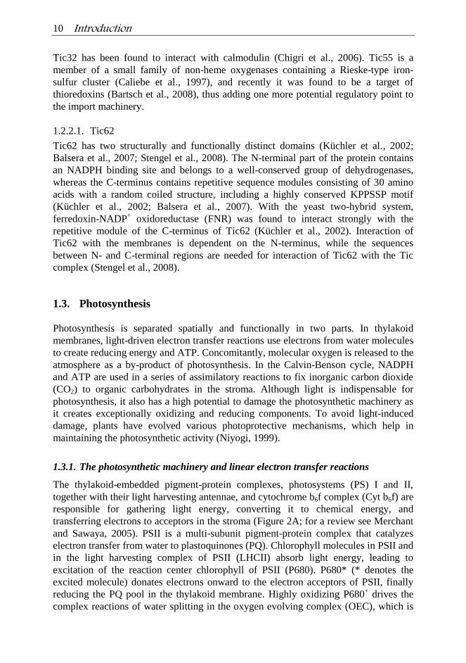
Chloroplasts are surrounded by a double lipid bilayer – an envelope composed of the
outer and the inner membrane – and they contain a specialized internal membrane
system, the thylakoid membrane, where the photosynthetic electron transfer reactions
occur (Figure 1). Thylakoid membranes are highly organized into appressed
membranes, called grana stacks, and non-appressed membranes called stroma
thylakoids, which connect the grana stacks. The soluble compartment inside the sack-
like thylakoid membranes is called the lumen (Figure 1). Most of the metabolic
reactions – e.g. photosynthetic carbon fixation reactions – take place in the stroma,
which is a soluble compartment of the chloroplasts that surrounds thylakoid
membranes (Figure 1).
Figure 1. Schematic representation of a
chloroplast. Chloroplasts are surrounded by a
double membrane, the outer and inner envelope
membranes. Thylakoid membranes are
arranged in piled grana stacks and connecting
stroma thylakoids. The soluble compartment
around the thylakoid network is called the
stroma and the soluble compartment enclosed
by thylakoid membranes is called the lumen.
During the evolution of chloroplasts, the majority of genes originating from the
bacterial ancestor of the chloroplasts were lost or transferred to the nucleus of the host
cell (Martin and Herrmann, 1998; Kleine et al., 2009). Since the circular genome of
chloroplasts, the plastome, contains less than 100 protein-coding genes and the
chloroplast proteome is estimated to consist of 3,500–4,000 polypeptides (Leister
2003), several mechanisms for re-targeting and transport of nuclear-encoded, cytosol-
translated, but chloroplast-located proteins had to be developed. Most of the nuclear-

Introduction 9
encoded chloroplast proteins are transported to chloroplasts through the general import
apparatus, which is composed of translocons of the outer and inner envelope
membranes of chloroplasts (Toc and Tic, respectively; Vothknecht and Soll, 2005).
Import-competent forms of the proteins, „preproteins‟, contain an N-terminal transit
peptide, which is needed for sorting and recognition and is cleaved off by the stromal
processing peptidase after import (Bruce, 2001).
1.2. Protein import apparatus
1.2.1. The Toc complex
At the outer envelope membrane, preproteins are recognized by the Toc complex and
import is initiated in a GTP/GDP-dependent manner (Agne and Kessler, 2009). The core
of the Toc complex is composed of three proteins: Toc159, Toc75, and Toc34 (Schnell et
al., 1997; Schleiff et al., 2003), which form a complex that is responsible for recognition
of the preprotein and its translocation across the outer envelope membrane. In addition,
two more proteins with less well known functions, Toc64 and Toc12, are associated with
the Toc complex (Sohrt and Soll, 2000; Becker et al., 2004a; 2004b).
1.2.2. The Tic complex
Although seven proteins have been identified so far in the Tic complex, the exact
composition of the complex has not yet been resolved. Tic110 is one of the most
abundant proteins in the inner envelope membrane and it is able to form a cation active
channel (Balsera et al., 2009). In addition to Tic110, Tic20 has also been predicted to
form a protein-selective channel at the inner envelope membrane (Heins, et al 2002;
Kessler and Blobel, 1996; Balsera et al., 2009; Ma et al., 1996; Kouranov and Schnell,
1997). Interaction of the preprotein with the Tic complex and translocation through the
inner envelope membrane requires high concentrations of ATP (Grossman et al., 1980).
The motor complex of Tic has been predicted to be composed of Tic40 and stromal
chaperones with ATPase activity (Chou et al., 2003). Tic22 is a peripheral subunit
located at the intermembrane face of the inner envelope membrane (Kouranov et al.,
1998). It has been shown to interact with Toc64 and Toc12 (Becker et al., 2004b),
indicating that it may be involved in promoting the contact site between the Toc and Tic
complexes, or it may have a role as a preprotein receptor at the intermembrane space.
For efficient protein import, additional regulatory subunits have evolved. Based on
their redox-active domains, Tic32, Tic55 and Tic62 have been predicted to form a
redox regulon of the Tic complex, which can sense and quickly react to signals that
give information about chloroplast status. Tic32 and Tic62 belong to an extended
family of short-chain dehydrogenases containing NADP(H)-binding domains and
showing enzymatic activity (Chigri et al., 2006; Stengel et al., 2008). The composition
of the Tic complex changes according to the stromal NADP+/NADPH ratio, as reduced
conditions dissociate Tic32 and Tic62 from the Tic complex (Chigri et al., 2006;
Stengel et al. 2008). Additionally, calcium signaling may regulate protein import since
10 Introduction
Tic32 has been found to interact with calmodulin (Chigri et al., 2006). Tic55 is a
member of a small family of non-heme oxygenases containing a Rieske-type iron-
sulfur cluster (Caliebe et al., 1997), and recently it was found to be a target of
thioredoxins (Bartsch et al., 2008), thus adding one more potential regulatory point to
the import machinery.
1.2.2.1. Tic62
Tic62 has two structurally and functionally distinct domains (Küchler et al., 2002;
Balsera et al., 2007; Stengel et al., 2008). The N-terminal part of the protein contains
an NADPH binding site and belongs to a well-conserved group of dehydrogenases,
whereas the C-terminus contains repetitive sequence modules consisting of 30 amino
acids with a random coiled structure, including a highly conserved KPPSSP motif
(Küchler et al., 2002; Balsera et al., 2007). With the yeast two-hybrid system,
ferredoxin-NADP+ oxidoreductase (FNR) was found to interact strongly with the
repetitive module of the C-terminus of Tic62 (Küchler et al., 2002). Interaction of
Tic62 with the membranes is dependent on the N-terminus, while the sequences
between N- and C-terminal regions are needed for interaction of Tic62 with the Tic
complex (Stengel et al., 2008).
1.3. Photosynthesis
Photosynthesis is separated spatially and functionally in two parts. In thylakoid
membranes, light-driven electron transfer reactions use electrons from water molecules
to create reducing energy and ATP. Concomitantly, molecular oxygen is released to the
atmosphere as a by-product of photosynthesis. In the Calvin-Benson cycle, NADPH
and ATP are used in a series of assimilatory reactions to fix inorganic carbon dioxide
(CO2) to organic carbohydrates in the stroma. Although light is indispensable for
photosynthesis, it also has a high potential to damage the photosynthetic machinery as
it creates exceptionally oxidizing and reducing components. To avoid light-induced
damage, plants have evolved various photoprotective mechanisms, which help in
maintaining the photosynthetic activity (Niyogi, 1999).
1.3.1. The photosynthetic machinery and linear electron transfer reactions
The thylakoid-embedded pigment-protein complexes, photosystems (PS) I and II,
together with their light harvesting antennae, and cytochrome b6f complex (Cyt b6f) are
responsible for gathering light energy, converting it to chemical energy, and
transferring electrons to acceptors in the stroma (Figure 2A; for a review see Merchant
and Sawaya, 2005). PSII is a multi-subunit pigment-protein complex that catalyzes
electron transfer from water to plastoquinones (PQ). Chlorophyll molecules in PSII and
in the light harvesting complex of PSII (LHCII) absorb light energy, leading to
excitation of the reaction center chlorophyll of PSII (P680). P680* (* denotes the
excited molecule) donates electrons onward to the electron acceptors of PSII, finally
reducing the PQ pool in the thylakoid membrane. Highly oxidizing P680+ drives the
complex reactions of water splitting in the oxygen evolving complex (OEC), which is
Introduction 11
located on the lumenal side of PSII (Renger and Renger, 2008). Oxidation of water
leads to a release of molecular oxygen and protons into the lumen. Electrons released
from water are used to fill the electron hole of P680+. From PSII, electrons are
transferred further via the PQ pool to the Cyt b6f complex. In the Cyt b6f complex, one
of the two electrons from fully reduced and protonated PQH2 is transferred to the small
lumenal electron carrier protein plastocyanin (PC), and the other back to an oxidized
plastoquinone in the so-called Q-cycle. PC mediates electrons to PSI where the
oxidized reaction center chlorophyll of PSI (P700+) is the electron acceptor. Electrons
in PSI are transferred from excited P700* via various electron carriers to the stromal
side of the thylakoid membrane, and are used for reduction of the small soluble protein
ferredoxin (Fd). In the last step of linear photosynthetic electron transfer reactions, the
flavoenzyme ferredoxin-NADP+ oxidoreductase (FNR) reduces NADP
+ to NADPH.
Water splitting and the action of the electron transfer reactions lead to the formation of
a proton gradient ( pH) across the thylakoid membrane, which is used to drive ATP
synthase and production of ATP (Dekker and Boekema, 2005). Photosynthetic protein
complexes are composed of nuclear- and plastome-encoded proteins, and therefore the
mechanisms for coordination of gene expression in the plastid and the nucleus are
required to maintain organelle function.
The photosynthetic protein complexes are not uniformly arranged in the thylakoid
membrane. PSII and LHCII are mainly located in grana stacks whereas PSI, LHCI, and
ATP synthase are located in stroma lamellae (Andersson and Anderson, 1980;
Danielsson et al., 2004). The Cyt b6f complex, however, is distributed more evenly
along the thylakoid membrane (Albertsson et al., 1991; Vallon et al., 1991).
1.3.2. Alternative electron transfer routes
In addition to the linear photosynthetic electron transfer reactions, cyclic and pseudo-
cyclic electron transfer reactions – coupled to ATP synthesis without net accumulation
of reducing energy – also take place in chloroplasts. The relevance and molecular
composition of these reactions is not yet clear, but the reactions could balance the
NADPH/ATP ratio to meet the metabolic demands of chloroplasts (Shikanai, 2007).
1.3.2.1. Cyclic electron transfer around PSI
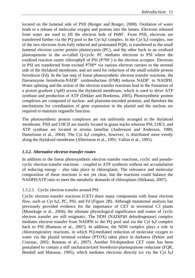
Cyclic electron transfer reactions (CET) share many components with linear electron
flow, such as Cyt b6f, PC, PSI, and Fd (Figure 2B). Although mutational analysis has
previously provided evidence for the importance of CET in terrestrial C3 plants
(Munekage et al., 2004), the ultimate physiological significance and routes of cyclic
electron transfer are still enigmatic. The NDH (NAD(P)H dehydrogenase) complex
mediates electron transfer from NADPH to the PQ pool and via the Cyt b6f complex
back to PSI (Rumeau et al., 2007). In addition, the NDH complex plays a role in
chlororespiratory reactions, in which PQ-mediated reduction of molecular oxygen to
water via the plastid terminal oxidase (PTOX) takes place in darkness (Peltier and
Cournac, 2002; Rumeau et al., 2007). Another Fd-dependent CET route has been
postulated to contain a still uncharacterized ferredoxin-plastoquinone reductase (FQR,
Bendall and Manasse, 1995), which mediates electrons directly (or via the Cyt b6f
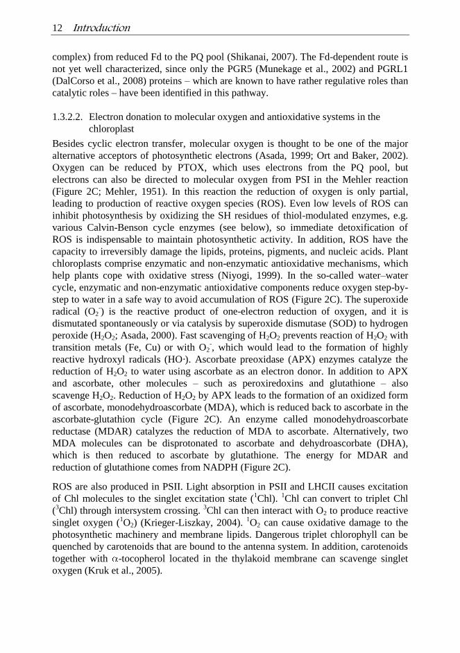
12 Introduction
complex) from reduced Fd to the PQ pool (Shikanai, 2007). The Fd-dependent route is
not yet well characterized, since only the PGR5 (Munekage et al., 2002) and PGRL1
(DalCorso et al., 2008) proteins – which are known to have rather regulative roles than
catalytic roles – have been identified in this pathway.
1.3.2.2. Electron donation to molecular oxygen and antioxidative systems in the
chloroplast
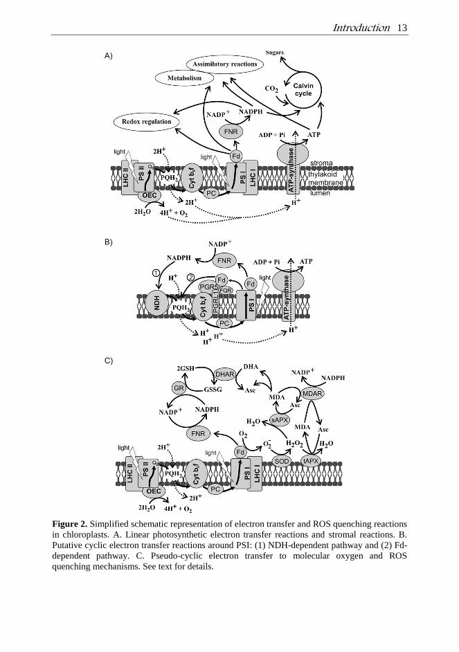
Besides cyclic electron transfer, molecular oxygen is thought to be one of the major
alternative acceptors of photosynthetic electrons (Asada, 1999; Ort and Baker, 2002).
Oxygen can be reduced by PTOX, which uses electrons from the PQ pool, but
electrons can also be directed to molecular oxygen from PSI in the Mehler reaction
(Figure 2C; Mehler, 1951). In this reaction the reduction of oxygen is only partial,
leading to production of reactive oxygen species (ROS). Even low levels of ROS can
inhibit photosynthesis by oxidizing the SH residues of thiol-modulated enzymes, e.g.
various Calvin-Benson cycle enzymes (see below), so immediate detoxification of
ROS is indispensable to maintain photosynthetic activity. In addition, ROS have the
capacity to irreversibly damage the lipids, proteins, pigments, and nucleic acids. Plant
chloroplasts comprise enzymatic and non-enzymatic antioxidative mechanisms, which
help plants cope with oxidative stress (Niyogi, 1999). In the so-called water–water
cycle, enzymatic and non-enzymatic antioxidative components reduce oxygen step-by-
step to water in a safe way to avoid accumulation of ROS (Figure 2C). The superoxide
radical (O2-) is the reactive product of one-electron reduction of oxygen, and it is
dismutated spontaneously or via catalysis by superoxide dismutase (SOD) to hydrogen
peroxide (H2O2; Asada, 2000). Fast scavenging of H2O2 prevents reaction of H2O2 with
transition metals (Fe, Cu) or with O2-, which would lead to the formation of highly
reactive hydroxyl radicals (HO·). Ascorbate preoxidase (APX) enzymes catalyze the
reduction of H2O2 to water using ascorbate as an electron donor. In addition to APX
and ascorbate, other molecules – such as peroxiredoxins and glutathione – also
scavenge H2O2. Reduction of H2O2 by APX leads to the formation of an oxidized form
of ascorbate, monodehydroascorbate (MDA), which is reduced back to ascorbate in the
ascorbate-glutathion cycle (Figure 2C). An enzyme called monodehydroascorbate
reductase (MDAR) catalyzes the reduction of MDA to ascorbate. Alternatively, two
MDA molecules can be disprotonated to ascorbate and dehydroascorbate (DHA),
which is then reduced to ascorbate by glutathione. The energy for MDAR and
reduction of glutathione comes from NADPH (Figure 2C).
ROS are also produced in PSII. Light absorption in PSII and LHCII causes excitation
of Chl molecules to the singlet excitation state (1Chl).
1Chl can convert to triplet Chl
(3Chl) through intersystem crossing.
3Chl can then interact with O2 to produce reactive
singlet oxygen (1O2) (Krieger-Liszkay, 2004).
1O2 can cause oxidative damage to the
photosynthetic machinery and membrane lipids. Dangerous triplet chlorophyll can be
quenched by carotenoids that are bound to the antenna system. In addition, carotenoids
together with -tocopherol located in the thylakoid membrane can scavenge singlet
oxygen (Kruk et al., 2005).
Introduction 13
Figure 2. Simplified schematic representation of electron transfer and ROS quenching reactions
in chloroplasts. A. Linear photosynthetic electron transfer reactions and stromal reactions. B.
Putative cyclic electron transfer reactions around PSI: (1) NDH-dependent pathway and (2) Fd-
dependent pathway. C. Pseudo-cyclic electron transfer to molecular oxygen and ROS
quenching mechanisms. See text for details.
14 Introduction
The non-photochemical quenching of chlorophyll fluorescence (NPQ) dissipates
excess excitation energy as heat in LHCII, and it is considered to be an important
photoprotective mechanism preventing the formation of singlet oxygen and
overreduction of intersystem electron carriers. Acidification of the thylakoid lumen,
due to a high proton gradient across the thylakoid membrane, promotes NPQ by
activating violaxanthin de-epoxidase enzyme (VDE) and by protonating the PsbS
protein. VDE catalyzes the conversion of violazanthin to antheraxanthin and
zeaxanthin (Niyogi et al., 1998; Li et al., 2000; 2002). In turn, protonation of PsbS
induces structural changes in the LHCII (Li et al., 2000; Kiss et al., 2008), which
accompanies quenching with the help of zeaxanthin.
1.3.3. Reductive metabolism in the stroma
The end products of the electron transfer reactions are mostly used in the Calvin-
Benson cycle. This is composed of 11 stromal enzymes that fix atmospheric carbon
dioxide (CO2) into carbon skeletons that are used directly for starch and sucrose
biosynthesis (Raines, 2003). Additionally, the reducing power from ferredoxin and
NADPH together with ATP are used in various other biosynthetic, assimilatory, and
redox reactions including fatty acid biosynthesis, nitrite and sulfate reduction, and
amino acid biosynthesis (Figure 2A).
The activity of several enzymes is regulated via redox exchange of thiol groups (S-S ↔
2SH) according to the reducing environment. Thioredoxins (Trx) are small universal
proteins containing active thiol/disulfide exchange motif that can reduce thiol groups in
enzymes. In chloroplasts, the thioredoxin-dependent regulation is closely connected to
photosynthesis, as the enzyme that reduces thioredoxins, the ferredoxin:thioredoxin
reductase (FTR; Buchanan and Balmer, 2005), accepts electrons from reduced
ferredoxin. Four enzymes of the Calvin-Benson cycle are activated through light-
dependent reduction by Trx, making Trx regulation indispensable for efficient
photosynthesis. In addition, the chloroplasts enzyme NADPH-dependent malate
dehydrogenase (MDH) is activated by Trx (Scheibe, 2004). NADP-MDH shuttles
NADPH from chloroplasts to the cytosol. The enzyme is activated in light by Trx and
inhibited by NADP+, thus switching off its own activity when all the NADPH is used
for assimilatory processes.
1.3.4. Ferredoxin
Ferredoxins (Fd) are small iron-sulfur [2Fe-2S] cluster-containing electron carrier
proteins that are important electron distributors in photosynthetic and non-
photosynthetic plastids. In photosynthetic tissue, Fd is reduced by PSI and, in addition
to NADP+ reduction via FNR, electrons from Fd can be directed to various metabolic
and signalling processes. Apart from its function in linear electron transfer reactions,
Fd has been predicted to have a role in cyclic electron transfer reactions around PSI,
and it is an important component of redox regulation via the Trx system in
chloroplasts. In addition FNR (NADPH) is capable of reducing Fd, which then donates
electrons to the reductive metabolism including nitrogen assimilation (nitrite

Introduction 15
reductase), sulphur assimilation (sulfite reductase), amino acid synthesis (Gln-
oxoglutarate amino transferase), and fatty acid synthesis (fatty acid desaturase). In
higher plants, a nuclear gene family encodes distinct leaf-type and root-type ferredoxin
isoforms, which have different redox potentials – being specific either for FNR or for
other enzymes (Bertini et al., 2002; Onda et al., 2000; Hanke et al., 2004).
1.3.5. Ferredoxin-NADP+ oxidoreductase
The enzyme ferredoxin-NADP+ oxidoreductase (FNR) harbors non-covalently bound
flavin adenine dinucleotide (FAD) as a redox center, which functions as a general
electron splitter, transferring electrons between obligatory one- and two-electron
carriers (Arakaki et al., 1997; Pschorn et al., 1988). FNR catalyzes the reversible
electron transfer reaction between two independent Fd molecules and a single molecule
of NADP(H). In plant plastids, FNR can be assigned to leaf-type (or photosynthetic;
LFNR) and root-type (or non-photosynthetic; RFNR) isoforms. LFNR catalyzes the
final step of photosynthetic electron transfer reactions reducing NADP+ to NADPH
(Shin and Arnon, 1965; Arakaki et al., 1997), whereas RFNR catalyzes the reverse
reaction, oxidizing NADPH to provide reduced Fd for various enzymes. The molecular
size of FNR is around 33 kDa and it has two structural domains (Pschorn et al., 1988).
The redox-active FAD is tightly associated with the N-terminal part of the protein
while the NADP+-binding domain is in the C-terminal part. However, based on the
crystal structure (Bruns and Karplus, 1995; Deng et al., 1999; Kurisu et al., 2001;
Dorowski et al., 2001), the tertiary structure of FNR is folded so that the FAD and
NADP+ binding sites are in close proximity. To ensure efficient electron transfer
between the substrates, Fd is also bound in close proximity to FAD and NADP+
(Martínez-Júlvez et al., 1999; Kurisu et al., 2001).
In Arabidopsis thaliana, FNR proteins are encoded by a small family of nuclear genes.
Two genes, At5g66190 and At1g20020, code for the leaf-targeted forms of FNR
(LFNR1 and LFNR2, respectively) and two genes, At4g05390 and At1g30510, code
for root plastid-targeted forms of FNR (RFNR1 and RFNR2, respectively) (Hanke et
al., 2005). In my thesis, I studied the LFNRs in Arabidopsis thaliana and the thesis will
only deal with the leaf-type FNRs. Several LFNR isoforms have been isolated and
characterized from maize, Arabidopsis and wheat. In maize leaves, three isoforms of
FNR vary in location; ZmFNR1 is restricted to the thylakoid membranes, ZmFNR3 is
a soluble enzyme in the stroma, and ZmFNR2 is present in both fractions (Okutani et
al., 2005). Hanke and co-workers (2005) identified the two leaf-targeted FNR isoforms
in green tissue of Arabidopsis plants, both of which have more than 80% identity to
maize leaf FNR. In Arabidopsis, the two leaf-targeted FNR isoforms (FNR1 and
FNR2) are present in chloroplasts in three different compartments: the thylakoid
membrane, the stroma, and the inner envelope membrane (Hanke et al., 2005; Küchler
et al., 2002). The membrane-bound pool can be divided further into tightly bound and
loosely bound pools (Matthijs et al., 1986). The loosely bound pool of FNR can be
washed out from thylakoids with salt, whereas the tightly bound pool needs detergent
treatment to dissociate from the membrane (Matthijs et al., 1986). The physiological
role of the different FNR pools is not clear, but it has been postulated that the
16 Introduction
thylakoid-bound pool of FNR is photosynthetically the most active one (Forti et al.,
1983; 1984). In wheat (Triticum aestivum) leaves, the acidic and more basic isoforms
of leaf-type FNRs are present. Post-translational modifications of the FNR isoforms
result in the formation of eight distinct spots after 2-dimensional (2-D) SDS-PAGE
(Grzyb et al., 2008a; Gummodova et al., 2007; Moolna and Bowsher, 2010). These
different wheat FNR isoforms differ in their response to physiological parameters of
chloroplast maturity, nitrogen regime, and oxidative stress (Moolna and Bowsher,
2010).
FNR is a soluble protein and it has been predicted to associate with the thylakoid
membrane by way of various protein complexes or binding proteins. Early studies
proposed the presence of a 17.5-kDa binding protein (Carrillo and Vallejos, 1982;
Vallejos et al., 1984), which forms a trimeric complex by binding two FNR subunits at
the thylakoid membrane (Ceccarelli et al., 1985). Another small 10-kDa protein, called
connectein, was found to be involved in thylakoid binding of FNR in maize (Shin et
al., 1985), but the detailed identity of these proteins has not been resolved.
Furthermore, FNR has been postulated to be connected to the thylakoid membrane via
the PsaE subunit of PSI (Andersen et al., 1992). This interaction was believed to bring
FNR in close proximity to ferredoxin, which is docked to PSI via PsaD, a subunit
located close to PsaE on the stromal side of PSI. The interaction between FNR and the
NDH complex has been suggested to allow the NDH complex to use NADPH in
chloroplasts for cyclic electron transfer (Quiles and Cuello, 1998; Guedeney et al.,
1996). Also, co-purification of FNR with the Cyt b6f complex (Zhang et al., 2001) and
association with the PGRL1 protein (DalCorso et al., 2008) are presumed to affiliate
FNR with cyclic electron transfer reactions. In addition to the proposed binding
proteins, direct association of FNR with membranes has also been suggested (Grzyb et
al., 2007b).
Aims of the Study 17
2. AIMS OF THE STUDY
Genome sequencing of Arabidopsis thaliana has revealed that several chloroplast
proteins, including FNR, are encoded by a small family of nuclear genes. The FNR
gene family encodes two highly homologous leaf-targeted FNR isoforms. In
chloroplasts, FNR is located in different pools but the functional differences between
the soluble stromal pool and the membrane-bound pool are unclear. The attachment
site of FNR at thylakoid membranes has been under intense study for decades. Despite
the fact that there have been several putative interaction candidates, the mode of
interaction has remained elusive. In my thesis work, I aimed (i) to determine whether
the two leaf-targeted FNR isoforms in Arabidopsis thaliana have specific functions in
the chloroplasts – one, for example, being involved in linear electron transfer reactions
and the other in cyclic electron transfer reactions; (ii) to clarify the functional
differences between soluble and membrane-bound pools of FNR; and (iii) to study in
more detail how the binding of FNR to the thylakoid membrane is mediated.
18 Methodological Aspects
3. METHODOLOGICAL ASPECTS
3.1. Plant material and growth conditions
Arabidopsis thaliana ecotype Columbia was used in most of the experiments.
Additionally, pea (Pisum sativum) was used for biochemical experiments in Paper III.
Transfer DNA (T-DNA) insertion mutant plants fnr1 and tic62 were purchased from
the SALK and GABI-Kat collections (Alonso et al., 2003; Rosso et al., 2003) and
RNAi silencing line fnr2 was purchased from AGRIKOLA (Hilson et al., 2004). In the
T-DNA insertion lines (fnr1a, fnr1b, tic62-1, and tic62-2), expression of the respective
gene is prevented by insertion of a foreign DNA sequence into the coding region of the
gene. In RNAi line fnr2, expression of the gene is silenced by a gene-specific RNAi
construct inserted randomly in the genome. fnr1/FNR1 fnr2(RNAi), hereafter referred
as fnr1xfnr2, was produced by crossing fnr1 and fnr2 plants and represents the F1
generation. fnr1 fnr2 double-mutant plants were screened from the segregating F2
generation. Photoautotrophic and heterotrophic growth conditions and the light
treatments of the plants are described in the respective papers.
3.2. Analysis of chloroplast proteins
Total soluble proteins and thylakoid membrane proteins were isolated according to
standard protocols described in the papers. Intact chloroplasts were isolated using
Percoll step gradients, and chloroplasts and thylakoid membranes were subfractionated
as in Paper III and references therein. Chlorophyll content of the isolated thylakoid
membranes was measured as in Porra et al. (1989) and the protein content of soluble
and membrane fractions was measured using the BioRad Protein Assay Kit. Distinct
gel electrophoresis methods were used for protein separation. Denaturating SDS-PAGE
was used to study the overall content of chloroplast proteins by staining gels with
Coomassie blue, or detecting proteins by western blotting using protein-specific
antibodies (see papers for details). For identification, proteins were separated by
isoelectric focusing according to the isoelectric point of the proteins, followed by SDS-
PAGE (Paper I). Thereafter, individual spots were identified by immunoblotting or by
mass spectrometric analysis after silver staining of the gels (Paper I). Treatment of
proteins with SDS denatures proteins and adds negative charge to unfolded
polypeptides, which allows to separation of the proteins according to their molecular
mass in SDS-PAGE. When proteins are separated in their native form, no ionic
detergent (such as SDS) is applied to protein samples and they are therefore separated
according to their native charge (isoelectric point). Native gel electrophoresis was used
to separate the FNR isoforms from each other (Papers II, III, and IV). Blue native (BN)
PAGE was used to separate protein complexes by mildly solubilizing the thylakoid
membranes with non-ionic detergent prior to gel electrophoresis (Papers II, III, and
IV). This allows to study the accumulation of thylakoid protein complexes in
chloroplasts and analysis of complex formation of the proteins of interest when
combined with immunodetection.
Methodological Aspects 19
3.3. Measurements of photosynthetic parameters
PSII performance was measured as chlorophyll a fluorescence using PAM
fluorometers (Walz) or with a Plant Efficiency Analyzer (PEA, Hansatech
Instruments). The maximum quantum efficiency of PSII was monitored as FV/FM,
excitation pressure of PSII (1-qP) as 1-[(FM′-FS)/(FM′-F0′)], and quantum yield of PSII
(Y(PSII)) as (FM′-FS)/FM′. Non-photochemical quenching (NPQ) was determined as
[FM-FM′]/FM′. Light-saturated rate of PSII oxygen evolution was measured by
Hansatech oxygen electrode using DMBQ as an electron acceptor, while PSI electron
transfer activity from DCPIPH2 to methyl viologen (MV) was measured as oxygen
consumption.
Cyclic electron transfer around PSI was estimated using three distinct methods. PAM-
101/102/103 equipped with an ED-P700DW-E emitter-detector unit or JTS-10
(Biologic) was used to monitor the redox state of PSI reaction center chlorophyll P700
as changes in absorbance at 810 nm (820 nm with JTS-10), using 860 nm as reference
wavelength (880 nm in the case of JTS-10) as reference. P700 was oxidized by 30-s FR
light illumination of the dark-adapted leaves, and the subsequent re-reduction of P700+
was monitored in darkness. The thermoluminescence AG signal was measured with the
PSI Thermoluminescence System TL200/PMT (Photon Systems Instruments). Leaf
discs were dark-adapted at +10°C for 30 s, illuminated with FR light for 60 s at the
same temperature, and the thermoluminescence signal was recorded immediately after
illumination. The transient post-illumination increase in chlorophyll fluorescence (F0
rise) was measured after turning off the actinic light as described in Allahverdiyeva et
al. (2005).
The CO2 assimilation rate of the intact plants was determined with the Ciras-1
photosynthesis system (PP Systems) at atmospheric CO2 concentration (360 ppm).
3.4. Enzyme activity measurements
FNR activity was measured as ferredoxin-dependent cytochrome C reduction assay in
the presence of NADPH (Papers III and IV). In Paper I, the relative NADP-malate
dehydrogenase (MDH) activity was measured by a spectrophotometric assay as
previously described (Scheibe and Stitt, 1988). In Papers II and IV, the ascorbate
peroxidase (APX) and glutathione reductase (GR) activities were measured as
previously described (Foyer et al., 1989), with the modifications of Pätsikkä et al.
(2002). In Papers I and II, nitrate reductase activity was measured as previously
described (Marton et al., 1982).
3.5. Pigment analysis
Chlorophyll content per leaf area was measured as described in Inskeep and Bloom
(1985). In Paper IV, chlorophylls, carotenoids, and -tocopherol were extracted from
20 Methodological Aspects
leaf discs using methanol and separated and analyzed according to Gilmore and
Yamamoto (1991) using Agilent 1100 HPLC and Agilent ChemStation software.
3.6. Transcriptional analysis
In Paper I, the Arabidopsis cDNA microarray chips based on the GEM1 clone set from
InCyte Genomics (http://www.incyte.com/) were used to study the gene expression
profiles of the plants. RNA extraction, labeling, and hybridization were performed as
in Piippo et al. (2006). For Paper II, Arabidopsis 24-K oligonucleotide arrays (MWG
Biotech, http://www.mwg-biotech.com; ArrayExpress database accession number A-
ATMX-2, http://www.ebi.ac.uk/arrayexpress) were used to study the gene expression
profiles of the plants. cDNA synthesis, labeling, hybridization, and analysis were
performed as in Kangasjärvi et al. (2008). For quantitative real-time PCR (q-RT-PCR)
the cDNA synthesis was carried out with the iScriptTM cDNA Synthesis Kit (Bio-Rad
Laboratories) and RT-PCR reactions were done using iQ SYBR Green Supermix (Bio-
Rad Laboratories). Data analysis is described in Paper II.
3.7. Structural analysis of proteins
The structural model of FNR1 and FNR2 was constructed by comparing the amino
acid sequences of Arabidopsis FNR1 and FNR2 to those of maize FNR1, FNR2, and
FNR3, with the program MALIGN (Johnson and Overington, 1993) in the Bodil
visualization and modeling package (Lehtonen et al., 2004) at the Department of
Biochemistry and Pharmacy of Åbo Akademi University, Turku, Finland. Methods are
described in more detail in Paper I. Chemical shift mapping by nuclear magnetic
resonance (NMR) spectroscopy was used to study complex formation between FNR
and the 30 amino acids repetitive C-terminal sequence of Tic62 at the Institute for
Protein Research, Osaka University and CREST, Osaka, Japan. Analytical
ultracentrifugation, atomic force microscopy, and electron microscopic images were
used in the sedimentation equilibrium measurements for analytical ultracentrifugation
(SE-AUC) to study the binding equilibrium of FNR and the C-terminus of Tic62 at the
Institute for Protein Research, Osaka University and CREST, Osaka, Japan. Methods
for NMR and AUC are described in more detail in Paper III.
3.8. Measurements of redox status of the plants
Relative ion leakage from detached rosettes was measured as described in Kangasjärvi
et al. (2008) from plants with or without pretreatment with methyl viologen (MV). MV
creates a high degree of oxidative stress by accepting electrons from PSI and donating
them further to oxygen, thus resulting in formation of reactive oxygen species. High
ion leakage from cells is a sign of oxidative stress-induced membrane damage and cell
death. Ascorbate content of leaves was determined as in Foyer et al. (1983). Oxidative
Methodological Aspects 21
modifications of thylakoid proteins were studied using the OxyBlotTM
Protein
Oxidation Detection Kit (Millipore).
3.9. Microscopic analysis of the leaf sections
Preparation and fixation of samples for light and electron microscopy were performed
as in Pätsikkä et al. (2002)
3.10. In vitro transcription, translation, and preprotein import into chloroplasts
For in vitro transcription, the coding region of the genes (FNR1, LHCB1.3, and GAP-
B) including transit peptide were cloned into the vector pSP65 (Promega) and
transcription was carried as described in Firlej-Kwoka et al. (2008). Translation was
performed using the Wheat Germ Extract Translation Kit (Promega) or the Flexi
Rabbit Reticulocyte Lysate System (Promega) in the presence of [35
S]-methionine.
Import into isolated chloroplasts was performed in the presence of ATP and [35
S]-
labeled translation products at 25°C.
3.11. Protein interaction studies
DNA sequences corresponding to leaf-targeted FNR isoforms from Arabidopsis (Paper
IV) and pea (Paper III), and to FBPase and C-terminus of AtTic62 from Arabidopsis,
were cloned and used for transformation of (and expression in) Escherichia coli. The
proteins were purified via their polyhistidine tags using Ni-NTA Sepharose (GE
Healthcare) and eluted with imidazole. Proteins were then concentrated and the buffer
was exchanged by dialysis prior to analysis.
Ectopically expressed and purified proteins were bound via their N- or C-terminal His-
tag to Ni-NTA Sepharose blocked with egg albumin. [35
S]-Met labeled translation
product of FNR (Paper IV) or concentrated stroma from tic62 Arabidopsis plants
(Paper III) were loaded onto columns. After incubation and subsequent washing, the
proteins were eluted with imidazole (Paper IV). In Paper III, the proteins were eluted
by sequential addition of salt, urea, and imidazole at increasing molarities. Eluted
fractions were separated by SDS-PAGE and the proteins were detected either by
autoradiography (Paper IV) or by immunoblotting (Paper III).
22 Results
4. RESULTS
4.1. FNR isoforms in Arabidopsis chloroplasts
Two leaf-targeted FNR isoforms were detected from the thylakoid membrane of wild-
type Arabidopsis as four protein spots after isoelectric focusing (IEF) followed by
SDS-PAGE. Two spots with an isoelectric point (pI) of around 5 were identified as
LFNR1, and two spots with a more basic pI were identified as LFNR2 by MALDI-
TOF mass spectrometric analysis (Paper I). Apart from thylakoid membranes, the leaf-
targeted FNR could also be found in the stroma (Paper I) and attached to the inner
envelope membrane (Paper III). Leaf-targeted FNR isoforms have almost identical
molecular mass, but the difference in pI allows separation of the FNR1 and FNR2
isoforms by native gel electrophoresis, and the presence of both isoforms in stroma and
at thylakoid membranes could therefore be shown. The incomplete solubilization of the
proteins did, however, prevent quantitative comparison of the isoforms (Paper II). The
transcriptional analysis of FNR pool sizes from wild-type leaves by q-RT-PCR
revealed that under standard growth conditions, the FNR1 transcripts constitute 68% of
the total FNR mRNA pool and FNR2 transcripts constitute only 32% (Paper II).
Separation of thylakoid protein complexes by blue native (BN) PAGE revealed that the
thylakoid-bound FNR is present in several high molecular weight (HMW) complexes
of around 200–500 kDa (Papers II and III).
4.2. Characterization of the mutant plants containing different amounts of FNR
In my thesis work, three Arabidopsis single-mutant plant lines were characterized. fnr1
mutants are devoid of FNR1 protein, as the FNR1 gene is interrupted by T-DNA
insertion (Paper I). The expression of the other isoform, FNR2, was unchanged in the
fnr1 mutant plants, and consequently the total amount of FNR was clearly reduced
when compared to wild-type plants (Papers I and II). In fnr2 mutant plants, the total
amount of FNR was also clearly smaller than in wild-type plants, and the RNAi
construct of FNR2 silenced specifically the expression of the FNR2 gene, resulting in
the loss of FNR2 protein with no marked downregulation (or upregulation) of the
FNR1 gene (Paper II). The third single-mutant plant, T-DNA insertion line tic62,
lacking the Tic62 protein, was also characterized (Paper III). The amount of both leaf-
targeted FNR isoforms was equally downregulated in the tic62 mutant plants. In
addition, double mutants from the crossing of fnr1 and fnr2 were characterized (Paper
IV). The F1 generation, genotypically double heterozygous plants (hereafter referred as
fnr1xfnr2) expressed only FNR1 protein, although in smaller quantity than the fnr2
single mutant. These results indicate that one RNAi construct of FNR2 is able to totally
silence the expression of FNR2 and that one functional gene of FNR1 is not able to
produce the protein at wild-type levels. Mutant plants completely devoid of leaf-type
FNR (fnr1 fnr2), segregated from the F2 generation, could also be obtained (Paper IV).
Results 23
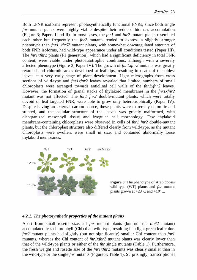
Both LFNR isoforms represent photosynthetically functional FNRs, since both single
fnr mutant plants were highly viable despite their reduced biomass accumulation
(Figure 3; Papers I and II). In most cases, the fnr1 and fnr2 mutant plants resembled
each other but frequently the fnr2 mutants tended to express a slightly stronger
phenotype than fnr1. tic62 mutant plants, with somewhat downregulated amounts of
both FNR isoforms, had wild-type appearance under all conditions tested (Paper III).
The fnr1xfnr2 plants (F1 generation), which had a significant deficiency in total FNR
content, were viable under photoautotrophic conditions, although with a severely
affected phenotype (Figure 3; Paper IV). The growth of fnr1xfnr2 mutants was greatly
retarded and chlorotic areas developed at leaf tips, resulting in death of the oldest
leaves at a very early stage of plant development. Light micrographs from cross
sections of wild-type and fnr1xfnr2 leaves revealed that limited numbers of small
chloroplasts were arranged towards anticlinal cell walls of the fnr1xfnr2 leaves.
However, the formation of granal stacks of thylakoid membranes in the fnr1xfnr2
mutant was not affected. The fnr1 fnr2 double-mutant plants, which were totally
devoid of leaf-targeted FNR, were able to grow only heterotrophically (Paper IV).
Despite having an external carbon source, these plants were extremely chlorotic and
stunted, and the cellular structure of the leaves was greatly malformed, with
disorganized mesophyll tissue and irregular cell morphology. Few thylakoid
membrane-containing chloroplasts were observed in cells of fnr1 fnr2 double-mutant
plants, but the chloroplast structure also differed clearly from wild-type, as the mutant
chloroplasts were swollen, were small in size, and contained abnormally loose
thylakoid membranes.
Figure 3. The phenotype of Arabidopsis
wild-type (WT) plants and fnr mutant
plants grown at +23°C and +10°C.
4.2.1. The photosynthetic properties of the mutant plants
Apart from small rosette size, all fnr mutant plants (but not the tic62 mutant)
accumulated less chlorophyll (Chl) than wild-type, resulting in a light green leaf color.
fnr2 mutant plants had slightly (but not significantly) smaller Chl content than fnr1
mutants, whereas the Chl content of fnr1xfnr2 mutant plants was clearly lower than
that of the wild-type plants or either of the fnr single mutants (Table 1). Furthermore,
the fresh weight and rosette size of the fnr1xfnr2 mutants was clearly smaller than in
the wild-type or the single fnr mutants (Figure 3; Table 1). Surprisingly, transcriptional
24 Results
analysis revealed upregulation of photosynthetic genes in the single fnr mutant plants,
such as those encoding the light harvesting antenna proteins of PSI and PSII. Genes
encoding the small subunit of Rubisco, and a gene encoding plastocyanin protein were
also upregulated in the fnr1 and fnr2 mutants relative to wild-type plants (Papers I and
II). However, in line with the low chlorophyll content, photosynthetic pigment-protein
complexes of all fnr mutant plants were downregulated, and less D1, PsaD, Lhcb1, and
Lhcb2 accumulated in thylakoid membranes of the mutant plants than in wild-type
plants (Table 1). Again, the accumulation of thylakoid proteins was most affected in
the fnr1xfnr2 mutant, although no differences in accumulation of ATP synthase
subunits (CF1 or ATP- ) were detected between the wild-type and mutant plants
(Papers I, II, and IV). Moreover, even though the amount of photosynthetic proteins
was extremely low in fnr1 fnr2 double-mutant plants, no such reduction in ATP
synthase was observed (Paper IV). In line with the low accumulation of protein
subunits of photosynthetic pigment-protein complexes, also small changes in the
composition of entire, functional protein complexes were observed, as PSII and LHCII
were found to assemble in smaller complexes in fnr1 and fnr2 mutant plants than in
wild-type plants (Paper II).
The capacity of PSII in the mutant plants (fnr and tic62) was not different from that of
the wild-type plants, although the PSII quantum yield (Y(PSII)) was slightly lower in
the fnr1xfnr2 mutant plants than in wild-type plants. However, when the susceptibility
of PSII to high light irradiance was studied, clearly faster photoinhibition of PSII was
detected in the fnr1xfnr2 mutants, both in the presence and absence of lincomycin,
which is a specific inhibitor of chloroplast protein translation (Paper IV). During high
light illumination, the D1 protein in thylakoid membranes was also quickly degraded.
Intriguingly, recovery from photoinhibition was equally fast in wild-type plants and
fnr1xfnr2 mutant plants. Although the size of the PQ pool and the excitation pressure
of PSII (1-qP), which can be used to estimate the redox-state of the PQ pool, were
smaller in the fnr1 single mutant than in wild-type plants (Paper I), the PQ pool of the
fnr1xfnr2 mutant plants was more reduced than in wild-type plants (Paper IV). In
addition, fnr1xfnr2 mutants had a higher capacity for non-photochemical quenching
(NPQ) under high actinic light illumination compared to wild-type plants (Paper IV).
In line with increased NPQ, the de-epoxidation state of xanthophyl cycle pigments in
fnr1xfnr2 mutant plants was high, as the mutant plants accumulated high content of
zeaxanthin even under standard growth conditions (Paper IV). Although the tic62
mutants did not show any difference in Y(PSII), 1-qP, NPQ, or in accumulation of
photosynthetic proteins when compared to wild-type plants, TIC62 did show high co-
expression with a number of photosynthetic genes (Paper III).
4.2.2. Alternative electron transfer reactions in the fnr mutant plants
No generally applicable methods to measure cyclic electron transfer pathways around
PSI have been presented so far. In this thesis, three different methods were used to
estimate the cyclic electron transfer capacity of the plants. First, the rate of cyclic
electron flow was determined from the kinetics of absorbance changes at 810 nm
resulting from P700+ re-reduction in darkness after far red illumination. Both fnr single
Results 25
mutants showed slower decay of P700+ than the wild-type plants when grown under
standard growth conditions (Paper I, data not shown for fnr2). Two other methods
applied were the transient post-illumination increase in chlorophyll a fluorescence and
the far-red light-induced thermoluminescence afterglow band, both of which have been
suggested to measure mostly the NDH-dependent cyclic electron flow (Shikanai et al.,
1998; Havaux et al., 2005a). Both latter methods gave results similar to that of P700+
re-reduction, revealing reduced amplitudes of the representative curves (Paper II).
These results showed that the single fnr mutant plants had a reduced ability to perform
cyclic electron transfer. However, when the fnr1 and fnr2 single-mutant plants were
grown under low-temperature conditions (+10°C), a faster decay in P700+ re-reduction
was observed compared to wild-type plants (Paper II). Similarly, in fnr1xfnr2 mutant
plants, the cyclic electron transfer – measured as P700+ re-reduction – was found to be
faster than in wild-type plants even under standard growth conditions (Paper IV).
Moreover, the fnr1xfnr2 mutant plants accumulated high levels of PGRL1 protein,
which has been shown to have a role in Fd-dependent cyclic electron transfer reactions
(DalCarso et al., 2008). No difference in P700+ re-reduction or transient post-
illumination increase in chlorophyll fluorescence was observed between tic62 mutant
and wild-type plants (Paper III).
Besides upregulated cyclic electron transfer reactions, other alternative electron
acceptors were also upregulated in fnr mutant plants. Transcript profiling of fnr1 and
fnr2 plants revealed upregulation of genes encoding nitrate reductase (NR), and several
nitrate transporters, and glutamine synthases (Papers I and II), which use high amounts
of reducing energy. Transcriptional upregulation was reflected in later steps of the
metabolism and more nitrite accumulated in fnr1 and fnr2 leaves than in wild-type
leaves, indicating an increased amount or activity of NR (Papers I and II). Based on the
chlorotic phenotype of the fnr mutant plants and upregulation of plastid terminal
oxidase (PTOX) in fnr1xfnr2 mutants (Paper IV), the direction of photosynthetic
electrons to molecular oxygen via PSI or the PQ pool appeared to be increased in the
mutant plants. In single-mutant fnr plants, the capacity to cope with sudden exposure to
oxidative stress was lower and more ions leaked from intact leaves after methyl
viologen- (MV-) induced oxidative stress than from wild-type leaves (Paper II). In
fnr1xfnr2 mutant plants, an extensive ion leakage from leaves was evident even under
steady-state conditions without any additional induction of oxidative stress (Paper IV).
4.2.3. Oxidative stress and the antioxidative properties of the mutant plants
In paper II, the acclimation of single-mutant fnr plants to oxidative stress and low-
temperature growth conditions was investigated. Under standard growth conditions, in
addition to increased ion leakage, single fnr mutant plants had higher levels of oxidized
thylakoid proteins than wild-type plants. Acclimation of wild-type and single fnr
mutants to low growth temperature concomitantly led to an increased tolerance to
oxidative stress. Under low temperature conditions, only fnr2 plants had a visual
phenotype, low Chl content, and low accumulation of photosynthetic proteins, while
fnr1 plants had no distinct phenotype. In addition, under low temperature fnr2 plants
had increased tolerance to oxidative stress, as demonstrated by lower ion leakage from

26 Results
cells compared to wild-type and fnr1 plants. Moreover, low-temperature grown fnr2
mutant plants contained less oxidized thylakoid proteins and accumulated more
antioxidative enzymes compared to wild-type plants and fnr1 mutant plants. The
enhanced tolerance of low-temperature grown fnr2 mutants to oxidative stress was
reflected in the stromal redox status, since in the mutant leaves the ascorbate pool was
in a more reduced state than in wild-type leaves.
The fnr1xfnr2 mutants differed from single fnr mutants in terms of protection against
oxidative stress (Paper IV). Already, the visual phenotype of the fnr1xfnr2 mutant
plants – the pale green color, stunted growth, and death of the oldest leaves at a very
early state – indicated problems in the ability to cope with increased accumulation of
ROS. Not only the upregulation of alternative electron acceptors (e.g. CET and O2) but
also the induction of various antioxidative mechanisms in fnr1xfnr2 plants was quite
obvious. More APX enzymes accumulated in the fnr1xfnr2 chloroplasts, although no
differences in total activity of APX or GR were detected compared to wild-type
chloroplasts. Moreover, fnr1xfnr2 mutant plants accumulated high relative contents of
-tocopherol and xanthophyll cycle pigments displaying high de-epoxidation state. In
fnr1xfnr2 mutant plants, a high content of zeaxanthin was detected even under standard
growth conditions; yet, the xanthophyll cycle responded normally to changes in light
intensity.
Table 1. Summary of the phenotype of fnr mutant plants grown at +23°C and +10°C.
100 µmol photons m-2
s-1
, +23°C 100 µmol photons m-2
s-1, +10°C
fnr1 fnr2 fnr1xfnr2 fnr1 fnr2
Chl - - - - +/- -
Photosynthetic proteins - - - - +/- -
Excitation pressure - n.a. + n.a. n.a.
Cyclic electron transfer - - + + +
Relative ion leakage + + ++ +/- - + parameter is upregulated in the mutant plant indicated, as compared to wild type; - parameter is
downregulated in the mutant plant relative to wild type; +/- no difference in parameter from wild type; a
double symbol indicates up- or downregulation of the parameter in the mutant plant compared to wild-type
and other fnr mutants; n.a. data not available.
4.3. FNR forms dimers and high molecular weight complexes at thylakoid membranes
As mentioned above, both FNR isoforms are found in the membrane and in soluble
pool in wild-type chloroplasts. On the other hand, fnr1 mutant plants have the FNR2
protein only in the soluble pool, while the FNR1 in fnr2 and fnr1xfnr2 mutant plants is
distributed between both the thylakoid pool and the stromal pool of chloroplasts
(Papers I, II, and IV). These results indicate that the FNR1 protein is required for
thylakoid attachment of the FNR2 protein, most probably through (hetero)dimer
formation. Crystal structures of Anabaena FNR and ZmLFNR1 from maize have been
solved both in the “free state” and in complex with Fd (Kurisu et al., 2001; Morales et
al., 2000; Serre et al., 1996). Both of these complexes contain two FNRs and one Fd.
Results 27
We found that the other FNR, predicted to be in the “free state” in the maize structure,
was able to associate with the FNR-Fd complex similarly to Anabaena FNR (Paper I;
Kurisu et al., 2001). The ZmFNR dimer in complex with Fd was then used as a
template when the structural model of the Arabidopsis FNR dimer was constructed.
Computational analysis of the FNR proteins revealed that there were no structural
hindrances that would prevent the formation of either homodimers or herterodimers of
Arabidopsis FNR1 and FNR2 (Paper I). In paper IV, the dimer formation was tested by
in vitro binding assay, in which either of the heterologously expressed and purified
Arabidopsis FNR proteins was bound via its (His)6-tag to nickel affinity matrix
together with the in vitro translated [35
S]-labeled FNR isoform. The analysis of elution
fractions revealed that, in line with the structural model, both homodimeric and
heterodimeric associations in all combinations of FNR isoforms were equally possible,
although very weak. The in vitro activities of FNR1 and FNR2, measured as Cyt C
reduction assays, revealed that FNR1 was more active than FNR2 but there were no
apparent difference in activity between monomeric/homodimeric and heterodimeric
enzyme (Paper IV).
Not only FNR dimers but also larger protein complexes containing FNR were present
in wild-type thylakoid membrane (Papers II, III, and IV). These complexes did not
show co-migration with PSI, Cyt b6f, or NDH complexes in BN-PAGE (Paper III),
although in earlier studies these protein complexes were predicted to act as thylakoid
platforms for FNR. In Paper III, Tic62 was shown to be present both at the inner
envelope membrane and in the stroma, and additionally at the thylakoid membrane –
and indeed, FNR was shown to have a highly similar migration pattern to that of Tic62
in BN-PAGE.
4.3.1. Interaction of Tic62 and FNR at the thylakoid membrane
When wild-type and tic62 chloroplasts were fractionated into stroma and the envelope
and thylakoid membranes, it was obvious that the amount of FNR was smaller in the
membrane fractions of tic62 than in those of the wild-type chloroplasts (Paper III).
Further fractionation of thylakoid membranes by differential centrifugation to grana
thylakoids, grana margins, and stroma lamellae revealed that in wild-type chloroplasts,
both Tic62 and FNR are enriched in stroma lamellae although FNR in particular is
distributed in other thylakoid fractions as well. In tic62 chloroplasts, only small amount
of FNR was present in the stroma lamellae fraction. The amount of FNR in tic62
stroma was, however, at levels similar to those in wild-type stroma.
The fnr mutants were found to have a reciprocal molecular phenotype to that in tic62
as in fnr1 and fnr2 mutants the total amount of Tic62 was reduced. Moreover, the
formation of FNR and Tic62 high molecular weight (HMW) complexes at the
thylakoid membrane was prevented in fnr1, fnr2, and tic62 mutant plants, indicating
that there is a very specific interaction between these proteins (Paper III). Freely
running FNR was, however, detected in the lower part of BN gels in wild-type, tic62,
and fnr2 plants, indicating that FNR has the ability to bind to the thylakoid membrane
also independently of Tic62. To test whether the mutant plants lacking one of the
predicted FNR-interacting proteins would have differential accumulation of thylakoid-
28 Results
bound FNR, the thylakoid proteins were applied to BN-PAGE. The mutant plants
lacking PsaE, NdhO, or PGR5 protein did not show any changes in patterns of FNR in
BN-PAGE compared to wild-type plants (Paper IV). However, the mutant plants
lacking the newly discovered FNR binding protein TROL (Juric et al. 2009) lacked one
of the smallest FNR complexes, which was different from FNR-Tic62 complexes
(Paper IV).
4.3.2. Tic62 binds FNR in a high-salt insensitive manner at a site of FNR that is
distinct from its catalytically important regions
The interaction between Tic62 and FNR was studied in Paper III. After high ionic
strength washing of wild-type and tic62 thylakoid membranes, it was obvious that FNR
is bound more strongly to membranes in the presence of Tic62. Moreover, in vitro
binding assays revealed that there was a strong interaction between Tic62 and FNR,
since most of the FNR bound to His-tagged Tic62 could be eluted from Ni2+
matrix
only by denaturation with urea.
Chemical shift mapping by nuclear magnetic resonance (NMR) spectroscopy together
with analytical ultracentrifugation analysis at sedimentation equilibrium (AUC-SE)
was used to investigate the interaction between FNR and Tic62. The C-terminal 30-
amino acid repeat of Tic62 (R1 peptide) – which is known to be the specific FNR
binding motif (Küchler et al. 2002) – instead of whole-length Tic62 was used in the
studies because the high molecular mass of molecules often hampers the resolution and
sensitivity of resonance. A number of spectra of uniformly labeled FNR changed
significantly upon addition of R1 and strikingly many peaks of free FNR disappeared.
At the same time, new peaks appeared at different resonance positions with strong
binding mode. Chemical shift perturbations resulting from addition of R1 peptide were
mapped onto the tertiary structure of FNR. The residues affected belonged to a large
area on only one side of FNR, which was located on the side opposite to the Fd and
NADP+ binding areas. AUC-SE analysis revealed that FNR and R1 peptide form a
complex with a stoichiometry of about 2:1. However, steric hindrances do not allow
full saturation of FNR binding sites in the C-terminal part of Tic62, which contains
several repeats depending on plant species (Balsera et al., 2007).
4.3.3. Regulation of Tic62-FNR complex formation
Regulation of formation of the Tic62-FNR complex at the thylakoid membrane was
investigated in Paper III. It was found that the complex formation between FNR and
Tic62 is regulated by light despite the total lack of any photosynthetic phenotype of
tic62 plants. At wild-type thylakoid membranes isolated from dark-adapted leaves,
FNR was strongly present in HMW complexes together with Tic62. Light treatment of
the leaves resulted in detachment of FNR from HMW complexes at the thylakoid
membrane. Similarly, the amount of Tic62 also became reduced in HMW complexes
but the effect was not as intense as for FNR. In order to mimic pH changes during the
dark-to-light transition, the thylakoids were isolated from dark-adapted leaves and the
pH was adjusted to different values. At pH 6, which mimics the stromal environment in
Results 29
darkness, most of the Tic62 and FNR was present in the membrane fraction. Treatment
of thylakoids with more alkaline pH released a proportion of Tic62 and FNR to the
soluble fraction. Again, FNR was more easily dissolved from the thylakoid membrane
than Tic62. Accordingly, the catalytic activity of heterologously expressed and purified
FNR was measured in vitro in the presence and absence of varying amounts of
recombinant Tic62 protein. The activity measured after overnight preincubation
revealed a loss of most of the activity from samples containing only FNR, whereas the
presence of full-length Tic62 or its C-terminus specifically retained most of the FNR
activity. These results indicate that Tic62 is able to stabilize FNR.
30 Discussion
5. DISCUSSION
5.1. Both FNR isoforms are functional, and partially redundant
The triple location of FNR in the stroma, at the thylakoid membranes, and at the inner
envelope membrane was confirmed in this thesis work. As revealed in Paper I, both
Arabidopsis isoforms at the thylakoid membrane are represented by two separate spots
after 2-D gel electrophoresis. These protein spots corresponded to similar molecular
masses, but there was a small shift in pI. This suggests that proteins undergo post-
translational modifications; such changes are known to induce changes in the net
charge of proteins. In wheat, the acidic and more basic isoforms of leaf-type FNRs are
post-translationally modified by alternative N-terminal cleavage and/or by
phosphorylation to form eight distinct isoforms in total (Gummadova et al., 2007;
Moolna and Bowsher, 2010). Four putative phosphorylation sites on the surfaces of
Arabidopsis FNR isoforms were predicted from structural modeling (Paper I). Moolna
and Bowsher (2010) predicted slightly different phosphorylation pattern of Arabidopsis
FNR based on the conservation of predicted phosphorylation sites between wheat and
Arabidopsis FNRs. However, no evidence of in vivo phosphorylation of Arabidopsis or
wheat FNR has been observed so far.
The high viability of both single fnr mutant plants indicates that both isoforms are
functional and photosynthetically active. However, the phenotypic deficiencies of the
single fnr mutant plants indicate that both isoforms are needed to guarantee efficient
photosynthesis (Papers I and II). Mutant plants lacking the FNR2 gene product and
containing only a small quantity of FNR1 protein (fnr1xfnr2) are also viable under
photoautotrophic growth conditions, whereas the mutant plants that are completely
devoid of FNR are not able to grow autotrophically due to a serious lack of NADPH
(Paper IV).
5.2. Lack of terminal photosynthetic electron acceptor leads to downregulation of the photosynthetic machinery
Downregulation of photosynthetic pigments and pigment-protein complexes in single
and double fnr mutant plants indicates that depletion of FNR leads to downregulation
of the entire photosynthetic machinery to avoid excess chlorophyll excitation,
overreduction of the electron transfer chain, and production of ROS. The amount of
FNR has been shown to be proportional to the degree of downregulation of
photosynthetic protein complexes (Hajirezaei et al., 2002) and mutant plants that are
totally devoid of FNR accumulate barely detectable amounts of photosynthetic
thylakoid proteins (Paper IV).
Particularly in the early literature, it was emphasized that the thylakoid pool of FNR is
the photosynthetically active one, whereas in the stroma the FNR pool would catalyze
a reverse reaction towards reduction of ferredoxin (Forti et al., 1983; Forti and Bracale,
Discussion 31
1984). Our results, however, clearly undercut these assumptions, as fnr1 mutant plants
– totally devoid of the thylakoid-bound pool of FNR – are highly viable (Papers I and
II). Moreover, tic62 mutant plants with a clear defect in the membrane-bound pool of
FNR show no photosynthetic phenotype (Paper III). Accumulation of FNR at the
thylakoid membrane in darkness and release of the protein to the stroma under
illumination rather indicates that most of the thylakoid-bound pool of FNR is
photosynthetically inactive (Paper III).
5.3. Alternative electron transfer reactions are upregulated when the amount of FNR limits linear electron flow
Despite the fact that there has been intense research in the field, the role of FNR in
cyclic electron transfer reactions has remained controversial. Discovery of the NDH
complex from the thylakoid membrane of higher plant chloroplasts, and its co-
migration with FNR in native gels indicated that FNR may function as an electron
input unit for the NDH complex (Quiles and Cuello, 1998; Guedeney et al., 1996).
However, the activity of NDH complex has been shown to be independent of FNR
activity (Teicher and Scheller, 1998; Endo et al., 1997). Despite the discovery of
multiple novel subunits in the NDH complex (see Suorsa et al., 2009 for a review), the
electron input unit has not been identified. More recent publications have indicated the
formation of cyclic electron transfer specific supercomplexes. NDH complex has been
shown to form a supercomplex with PSI in plant chloroplasts (Peng et al., 2008; Sirpiö
et al., 2009), and this was hypothesized to be the CET-acting form of NDH complex.
However, the presence of FNR in this complex has not been proven and recently there
have been speculations about electron donors other than NAD(P)H for NDH, such as
Fd.
It has been suggested that FNR might function in cyclic electron transfer reactions,
acting as a still uncharacterized ferredoxin-plastoquinone reductase (FQR). Firstly,
FNR has been shown to have diaphorase activity (Bojko and Wieckowski, 1995;
Melamed-Harel et al., 1984), and it has been shown to be able to donate electrons to
the plastoquinone pool under in vitro conditions (Bojko et al., 2003). Secondly, FNR
has been co-purified with the Cyt b6f complex (Okutani et al., 2005; Zhang et al.,
2001), suggesting the formation of a speculative supercomplex containing PSI, Cyt b6f
complex, and plastocyanin (Joliot and Joliot, 2002). This complex has, however,
remained hypothetical due to the lack of biochemical identification until recently.
Interaction of PGR5 with Fd, Cyt b6, and PGRL1, and interaction of PGRL1
additionally with PsaD, FNR1, and FNR2 in the yeast 2-hybrid system and split-
ubiquitine assays, caused authors to propose that a CET supercomplex containing PSI,
Cyt b6f, PGRL1, PGR5, Fd, and FNR (and predicted FQR) may indeed exist (DalCorso
et al., 2008). However, such a complex has not been identified in plants. In addition,
loss of PGR5 or PGRL1 did not affect the accumulation of PSI, Cyt b6f, Fd ,or FNR;
moreover, PGR5 and PGRL1 accumulated independently of each of the proteins and
protein complexes previously mentioned (DalCorso et al., 2008). This means that Fd-
dependent CET is able to operate without the formation of such a supercomplex, and/or
32 Discussion
that the complex is only transient. Very recently, a CET-dependent supercomplex
containing PSI-LHCI, LHCII, Cyt b6f, FNR, and PGRL1 in near stoichiometric
amounts was isolated from the unicellular green alga Chlamydomonas reinhardtii (Iwai
et al., 2010). In Chlamydomonas, the induction of CET is closely connected to state
transition (Rochaix, 2007), an event in which excitation energy is balanced by physical
rearrangement of LHCII between PSII and PSI (Kargul and Barber, 2008). This
isolated CET complex was shown to be able to mediate electron transfer between PSI
and Cyt b6f under state 2 conditions, indicating CET activity of the supercomplex.
When the amount of FNR is only partially limiting (as in the fnr1, fnr2, and tic62
mutants), there is only minor downregulation or no change in cyclic electron transfer
reactions (Papers I, II, and III). Altered CET capacity is most probably a secondary
effect resulting from changes in the stromal redox state in fnr mutant plants. However,
in the fnr1xfnr2 mutant the content of the terminal acceptor clearly limits the linear
electron transfer, and the reducing energy is directed to alternative electron transfer
reactions, e.g. to Fd-dependent cyclic electron transfer and molecular oxygen via
PTOX or via PSI in the Mehler reaction, to prevent major over-reduction of the inter-
system electron carriers and production of ROS (Paper IV; see section 5.4). The same
phenomenon can be seen when single fnr mutant plants grow under low-temperature
conditions (Paper II). Most probably, the changes in the stromal redox state and an
inability of ferredoxin to direct electrons to NADP+ reduction constitute the driving
force for the upregulation of cyclic electron transfer reactions in the fnr mutants.
LFNR isoforms can be divided into acidic and more basic forms, which show different
responses to the nitrogen regime. Since FNR2 is expressed at high levels under high
nitrate content in growth media, it is conceivable that FNR2 provides the additional
reducing power required for assimilation of nitrate (Hanke et al., 2005). In Papers I and
II, the upregulation of genes encoding enzymes involved in nitrogen metabolism was
detected in both fnr mutant plants. Nitrate reductase activity is also increased in mutant
plants relative to wild-type plants. In the cytosol, NR reduces nitrate to nitrite – which
is a toxic compound, and is immediately imported to plastids where Fd-dependent
nitrite reductase reduces it to ammonium. Ammonium fixation into amino acids is then
performed by Fd-dependent glutamate synthase. Although nitrogen fixation in
chloroplasts mostly uses Fd as a reducing power, assimilation of nitrate requires export
of NADPH from chloroplasts to the cytosol via the malate shuttle. Low activity of
NADPH-MDH in fnr1 mutant plants (Paper I) is most probably due to an increased
NADP+/NADPH ratio, as it is known that a high content of NADP
+ reduces the activity
of MDH by a feedback mechanism (Scheibe, 1991). Altogether, it is conceivable that
the export of reductants to the cytosol is depressed due to deficiency in FNR which
raises the question of how the energy needed to run NR in the cytosol is guaranteed in
fnr mutant plants.
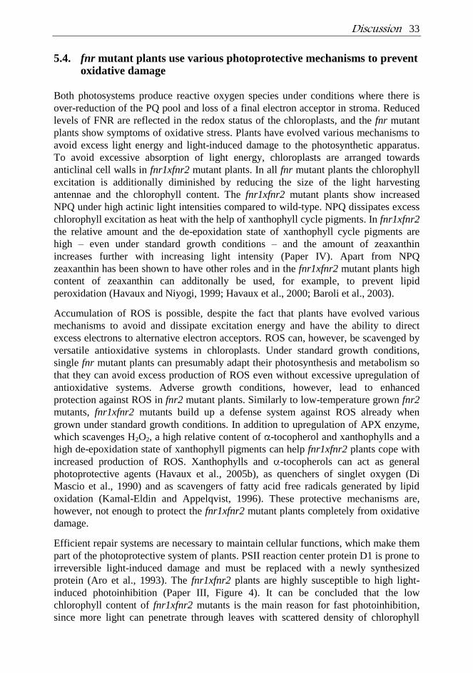
Discussion 33
5.4. fnr mutant plants use various photoprotective mechanisms to prevent oxidative damage
Both photosystems produce reactive oxygen species under conditions where there is
over-reduction of the PQ pool and loss of a final electron acceptor in stroma. Reduced
levels of FNR are reflected in the redox status of the chloroplasts, and the fnr mutant
plants show symptoms of oxidative stress. Plants have evolved various mechanisms to
avoid excess light energy and light-induced damage to the photosynthetic apparatus.
To avoid excessive absorption of light energy, chloroplasts are arranged towards
anticlinal cell walls in fnr1xfnr2 mutant plants. In all fnr mutant plants the chlorophyll
excitation is additionally diminished by reducing the size of the light harvesting
antennae and the chlorophyll content. The fnr1xfnr2 mutant plants show increased
NPQ under high actinic light intensities compared to wild-type. NPQ dissipates excess
chlorophyll excitation as heat with the help of xanthophyll cycle pigments. In fnr1xfnr2
the relative amount and the de-epoxidation state of xanthophyll cycle pigments are
high – even under standard growth conditions – and the amount of zeaxanthin
increases further with increasing light intensity (Paper IV). Apart from NPQ
zeaxanthin has been shown to have other roles and in the fnr1xfnr2 mutant plants high
content of zeaxanthin can additonally be used, for example, to prevent lipid
peroxidation (Havaux and Niyogi, 1999; Havaux et al., 2000; Baroli et al., 2003).
Accumulation of ROS is possible, despite the fact that plants have evolved various
mechanisms to avoid and dissipate excitation energy and have the ability to direct
excess electrons to alternative electron acceptors. ROS can, however, be scavenged by
versatile antioxidative systems in chloroplasts. Under standard growth conditions,
single fnr mutant plants can presumably adapt their photosynthesis and metabolism so
that they can avoid excess production of ROS even without excessive upregulation of
antioxidative systems. Adverse growth conditions, however, lead to enhanced
protection against ROS in fnr2 mutant plants. Similarly to low-temperature grown fnr2
mutants, fnr1xfnr2 mutants build up a defense system against ROS already when
grown under standard growth conditions. In addition to upregulation of APX enzyme,
which scavenges H2O2, a high relative content of -tocopherol and xanthophylls and a
high de-epoxidation state of xanthophyll pigments can help fnr1xfnr2 plants cope with
increased production of ROS. Xanthophylls and -tocopherols can act as general
photoprotective agents (Havaux et al., 2005b), as quenchers of singlet oxygen (Di
Mascio et al., 1990) and as scavengers of fatty acid free radicals generated by lipid
oxidation (Kamal-Eldin and Appelqvist, 1996). These protective mechanisms are,
however, not enough to protect the fnr1xfnr2 mutant plants completely from oxidative
damage.
Efficient repair systems are necessary to maintain cellular functions, which make them
part of the photoprotective system of plants. PSII reaction center protein D1 is prone to
irreversible light-induced damage and must be replaced with a newly synthesized
protein (Aro et al., 1993). The fnr1xfnr2 plants are highly susceptible to high light-
induced photoinhibition (Paper III, Figure 4). It can be concluded that the low
chlorophyll content of fnr1xfnr2 mutants is the main reason for fast photoinhibition,
since more light can penetrate through leaves with scattered density of chlorophyll
34 Discussion
(Pätsikkä et al., 2002). Despite fast photoinhibition, the recovery of PSII is highly
efficient in fnr1xfnr2 plants. High accumulation of -tocopherol, as shown in Paper IV,
may protect the chloroplast translation machinery responsible for the PSII repair cycle
in fnr1xfnr2. In accordance with this result, Arabidopsis -tocopherol deficient mutant
plants have a slow PSII repair cycle (personal communication with E. Tyystjärvi).
Altogether, the loss of FNR results in changes in various photosynthetic and metabolic
reactions, finally leading to defects in plant growth and development. Most of these
changes are likely to be due to secondary effects, as a result of changes in electron
transfer properties and stromal redox state. Figure 4 gathers together the results
obtained using the fnr mutant plants and includes a proposal for the network that leads
to the observed phenotype of the mutant plants.
Figure 4. Schematic representation of the modulation of chloroplast functions by
downregulation of FNR expression. Downregulation of FNR leads to induction (arrows) and
suppression (lines with blunt ends) of various reactions in the chloroplast. Hindrance of NADP+
photoreduction leads to downregulation of NADP+-dependent enzymes (e.g. Calvin-Benson
cycle enzymes). Defects in carbon fixation then lead to retardation of plant growth and to
downregulation of linear electron transfer reactions as a feed-back mechanism. High excitation
pressure leads to accumulation of reduced Fd (Fdred) and upregulation of alternative electron
transfer routes, such as cylic electron transfer (CET) and the Mehler reaction. Direction of
electrons to oxygen in the Mehler reaction leads to enhanced production of reactive oxygen
species (ROS). Alternative electron transfer reactions further promote proton gradient ( pH)
formation across the thylakoid membrane. pH allows sufficient accumulation of ATP synthase
and keeps the de-epoxidation state of xanthophyll cycle pigments high, which then promotes
non-photochemical quenching of chlorophyll fluorescence (NPQ).
5.5. Functional differences between FNR1 and FNR2
Although both Arabidopsis leaf-targeted FNR isoforms are functional, FNR1 and
FNR2 are not totally redundant, which can be deduced from the phenotypic
deficiencies of the mutant plants. Since the fnr1 and fnr2 mutants possess similar
photosynthetic properties, it can be concluded that one isoform is not solely restricted
Discussion 35
to linear electron flow and the other to cyclic electron flow. According to q-RT-PCR
analysis of the FNR pool, FNR1 is more abundant than FNR2 (Paper II). Moreover, the
activity of FNR1 is higher than that of FNR2 (Paper IV, Hanke et al., 2005). It is
therefore surprising that the phenotype of the fnr2 mutant is stronger than that of the
fnr1 mutant; thus the definition of specific roles of FNR1 and FNR2 still requires
further study.
In an earlier study, FNR1 was found to be a high-salt eluting protein and FNR2 to be
low-salt eluting protein, indicating different membrane binding abilities of the proteins
(Hanke et al., 2005). Moreover, FNR1 was found to be more abundant in the
membrane fraction than FNR2 (Hanke et al., 2005). These results were corroborated in
my thesis work, and indeed I found that FNR1 is indispensable for thylakoid
association of FNR in Arabidopsis. Attachment of FNR2 to the thylakoid membrane
most probably occurs through heterodimer formation, FNR1 serving as a membrane
anchor. In Anabaena (Morales et al., 2000) and in maize (Kurisu et al., 2001; Paper I),
a possible formation of an FNR dimer – containing two FNR molecules binding one
ferredoxin, forming a “front–front” dimer of FNR (see below) – was predicted based
on crystallographic studies. According to structural modeling, the formation of FNR
heterodimers and homodimers in Arabidopsis is equally probable (Paper I). The ability
of the two FNR isoforms to form dimers was also studied in vitro. Formation of both
heterodimers and homodimers was shown to be equally possible; nevertheless, the in
vitro dimer formation was very weak (less than 5%). It would be interesting to
investigate how addition of ferredoxin might affect FNR dimer formation in vitro.
Moreover, it would be interesting to study whether the dimer formation would have
any direct consequences, for example in changing the electron transfer properties of the
enzyme.
5.6. Tic62 is the major thylakoid membrane anchor for FNR
Over the past three decades, various binding proteins and protein complexes have been
proposed to tether FNR at the thylakoid membrane. However, identification of
molecular interactions and the crystal structure of FNR and predicted binding proteins
have constantly failed, keeping the question of how a soluble FNR is strongly bound to
the thylakoid membrane still open.
In addition to dimers, high molecular weight complexes containing FNR were detected
at the thylakoid membranes of wild-type plants (Paper II). Previously, FNR was
thought to co-migrate with the PSI, the NDH-complex, and the Cyt b6f complex in BN
gels, but closer examination of the gels and immunoblots revealed that there was no
clear co-migration of FNR with any of the above-mentioned complexes (Paper III).
Moreover, the pattern of FNR complexes in BN gels in the plants lacking NdhO, PsaE,
or PGR5 protein did not differ from that in wild-type plants (Paper IV). Interestingly,
Tic62 showed a similar migration pattern to that of FNR in BN gels, and specific loss
of FNR high molecular weight complexes from tic62 thylakoids indicates that there is
a strong interaction between FNR and Tic62 at the thylakoid membrane (Paper III).
36 Discussion
FNR and Tic62 interact via a C-terminal repetitive sequence in Tic62 (the R1 peptide)
and a novel binding site in FNR opposite to the NADP+- and FAD-binding sites (Paper
III). Crystallographic analysis of FNR has revealed a large hydrophobic cavity of
unknown function far from the active sites (Bruns and Karplus, 1995; Serre et al.,
1996). This region was predicted to be responsible for the membrane association of
FNR, and may be the same as that shown to be essential for binding to the repetitive
sequence of Tic62 (Paper III). The NMR and AUC-SE analyses revealed a binding
stoichiometry of 2:1 for FNR and R1 peptide, respectively, possibly indicating binding
of two FNR isoforms to one R1 peptide (Paper III). The strong binding stoichiometry
between FNR and R1 peptide has now been shown by crystallographic studies also
(unpublished results: personal communication with Prof. Soll). In that study, the FNR
dimer without R1 peptide failed to form a crystal. This, and the very weak interaction
between the two FNR isoforms detected by biochemical in vitro experiments (Paper
IV), could be explained by the fact that the FNR front-front dimer requires the
presence of an Fd molecule (unpublished results). The two different types of FNR
dimers, one with Tic62 (via the back site of FNR) and the other with Fd (forming
front-front dimer), would make possible the formation of large complexes containing
only FNR, Tic62, and Fd as the C-terminus of Tic62 contains several repetitive
sequences.
During the preparation of Paper III, an article describing the second FNR binding
protein was published (Juric et al. 2009). TROL (thylakoid-rhodanese-like) protein is a
novel nuclear-encoded integral thylakoid protein. It contains two modules: rhodanese-
like domain and a C-terminal repetitive FNR binding sequence similar to that in Tic62.
Unlike Tic62, which contains several repetitive sequences, TROL contains only one,
which is, however, able to bind strongly FNR (Juric et al., 2009) and loss of TROL
resulted in only a slight decrease in the content of FNR at the thylakoid membrane
(Paper IV, Juric et al., 2009). In contrast to tic62 mutant plants, T-DNA insertion
mutants of TROL were found to have impaired capacity in linear electron transfer
reactions, as deduced by relative ETR values and increased NPQ under high light
conditions (Juric et al., 2009).
Using the FNR antibody and the tic62 and trol mutant plants, we could identify bands
consisting of FNR together with Tic62 and ones containing FNR together with TROL
in BN gels (Paper IV). FNR and Tic62 form the four largest complexes, whereas FNR
and TROL form only one smaller complex of molecular weight around 200 kDa. Most
of the FNR is bound to thylakoid membranes via Tic62, and only a relatively small
amount via TROL. A proportion of FNR is also present at thylakoid membranes as a
monomer/dimer. Although Tic62 and TROL appear to be the only components that
strongly bind to FNR at thylakoid membranes, more transient interactions of FNR with
other proteins at the thylakoid membrane cannot be ruled out. Moreover, these
interactions are most probably regulated differently to the interactions between FNR
and Tic62 or TROL.
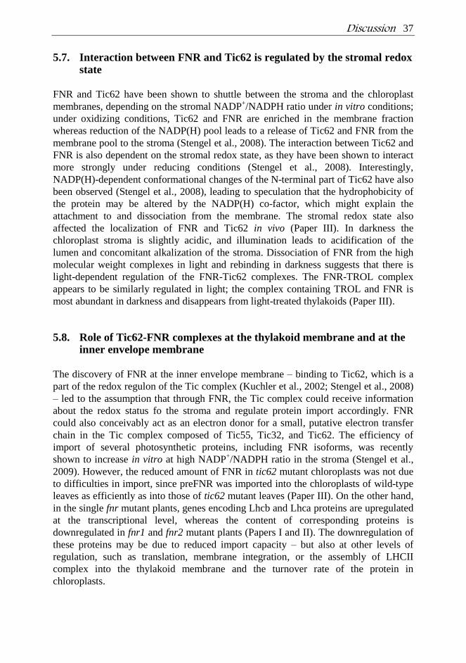
Discussion 37
5.7. Interaction between FNR and Tic62 is regulated by the stromal redox state
FNR and Tic62 have been shown to shuttle between the stroma and the chloroplast
membranes, depending on the stromal NADP+/NADPH ratio under in vitro conditions;
under oxidizing conditions, Tic62 and FNR are enriched in the membrane fraction
whereas reduction of the NADP(H) pool leads to a release of Tic62 and FNR from the
membrane pool to the stroma (Stengel et al., 2008). The interaction between Tic62 and
FNR is also dependent on the stromal redox state, as they have been shown to interact
more strongly under reducing conditions (Stengel et al., 2008). Interestingly,
NADP(H)-dependent conformational changes of the N-terminal part of Tic62 have also
been observed (Stengel et al., 2008), leading to speculation that the hydrophobicity of
the protein may be altered by the NADP(H) co-factor, which might explain the
attachment to and dissociation from the membrane. The stromal redox state also
affected the localization of FNR and Tic62 in vivo (Paper III). In darkness the
chloroplast stroma is slightly acidic, and illumination leads to acidification of the
lumen and concomitant alkalization of the stroma. Dissociation of FNR from the high
molecular weight complexes in light and rebinding in darkness suggests that there is
light-dependent regulation of the FNR-Tic62 complexes. The FNR-TROL complex
appears to be similarly regulated in light; the complex containing TROL and FNR is
most abundant in darkness and disappears from light-treated thylakoids (Paper III).
5.8. Role of Tic62-FNR complexes at the thylakoid membrane and at the inner envelope membrane
The discovery of FNR at the inner envelope membrane – binding to Tic62, which is a
part of the redox regulon of the Tic complex (Kuchler et al., 2002; Stengel et al., 2008)
– led to the assumption that through FNR, the Tic complex could receive information
about the redox status fo the stroma and regulate protein import accordingly. FNR
could also conceivably act as an electron donor for a small, putative electron transfer
chain in the Tic complex composed of Tic55, Tic32, and Tic62. The efficiency of
import of several photosynthetic proteins, including FNR isoforms, was recently
shown to increase in vitro at high NADP+/NADPH ratio in the stroma (Stengel et al.,
2009). However, the reduced amount of FNR in tic62 mutant chloroplasts was not due
to difficulties in import, since preFNR was imported into the chloroplasts of wild-type
leaves as efficiently as into those of tic62 mutant leaves (Paper III). On the other hand,
in the single fnr mutant plants, genes encoding Lhcb and Lhca proteins are upregulated
at the transcriptional level, whereas the content of corresponding proteins is
downregulated in fnr1 and fnr2 mutant plants (Papers I and II). The downregulation of
these proteins may be due to reduced import capacity – but also at other levels of
regulation, such as translation, membrane integration, or the assembly of LHCII
complex into the thylakoid membrane and the turnover rate of the protein in
chloroplasts.
38 Discussion
It can be concluded that the FNR-Tic62 complexes do not have any photosynthetic
role, as the complexes were found to accumulate at the thylakoid membrane in
darkness. Instead, Tic62 keeps FNR active during a long incubation time in darkness in
vitro. The C-terminal part of Tic62 (containing the FNR-binding repetitive sequences)
is relatively young from an evolutionary standpoint, being present only in flowering
plants (Balsera et al., 2007). Since the plants completely lacking the thylakoid-bound
FNR (Papers I and II) or the pool of FNR that associates with thylakoids via Tic62
(Paper III) were found to be healthy under the conditions used in the study, it is
enigmatic that such a large amount of FNR is relocated to the thylakoid membrane in
wild-type plants. There are a few possible reasons that could explain the interaction
between FNR and Tic62 at the thylakoid membrane: (i) for storage and safe-keeping,
(ii) for regulation of the amount of available enzyme in the stroma, or (iii) for some
other as yet unknown function(s). It is obvious nowadays that the reducing energy in
the form of reduced Fd and NADPH is used in several stromal metabolic,
antioxidative, and signaling events in addition to the Calvin-Benson cycle. Thus,
allocation of FNR for NADP+ reduction must be quickly controlled under the ever-
changing environment that plants experience in nature. The rebinding of FNR to the
thylakoid membrane after plants are transferred from light to darkness occurs in a
matter of minutes (unpublished results), indicating that quick removal of FNR from the
stroma would allow other enzymes to use reducing power from Fd according to
environmental cues. In addition, stabilization of FNR by Tic62 may indicate that Tic62
has a chaperone-related role in protecting the enzyme from adverse environmental
conditions in vivo.
Concluding Remarks 39
6. CONCLUDING REMARKS
In this thesis I have shown that both Arabidopsis leaf-targeted FNR isoforms are
photosynthetically active, although loss of one isoform leads to downregulation of
photosynthesis. The isoforms are highly redundant in terms of function but they cannot
completely compensate for each other. FNR1 is indispensable for thylakoid association
of FNR2 evidencing dimer formation between the isoforms. Tic62 was found to have a
major role in tethering FNR to the thylakoid membrane. The thylakoidal pool of FNR
was shown to have roles that are not necessarily involved in photosynthesis, and the
soluble pool of FNR was shown to be photosynthetically active. The exact role of FNR
and Tic62 (or TROL) complexes at the thylakoid membrane still requires investigation,
but based on the results in this thesis, I can propose that Tic62 has a chaperone-like
function – keeping FNR active at the thylakoid membrane during the
photosynthetically inactive periods.
40 Acknowledgements
ACKNOWLEDGEMENTS
This work was carried out in the Laboratory of Molecular Plant Biology at the
University of Turku. Financial support from Academy of Finland, Finnish Graduate
School of Plant Biology and Turku University Foundation are acknowledged.
Professor Eevi Rintamäki is warmly thanked for introducing me for a science and for
excellent guidance throughout my studies. Professor Eva-Mari Aro is thanked for
giving me an opportunity to work in excellent working facilities at Biocity and for
scientific advises. My supervisor Docent Paula Mulo is thanked for never ending
positive attitude, for patience, and for encouraging me in so many ways throughout my
studies. I am grateful for Professors Elina Oksanen and Renate Scheibe for critically
reviewing the thesis and to Hugh Kidd for revising the language.
I would like to acknowledge all my co-authors for the excellent collaboration. Special
thanks go to Professor Jürgen Soll and to his “Tic62-group”, Philipp, Anna, Eike and
Bettina. Philipp and Anna I want to thank for sharing your knowledge, for the passion
to reach the same goal from a different point of view, and for fun. Warm thanks go to
Mika for helping me with small and big issues with equipments of all kinds. Kurt,
Martti, Mirja, and Ulla-Maija are thanked for keeping the laboratory working and
making it possible for us to concentrate on science.
Enormous thanks to all past and present work mates in the laboratory. It has been a
pleasure to get to know all these great persons from different cultures and backgrounds.
Andrea, Anna, Dalton, Fernando, Greg, Hanna, Henna, Ilaf, Irina, Isamu, Jatta, Maija
P, Markus, Michele, Mikko, Moona, Satu, Natasha, Yagut and all the others, you have
made the working environment extremely pleasant with your never-ending help and by
shearing the social life in the coffee room. I want cordially thank Eve, Maija Hoo,
Marion, Marja, Ninni, and Susa for your love and friendship.
My family and friends are warmly thanked for sharing the life outside of science, but
most of all I want to thank Teemu, for showing me what is really important in life.
Turku October 2010
References 41
REFERENCES
Agne, B. and Kessler, F. (2009) Protein transport
in organelles: The toc complex way of preprotein
import. FEBS J., 276:1156-1165.
Albertsson, P., Andreasson, E., Svensson, P. and
Yu, S. (1991) Localization of cytochrome f in the
thylakoid membrane: Evidence for multiple
domains. Biochim. Biophys. Acta, 1098:90-94.
Allahverdiyeva, Y., Mamedov, F., Mäenpaa, P.,
Vass, I. and Aro, E. M. (2005) Modulation of
photosynthetic electron transport in the absence
of terminal electron acceptors: Characterization
of the rbcL deletion mutant of tobacco. Biochim.
Biophys. Acta, 1709:69-83.
Alonso, J.M., Stepanova, A. N., Leisse, T. J.,
Kim, C. J., Chen, H. M., Shinn, P., Stevenson,
D. K., Zimmerman, J., Barajas, P., Cheuk, R.,
Gadrinab, C., Heller, C., Jeske, A., Koesema,
E., Meyers, C. C., Parker, H., Prednis, L.,
Ansari, Y., Choy, N., Deen, H., Geralt, M.,
Hazari, N., Hom, E., Karnes, M., Mulholland,
C., Ndubaku, R., Schmidt, I., Guzman, P.,
Aguilar-Henonin, L., Schmid, M., Weigel, D.,
Carter, D. E., Marchand, T., Risseeuw, E.,
Brogden, D., Zeko, A., Crosby, W. L., Berry,
C. C. and Ecker, J. R. (2003) Genome-wide
insertional mutagenesis of arabidopsis thaliana.
Science, 301:653-657.
Andersen, B., Scheller, H. V. and Moller, B. L. (1992) The PSI-E subunit of photosystem I binds
ferredoxin:NADP+ oxidoreductase. FEBS Lett.,
311:169-173.
Andersson, B. and Anderson, J. M. (1980) Lateral
heterogeneity in the distribution of chlorophyll-
protein complexes of the thylakoid membranes of
spinach chloroplasts. Biochim. Biophys. Acta,
593:427-440.
Arakaki, A.K., Ceccarelli, E. A. and Carrillo, N. (1997) Plant-type ferredoxin-NADP+ reductases:
A basal structural framework and a multiplicity of
functions. FASEB J., 11:133-140.
Aro, E.M., Virgin, I. and Andersson, B. (1993)
Photoinhibition of photosystem II. inactivation,
protein damage and turnover. Biochim. Biophys.
Acta, 1143:113-134.
Asada, K. (2000) The water-water cycle as
alternative photon and electron sinks. Philos.
Trans. R. Soc. Lond. B. Biol. Sci., 355:1419-
1431.
Asada, K. (1999) THE WATER-WATER CYCLE
IN CHLOROPLASTS: Scavenging of active
oxygens and dissipation of excess photons. Annu.
Rev. Plant Physiol. Plant Mol. Biol., 50:601-639.
Balsera, M., Stengel, A., Soll, J. and Bölter, B. (2007) Tic62: A protein family from metabolism
to protein translocation. BMC Evol. Biol., 7:43.
Balsera, M., Goetze, T. A., Kovacs-Bogdan, E.,
Schurmann, P., Wagner, R., Buchanan, B. B.,
Soll, J. and Bölter, B. (2009) Characterization of
Tic110, a channel-forming protein at the inner
envelope membrane of chloroplasts, unveils a
response to ca(2+) and a stromal regulatory
disulfide bridge. J. Biol. Chem., 284:2603-2616.
Baroli, I., Do, A. D., Yamane, T. and Niyogi, K.
K. (2003) Zeaxanthin accumulation in the
absence of a functional xanthophyll cycle protects
chlamydomonas reinhardtii from photooxidative
stress. Plant Cell, 15:992-1008.
Bartsch, S., Monnet, J., Selbach, K., Quigley, F.,
Gray, J., von Wettstein, D., Reinbothe, S. and
Reinbothe, C. (2008) Three thioredoxin targets
in the inner envelope membrane of chloroplasts
function in protein import and chlorophyll
metabolism. Proc. Natl. Acad. Sci. U. S. A.,
105:4933-4938.
Becker, T., Jelic, M., Vojta, A., Radunz, A., Soll,
J. and Schleiff, E. (2004a) Preprotein recognition
by the toc complex. EMBO J., 23:520-530.
Becker, T., Hritz, J., Vogel, M., Caliebe, A.,
Bukau, B., Soll, J. and Schleiff, E. (2004b)
Toc12, a novel subunit of the intermembrane
space preprotein translocon of chloroplasts. Mol.
Biol. Cell, 15:5130-5144.
Bendall, D. and Manasse, R. (1995) Cyclic
photophosphorylation and electron transport.
Biochim. Biophys. Acta, 1229:23-38.
Bertini, I., Luchinat, C., Provenzani, A., Rosato,
A. and Vasos, P. R. (2002) Browsing gene banks
for Fe2S2 ferredoxins and structural modeling of
88 plant-type sequences: An analysis of fold and
function. Proteins, 46:110-127.
Bojko, M. and Wieckowski, S. (1995) Diaphorase
activity of ferredoxin: NADP oxidoreductase in
the presence of dibromothymoquinone.
Phytochemistry, 40:661-665.
Bojko, M., Kruk, J. and Wieckowski, S. (2003)
Plastoquinones are effectively reduced by
ferredoxin:NADP+ oxidoreductase in the
presence of sodium cholate micelles. significance
for cyclic electron transport and chlororespiration.
Phytochemistry, 64:1055-1060.
42 References
Briantais, J.M. (1994) Light harvesting chlorophyll
a-b complex requirement for regulation of
photosystem II photochemistry by non-
photochemical quenching. Photosynthesis
Research, 40:287-294.
Bruce, B.D. (2001) The paradox of plastid transit
peptides: Conservation of function despite
divergence in primary structure. Biochim.
Biophys. Acta, 1541:2-21.
Bruns, C.M. and Karplus, P. A. (1995) Refined
crystal structure of spinach ferredoxin reductase
at 1.7 A resolution: Oxidized, reduced and 2'-
phospho-5'-AMP bound states. J. Mol. Biol.,
247:125-145.
Buchanan, B.B. and Balmer, Y. (2005) Redox
regulation: A broadening horizon. Annu. Rev.
Plant. Biol., 56:187-220.
Caliebe, A., Grimm, R., Kaiser, G., Lubeck, J.,
Soll, J. and Heins, L. (1997) The chloroplastic
protein import machinery contains a rieske-type
iron-sulfur cluster and a mononuclear iron-
binding protein. EMBO J., 16:7342-7350.
Carrillo N. and Vallejos R.H. (1987) Ferredoxin-
NADP+ oxidoreductase. In The light reactions.
Topics in Photosynthesis. (Barber J., ed)
Amsterdam: Elsevier.
Ceccarelli, E., Chan, R. and Vallejos, R. H. (1985) Trimeric structure and other properties of
the chloroplast reductase binding protein. FEBS
Lett., 190:165-168.
Chigri, F., Hormann, F., Stamp, A., Stammers,
D. K., Bölter, B., Soll, J. and Vothknecht, U. C. (2006) Calcium regulation of chloroplast protein
translocation is mediated by calmodulin binding
to Tic32. Proc. Natl. Acad. Sci. U. S. A.,
103:16051-16056.
Chou, M.L., Fitzpatrick, L. M., Tu, S. L.,
Budziszewski, G., Potter-Lewis, S., Akita, M.,
Levin, J. Z., Keegstra, K. and Li, H. M. (2003)
Tic40, a membrane-anchored co-chaperone
homolog in the chloroplast protein translocon.
EMBO J., 22:2970-2980.
DalCorso, G., Pesaresi, P., Masiero, S., Aseeva,
E., Schunemann, D., Finazzi, G., Joliot, P.,
Barbato, R. and Leister, D. (2008) A complex
containing PGRL1 and PGR5 is involved in the
switch between linear and cyclic electron flow in
arabidopsis. Cell, 132:273-285.
Danielsson, R., Albertsson, P.Å., Mamedov, F. (2004) Quantification of photosystem I and II in
different parts of the thylakoid membrane from
spinach. Biochim. Biophys. Acta, 1608:53-61.
Dekker, J.P. and Boekema, E. J. (2005)
Supramolecular organization of thylakoid
membrane proteins in green plants. Biochim.
Biophys. Acta, 1706:12-39.
Deng, Z., Aliverti, A., Zanetti, G., Arakaki, A.
K., Ottado, J., Orellano, E. G., Calcaterra, N.
B., Ceccarelli, E. A., Carrillo, N. and Karplus,
P. A. (1999) A productive NADP+ binding mode
of ferredoxin-NADP + reductase revealed by
protein engineering and crystallographic studies.
Nat. Struct. Biol., 6:847-853.
Di Mascio, P., Devasagayam, T. P., Kaiser, S.
and Sies, H. (1990) Carotenoids, tocopherols and
thiols as biological singlet molecular oxygen
quenchers. Biochem. Soc. Trans., 18:1054-1056.
Dorowski, A., Hofmann, A., Steegborn, C.,
Boicu, M. and Huber, R. (2001) Crystal
structure of paprika ferredoxin-NADP+
reductase. implications for the electron transfer
pathway. J. Biol. Chem., 276:9253-9263.
Endo, T., Mi, H., Shikanai, T. and Asada, K. (1997) Donation of electrons to plastoquinone by
NAD(P)H dehydrogenase and by ferredoxin-
quinone reductase in spinach chloroplasts. Plant
Cell Physiol., 38:1272-1277.
Firlej-Kwoka, E., Strittmatter, P., Soll, J. and
Bölter, B. (2008) Import of preproteins into the
chloroplast inner envelope membrane. Plant Mol.
Biol., 68:505-519.
Forti, G., Cappelletti, A., Nobili, R. L., Garlaschi,
F. M., Gerola, P. D. and Jennings, R. C. (1983)
Interaction of ferredoxin and ferredoxin-NADP
reductase with thylakoids. Arch. Biochem.
Biophys., 221:507-513.
Forti, G. and Bracale, M. (1984) Ferredoxin-
ferredoxin NADP redcutase interaction: Catalytic
differences between the soluble and thylakoid-
bound complex. FEBS Lett., 166:81-84.
Foyer, C., Rowell, J. and Walker, D. (1983) The
effect of sucrose on the rate of de novo sucrose
biosynthesis in leaf protoplasts from spinach,
wheat and barley. Arch. Biochem. Biophys.,
220:232-238.
Foyer, C.H., Furbank, R. T. and Walker, D. A. (1989) Coregulation of electron transport and
benson-calvin cycle activity in isolated spinach
chloroplasts: Studies on glycerate 3-phosphate
reduction. Arch. Biochem. Biophys., 268:687-
697.
Gilmor, A.M., Itoh, S. and Govindjee (2000)
Global spectral-kinetic analysis of room
temperature chlorophyll a fluorescence from
light-harvesting antenna mutants of barley.
References 43
Philos. Trans. R. Soc. Lond. B. Biol. Sci.,
355:1371-1384.
Gilmore, A.M. and Yamamoto, H. Y. (1991)
Resolution of lutein and zeaxanthin using a non-
endcapped, lightly carbon-loaded C18 high-
performance liquid chromatographic column.
Journal of Chromatography, 543:137-145.
Grossman, A.R., Bartlett, S. G., Schmidt, G. W.
and Chua, N. H. (1980) Post-translational uptake
of cytoplasmically synthesized proteins by intact
chloroplasts in vitro. Ann. N. Y. Acad. Sci.,
343:266-274.
Grzyb, J., Malec, P., Rumak, I., Garstka, M. and
Strzalka, K. (2008a) Two isoforms of
ferredoxin:NADP(+) oxidoreductase from wheat
leaves: Purification and initial biochemical
characterization. Photosynth Res., 96:99-112.
Grzyb, J., Gagos, M., Gruszecki, W. I., Bojko,
M. and Strzalka, K. (2008b) Interaction of
ferredoxin:NADP+ oxidoreductase with model
membranes. Biochim. Biophys. Acta, 1778:133-
142.
Guedeney, G., Corneille, S., Cuine, S. and
Peltier, G. (1996) Evidence for an association of
ndh B, ndh J gene products and ferredoxin-
NADP-reductase as components of a
chloroplastic NAD(P)H dehydrogenase complex.
FEBS Lett., 378:277-280.
Gummadova, J.O., Fletcher, G. J., Moolna, A.,
Hanke, G. T., Hase, T. and Bowsher, C. G. (2007) Expression of multiple forms of
ferredoxin NADP+ oxidoreductase in wheat
leaves. J. Exp. Bot., 58:3971-3985.
Hajirezaei, M.R., Peisker, M., Tschiersch, H.,
Palatnik, J. F., Valle, E. M., Carrillo, N. and
Sonnewald, U. (2002) Small changes in the
activity of chloroplastic NADP(+)-dependent
ferredoxin oxidoreductase lead to impaired plant
growth and restrict photosynthetic activity of
transgenic tobacco plants. Plant J., 29:281-293.
Hanke, G.T., Kimata-Ariga, Y., Taniguchi, I.
and Hase, T. (2004) A post genomic
characterization of arabidopsis ferredoxins. Plant
Physiol., 134:255-264.
Hanke, G.T., Okutani, S., Satomi, Y., Takao, T.,
Suzuki, A. and Hase, T. (2005) Multiple iso-
proteins of FNR in arabidopsis: Evidence for
different contributions to chloroplast function and
nitrogen assimilation. Plant Cell and
Environment, 28:1146-1157.
Härtel, H. and Lokstein, H. (1995) Relationship
between quenching of maximum and dark-level
chlorophyll fluorescence in vivo: Dependenet on
photosystem II antenna size. Biochim. Biophys.
Acta, 1228:91-94.
Havaux, M. and Niyogi, K. K. (1999) The
violaxanthin cycle protects plants from
photooxidative damage by more than one
mechanism. Proc. Natl. Acad. Sci. U. S. A.,
96:8762-8767.
Havaux, M., Bonfils, J. P., Lutz, C. and Niyogi,
K. K. (2000) Photodamage of the photosynthetic
apparatus and its dependence on the leaf
developmental stage in the npq1 arabidopsis
mutant deficient in the xanthophyll cycle enzyme
violaxanthin de-epoxidase. Plant Physiol.,
124:273-284.
Havaux, M., Rumeau, D. and Ducruet, J. M. (2005a) Probing the FQR and NDH activities
involved in cyclic electron transport around
photosystem I by the 'afterglow' luminescence.
Biochim. Biophys. Acta, 1709:203-213.
Havaux, M., Eymery, F., Porfirova, S., Rey, P.
and Dormann, P. (2005b) Vitamin E protects
against photoinhibition and photooxidative stress
in arabidopsis thaliana. Plant Cell, 17:3451-3469.
Hedges, S.B., Blair, J. E., Venturi, M. L. and
Shoe, J. L. (2004) A molecular timescale of
eukaryote evolution and the rise of complex
multicellular life. BMC Evol. Biol., 4:2.
Heins, L., Mehrle, A., Hemmler, R., Wagner, R.,
Kuchler, M., Hormann, F., Sveshnikov, D. and
Soll, J. (2002) The preprotein conducting channel
at the inner envelope membrane of plastids.
EMBO J., 21:2616-2625.
Hilson, P., Allemeersch, J., Altmann, T.,
Aubourg, S., Avon, A., Beynon, J., Bhalerao,
R. P., Bitton, F., Caboche, M., Cannoot, B.,
Chardakov, V., Cognet-Holliger, C., Colot, V.,
Crowe, M., Darimont, C., Durinck, S.,
Eickhoff, H., de Longevialle, A. F., Farmer, E.
E., Grant, M., Kuiper, M. T., Lehrach, H.,
Leon, C., Leyva, A., Lundeberg, J., Lurin, C.,
Moreau, Y., Nietfeld, W., Paz-Ares, J.,
Reymond, P., Rouze, P., Sandberg, G., Segura,
M. D., Serizet, C., Tabrett, A., Taconnat, L.,
Thareau, V., Van Hummelen, P., Vercruysse,
S., Vuylsteke, M., Weingartner, M., Weisbeek,
P. J., Wirta, V., Wittink, F. R., Zabeau, M.
and Small, I. (2004) Versatile gene-specific
sequence tags for arabidopsis functional
genomics: Transcript profiling and reverse
genetics applications. Genome Res., 14:2176-
2189.
Inskeep, W.P. and Bloom, P. R. (1985) Extinction
coefficients of chlorophyll-A and chlorophyll-B
in N,N-dimethylformamide and 80-percent
acetone. Plant Physiol., 77:483-485.
44 References
Iwai, M., Takizawa, K., Tokutsu, R., Okamuro,
A., Takahashi, Y. and Minagawa, J. (2010)
Isolation of the elusive supercomplex that drives
cyclic electron flow in photosynthesis. Nature,
464:1210-1213.
Johnson, M.S. and Overington, J. P. (1993) A
structural basis for sequence comparisons - an
evaluation of scoring methodologies. J. Mol.
Biol., 233:716-738.
Joliot, P. and Joliot, A. (2002) Cyclic electron
transfer in plant leaf. Proc. Natl. Acad. Sci. U. S.
A., 99:10209-10214.
Juric, S., Hazler-Pilepic, K., Tomasic, A., Lepedus,
H., Jelicic, B., Puthiyaveetil, S., Bionda, T.,
Vojta, L., Allen, J. F., Schleiff, E. and Fulgosi,
H. (2009) Tethering of ferredoxin:NADP+
oxidoreductase to thylakoid membranes is
mediated by novel chloroplast protein TROL. Plant
J., 60:783-794.
Kamal-Eldin, A. and Appelqvist, L. A. (1996)
The chemistry and antioxidant properties of
tocopherols and tocotrienols. Lipids, 31:671-701.
Kangasjärvi, S., Lepistö, A., Hännikäinen, K.,
Piippo, M., Luomala, E. M., Aro, E. M. and
Rintamäki, E. (2008) Diverse roles for
chloroplast stromal and thylakoid-bound
ascorbate peroxidases in plant stress responses.
Biochem. J., 412:275-285.
Kargul, J. and Barber, J. (2008) Photosynthetic
acclimation; structural reorganization of light
harvesting antenna--role of redox-dependent
phosphorylation of major and minor chlorophyll
a/b binding proteins. FEBS J., 275:1056-1068.
Kessler, F. and Blobel, G. (1996) Interaction of the
protein import and folding machineries of the
chloroplast. Proc. Natl. Acad. Sci. U. S. A.,
93:7684-7689.
Kiss, A.Z., Ruban, A. V. and Horton, P. (2008)
The PsbS protein controls the organization of the
photosystem II antenna in higher plant thylakoid
membranes. Journal of Biological Chemistry,
283:3972-3978.
Kleine, T., Voigt, C. and Leister, D. (2009) Plastid
signalling to the nucleus: Messengers still lost in
the mists? Trends Genet., 25:185-192.
Kouranov, A. and Schnell, D. J. (1997) Analysis
of the interactions of preproteins with the import
machinery over the course of protein import into
chloroplasts. J. Cell Biol., 139:1677-1685.
Kouranov, A., Chen, X., Fuks, B. and Schnell, D.
J. (1998) Tic20 and Tic22 are new components
of the protein import apparatus at the chloroplast
inner envelope membrane. J. Cell Biol., 143:991-
1002.
Krieger-Liszkay, A. (2005) Singlet oxygen
production in photosynthesis. J. Exp. Bot.,
56:337-346.
Kruk, J., Hollander-Czytko, H., Oettmeier, W.
and Trebst, A. (2005) Tocopherol as singlet
oxygen scavenger in photosystem II. J. Plant
Physiol., 162:749-757.
Küchler, M., Decker, S., Hormann, F., Soll, J.
and Heins, L. (2002) Protein import into
chloroplasts involves redox-regulated proteins.
EMBO J., 21:6136-6145.
Kurisu, G., Kusunoki, M., Katoh, E., Yamazaki,
T., Teshima, K., Onda, Y., Kimata-Ariga, Y.
and Hase, T. (2001) Structure of the electron
transfer complex between ferredoxin and
ferredoxin-NADP(+) reductase. Nat. Struct. Biol.,
8:117-121.
Lehtonen, J.V., Still, D.J., Rantanen, V.V.,
Ekhlom, J., Björklund, D., Iftikhar, Z.,
Huhtala, M., Repo, S., Jussila, A., Jaakkola, J.,
Pentikäinen, O., Nyrönen, T., Salminen, T.,
Gyllenberg, M., Johnson, M.S. (2004) BODIL:
A molecular modeling environment for structure-
function analysis and drug design. J. Comput.
Aided Mol. Des., 18:401-419.
Leister, D. (2003) Chloroplast research in the
genomic age. Trends Genet. 19:47-56.
Li, X.P., Bjorkman, O., Shih, C., Grossman, A.
R., Rosenquist, M., Jansson, S. and Niyogi, K.
K. (2000) A pigment-binding protein essential for
regulation of photosynthetic light harvesting.
Nature, 403:391-395.
Li, X.P., Phippard, A., Pasari, J. and Niyogi, K.
K. (2002) Structure-function analysis of
photosystem II subunit S (PsbS) in vivo.
Functional Plant Biology, 29:1131-1139.
Ma, Y., Kouranov, A., LaSala, S. E. and Schnell,
D. J. (1996) Two components of the chloroplast
protein import apparatus, IAP86 and IAP75,
interact with the transit sequence during the
recognition and translocation of precursor
proteins at the outer envelope. J. Cell Biol.,
134:315-327.
Martin, W. and Herrmann, R. G. (1998) Gene
transfer from organelles to the nucleus: How
much, what happens, and why? Plant Physiol.,
118:9-17.
Martinez-Julvez, M., Medina, M. and Gomez-
Moreno, C. (1999) Ferredoxin-NADP(+)
reductase uses the same site for the interaction
References 45
with ferredoxin and flavodoxin. J. Biol. Inorg.
Chem., 4:568-578.
Marton, L., Sidorov, V., Biasini, G. and Maliga,
P. (1982) Complementation in somatic hybrids
indicates 4 types of nitrate reductase deficient
lines in nicotiana-plumbaginifolia. Molecular &
General Genetics, 187:1-3.
Matthijs, H.C.P., Coughlan, S. J. and Hind, G. (1986) Removal of ferredoxin - nadp+
oxidoreductase from thylakoid membranes,
rebinding to depleted membranes, and
identification of the binding-site. J. Biol. Chem.,
261:2154-2158.
Mehler, A.H. (1951) Studies on reactions of
illuminated chloroplasts. II. stimulation and
inhibition of the reaction with molecular oxygen.
Arch. Biochem., 34:339-351.
Melamed-Harel, H., Tel-Or, E., San Pietro, A. (1985) Effect of ferredoxin on the diaphorase
activity of cyanobacterial ferredoxin-NADP
redcutase. Plant Physiol., 77:229-231.
Merchant, S. and Sawaya, M. R. (2005) The light
reactions: A guide to recent acquisitions for the
picture gallery. Plant Cell, 17:648-663.
Moolna, A. and Bowsher, C. G. (2010) The
physiological importance of photosynthetic
ferredoxin NADP+ oxidoreductase (FNR)
isoforms in wheat. J. Exp. Bot., 61:2669-2681.
Morales, R., Kachalova, G., Vellieux, F., Charon,
M.H., Frey, M. (2000) Chrystallographic studies
of the interaction between the ferredoxin-NADP+
reductase and ferredoxin from the
cyanobacterium Anabaena: looking for the
elusive ferredoxin molecule. Acta Cryst.
D56:1408-1412
Munekage, Y., Hojo, M., Meurer, J., Endo, T.,
Tasaka, M. and Shikanai, T. (2002) PGR5 is
involved in cyclic electron flow around
photosystem I and is essential for photoprotection
in arabidopsis. Cell, 110:361-371.
Munekage, Y., Hashimoto, M., Miyake, C.,
Tomizawa, K., Endo, T., Tasaka, M. and
Shikanai, T. (2004) Cyclic electron flow around
photosystem I is essential for photosynthesis.
Nature, 429:579-582.
Niyogi, K.K., Grossman, A. R. and Bjorkman, O. (1998) Arabidopsis mutants define a central role
for the xanthophyll cycle in the regulation of
photosynthetic energy conversion. Plant Cell,
10:1121-1134.
Niyogi, K.K. (1999) PHOTOPROTECTION
REVISITED: Genetic and molecular approaches.
Annu. Rev. Plant Physiol. Plant Mol. Biol.,
50:333-359.
Okutani, S., Hanke, G. T., Satomi, Y., Takao, T.,
Kurisu, G., Suzuki, A. and Hase, T. (2005)
Three maize leaf ferredoxin:NADPH
oxidoreductases vary in subchloroplast location,
expression, and interaction with ferredoxin. Plant
Physiol., 139:1451-1459.
Onda, Y., Matsumura, T., Kimata-Ariga, Y.,
Sakakibara, H., Sugiyama, T. and Hase, T. (2000) Differential interaction of maize root
ferredoxin:NADP(+) oxidoreductase with
photosynthetic and non-photosynthetic ferredoxin
isoproteins. Plant Physiol., 123:1037-1045.
Ort, D.R. and Baker, N. R. (2002) A
photoprotective role for O(2) as an alternative
electron sink in photosynthesis? Curr. Opin. Plant
Biol., 5:193-198.
Pätsikkä, E., Kairavuo, M., Sersen, F., Aro, E. M.
and Tyystjärvi, E. (2002) Excess copper
predisposes photosystem II to photoinhibition in
vivo by outcompeting iron and causing decrease in leaf chlorophyll. Plant Physiol., 129:1359-1367.
Peltier, G. and Cournac, L. (2002) Chlororespiration.
Annu. Rev. Plant. Biol., 53:523-550.
Peng, L., Shimizu, H., Shikanai, T. (2008) The
chloroplast NAD(P)H dehydrogenase complex
interacts with photosystem I in arabidopsis. J.
Biol. Chem., 283:34873-34879.
Piippo, M., Allahverdiyeva, Y., Paakkarinen, V.,
Suoranta, U. M., Battchikova, N. and Aro, E.
M. (2006) Chloroplast-mediated regulation of
nuclear genes in arabidopsis thaliana in the
absence of light stress. Physiological Genomics,
25:142-152.
Porra, R.J., Thompson, W. A. and Kriedemann,
P. E. (1989) Determination of accurate extinction
coefficients and simultaneous-equations for
assaying chlorophyll-A and chlorophyll-B
extracted with 4 different solvents - verification
of the concentration of chlorophyll standards by
atomic-absorption spectroscopy. Biochim.
Biophys. Acta, 975:384-394.
Pschorn, R., Rühle, W. and Wild, A. (1988)
Structure and function of ferredoxin-NADP+-
oxidoreductase. Photosynth Res., 17:217-229.
Quiles, M.J. and Cuello, J. (1998) Association of
ferredoxin-NADP oxidoreductase with the
chloroplastic pyridine nucleotide dehydrogenase
complex in barley leaves. Plant Physiol.,
117:235-244.
Raines, C.A. (2003) The calvin cycle revisited.
Photosynth Res., 75:1-10.
46 References
Renger, G. and Renger, T. (2008) Photosystem II:
The machinery of photosynthetic water splitting.
Photosynth Res., 98:53-80.
Rochaix, J.D. (2007) Role of thylakoid protein
kinases in photosynthetic acclimation. FEBS
Lett., 581:2768-2775.
Rosso, M.G., Li, Y., Strizhov, N., Reiss, B., Dekker,
K. and Weisshaar, B. (2003) An arabidopsis
thaliana T-DNA mutagenized population (GABI-
kat) for flanking sequence tag-based reverse
genetics. Plant Mol. Biol., 53:247-259.
Rumeau, D., Peltier, G. and Cournac, L. (2007)
Chlororespiration and cyclic electron flow around
PSI during photosynthesis and plant stress
response. Plant. Cell. Environ., 30:1041-1051.
Scheibe, R. (2004) Malate valves to balance
cellular energy supply. Physiol. Plant., 120:21-26.
Scheibe, R. (1991) Redox-modulation of
chloroplast enzymes : A common principle for
individual control. Plant Physiol., 96:1-3.
Scheibe, R. and Stitt, M. (1988) Comparison of
nadp-malate dehydrogenase activation, qa
reduction and O-2 evolution in spinach leaves.
Plant Physiology and Biochemistry, 26:473-481.
Schleiff, E., Soll, J., Küchler, M., Kuhlbrandt,
W. and Harrer, R. (2003) Characterization of
the translocon of the outer envelope of
chloroplasts. J. Cell Biol., 160:541-551.
Schnell, D.J., Blobel, G., Keegstra, K., Kessler,
F., Ko, K. and Soll, J. (1997) A consensus
nomenclature for the protein-import components
of the chloroplast envelope. Trends Cell Biol.,
7:303-304.
Serre, L., Vellieux, F. M. D., Medina, M.,
GomezMoreno, C., FontecillaCamps, J. C. and
Frey, M. (1996) X-ray structure of the
ferredoxin:NADP(+) reductase from the
cyanobacterium anabaena PCC 7119 at 1.8
angstrom resolution, and crystallographic studies
of NADP(+) binding at 2.25 angstrom resolution.
J. Mol. Biol., 263:20-39.
Shikanai, T. (2007) Cyclic electron transport
around photosystem I: Genetic approaches. Annu.
Rev. Plant. Biol., 58:199-217.
Shikanai, T., Endo, T., Hashimoto, T., Yamada,
Y., Asada, K. and Yokota, A. (1998) Directed
disruption of the tobacco ndhB gene impairs
cyclic electron flow around photosystem I. Proc.
Natl. Acad. Sci. U. S. A., 95:9705-9709.
SHIN, M. and ARNON, D. I. (1965) Enzymic
mechanisms of pyridine nucleotide reduction in
chloroplasts. J. Biol. Chem., 240:1405-1411.
Shin, M., Ishida, H. and Nozaki, Y. (1985) A new
protein factor, connectein, as a constituent of the
large form of ferredoxin-NADP reductase. Plant
Cell Physiol., 26:559-563.
Sirpiö, S., Allahverdiyeva, Y., Holmström, M.,
Khrouchtchova, A., Haldrup, A., Battchikova,
N. and Aro, E. M. (2009) Novel nuclear-
encoded subunits of the chloroplast NAD(P)H
dehydrogenase complex. J. Biol. Chem.,
284:905-912.
Sohrt, K. and Soll, J. (2000) Toc64, a new
component of the protein translocon of
chloroplasts. J. Cell Biol., 148:1213-1221.
Stengel, A., Benz, P., Balsera, M., Soll, J. and
Bolter, B. (2008) TIC62 redox-regulated
translocon composition and dynamics. J. Biol.
Chem., 283:6656-6667.
Stengel, A., Benz, J. P., Buchanan, B. B., Soll, J.
and Bölter, B. (2009) Preprotein import into
chloroplasts via the toc and tic complexes is
regulated by redox signals in pisum sativum.
Mol. Plant., 2:1181-1197.
Suorsa, M., Sirpiö, S. and Aro, E. M. (2009)
Towards characterization of the chloroplast
NAD(P)H dehydrogenase complex. Mol. Plant.,
2:1127-1140.
Teicher, H.B. and Scheller, H. V. (1998) The
NAD(P)H dehydrogenase in barley thylakoids is
photoactivatable and uses NADPH as well as
NADH. Plant Physiol., 117:525-532.
Vallejos, R.H., Ceccarelli, E. and Chan, R. (1984)
Evidence for the existence of a thylakoid intrinsic
protein that binds ferredoxin-NADP+
oxidoreductase. J. Biol. Chem., 259:8048-8051.
Vallon, O., Bulte, L., Dainese, P., Olive, J., Bassi,
R. and Wollman, F. A. (1991) Lateral
redistribution of cytochrome b6/f complexes
along thylakoid membranes upon state
transitions. Proc. Natl. Acad. Sci. U. S. A.,
88:8262-8266.
Vothknecht, U.C. and Soll, J. (2005) Chloroplast
membrane transport: Interplay of prokaryotic and
eukaryotic traits. Gene, 354:99-109.
Yoon, H.S., Hackett, J. D., Ciniglia, C., Pinto, G.
and Bhattacharya, D. (2004) A molecular
timeline for the origin of photosynthetic
eukaryotes. Mol. Biol. Evol., 21:809-818.
Zhang, H., Whitelegge, J. P. and Cramer, W. A. (2001) Ferredoxin:NADP+ oxidoreductase is a
subunit of the chloroplast cytochrome b6f
complex. J. Biol. Chem., 276:38159-38165.