Embed Size (px)
Citation preview
L.D.R. Thompson 14MAY2018
Spooky Spindled Sinonasal SpectresNapa Valley Pathology Conference 1
Spooky Spindled Sinonasal Spectres
Lester D. R. Thompson
www.lester-thompson.com
2
Learning Objectives
A presentation of selected spindled cell lesions involving the mucosa and soft tissue of the sinonasal tract and larynx
Emphasis will be placed on a practical approach to identify important histologic criteria to separate these spindle cell lesions
Pertinent clinical and immunohistochemical features of diagnostic or prognostic significance will be highlighted
Select and integrate special studies that aid in diagnosis
3
Differential Diagnostic Considerations
Biphenotypic sinonasal sarcoma Rhabdomyosarcoma Respiratory epithelial adenomatoid hamartoma Angiofibroma Synovial sarcoma Solitary fibrous tumor Spindle cell squamous cell carcinoma Spindle cell mucosal melanoma Mycobacterial spindle cell tumor Peripheral nerve sheath tumor (benign or malignant) / PEComa Leiomyosarcoma/leiomyoma Glomangiopericytoma Fibrosarcoma/fibromatosis
4
Glomangiopericytoma
A tumor demonstrating perivascular myoid phenotype (myopericyte) A modified smooth muscle cell similar to glomus tumors
Sex: Female > Male (1.2:1)Age: Peak in the 7th decade Symptoms: A short duration of nasal obstruction, mass, and
epistaxis Rare association with osteomalacia
Site: Unilaterally in the nasal cavity alone May extend into paranasal sinuses
Surgical excision with excellent outcome Recurrence in about 18% of cases
5
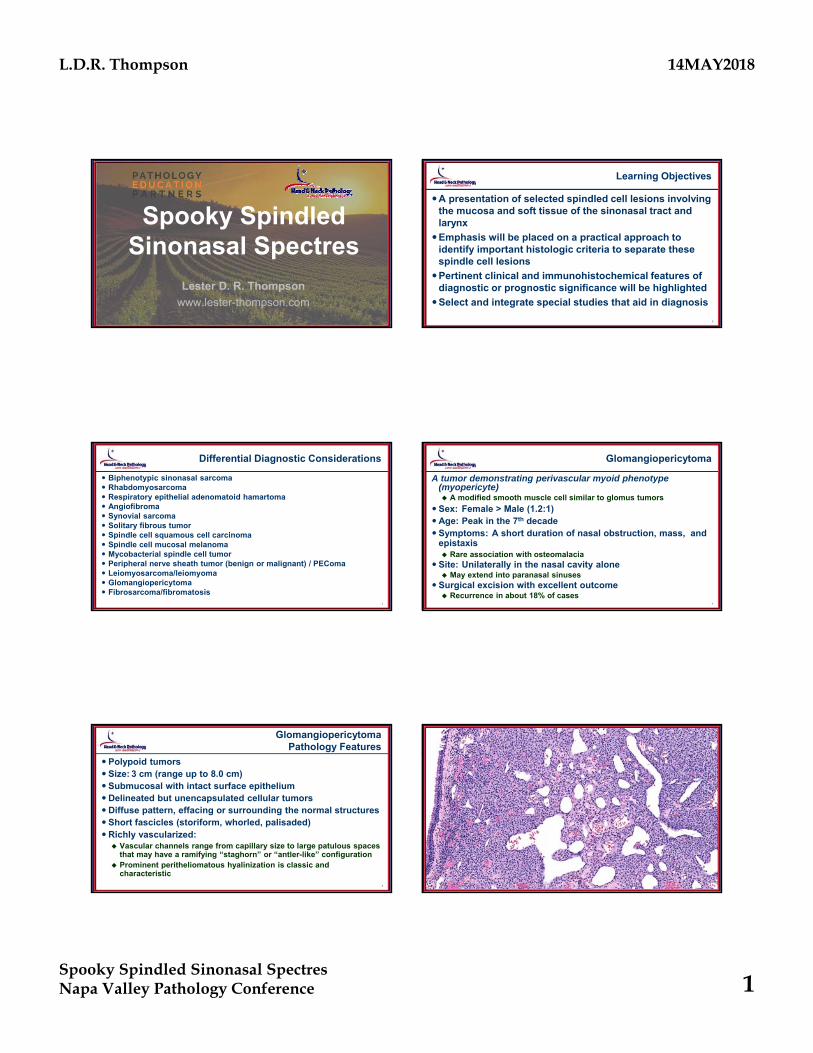
GlomangiopericytomaPathology Features
Polypoid tumors Size: 3 cm (range up to 8.0 cm) Submucosal with intact surface epitheliumDelineated but unencapsulated cellular tumorsDiffuse pattern, effacing or surrounding the normal structures Short fascicles (storiform, whorled, palisaded)Richly vascularized: Vascular channels range from capillary size to large patulous spaces
that may have a ramifying “staghorn” or “antler-like” configuration Prominent peritheliomatous hyalinization is classic and
characteristic6
L.D.R. Thompson 14MAY2018
Spooky Spindled Sinonasal SpectresNapa Valley Pathology Conference 2
7 8
9 10
GlomangiopericytomaMicroscopic
Closely packed syncytium of uniform oval to elongate cells Indistinct cell bordersVesicular to hyperchromatic, round to oval to spindle-
shaped nucleiMild nuclear pleomorphismMitotic figures may be presentExtravasated erythrocytesMast cells and eosinophils usually prominentGiant cells and fat are rarely reportedRare malignant cases develop
11 12
L.D.R. Thompson 14MAY2018
Spooky Spindled Sinonasal SpectresNapa Valley Pathology Conference 3
13CD117
14
15
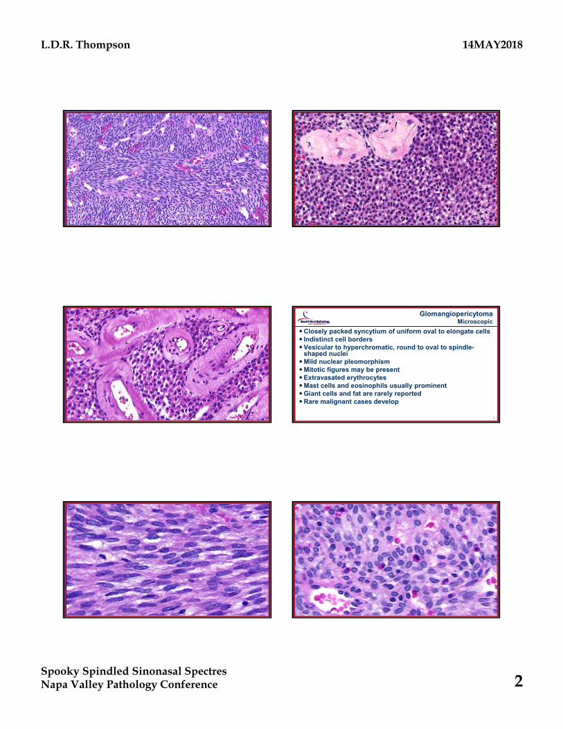
GlomangiopericytomaImmunohistochemistry
Positive: Actins (SMA>MSA), nuclear β-catenin Negative: CD34, CD31, CD117, STAT6, EMA, keratin,
S100 protein, GFAP, FVIIIR-Ag, CD99, desmin
Genetics: Somatic, single nucleotide substitution heterozygous
mutations in CTNNB1 gene encoding β-catenin, specifically in GSK3β region (encoded by exon 3)
Mutations constitutively active β-catenin with cyclin D1 over expression, and aberrant nuclear accumulation due to nuclear translocation of membrane protein
16CD34
17SMA ß-catenin
18
Rhabdomyosarcoma
Malignant mesenchymal neoplasm with skeletal muscle differentiation Embryonal rhabdomyosarcoma (botryoid, anaplastic)
Alveolar rhabdomyosarcoma (anaplastic, botryoid, spindle)
Incidence Most common soft tissue sarcoma in head and neck
Most common soft tissue sarcoma in children and adolescents
Embryonal: ear Alveolar: sinonasal tract
Age: Embryonal (usually < 20 yrs.); Alveolar: Adults
Sex: Male > Female (1.2:1)
L.D.R. Thompson 14MAY2018
Spooky Spindled Sinonasal SpectresNapa Valley Pathology Conference 4
19
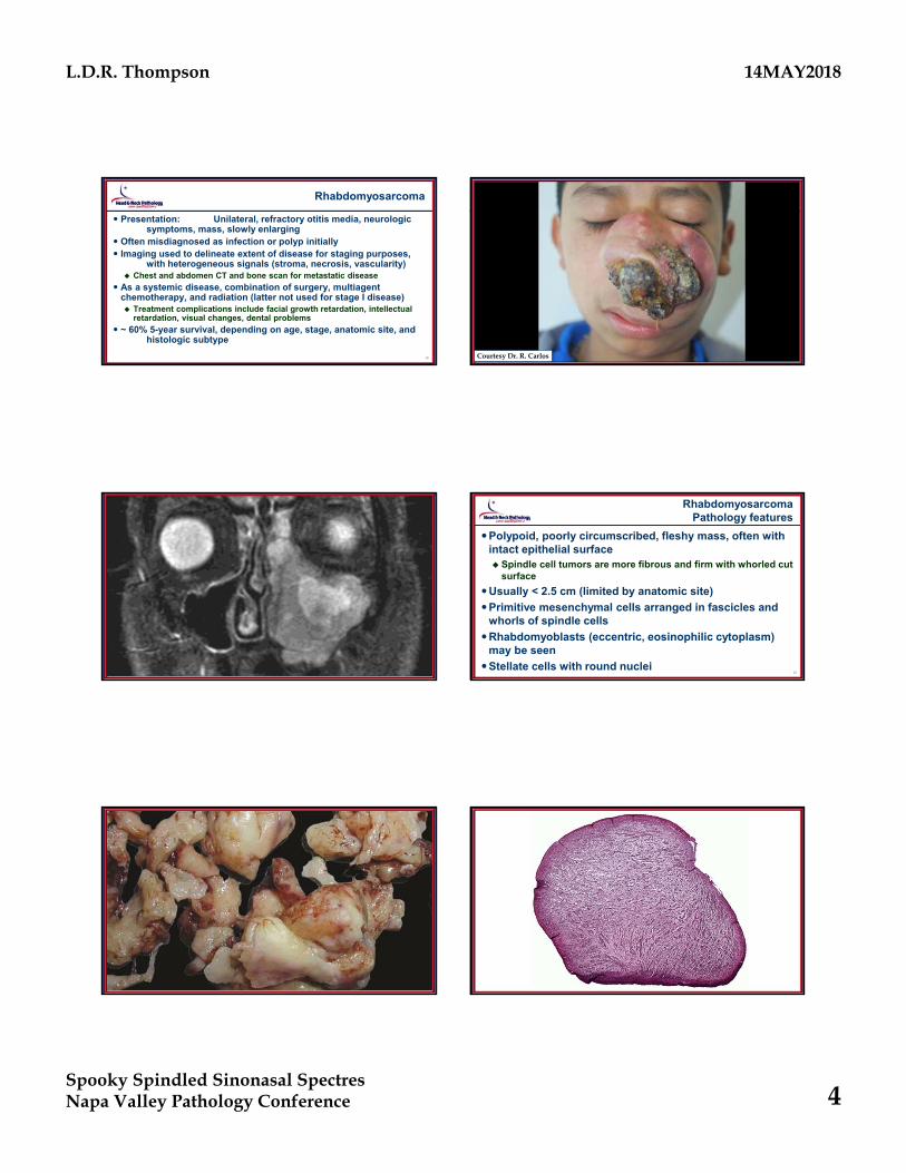
Rhabdomyosarcoma
Presentation: Unilateral, refractory otitis media, neurologic symptoms, mass, slowly enlarging
Often misdiagnosed as infection or polyp initially Imaging used to delineate extent of disease for staging purposes,
with heterogeneous signals (stroma, necrosis, vascularity) Chest and abdomen CT and bone scan for metastatic disease
As a systemic disease, combination of surgery, multiagent chemotherapy, and radiation (latter not used for stage I disease) Treatment complications include facial growth retardation, intellectual
retardation, visual changes, dental problems
~ 60% 5-year survival, depending on age, stage, anatomic site, and histologic subtype
20Courtesy Dr. R. Carlos
21 22
RhabdomyosarcomaPathology features
Polypoid, poorly circumscribed, fleshy mass, often with intact epithelial surface Spindle cell tumors are more fibrous and firm with whorled cut
surface
Usually < 2.5 cm (limited by anatomic site)
Primitive mesenchymal cells arranged in fascicles and whorls of spindle cells
Rhabdomyoblasts (eccentric, eosinophilic cytoplasm) may be seen
Stellate cells with round nuclei
23
L.D.R. Thompson 14MAY2018
Spooky Spindled Sinonasal SpectresNapa Valley Pathology Conference 5
25 26
27 28
RhabdomyosarcomaPathology features
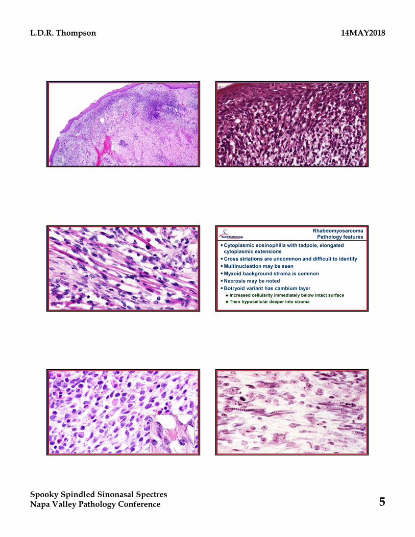
Cytoplasmic eosinophilia with tadpole, elongated cytoplasmic extensions
Cross striations are uncommon and difficult to identify
Multinucleation may be seen
Myxoid background stroma is common
Necrosis may be noted
Botryoid variant has cambium layer Increased cellularity immediately below intact surface
Then hypocellular deeper into stroma
29 30
L.D.R. Thompson 14MAY2018
Spooky Spindled Sinonasal SpectresNapa Valley Pathology Conference 6
31 32
Special studies
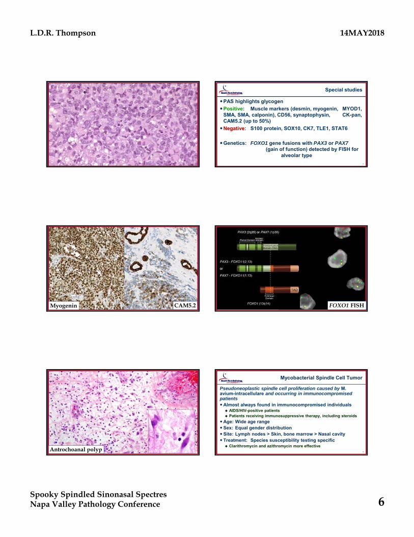
PAS highlights glycogen
Positive: Muscle markers (desmin, myogenin, MYOD1, SMA, SMA, calponin), CD56, synaptophysin, CK-pan, CAM5.2 (up to 50%)
Negative: S100 protein, SOX10, CK7, TLE1, STAT6
Genetics: FOXO1 gene fusions with PAX3 or PAX7(gain of function) detected by FISH for
alveolar type
33Myogenin CAM5.2
34FOXO1 FISH
35Antrochoanal polyp
36
Mycobacterial Spindle Cell Tumor
Pseudoneoplastic spindle cell proliferation caused by M. avium-intracellulare and occurring in immunocompromised patientsAlmost always found in immunocompromised individuals AIDS/HIV-positive patients Patients receiving immunosuppressive therapy, including steroids
Age: Wide age range Sex: Equal gender distribution Site: Lymph nodes > Skin, bone marrow > Nasal cavity Treatment: Species susceptibility testing specific Clarithromycin and azithromycin more effective
L.D.R. Thompson 14MAY2018
Spooky Spindled Sinonasal SpectresNapa Valley Pathology Conference 7
37
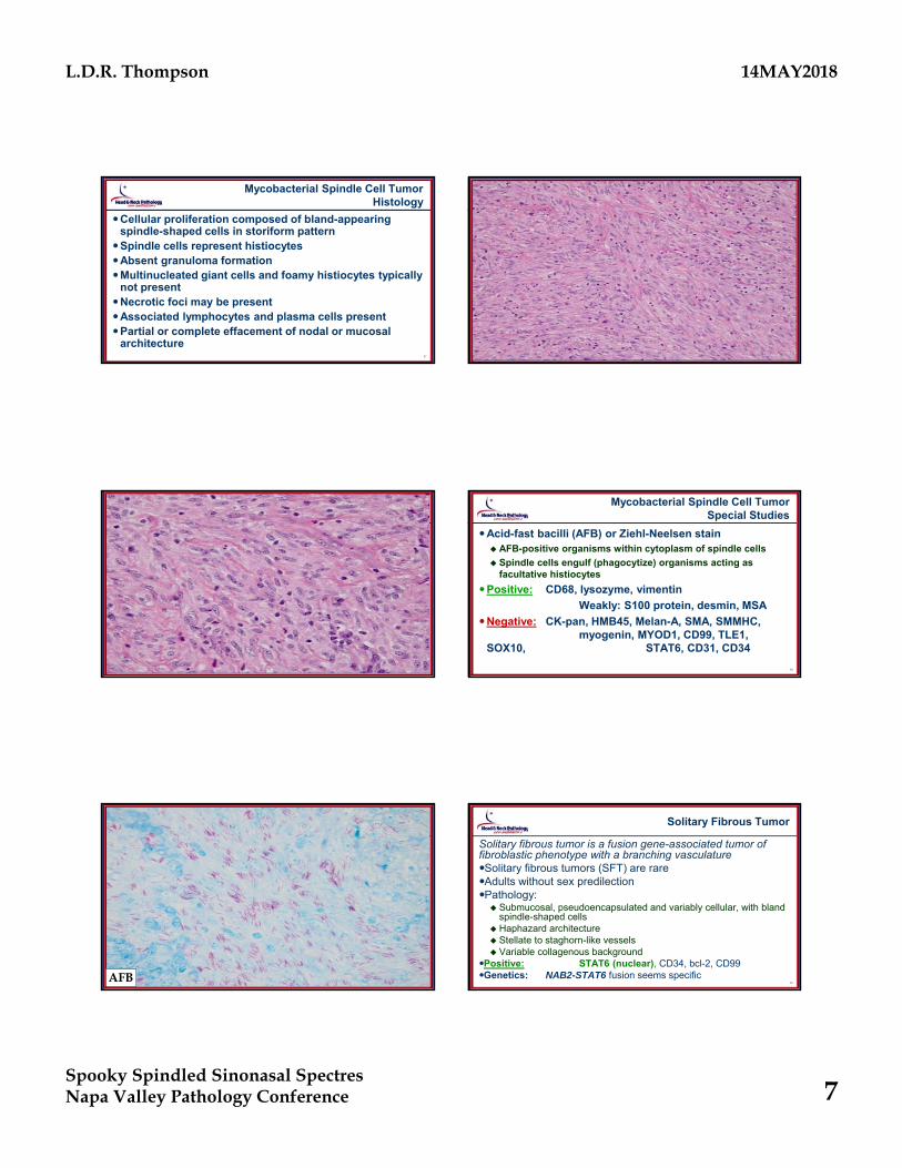
Mycobacterial Spindle Cell TumorHistology
Cellular proliferation composed of bland-appearing spindle-shaped cells in storiform pattern
Spindle cells represent histiocytesAbsent granuloma formationMultinucleated giant cells and foamy histiocytes typically
not presentNecrotic foci may be presentAssociated lymphocytes and plasma cells presentPartial or complete effacement of nodal or mucosal
architecture 38
39 40
Mycobacterial Spindle Cell TumorSpecial Studies
Acid-fast bacilli (AFB) or Ziehl-Neelsen stain AFB-positive organisms within cytoplasm of spindle cells
Spindle cells engulf (phagocytize) organisms acting as facultative histiocytes
Positive: CD68, lysozyme, vimentin
Weakly: S100 protein, desmin, MSA
Negative: CK-pan, HMB45, Melan-A, SMA, SMMHC, myogenin, MYOD1, CD99, TLE1,
SOX10, STAT6, CD31, CD34
41AFB
42
Solitary Fibrous Tumor
Solitary fibrous tumor is a fusion gene-associated tumor of fibroblastic phenotype with a branching vasculatureSolitary fibrous tumors (SFT) are rareAdults without sex predilectionPathology: Submucosal, pseudoencapsulated and variably cellular, with bland
spindle-shaped cells Haphazard architecture Stellate to staghorn-like vessels Variable collagenous background
Positive: STAT6 (nuclear), CD34, bcl-2, CD99Genetics: NAB2-STAT6 fusion seems specific
L.D.R. Thompson 14MAY2018
Spooky Spindled Sinonasal SpectresNapa Valley Pathology Conference 8
43 44
45 46
47 48CD34
L.D.R. Thompson 14MAY2018
Spooky Spindled Sinonasal SpectresNapa Valley Pathology Conference 9
49STAT6
50
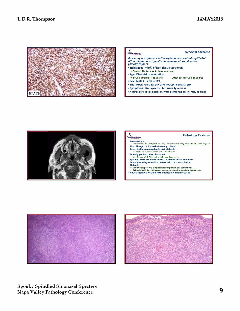
Synovial sarcoma
Mesenchymal spindled cell neoplasm with variable epithelial differentiation and specific chromosomal translocation: t(X;18)(p11;q11) Incidence: ~10% of soft tissue sarcomas About 10% develop in head and neck
Age: Bimodal presentation Young adults (15-35 years) Older age (around 50 years)
Sex: Male > Female (3:1) Site: Neck, oropharynx and hypopharynx/larynx Symptoms: Nonspecific, but usually a massAggressive local excision with combination therapy is best
51 52
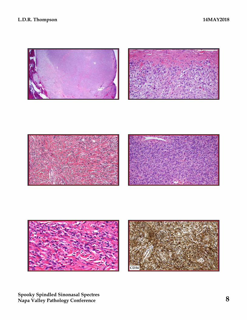
Pathology Features
Macroscopic: Pedunculated or polypoid, usually circumscribed; may be multinodular and cystic
Size: Range: 1-12 cm (but usually < 5 cm) Separated into monophasic and biphasic
Monophasic most common in head and neck Densely packed, short fascicles
May be marbled: Alternating light and dark areas Spindled cells are uniform with indistinct cell boundaries Hemangiopericytoma-like pattern with rich vascularity Biphasic
Variable proportions of epithelial and spindled cell components Epithelial cells have abundant cytoplasm, creating glandular appearance
Mitotic figures are identified, but usually not increased
53 54
L.D.R. Thompson 14MAY2018
Spooky Spindled Sinonasal SpectresNapa Valley Pathology Conference 10
55 56
57 58
59 60
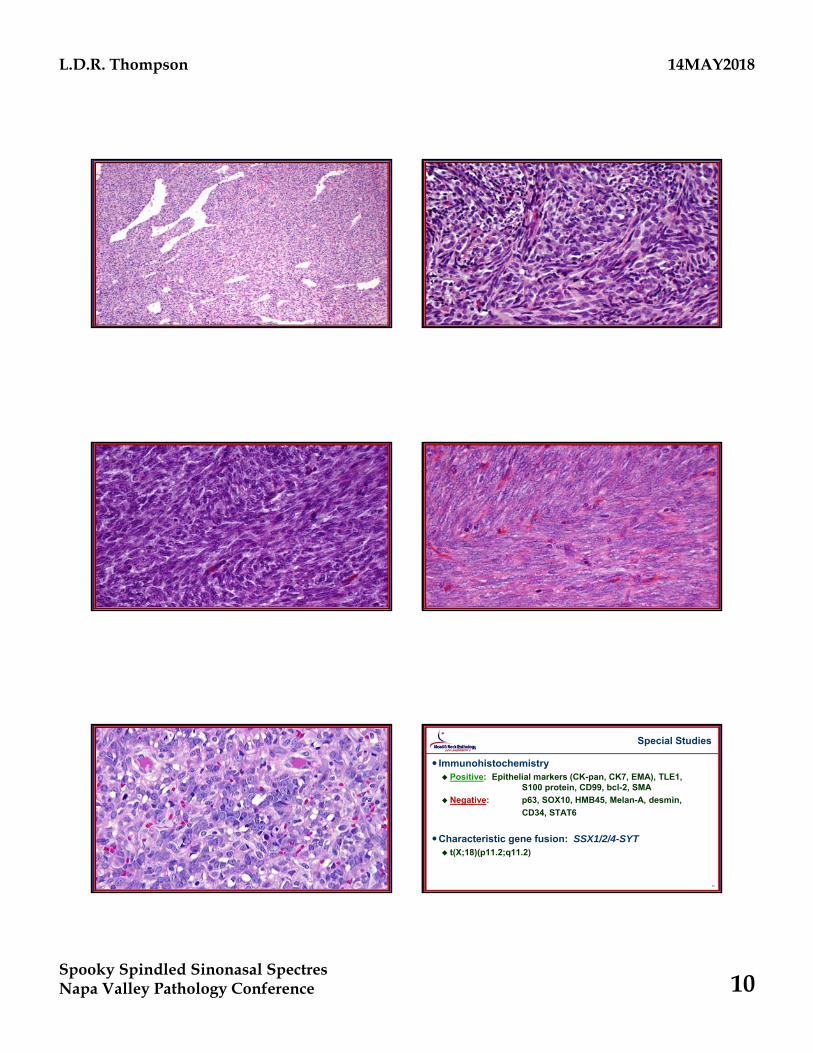
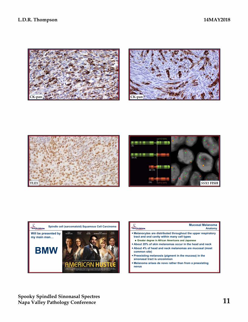
Special Studies
Immunohistochemistry Positive: Epithelial markers (CK-pan, CK7, EMA), TLE1,
S100 protein, CD99, bcl-2, SMA
Negative: p63, SOX10, HMB45, Melan-A, desmin,
CD34, STAT6
Characteristic gene fusion: SSX1/2/4-SYT t(X;18)(p11.2;q11.2)
L.D.R. Thompson 14MAY2018
Spooky Spindled Sinonasal SpectresNapa Valley Pathology Conference 11
61CK-pan
62CK-pan
63TLE1
64SSX1 FISH
65
Spindle cell (sarcomatoid) Squamous Cell Carcinoma
Will be presented by my main man…
BMW
66
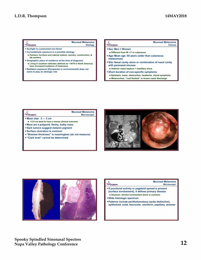
Mucosal MelanomaAnatomy
Melanocytes are distributed throughout the upper respiratory tract and oral cavity within many cell types Greater degree in African Americans and Japanese
About 20% of skin melanomas occur in the head and neck
About 4% of head and neck melanomas are mucosal (most common site)
Preexisting melanosis (pigment in the mucosa) in the sinonasal tract is uncommon
Melanoma arises de novo rather than from a preexisting nevus
L.D.R. Thompson 14MAY2018
Spooky Spindled Sinonasal SpectresNapa Valley Pathology Conference 12
67
Mucosal MelanomaEtiology
Sunlight is a presumed non-factor
Formaldehyde exposure is a possible etiology Painters, furniture and cabinet makers, laundry, construction, &
lab workers
Geographic place of residence at the time of diagnosis Living in southern latitudes (defined as < 40°N in North America)
have increased incidence of melanoma
Radiation exposure (therapeutic or environmental) does not seem to play an etiologic role
68
Mucosal MelanomaClinical
Sex: Men = Women Different from M > F in cutaneous
Age:Mean age: 65 years (older than cutaneous melanomas)
Site:Nasal cavity alone or combination of nasal cavity with paranasal sinuses Anterior nasal septum > maxillary sinus
Short duration of non-specific symptoms Epistaxis, mass, obstruction, headache, visual symptoms
Melanorrhea: “coal flecked” or brown nasal discharge
69
Mucosal MelanomaMacroscopic
Mean size: 2 — 3 cm >3.0 cm tend to have a worse clinical outcome
Most are a polypoid, fleshy, bulky massDark tumors suggest melanin pigmentSurface ulceration is common“Breslow thickness” is meaningless (do not measure)“Clark level” cannot be determined
70
71 72
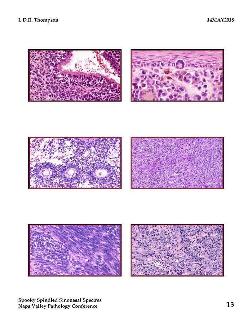
Mucosal MelanomaMicroscopic
If junctional activity or pagetoid spread is present (surface involvement), it defines primary disease However, stroma involvement alone is common
Wide histologic spectrum
Patterns include peritheliomatous (quite distinctive), epithelioid, solid, fascicular, storiform, papillary, alveolar
L.D.R. Thompson 14MAY2018
Spooky Spindled Sinonasal SpectresNapa Valley Pathology Conference 13
73 74
75 76
77 78
L.D.R. Thompson 14MAY2018
Spooky Spindled Sinonasal SpectresNapa Valley Pathology Conference 14
79 80
81
Mucosal MelanomaMicroscopic
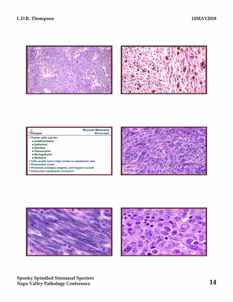
Tumor cells can be: Undifferentiated Epithelioid Spindled PlasmacytoidMeningothelial Rhabdoid
Cells usually have a high nuclear to cytoplasmic ratio Pleomorphic nuclei Prominent, enlarged, magenta, and irregular nucleoli Intranuclear cytoplasmic inclusions
82
83 84
L.D.R. Thompson 14MAY2018
Spooky Spindled Sinonasal SpectresNapa Valley Pathology Conference 15
85
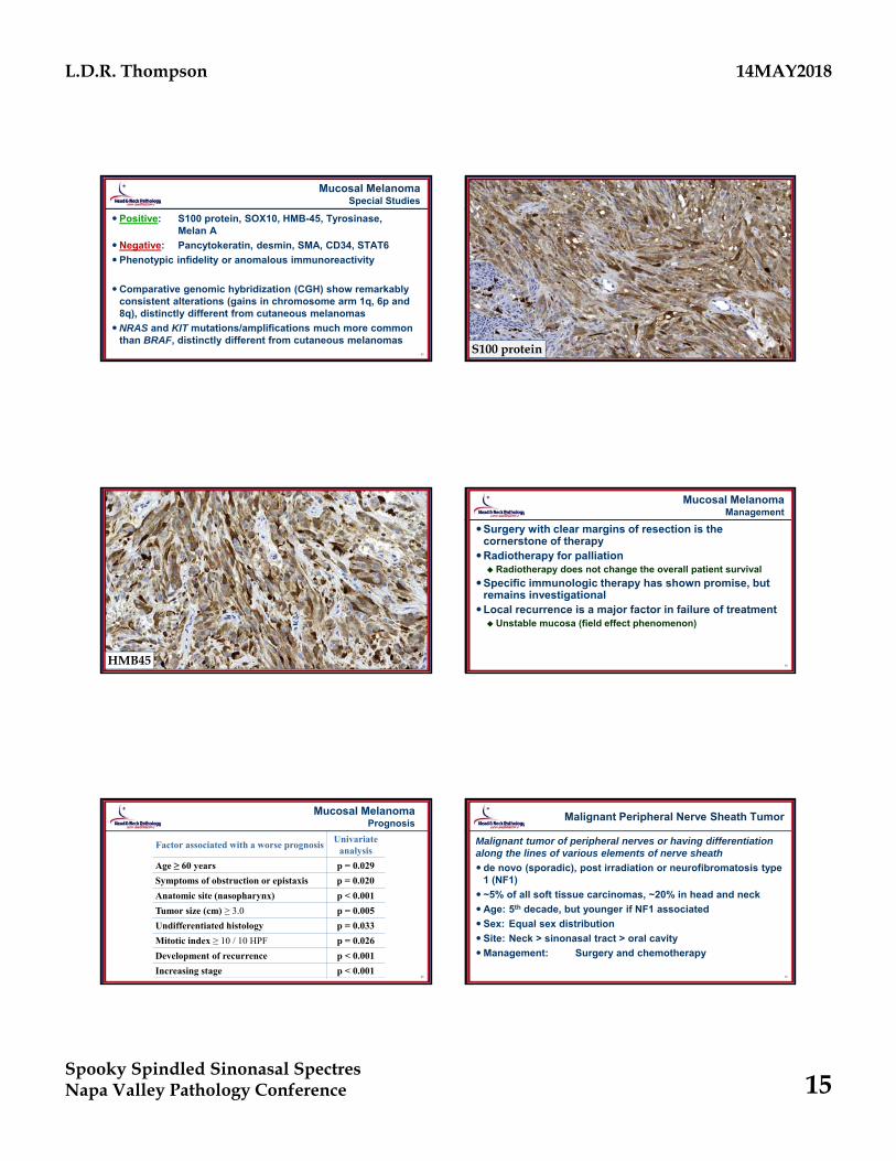
Mucosal MelanomaSpecial Studies
Positive: S100 protein, SOX10, HMB-45, Tyrosinase, Melan A
Negative: Pancytokeratin, desmin, SMA, CD34, STAT6
Phenotypic infidelity or anomalous immunoreactivity
Comparative genomic hybridization (CGH) show remarkably consistent alterations (gains in chromosome arm 1q, 6p and 8q), distinctly different from cutaneous melanomas
NRAS and KIT mutations/amplifications much more common than BRAF, distinctly different from cutaneous melanomas
86S100 protein
87HMB45
88
Mucosal MelanomaManagement
Surgery with clear margins of resection is the cornerstone of therapy
Radiotherapy for palliation Radiotherapy does not change the overall patient survival
Specific immunologic therapy has shown promise, but remains investigational
Local recurrence is a major factor in failure of treatment Unstable mucosa (field effect phenomenon)
89
Mucosal MelanomaPrognosis
Factor associated with a worse prognosisUnivariate
analysis
Age ≥ 60 years p = 0.029
Symptoms of obstruction or epistaxis p = 0.020
Anatomic site (nasopharynx) p < 0.001
Tumor size (cm) ≥ 3.0 p = 0.005
Undifferentiated histology p = 0.033
Mitotic index ≥ 10 / 10 HPF p = 0.026
Development of recurrence p < 0.001
Increasing stage p < 0.00190
Malignant Peripheral Nerve Sheath Tumor
Malignant tumor of peripheral nerves or having differentiation along the lines of various elements of nerve sheath
de novo (sporadic), post irradiation or neurofibromatosis type 1 (NF1)
~5% of all soft tissue carcinomas, ~20% in head and neck
Age: 5th decade, but younger if NF1 associated
Sex: Equal sex distribution
Site: Neck > sinonasal tract > oral cavity
Management: Surgery and chemotherapy
L.D.R. Thompson 14MAY2018
Spooky Spindled Sinonasal SpectresNapa Valley Pathology Conference 16
91
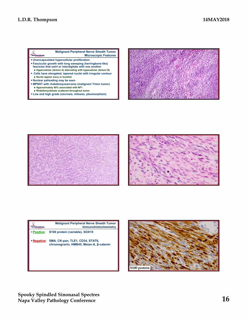
Malignant Peripheral Nerve Sheath TumorMicroscopic Features
Unencapsulated hypercellular proliferation Fascicular growth with long sweeping (herringbone-like)
fascicles that swirl or interdigitate with one another Hypercellular (Antoni A) alternating with hypocellular (Antoni B)
Cells have elongated, tapered nuclei with irregular contour Nuclei appear wavy or buckled
Nuclear palisading may be seenMPNST with rhabdomyosarcoma (malignant Triton tumor) Approximately 60% associated with NF1 Rhabdomyoblasts scattered throughout tumor
Low and high grade (necrosis, mitoses, pleomorphism)92
93 94
95
Malignant Peripheral Nerve Sheath TumorImmunohistochemistry
Positive: S100 protein (variable), SOX10
Negative: SMA, CK-pan, TLE1, CD34, STAT6, chromogranin, HMB45, Melan-A, β-catenin
96S100 protein
L.D.R. Thompson 14MAY2018
Spooky Spindled Sinonasal SpectresNapa Valley Pathology Conference 17
97
PEComa
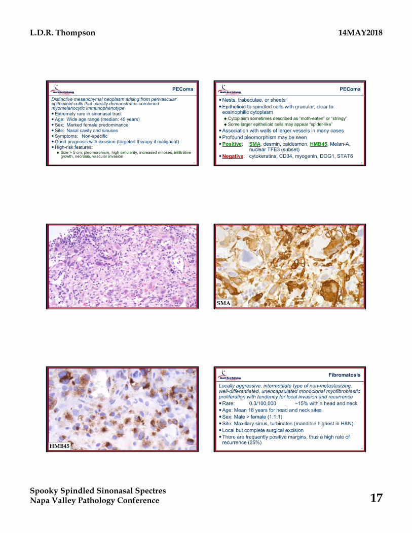
Distinctive mesenchymal neoplasm arising from perivascular epithelioid cells that usually demonstrates combined myomelanocytic immunophenotype Extremely rare in sinonasal tract Age: Wide age range (median: 45 years) Sex: Marked female predominance Site: Nasal cavity and sinuses Symptoms: Non-specificGood prognosis with excision (targeted therapy if malignant)High-risk features: Size > 5 cm, pleomorphism, high cellularity, increased mitoses, infiltrative
growth, necrosis, vascular invasion98
PEComa
Nests, trabeculae, or sheetsEpithelioid to spindled cells with granular, clear to
eosinophilic cytoplasm Cytoplasm sometimes described as “moth-eaten” or “stringy” Some larger epithelioid cells may appear “spider-like”
Association with walls of larger vessels in many casesProfound pleomorphism may be seenPositive: SMA, desmin, caldesmon, HMB45, Melan-A,
nuclear TFE3 (subset)Negative: cytokeratins, CD34, myogenin, DOG1, STAT6
99 100SMA
101HMB45
102
Fibromatosis
Locally aggressive, intermediate type of non-metastasizing, well-differentiated, unencapsulated monoclonal myofibroblastic proliferation with tendency for local invasion and recurrenceRare: 0.3/100,000 ~15% within head and neckAge: Mean 18 years for head and neck sitesSex: Male > female (1.1:1)Site: Maxillary sinus, turbinates (mandible highest in H&N)Local but complete surgical excisionThere are frequently positive margins, thus a high rate of
recurrence (25%)
L.D.R. Thompson 14MAY2018
Spooky Spindled Sinonasal SpectresNapa Valley Pathology Conference 18
103
Fibromatosis
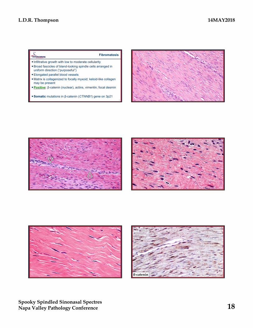
Infiltrative growth with low to moderate cellularity
Broad fascicles of bland-looking spindle cells arranged in uniform direction (“purposeful”)
Elongated parallel blood vessels
Matrix is collagenized to focally myxoid; keloid-like collagen may be present
Positive: β-catenin (nuclear), actins, vimentin; focal desmin
Somatic mutations in β-catenin (CTNNB1) gene on 3p21104
105 106
107 108ß-catenin
L.D.R. Thompson 14MAY2018
Spooky Spindled Sinonasal SpectresNapa Valley Pathology Conference 19
109
Fibrosarcoma
Malignant neoplasm with only fibroblastic &/or myofibroblastic differentiation (a diagnosis of exclusion) Incidence: Uncommon (~ 3% of sinonasal malignancies) BUT: Many are reclassified into other categories now
Age: Peak: 5th to 6th decades Sex: Female > Male (3:2) Site: One or more paranasal sinuses (maxillary, ethmoid) Symptoms: Short duration of nasal obstruction, epistaxis Treatment: En-bloc resection and radiation yield best
outcome (75% 5 year survival)High incidence of recurrence (up to 60%)
110
Fibrosarcoma
Malignant neoplasm with only fibroblastic &/or myofibroblastic differentiation (a diagnosis of exclusion) Incidence: Uncommon (~ 3% of sinonasal malignancies) BUT: Many are reclassified into other categories now
Age: Peak: 5th to 6th decades Sex: Female > Male (3:2) Site: One or more paranasal sinuses (maxillary, ethmoid) Symptoms: Short duration of nasal obstruction, epistaxis Treatment: En-bloc resection and radiation yield best
outcome (75% 5 year survival)High incidence of recurrence (up to 60%)
111 112
Fibrosarcoma
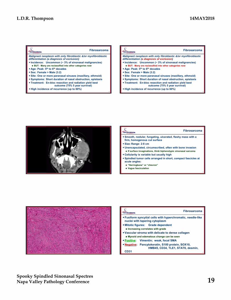
Smooth, nodular, fungating, ulcerated, fleshy mass with a firm, homogenous cut surface
Size: Range: 2-8 cm
Unencapsulated, circumscribed, often with bone invasion If surface invaginations, think biphenotypic sinonasal sarcoma
Cellularity is variable but usually high
Spindled tumor cells arranged in short, compact fascicles at acute angles: “Herringbone” or “chevron”
Vague fasciculation
113
Herringbone Pattern
114
Fibrosarcoma
Fusiform syncytial cells with hyperchromatic, needle-like nuclei with tapering cytoplasm
Mitotic figures: Grade dependent Increasing correlates with grade
Vascular stroma with delicate to dense collagenMyxoid and edematous change can be seen
Positive: Vimentin; weak, focal SMA
Negative: Pancytokeratin, S100 protein, SOX10, HMB45, CD34, TLE1, STAT6, desmin,
CD31
L.D.R. Thompson 14MAY2018
Spooky Spindled Sinonasal SpectresNapa Valley Pathology Conference 20
115 116
117 118
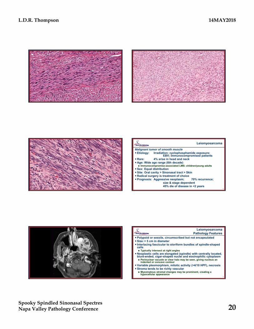
Leiomyosarcoma
Malignant tumor of smooth muscle Etiology: Irradiation; cyclophosphamide exposure;
EBV; immunocompromised patientsRare: 4% arise in head and neckAge: Wide age range (6th decade) Immunocompromise-associated LMS: children/young adults
Sex: Equal distribution Site: Oral cavity > Sinonasal tract > SkinRadical surgery is treatment of choice Prognosis: Aggressive neoplasm; 70% recurrence;
size & stage dependent45% die of disease in <2 years
119 120
LeiomyosarcomaPathology Features
Polypoid or sessile, circumscribed but not encapsulated Size: > 5 cm in diameter Interlacing fascicular to storiform bundles of spindle-shaped
cells Typically intersect at right angles
Neoplastic cells are elongated (spindle) with centrally located, blunt-ended, cigar-shaped nuclei and eosinophilic cytoplasm Perinuclear vacuole or clear halo may be seen, giving nucleus an
indented or concave contour Variable pleomorphism, mitotic activity (>4/10 HPF), necrosis Stroma tends to be richly vascular Myxomatous stromal changes may be prominent, creating a
hypocellular appearance
L.D.R. Thompson 14MAY2018
Spooky Spindled Sinonasal SpectresNapa Valley Pathology Conference 21
121 122
123 124
125 126
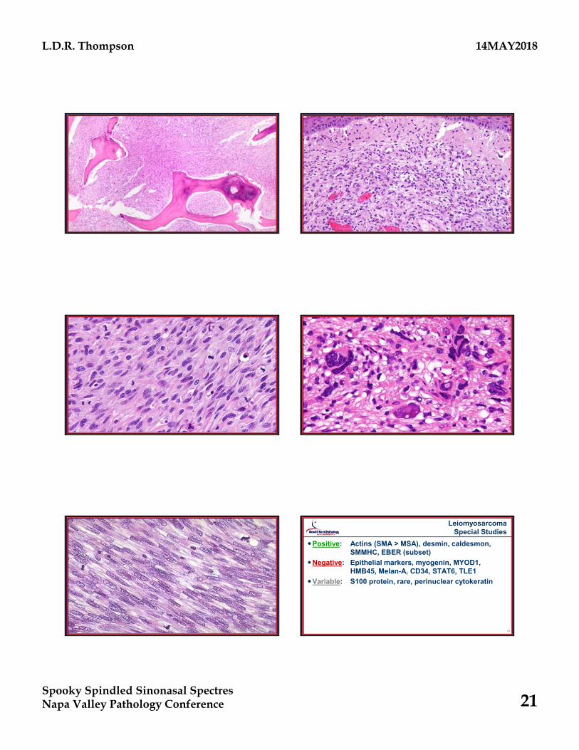
LeiomyosarcomaSpecial Studies
Positive: Actins (SMA > MSA), desmin, caldesmon, SMMHC, EBER (subset)
Negative: Epithelial markers, myogenin, MYOD1, HMB45, Melan-A, CD34, STAT6, TLE1
Variable: S100 protein, rare, perinuclear cytokeratin
L.D.R. Thompson 14MAY2018
Spooky Spindled Sinonasal SpectresNapa Valley Pathology Conference 22
127SMA
128Desmin
129
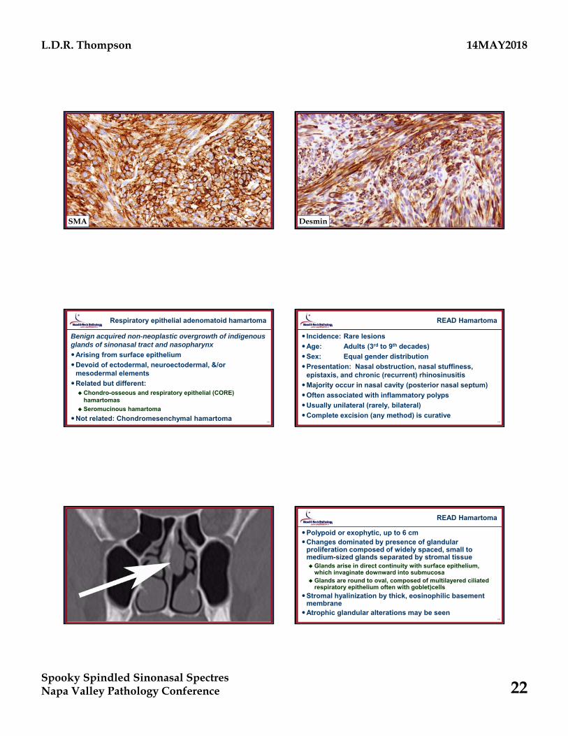
Respiratory epithelial adenomatoid hamartoma
Benign acquired non-neoplastic overgrowth of indigenous glands of sinonasal tract and nasopharynx
Arising from surface epithelium
Devoid of ectodermal, neuroectodermal, &/or mesodermal elements
Related but different: Chondro-osseous and respiratory epithelial (CORE)
hamartomas
Seromucinous hamartoma
Not related: Chondromesenchymal hamartoma130
READ Hamartoma
Incidence: Rare lesions
Age: Adults (3rd to 9th decades)
Sex: Equal gender distribution
Presentation: Nasal obstruction, nasal stuffiness, epistaxis, and chronic (recurrent) rhinosinusitis
Majority occur in nasal cavity (posterior nasal septum)
Often associated with inflammatory polyps
Usually unilateral (rarely, bilateral)
Complete excision (any method) is curative
131 132
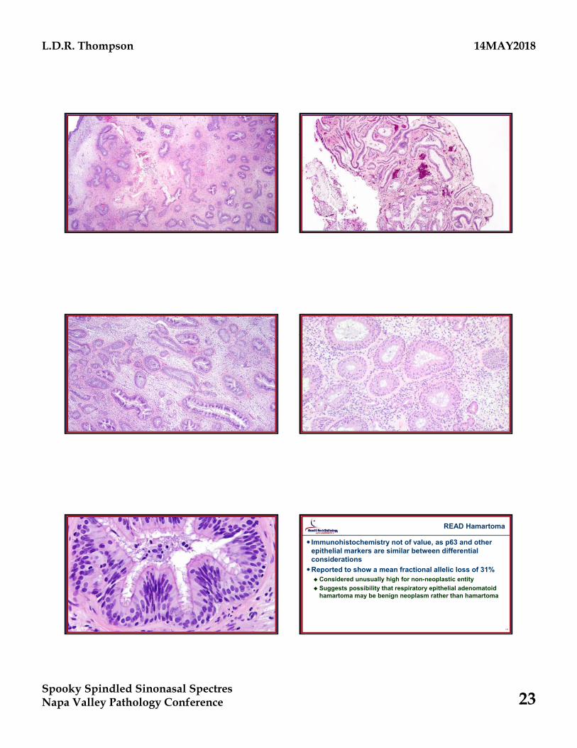
READ Hamartoma
Polypoid or exophytic, up to 6 cm Changes dominated by presence of glandular
proliferation composed of widely spaced, small to medium-sized glands separated by stromal tissueGlands arise in direct continuity with surface epithelium,
which invaginate downward into submucosaGlands are round to oval, composed of multilayered ciliated
respiratory epithelium often with goblet)cells
Stromal hyalinization by thick, eosinophilic basement membrane
Atrophic glandular alterations may be seen
L.D.R. Thompson 14MAY2018
Spooky Spindled Sinonasal SpectresNapa Valley Pathology Conference 23
133 134
135 136
137 138
READ Hamartoma
Immunohistochemistry not of value, as p63 and other epithelial markers are similar between differential considerations
Reported to show a mean fractional allelic loss of 31% Considered unusually high for non-neoplastic entity
Suggests possibility that respiratory epithelial adenomatoid hamartoma may be benign neoplasm rather than hamartoma
L.D.R. Thompson 14MAY2018
Spooky Spindled Sinonasal SpectresNapa Valley Pathology Conference 24
139
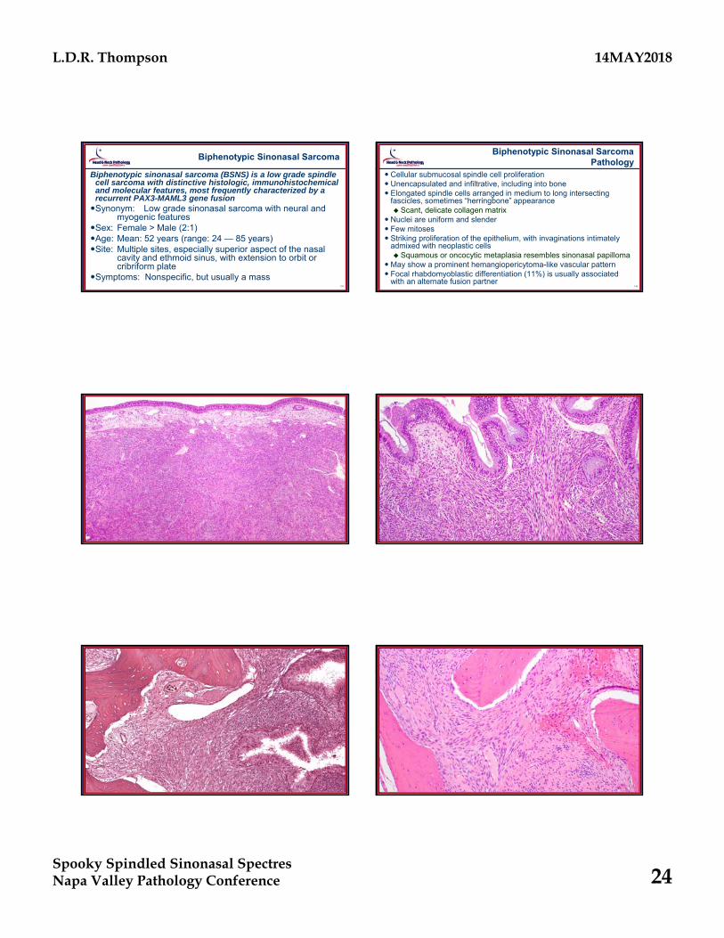
Biphenotypic Sinonasal Sarcoma
Biphenotypic sinonasal sarcoma (BSNS) is a low grade spindle cell sarcoma with distinctive histologic, immunohistochemical and molecular features, most frequently characterized by a recurrent PAX3-MAML3 gene fusionSynonym: Low grade sinonasal sarcoma with neural and
myogenic featuresSex: Female > Male (2:1) Age: Mean: 52 years (range: 24 — 85 years)Site: Multiple sites, especially superior aspect of the nasal
cavity and ethmoid sinus, with extension to orbit or cribriform plate
Symptoms: Nonspecific, but usually a mass140
Biphenotypic Sinonasal SarcomaPathology
Cellular submucosal spindle cell proliferation Unencapsulated and infiltrative, including into bone Elongated spindle cells arranged in medium to long intersecting
fascicles, sometimes “herringbone” appearance Scant, delicate collagen matrix
Nuclei are uniform and slender Few mitoses Striking proliferation of the epithelium, with invaginations intimately
admixed with neoplastic cells Squamous or oncocytic metaplasia resembles sinonasal papilloma
May show a prominent hemangiopericytoma-like vascular pattern Focal rhabdomyoblastic differentiation (11%) is usually associated
with an alternate fusion partner
141 142
143 144
L.D.R. Thompson 14MAY2018
Spooky Spindled Sinonasal SpectresNapa Valley Pathology Conference 25
145 146
147 148
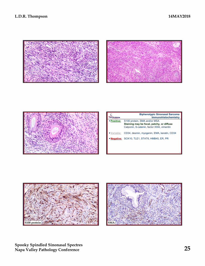
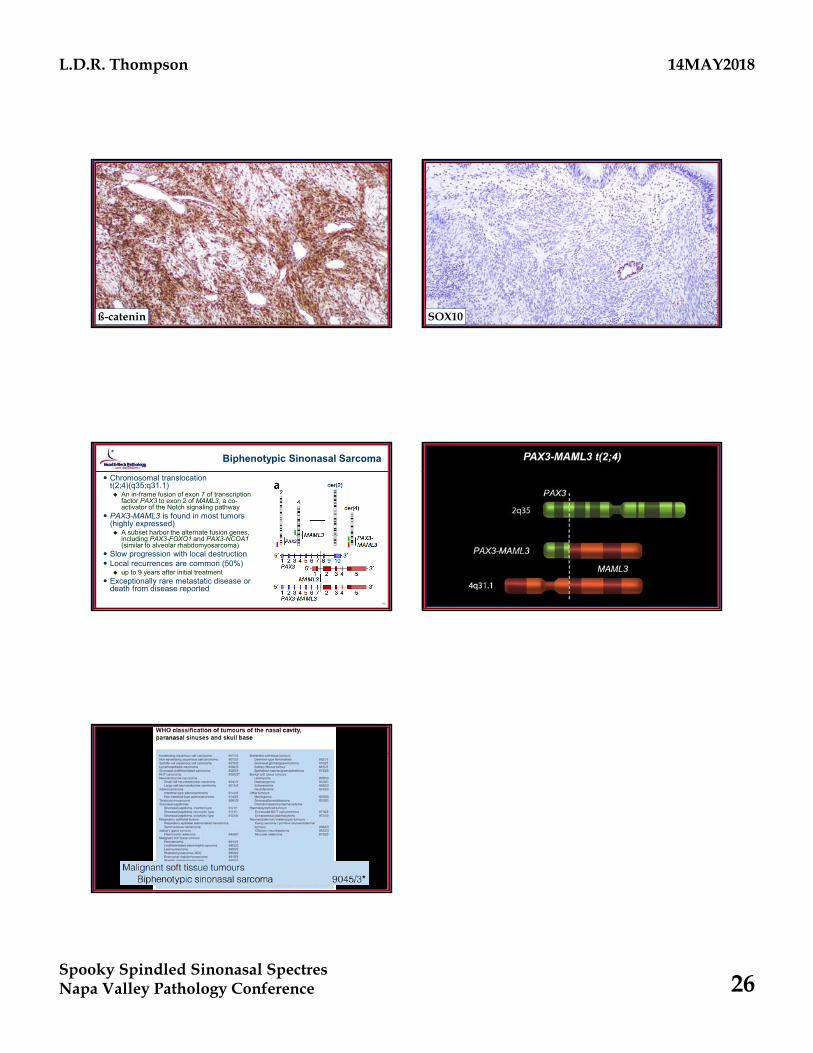
Biphenotypic Sinonasal SarcomaImmunohistochemistry
Positive: S100 protein, SMA and/or MSAStaining may be focal, patchy, or diffuseCalponin, ß-catenin, factor XIIIA, vimentin
Variable: CD34, desmin, myogenin, EMA, keratin, CD34
Negative: SOX10, TLE1, STAT6, HMB45, ER, PR
149S100 protein
150SMA
L.D.R. Thompson 14MAY2018
Spooky Spindled Sinonasal SpectresNapa Valley Pathology Conference 26
151ß-catenin
152SOX10
153
Biphenotypic Sinonasal Sarcoma
Chromosomal translocation t(2;4)(q35;q31.1) An in-frame fusion of exon 7 of transcription
factor PAX3 to exon 2 of MAML3, a co-activator of the Notch signaling pathway
PAX3-MAML3 is found in most tumors (highly expressed) A subset harbor the alternate fusion genes,
including PAX3-FOXO1 and PAX3-NCOA1(similar to alveolar rhabdomyosarcoma)
Slow progression with local destruction Local recurrences are common (50%)
up to 9 years after initial treatment Exceptionally rare metastatic disease or
death from disease reported
154
155