Embed Size (px)
Citation preview
1
1
Lecture 5 (Ch6) - Viruses
• Topics
– Characteristics
– Structure/Classification
– Multiplication
– Cultivation and replication
– Non-viral infectious agents
– Treatment
2
Virus Characteristics
• obligate intracellular parasites
• not cells
• tiny! - 20nm -450nm (no light scope)
• do not independently fulfill characteristics of life
• active only inside the cell
• surface molecules confer high specificity
• use hosts genetic material
• lack enzymes or machinery for synthesis
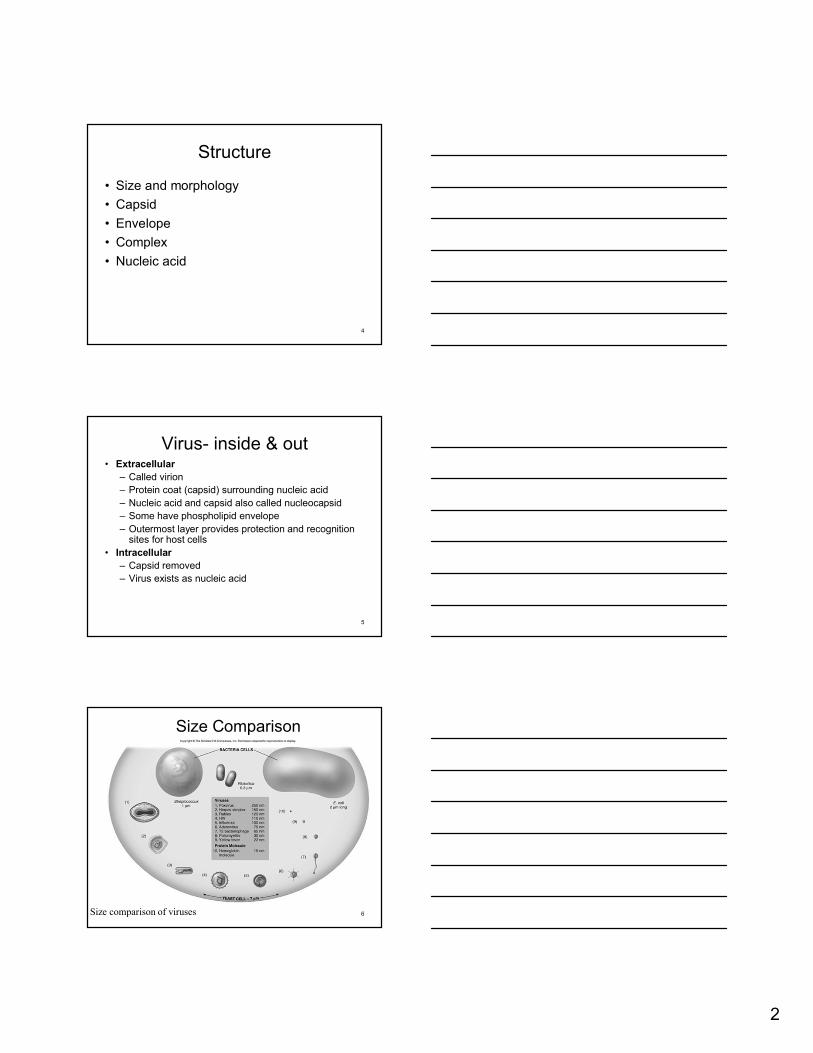
Viral Host Range
3
Most infect only specific host (attachment)Can be so specific only infect specific type of cell in specific hostSome generalists – infect many kinds of cells in many different hosts
2
4
Structure
• Size and morphology
• Capsid
• Envelope
• Complex
• Nucleic acid
Virus- inside & out• Extracellular
– Called virion
– Protein coat (capsid) surrounding nucleic acid
– Nucleic acid and capsid also called nucleocapsid
– Some have phospholipid envelope
– Outermost layer provides protection and recognition sites for host cells
• Intracellular
– Capsid removed
– Virus exists as nucleic acid
5
6
Size Comparison
Size comparison of viruses
3
More Size…
7
8
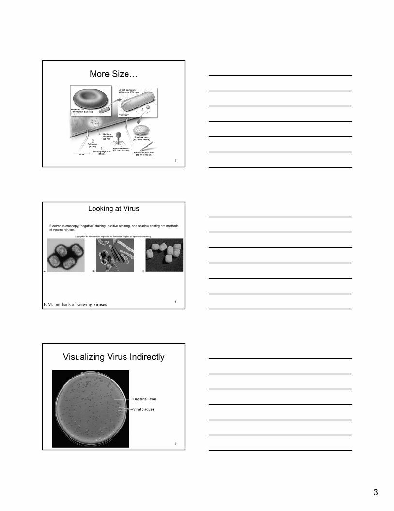
Looking at Virus
E.M. methods of viewing viruses
Electron microscopy, “negative” staining, positive staining, and shadow casting are methods of viewing viruses.
Visualizing Virus Indirectly
9

4
10
The two major structure types for viruses:
Generalized viral structures
naked nucleocapsid virus enveloped virus
11
Capsid
• Protective outer shell that surrounds viral nucleic acid
• Capsid spikes
• Composed of capsomer subunits
• Two types of capsids (based on shape):
– Helical
– Icosahedral
12
Helical capsid
• Naked helical virus
– Nucleocapsid is rigid and tightly wound into a cylinder-shaped package
– Example: Tobacco mosaic virus
• Enveloped helical virus
– Nucleocapsid is more flexible than naked virus
– Examples: Influenza, measles, rabies
5
13
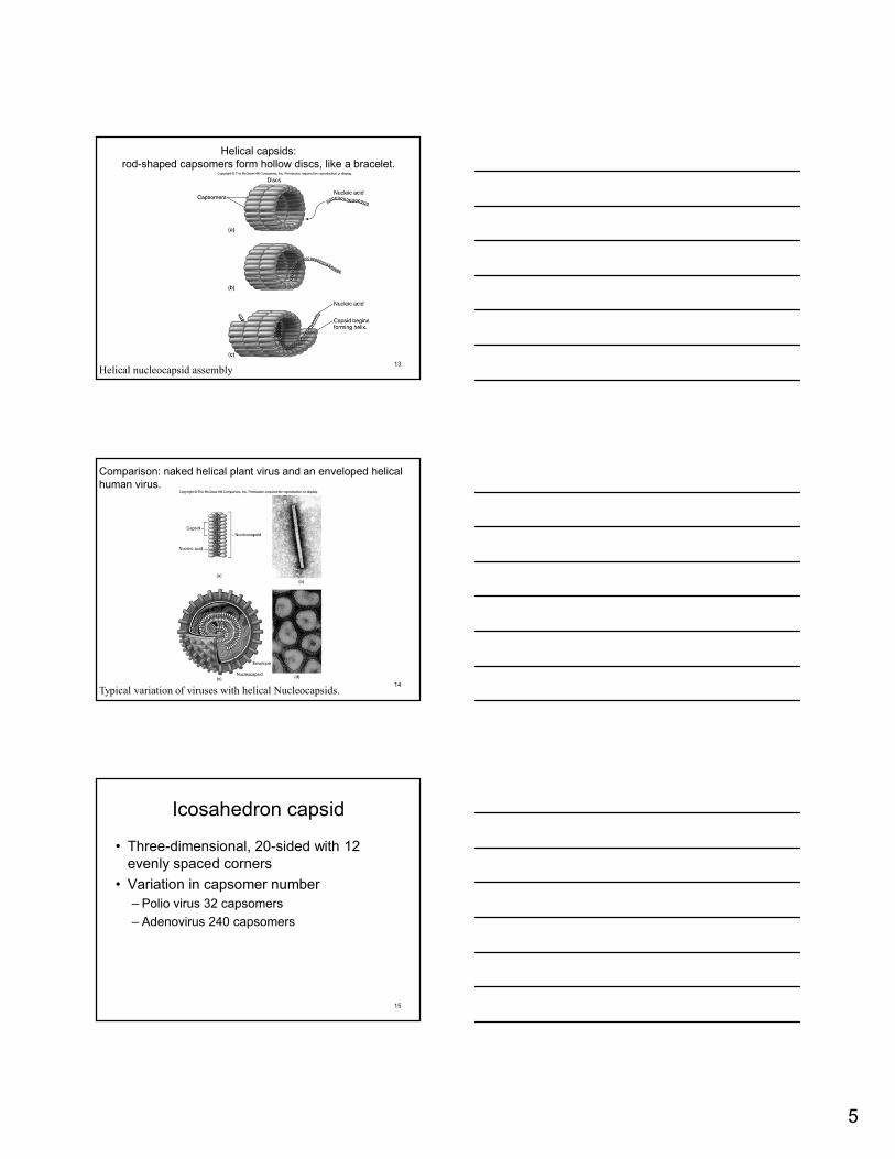
Helical capsids:rod-shaped capsomers form hollow discs, like a bracelet.
Helical nucleocapsid assembly
14
Comparison: naked helical plant virus and an enveloped helical human virus.
Typical variation of viruses with helical Nucleocapsids.
15
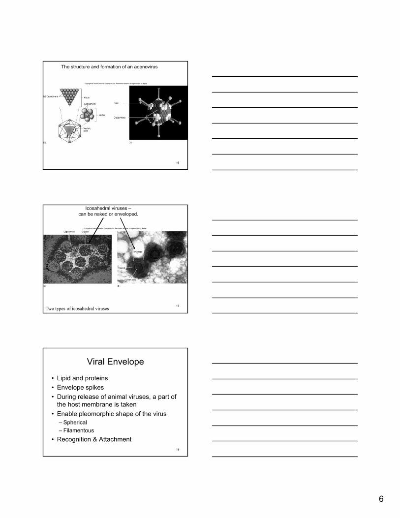
Icosahedron capsid
• Three-dimensional, 20-sided with 12 evenly spaced corners
• Variation in capsomer number
– Polio virus 32 capsomers
– Adenovirus 240 capsomers
6
16
The structure and formation of an adenovirus
17
Icosahedral viruses –can be naked or enveloped.
Two types of icosahedral viruses
18
Viral Envelope
• Lipid and proteins
• Envelope spikes
• During release of animal viruses, a part of the host membrane is taken
• Enable pleomorphic shape of the virus
– Spherical
– Filamentous
• Recognition & Attachment
7
19
Function of the capsid/envelope
• Protect nucleic acid from the host’s acid-and protein-digesting enzymes
• Assist in binding and penetrating host cell
• Stimulate the host’s immune system
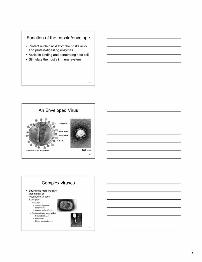
An Enveloped Virus
20
21
Complex viruses
• Structure is more intricate than helical or icosahedral viruses. Examples:– Pox virus
• Several layers of lipoproteins
• Course surface fibrils
– Bacteriophage (next slide)
• Polyhedral head
• Helical tail
• Fibers for attachment
8
22
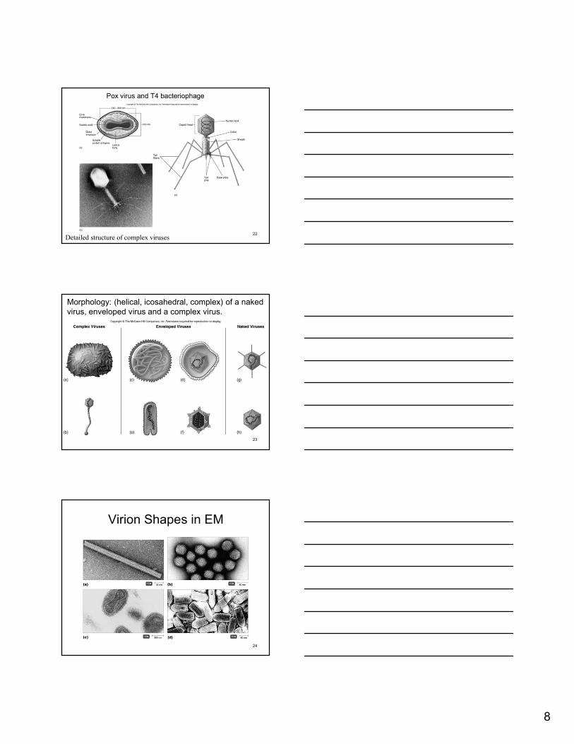
Pox virus and T4 bacteriophage
Detailed structure of complex viruses
23
Morphology: (helical, icosahedral, complex) of a naked virus, enveloped virus and a complex virus.
Virion Shapes in EM
24
9
25
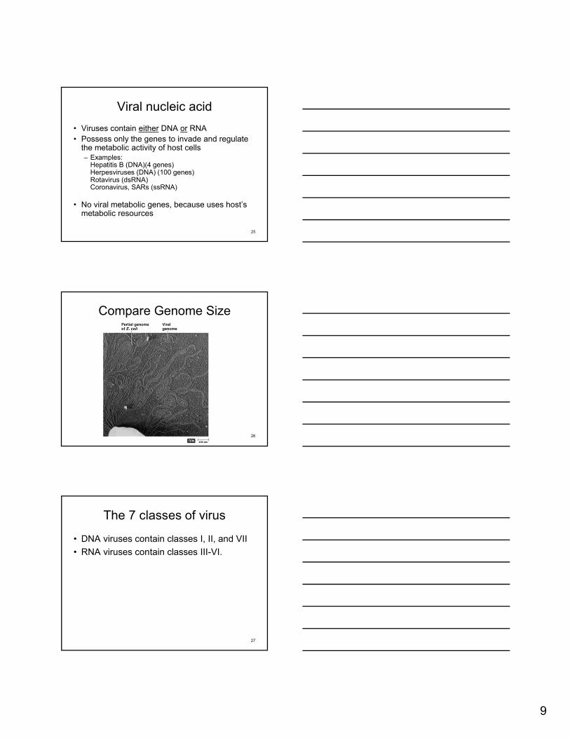
Viral nucleic acid
• Viruses contain either DNA or RNA
• Possess only the genes to invade and regulate the metabolic activity of host cells– Examples:
Hepatitis B (DNA)(4 genes) Herpesviruses (DNA) (100 genes)Rotavirus (dsRNA)Coronavirus, SARs (ssRNA)
• No viral metabolic genes, because uses host’s metabolic resources
Compare Genome Size
26
27
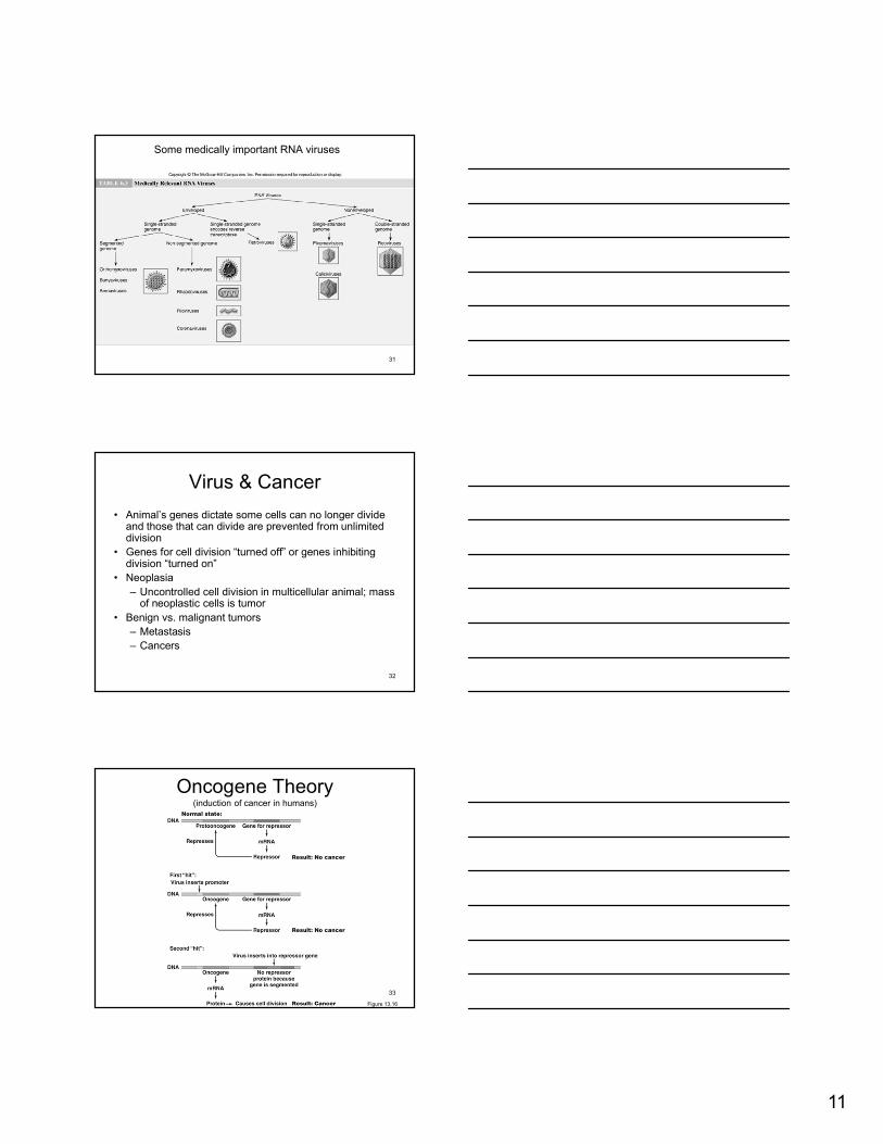
The 7 classes of virus
• DNA viruses contain classes I, II, and VII
• RNA viruses contain classes III-VI.

10
28
DNA virus classes (I, II, VII)• Class I viruses:
– double-stranded DNA (dsDNA) genome– Examples:
• Some phages• Family Herpesviridae (includes human herpesviruses),
Varicella Zoster, Poxviridae, JC, papilloma
• Class II viruses: – +sense single-stranded DNA (ssDNA) genome.– Example: Parvoviridae
• Class VII viruses:– double-stranded, reverse transcriptase (dsDNA-RT)
genome.– Example: Hepadenovirus
29
RNA virus classes (III-VI)• Class III viruses:
– double-stranded RNA (dsRNA) genome.
– Example: Reovirus
• Class IV viruses: – +sense single-stranded RNA (ssRNA) genome (acts as mRNA).
Example: Picornaviruses,
• Class V viruses: – -sense single-stranded RNA (ssRNA) genome used as a
template for mRNA synthesis.
– Example: Rhabdovirus
• Class VI viruses: – +sense single- stranded reverse transcriptase RNA (ssRNA-RT)
genome (with DNA intermediate in replication and also mRNA synthesis).
– Example: Retroviridae
30
Examples of medically important DNA viruses
11
31
Some medically important RNA viruses
Virus & Cancer
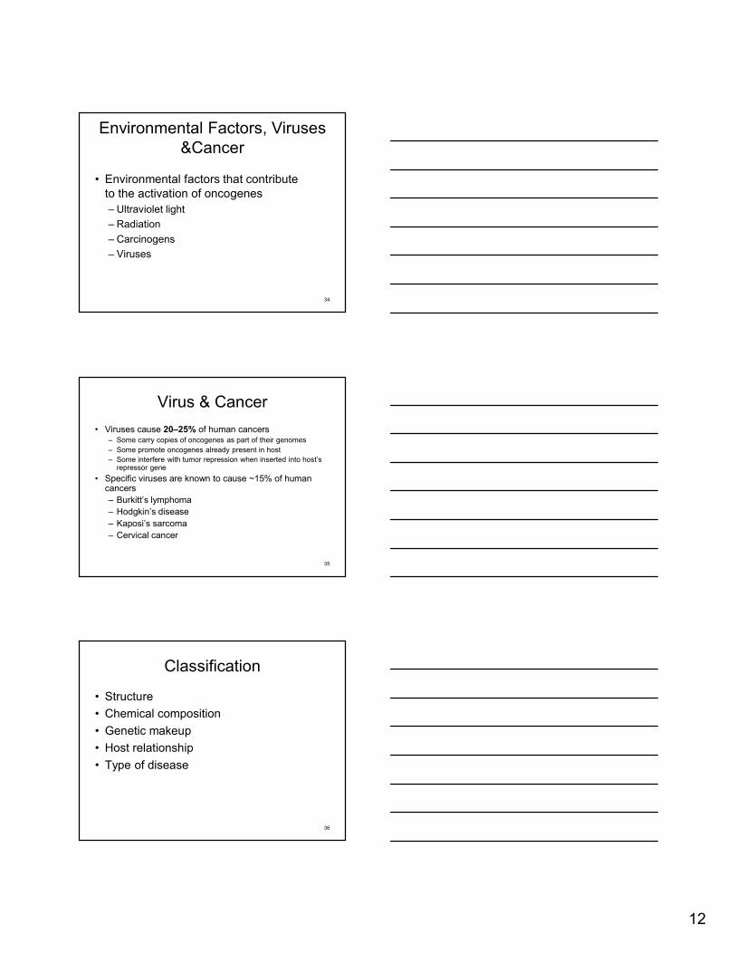
• Animal’s genes dictate some cells can no longer divide and those that can divide are prevented from unlimited division
• Genes for cell division “turned off” or genes inhibiting division “turned on”
• Neoplasia
– Uncontrolled cell division in multicellular animal; mass of neoplastic cells is tumor
• Benign vs. malignant tumors
– Metastasis
– Cancers
32
Oncogene Theory (induction of cancer in humans)
Figure 13.16
33
12
Environmental Factors, Viruses &Cancer
• Environmental factors that contribute to the activation of oncogenes
– Ultraviolet light
– Radiation
– Carcinogens
– Viruses
34
Virus & Cancer
• Viruses cause 20–25% of human cancers– Some carry copies of oncogenes as part of their genomes
– Some promote oncogenes already present in host
– Some interfere with tumor repression when inserted into host’s repressor gene
• Specific viruses are known to cause ~15% of human cancers
– Burkitt’s lymphoma
– Hodgkin’s disease
– Kaposi’s sarcoma
– Cervical cancer
35
36
Classification
• Structure
• Chemical composition
• Genetic makeup
• Host relationship
• Type of disease
13
37
Three orders of viruses developed for classification.
38
Classification of important human viruses
39
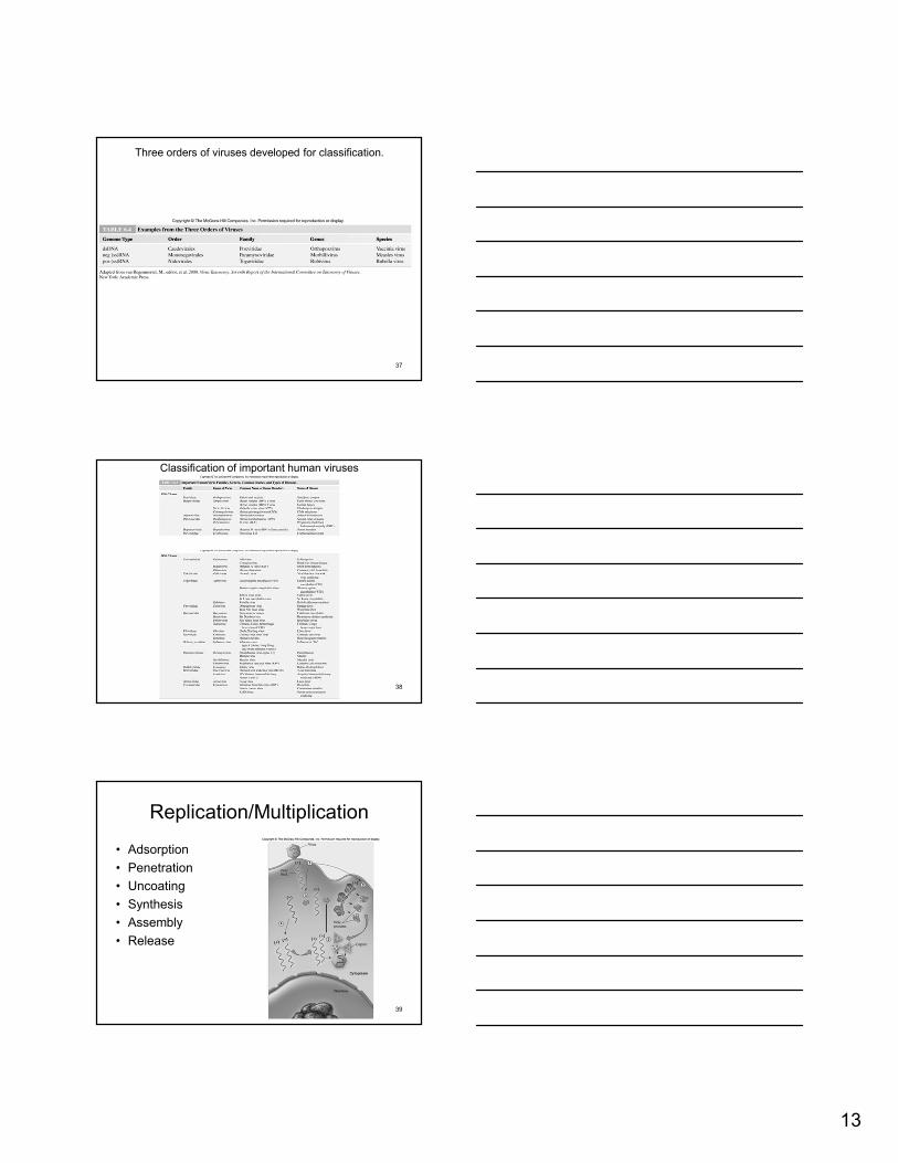
Replication/Multiplication
• Adsorption
• Penetration
• Uncoating
• Synthesis
• Assembly
• Release
14
Virions in Persistent Infection
40
41
Adsorption to Host Cell
enveloped spike virus naked capsid spike virus.
42
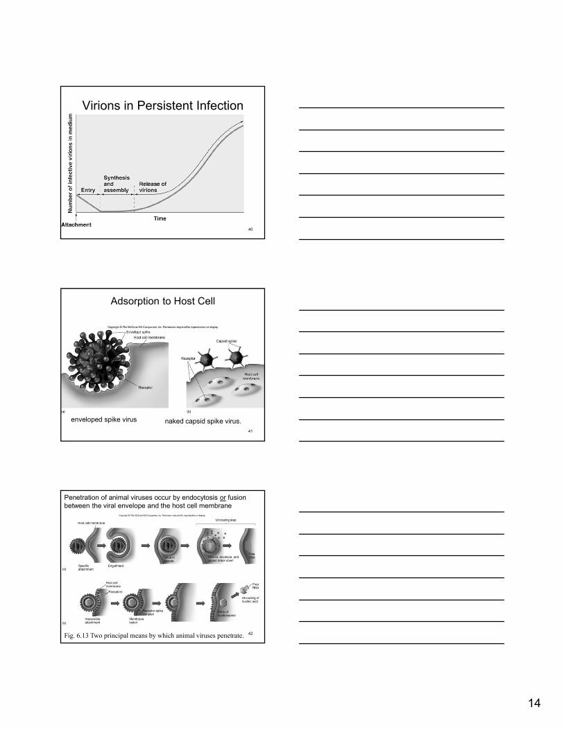
Penetration of animal viruses occur by endocytosis or fusion between the viral envelope and the host cell membrane
Fig. 6.13 Two principal means by which animal viruses penetrate.
15
43
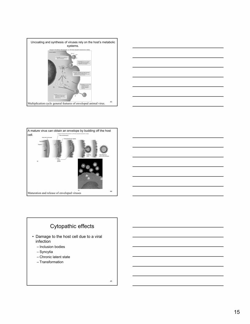
Uncoating and synthesis of viruses rely on the host’s metabolic systems.
Multiplication cycle general features of enveloped animal virus.
44
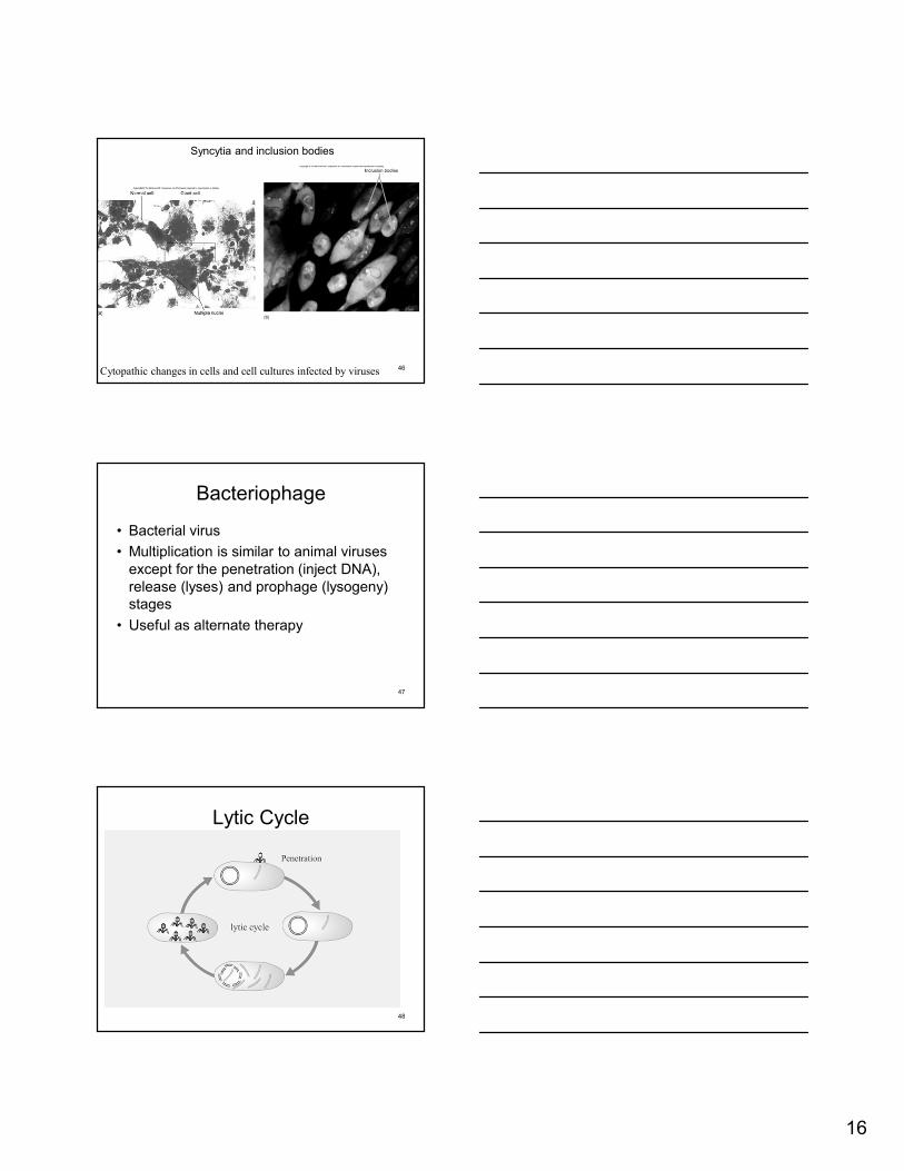
A mature virus can obtain an envelope by budding off the host
cell.
Maturation and release of enveloped viruses
45
Cytopathic effects
• Damage to the host cell due to a viral infection
– Inclusion bodies
– Syncytia
– Chronic latent state
– Transformation
16
46
Syncytia and inclusion bodies
Cytopathic changes in cells and cell cultures infected by viruses
47
Bacteriophage
• Bacterial virus
• Multiplication is similar to animal viruses except for the penetration (inject DNA), release (lyses) and prophage (lysogeny) stages
• Useful as alternate therapy
Lytic Cycle
48

17
Viral Lytic Cycle
49
Viral Lytic Cycle
50
Lysogeny
51
•Modified replication cycle•Infected host cells grow and reproduce normally for generations before they lyse•Temperate phages
Prophages – inactive phages•Lysogenic conversion results when phages carry genes that alter phenotype of a bacterium

18
52
Lysogeny is when the bacteriophage inserts its DNA into the bacterial host genome.
The lysogenic state in bacteria
Lytic to Lysogenic
53
54
T-even bacteriophage penetrate the host cell by specifically binding and injecting their DNA into the host cell.
Penetration of a bacterial cell by a T-even bacteriophage.
19
55
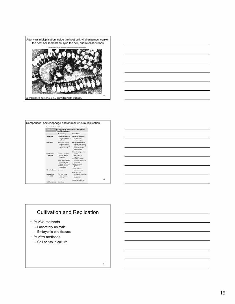
After viral multiplication inside the host cell, viral enzymes weaken the host cell membrane, lyse the cell, and release virions
A weakened bacterial cell, crowded with viruses.
56
Comparison: bacteriophage and animal virus multiplication
57
Cultivation and Replication
• In vivo methods
– Laboratory animals
– Embryonic bird tissues
• In vitro methods
– Cell or tissue culture

20
58
Early developing bird embryos have a protective case that provides an ideal viral propagation environment
Cultivating animal viruses in a developingbird embryo
59
A monolayer of monkey kidney cells is a cell culture that enables virus propagation
Normal and infected cell cultures
60
Non-cellular Infectious Agents
• Prions (naked proteins)
• Satellite viruses (usually plant virus associated for purpose of replication)
• Viroids (unique plant pathogens, small, single-stranded, circular RNA )
21
61
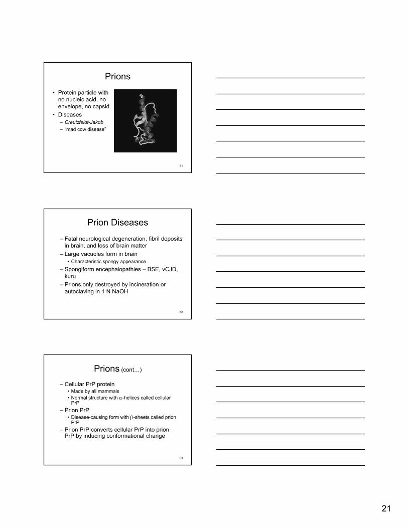
Prions
• Protein particle with no nucleic acid, no envelope, no capsid
• Diseases
– Creutzfeldt-Jakob
– “mad cow disease”
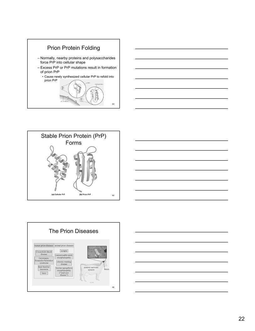
Prion Diseases
– Fatal neurological degeneration, fibril deposits in brain, and loss of brain matter
– Large vacuoles form in brain
• Characteristic spongy appearance
– Spongiform encephalopathies – BSE, vCJD, kuru
– Prions only destroyed by incineration or autoclaving in 1 N NaOH
62
Prions (cont…)
– Cellular PrP protein• Made by all mammals
• Normal structure with -helices called cellular PrP
– Prion PrP• Disease-causing form with -sheets called prion
PrP
– Prion PrP converts cellular PrP into prion PrP by inducing conformational change
63
22
Prion Protein Folding
– Normally, nearby proteins and polysaccharides force PrP into cellular shape
– Excess PrP or PrP mutations result in formation of prion PrP
• Cause newly synthesized cellular PrP to refold into prion PrP
64
Stable Prion Protein (PrP) Forms
65
The Prion Diseases
66

23
67
Satellite viruses
• Dependent on other viruses for replication
• Ex. Delta agent, which is only expressed in the presence of hepatitis B virus, depend on it for replication- the only viroid like infectious agent of animals.
68
Viroids
• Plant pathogens
– Tomatoes, potatoes, cucumbers.
• 1/10th the size of normal viruses
• Naked strands of RNA, no capsid
Example Viroid Effects
69