Embed Size (px)
Citation preview
Lecture 6 Intracellular accumulations
Aging/Senescence
Associate Professor Dr. Alexey Podcheko
Spring 2015
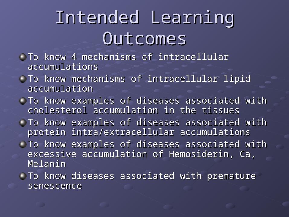
Intended Learning OutcomesIntended Learning Outcomes
To know 4 mechanisms of intracellular accumulationsTo know 4 mechanisms of intracellular accumulationsTo know mechanisms of intracellular lipid accumulationTo know mechanisms of intracellular lipid accumulationTo know examples of diseases associated with To know examples of diseases associated with cholesterol accumulation in the tissuescholesterol accumulation in the tissuesTo know examples of diseases associated with protein To know examples of diseases associated with protein intra/extracellular accumulationsintra/extracellular accumulationsTo know examples of diseases associated with To know examples of diseases associated with excessive accumulation of Hemosiderin, Ca, Melaninexcessive accumulation of Hemosiderin, Ca, MelaninTo know diseases associated with premature To know diseases associated with premature senescencesenescence
Intracellular AccumulationsIntracellular Accumulations
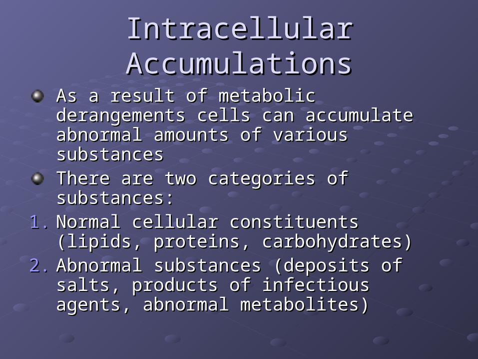
As a result of metabolic derangements As a result of metabolic derangements cells can accumulate abnormal amounts cells can accumulate abnormal amounts of various substancesof various substancesThere are two categories of substances: There are two categories of substances:
1.1. Normal cellular constituents (lipids, Normal cellular constituents (lipids, proteins, carbohydrates)proteins, carbohydrates)
2.2. Abnormal substances (deposits of salts, Abnormal substances (deposits of salts, products of infectious agents, abnormal products of infectious agents, abnormal metabolites)metabolites)
First main mechanisms of First main mechanisms of intracellular accumulation (out of 4) intracellular accumulation (out of 4)
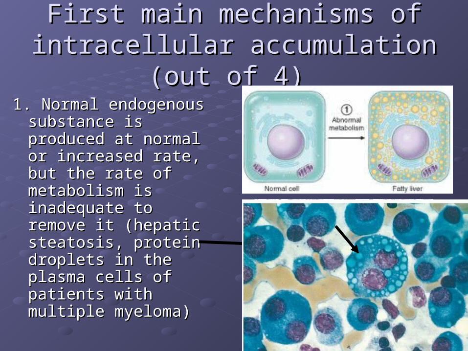
1. Normal endogenous 1. Normal endogenous substance is produced substance is produced at normal or increased at normal or increased rate, but the rate of rate, but the rate of metabolism is metabolism is inadequate to remove inadequate to remove it (hepatic steatosis, it (hepatic steatosis, protein droplets in the protein droplets in the plasma cells of plasma cells of patients with multiple patients with multiple myeloma)myeloma)
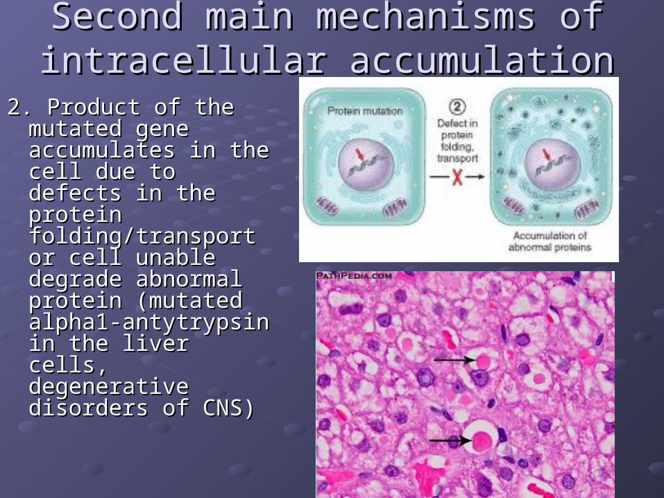
2. Product of the 2. Product of the mutated gene mutated gene accumulates in the accumulates in the cell due to defects in cell due to defects in the protein the protein folding/transport or folding/transport or cell unable degrade cell unable degrade abnormal protein abnormal protein (mutated alpha1-(mutated alpha1-antytrypsin in the antytrypsin in the liver cells, liver cells, degenerative degenerative disorders of CNS)disorders of CNS)
Second main mechanisms of Second main mechanisms of intracellular accumulation intracellular accumulation
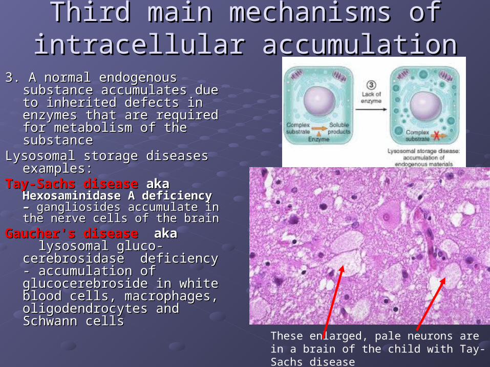
3. A normal endogenous substance 3. A normal endogenous substance accumulates due to inherited accumulates due to inherited defects in enzymes that are defects in enzymes that are required for metabolism of the required for metabolism of the substancesubstance
Lysosomal storage diseases Lysosomal storage diseases examples:examples:
Tay-Sachs diseaseTay-Sachs disease aka aka Hexosaminidase A deficiency – Hexosaminidase A deficiency – gangliosides accumulate in gangliosides accumulate in the nerve cells of the brainthe nerve cells of the brain
Gaucher's diseaseGaucher's disease aka aka lysosomal gluco- lysosomal gluco-cerebrosidase deficiency - cerebrosidase deficiency - accumulation of accumulation of glucocerebroside in white blood glucocerebroside in white blood cells, macrophages, cells, macrophages, oligodendrocytes and Schwann oligodendrocytes and Schwann cellscells
Third main mechanisms of Third main mechanisms of intracellular accumulation intracellular accumulation
These enlarged, pale neurons are in a brain of the child with Tay-Sachs disease
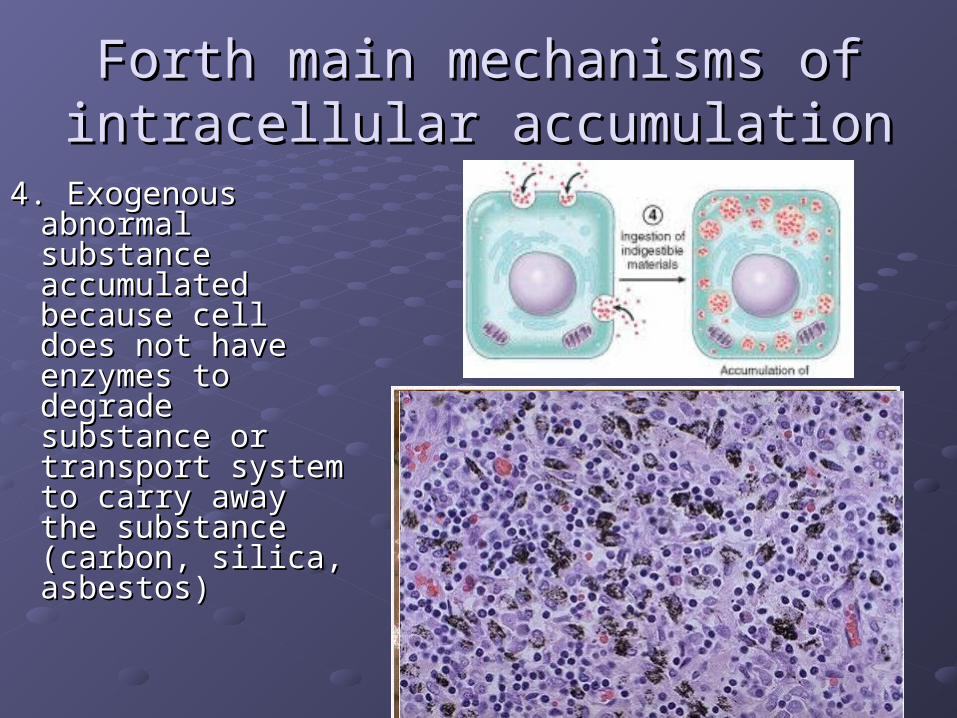
4. Exogenous 4. Exogenous abnormal abnormal substance substance accumulated accumulated because cell does because cell does not have enzymes not have enzymes to degrade to degrade substance or substance or transport system transport system to carry away the to carry away the substance substance (carbon, silica, (carbon, silica, asbestos)asbestos)
Forth main mechanisms of Forth main mechanisms of intracellular accumulation intracellular accumulation
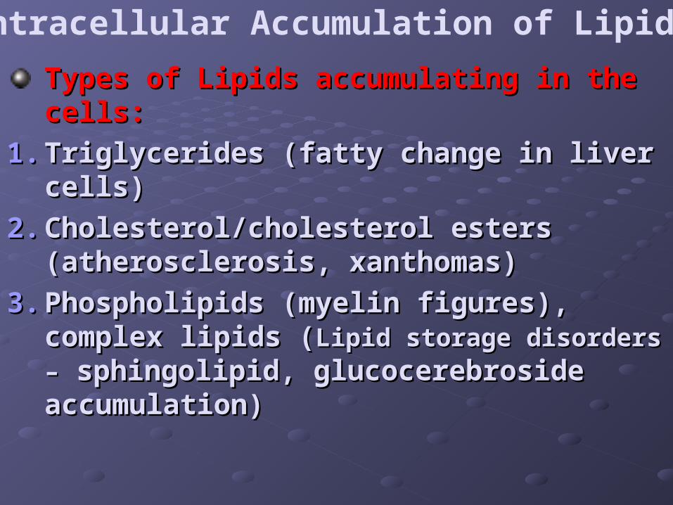
Intracellular Accumulation of Lipids
Types of Lipids accumulating in the cells: Types of Lipids accumulating in the cells:
1.1. Triglycerides (fatty change in liver cells)Triglycerides (fatty change in liver cells)
2.2. Cholesterol/cholesterol esters Cholesterol/cholesterol esters (atherosclerosis, xanthomas)(atherosclerosis, xanthomas)
3.3. Phospholipids (myelin figures), complex Phospholipids (myelin figures), complex lipids (lipids (Lipid storage disorders –Lipid storage disorders – sphingolipid, glucocerebroside sphingolipid, glucocerebroside accumulation) accumulation)
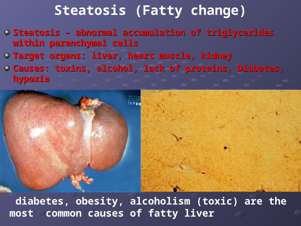
Steatosis – abnormal accumulation of triglycerides within Steatosis – abnormal accumulation of triglycerides within parenchymal cellsparenchymal cells
Target organs: liver, heart muscle, kidneyTarget organs: liver, heart muscle, kidney
Causes: toxins, alcohol, lack of proteins, Diabetes, hypoxiaCauses: toxins, alcohol, lack of proteins, Diabetes, hypoxia
diabetes, obesity, alcoholism (toxic) are the most common causes of fatty liver
Steatosis (Fatty change)
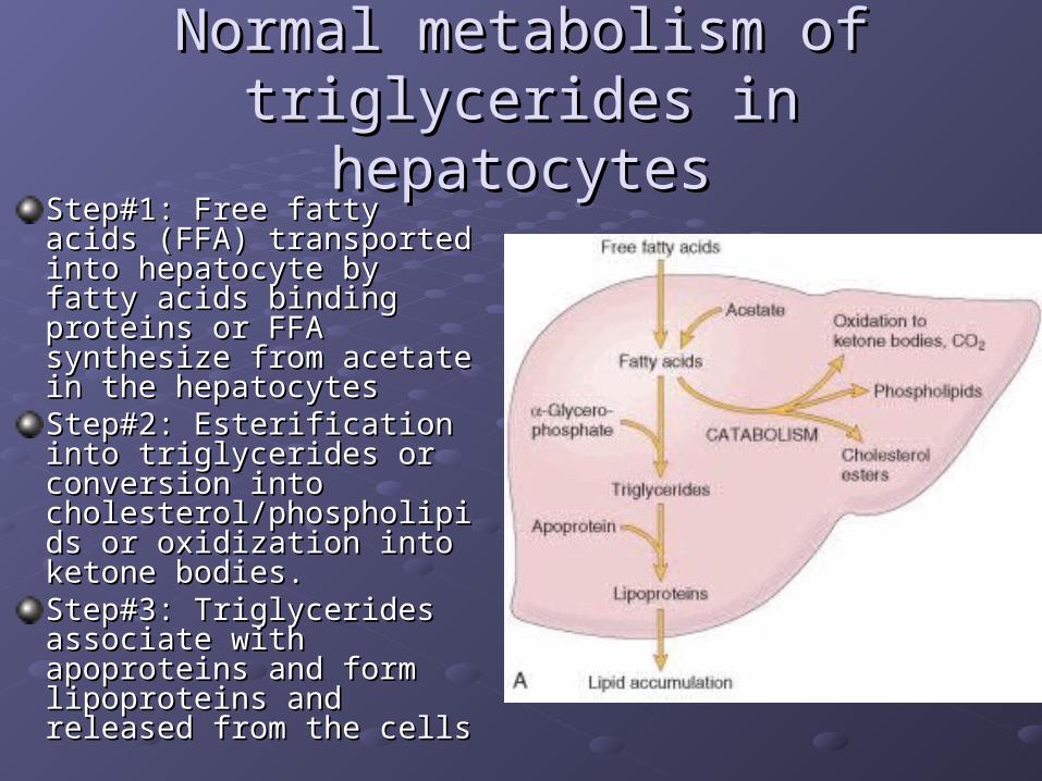
Normal metabolism of triglycerides Normal metabolism of triglycerides in hepatocytesin hepatocytes
Step#1: Free fatty acids Step#1: Free fatty acids (FFA) transported into (FFA) transported into hepatocyte by fatty acids hepatocyte by fatty acids binding proteins or FFA binding proteins or FFA synthesize from acetate in synthesize from acetate in the hepatocytesthe hepatocytesStep#2: Esterification into Step#2: Esterification into triglycerides or conversion triglycerides or conversion into into cholesterol/phospholipids cholesterol/phospholipids or oxidization into ketone or oxidization into ketone bodies. bodies. Step#3: Triglycerides Step#3: Triglycerides associate with apoproteins associate with apoproteins and form lipoproteins and and form lipoproteins and released from the cells released from the cells
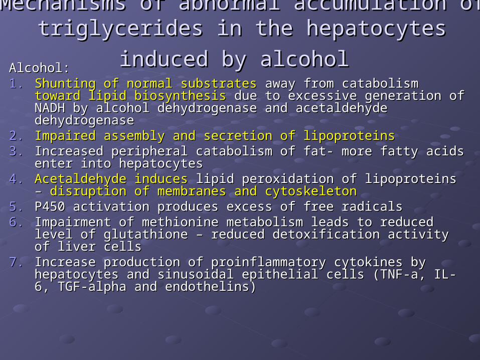
Mechanisms of abnormal accumulation of Mechanisms of abnormal accumulation of
triglycerides in the hepatocytes induced by alcoholtriglycerides in the hepatocytes induced by alcohol Alcohol: Alcohol: 1.1. Shunting of normal substratesShunting of normal substrates away from catabolism away from catabolism toward lipid toward lipid
biosynthesisbiosynthesis due to excessive generation of NADH by alcohol due to excessive generation of NADH by alcohol dehydrogenase and acetaldehyde dehydrogenasedehydrogenase and acetaldehyde dehydrogenase
2.2. Impaired assembly and secretion of lipoproteinsImpaired assembly and secretion of lipoproteins3.3. Increased peripheral catabolism of fat- more fatty acids enter into Increased peripheral catabolism of fat- more fatty acids enter into
hepatocyteshepatocytes4.4. Acetaldehyde inducesAcetaldehyde induces lipid peroxidation of lipoproteins – lipid peroxidation of lipoproteins –
disruption of membranes and cytoskeletondisruption of membranes and cytoskeleton5.5. P450 activation produces excess of free radicalsP450 activation produces excess of free radicals6.6. Impairment of methionine metabolism leads to reduced level of Impairment of methionine metabolism leads to reduced level of
glutathione – reduced detoxification activity of liver cellsglutathione – reduced detoxification activity of liver cells7.7. Increase production of proinflammatory cytokines by hepatocytes Increase production of proinflammatory cytokines by hepatocytes
and sinusoidal epithelial cells (TNF-a, IL-6, TGF-alpha and and sinusoidal epithelial cells (TNF-a, IL-6, TGF-alpha and endothelins)endothelins)
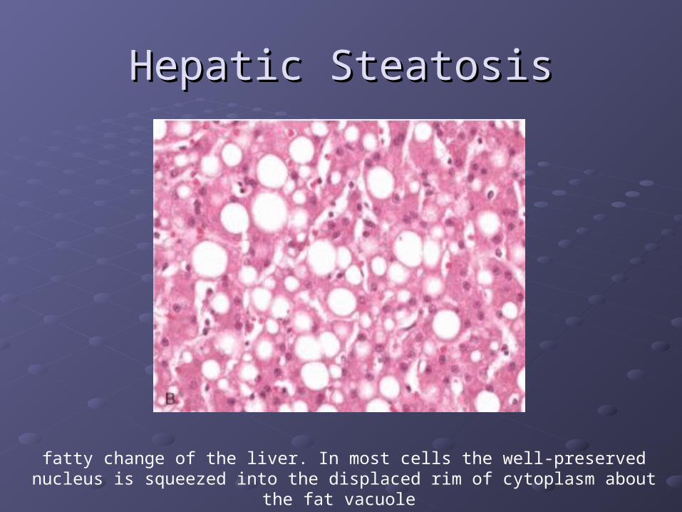
Hepatic SteatosisHepatic Steatosis
fatty change of the liver. In most cells the well-preserved nucleus is squeezed into the displaced rim of cytoplasm about the fat vacuole
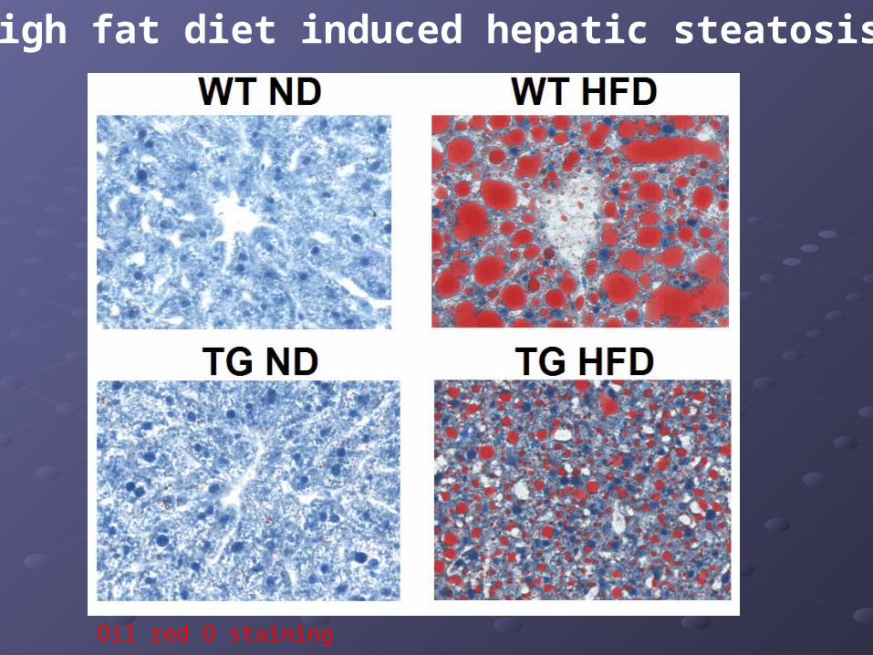
High fat diet induced hepatic steatosis
Oil red O staining
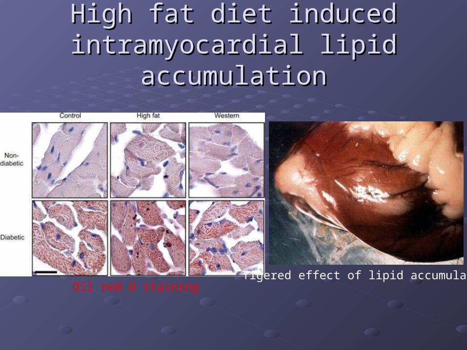
High fat diet induced High fat diet induced intramyocardial lipid accumulationintramyocardial lipid accumulation
Oil red O stainingTigered effect of lipid accumulation
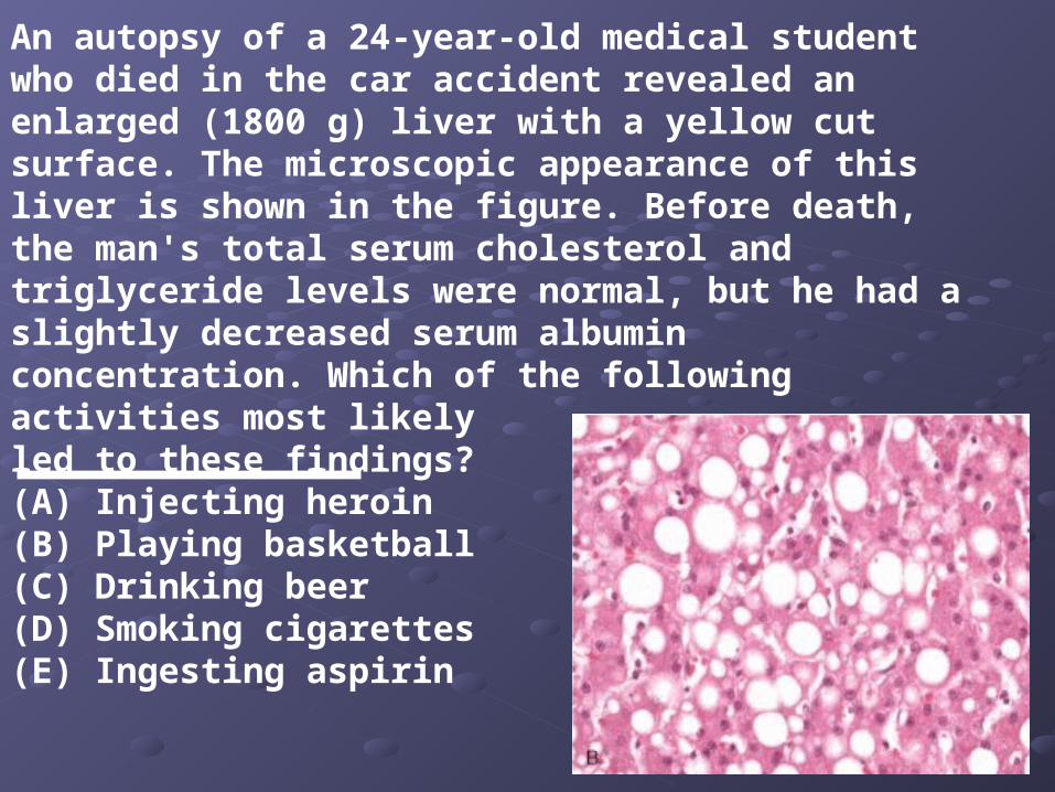
An autopsy of a 24-year-old medical student who died in the car accident revealed an enlarged (1800 g) liver with a yellow cut surface. The microscopic appearance of this liver is shown in the figure. Before death, the man's total serum cholesterol and triglyceride levels were normal, but he had a slightly decreased serum albumin concentration. Which of the following activities most likelyled to these findings?(A) Injecting heroin(B) Playing basketball(C) Drinking beer(D) Smoking cigarettes(E) Ingesting aspirin
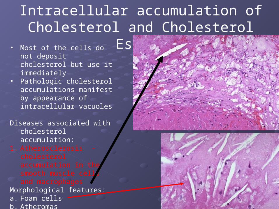
• Most of the cells do not deposit cholesterol but use it immediately
• Pathologic cholesterol accumulations manifest by appearance of intracellular vacuoles
Diseases associated with cholesterol accumulation:
1. Atherosclerosis - cholesterol accumulation in the smooth muscle cells and macrophages
Morphological features:a. Foam cellsb. Atheromasc. Cholesterol Clefts- crystals of
cholesterol
Intracellular accumulation of Cholesterol and Cholesterol Esters
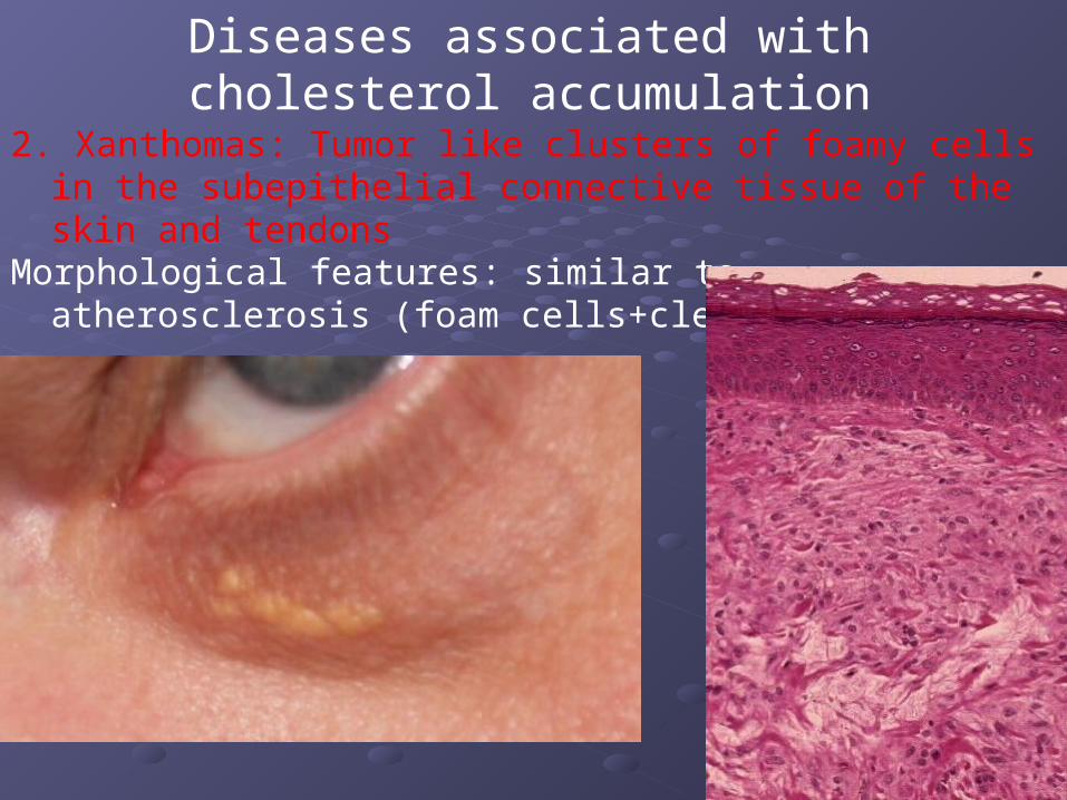
2. Xanthomas: Tumor like clusters of foamy cells in the subepithelial connective tissue of the skin and tendons
Morphological features: similar to atherosclerosis (foam cells+clefts)
Diseases associated with cholesterol accumulation
Diseases associated with cholesterol accumulation
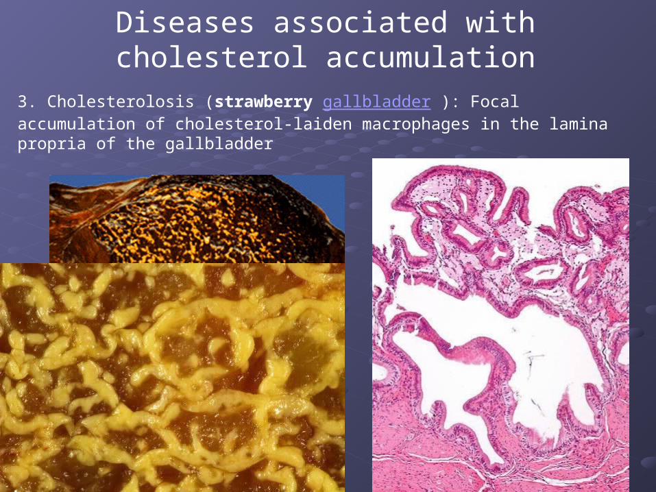
3. Cholesterolosis (strawberry gallbladder ): Focal accumulation of cholesterol-laiden macrophages in the lamina propria of the gallbladder
Diseases associated with cholesterol accumulation
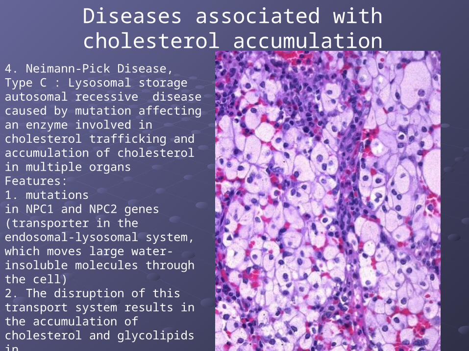
4. Neimann-Pick Disease, Type C : Lysosomal storage autosomal recessive disease caused by mutation affecting an enzyme involved in cholesterol trafficking and accumulation of cholesterol in multiple organsFeatures: 1. mutations in NPC1 and NPC2 genes (transporter in the endosomal-lysosomal system, which moves large water-insoluble molecules through the cell)2. The disruption of this transport system results in the accumulation of cholesterol and glycolipids in lysosomes
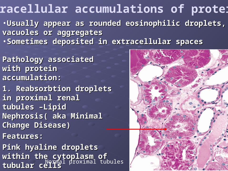
Intracellular accumulations of proteins
Pathology associated with Pathology associated with protein accumulation:protein accumulation:
1. Reabsorbtion droplets in 1. Reabsorbtion droplets in proximal renal tubules –Lipid proximal renal tubules –Lipid Nephrosis( aka Minimal Nephrosis( aka Minimal Change Disease)Change Disease)
Features: Features:
Pink hyaline droplets within Pink hyaline droplets within the cytoplasm of tubular cellsthe cytoplasm of tubular cells
•Usually appear as rounded eosinophilic droplets, vacuoles Usually appear as rounded eosinophilic droplets, vacuoles or aggregatesor aggregates•Sometimes deposited in extracellular spacesSometimes deposited in extracellular spaces
Normal proximal tubules
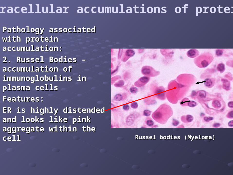
Intracellular accumulations of proteins
Pathology associated with Pathology associated with protein accumulation:protein accumulation:
2. Russel Bodies – 2. Russel Bodies – accumulation of accumulation of immunoglobulins in plasma immunoglobulins in plasma cells cells
Features: Features:
ER is highly distended and ER is highly distended and looks like pink aggregate looks like pink aggregate within the cellwithin the cell
Russel bodies (Myeloma)Russel bodies (Myeloma)
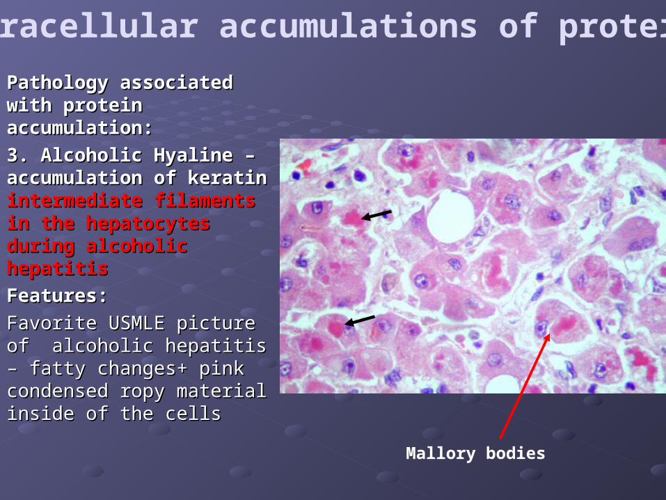
Intracellular accumulations of proteins
Pathology associated Pathology associated with protein with protein accumulation:accumulation:
3. Alcoholic Hyaline – 3. Alcoholic Hyaline – accumulation of keratin accumulation of keratin intermediate filaments in intermediate filaments in the hepatocytes during the hepatocytes during alcoholic hepatitisalcoholic hepatitis
Features: Features:
Favorite USMLE picture of Favorite USMLE picture of alcoholic hepatitis – fatty alcoholic hepatitis – fatty changes+ pink condensed changes+ pink condensed ropy material inside of the ropy material inside of the cellscells
Mallory bodies
AmyloidAmyloid
Hyaline arteriolosclerosis Hyaline arteriolosclerosis
Hyaline membrane disease of the Hyaline membrane disease of the newbornnewborn
Extracellular accumulations of proteins
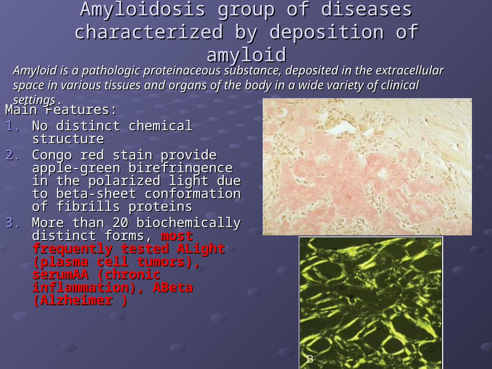
Amyloidosis group of diseases characterized by Amyloidosis group of diseases characterized by deposition of amyloiddeposition of amyloid
Main Features: Main Features: 1.1. No distinct chemical structureNo distinct chemical structure2.2. Congo red stain provide apple-Congo red stain provide apple-
green birefringence in the green birefringence in the polarized light due to beta-polarized light due to beta-sheet conformation of fibrills sheet conformation of fibrills proteinsproteins
3.3. More than 20 biochemically More than 20 biochemically distinct forms, distinct forms, most frequently most frequently tested ALight (plasma cell tested ALight (plasma cell tumors), serumAA (chronic tumors), serumAA (chronic inflammation), ABeta inflammation), ABeta (Alzheimer )(Alzheimer )
Amyloid is a pathologic proteinaceous substance, deposited in the extracellular Amyloid is a pathologic proteinaceous substance, deposited in the extracellular space in various tissues and organs of the body in a wide variety of clinical settingsspace in various tissues and organs of the body in a wide variety of clinical settings..
Hyaline arteriolosclerosisHyaline arteriolosclerosisHyaline is any deposition of protein in the extracellular Hyaline is any deposition of protein in the extracellular space that gives a homogeneous, glassy pink space that gives a homogeneous, glassy pink appearance on H&E stainingappearance on H&E staining
Hyaline arteriolosclerosis is the deposition of proteins in Hyaline arteriolosclerosis is the deposition of proteins in the arterioles the arterioles Mechanism: Mechanism:
1.1. plasma protein leakage across the endothelium due to plasma protein leakage across the endothelium due to increased hydrostatic pressure andincreased hydrostatic pressure and
2.2. increased production of cell matrix by smooth muscle increased production of cell matrix by smooth muscle cells in vessel wallcells in vessel wall
3.3. Dysfunction of endothelial cells induced by Dysfunction of endothelial cells induced by hyperglycemiahyperglycemia
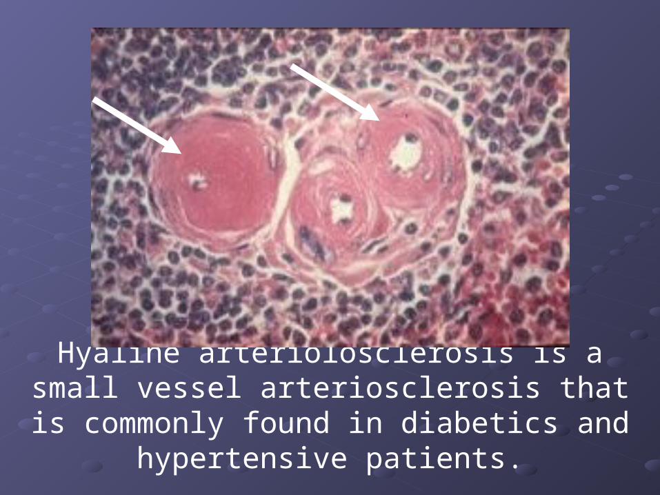
Hyaline arteriolosclerosis is a small vessel arteriosclerosis that is commonly found in
diabetics and hypertensive patients.
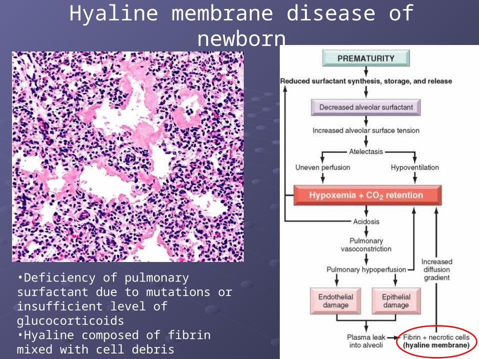
Hyaline membrane disease of newborn
•Deficiency of pulmonary surfactant due to mutations or insufficient level of glucocorticoids•Hyaline composed of fibrin mixed with cell debris
Other Cellular Alterations During Injury
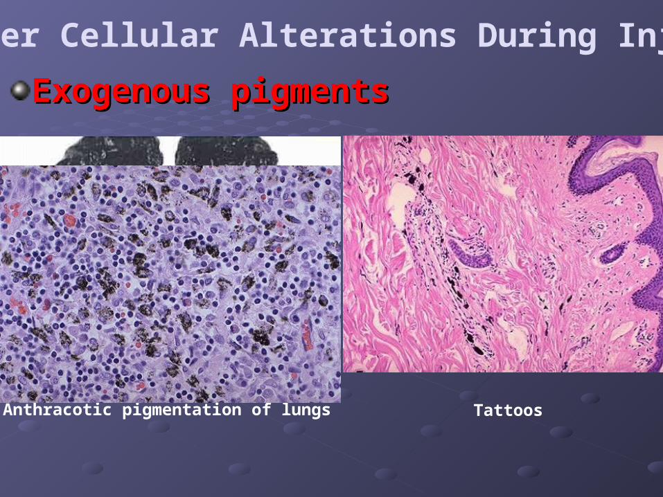
Exogenous pigmentsExogenous pigments
Anthracotic pigmentation of lungs Tattoos
Other Cellular Alterations During Injury
Endogenous pigmentsEndogenous pigments
1. Lipofuscin1. Lipofuscin
2. Melanin2. Melanin
3. Hemosiderin3. Hemosiderin
•Pigments are colored substances which accumulate under special circumstances, can be endogenous and exogenous
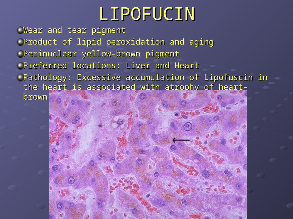
LIPOFUCINLIPOFUCINWear and tear pigmentWear and tear pigment
Product of lipid peroxidation and agingProduct of lipid peroxidation and aging
Perinuclear yellow-brown pigment Perinuclear yellow-brown pigment
Preferred locations: Liver and HeartPreferred locations: Liver and Heart
Pathology: Excessive accumulation of Lipofuscin in the Pathology: Excessive accumulation of Lipofuscin in the heart is associated with atrophy of heart- brown atrophyheart is associated with atrophy of heart- brown atrophy
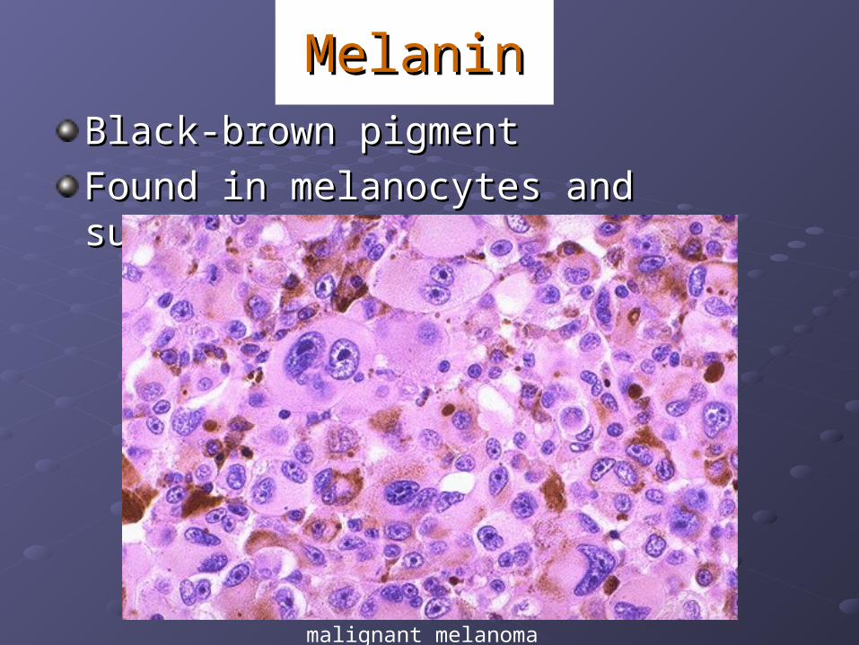
MelaninMelanin
Black-brown pigmentBlack-brown pigment
Found in melanocytes and substantia nigraFound in melanocytes and substantia nigra
malignant melanoma
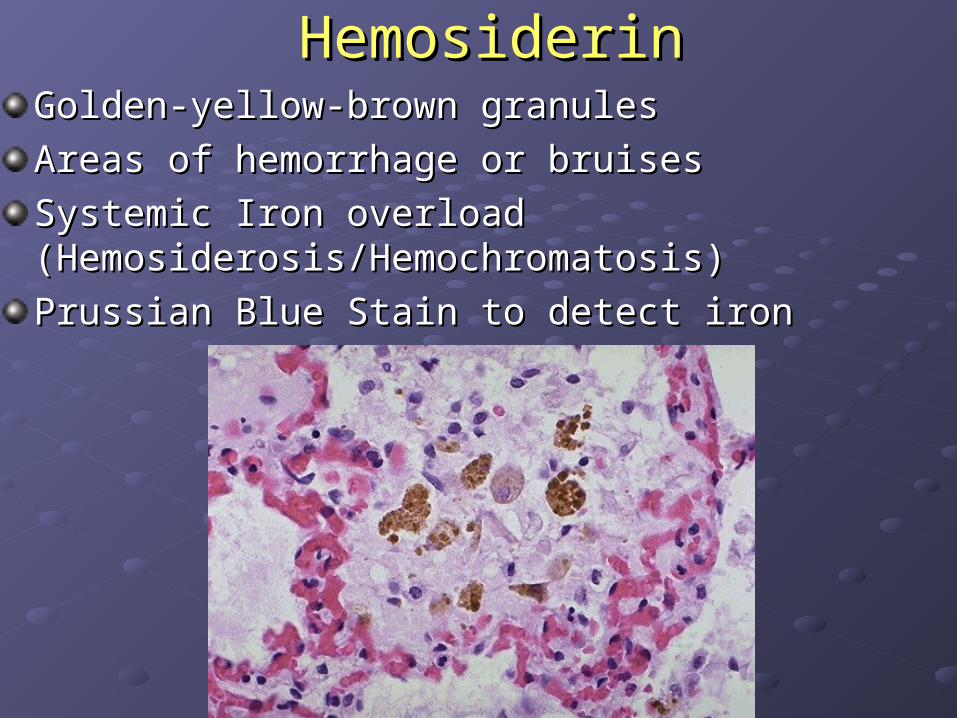
HemosiderinHemosiderinGolden-yellow-brown granulesGolden-yellow-brown granules
Areas of hemorrhage or bruisesAreas of hemorrhage or bruises
Systemic Iron overload Systemic Iron overload (Hemosiderosis/Hemochromatosis)(Hemosiderosis/Hemochromatosis)
Prussian Blue Stain to detect ironPrussian Blue Stain to detect iron
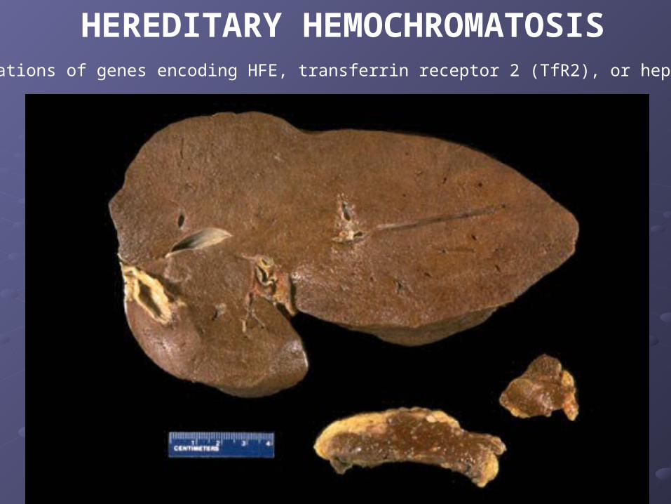
HEREDITARY HEMOCHROMATOSISMutations of genes encoding HFE, transferrin receptor 2 (TfR2), or hepcidin
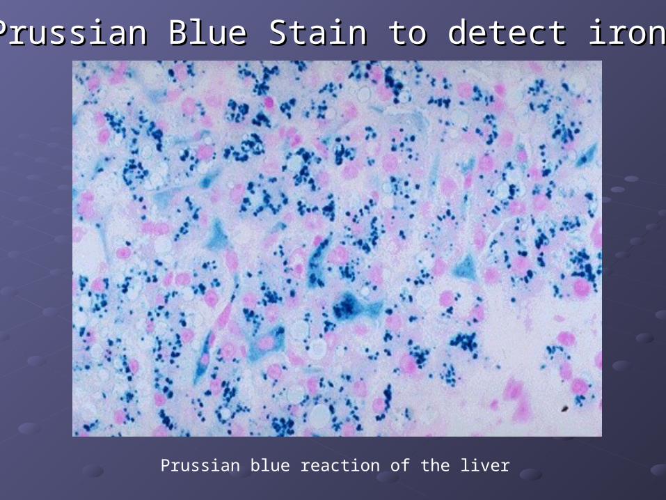
Prussian Blue Stain to detect ironPrussian Blue Stain to detect iron
Prussian blue reaction of the liver
Pathologic forms of CalcificationPathologic forms of Calcification
Dystrophic calcification: precipitation of Dystrophic calcification: precipitation of Ca3(PO4)2 in dying or necrotic tissues Ca3(PO4)2 in dying or necrotic tissues (saponification)(saponification)
Metastatic calcification: precipitation of Metastatic calcification: precipitation of Ca3(PO4)2 in normal tissues due to Ca3(PO4)2 in normal tissues due to hypercalciemiahypercalciemia
Dystrophic calcification examplesDystrophic calcification examples
Fat Necrosis and saponificationFat Necrosis and saponification
Psammoma bodies –laminated calcification Psammoma bodies –laminated calcification ((papillary cancer of thyroid, ovaries serous papillary cancer of thyroid, ovaries serous cystadenocarcinoma , meningiomascystadenocarcinoma , meningiomas))
Monckeberg’s medial calcific sclerosisMonckeberg’s medial calcific sclerosis
Atherosclerotic plaques Atherosclerotic plaques
Oligodendrocytoma and craniopharyngioma Oligodendrocytoma and craniopharyngioma associated with calcification of brain tissue associated with calcification of brain tissue
Psammoma bodies:Psammoma bodies:
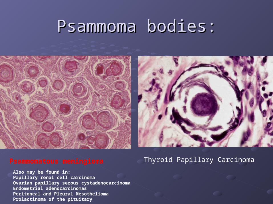
Psammomatous meningioma Thyroid Papillary Carcinoma
Also may be found in:Papillary renal cell carcinoma Ovarian papillary serous cystadenocarcinoma Endometrial adenocarcinomasPeritoneal and Pleural Mesothelioma Prolactinoma of the pituitary
Metastatic calcification examplesMetastatic calcification examples
Hyperparathyroidism (primary)Hyperparathyroidism (primary)
Parathyroid adenomasParathyroid adenomas
Renal failure (due to accumulation of phosphates!)Renal failure (due to accumulation of phosphates!)
Paraneoplastic syndromeParaneoplastic syndrome
Vit D intoxicationVit D intoxication
Milk-alkali syndromeMilk-alkali syndrome
SarcoidosisSarcoidosis
Paget DiseasePaget Disease
Multiple Myeloma to the boneMultiple Myeloma to the bone
Metastatic cancer to the boneMetastatic cancer to the bone
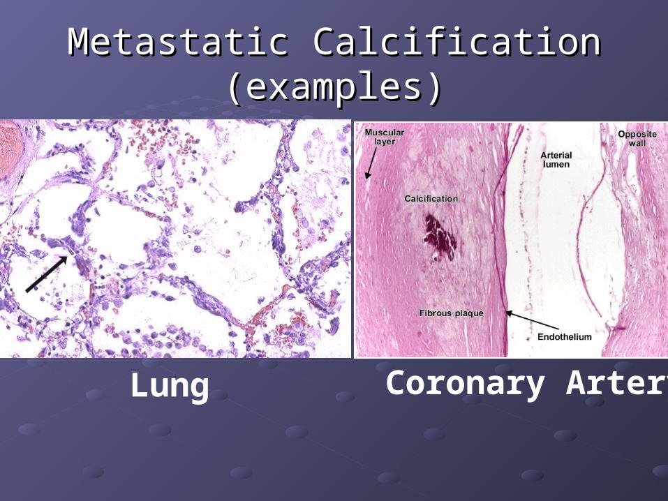
Metastatic Calcification (examples)Metastatic Calcification (examples)
Lung Coronary Artery
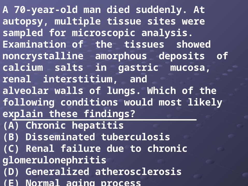
A 70-year-old man died suddenly. At autopsy, multiple tissue sites were sampled for microscopic analysis. Examination of the tissues showed noncrystalline amorphous deposits of calcium salts in gastric mucosa, renal interstitium, andalveolar walls of lungs. Which of the following conditions would most likely explain these findings?(A) Chronic hepatitis(B) Disseminated tuberculosis(C) Renal failure due to chronic glomerulonephritis(D) Generalized atherosclerosis(E) Normal aging process
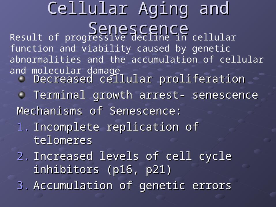
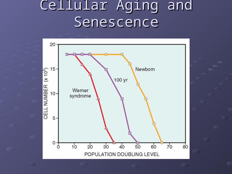
Cellular Aging and SenescenceCellular Aging and Senescence
Decreased cellular proliferationDecreased cellular proliferation
Terminal growth arrest- senescenceTerminal growth arrest- senescence
Mechanisms of Senescence:Mechanisms of Senescence:
1.1. Incomplete replication of telomeresIncomplete replication of telomeres
2.2. Increased levels of cell cycle inhibitors Increased levels of cell cycle inhibitors (p16, p21)(p16, p21)
3.3. Accumulation of genetic errorsAccumulation of genetic errors
Result of progressive decline in cellular function and viability caused by genetic abnormalities and the accumulation of cellular and molecular damage
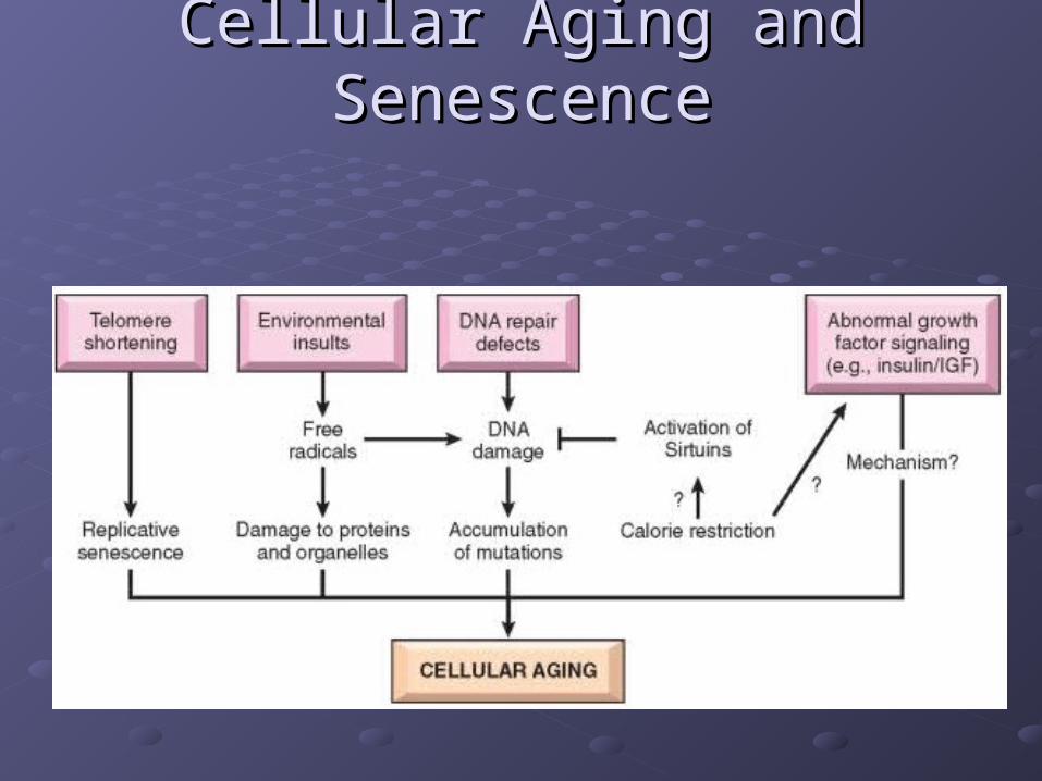
Cellular Aging and SenescenceCellular Aging and Senescence
Cellular Aging and SenescenceCellular Aging and Senescence

Role of Telomeres in replicative senescence
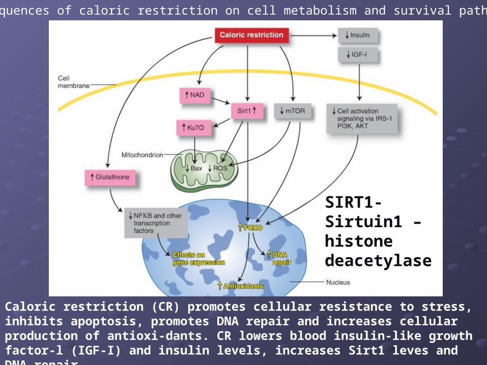
Consequences of caloric restriction on cell metabolism and survival pathways.
Caloric restriction (CR) promotes cellular resistance to stress, inhibits apoptosis, promotes DNA repair and increases cellular production of antioxi-dants. CR lowers blood insulin-like growth factor-l (IGF-I) and insulin levels, increases Sirt1 leves and DNA repair
SIRT1- Sirtuin1 – histone deacetylase
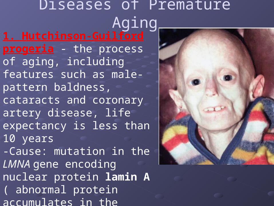
Diseases of Premature Aging1. Hutchinson-Guilford progeria - the process of aging, including features such as male-pattern baldness, cataracts and coronary artery disease, life expectancy is less than 10 years -Cause: mutation in the LMNA gene encoding nuclear protein lamin A ( abnormal protein accumulates in the nucleus and affect mitotic activity)
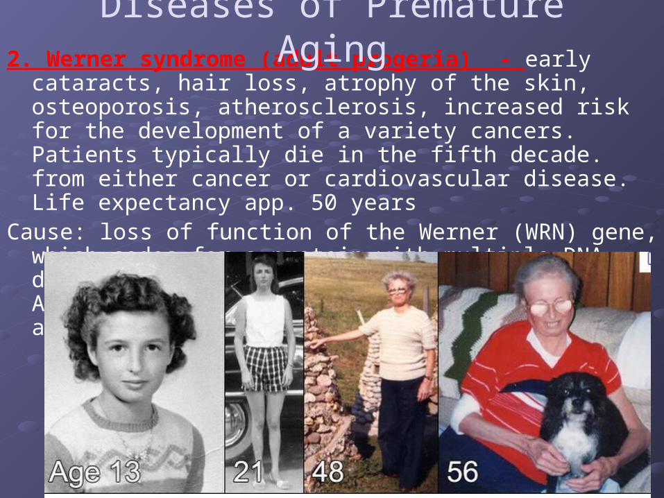
2. Werner syndrome (adult progeria) - early cataracts, hair loss, atrophy of the skin, osteoporosis, atherosclerosis, increased risk for the development of a variety cancers. Patients typically die in the fifth decade. from either cancer or cardiovascular disease. Life expectancy app. 50 years
Cause: loss of function of the Werner (WRN) gene, which codes for a protein with multiple DNA-dependent enzymatic activities, including ATPase, helicase, exonuclease and strand annealing
Diseases of Premature Aging
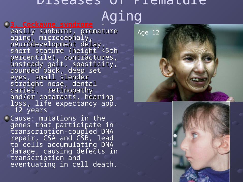
3. Cockayne syndrome - easily sunburns, premature easily sunburns, premature aging, microcephaly, aging, microcephaly, neurodevelopment delay, neurodevelopment delay, short stature (height <5th short stature (height <5th percentile), contractures, percentile), contractures, unsteady gait, spasticity, unsteady gait, spasticity, rounded back, deep set eyes, rounded back, deep set eyes, small slender straight nose, small slender straight nose, dental caries, retinopathy dental caries, retinopathy and/or cataracts, hearing loss, and/or cataracts, hearing loss, life expectancy app. 12 yearsCause: mutations in the genes that participate in transcription-coupled DNA repair, CSA and CSB, lead to cells accumulating DNA damage, causing defects in transcription and eventuating in cell death.
Diseases of Premature Aging
Age 12

Name the three most common causes of fatty liver: Name the three most common causes of fatty liver:
1. Diabetes 1. Diabetes
2. Obesity 2. Obesity
3. Alcoholism 3. Alcoholism
Objectives Review:
To know 2 morphological signs of alcoholic To know 2 morphological signs of alcoholic hepatitishepatitis
Objectives Review:
1. fatty changes2. Alcoholic hyaline - pink condensed ropy material
inside of the cells
Be able to name 2 diseases associated with Be able to name 2 diseases associated with hyaline changeshyaline changes
Objectives Review:
1.1. Hyaline arteriosclerosis (Essential Hyaline arteriosclerosis (Essential Hypertension)Hypertension)
2.2. Hyaline membrane disease of the newborn- Hyaline membrane disease of the newborn- (Neonatal Respiratory Distress Syndrome(Neonatal Respiratory Distress Syndrome ))
To know main causes of anthracotic To know main causes of anthracotic pigmentation of lungspigmentation of lungs
Objectives Review:
1. Smoking2. Inhalation of carbon dust in big cities or occupational
hazard
List at least one tissue where is possible to find List at least one tissue where is possible to find deposition of Lipofuscin, Melanin and Hemosiderindeposition of Lipofuscin, Melanin and Hemosiderin
Objectives Review:
1. Lipofuscin – Liver, Heart2. Melanin – Melanoma3. Hemosiderin – Liver, Lymph nodes.
List 3 types of tumors morphologically List 3 types of tumors morphologically associated with psammoma bodiesassociated with psammoma bodies
Objectives Review:
Papillary Thyroid CancerPapillary Ovarian CancerMeningioma
Name at least 2 diseases associated with Name at least 2 diseases associated with metastatic calcification metastatic calcification
Objectives Review:
1 hyperparathyroidism (parathyroid gland tumors, renal failure)
2 diffuse skeletal metastasis (multiple myeloma) 3 vitamin D intoxication4 renal failure

Please, take your pen and answer Please, take your pen and answer on the following questionson the following questions
You have 80 seconds to answer You have 80 seconds to answer each questioneach question
2:001:591:581:571:561:551:541:531:521:511:501:491:481:471:461:451:441:431:421:411:401:391:381:371:361:351:341:331:321:311:301:291:281:271:261:251:241:231:221:211:201:191:181:171:161:151:141:131:121:111:101:091:081:071:061:051:041:031:021:011:000:590:580:570:560:550:540:530:520:510:500:490:480:470:460:450:440:430:420:410:400:390:380:370:360:350:340:330:320:310:300:290:280:270:260:250:240:230:220:210:200:190:180:170:160:150:140:130:120:110:100:090:080:070:060:050:040:030:020:01End
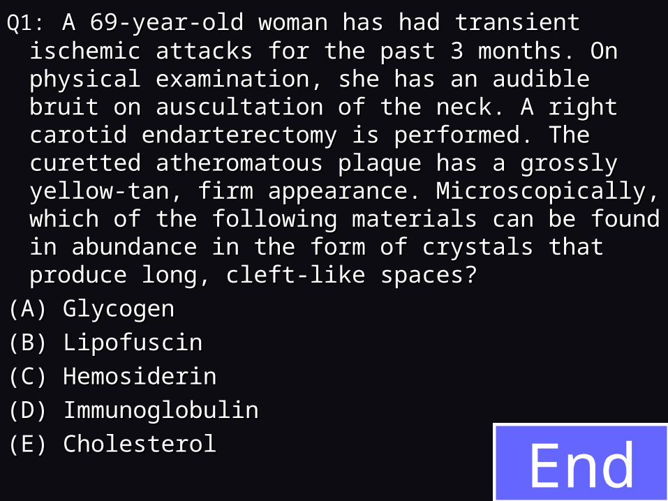
Q1:Q1: A 69-year-old woman has had transient ischemic A 69-year-old woman has had transient ischemic attacks for the past 3 months. On physical attacks for the past 3 months. On physical examination, she has an audible bruit on auscultation examination, she has an audible bruit on auscultation of the neck. A right carotid endarterectomy is of the neck. A right carotid endarterectomy is performed. The curetted atheromatous plaque has a performed. The curetted atheromatous plaque has a grossly yellow-tan, firm appearance. Microscopically, grossly yellow-tan, firm appearance. Microscopically, which of the following materials can be found in which of the following materials can be found in abundance in the form of crystals that produce long, abundance in the form of crystals that produce long, cleft-like spaces?cleft-like spaces?
(A) Glycogen(A) Glycogen
(B) Lipofuscin(B) Lipofuscin
(C) Hemosiderin(C) Hemosiderin
(D) Immunoglobulin(D) Immunoglobulin
(E) Cholesterol(E) Cholesterol
2:001:591:581:571:561:551:541:531:521:511:501:491:481:471:461:451:441:431:421:411:401:391:381:371:361:351:341:331:321:311:301:291:281:271:261:251:241:231:221:211:201:191:181:171:161:151:141:131:121:111:101:091:081:071:061:051:041:031:021:011:000:590:580:570:560:550:540:530:520:510:500:490:480:470:460:450:440:430:420:410:400:390:380:370:360:350:340:330:320:310:300:290:280:270:260:250:240:230:220:210:200:190:180:170:160:150:140:130:120:110:100:090:080:070:060:050:040:030:020:01End
Q2. An experiment analyzes cells for enzyme activity associated with sustained cellular proliferation. Which of the followingcells is most likely to have the highest telomerase activity?(A) Endothelial cells(B) Germ cells(C) Neurons(D) Neutrophils(E) Erythrocytes
2:001:591:581:571:561:551:541:531:521:511:501:491:481:471:461:451:441:431:421:411:401:391:381:371:361:351:341:331:321:311:301:291:281:271:261:251:241:231:221:211:201:191:181:171:161:151:141:131:121:111:101:091:081:071:061:051:041:031:021:011:000:590:580:570:560:550:540:530:520:510:500:490:480:470:460:450:440:430:420:410:400:390:380:370:360:350:340:330:320:310:300:290:280:270:260:250:240:230:220:210:200:190:180:170:160:150:140:130:120:110:100:090:080:070:060:050:040:030:020:01End
Q3: A 22-year-old woman has a congenital anemia that has required multiple transfusions of RBCs for many years. On physical examination, she now has no significant findings; however, liver function tests show reduced serum albumin and high level of iron. Which of the following findings would most likely appear in a liver biopsy specimen?(A) Steatosis in hepatocytes(B) Bilirubin in canaliculi(C) Glycogen in hepatocytes(D) Amyloid in portal triads(E) Hemosiderin in hepatocytes