Embed Size (px)
Citation preview
Lecture #7
Angiosperms: Form & Function
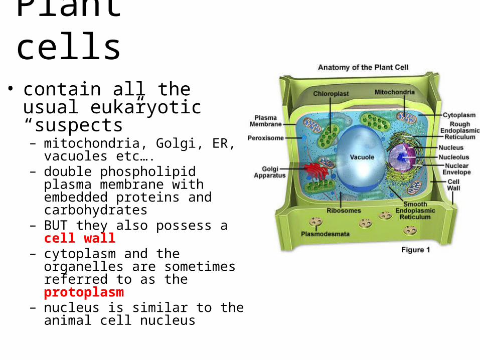
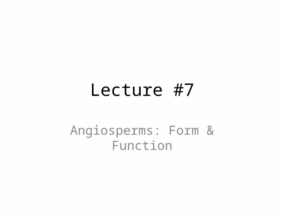
Plant cells
• contain all the usual eukaryotic “suspects”– mitochondria, Golgi, ER, vacuoles
etc….– double phospholipid plasma
membrane with embedded proteins and carbohydrates
– BUT they also possess a cell wall– cytoplasm and the organelles are
sometimes referred to as the protoplasm
– nucleus is similar to the animal cell nucleus
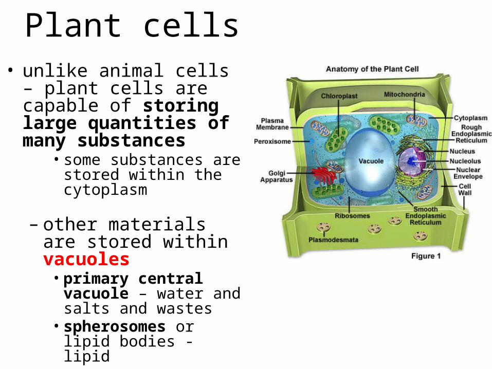
Plant cells• unlike animal cells – plant
cells are capable of storing large quantities of many substances
• some substances are stored within the cytoplasm
– other materials are stored within vacuoles• primary central vacuole –
water and salts and wastes• spherosomes or lipid
bodies - lipid
Plastids• plant cells also possess specialized structures
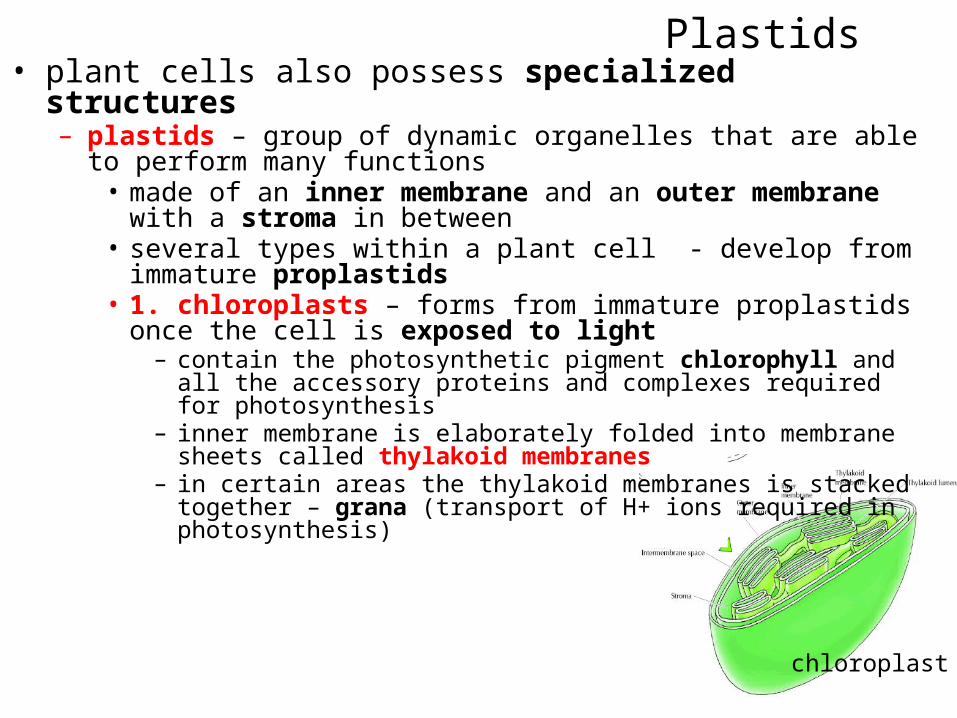
– plastids – group of dynamic organelles that are able to perform many functions• made of an inner membrane and an outer membrane with a stroma in
between• several types within a plant cell - develop from immature proplastids• 1. chloroplasts – forms from immature proplastids once the cell is
exposed to light– contain the photosynthetic pigment chlorophyll and all the accessory proteins
and complexes required for photosynthesis– inner membrane is elaborately folded into membrane sheets called thylakoid
membranes – in certain areas the thylakoid membranes is stacked together – grana
(transport of H+ ions required in photosynthesis)
chloroplast

Plastids• 2. amyloplasts – in plants tissues that can’t
photosynthesize– roots, bark and wood cells– accumulate sugar and store it as starch
• 3. chromoplasts – e.g. in tomatoes and yellow squash– bright red, yellow and orange lipids accumulated here
• 4. leucoplasts – large and unpigmented plastids– no chlorophyll or lipid pigments– involved in the synthesis of fats and phospholipids
chloroplast
The Cell Wall
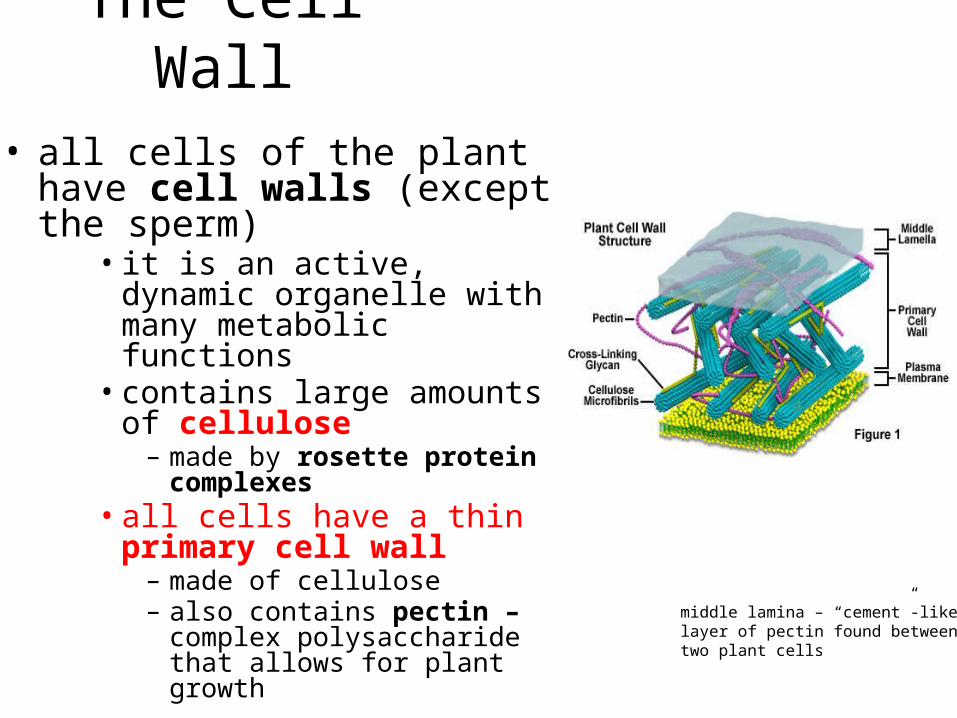
• all cells of the plant have cell walls (except the sperm)
• it is an active, dynamic organelle with many metabolic functions• contains large amounts of
cellulose– made by rosette protein complexes
• all cells have a thin primary cell wall– made of cellulose– also contains pectin – complex
polysaccharide that allows for plant growth
middle lamina – “cement”-likelayer of pectin found betweentwo plant cells
The Cell Wall
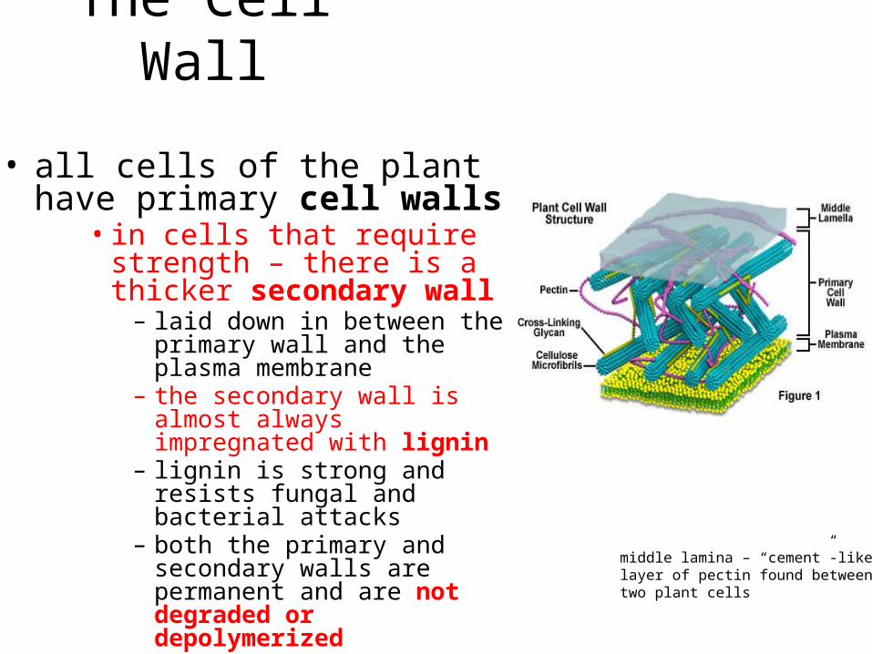
• all cells of the plant have primary cell walls
• in cells that require strength – there is a thicker secondary wall – laid down in between the primary
wall and the plasma membrane– the secondary wall is almost always
impregnated with lignin– lignin is strong and resists fungal
and bacterial attacks– both the primary and secondary
walls are permanent and are not degraded or depolymerized
middle lamina – “cement”-likelayer of pectin found betweentwo plant cells
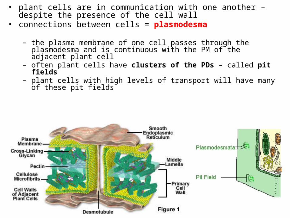
• plant cells are in communication with one another – despite the presence of the cell wall
• connections between cells = plasmodesma
– the plasma membrane of one cell passes through the plasmodesma and is continuous with the PM of the adjacent plant cell
– often plant cells have clusters of the PDs – called pit fields– plant cells with high levels of transport will have many of these pit fields
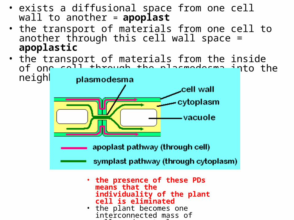
• exists a diffusional space from one cell wall to another = apoplast• the transport of materials from one cell to another through this
cell wall space = apoplastic• the transport of materials from the inside of one cell through the
plasmodesma into the neighbouring cell = symplastic
• the presence of these PDs means that the individuality of the plant cell is eliminated
• the plant becomes one interconnected mass of cells and protoplasm = symplast
Plant Cells
• three types of plant cells:• classified based on the nature of their cell walls
• 1. parenchyma – only have thin primary walls– undifferentiated cells – differentiation leads to the other two cell types
• 2. collenchyma – primary cell walls thin in some areas, thick in others
• 3. sclerenchyma – primary and secondary walls containing lignin
1. Parenchyma
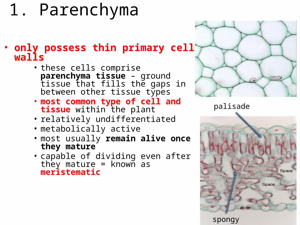
• only possess thin primary cell walls• these cells comprise parenchyma tissue –
ground tissue that fills the gaps in between other tissue types
• most common type of cell and tissue within the plant
• relatively undifferentiated• metabolically active• most usually remain alive once they mature• capable of dividing even after they mature =
known as meristematic
spongy
palisade
1. Parenchyma
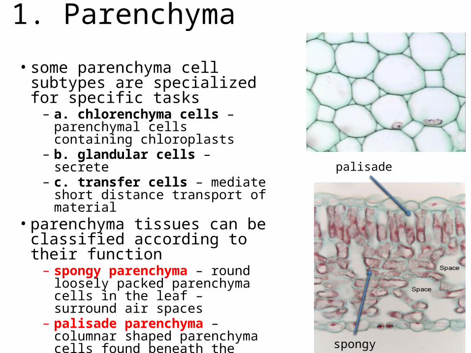
• some parenchyma cell subtypes are specialized for specific tasks– a. chlorenchyma cells – parenchymal
cells containing chloroplasts – b. glandular cells – secrete – c. transfer cells – mediate short distance
transport of material • parenchyma tissues can be classified
according to their function– spongy parenchyma – round loosely
packed parenchyma cells in the leaf – surround air spaces
– palisade parenchyma – columnar shaped parenchyma cells found beneath the epidermis of the leaf
spongy
palisade

– thin primary cell wall in some areas• in other areas the cell wall thickens –
most often the corners of the cell• cells exhibit plasticity – the ability to
become deformed by pressure or tension and to retain this new shape once this force is removed• collenchyma tissue - usually only
produced in elongating shoot tips – give the tips strength as it elongates but it can be stretched– found just underneath the epidermis
2. Collenchyma
sclerenchyma
collenchyma
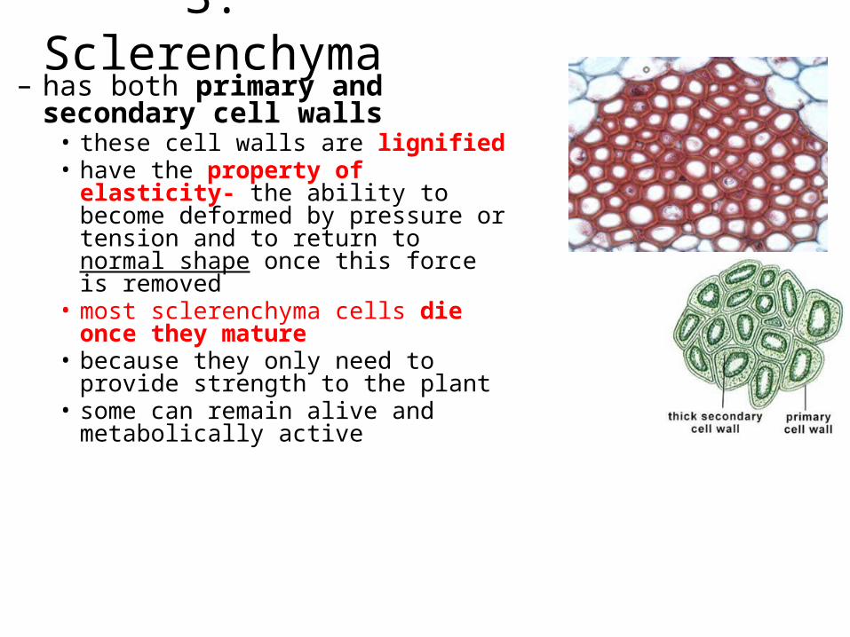
– has both primary and secondary cell walls • these cell walls are lignified• have the property of elasticity- the ability
to become deformed by pressure or tension and to return to normal shape once this force is removed
• most sclerenchyma cells die once they mature
• because they only need to provide strength to the plant
• some can remain alive and metabolically active
3. Sclerenchyma
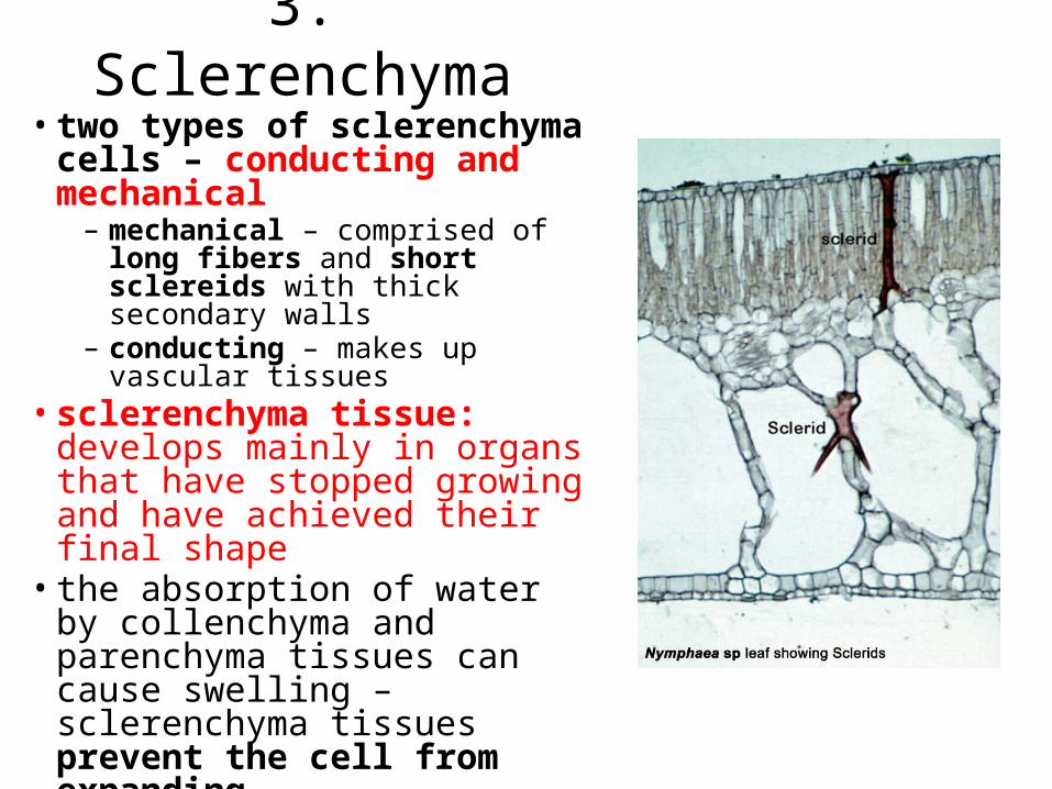
• two types of sclerenchyma cells – conducting and mechanical– mechanical – comprised of long fibers
and short sclereids with thick secondary walls
– conducting – makes up vascular tissues• sclerenchyma tissue: develops
mainly in organs that have stopped growing and have achieved their final shape• the absorption of water by
collenchyma and parenchyma tissues can cause swelling – sclerenchyma tissues prevent the cell from expanding
3. Sclerenchyma
Plant Tissues
• found throughout the plant– i.e. in roots, stems and leaves– so each forms a tissue system that is continuous through
all parts of the plant
• 1. dermal • 2. vascular• 3. ground
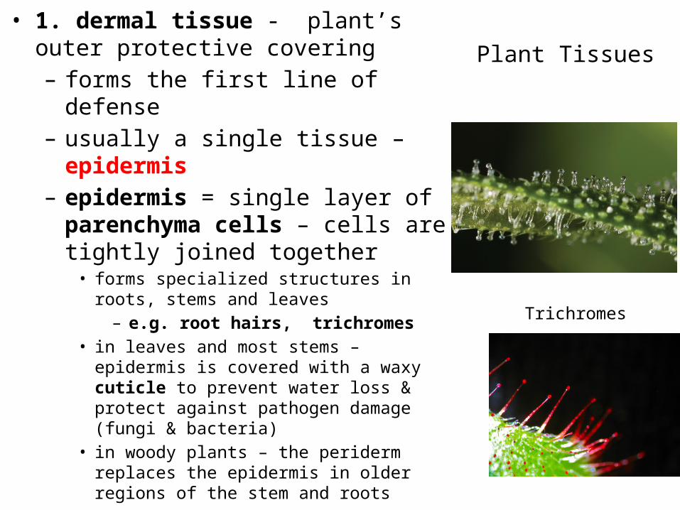
Plant Tissues• 1. dermal tissue - plant’s outer protective
covering– forms the first line of defense– usually a single tissue – epidermis– epidermis = single layer of parenchyma
cells – cells are tightly joined together• forms specialized structures in roots, stems and
leaves– e.g. root hairs, trichromes
• in leaves and most stems – epidermis is covered with a waxy cuticle to prevent water loss & protect against pathogen damage (fungi & bacteria)
• in woody plants – the periderm replaces the epidermis in older regions of the stem and roots
Trichromes
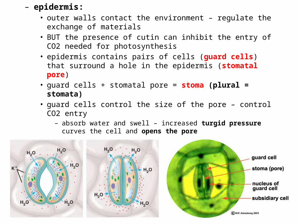
– epidermis:• outer walls contact the environment – regulate the exchange of materials• BUT the presence of cutin can inhibit the entry of CO2 needed for
photosynthesis• epidermis contains pairs of cells (guard cells) that surround a hole in the
epidermis (stomatal pore)• guard cells + stomatal pore = stoma (plural = stomata)• guard cells control the size of the pore – control CO2 entry
– absorb water and swell – increased turgid pressure curves the cell and opens the pore
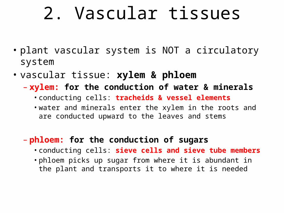
• plant vascular system is NOT a circulatory system• vascular tissue: xylem & phloem– xylem: for the conduction of water & minerals
• conducting cells: tracheids & vessel elements• water and minerals enter the xylem in the roots and are
conducted upward to the leaves and stems
– phloem: for the conduction of sugars• conducting cells: sieve cells and sieve tube members• phloem picks up sugar from where it is abundant in the plant
and transports it to where it is needed
2. Vascular tissues

XylemPARENCHYMA CELLS
WATER-CONDUCTING CELLS OF THE XYLEM
Vessel elements withperforated end walls
Vesselelement
Tracheids
Pits
Tracheids and vessels(colorized SEM)
TracheidsVessel 100 µm
– two types of conducting cells: tracheids and vessel elements• either type of cell can be called a “tracheary
element”– both are types of conducting
sclerenchyma cells– tracheids and vessel elements develop
first as immature parenchyma cells with thin primary walls
– cell elongates and deposits and secondary wall
– cell then dies and the protoplasm degenerates – leaving a hollow dead cell comprised of two cell walls
– now a type of sclerenchyma cell
aligned perforations
pit-pair
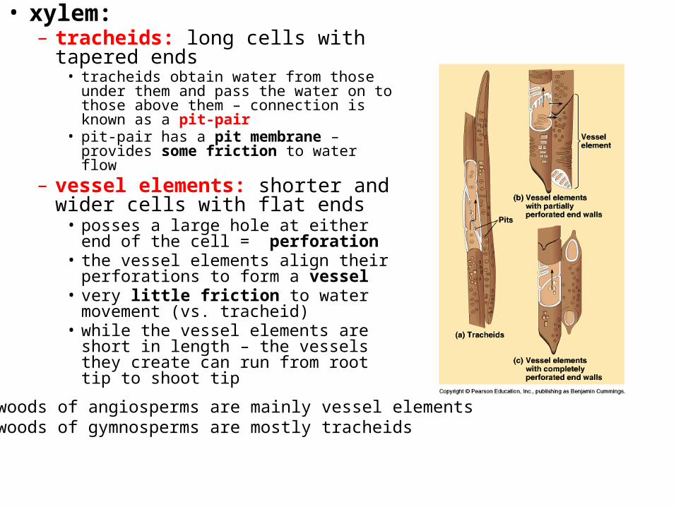
• xylem:– tracheids: long cells with tapered
ends• tracheids obtain water from those under
them and pass the water on to those above them – connection is known as a pit-pair
• pit-pair has a pit membrane – provides some friction to water flow
– vessel elements: shorter and wider cells with flat ends • posses a large hole at either end of the
cell = perforation• the vessel elements align their
perforations to form a vessel• very little friction to water movement
(vs. tracheid)• while the vessel elements are short in
length – the vessels they create can run from root tip to shoot tip
hardwoods of angiosperms are mainly vessel elementssoftwoods of gymnosperms are mostly tracheids

PARENCHYMA CELLS
• xylem:– many tracheids or vessel elements
can be identified by their secondary wall
– secondary wall is organized as rings = annular thickenings • the majority of the primary wall is
uncovered - for water entry and exit• large surface area for water uptake and
movement– the strongest type of xylem cell is
called a pitted tracheary element• the majority of the primary wall is covered
with secondary wall except for small regions called circular bordered pits
• water uptake and movement through these is slow
tracheidsvessel elements
alignedperforations
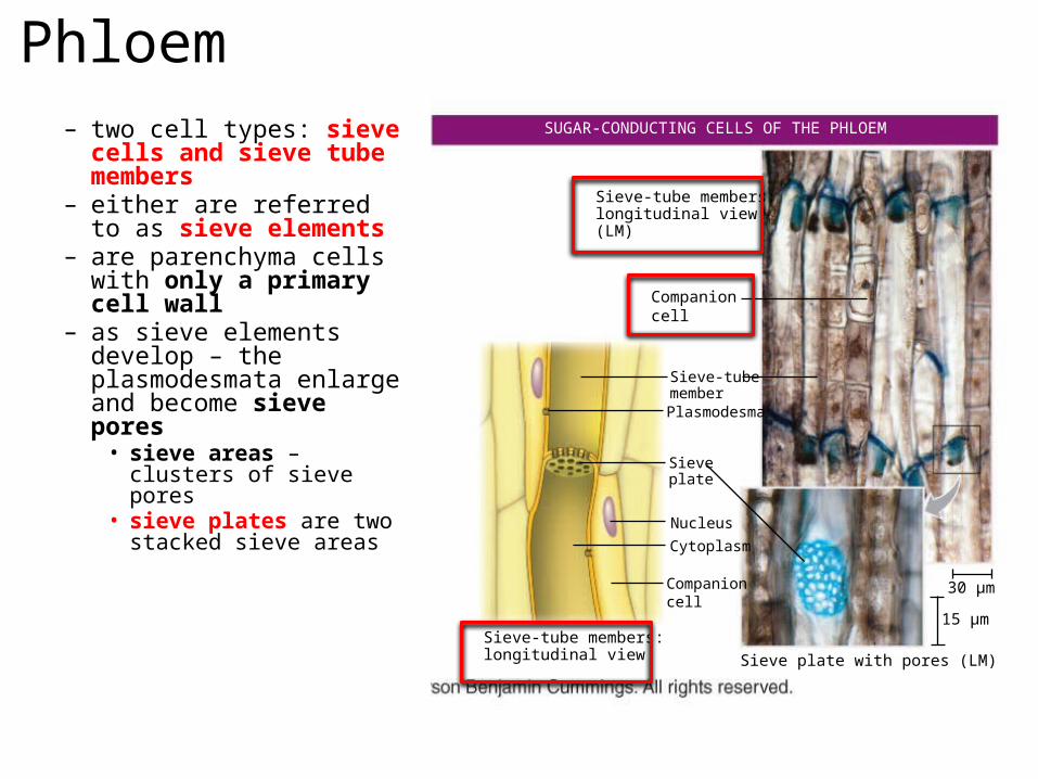
Phloem– two cell types: sieve cells
and sieve tube members– either are referred to as
sieve elements– are parenchyma cells
with only a primary cell wall
– as sieve elements develop – the plasmodesmata enlarge and become sieve pores• sieve areas – clusters of
sieve pores• sieve plates are two
stacked sieve areas
SUGAR-CONDUCTING CELLS OF THE PHLOEM
Sieve-tube members:longitudinal view
30 µm
15 µm
Companioncell
Companioncell
Sieve-tubemember
Plasmodesma
Sieveplate
Sieve plate with pores (LM)
NucleusCytoplasm
Sieve-tube members:longitudinal view(LM)
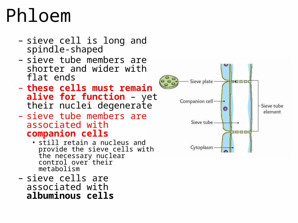
Phloem– sieve cell is long and spindle-
shaped– sieve tube members are shorter
and wider with flat ends– these cells must remain alive
for function – yet their nuclei degenerate
– sieve tube members are associated with companion cells • still retain a nucleus and provide the
sieve cells with the necessary nuclear control over their metabolism
– sieve cells are associated with albuminous cells

Vascular Bundles• xylem and phloem occur
together in roots and stems as vascular bundles
• bundles are located just interior to the cortex
• the arrangement of the vascular bundles helps define a monocot stem from a eudicot stem
• also arranged differently according to whether they are in a root or a stem– e.g. in angiosperms there is a
solid central vascular cylinder called a stele
• tissues that are neither dermal or vascular– includes cells specialized for storage, photosynthesis,
and support– made up of:• 1. parenchyma – made up of parenchyma cells
– many parenchyma cells contain chloroplasts
• 2. sclerenchyma – made up of sclerenchyma cells– provides support to the plant
• 3. collenchyma- made up of collenchyma cells
– found in the cortex and pith of stem, the cortex of the root, the mesophyll of leaves and the endosperm of seeds
3. Ground Tissue
3 Plant organs
Shootsystem
Rootsystem
Reproductive shoot (flower)
Terminal bud
NodeInternode
Blade
Vegetableshoot
Terminalbud
Petiole
Axillarybud
Stem
Leaf
Taproot
Lateral roots
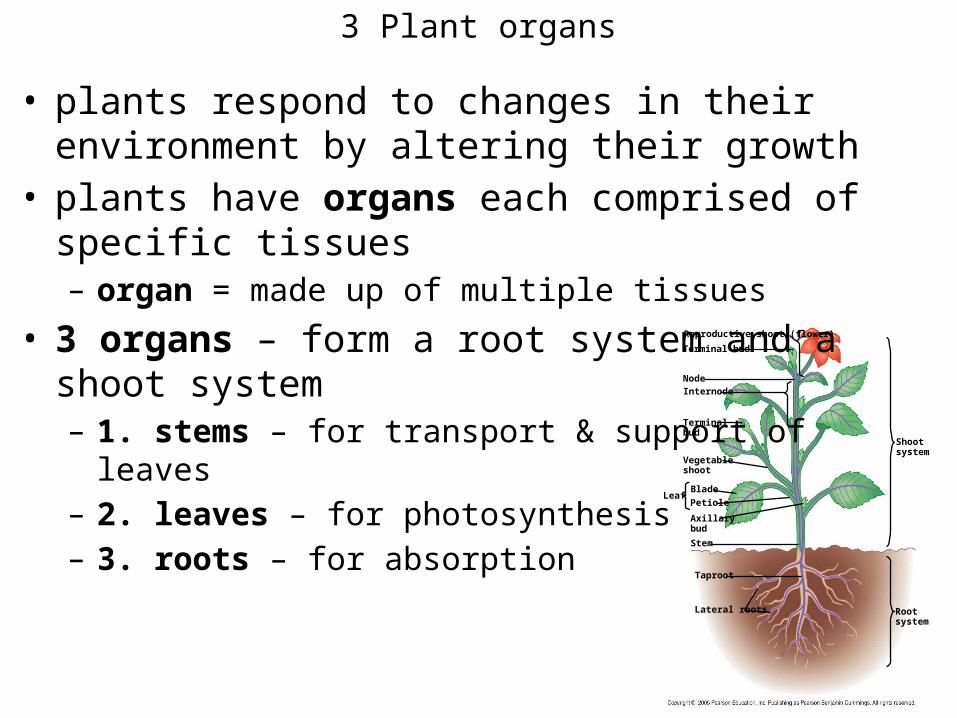
• plants respond to changes in their environment by altering their growth
• plants have organs each comprised of specific tissues– organ = made up of multiple tissues
• 3 organs – form a root system and a shoot system– 1. stems – for transport & support of leaves– 2. leaves – for photosynthesis– 3. roots – for absorption
Stems
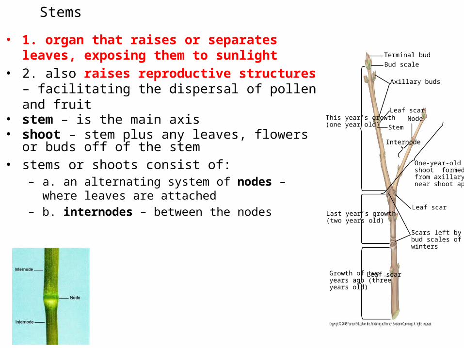
• 1. organ that raises or separates leaves, exposing them to sunlight
• 2. also raises reproductive structures – facilitating the dispersal of pollen and fruit
• stem – is the main axis• shoot – stem plus any leaves, flowers or buds off of the
stem• stems or shoots consist of:
– a. an alternating system of nodes – where leaves are attached
– b. internodes – between the nodes
This year’s growth(one year old)
Terminal bud
Leaf scarGrowth of twoyears ago (three years old)
Last year’s growth(two years old)
One-year-old sshoot formedfrom axillary budnear shoot apex
Leaf scar
Bud scale
Axillary buds
Internode
StemNode
Leaf scar
Scars left by terminalbud scales of previouswinters
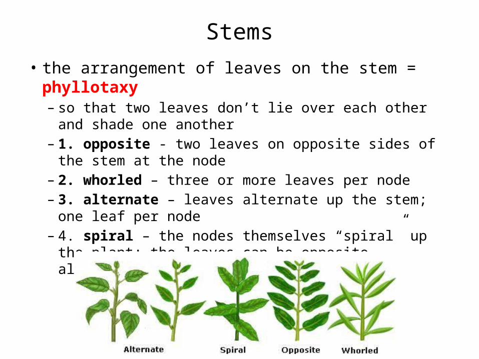
Stems• the arrangement of leaves on the stem = phyllotaxy
– so that two leaves don’t lie over each other and shade one another
– 1. opposite - two leaves on opposite sides of the stem at the node
– 2. whorled – three or more leaves per node– 3. alternate – leaves alternate up the stem; one leaf per
node– 4. spiral – the nodes themselves “spiral” up the plant; the
leaves can be opposite, alternate or whorled at the nodes
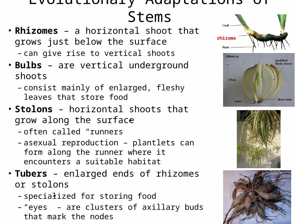
Evolutionary Adaptations of Stems• Rhizomes – a horizontal shoot that grows just
below the surface– can give rise to vertical shoots
• Bulbs – are vertical underground shoots– consist mainly of enlarged, fleshy leaves that store
food• Stolons – horizontal shoots that grow along
the surface– often called “runners” – asexual reproduction – plantlets can form along
the runner where it encounters a suitable habitat• Tubers – enlarged ends of rhizomes or stolons
– specialized for storing food– “eyes” – are clusters of axillary buds that mark the
nodes
rhizome
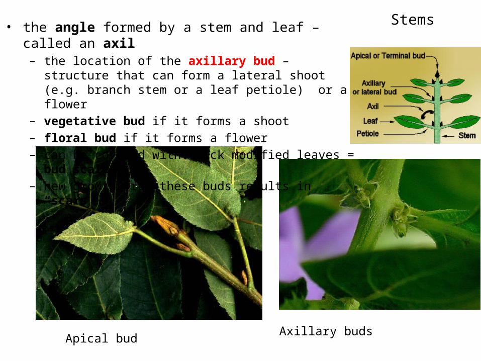
Stems• the angle formed by a stem and leaf – called an axil– the location of the axillary bud – structure that can form a
lateral shoot (e.g. branch stem or a leaf petiole) or a flower– vegetative bud if it forms a shoot – floral bud if it forms a flower– can be covered with thick modified leaves = bud scales– new growth from these buds results in “scars”
Axillary budsApical bud
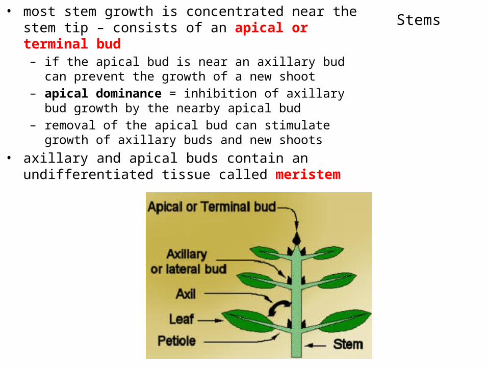
Stems• most stem growth is concentrated near the stem tip –
consists of an apical or terminal bud– if the apical bud is near an axillary bud can prevent the growth
of a new shoot– apical dominance = inhibition of axillary bud growth by the
nearby apical bud– removal of the apical bud can stimulate growth of axillary buds
and new shoots • axillary and apical buds contain an undifferentiated tissue
called meristem

Stem growth:Apical meristems
• stems grow longer by creating new cells at their tips
• growth is at regions known as shoot apical meristems (SAMs)
• growth is via mitosis• as the small daughter cells grow to
the size of the parent – they push the meristem upward – lower and older cells mature and become part of the growing stem
• region below the apical meristem = subapical meristem– site of differentiation
• apical meristem is flanked by small, developing leaf primordia which protect the AM
apical meristem
axillary bud
developingvascular tissue
developing leaf primordia
subapicalmeristem
the apical meristem and leaf primordia = bud

• primary growth in the AM leads to the formation of the subapical meristem
• Subapical Meristem is composed of three types of subapical cells– 1. protoderm – gives rise to the epidermis– 2. provascular tissue – gives rise to primary xylem
and primary phloem – 3. ground meristem – gives rise to pith and cortex
Stem growth:Primary and Secondary growth
• primary growth is followed by secondary growth – continued differentiation– 1. in some plants - epidermis becomes the cork
cambium – 2. provascular tissue gives rise to the vascular
cambium - becomes the secondary xylem and phloem (woody tissues)
– 3. pith becomes the interfasicular cambium and the cortex forms the cork cambium (which forms cork)
Stem growth:Primary and Secondary growth
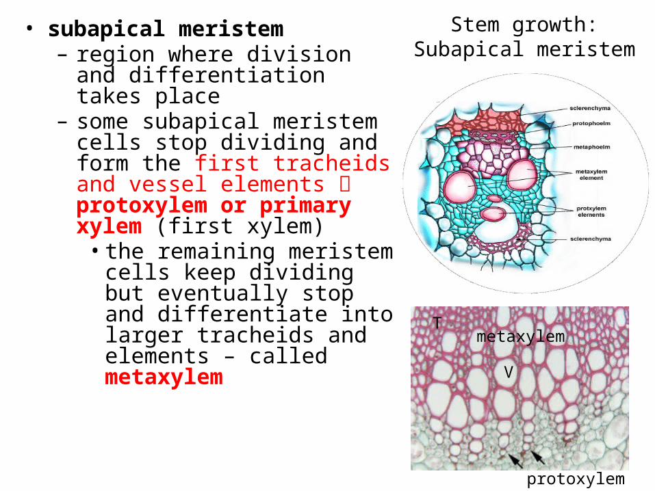
• subapical meristem – region where division and
differentiation takes place– some subapical meristem cells stop
dividing and form the first tracheids and vessel elements protoxylem or primary xylem (first xylem)• the remaining meristem cells
keep dividing but eventually stop and differentiate into larger tracheids and elements – called metaxylem
protoxylem
V
Tmetaxylem
Stem growth:Subapical meristem
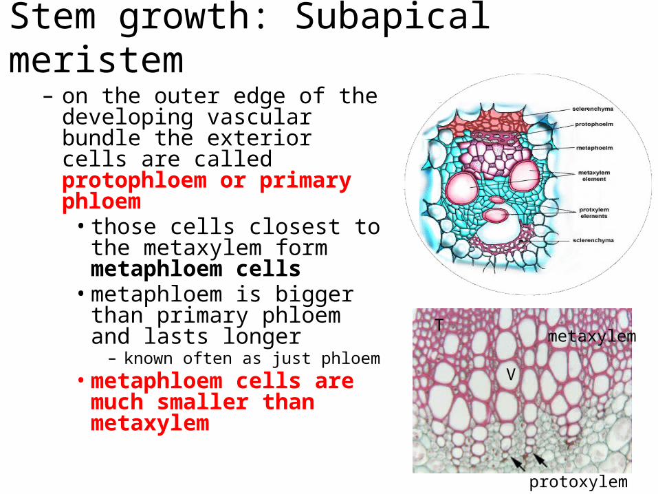
– on the outer edge of the developing vascular bundle the exterior cells are called protophloem or primary phloem• those cells closest to the
metaxylem form metaphloem cells• metaphloem is bigger than
primary phloem and lasts longer– known often as just phloem
• metaphloem cells are much smaller than metaxylem
protoxylem
V
Tmetaxylem
Stem growth: Subapical meristem
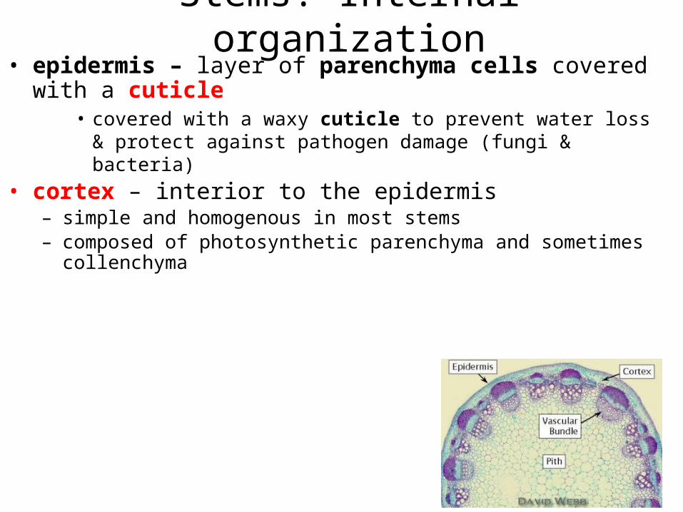
Stems: Internal organization• epidermis – layer of parenchyma cells covered with a cuticle
• covered with a waxy cuticle to prevent water loss & protect against pathogen damage (fungi & bacteria)
• cortex – interior to the epidermis– simple and homogenous in most stems– composed of photosynthetic parenchyma and sometimes collenchyma
• vascular bundles – xylem and phloem– unique arrangement depending on whether the plant is a eudicot or a
monocot
• pith – most interior portion of the stem– region of parenchyma– similar to the parenchyma of the cortex
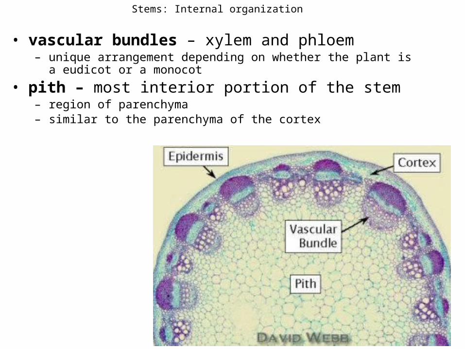
Stems: Internal organization
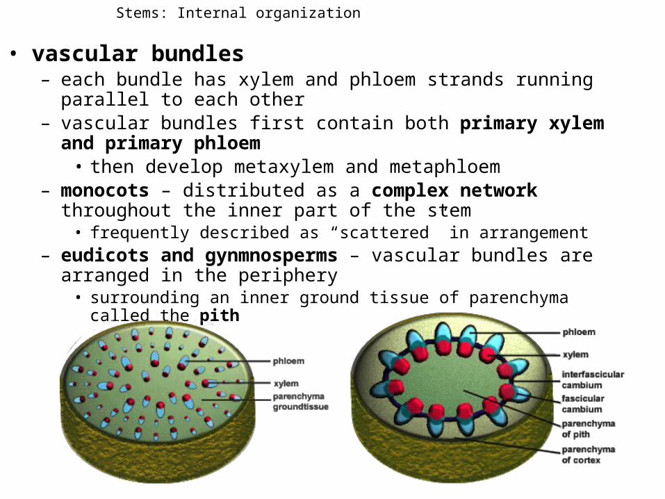
• vascular bundles– each bundle has xylem and phloem strands running parallel to each other– vascular bundles first contain both primary xylem and primary phloem
• then develop metaxylem and metaphloem– monocots – distributed as a complex network throughout the inner part of
the stem • frequently described as “scattered” in arrangement
– eudicots and gynmnosperms – vascular bundles are arranged in the periphery• surrounding an inner ground tissue of parenchyma called the pith
Stems: Internal organization
Key
Dermal
Ground
Vascular
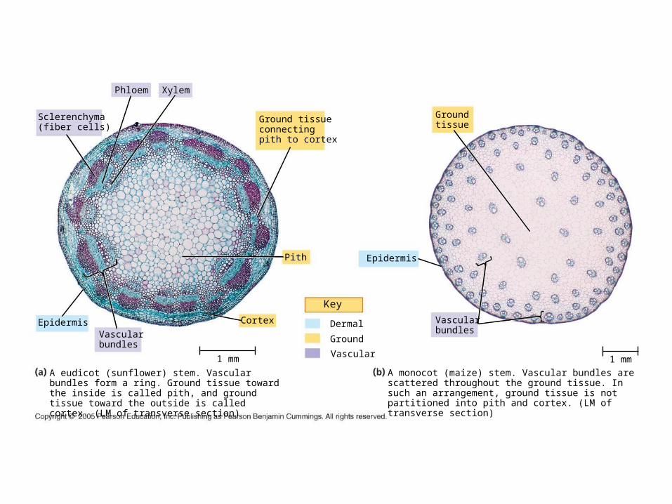
Epidermis Cortex
A eudicot (sunflower) stem. Vascular bundles form a ring. Ground tissue toward the inside is called pith, and ground tissue toward the outside is called cortex. (LM of transverse section)
XylemPhloem
Pith
Vascularbundles
Epidermis
Vascularbundles
1 mm
Sclerenchyma(fiber cells)
Ground tissueconnectingpith to cortex
Ground tissue
A monocot (maize) stem. Vascular bundles are scattered throughout the ground tissue. In such an arrangement, ground tissue is not partitioned into pith and cortex. (LM of transverse section)
1 mm

• vascular bundles of monocots– between the bundles is parenchyma– frequently described as “scattered” in arrangement– more complex than a random arrangement
• vascular bundles of eudicots and gymnosperms– vascular bundles are arranged in the periphery
surrounding an inner tissue of parenchyma called the pith (ground tissue)
Dicot
MedullaryRays

Monocot Stem Vascular Bundles
• cap of sclerenchyma on top of primary phloem and metaphloem– called phloem in the figure
• below this is a region of large tracheids & vessel elements = metaxylem – called xylem in the figure
• smaller tracheids and vessel elements below this is primary xylem
• most interior layer is another layer of sclerenchyma

• cap of sclerenchyma• region of phloem (mostly
metaphloem)• below this is a region of
fascicular/vascular cambium– play a role in secondary growth
of a dicot stem– produces secondary xylem and
phloem in the “woody” stem– no VC is mature monocot stems
(no secondary growth)
Eudicot Stem Vascular Bundles
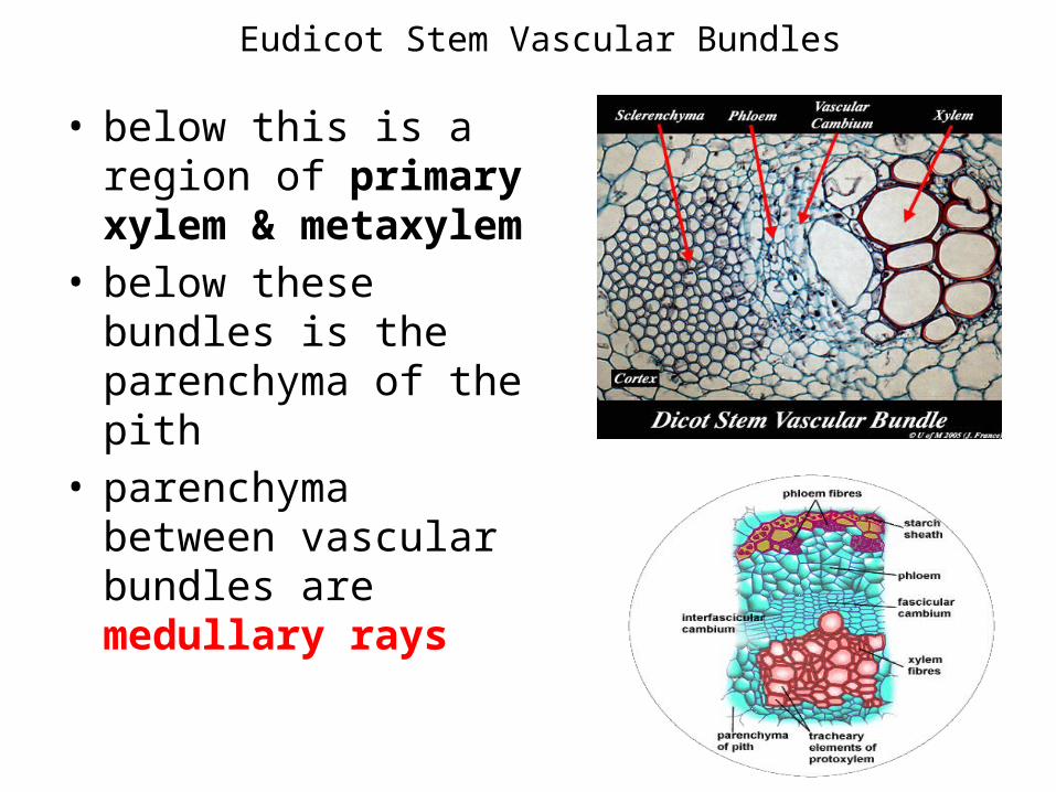
• below this is a region of primary xylem & metaxylem
• below these bundles is the parenchyma of the pith
• parenchyma between vascular bundles are medullary rays
Eudicot Stem Vascular Bundles
Leaves
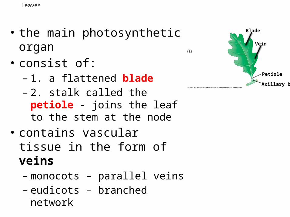
• the main photosynthetic organ• consist of:– 1. a flattened blade– 2. stalk called the petiole - joins
the leaf to the stem at the node• contains vascular tissue in the
form of veins– monocots – parallel veins– eudicots – branched network
Axillary bud
Petiole
Blade
Vein
Leaves
Simple leaf
Axillary bud
Petiole
Compound leaf
Axillary bud
Petiole
Leaflet
Doubly compound leaf
Axillary budPetiole
Leaflet
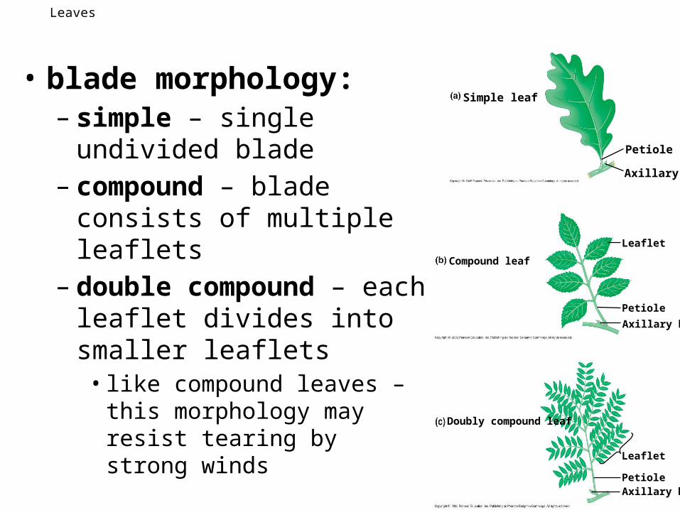
• blade morphology:– simple – single undivided blade– compound – blade consists of
multiple leaflets– double compound – each leaflet
divides into smaller leaflets• like compound leaves – this
morphology may resist tearing by strong winds

Evolutionary Adaptations of Leaves
Tendrils.
Spines.
Storage leaves.
Bracts.
• tendrils –modified leaves or lateral branches capable of wrapping around small objects– e.g. pea plants, ivy
• spines – non-photosynthetic– e.g. cacti– photosynthesis carried out by the stem
• needles – capable of photosynthesis– usually seen in gymnosperms
• storage leaves – adapted to storing water– e.g. succulents
• reproductive – leaves that produce adventitious plantlets– e.g. succulents
• bracts – often mistaken for petals; modified leaves that surround a group of flowers– e.g. pointsettia
Keyto labels
DermalGroundVascular
Guardcells
Epidermalcells
Stomatal pore
Surface view of a spiderwort(Tradescantia) leaf (LM)
50 µm
Upperepidermis
Stoma
Lowerepidermis
Palisademesophyll
Spongymesophyll
Air spacesVein Guard cells
Transverse section of a lilac(Syringa) leaf (LM)
100 µm
Bundle-sheathcell
XylemPhloem Guard
cells
Vein
Cuticle
CuticleSclerenchymafibers
Guardcells
Cutaway drawing of leaf tissues
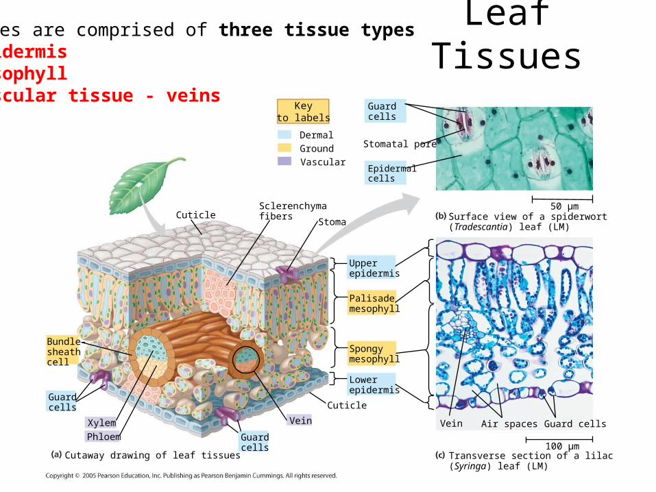
Leaf Tissues-leaves are comprised of three tissue types1. epidermis2. mesophyll3. vascular tissue - veins
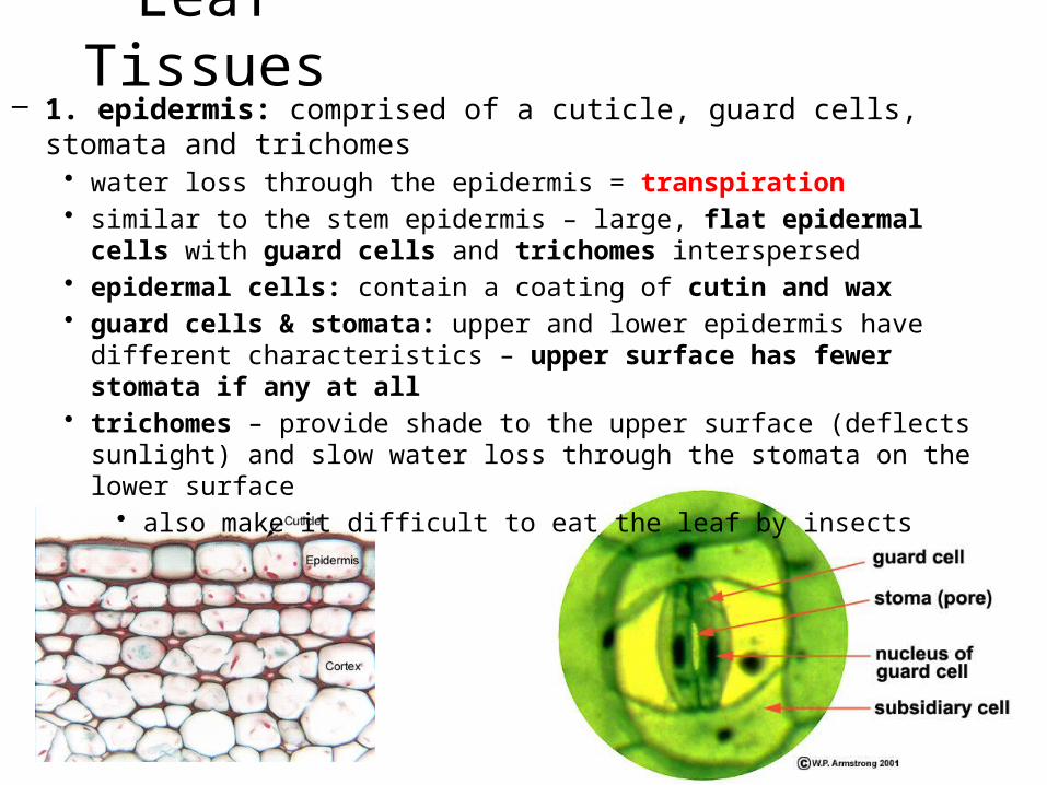
Leaf Tissues– 1. epidermis: comprised of a cuticle, guard cells, stomata and trichomes
• water loss through the epidermis = transpiration• similar to the stem epidermis – large, flat epidermal cells with guard cells and
trichomes interspersed• epidermal cells: contain a coating of cutin and wax• guard cells & stomata: upper and lower epidermis have different characteristics –
upper surface has fewer stomata if any at all• trichomes – provide shade to the upper surface (deflects sunlight) and slow water
loss through the stomata on the lower surface• also make it difficult to eat the leaf by insects
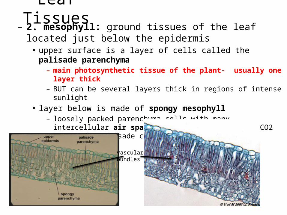
Leaf Tissues– 2. mesophyll: ground tissues of the leaf located just below the
epidermis• upper surface is a layer of cells called the palisade parenchyma
– main photosynthetic tissue of the plant- usually one layer thick– BUT can be several layers thick in regions of intense sunlight
• layer below is made of spongy mesophyll– loosely packed parenchyma cells with many intercellular air spaces to permit
diffusion of CO2 toward the palisade cells – for photosynthesis
vascular bundles
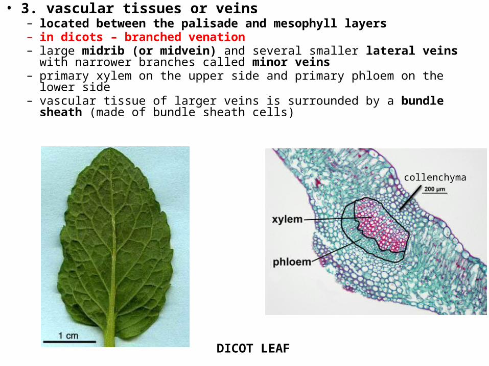
• 3. vascular tissues or veins– located between the palisade and mesophyll layers– in dicots – branched venation– large midrib (or midvein) and several smaller lateral veins with narrower
branches called minor veins– primary xylem on the upper side and primary phloem on the lower side– vascular tissue of larger veins is surrounded by a bundle sheath (made of
bundle sheath cells)
collenchyma
DICOT LEAF
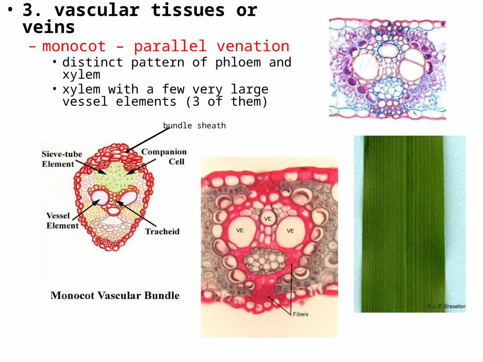
• 3. vascular tissues or veins– monocot – parallel venation
• distinct pattern of phloem and xylem• xylem with a few very large vessel
elements (3 of them)
bundle sheath
Roots
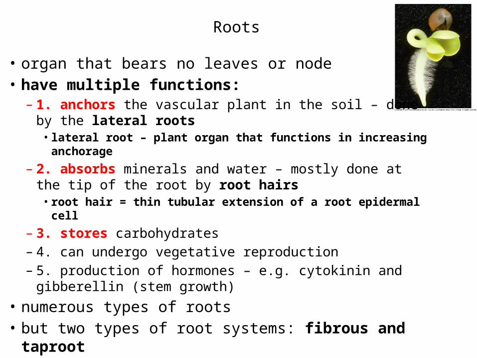
• organ that bears no leaves or node• have multiple functions:
– 1. anchors the vascular plant in the soil – done by the lateral roots• lateral root – plant organ that functions in increasing anchorage
– 2. absorbs minerals and water – mostly done at the tip of the root by root hairs• root hair = thin tubular extension of a root epidermal cell
– 3. stores carbohydrates– 4. can undergo vegetative reproduction– 5. production of hormones – e.g. cytokinin and gibberellin
(stem growth)• numerous types of roots• but two types of root systems: fibrous and taproot

Roots
• numerous types of roots– adventitious – form from unusual
locations – e.g. leaves or stems– aerial – growth above ground
• e.g. orchids
– aerating – grow up above the ground or water
– coarse – undergo secondary thickening and can be woody
– haustorial - seen in parasitic plants; substrate is the body of another plant• roots known as haustoria
Storage roots.
“Strangling” aerial roots.Aerating
Prop roots.
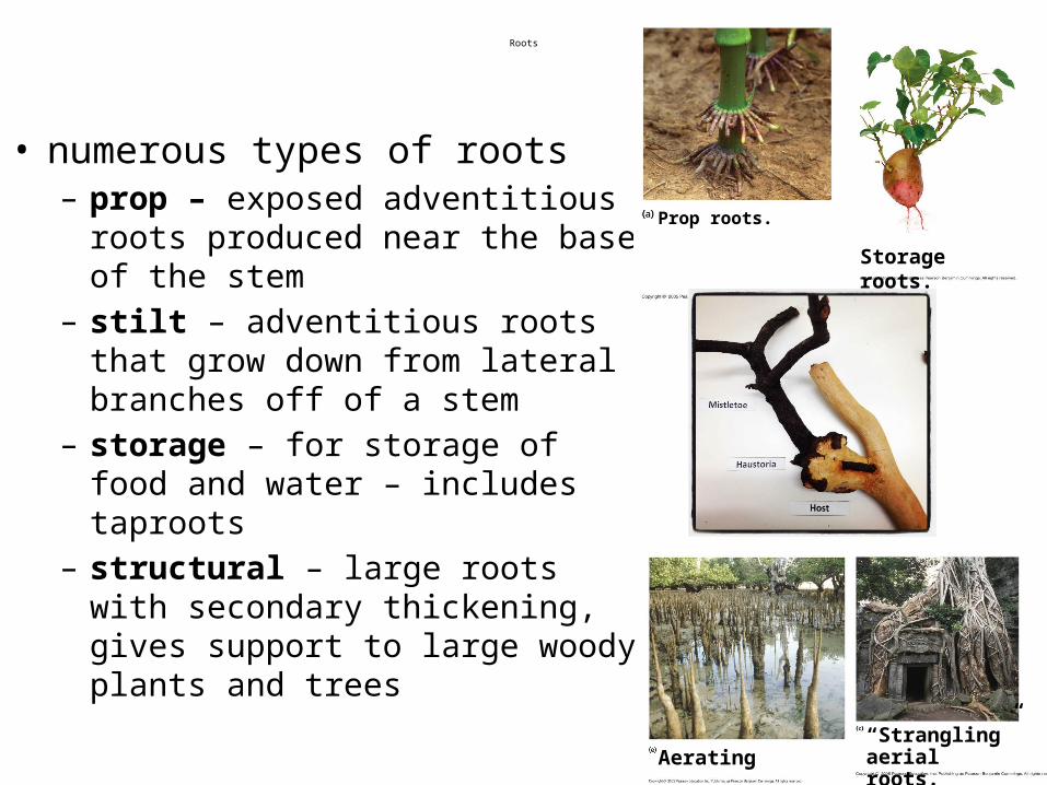
Roots
• numerous types of roots– prop – exposed adventitious roots
produced near the base of the stem– stilt – adventitious roots that grow
down from lateral branches off of a stem
– storage – for storage of food and water – includes taproots
– structural – large roots with secondary thickening, gives support to large woody plants and trees
Storage roots.
“Strangling” aerial roots.Aerating
Prop roots.
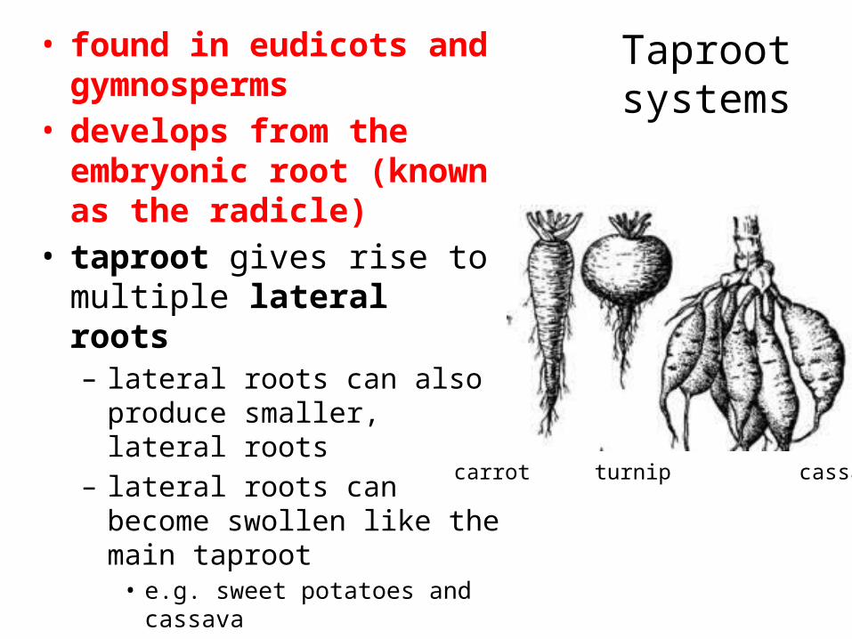
Taproot systems
• found in eudicots and gymnosperms
• develops from the embryonic root (known as the radicle)
• taproot gives rise to multiple lateral roots– lateral roots can also produce
smaller, lateral roots– lateral roots can become swollen
like the main taproot• e.g. sweet potatoes and cassava
• generally penetrate deeply • are well adapted to deep soils
where groundwater is not close to the surface
carrot turnip cassava
Fibrous Root Systems
• most monocots – e.g. grasses• mat of generally thin roots that spread out
below the soil surface• most of the roots are similarly sized• the embryonic root (radicle) dies early on
and doesn’t form a taproot• many small roots emerge from primordial
tissues found in the stem• each small root forms multiple lateral roots• does not penetrate the soil deeply• excellent at holding topsoil in place
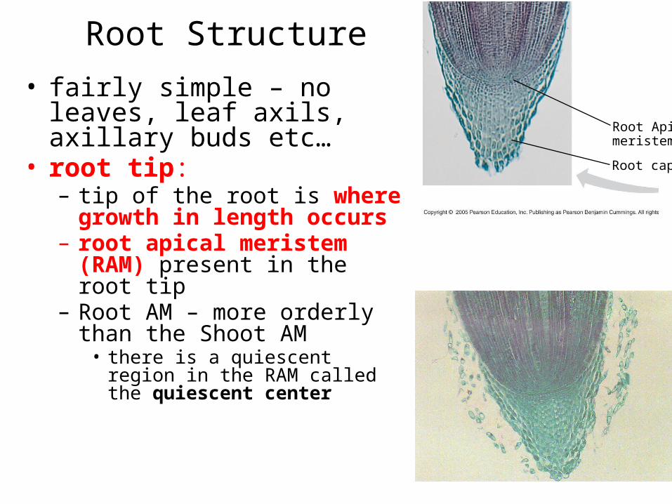
Root Structure• fairly simple – no leaves, leaf
axils, axillary buds etc…• root tip:– tip of the root is where growth in
length occurs– root apical meristem (RAM)
present in the root tip– Root AM – more orderly than the
Shoot AM• there is a quiescent region in the RAM
called the quiescent center
Root Apicalmeristem
Root cap

Root Structure
• root cap:– the RAM is protected by a root
cap as the root pushes through soil
– golgi of the root cap cells secrete a mucilage or slime to help the root push through the soil
– cells are small and meristematic
Root Apicalmeristem
Root cap
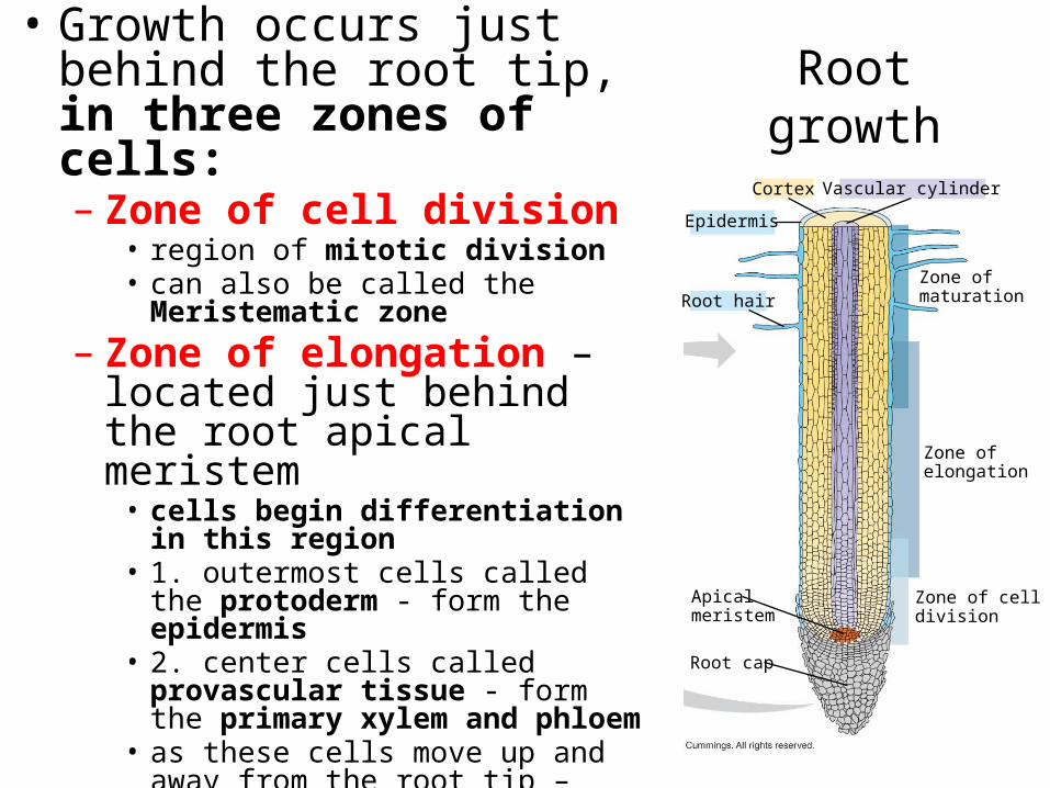
Root growth• Growth occurs just behind the
root tip, in three zones of cells:– Zone of cell division
• region of mitotic division• can also be called the Meristematic zone
– Zone of elongation – located just behind the root apical meristem• cells begin differentiation in this region• 1. outermost cells called the protoderm -
form the epidermis• 2. center cells called provascular tissue -
form the primary xylem and phloem• as these cells move up and away from the
root tip – primary X and P becomes metaxylem and metaphloem
• permeable to water
Epidermis
Root hair
Cortex Vascular cylinder
Zone ofmaturation
Zone ofelongation
Zone of celldivision
Apicalmeristem
Root cap
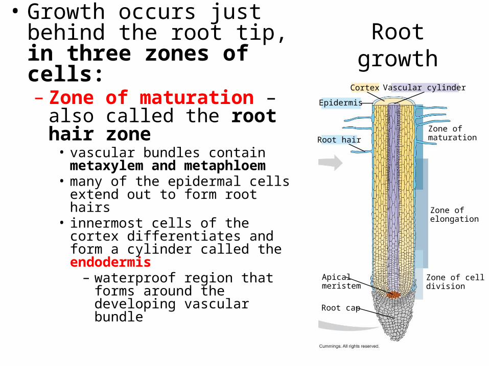
Root growth• Growth occurs just behind the
root tip, in three zones of cells:– Zone of maturation – also called
the root hair zone• vascular bundles contain metaxylem and
metaphloem• many of the epidermal cells extend out to
form root hairs • innermost cells of the cortex
differentiates and form a cylinder called the endodermis – waterproof region that forms around
the developing vascular bundle
Epidermis
Root hair
Cortex Vascular cylinder
Zone ofmaturation
Zone ofelongation
Zone of celldivision
Apicalmeristem
Root cap
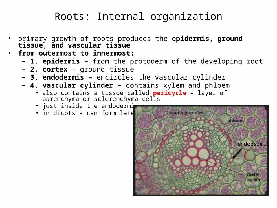
Roots: Internal organization
• primary growth of roots produces the epidermis, ground tissue, and vascular tissue• from outermost to innermost:
– 1. epidermis – from the protoderm of the developing root– 2. cortex – ground tissue– 3. endodermis – encircles the vascular cylinder – 4. vascular cylinder – contains xylem and phloem
• also contains a tissue called pericycle – layer of parenchyma or sclerenchyma cells• just inside the endodermis • in dicots – can form lateral roots
endodermis
Key
DermalGroundVascular
Epidermis
Cortex
Vascular cylinder
Endodermis
Core ofparenchymacells
Pericycle
Xylem
Phloem100 µm
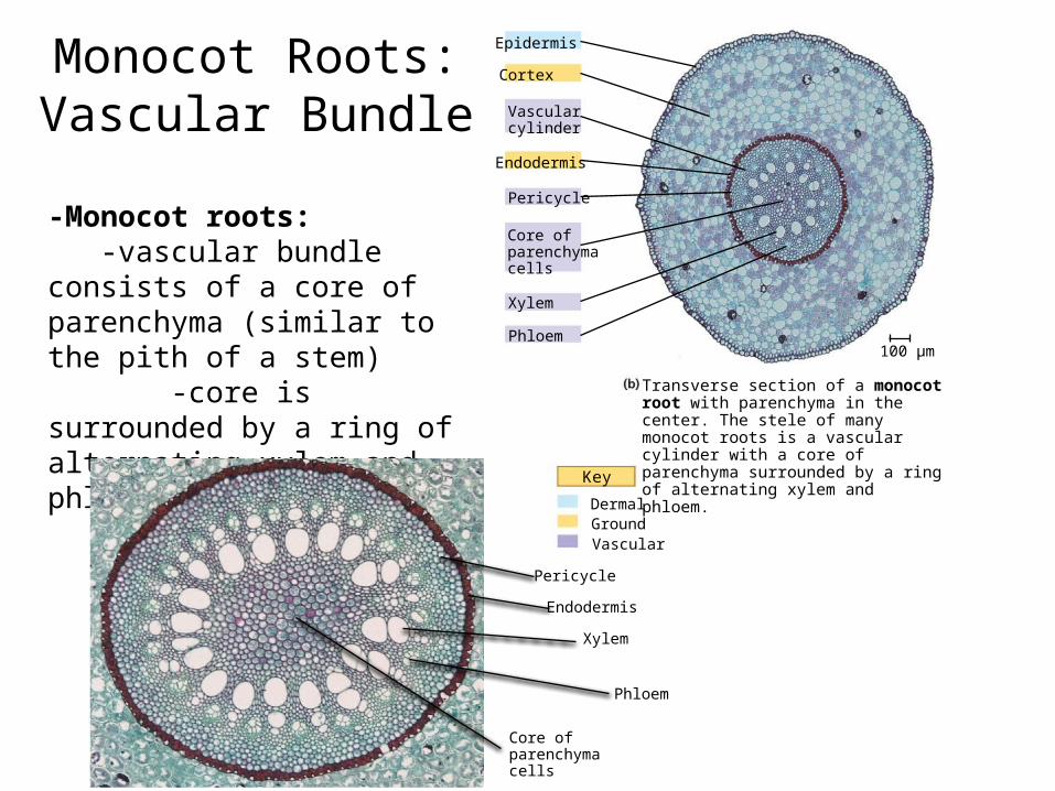
Transverse section of a monocot root with parenchyma in the center. The stele of many monocot roots is a vascular cylinder with a core of parenchyma surrounded by a ring of alternating xylem and phloem.
Monocot Roots: Vascular Bundle
-Monocot roots:-vascular bundle consists of
a core of parenchyma (similar to the pith of a stem) -core is surrounded by a ring of alternating xylem and phloem tissue
Phloem
Xylem
Pericycle
Core ofparenchymacells
Endodermis
Key
DermalGroundVascular
Epidermis
Cortex
Vascular cylinder
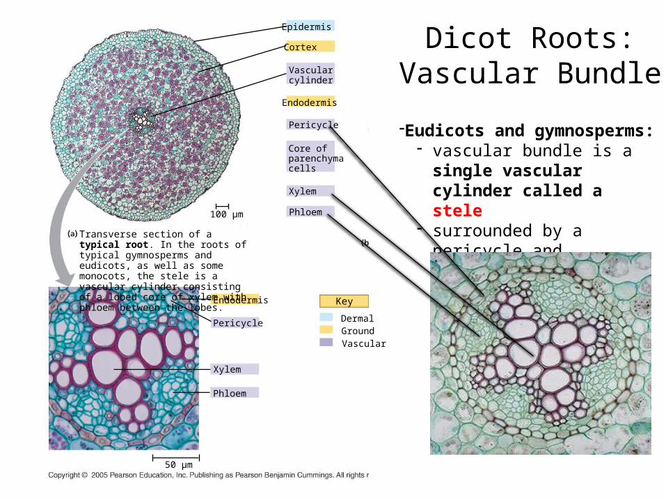
Transverse section of a typical root. In the roots of typical gymnosperms and eudicots, as well as some monocots, the stele is a vascular cylinder consisting of a lobed core of xylem with phloem between the lobes.
100 µm
Endodermis
Core ofparenchymacells
Pericycle
Xylem
Phloem
Endodermis
Pericycle
Xylem
Phloem
50 µm
-Eudicots and gymnosperms:- vascular bundle is a single
vascular cylinder called a stele- surrounded by a pericycle and
endodermis
Dicot Roots: Vascular Bundle
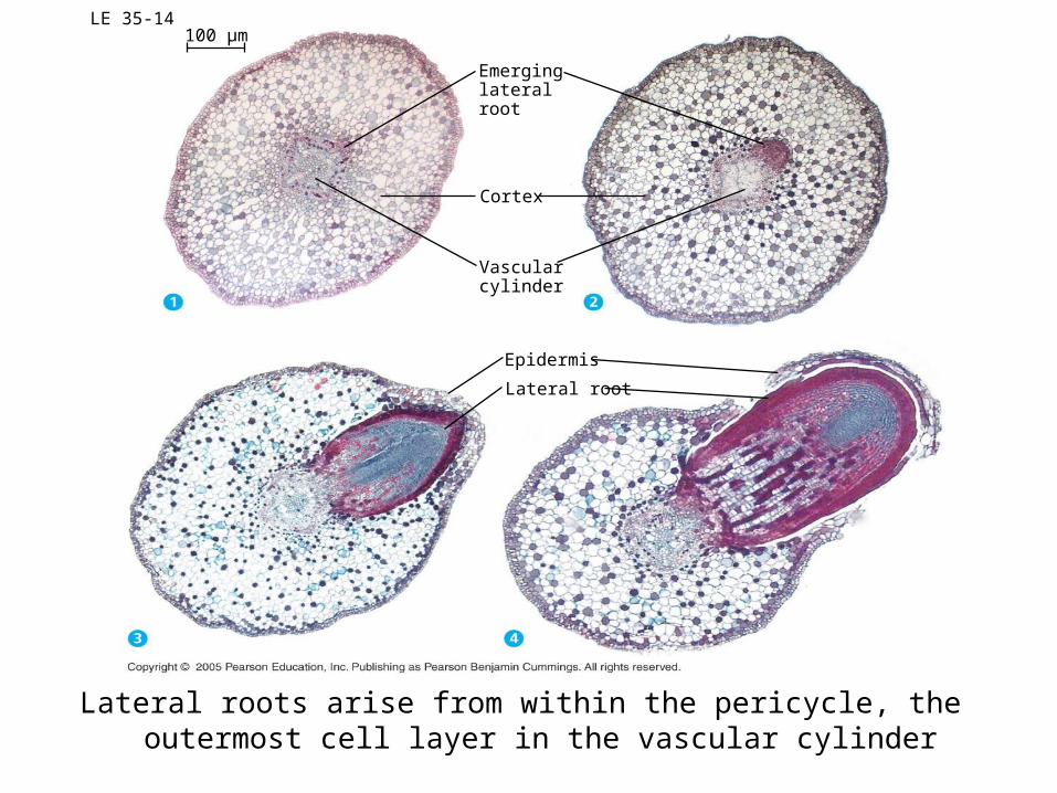
LE 35-14
Emerginglateralroot
100 µm
Cortex
Vascularcylinder
Epidermis
Lateral root
Lateral roots arise from within the pericycle, the outermost cell layer in the vascular cylinder

Plants
• two designations: herbaceous (herb – non-woody) and woody plants
• wood: considered to be made of secondary xylem

Vascular Cambium• meristematic tissue that forms during primary growth
– also called a lateral meristem– source of secondary xylem and phloem– VC located between the xylem and phloem of the vascular bundle– in non-woody plants – the cambium ultimately stops dividing and
differentiates into more xylem and phloem
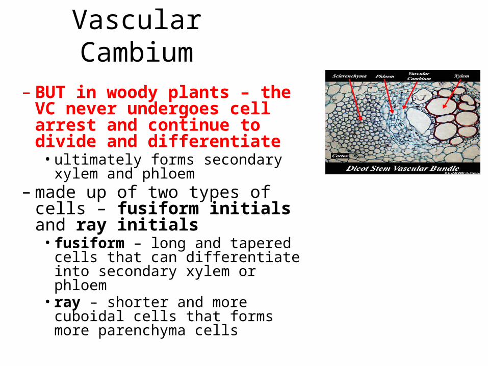
Vascular Cambium
– BUT in woody plants – the VC never undergoes cell arrest and continue to divide and differentiate• ultimately forms secondary xylem and
phloem– made up of two types of cells –
fusiform initials and ray initials• fusiform – long and tapered cells that
can differentiate into secondary xylem or phloem• ray – shorter and more cuboidal cells
that forms more parenchyma cells
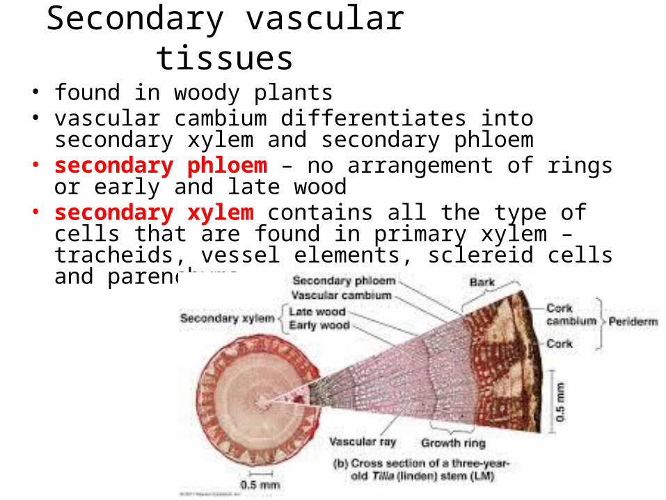
Secondary vascular tissues• found in woody plants• vascular cambium differentiates into secondary xylem and
secondary phloem• secondary phloem – no arrangement of rings or early and late
wood• secondary xylem contains all the type of cells that are found in
primary xylem – tracheids, vessel elements, sclereid cells and parenchyma
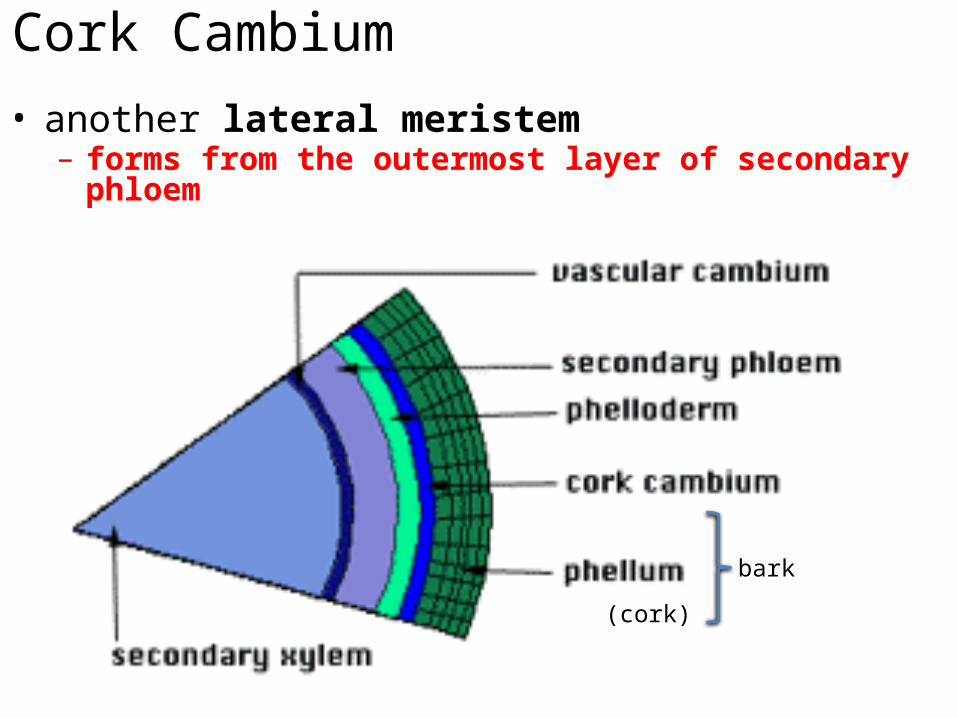
Cork Cambium• another lateral meristem– forms from the outermost layer of secondary phloem
(cork)
bark
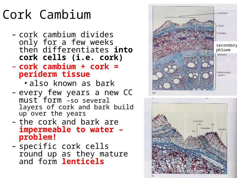
Cork Cambium– cork cambium divides only for a few
weeks then differentiates into cork cells (i.e. cork)
– cork cambium + cork = periderm tissue• also known as bark
– every few years a new CC must form –so several layers of cork and bark build up over the years
– the cork and bark are impermeable to water – problem!
– specific cork cells round up as they mature and form lenticels
secondaryphloem
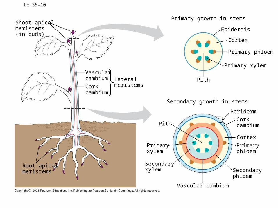
LE 35-10
Shoot apicalmeristems(in buds)
Vascularcambium
Corkcambium
Lateralmeristems
Primaryphloem
PeridermCorkcambium
Secondaryxylem
Primaryxylem
Pith
Pith
Cortex
Secondary growth in stems
Secondaryphloem
Vascular cambium
Primary phloem
Primary xylem
Cortex
Primary growth in stems
Epidermis
Root apicalmeristems
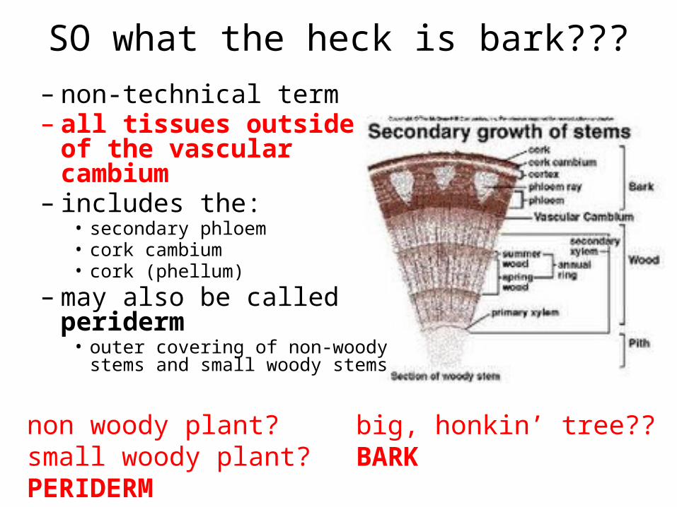
SO what the heck is bark???
non woody plant?small woody plant?PERIDERM
big, honkin’ tree??BARK
– non-technical term– all tissues outside of the
vascular cambium– includes the:
• secondary phloem• cork cambium• cork (phellum)
– may also be called periderm• outer covering of non-woody stems
and small woody stems

Secondary growth: Wood• most commercial dicot woods are strong and
tough (hardwoods)• wood from conifers have a softer consistency
– softwoods• annual rings – vascular cambium becomes
quiescent during times of stress (extreme hot and cold) and stops forming new cells
• when the VC resumes its growth and starts to form new secondary X and P – forms new rings of secondary xylem or wood– first wood that forms – early wood or spring
wood – later in the season – late wood or summer
wood• late wood + early wood = annual ring
• early years of a tree are characterized by vigorous growth – innermost annual rings are wider – more secondary xylem
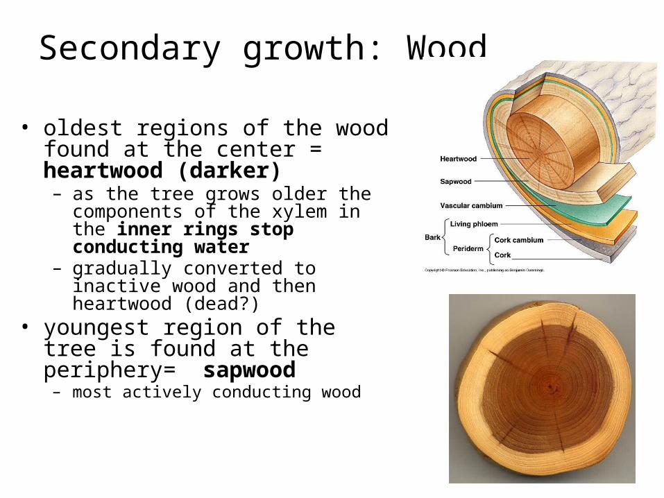
Secondary growth: Wood
• oldest regions of the wood found at the center = heartwood (darker)– as the tree grows older the components of
the xylem in the inner rings stop conducting water
– gradually converted to inactive wood and then heartwood (dead?)
• youngest region of the tree is found at the periphery= sapwood– most actively conducting wood

Some websites to check out
• http://www.slideshare.net/apurvanagvenker/anatomy-of-dicot-monocot-leaf
• http://waynesword.palomar.edu/index.htm• https
://www.youtube.com/watch?v=33H93Rlzk2w