Embed Size (px)
Citation preview

MEDICAL IMAGE COMPUTING (CAP 5937)- SPRING 2016
LECTURE 1: Introduction
Dr. Ulas BagciHEC 221, Center for Research in Computer Vision (CRCV), University of Central Florida (UCF), Orlando, FL [email protected] or [email protected]
1

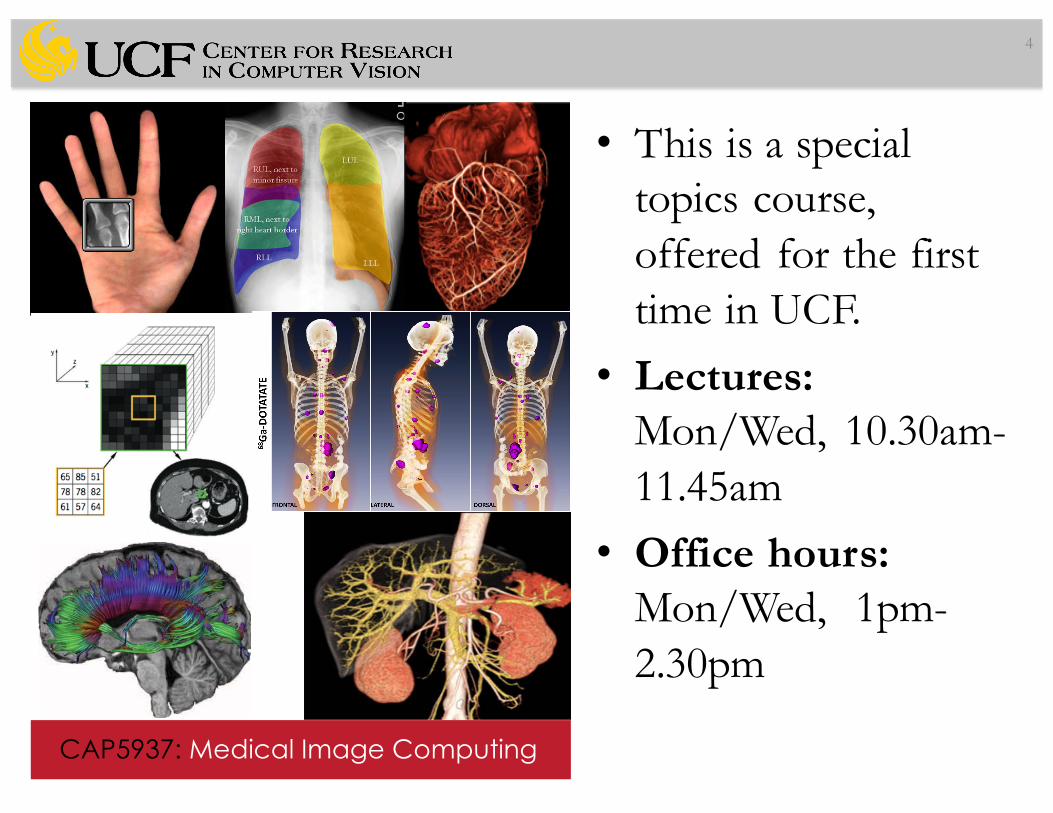
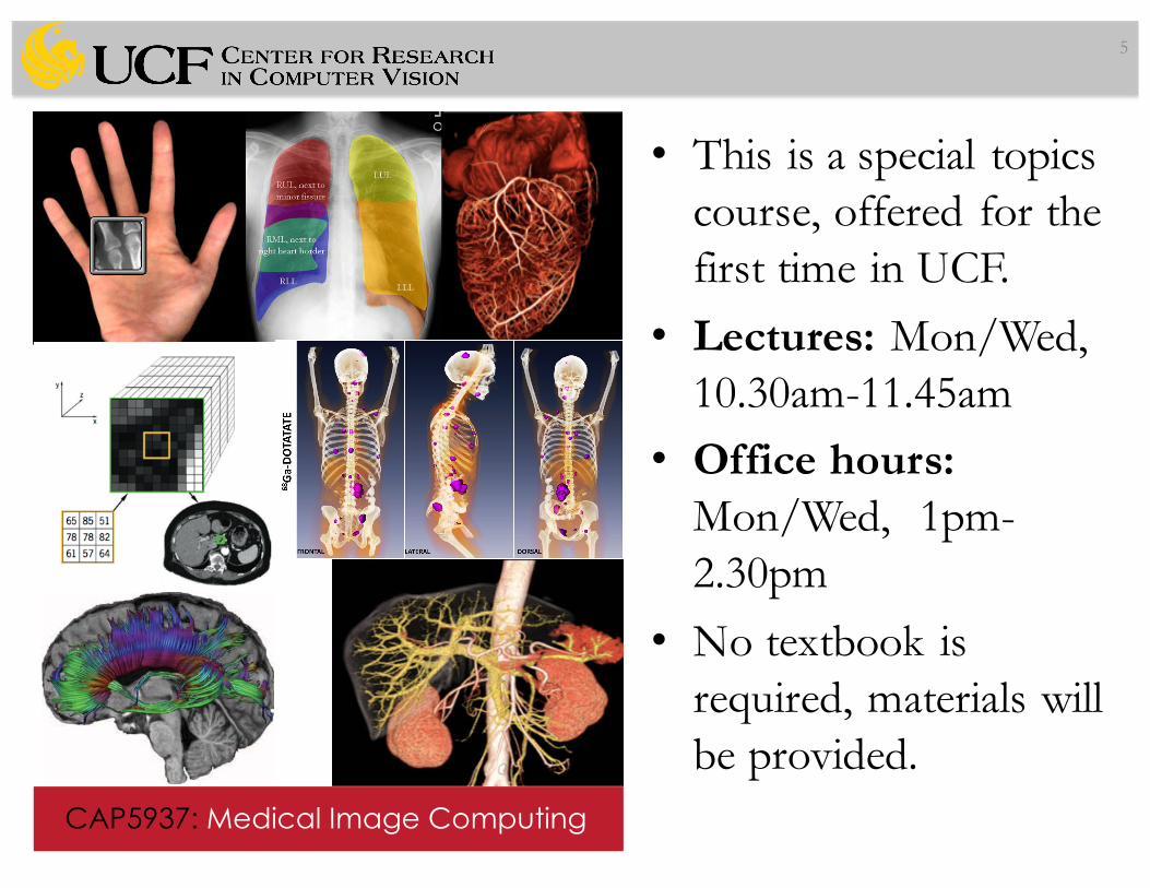
• This is a special topics course, offered for the first time in UCF.
2
CAP5937: Medical Image Computing
Lorem Ipsum Dolor Sit Amet

• This is a special topics course, offered for the first time in UCF.
• Lectures: Mon/Wed, 10.30am-11.45am
3
CAP5937: Medical Image Computing
Lorem Ipsum Dolor Sit Amet
• This is a special topics course, offered for the first time in UCF.
• Lectures: Mon/Wed, 10.30am-11.45am
• Office hours: Mon/Wed, 1pm-2.30pm
4
CAP5937: Medical Image Computing
Lorem Ipsum Dolor Sit Amet
• This is a special topics course, offered for the first time in UCF.
• Lectures: Mon/Wed, 10.30am-11.45am
• Office hours: Mon/Wed, 1pm-2.30pm
• No textbook is required, materials will be provided.
5
CAP5937: Medical Image Computing
Lorem Ipsum Dolor Sit Amet

6
Medical Image
Computing
Image Processing Computer
Vision
Machine Learning
Imaging Sciences
(Radiology, Biomedical)

Motivation• Imaging sciences is experiencing a tremendous
growth in the U.S. The NYT recently ranked biomedical jobs as the number one fastest growing career field in the nation and listed bio-medical imaging as the primary reason for the growth.
7

Motivation• Imaging sciences is experiencing a tremendous
growth in the U.S. The NYT recently ranked biomedical jobs as the number one fastest growing career field in the nation and listed bio-medical imaging as the primary reason for the growth.
• Biomedical imaging and its analysis are fundamental to (1) understanding, (2) visualizing, and (3) quantifying information.
8

Motivation• Imaging sciences is experiencing a tremendous
growth in the U.S. The NYT recently ranked biomedical jobs as the number one fastest growing career field in the nation and listed bio-medical imaging as the primary reason for the growth.
• Biomedical imaging and its analysis are fundamental to (1) understanding, (2) visualizing, and (3) quantifying information.
• This course will mostly focus on analysis of biomedical images, and imaging part will be briefly taught!
9

Syllabus• Basics of Radiological Images, Imaging, and Their
Clinical Use– X-Ray, CT, MRI, fMRI, DTI, DWI, PET, dPET, PET/CT,
MRI/PET,…• Image Enhancement and Pre-processing– Spatial and Frequency Domain Filtering
• Medical Image Registration/Alignment– Atlas construction, disease tracking, severity analysis,…
• Medical Image Segmentation– Extraction of object information, volumetry, morphometry,..
• Medical Image Visualization• Machine Learning for Medical Imaging
10

Syllabus• Grading:– 1 Mid-term exam at the classroom, written (20%)– 3 Programming Assignments (each 10%, total 30%)• ITK/VTK packages should be used• ITK and VTK provide necessary codes/libraries for medical
image processing and analysis. • C/C++ or Python can be used and call ITK/VTK functions• In-class collaboration is encouraged, but individual submission
is required.– 1 Individual Project (50%)• Will be selected from a list of projects• A short presentation (15%), coding/method (25%), results
(10%)
11

Optional Reading List• Image Processing, Analysis, and Machine Vision. M. Sonka, V.
Hlavac, R. Boyle. Nelson Engineering, 2014.• Level-set Methods, by J. A. Sethian, Cambridge University Press.• Visual Computing for Medicine: Theory, Algorithms, and
Applications. B. Preim, C. Botha. Morgan Kaufmann, 2013.• Medical Image Registration. J. Hajnal, D. Hill, D. Hawkes (eds).
CRC Press, 2001.• Pattern Recognition and Machine Learning. C. Bishop. Springer,
2007.• Insight into Images: Principles and Practice for Segmentation,
Registration and Image Analysis, Terry S. Yoo (Editor) (FREE)• Algorithms for Image Processing and Computer Vision, J. R. Parker• Medical Imaging Signals and Systems, by Jerry Prince & Jonathan
Links, Publisher: Prentice Hall
12
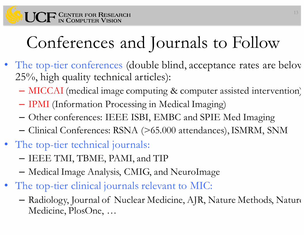
Conferences and Journals to Follow• The top-tier conferences (double blind, acceptance rates are below
25%, high quality technical articles):– MICCAI (medical image computing & computer assisted intervention)– IPMI (Information Processing in Medical Imaging)– Other conferences: IEEE ISBI, EMBC and SPIE Med Imaging– Clinical Conferences: RSNA (>65.000 attendances), ISMRM, SNM
• The top-tier technical journals:– IEEE TMI, TBME, PAMI, and TIP– Medical Image Analysis, CMIG, and NeuroImage
• The top-tier clinical journals relevant to MIC:– Radiology, Journal of Nuclear Medicine, AJR, Nature Methods, Nature
Medicine, PlosOne, …
13

Required skill set• Basic programming experience (any language is fine)• Linear Algebra/Matrix Algebra• Differential Equations• Basic Statistics
14

Biomedical Images• (Bio)medical images are different from other
pictures
15

Biomedical Images• (Bio)medical images are different from other
pictures– They depict distributions of various physical features
measured from the human body (or animal).
16

Biomedical Images• (Bio)medical images are different from other
pictures– They depict distributions of various physical features
measured from the human body (or animal body).• Analysis of biomedical images is guided by very
specific expectations
17

Biomedical Images• (Bio)medical images are different from other pictures– They depict distributions of various physical features
measured from the human body (or animal).
• Analysis of biomedical images is guided by very specific expectations– Automatic detection of tumors, characterizing their types,– Measurement of normal/abnormal structures,– Visualization of anatomy, surgery guidance, therapy
planning, – Exploring relationship between clinical, genomic, and
imaging based markers
18

Free Software to Use in this course• ImageJ (and/or FIJI)• ITK-Snap• SimpleITK• MITK• FreeSurfer• SLICER• OsiriX• An extensive list of software: www.idoimaging.com and
blue: will be frequently used in this course
19

• Thank you for your attention!
20


![[5937 - 19334]Auditoria de Prestacao de Contas Publicas](https://img.pdfslide.net/doc/110x75/55cf9b98550346d033a6aaf0/5937-19334auditoria-de-prestacao-de-contas-publicas.jpg)

