Embed Size (px)
Citation preview
Legg-Perthes Disease in the Dog: The Histo ogical and
Associated Radiological Changes
ROBIN LEE]
INTRODUCTION
The literature relating to the histological changes occurring in Legg-Perthes Disease in the dog was reviewed in 1967 (2) and again in 1970 (1) by the author in an initial study reported previously. This study has now been continued and a total of over eighty dogs with Perthes Disease have now been studied, and the femoral heads, obtained after excision arthroplasty, studied histologically. The sequence of histological changes has been related to the duration of lesions as determined from the clinical history and an attempt made to explain the radiological features observed. From these studies a thesis to explain the sequence of events is proposed.
The initial event appears to be ischemic necrosis of the otherwise normal bone and marrow tissue of the femoral capital epiph- ysis (Fig. 1). There may be little clini- cal evidence of lameness or radiological abnormality a t this stage. Continued growth of the deeper layers of the articular cartilage, but failure of the ossific nucleus to grow, results in an increase in thickness of the articular cartilage. This probably accounts for the early increase in the width of the joint space observed on the radio- graph (Fig. 2).
As a result of continued weight bearing, trabecular fragmentation, deformity and cavitation occur, resulting in uneven femo- ral head density and deformity seen on the radiograph. Probably concurrent with this stage of deformation there is the begin- ning of vascular invasion with hyperemia of the metaphysis and the soft tissues of the
joint with a resultant lateral displacement of the femoral head (Fig. 2).
Highly vascular granulation tissue then penetrates the growth plate and fairly rapidly results in revascularization and re- placement of the dead tissue by a process of “creeping substitution” (Figs. 3-5). Some of the dead bone is actively removed by osteoclastic activity, while in some in- stances dead trabeculae persist and become encapsulated by newly deposited bone. This vascular invasion probably occurs coincident with the natural closure of the growth plate. The removal of bone and deposition of foci of closely trabeculated bone and areas of fibrous tissue results in a persistence of the uneven density on radio- graphic examination (Fig. 2).
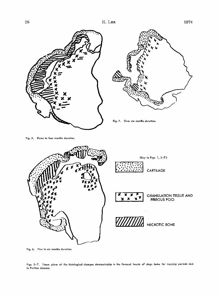
During early deformation of the femoral head a very regular feature is the formation of linear subchondral cavities which may be identified on the radiographs in certain instances. These cavities persist for a long time and the dead bone on the deep face of the osteochondral flap persists after revascularization of the remainder of the femoral epiphysis and may be observed in cases of several months duration (Figs. 3-7).
The revascularized head remains de- formed and there is associated osteophytic proliferation around the femoral neck and acetabular rim. There may be persistence of foci of fibrous tissue or of cartilage remnants which all contribute to the con- tinued uneven density (Figs. 6, 7).
Not all dogs studied fit accurately into this proposed sequence, but this may be due to initially vague or absent clinical
Lecturer in Veterinary Surgery, Faculty of Veterinary Medicine, University of Glasgow, Glasgow, This manuscript is based on a paper presented at the third Conference of the International Scotland.
Veterinary Radiology Association in Washington, D. C., Sept. 5-7, 1973.
24

VOL. xv LEGG-PERTHES DISEASE IN THE DOG 25
Fig. 1. Tissue plan of the histological changes occurring in the femoral head of a dog with Perthes disease and exhibiting signs of lameness for only one week. (See explanatory key p. 26, for Figs. 1, 3-7).
Fig. 2. Ventrodorsol radiograph of the hip of a dog showing the radiographic features of Perthes disease. Note the un- even density, increase in widjh of the joint space, lateral dis- placement of the femoral head, the irregular articular sur- face, and acetabular reaction. A linear subchondral defect is also visible. Fig. 4. Two to three months duration.
a ;=!
.Om
a
t 0,
5 z a n
s.
b
w
n
a 9 -n -.
n 'p cn s m a 0" 5 z z a Q
5 %.
3
3, p
VOL. xv LEGG-PERTHES DISEASE I N THE DOG 27
signs which only become significant and allow a diagnosis to be made when there is marked deformity. Possibly there may be only partial involvement of the epiph7 ysis. This partial involvement has been reported to occur in human Perthes Dis- ease and has been suspected in a small number of dogs included in this report. There may also be repeated episodes of ischemia, trabecular damage and necrosis which would serve to distort this simplified sequence of events.
SUMMARY
On the basis of the histological examina- tion of over 80 dogs with naturally occur- ring Legg-Perthes Disease, the sequence of the histological changes has been re- lated to the duration of the lesion and to the radiological features observed. The initial event appears to be ischemic necrosis followed by trabecular fragmentation with deformity and cavitation. This is rapidly followed by revascularization and repair, However, linear subchondral cavities per- sist for a long time as does the deformity of the femoral head.
Department of Veterinary Surgery University of Glasgow Veterinary School Bearsden Road, Bearsden Glasgow, G61 lQH, Scotland
REFERENCES 1. Lee, R.: A Study of the Radiographic and
Histological Changes Occurring in Legg-Calve- Perthes Disease (LCP) in the Dog. J. Small Anim. Pract. 11: 621-638, 1970.
2. Ljunggren, G.: Legg-Perthes Disease in the Dog. Ac ta Orthrop. Scand. (Supplement No. 95), 1967.
ZUSAMMEN FASSUNG Auf Grund der histologischen Untersuchung von
uber 80 Hunden mit natiirlich vorkommender Legg-CalvC-Perthes’ scher Krankheit, ist die Sequenz von histologischen Veranderungen in Bezug gesetzt worden zu der Dauer der Verletzung und zu den beobachteten rontgenologischen Merk- malen. Der Ausgang schejnt ischaemische Nekro- sis zu sein, gefolgt von trabekularer Fragmentation mit Verformung und Hohlen-bildung. Dies wird rapide gefolgt von Re-Vaskularization und Heilung. Jedoch halten lineare sub-chondrale Hohlen lange Zeit an, sowie die Verformung des femoralen Kop- fes.
RESUME A partir de l’examen histologique de plus de 80
chiens chez lesquels la maladie de Legg-Perthes- Calve est survenue naturellement, la sequence des changements histologiques a BtB like ii la durCe de la lesion et aux caracthres radiologiques observes. L’QvBnement initial apparait Etre la necrose isch- Bmique suivie de la fragmentation trabeculaire et de la cavitation. Ceci est rapidement suivi d’une revascularisation et d’un retablissement. NBan- moins, les cavitks lineaires sous-chondrales per- sistent pendant longtemps comme la difformite de la tEte fernorale.