Embed Size (px)
Citation preview
Leishmania major: Promastigotes Induce Expression of a Subset ofChemokine Genes in Murine Macrophages
ESTHERL. RACOOSIN1
AND STEPHENM. BEVERLEY
Department of Biological Chemistry and Molecular Pharmacology, Harvard Medical School,Boston, Massachusetts 02115 U.S.A.
RACOOSIN, E. L., AND BEVERLEY, S. M. 1997.Leishmania major:Promastigotes induce expressionof a subset of chemokine genes in murine macrophages.Experimental Parasitology85, 283–295.Recent studies suggest thatLeishmania majorpromastigotes infect cultured macrophages in astealthy fashion, activating little or no host gene expression and often interfering with the host’sability to respond to further stimulation. Here we examined macrophage transcription at early timesfollowing infection, when virulent parasites must execute steps required for survival. Stationary-phase promastigotes induced rapid and transient expression of transcripts of the chemokines JE(human MCAF/MCP-1) and KC (human GRO) in bone marrow-derived macrophages from BALB/cmice. JE and KC expression rose four- to sixfold shortly after infection and returned to uninducedlevels by 4–24 hr. In contrast, chemokines MIP-1a, C10, and RANTES were not induced, nor wereTGF-b, IL-10, IL-12, or i-NOS. Chemokine induction did not occur following ingestion of latexbeads, implicating a parasite-specific stimulus. Elevated expression of a subset of chemokines is theearliest known transcriptional response of macrophages toL. major infection and potentially mayprovide a signal for the initiation of downstream immunological responses which occurin vivo,suchas cytokine induction and chemotaxis of monocytes and macrophages. Thus,Leishmaniahas aremarkable ability to take an active role in either inducing or preventing the expression of distinct setsof host genes during macrophage invasion and successful intracellular parasitism.© 1997 Academic
Press
INDEX DESCRIPTORS ANDABBREVIATIONS: Leishmania;chemokines; MCP-1, monocyte chemoat-tractant protein-1; FBS, fetal bovine serum; BMM, bone marrow-derived macrophages; M-CSF,macrophage colony-stimulating factor; MIP, macrophage inflammatory protein; nt, nucleotide; iNOS,inducible nitric oxide synthase; LPG, lipophosphoglycan.
INTRODUCTION
Virulent metacyclic promastigotes ofLeish-mania are introduced into host tissues by thebite of an infected phlebotomine sand fly andare subsequently phagocytosed by tissue mac-rophages, where differentiation into the amasti-gote stage occurs. In order to survive within thehost,Leishmaniapromastigotes express severalabundant surface molecules, including a glyco-lipid termed lipophosphoglycan (LPG) and asurface protease, gp63 (Chang and Chaudhuri1990; Turco and Descoteaux 1992). LPG, forexample, participates in binding to the macro-
phage and phagocytosis and protects the para-site from lysis by complement, damage by re-active oxygen intermediates, and hydrolytic en-zymes of the macrophage phagolysosome(Chenet al.1989; Cooperet al.1988; daSilvaetal. 1989; Eilamet al. 1985; Talamas-Rohanaetal. 1990).
Leishmaniainfection may also affect the pat-tern of gene expression in the macrophage.The effects reported vary greatly among dif-ferent Leishmaniaspecies, source of macro-phages, and laboratories. Many workers havefound that, unlike most other pathogens, there issurprisingly little induction of macrophagegene expression immediately following infec-tion with Leishmania majorpromastigotesinvitro. This included studies of IL-1, IL-10, IL-12, TNF-a, i-NOS, MIP-1a, and TGF-b (Car-rera et al. 1996; Reineret al. 1994). Yet this
1 To whom correspondence should be addressed at pre-sent address: Department of Microbiology and Immunol-ogy, College of Veterinary Medicine, Cornell University,Ithaca NY 14853-6401. Fax: (607) 253-3384. E-mail:[email protected].
EXPERIMENTAL PARASITOLOGY 85, 283–295 (1997)ARTICLE NO. PR964139
2830014-4894/97 $25.00Copyright © 1997 by Academic PressAll rights of reproduction in any form reserved.
‘‘stealthy’’ mode of invasion may be mislead-ing, asLeishmaniainfection actively interfereswith the macrophage’s ability to respond toother immunological stimuli, such as LPS orother antigens. Bone marrow-derived macro-phages (BMM) infected withL. majorwere de-fective in LPS-induced expression of IL-12(Carreraet al.1996), and macrophages infectedwith L. donovani were defective in LPS-induced c-fos and TNF-a gene expression(Descoteaux and Matlashewski 1989) and inter-feron-g induced MHC class II antigen expres-sion (Reineret al. 1988). In some instances,gene expression in infected macrophages didnot commence until 24 hr after infection. Forexample, in peritoneal macrophages isolatedfrom BALB/c mice, expression of IL-1 wasstimulated byL. major infection (Cillari et al.1989) and expression of TGF-b was stimulatedby L. amazonensisandL. braziliensis(Barraletal. 1993; Barral-Nettoet al.1992). In BMM,L.donovanipromastigotes induced expression ofGM-CSF, TNF-a, TGF-b, and IL-6 (Moore andMatlashewski 1994). Significantly, all of theseeffects on macrophage gene expression couldaffect the ability of theLeishmaniato establishinfection and induce host T-cell responses dur-ing infections in vivo (Reiner et al. 1994;Scharten and Scott 1993).
Many studies of the effects ofLeishmaniaonhost gene expression examined periods well af-ter macrophage invasion (usually more than 24hr). However, here we showed that virulentL.major must successfully execute steps requiredfor establishment of infection within 24 hr. Thissuggested that it would be important to examinemacrophage responses during this critical pe-riod, especially for genes expressed only tran-siently. We used Northern blot analysis to fol-low gene expression at early times after infec-tion and additionally compared the expressioninduced by avirulent vs. virulentL. majorstrains, since this could potentially assist inidentifying key host pathways intercepted byvirulent parasites. Significantly, we found thatLeishmaniainduces rapid and transient expres-sion of only a subset of macrophage chemo-
kines, specifically JE and KC (the murine ho-mologs of human MCAF/MCP-1 and GRO, re-spectively). In addition, our results confirmedand extended studies showing that macrophagesinfected in vitro do not express a variety ofgenes shown under other circumstances to beinvolved in the host response toLeishmaniain-fection (Carreraet al.1996; Reineret al.1994).
MATERIALS AND METHODS
Macrophage culture.Murine BMM were obtained frombone marrow extruded from the femurs of female BALB/cmice (National Institute of Health, National Cancer Insti-tute, Frederick, MD) as described (Racoosin and Swanson1989). BMM were cultured in DME-10F, which is Dulbec-co’s modified Eagle’s medium (DMEM; GIBCO BRL, Gai-thersburg, MD) supplemented with 10% heat-inactivatedfetal bovine serum (FBS; Hyclone, Logan, UT), and 100units/ml penicillin and 100mg/ml streptomycin (GIBCOBRL). To stimulate BMM growth, DME-10F was furthersupplemented with 30% L929 fibroblast-conditioned me-dium, which contains macrophage-CSF. Twenty-four hoursbefore infections, BMM were replated in either 100- or60-mm tissue culture dishes (Falcon, Franklin Lakes, NJ)containing DME-10F but lacking L929-conditioned me-dium. To monitorLeishmaniainfection, dishes often con-tained one 12-mm-diameter, No. 1 glass coverslip (FisherScientific, Pittsburgh, PA) that had been washed in 95%ethanol and double-distilled water and autoclaved previ-ously. RAW 264.7 cells (American Type Culture Collec-tion; Rockville, MD) were grown in DME-10F. L929 fibro-blasts (ATCC) were grown in MEM (GIBCO-BRL) + 10%FBS. All solutions and media used in growing murine mac-rophages were endotoxin-free, as reported by the manufac-turers.
Leishmaniastrains and culture.The L. major strainsFriedlin V1 (FV1), Friedlin A1 (FA1; (daSilva and Sacks1987)), LV39 clone 5 (Lc5), and LV39 clone 79 (Lc79;(Marchandet al. 1987)) were maintainedin vitro as pro-mastigotes in M199 medium as described (Kapleret al.1990). Promastigote cultures were routinely passaged whilein the logarithmic phase of growth. Virulent strains werepassaged periodically through BALB/c mice and were pas-saged no more than 10 timesin vitro.
Infection of macrophages.Promastigotes in the stationaryphase of growth were used to infect BMM at a final ratio of10 parasites per macrophage. Parasites were pelleted at5000g in a SS-34 rotor (Sorvall) for 15 min at room tem-perature and then resuspended in DMEM containing 4%complement fragment 5-deficient serum obtained fromstrain B10.D2/oSnJ female mice (Jackson Laboratory, BarHarbor, ME) at a concentration of 107–108 parasites/ml for15 min at 37°C. Opsonization with complement has beenshown to enhance phagocytosis of metacyclic promastigotes
RACOOSIN AND BEVERLEY284
(daSilvaet al. 1989; Mosser and Edelson 1987). Parasiteswere then washed twice in DMEM alone and resuspendedin DME-10F warmed to 37°C. Immediately prior to addi-tion of parasites, the macrophages were washed once withDME-10F. To initiate infection, promastigotes were addedto 3.6 × 10⁶ or 107 BMM in 60- or 100-mm dishes, respec-tively. The 100-mm dishes were used in experiments withtwo time points, and 60-mm dishes were used for largerexperiments. Where indicated, 6 × 10⁶ 4-mm-diameterwashed, sterile latex beads (Polysciences, Warrington, PA)were added to 3.6 × 10⁶ macrophages plated in 60-mmdishes. After addition of parasites or beads, the macro-phages were incubated at 37°C in 95% air, 5% CO2. Attimes indicated, the glass coverslip was removed andstained with Diff-Quik (Baxter Scientific, McGaw Park, IL)to monitor the infection. The remaining cells were taken forRNA analysis. For infections longer than 2 hr, infected mac-rophages were washed three times with DME-10F to re-move noninternalized parasites or beads, and fresh mediumwas added.
Controls for macrophage RNA transcripts.107 BMM orRAW 264.7 cells plated in 100-mm dishes were treated witheither 100 ng/ml lipopolysaccharide (LPS,Escherichia coliserotype 0127:B8 (SIGMA) or 100 ng/ml LPS + 500 unitsmurine interferon-g (Genzyme, Cambridge, MA). TotalRNA from L929 fibroblasts and/or BMM treated withDME-10F containing 30% L929-conditioned medium con-taining macrophage-CSF (M-CSF) were used as negativecontrols.
Northern blots.Total RNA was extracted using 1 or 2 mlRNAzol (Biotecx, Houston, TX) for a 60- or 100-mm tissueculture dish, respectively, according to the manufacturer’sinstructions. RNA resuspended in diethyl pyrocarbonate-treated water was quantitated by UV absorbance and stain-ing following gel separation. Samples containing 10mgtotal RNA were glyoxylated and electrophoresed at 4 V/cmon a 10 mM phosphate agarose gel, pH 7.0, with constantbuffer circulation (Brown 1994). RNA was transferred us-ing the Turbo Blot kit (Schleicher & Schuell, Keene, NH)onto Hybond N nylon membranes (Amersham, ArlingtonHeights, IL). Membranes were baked 2 hr at 80°C and thendeglyoxylated by boiling 30 min in 20 mM Tris–HCl, pH8.0. Prehybridization was performed at 67°C for 2 hr in 6×SSC (1× SSC is 0.15M sodium chloride, 0.015M sodiumcitrate, pH 7.0), 0.1–0.5% SDS, 200mg/ml denaturedsalmon sperm DNA, 0.04% Ficoll 400, 0.04% polyvinyl-pyrrolidone, and 0.04% bovine serum albumin (Brown1994). For hybridization, denatured double-stranded cDNAprobes (see below) labeled with [a32P]DCTP (Amersham)by the random primer method (Feinberg and Vogelstein1983) were added directly to prehybridized membranes andincubated for at least 18 hr. Membranes were then washedat room temperature in 2× SSC + 0.5% SDS, then at 67°Cin 0.1× SSC + 0.5% SDS, and were exposed first to X-LSor X-AR film (Kodak, Rochester, NY) and then to a phos-phorimager screen (Molecular Dynamics, Sunnyvale, CA).
Quantitation of RNA levels was performed using a Molecu-lar Dynamics Phosphorimager equipped with the ImageQuant 3.2 program, usingb-actin hybridization as an inter-nal control.
cDNA probes.Probes used for Northern hybridizationwere as follows: murine KC cDNA (Oquendoet al. 1989),murine JE cDNA (Rollinset al. 1988), murinei-NOS (Xieet al. 1992), murine IL-1b (Gray et al. 1986), rat TGF-b1(Qianet al. 1990), murine C10 (Orlofskyet al. 1994), mu-rine RANTES (Heegeret al. 1992), and murine IL-10(Moore et al. 1990). cDNA probes for murine actin andTNF-a were generated by polymerase chain reaction (PCR)using primer pairs purchased from Stratagene (San Diego,CA) and cDNA template prepared as described (Reineretal. 1993) from total RNA prepared from BMM treated withLPS for 6 hr. cDNA probes for murine IL-12 p40 mRNAand MIP-1a were generated by PCR using cDNA templatefrom BMM treated with LPS and IFN-g for 6 hr and thefollowing primer pairs: for IL-12, (59) cgggatccaccATGT-GTCCTCAGAAGCTAACC (SMB 85) and (59) cgg-gatcCTAGGATCGGACCCTGCAGGGAA (SMB 86) togenerate a fragment of 1050 nt (Schoenhautet al.1992); forMIP-1a, (59) GCCCTTGCTGTTCTTCTCTGT and (59)GGCAATAGTTCCAGGTCAGT to generate a fragment of300 nt (Davateliset al. 1988). Lowercase letters denotenucleotides not present in the gene.
RESULTS
Macrophages phagocytose comparable num-bers of avirulent and virulentL. major. We firstcompared the ability of BMM to phagocytosedifferent strains ofL. major. The virulentstrains, Friedlin V1 (FV1) and LV39 clone 5(Lc5), previously have been shown to replicatein macrophagesin vitro and rapidly induce le-sion formation following injection into BALB/cmouse footpads (daSilva and Sacks 1987; Shan-kar et al.1993). In contrast, the avirulent Fried-lin-derived clone A1 (FA1) derived by long-term passagein vitro does not form metacyclicpromastigotes and cannot infect mice (daSilvaand Sacks 1987). The attenuated LV39-derivedclone 79 (Lc79) derived by passage and muta-genesis is similarly avirulent, although infec-tions eventually occur after 6 months due toreversion to virulencein vivo (Shankaret al.1993).
BMM were infected with opsonized station-ary-phase promastigotes from these four strains,and the numbers of intracellular parasites deter-mined after 1 or 24 hr. At 1 hr, 83–95% of the
LeishmaniaINDUCE SPECIFIC CHEMOKINE mRNAs 285

macrophages contained 2.8–3.7 promastigotes,regardless of the infecting strain (Table I). After24 hr of infection, parasites from the virulentFV1 and Lc5 strains differentiated into amasti-gotes and survived in 57–67% of the BMM,whereas only 23% of the macrophages con-tained Lc79 parasites and 2% contained FA1parasites. Similar results were obtained in twoother independent experiments not shown.Thus, initially both virulent and avirulent para-sites are rapidly ingested by macrophages, butwithin 24 hr avirulent strains are destroyed.This suggests that during this interval virulentparasites must execute steps critical to survival.
Macrophages infected withL. majorpromas-tigotes exhibit rapid transient induction of che-mokine gene expression.BMM were incubatedfor increasing times in medium alone or in me-dium containing opsonized stationary-phasepromastigotes. Subsequently, total RNAs ex-tracted from uninfected and infected macro-phages from the same experiment were sub-jected to Northern blot analysis with a numberof cDNA probes. Hybridization of the same fil-ters with ab-actin probe was used as a control.
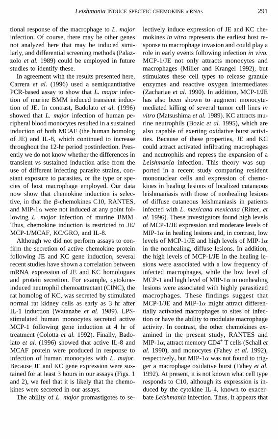
A comparison of cDNA probe hybridizationto RNA from uninfected and infected macro-phages showed that only JE and KC transcriptswere expressed at significantly higher levelsfollowing Leishmaniainvasion of BMM. Thisis shown in Fig. 1 for the Lc5 and Lc79 lines. Inthis experiment, expression of both chemokineswas detectable by 45 min postinfection and wasmaximal at 60 and 120 min for JE and KC,respectively. For JE, the maximal stimulationwas 4-fold for Lc5 and 12-fold for Lc79, andexpression returned to baseline levels by 3 hr(Figs. 1 and 2). For KC, maximal stimulationwas 12-fold for Lc5 and 30-fold for Lc79 andreturned to baseline values by 4 hr (Figs. 1 and2). These studies were repeated numerous timeswith both of the LV39 strains as well as thevirulent FV1 and avirulent FA1L. majorstrains(Northern blots not shown; Table II). On aver-age, the magnitude of JE and KC expressionwas higher with the avirulent than virulentstrains (Table II). For the LV39 lines, peak ex-pression of both JE and KC transcripts wasabout 2–3 times higher following infection withthe avirulent clone Lc79 than with virulent Lc5(Figs. 1B and 1C).
We observed considerable variability in themagnitude of the induction among experimentswith all strains, a finding also evident in the JEand IL-10 data of Carreraet al. (1996). This wasapparent in comparisons between individual ex-periments, with some showing generally low andsome showing generally high induction of bothgenes with all parasite lines tested. We attributethis to differences in BMM, which are primarycell cultures, or to differences among parasitepreparations. Nonetheless, in each experiment,there was greater induction of JE and KC expres-sion in infected BMM than in uninfected BMM.
In order to determine if chemokine inductionwas a general phenomenon followingLeish-maniainfection, we examined the expression ofseveral other members of theb-chemokine fam-ily. MIP-1a was expressed at a low constitutivelevel but was not elevated byLeishmaniainfec-tion of BMM (Figs. 1A, 2A, and 2F). Expres-sion of murine RANTES was undetectable ex-cept in the control, LPS-treated macrophages
TABLE IMurine Bone Marrow-Derived Macrophages Are InfectedEqually by Avirulent and VirulentL. majorPromastigotes
Time
L. major strain
FA1 FV1 Lc79 Lc5
1 hrPercentage macrophages
infecteda 86% 83% 95% 92%Parasites/macrophage 3.0 2.8 3.7 3.4
24 hrPercentage macrophages
infectedb 2% 57% 23% 67%Amastigotes/macrophage 0.3 1.2 0.5 2.2
Note.Data from one representative experiment of threetotal are shown.
a The ranges for the three experiments were FA1, 86–95%; FV1, 76–87%; Lc79, 88–98%; Lc5, 86–97%.
b For the 24-hr time point, the percentage of cells infectedand the number of parasites per macrophage were basedonly on amastigotes; promastigotes (if present) were notcounted. The ranges of percent cells infected for the threeexperiments were FA1, 0–2%; FV1, 55–60%; Lc79, 19–23%; Lc5, 65–72%.
RACOOSIN AND BEVERLEY286
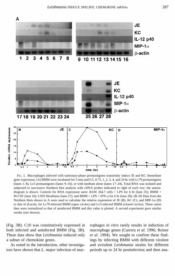
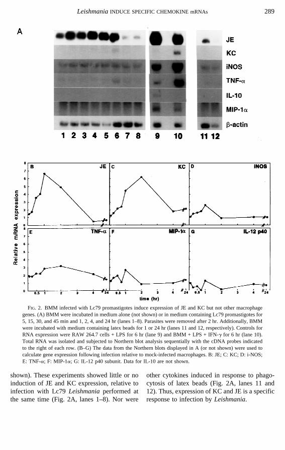
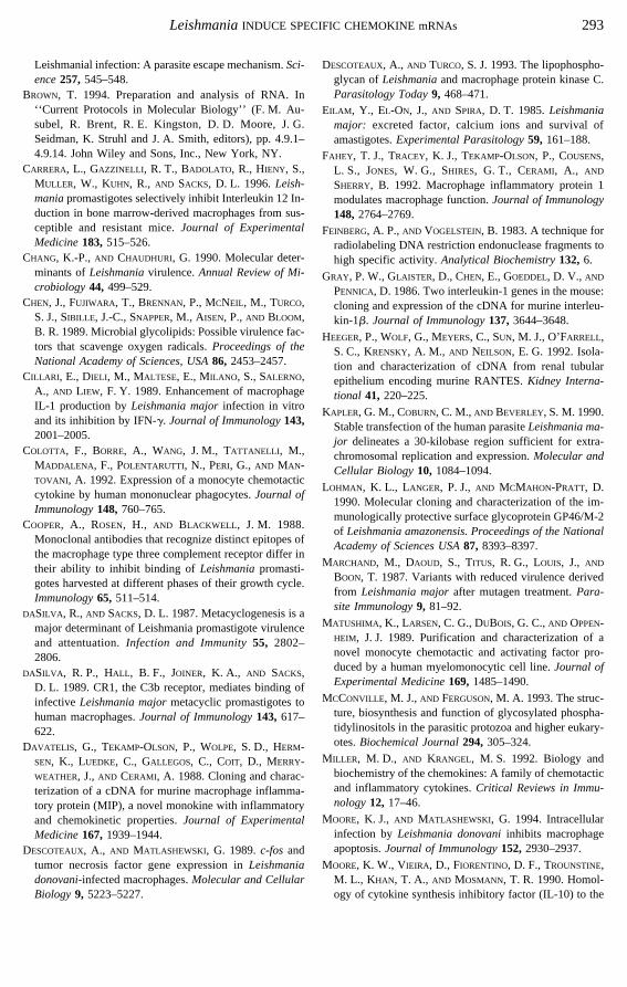
(Fig. 3B). C10 was constitutively expressed inboth infected and uninfected BMM (Fig. 3B).These data show thatLeishmaniainduced onlya subset of chemokine genes.
As noted in the introduction, other investiga-tors have shown thatL. major infection of mac-
rophagesin vitro rarely results in induction ofmacrophage genes (Carreraet al. 1996; Reineret al. 1994). We sought to confirm these find-ings by infecting BMM with different virulentand avirulentLeishmaniastrains for differentperiods up to 24 hr postinfection and then ana-
FIG. 1. Macrophages infected with stationary-phase promastigotes transiently induce JE and KC chemokinegene expression. (A) BMM were incubated for 5 min and 0.5, 0.75, 1, 2, 3, 4, and 24 hr with Lc79 promastigotes(lanes 1–8), Lc5 promastigotes (lanes 9–16), or with medium alone (lanes 17–24). Total RNA was isolated andsubjected to successive Northern blot analysis with cDNA probes indicated to right of each row; the autora-diogram is shown. Controls for RNA expression were: RAW 264.7 cells + LPS for 6 hr (lane 25); BMM +M-CSF (lane 26); L929 fibroblasts (lane 27); and BMM + LPS + IFN-g for 6 hr (lane 28). (B–D) Data from theNorthern blots shown in A were used to calculate the relative expression of JE (B), KC (C), and MIP-1a (D)to that ofb-actin, for Lc79-infected BMM (open circles) and Lc5-infected BMM (closed circles). These ratiosthen were normalized to that of uninfected BMM and this value is plotted. A second experiment gave similarresults (not shown).
LeishmaniaINDUCE SPECIFIC CHEMOKINE mRNAs 287
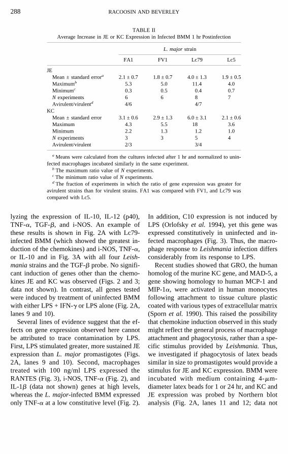
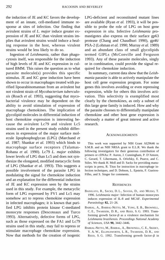
lyzing the expression of IL-10, IL-12 (p40),TNF-a, TGF-b, and i-NOS. An example ofthese results is shown in Fig. 2A with Lc79-infected BMM (which showed the greatest in-duction of the chemokines) and i-NOS, TNF-a,or IL-10 and in Fig. 3A with all fourLeish-maniastrains and the TGF-b probe. No signifi-cant induction of genes other than the chemo-kines JE and KC was observed (Figs. 2 and 3;data not shown). In contrast, all genes testedwere induced by treatment of uninfected BMMwith either LPS + IFN-g or LPS alone (Fig. 2A,lanes 9 and 10).
Several lines of evidence suggest that the ef-fects on gene expression observed here cannotbe attributed to trace contamination by LPS.First, LPS stimulated greater, more sustained JEexpression thanL. major promastigotes (Figs.2A, lanes 9 and 10). Second, macrophagestreated with 100 ng/ml LPS expressed theRANTES (Fig. 3), i-NOS, TNF-a (Fig. 2), andIL-1b (data not shown) genes at high levels,whereas theL. major-infected BMM expressedonly TNF-a at a low constitutive level (Fig. 2).
In addition, C10 expression is not induced byLPS (Orlofskyet al. 1994), yet this gene wasexpressed constitutively in uninfected and in-fected macrophages (Fig. 3). Thus, the macro-phage response toLeishmaniainfection differsconsiderably from its response to LPS.
Recent studies showed that GRO, the humanhomolog of the murine KC gene, and MAD-5, agene showing homology to human MCP-1 andMIP-1a, were activated in human monocytesfollowing attachment to tissue culture plasticcoated with various types of extracellular matrix(Spornet al. 1990). This raised the possibilitythat chemokine induction observed in this studymight reflect the general process of macrophageattachment and phagocytosis, rather than a spe-cific stimulus provided byLeishmania.Thus,we investigated if phagocytosis of latex beadssimilar in size to promastigotes would provide astimulus for JE and KC expression. BMM wereincubated with medium containing 4-mm-diameter latex beads for 1 or 24 hr, and KC andJE expression was probed by Northern blotanalysis (Fig. 2A, lanes 11 and 12; data not
TABLE IIAverage Increase in JE or KC Expression in Infected BMM 1 hr Postinfection
L. major strain
FA1 FV1 Lc79 Lc5
JEMean ± standard errora 2.1 ± 0.7 1.8 ± 0.7 4.0 ± 1.3 1.9 ± 0.5Maximumb 5.3 5.0 11.4 4.0Minimumc 0.3 0.5 0.4 0.7N experiments 6 6 8 7Avirulent/virulentd 4/6 4/7
KCMean ± standard error 3.1 ± 0.6 2.9 ± 1.3 6.0 ± 3.1 2.1 ± 0.6Maximum 4.3 5.5 18 3.6Minimum 2.2 1.3 1.2 1.0N experiments 3 3 5 4Avirulent/virulent 2/3 3/4
a Means were calculated from the cultures infected after 1 hr and normalized to unin-fected macrophages incubated similarly in the same experiment.
b The maximum ratio value ofN experiments.c The minimum ratio value ofN experiments.d The fraction of experiments in which the ratio of gene expression was greater for
avirulent strains than for virulent strains. FA1 was compared with FV1, and Lc79 wascompared with Lc5.
RACOOSIN AND BEVERLEY288
shown). These experiments showed little or noinduction of JE and KC expression, relative toinfection with Lc79 Leishmaniaperformed atthe same time (Fig. 2A, lanes 1–8). Nor were
other cytokines induced in response to phago-cytosis of latex beads (Fig. 2A, lanes 11 and12). Thus, expression of KC and JE is a specificresponse to infection byLeishmania.
FIG. 2. BMM infected with Lc79 promastigotes induce expression of JE and KC but not other macrophagegenes. (A) BMM were incubated in medium alone (not shown) or in medium containing Lc79 promastigotes for5, 15, 30, and 45 min and 1, 2, 4, and 24 hr (lanes 1–8). Parasites were removed after 2 hr. Additionally, BMMwere incubated with medium containing latex beads for 1 or 24 hr (lanes 11 and 12, respectively). Controls forRNA expression were RAW 264.7 cells + LPS for 6 hr (lane 9) and BMM + LPS + IFN-g for 6 hr (lane 10).Total RNA was isolated and subjected to Northern blot analysis sequentially with the cDNA probes indicatedto the right of each row. (B–G) The data from the Northern blots displayed in A (or not shown) were used tocalculate gene expression following infection relative to mock-infected macrophages. B: JE; C: KC; D: i-NOS;E: TNF-a; F: MIP-1a; G: IL-12 p40 subunit. Data for IL-10 are not shown.
LeishmaniaINDUCE SPECIFIC CHEMOKINE mRNAs 289
DISCUSSION
In this study we examined the effects ofL.major infection on gene expression in BMMduring the 24-hr period following infection,when virulent organisms must successfully ex-ecute steps required for intracellular parasitism.The results showed that BMM infected withL.major stationary-phase promastigotes selec-tively activated the KC and JE genes, membersof thea andb chemokine families, respectively.We showed that the induction of JE and KC wasrapid and transitory, peaking between 1 and 2 hrand disappearing within 3–4 hr. Significantly,JE and KC were induced specifically in re-sponse to parasite invasion, as ingestion of inertlatex beads did not result in induction of anygene examined.
Given that the induction of the chemokineswas rapid and transitory, we examined the in-duction of a number of cytokines at time periodsimmediately following Leishmania infection.We found thatL. major promastigotes did notsignificantly activate genes encoding TGF-b,IL-10, IL-12, TNF-a, and i-NOS at any periodup to 24 hr postinfection. Our findings were in
agreement with previous reports using PCR-based methods (Carreraet al. 1996); (Reineretal. 1994), although Carreraet al. found that insome of their experiments, metacyclicL. majorpromastigotes did induce considerable BMMexpression of IL-10 (Carreraet al. 1996). It isnotable that, in the latter study, metacyclic pro-mastigote induction of JE expression and slightinduction of TNF-a expression were correlatedwith a lack of IL-10 induction (Carreraet al.1996), which was also seen in the present study.Like Reineret al.,we found that promastigotesdid not induce BMM IL-12 expression. How-ever, Scharton-Kerstenet al. found that 24 hrafter injection ofL. major promastigotes intoBalb/C mice, IL-12 was produced within pop-liteal lymph nodes (Scharten-Kerstenet al.1995). Their result indicates that other immunecells present at the site ofL. major infectionmay play a role in subsequent cytokine geneinduction and protein synthesis. We surmisethat exposure of BMM to exogenous cytokinesis not required forin vitro expression of the JEand KC chemokines. Induction of chemokinegenes thus represents the first known transcrip-
FIG. 3. L. major-infected BMM induce JE but not TGF-b1 or otherb-chemokine gene expression. (A)Northern blot analysis of total BMM RNA after incubation for 1 hr (lanes 1–5) or 24 hr (lanes 6–10) withstationary-phase promastigotes of strains FA1 (lanes 1, 6); FV1 (lanes 2, 7); Lc5 (lanes 3, 8); Lc79 (lanes 4, 9),or with no promastigotes (lanes 5, 10). Controls for RNA expression are: BMM + M-CSF (lane 11); RAW 264.7cells + LPS for 1 hr (lane 12) or for 6 hr (lane 13). The blot was sequentially hybridized with rat TGF-b1 andb-actin cDNA probes. (B) Northern blot of total RNA from BMM incubated for 1 hr (lanes 1–5) or 24 hr (lanes9–13) with strain FA1 (lanes 1, 9); FV1 (lanes 2, 10); Lc79 (lanes 3, 11); Lc5 (lanes 4, 12), or with nopromastigotes (lanes 5, 13). Controls for RNA expression are: RAW 264.7 cells + LPS for 1 hr (lane 6); RAW264.7 cells + LPS for 6 hr (lane 7) and L929 fibroblasts (lane 8). The blot was sequentially hybridized with thecDNA probes indicated to the right of each row.
RACOOSIN AND BEVERLEY290
tional response of the macrophage toL. majorinfection. Of course, there may be other genesnot analyzed here that may be induced simi-larly, and differential screening methods (Palaz-zolo et al. 1989) could be employed in futurestudies to identify these.
In agreement with the results presented here,Carreraet al. (1996) used a semiquantitativePCR-based assay to show thatL. major infec-tion of murine BMM induced transient induc-tion of JE. In contrast, Badolatoet al. (1996)showed thatL. major infection of human pe-ripheral blood monocytes resulted in a sustainedinduction of both MCAF (the human homologof JE) and IL-8, which continued to increasethroughout the 12-hr period postinfection. Pres-ently we do not know whether the differences intransient vs sustained induction arise from theuse of different infecting parasite strains, con-stant exposure to parasites, or the type or spe-cies of host macrophage employed. Our datanow show that chemokine induction is selec-tive, in that theb-chemokines C10, RANTES,and MIP-1a were not induced at any point fol-lowing L. major infection of murine BMM.Thus, chemokine induction is restricted to JE/MCP-1/MCAF, KC/GRO, and IL-8.
Although we did not perform assays to con-firm the secretion of active chemokine proteinfollowing JE and KC gene induction, severalrecent studies have shown a correlation betweenmRNA expression of JE and KC homologuesand protein secretion. For example, cytokine-induced neutrophil chemoattractant (CINC), therat homolog of KC, was secreted by stimulatednormal rat kidney cells as early as 3 hr afterIL-1 induction (Watanabeet al. 1989). LPS-stimulated human monocytes secreted activeMCP-1 following gene induction at 4 hr oftreatment (Colottaet al. 1992). Finally, Bado-lato et al. (1996) showed that active IL-8 andMCAF protein were produced in response toinfection of human monocytes withL. major.Because JE and KC gene expression were sus-tained for at least 3 hours in our assays (Figs. 1and 2), we feel that it is likely that the chemo-kines were secreted in our assays.
The ability of L. major promastigotes to se-
lectively induce expression of JE and KC che-mokinesin vitro represents the earliest host re-sponse to macrophage invasion and could play arole in early events following infectionin vivo.MCP-1/JE not only attracts monocytes andmacrophages (Miller and Krangel 1992), butstimulates these cell types to release granuleenzymes and reactive oxygen intermediates(Zachariaeet al. 1990). In addition, MCP-1/JEhas also been shown to augment monocyte-mediated killing of several tumor cell linesinvitro (Matsushimaet al.1989). KC attracts mu-rine neutrophils (Bozicet al. 1995), which arealso capable of exerting oxidative burst activi-ties. Because of these properties, JE and KCcould attract activated infiltrating macrophagesand neutrophils and repress the expansion of aLeishmania infection. This theory was sup-ported in a recent study comparing residentmononuclear cells and expression of chemo-kines in healing lesions of localized cutaneousleishmaniasis with those of nonhealing lesionsof diffuse cutaneous leishmaniasis in patientsinfected withL. mexicana mexicana(Ritter, etal. 1996). These investigators found high levelsof MCP-1/JE expression and moderate levels ofMIP-1a in healing lesions and, in contrast, lowlevels of MCP-1/JE and high levels of MIP-1ain the nonhealing, diffuse lesions. In addition,the high levels of MCP-1/JE in the healing le-sions were associated with a low frequency ofinfected macrophages, while the low level ofMCP-1 and high level of MIP-1a in nonhealinglesions were associated with highly parasitizedmacrophages. These findings suggest thatMCP-1/JE and MIP-1a might attract differen-tially activated macrophages to sites of infec-tion or have the ability to modulate macrophageactivity. In contrast, the other chemokines ex-amined in the present study, RANTES andMIP-1a, attract memory CD4+ T cells (Schalletal. 1990), and monocytes (Faheyet al. 1992),respectively, but MIP-1a was not found to trig-ger a macrophage oxidative burst (Faheyet al.1992). At present, it is not known what cell typeresponds to C10, although its expression is in-duced by the cytokine IL-4, known to exacer-bateLeishmaniainfection. Thus, it appears that
LeishmaniaINDUCE SPECIFIC CHEMOKINE mRNAs 291
the induction of JE and KC favors the develop-ment of an innate, cell-mediated immune re-sponse at sites of infection. Our finding thatavirulent strains ofL. major induce greater ex-pression of JE and KC than virulent strains im-plies that avirulent strains would induce a heal-ing response in the host, whereas virulentstrains would be less likely to do so.
Ingestion ofLeishmania,rather than phago-cytosis itself, was responsible for the inductionof high levels of JE and KC expression in cul-tured BMM. This raises the question as to whatparasite molecule(s) provides this specificstimulus. JE and KC gene induction have beenshown to occur in macrophages treated with pu-rified lipoarabinomannan from an avirulent butnot virulent strain ofMycobacterium tuberculo-sis (Roachet al. 1993), suggesting that myco-bacterial virulence may be dependent on theability to avoid stimulation of expression ofmacrophage chemokines. The implication ofglycolipid molecules in differential induction ofhost chemokine expression is interesting be-cause the avirulent Lc79 and virulent Lc5strains used in the present study exhibit differ-ences in expression of the major surface mol-ecule lipophosphoglycan (LPG) (Marchandetal. 1987; Shankaret al. 1993) which binds tomacrophage surface receptors (Talamas-Rohanaet al. 1990). Lc79L. major exhibitslower levels of LPG than Lc5 and does not syn-thesize the elongated, modified metacyclic formof LPG (Shankaret al. 1993). This suggests apossible involvement of the parasite LPG inmodulating the signal for chemokine inductionand an explanation for the differential inductionof JE and KC expression seen by the strainsused in this study. For example, the metacyclicform of LPG expressed by FV1 and Lc5 maysomehow act to repress chemokine expressionin infected macrophages; it is known that puri-fied LPG inhibits protein kinase C-mediatedmonocyte responses (Descoteaux and Turco1993). Alternatively, defective forms of LPG,such as those expressed on the FA1 and Lc79strains used in this study, may fail to repress orstimulate macrophage chemokine expression.Now that methods for the creation of isogenic
LPG-deficient and reconstituted mutant linesare available (Ryanet al. 1993), it will be pos-sible to probe the role of LPG on host geneexpressionin situ. Infective Leishmaniapro-mastigotes also express on their surface gp63protease (Chang and Chaudhuri 1990), gp46/PSA-2 (Lohmanet al.1990; Murrayet al.1989)and an abundant class of small glycolipidstermed GIPLs (McConville and Ferguson1993). Any of these parasite molecules, singlyor in combination, could provide the signal re-quired for chemokine induction.
In summary, current data show that theLeish-maniaparasite is able to actively manipulate thehost cell gene expression program. For somegenes this involves avoiding or even repressingexpression, while for others this involves acti-vation. This selectivity is exemplified mostclearly by the chemokines, as only a subset ofthis large gene family is induced. How and whythe parasite chooses to differentially manipulatechemokine and other host gene expression isobviously a matter of great interest and activeresearch.
ACKNOWLEDGMENTS
This work was supported by NIH Grant AI29646 toS.M.B. and an NIH NRSA grant to E.L.R. We thank thefollowing investigators for their generous contribution ofprimers or cDNAs: P. Auron, J. Cunningham, P. D’Amore,C. Gerard, T. Libermann, A. Orlofsky, E. Pearce, and C.Stiles. We thank H. Moll and D. Sacks for providing manu-scripts in press, R. Titus for instruction in macrophage in-fection techniques, and D. Dobson, L. Epstein, F. Gueiros-Filho, and S. Singer for comments.
REFERENCES
BADOLATO, R., SACKS, D. L., SAVOIA , D., AND MUSSO, T.1996.Leishmania major:Infection of human monocytesinduces expression of IL-8 and MCAF.ExperimentalParasitology82, 21–26.
BARRAL, A., BARRAL-NETTO, M., YONG, E. B., BROWNELL,C. E., TWARDZIK, D. R., AND REED, S. G. 1993. Trans-forming growth factor-b as a virulence mechanism forLeishmania braziliensis. Proceedings National Academyof Sciences, USA90, 3442–3446.
BARRAL-NETTO, M., BARRAL, A., BROWNELL, C. E., SKEIKY ,Y. A. W., ELLINGSWORTH, L. R., TWARDZIK, D. R., AND
REED, S. G. 1992. Transforming growth factor-b in
RACOOSIN AND BEVERLEY292
Leishmanial infection: A parasite escape mechanism.Sci-ence257,545–548.
BROWN, T. 1994. Preparation and analysis of RNA. In‘‘Current Protocols in Molecular Biology’’ (F. M. Au-subel, R. Brent, R. E. Kingston, D. D. Moore, J. G.Seidman, K. Struhl and J. A. Smith, editors), pp. 4.9.1–4.9.14. John Wiley and Sons, Inc., New York, NY.
CARRERA, L., GAZZINELLI , R. T., BADOLATO, R., HIENY, S.,MULLER, W., KUHN, R., AND SACKS, D. L. 1996.Leish-maniapromastigotes selectively inhibit Interleukin 12 In-duction in bone marrow-derived macrophages from sus-ceptible and resistant mice.Journal of ExperimentalMedicine183,515–526.
CHANG, K.-P., AND CHAUDHURI, G. 1990. Molecular deter-minants ofLeishmaniavirulence.Annual Review of Mi-crobiology44, 499–529.
CHEN, J., FUJIWARA, T., BRENNAN, P., MCNEIL, M., TURCO,S. J., SIBILLE , J.-C., SNAPPER, M., AISEN, P.,AND BLOOM,B. R. 1989. Microbial glycolipids: Possible virulence fac-tors that scavenge oxygen radicals.Proceedings of theNational Academy of Sciences, USA86, 2453–2457.
CILLARI , E., DIELI, M., MALTESE, E., MILANO, S., SALERNO,A., AND LIEW, F. Y. 1989. Enhancement of macrophageIL-1 production byLeishmania majorinfection in vitroand its inhibition by IFN-g. Journal of Immunology143,2001–2005.
COLOTTA, F., BORRE, A., WANG, J. M., TATTANELLI , M.,MADDALENA , F., POLENTARUTTI, N., PERI, G., AND MAN-TOVANI, A. 1992. Expression of a monocyte chemotacticcytokine by human mononuclear phagocytes.Journal ofImmunology148,760–765.
COOPER, A., ROSEN, H., AND BLACKWELL , J. M. 1988.Monoclonal antibodies that recognize distinct epitopes ofthe macrophage type three complement receptor differ intheir ability to inhibit binding ofLeishmaniapromasti-gotes harvested at different phases of their growth cycle.Immunology65, 511–514.
DASILVA , R., AND SACKS, D. L. 1987. Metacyclogenesis is amajor determinant of Leishmania promastigote virulenceand attentuation.Infection and Immunity55, 2802–2806.
DASILVA , R. P., HALL , B. F., JOINER, K. A., AND SACKS,D. L. 1989. CR1, the C3b receptor, mediates binding ofinfective Leishmania majormetacyclic promastigotes tohuman macrophages.Journal of Immunology143, 617–622.
DAVATELIS, G., TEKAMP-OLSON, P., WOLPE, S. D., HERM-SEN, K., LUEDKE, C., GALLEGOS, C., COIT, D., MERRY-WEATHER, J.,AND CERAMI, A. 1988. Cloning and charac-terization of a cDNA for murine macrophage inflamma-tory protein (MIP), a novel monokine with inflammatoryand chemokinetic properties.Journal of ExperimentalMedicine167,1939–1944.
DESCOTEAUX, A., AND MATLASHEWSKI, G. 1989.c-fos andtumor necrosis factor gene expression inLeishmaniadonovani-infected macrophages.Molecular and CellularBiology 9, 5223–5227.
DESCOTEAUX, A., AND TURCO, S. J. 1993. The lipophospho-glycan ofLeishmaniaand macrophage protein kinase C.Parasitology Today9, 468–471.
EILAM , Y., EL-ON, J., AND SPIRA, D. T. 1985.Leishmaniamajor: excreted factor, calcium ions and survival ofamastigotes.Experimental Parasitology59, 161–188.
FAHEY, T. J., TRACEY, K. J., TEKAMP-OLSON, P., COUSENS,L. S., JONES, W. G., SHIRES, G. T., CERAMI, A., AND
SHERRY, B. 1992. Macrophage inflammatory protein 1modulates macrophage function.Journal of Immunology148,2764–2769.
FEINBERG, A. P.,AND VOGELSTEIN, B. 1983. A technique forradiolabeling DNA restriction endonuclease fragments tohigh specific activity.Analytical Biochemistry132,6.
GRAY, P. W., GLAISTER, D., CHEN, E., GOEDDEL, D. V., AND
PENNICA, D. 1986. Two interleukin-1 genes in the mouse:cloning and expression of the cDNA for murine interleu-kin-1b. Journal of Immunology137,3644–3648.
HEEGER, P., WOLF, G., MEYERS, C., SUN, M. J., O’FARRELL,S. C., KRENSKY, A. M., AND NEILSON, E. G. 1992. Isola-tion and characterization of cDNA from renal tubularepithelium encoding murine RANTES.Kidney Interna-tional 41, 220–225.
KAPLER, G. M., COBURN, C. M.,AND BEVERLEY, S. M. 1990.Stable transfection of the human parasiteLeishmania ma-jor delineates a 30-kilobase region sufficient for extra-chromosomal replication and expression.Molecular andCellular Biology10, 1084–1094.
LOHMAN, K. L., LANGER, P. J.,AND MCMAHON-PRATT, D.1990. Molecular cloning and characterization of the im-munologically protective surface glycoprotein GP46/M-2of Leishmania amazonensis. Proceedings of the NationalAcademy of Sciences USA87, 8393–8397.
MARCHAND, M., DAOUD, S., TITUS, R. G., LOUIS, J., AND
BOON, T. 1987. Variants with reduced virulence derivedfrom Leishmania majorafter mutagen treatment.Para-site Immunology9, 81–92.
MATUSHIMA, K., LARSEN, C. G., DUBOIS, G. C.,AND OPPEN-HEIM, J. J. 1989. Purification and characterization of anovel monocyte chemotactic and activating factor pro-duced by a human myelomonocytic cell line.Journal ofExperimental Medicine169,1485–1490.
MCCONVILLE, M. J.,AND FERGUSON, M. A. 1993. The struc-ture, biosynthesis and function of glycosylated phospha-tidylinositols in the parasitic protozoa and higher eukary-otes.Biochemical Journal294,305–324.
MILLER, M. D., AND KRANGEL, M. S. 1992. Biology andbiochemistry of the chemokines: A family of chemotacticand inflammatory cytokines.Critical Reviews in Immu-nology12, 17–46.
MOORE, K. J., AND MATLASHEWSKI, G. 1994. Intracellularinfection by Leishmania donovaniinhibits macrophageapoptosis.Journal of Immunology152,2930–2937.
MOORE, K. W., VIEIRA, D., FIORENTINO, D. F., TROUNSTINE,M. L., KHAN, T. A., AND MOSMANN, T. R. 1990. Homol-ogy of cytokine synthesis inhibitory factor (IL-10) to the
LeishmaniaINDUCE SPECIFIC CHEMOKINE mRNAs 293
Epstein-Barr virus gene BCRFI.Science248, 1230–1234.
MOSSER, D. M., AND EDELSON, P. J. 1987. The third com-ponent of complement (C3) is responsible for the intra-cellular survival ofLeishmania major. Nature327,329–331.
MURRAY, P. J., SPITHILL, T. W.,AND HANDMAN , E. W. 1989.The PSA-2 glycoprotein complex ofLeishmania majorisa glycosylphosphatidylinositol-linked promastigote sur-face antigen.Journal of Immunology143,4221–4226.
OQUENDO, P., ALBERTA, J., WEN, D., GRAYCAR, J. L.,DERYNCK, R., AND STILES, C. D. 1989. The platelet-derived growth factor-inducible KC gene encodes a se-cretory protein related to plateleta-granule proteins.Journal of Biological Chemistry264,4133–4137.
ORLOFSKY, A., LIN, E. Y., AND PRYSTOWSKY, M. B. 1994.Selective induction of the beta chemokine C10 by IL-4 inmouse macrophages.Journal of Immunology152,5084–5091.
PALAZZOLO, M. J., HYDE, D. R., VIJAYRAGHAVAN , K.,MECKLENBURG, K. K., BENZER, S., AND MEYEROWITZ, E.1989. Use of a new strategy to isolate and characterize436 Drosophila cDNA clones corresponding to RNAsdetected in adult heads but not in early embryos.Neuron3, 527–539.
QIAN, S. W., KONDAIAH, P., ROBERTS, A. B., AND SPORN,M. B. 1990. cDNA cloning by PCR of rat transforminggrowth factor-b1. Nucleic Acids Research18, 3059.
RACOOSIN, E. L., AND SWANSON, J. A. 1989. Macrophagecolony-stimulating factor (rM-CSF) stimulates pinocyto-sis in bone marrow-derived macrophages.Journal of Ex-perimental Medicine170,1635–1648.
REINER, N. E., NG, W., MA, T., AND MCMASTER, W. R.1988. Kinetics ofg-interferon binding and induction ofmajor histocompatibility complex class II mRNA inLeishmania-infected macrophages.Proceedings of theNational Academy of Sciences. USA85, 4330–4334.
REINER, S. L., ZHENG, S., CORRY, D. B., AND LOCKSLEY,R. M. 1993. Constructing polycompetitor cDNAs forquantitative PCR.Journal of Immunological Methods165,37–46.
REINER, S. L., ZHENG, S., WANG, Z.-E., STOWRING, L., AND
LOCKSLEY, R. M. 1994. Leishmania promastigotes evadeinterleukin 12 (IL-12) induction by macrophages andstimulate a broad range of cytokines from CD4+ T cellsduring initiation of infection.Journal of ExperimentalMedicine179,447–456.
RITTER, U., MOLL., H., LASKAY, T., BROCKER, E.-B.,VELAZCO, O., BECKER, I., AND GILLITZER, R. 1996. Dif-ferential expression of chemokines in patients with local-ized and diffuse cutaneous American Leishmaniasis.Journal of Infectious Diseases173,699–709.
ROACH, T. I. A., BARTON, C. H., CHATTERJEE, D., AND
BLACKWELL , J. M. 1993. Macrophage activation: Lipoar-
abinomannan from avirulent and virulent strains ofMy-cobacterium tuberculosisdifferentially induces the earlygenes c-fos, KC, JE, and tumor necrosis factor-a. Journalof Immunology150,1886–1896.
ROLLINS, B. J., MORRISON, E. D., AND STILES, C. D. 1988.Cloning and expression of JE, a gene inducible by plate-let-derived growth factor and whose product has cyto-kine-like properties.Proceedings of the National Acad-emy of Sciences USA85, 3738–3742.
RYAN, K., GARRAWAY, L. A., DESCOTEAUX, A., TURCO,S. J.,AND BEVERLEY, S. M. 1993. Isolation of virulencegenes directing surface glycosyl phosphatidyl inositolsynthesis by functional complementation of Leishmania.Proceedings of the National Academy of Sciences, USA90, 8609–8613.
SCHALL, T. J., BACON, K., TOY, K. J., AND GOEDDEL, D. V.1990. Selective attraction of monocytes and t lympho-cytes of the memory phenotype by cytokine RANTES.Nature347,669–671.
SCHARTEN, T. M., AND SCOTT, P. 1993. Natural killer cellsare a source of interferon-g that drives differentiation ofCD4 T cell subsets and induces early resistance toLeish-mania majorin mice.Journal of Experimental Medicine178,567–577.
SCHARTEN-KERSTEN, T., ALFONSO, L. C. C., WYSOCKA, M.,TRINCHIERI, G.,AND SCOTT, P. 1995. IL-12 is required fornatural killer cell activation and subsequent T helper 1cell development in experimental leishmaniasis.Journalof Immunology154,5320–5330.
SCHOENHAUT, D. S., CHUA, A. O., WOLITZKY , A. G., QUINN,P. M., DWYER, C. M., MCCOMAS, W., FAMILLETTI , P. C.,GATELY, M. K., AND GUBLER, U. 1992. Cloning and ex-pression of murine IL-12.Journal of Immunology148,3433–3440.
SHANKAR, A., MITCHEN, T. K., HALL , L. R., TURCO, S. J.,AND TITUS, R. G. 1993. Reversion to virulence inLeish-mania majorcorrelates with expression of surface lipo-phosphoglycan.Molecular and Biochemical Parasitol-ogy 61, 207–216.
SPORN, S. A., EIERMAN, D. F., JOHNSON, C. E., MORRIS, J.,MARTIN, G., LADNER, M., AND HASKILL , S. 1990. Mono-cyte adherence results in selective induction of novelgenes sharing homology with mediators of inflammationand tissue repair.Journal of Immunology144, 4434–4441.
TALAMAS -ROHANA, P., WRIGHT, S. D., LENNARTZ, M. R.,AND RUSSELL, D. G. 1990. Lipophosphoglycan fromLeishmania mexicanapromastigotes binds to members ofthe CR3, p150,95 and LFA-1 family of leukocyte inte-grins.Journal of Immunology144,4817–4824.
TURCO, S. J.,AND DESCOTEAUX, A. 1992. The lipophospho-glycan of Leishmania parasites.Annual Review of Micro-biology 46, 65–94.
WATANABE, K., KINOSHITA, S., AND NAKAGAWA , H. 1989.Purification and characterization of cytokine-induced
RACOOSIN AND BEVERLEY294
neutrophil chemoattractant produced by epithelioid cellline of normal rat kidney (NRK-52E cells).Biochemicaland Biophysical Research Communications161, 1093–1099.
XIE, Q.-W., CHO, H. J., CALAYCAY , J., MUMFORD, R. A.,SWIDEREK, K. M., LEE, T. D., DING, A., TROSO, T., AND
NATHAN, C. 1992. Cloning and characterization of induc-ible nitric oxide synthase from mouse macrophages.Sci-ence256,225–228.
ZACHARIAE, C. O. C., ANDERSON, A. O., THOMPSON, H. L.,APPELLA, E., MANTOVANI , A., OPPENHEIM, J. J., AND
MATSUSHIMA, K. 1990. Properties of monocyte chemo-tactic and activating factor (MCAF) purified from a hu-man fibrosarcoma cell line.Journal of ExperimentalMedicine171,2177–2182.
Received 16 August 1996; accepted with revision 12 De-cember 1996
LeishmaniaINDUCE SPECIFIC CHEMOKINE mRNAs 295