Embed Size (px)
Citation preview
Leishmaniasis
Leyi Lin, MDMAJ, MCWRAIR
July 2013
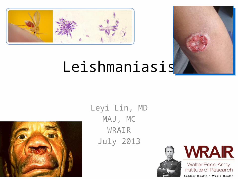
http://www.niaid.nih.gov/topics/leishmaniasis
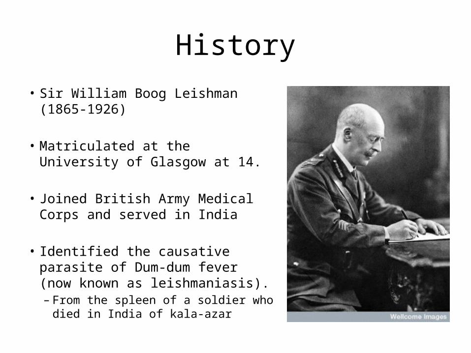
History• Sir William Boog Leishman (1865-1926)
• Matriculated at the University of Glasgow at 14.
• Joined British Army Medical Corps and served in India
• Identified the causative parasite of Dum-dum fever (now known as leishmaniasis). – From the spleen of a soldier who died in
India of kala-azar
Leishmaniasis• Group of disease caused by infection from one of
the obligate intracellular protozoan parasites of the genus Leishmania
• Designated one of the five most important diseases worldwide by WHO
• Leishmania threatens 350 million individuals in 88 countries (72 are “developing countries”).
Highly Endemic Areas
• 90% of cutaneous leishmaniasis occur in Afghanistan, Brazil, Iran, Peru, Saudi Arabia, and Syria.
• 90% mucocutaneous leishmaniasis occur in Bolivia, Brazil, and Peru
• 90% of all visceral leishmaniasis cases occur in Bangledesh, Brazil, India, Nepal, and Sudan
WHO Leishmaniasis: Burden of Disease
Question
• Leishmaniasis is acquired through the bite of ...
A). Anopheles mosquitoesB). Aedes mosquitoesC). Sand fliesD). Fleas
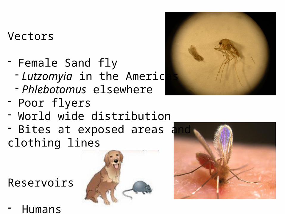
Vectors
- Female Sand fly- Lutzomyia in the Americas- Phlebotomus elsewhere
- Poor flyers- World wide distribution- Bites at exposed areas and clothing lines
Reservoirs
- Humans- Dogs- Rodents
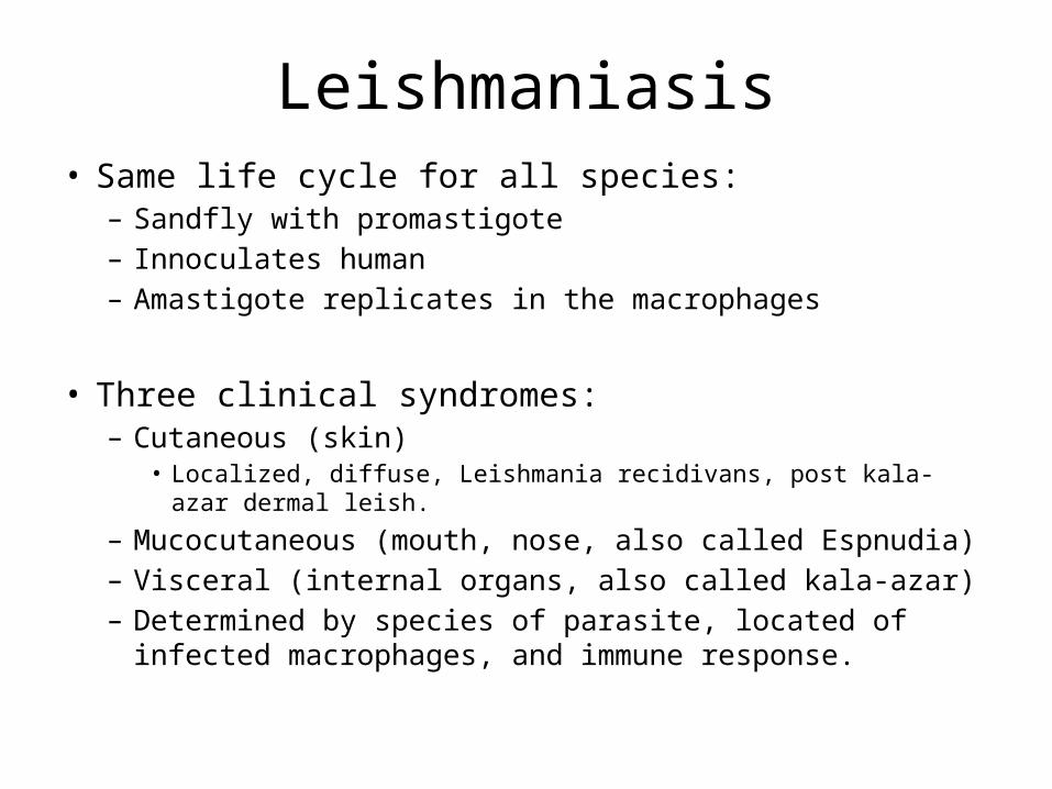
Leishmaniasis• Same life cycle for all species:
– Sandfly with promastigote– Innoculates human – Amastigote replicates in the macrophages
• Three clinical syndromes:– Cutaneous (skin)
• Localized, diffuse, Leishmania recidivans, post kala-azar dermal leish.
– Mucocutaneous (mouth, nose, also called Espnudia)– Visceral (internal organs, also called kala-azar)– Determined by species of parasite, located of infected
macrophages, and immune response.
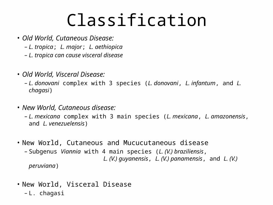
Classification• Old World, Cutaneous Disease:
– L. tropica; L. major; L. aethiopica – L. tropica can cause visceral disease
• Old World, Visceral Disease: – L. donovani complex with 3 species (L. donovani, L. infantum, and L. chagasi)
• New World, Cutaneous disease:– L. mexicana complex with 3 main species (L. mexicana, L. amazonensis, and L.
venezuelensis)
• New World, Cutaneous and Mucucutaneous disease– Subgenus Viannia with 4 main species (L. (V.) braziliensis, L.
(V.) guyanensis, L. (V.) panamensis, and L. (V.) peruviana)
• New World, Visceral Disease– L. chagasi
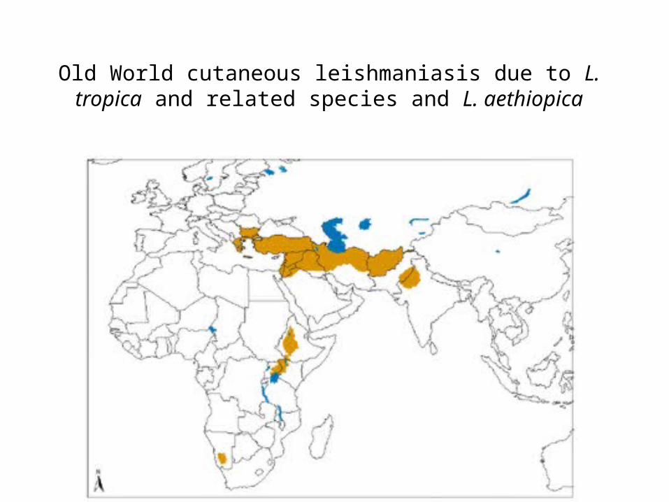
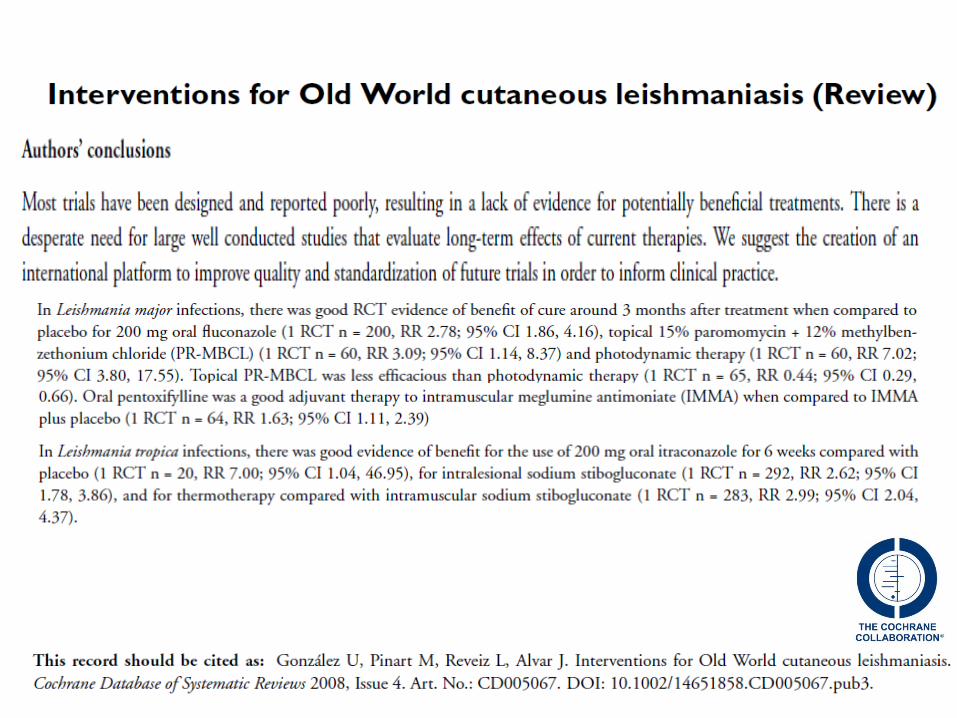
Old World cutaneous leishmaniasis due to L. tropica and related species and L. aethiopica
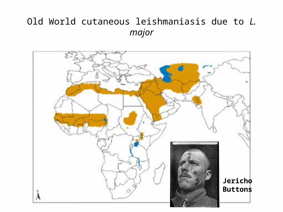
Old World cutaneous leishmaniasis due to L. major
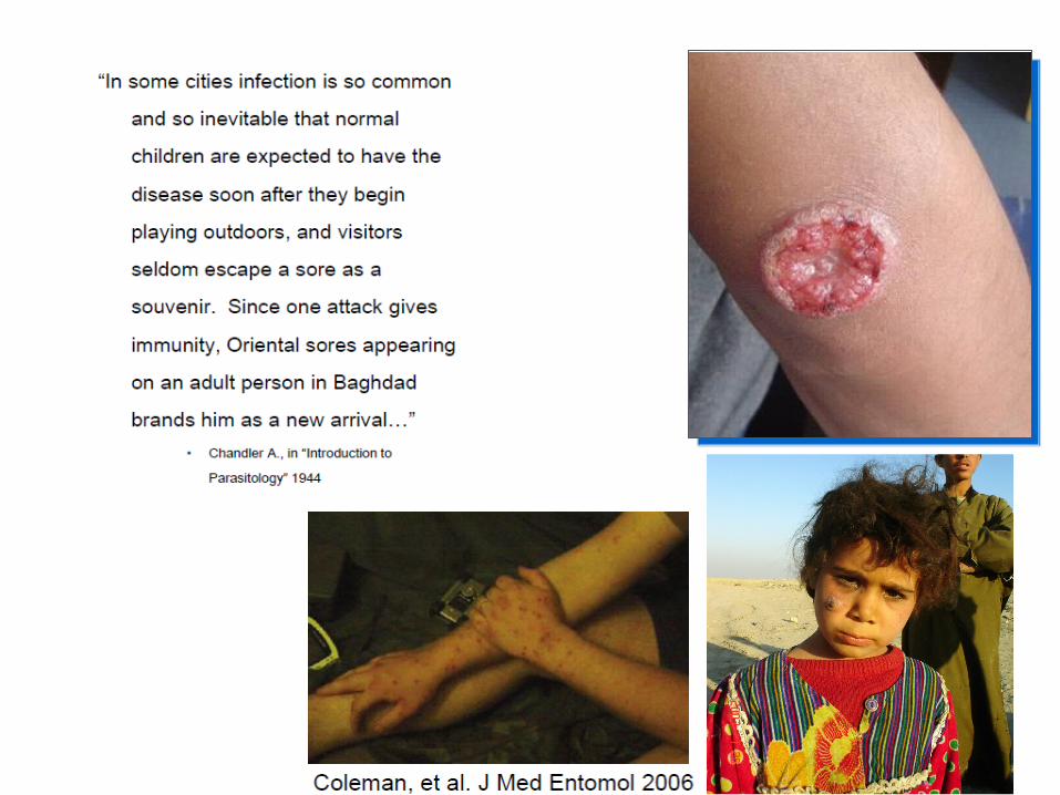
Jericho Buttons
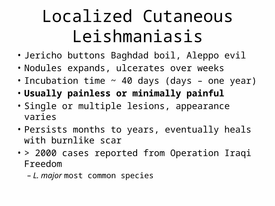
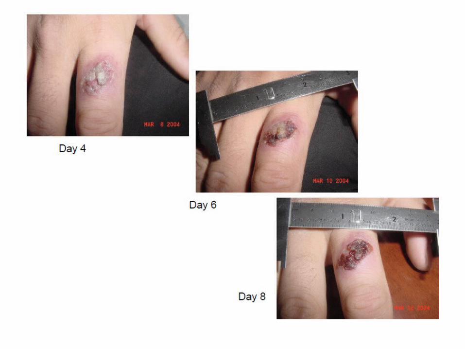
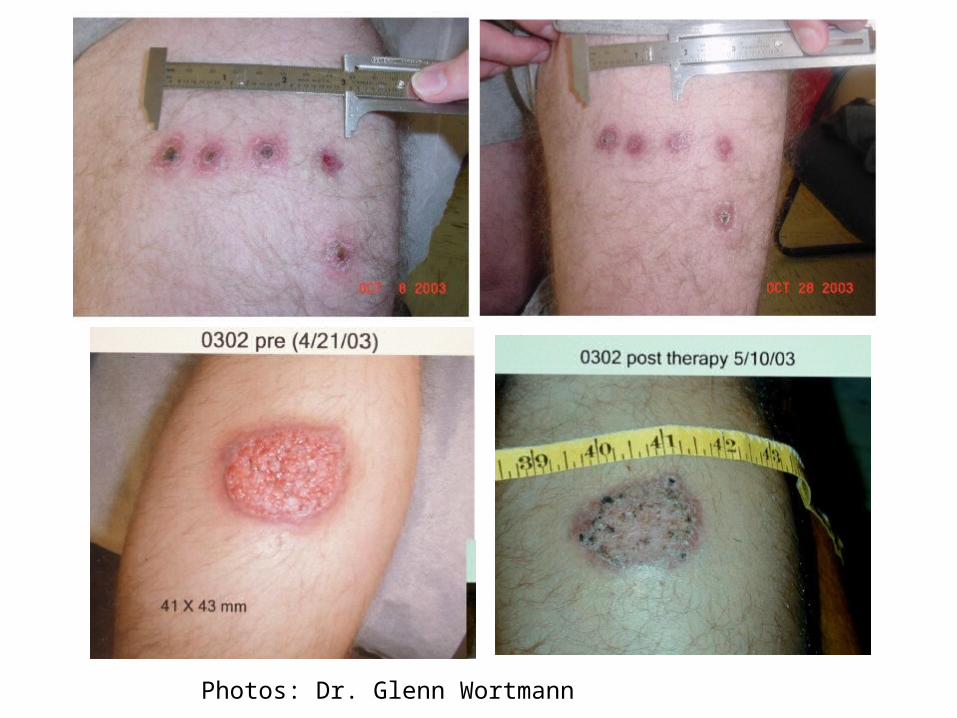
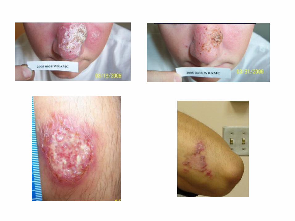
Localized Cutaneous Leishmaniasis
• Jericho buttons Baghdad boil, Aleppo evil• Nodules expands, ulcerates over weeks• Incubation time ~ 40 days (days – one year)• Usually painless or minimally painful• Single or multiple lesions, appearance varies• Persists months to years, eventually heals with
burnlike scar• > 2000 cases reported from Operation Iraqi Freedom– L. major most common species
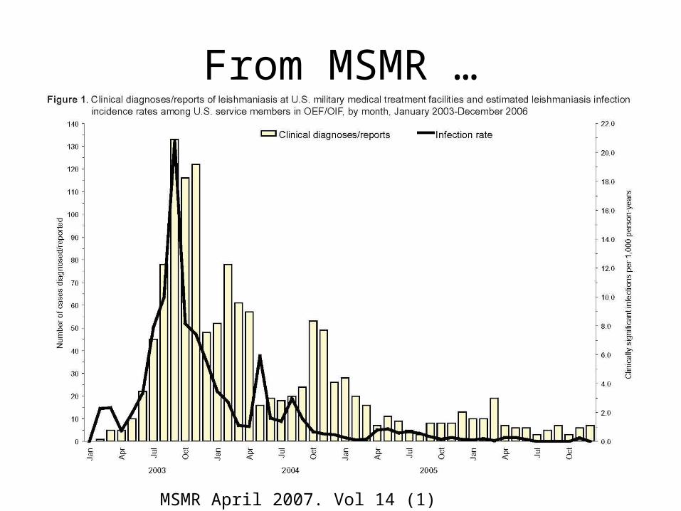
From MSMR …
MSMR April 2007. Vol 14 (1)
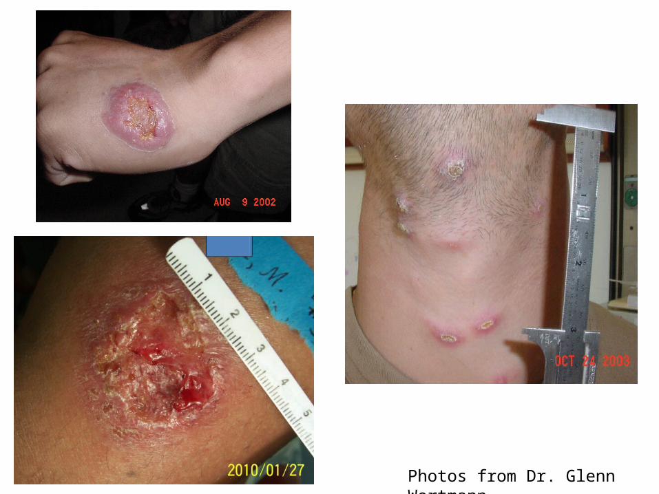
Photos from Dr. Glenn Wortmann

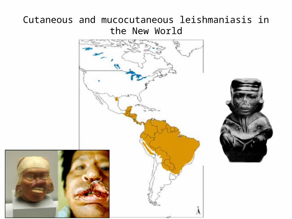
Cutaneous and mucocutaneous leishmaniasis in the New World
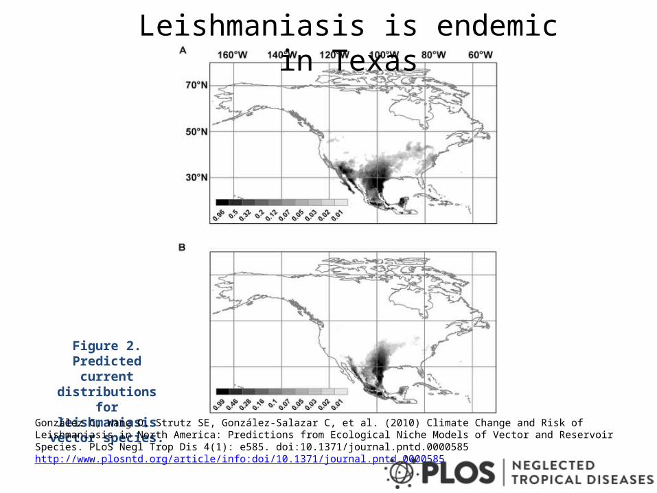
Figure 2. Predicted current distributions for
leishmaniasis vector species.
González C, Wang O, Strutz SE, González-Salazar C, et al. (2010) Climate Change and Risk of Leishmaniasis in North America: Predictions from Ecological Niche Models of Vector and Reservoir Species. PLoS Negl Trop Dis 4(1): e585. doi:10.1371/journal.pntd.0000585http://www.plosntd.org/article/info:doi/10.1371/journal.pntd.0000585
Leishmaniasis is endemic in Texas
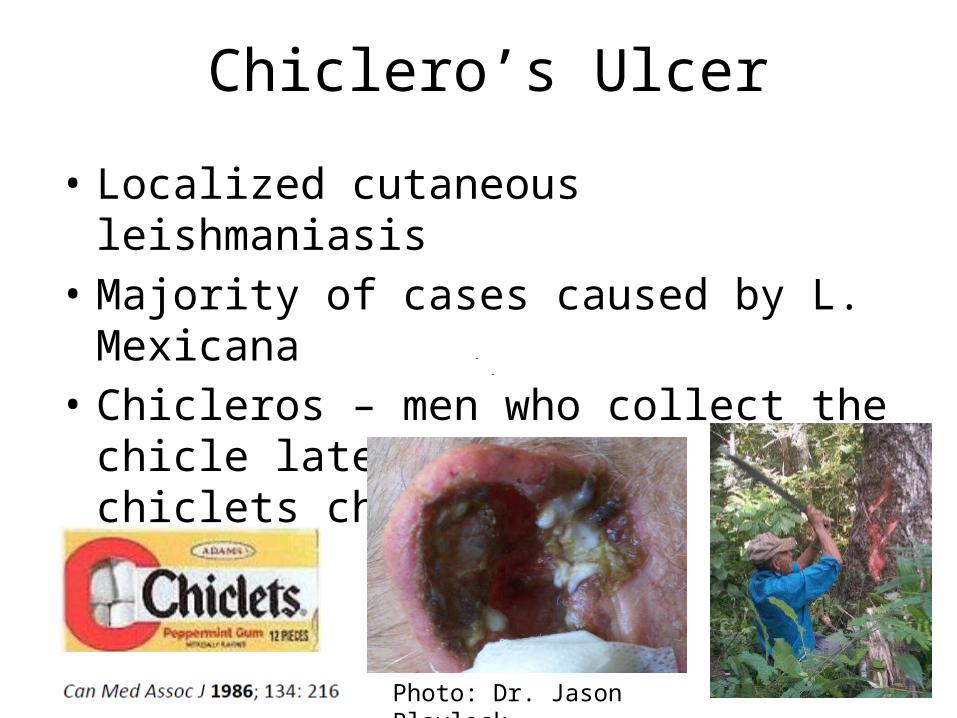
Chiclero’s Ulcer
• Localized cutaneous leishmaniasis• Majority of cases caused by L. Mexicana• Chicleros – men who collect the chicle latex
from which chiclets chewing gum is made
Photo: Dr. Jason Blaylock
South American Leishmaniasis associated with mucocutaneous disease
• Subgenus Viannia with 4 main species – 1). L. (V.) braziliensis– 2). L. (V.) guyanensis– 3). L. (V.) panamensis– 4). L. (V.) peruviana
• True incidence of mucus membrane involvement is well documented, estimated at least 5% - 25 %
• Destructive lesions, possibly immune-medicated• Delay in diagnosis
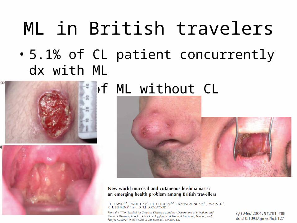
ML in British travelers • 5.1% of CL patient concurrently dx with ML• 2 cases of ML without CL
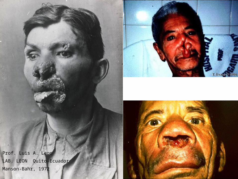
Prof. Luis A. Leon
LAB. LEON Quito-Ecuador
Manson-Bahr, 1972
Long-standing cases
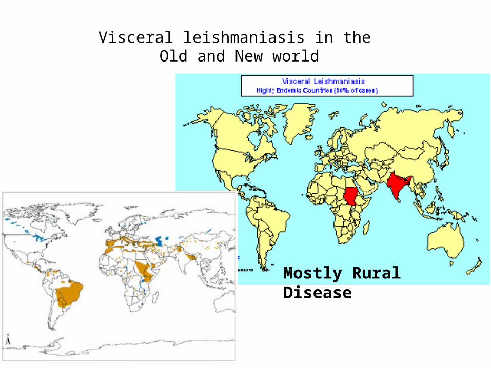
Visceral leishmaniasis in the Old and New world
Mostly Rural Disease
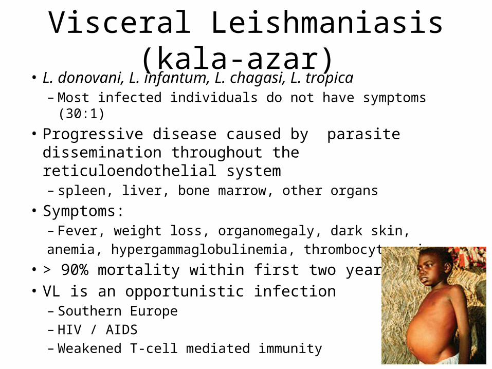
Visceral Leishmaniasis (kala-azar) • L. donovani, L. infantum, L. chagasi, L. tropica
– Most infected individuals do not have symptoms (30:1)• Progressive disease caused by parasite dissemination
throughout the reticuloendothelial system– spleen, liver, bone marrow, other organs
• Symptoms:– Fever, weight loss, organomegaly, dark skin, anemia, hypergammaglobulinemia, thrombocytopenia
• > 90% mortality within first two years• VL is an opportunistic infection
– Southern Europe– HIV / AIDS – Weakened T-cell mediated immunity
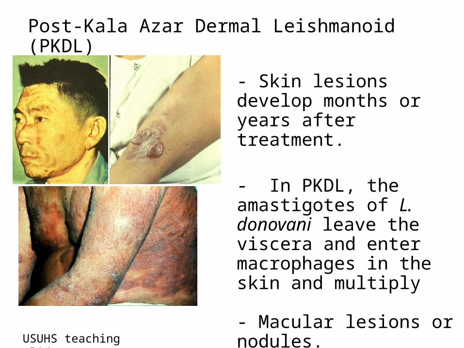
Post-Kala Azar Dermal Leishmanoid (PKDL)
- Skin lesions develop months or years after treatment.
- In PKDL, the amastigotes of L. donovani leave the viscera and enter macrophages in the skin and multiply
- Macular lesions or nodules.- Sandflies can transmit organisms from skin lesions.
USUHS teaching slides
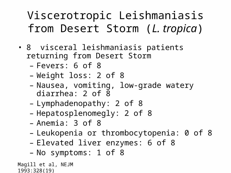
Viscerotropic Leishmaniasis from Desert Storm (L. tropica)
• 8 visceral leishmaniasis patients returning from Desert Storm– Fevers: 6 of 8– Weight loss: 2 of 8– Nausea, vomiting, low-grade watery diarrhea: 2 of 8– Lymphadenopathy: 2 of 8– Hepatosplenomegly: 2 of 8– Anemia: 3 of 8– Leukopenia or thrombocytopenia: 0 of 8– Elevated liver enzymes: 6 of 8– No symptoms: 1 of 8
Magill et al, NEJM 1993:328(19)
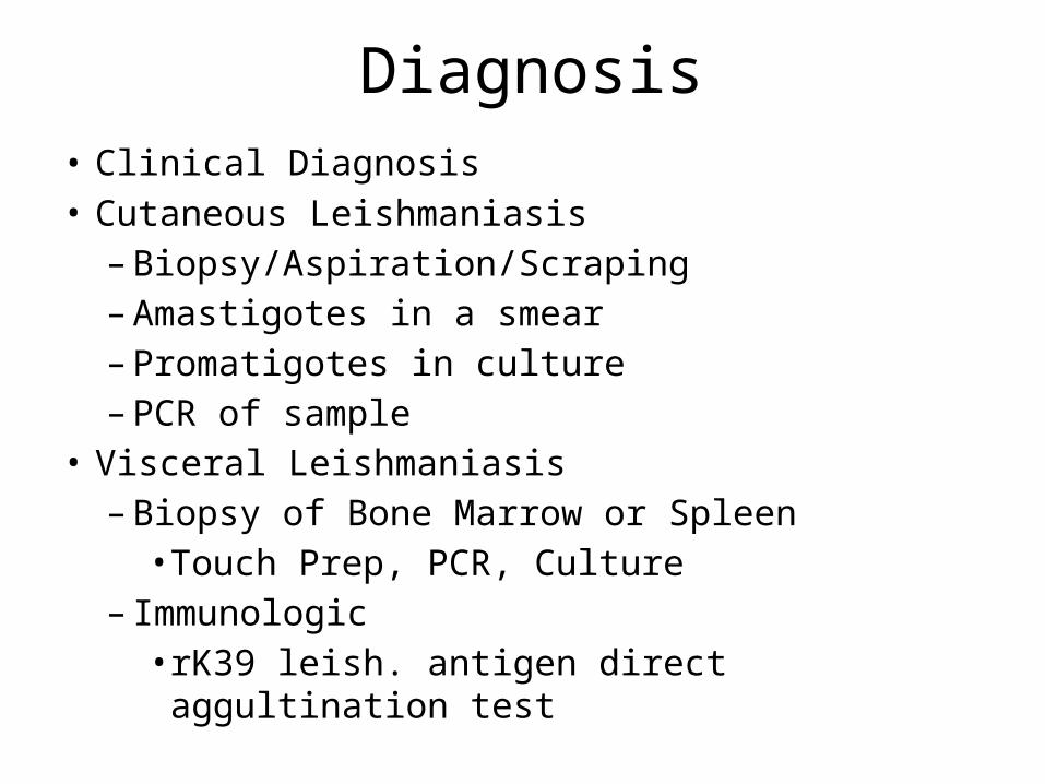
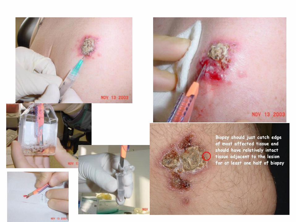
Diagnosis• Clinical Diagnosis• Cutaneous Leishmaniasis– Biopsy/Aspiration/Scraping– Amastigotes in a smear– Promatigotes in culture– PCR of sample
• Visceral Leishmaniasis– Biopsy of Bone Marrow or Spleen• Touch Prep, PCR, Culture
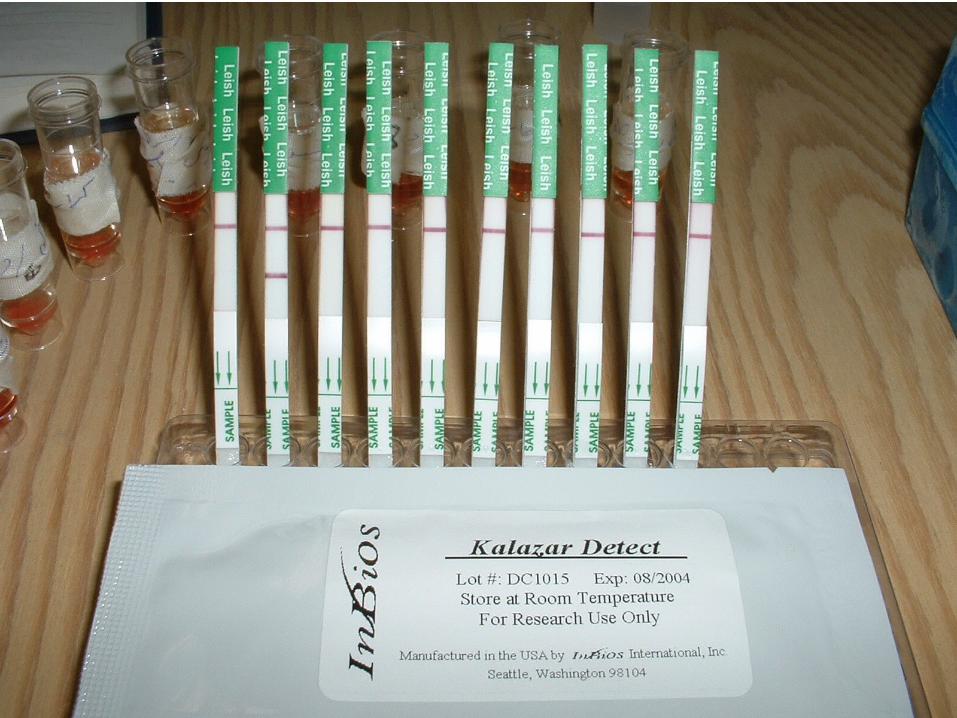
– Immunologic• rK39 leish. antigen direct aggultination test
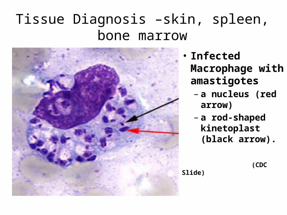
Tissue Diagnosis –skin, spleen, bone marrow
• Infected Macrophage with amastigotes– a nucleus (red
arrow)– a rod-shaped
kinetoplast (black arrow).
(CDC Slide)
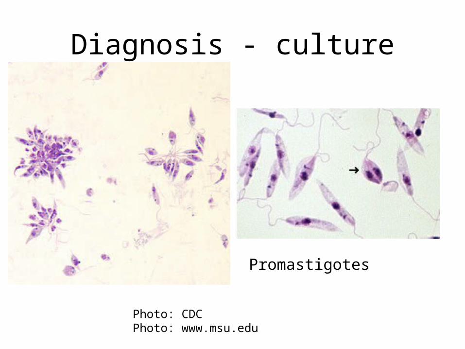
Diagnosis - culture
Photo: CDCPhoto: www.msu.edu
Promastigotes
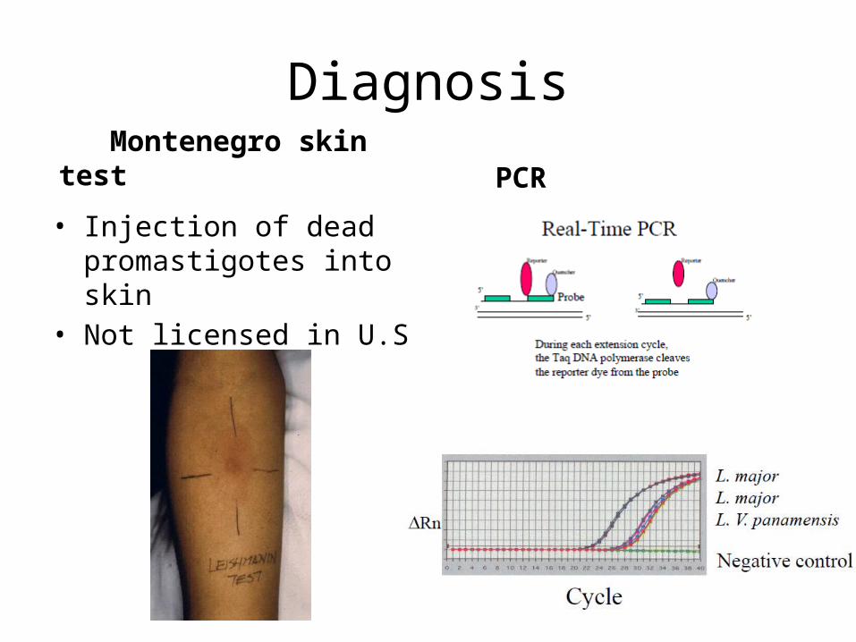
Diagnosis
• Injection of dead promastigotes into skin
• Not licensed in U.S
PCR Montenegro skin test
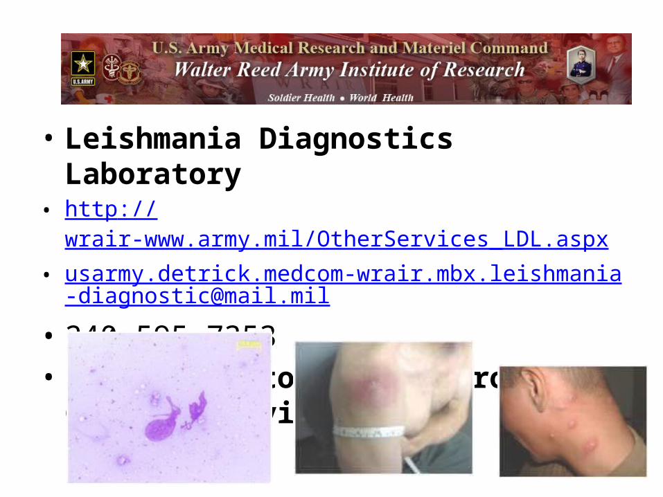
• Leishmania Diagnostics Laboratory• http://wrair-www.army.mil/OtherServices_LDL.aspx• [email protected]
• 240-595-7353• ID or Dermatology Electronic Consult Service

Question
• What’s the best way to treat Leishmaniasis ?1). Amphotericin2). Watch it3). Pentostam4). We don’t know5). Surgery

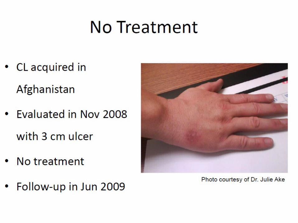
Treatment• CL
– Watchful waiting– Local destructive therapies
• Liquid NO2• Thermo-Med device
– Topical creams• Paromomycin
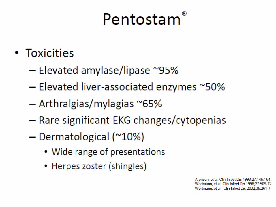
– Systemic treatment• Sodium stibogluconate (Pentostam)• Fluconazole• Amphotericins• Pentamidine• Miltefosin
• MCL, VL– Systemic therapy, usually Pentostam or amphotericinm
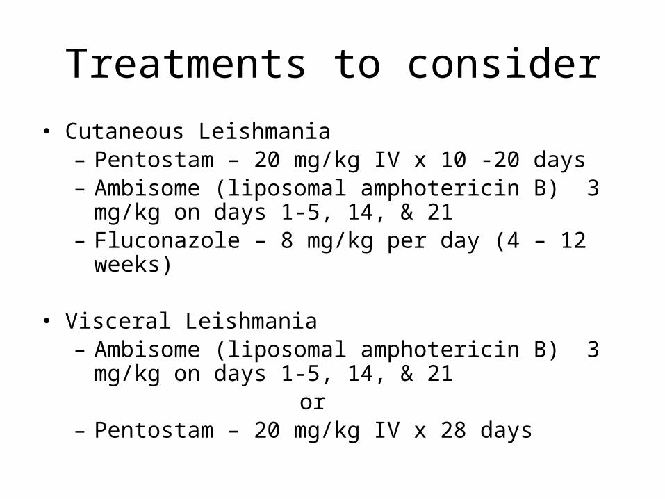
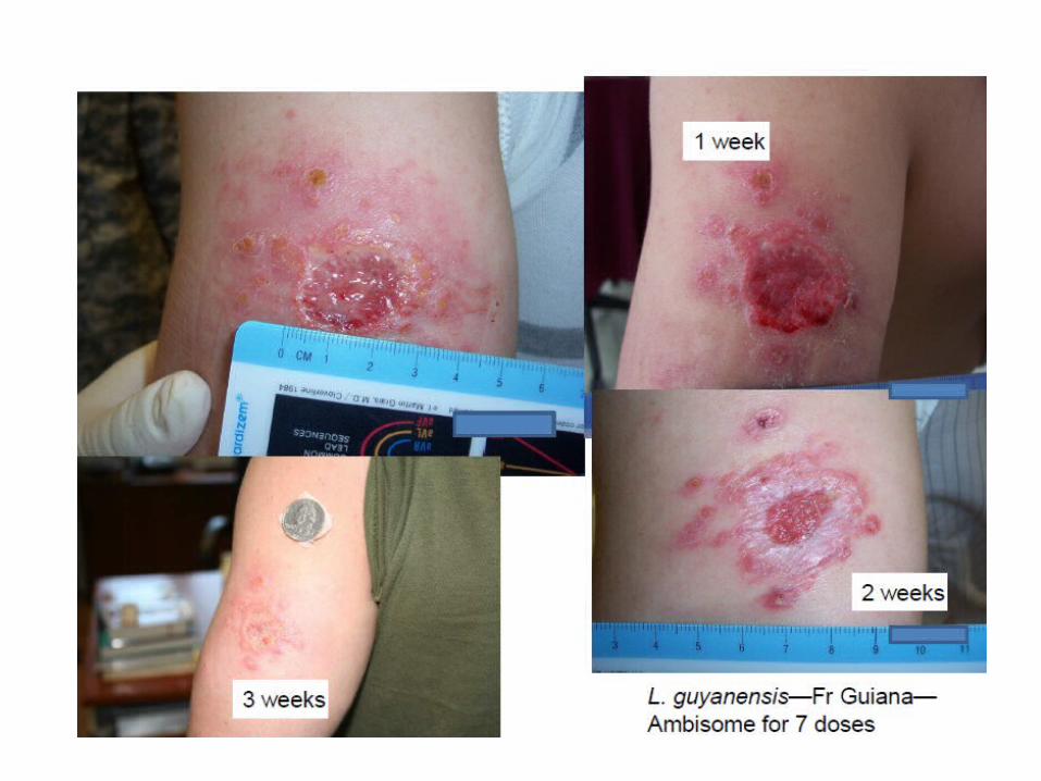
Treatments to consider• Cutaneous Leishmania– Pentostam – 20 mg/kg IV x 10 -20 days– Ambisome (liposomal amphotericin B) 3 mg/kg on days 1-
5, 14, & 21– Fluconazole – 8 mg/kg per day (4 – 12 weeks)
• Visceral Leishmania– Ambisome (liposomal amphotericin B) 3 mg/kg on days 1-
5, 14, & 21 or– Pentostam – 20 mg/kg IV x 28 days
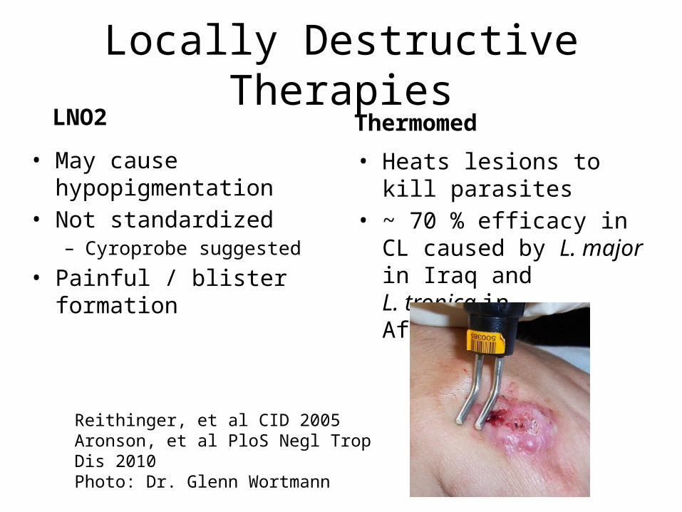
Locally Destructive Therapies LNO2
• May cause hypopigmentation
• Not standardized– Cyroprobe suggested
• Painful / blister formation
Thermomed
• Heats lesions to kill parasites
• ~ 70 % efficacy in CL caused by L. major in Iraq and L. tropica in Afghanistan
Reithinger, et al CID 2005Aronson, et al PloS Negl Trop Dis 2010Photo: Dr. Glenn Wortmann

Photos: Dr. Glenn Wortmann

Prevention• Sandflies bite and are active at night during warmer months• Stay indoors between dusk and dawn• Keep dogs and susceptible animals indoors at night• Sandflies are poor fliers and are deterred by wind; fans are
helpful• Sandflies are small and can get through mesh netting if not
extremely fine• House construction and modification; sandflies breed in cracks
of houses• Insecticides or people and animals• Help from entomologists• Dog vaccine available in Brazil
http://www.cfsph.iastate.edu/Factsheets/pdfs/leishmaniasis.pdf
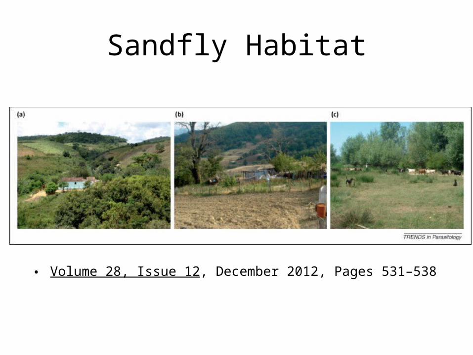
Sandfly Habitat
• Volume 28, Issue 12, December 2012, Pages 531–538
What South American disease is transmitted by sandflies ?
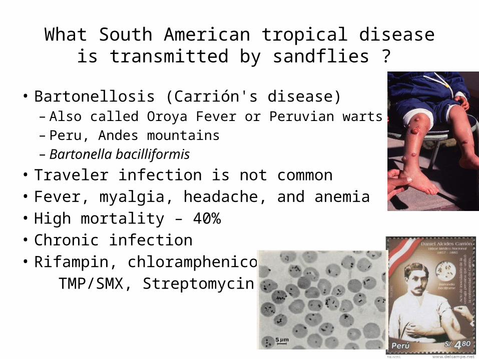
What South American tropical disease is transmitted by sandflies ?
• Bartonellosis (Carrión's disease)– Also called Oroya Fever or Peruvian warts– Peru, Andes mountains– Bartonella bacilliformis
• Traveler infection is not common• Fever, myalgia, headache, and anemia• High mortality – 40%• Chronic infection• Rifampin, chloramphenicol TMP/SMX, Streptomycin
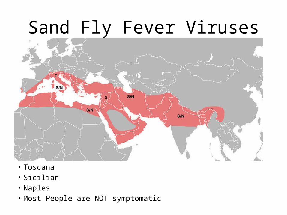
Sand Fly Fever Viruses
• Toscana• Sicilian• Naples• Most People are NOT symptomatic
Summary – Leishmaniasis
• World wide distribution• Many species with different disease
presentations• Cutaneous form may be self-limited• Think about mucocutaneous disease,
especially in South America• Resources available for diagnosis• Treatment response varies with species and
host
Thank You