Embed Size (px)
Citation preview
Lesson 22 Lesson Outline: General Phylogenetic Trends
Feeding Methods Specific Features and Phylogenetic Trends
o Food reception o Food conduction, storage and mechanical digestion (Foregut) o Chemical digestion and absorption (Midgut) o Water and ion absorption and defecation (Hindgut) o Dynamics of gut structure - influence of diet
Objectives: At the end of this lesson you should be able to:
Describe the different types of feeding methods found in animals and discuss the unique problems and solutions associated with each
• Understand the basic design of the digestive system • Know the three basic types of food processing • Be able to describe the basic functions of each region of the gut • Understand the basic structure/function relationships of different digestive systems • Describe the dynamic component of gut structure/function
References: Chapter 12: 265-291 Reading for Next Lesson: Chapter 12: 265-291
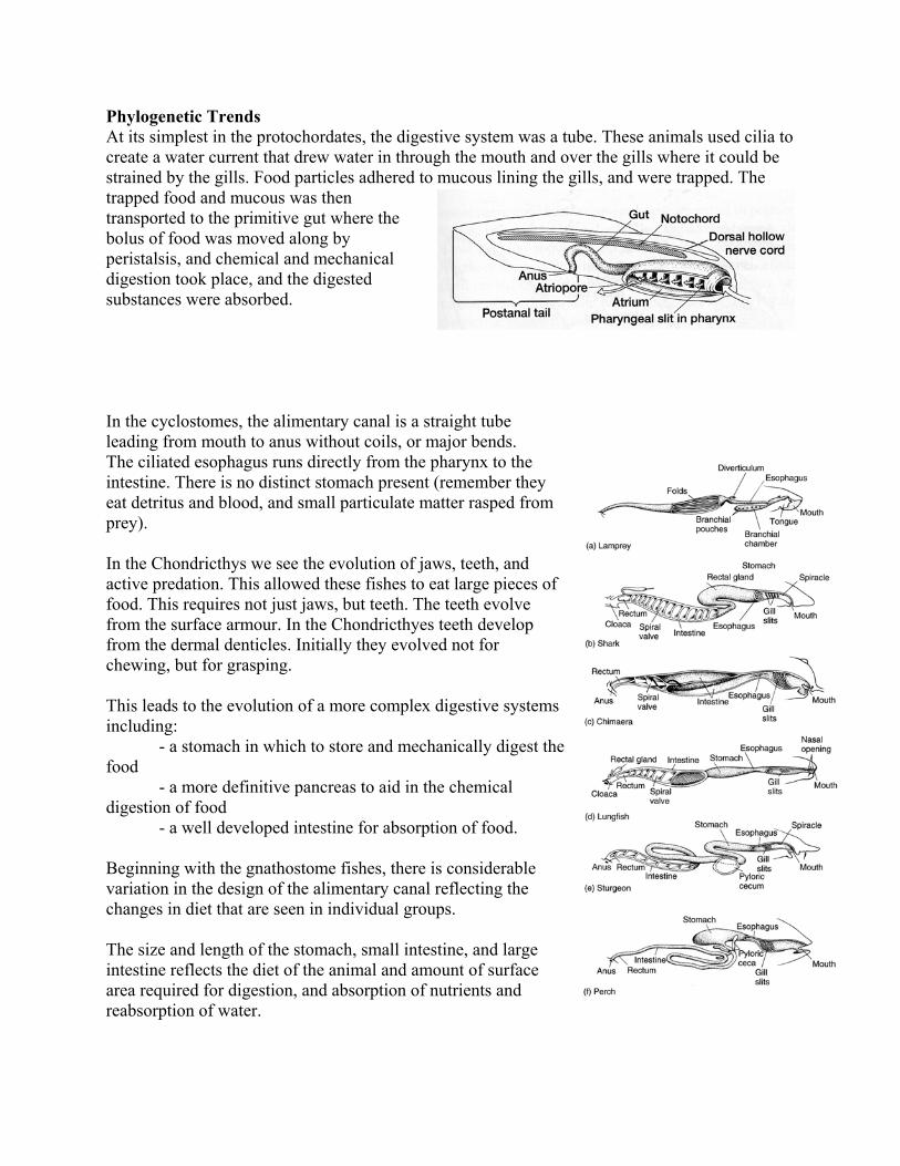
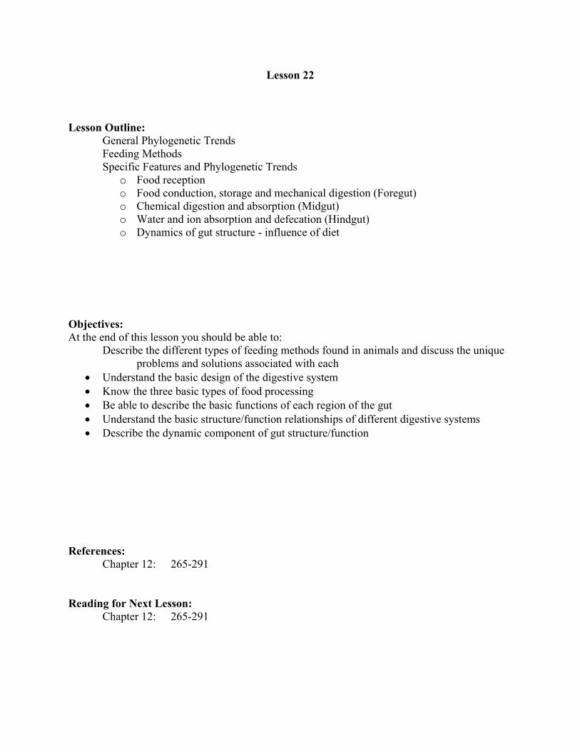
Phylogenetic Trends At its simplest in the protochordates, the digestive system was a tube. These animals used cilia to create a water current that drew water in through the mouth and over the gills where it could be strained by the gills. Food particles adhered to mucous lining the gills, and were trapped. The trapped food and mucous was then transported to the primitive gut where the bolus of food was moved along by peristalsis, and chemical and mechanical digestion took place, and the digested substances were absorbed. In the cyclostomes, the alimentary canal is a straight tube leading from mouth to anus without coils, or major bends. The ciliated esophagus runs directly from the pharynx to the intestine. There is no distinct stomach present (remember they eat detritus and blood, and small particulate matter rasped from prey). In the Chondricthys we see the evolution of jaws, teeth, and active predation. This allowed these fishes to eat large pieces of food. This requires not just jaws, but teeth. The teeth evolve from the surface armour. In the Chondricthyes teeth develop from the dermal denticles. Initially they evolved not for chewing, but for grasping. This leads to the evolution of a more complex digestive systems including:
- a stomach in which to store and mechanically digest tfood
he
- a more definitive pancreas to aid in the chemical digestion of food
- a well developed intestine for absorption of food. Beginning with the gnathostome fishes, there is considerable variation in the design of the alimentary canal reflecting the changes in diet that are seen in individual groups. The size and length of the stomach, small intestine, and large intestine reflects the diet of the animal and amount of surface area required for digestion, and absorption of nutrients and reabsorption of water.
Feeding Methods Acquiring food is probably the most time consuming activity of most animals. A variety of techniques, each with its own associated structures and physiology have evolved for this purpose. 1) Filter Feeding
Found in animals which ingest aquatic prey (although they don't necessarily live in aquatic environments).
Food is filtered from water by specialized entrapment devices whether on the body surface or within it.
Movement of water to the "filter" is usually achieved by muscular movement and muscular pumps.
In some, the same structure that is used to obtain food for metabolism is also used for gas exchange for oxygen to metabolize the food. i.e. - branchial baskets in sea squirts - gill rakers in tadpoles and fish
In most cases, mucus is used to entrap the food particles at the "filter" and the mucus is then transported to the gut for digestion - usually by cilia.
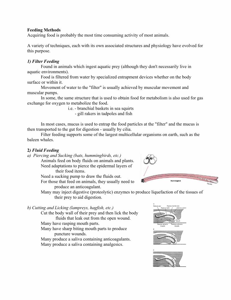
Filter feeding supports some of the largest multicellular organisms on earth, such as the baleen whales. 2) Fluid Feeding a) Piercing and Sucking (bats, hummingbirds, etc.)
Animals feed on body fluids on animals and plants. Need adaptations to pierce the epidermal layers of
their food items. Need a sucking pump to draw the fluids out. For those that feed on animals, they usually need to
produce an anticoagulant. Many may inject digestive (proteolytic) enzymes to produce liquefaction of the tissues of
their prey to aid digestion. b) Cutting and Licking (lampreys, hagfish, etc.) Cut the body wall of their prey and then lick the body
fluids that leak out from the open wound. Many have rasping mouth parts. Many have sharp biting mouth parts to produce
puncture wounds. Many produce a saliva containing anticoagulants. Many produce a saliva containing analgesics.

3) Seizing of Prey a) Jaws, Teeth and Beaks Found in animals that actually eat their prey. Are used for holding prey, primarily. Can only be
used for tearing and cutting prey once the prey item is immobilized.
Swallowing prey whole is very common. In many, all teeth are the same. In others there is a
high degree of differentiation. The beaks of birds are as diverse as the teeth of
mammals. Many use the crop or gizzard as a secondary
structure for mechanically grinding food. b) Toxins To immobilize large prey for subsequent tearing and
dismemberment, many animals use toxins. Most are neurotoxins acting on the nervous system. Some are hemolytic - destroy blood cells. Some are proteolytic. Must be stored in an inactive form. Predator must have proteolytic enzymes to digest the toxins
once it ingests the prey. 4) Herbivory and Grazing to Collect Food Usually requires rasping mouth parts if grazing on encrusting vegetation (not dissimilar to some cutting and licking strategies). Requires grinding mouth parts if grazing on taller plants. Teeth are either very resistant to wear or continuously growing and replaced.
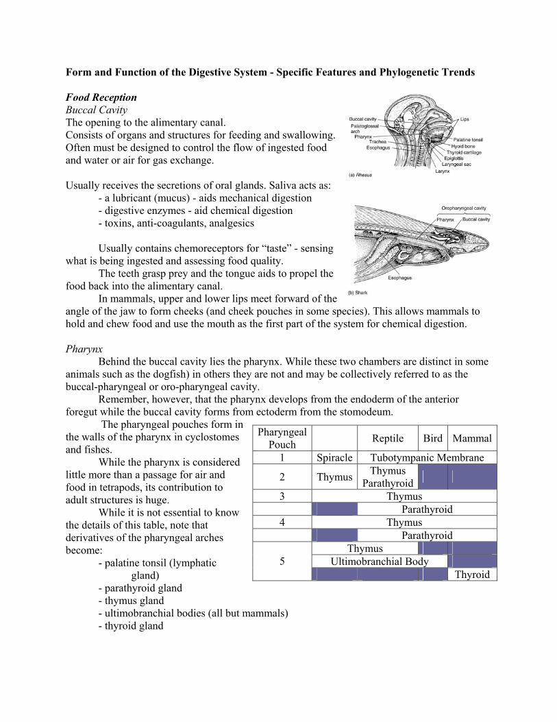
Form and Function of the Digestive System - Specific Features and Phylogenetic Trends Food Reception Buccal Cavity The opening to the alimentary canal. Consists of organs and structures for feeding and swallowing. Often must be designed to control the flow of ingested food and water or air for gas exchange. Usually receives the secretions of oral glands. Saliva acts as:
- a lubricant (mucus) - aids mechanical digestion - digestive enzymes - aid chemical digestion - toxins, anti-coagulants, analgesics
Usually contains chemoreceptors for “taste” - sensing
what is being ingested and assessing food quality. The teeth grasp prey and the tongue aids to propel the
food back into the alimentary canal. In mammals, upper and lower lips meet forward of the
angle of the jaw to form cheeks (and cheek pouches in some species). This allows mammals to hold and chew food and use the mouth as the first part of the system for chemical digestion. Pharynx Behind the buccal cavity lies the pharynx. While these two chambers are distinct in some animals such as the dogfish) in others they are not and may be collectively referred to as the buccal-pharyngeal or oro-pharyngeal cavity.
Remember, however, that the pharynx develops from the endoderm of the anterior foregut while the buccal cavity forms from ectoderm from the stomodeum.
The pharyngeal pouches form in the walls of the pharynx in cyclostomes and fishes.
PharyngealPouch Reptile Bird Mammal
1 Spiracle Tubotympanic Membrane
2 Thymus Thymus Parathyroid
3 Thymus Parathyroid
4 Thymus Parathyroid
Thymus Ultimobranchial Body 5 Thyroid
While the pharynx is considered little more than a passage for air and food in tetrapods, its contribution to adult structures is huge.
While it is not essential to know the details of this table, note that derivatives of the pharyngeal arches become:
- palatine tonsil (lymphatic gland)
- parathyroid gland - thymus gland
- ultimobranchial bodies (all but mammals) - thyroid gland
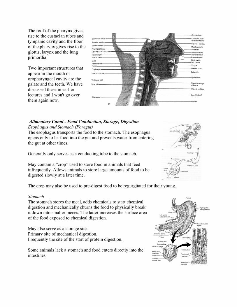
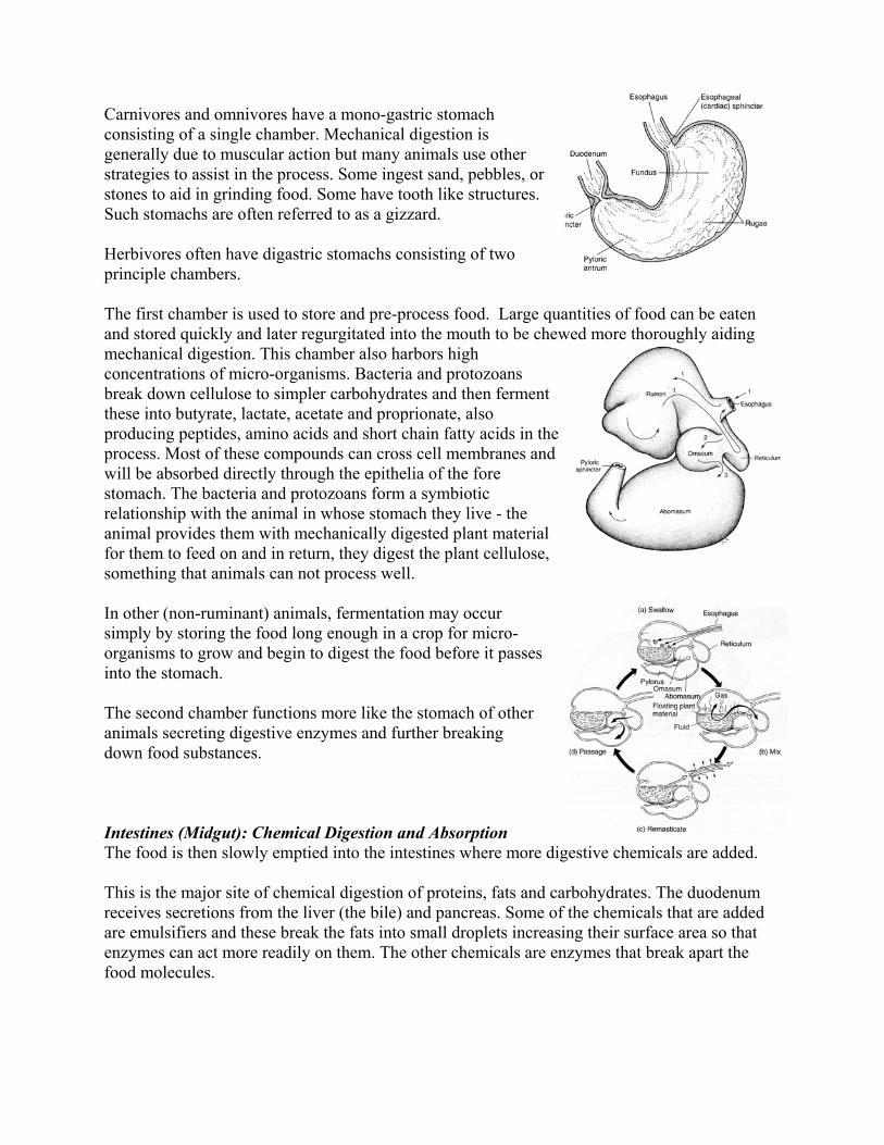
The roof of the pharynx gives rise to the eustacian tubes and tympanic cavity and the floor of the pharynx gives rise to the glottis, larynx and the lung primordia. Two important structures that appear in the mouth or oropharyngeal cavity are the palate and the teeth. We have discussed these in earlier lectures and I won't go over them again now. Alimentary Canal - Food Conduction, Storage, Digestion Esophagus and Stomach (Foregut) The esophagus transports the food to the stomach. The esophagus opens only to let food into the gut and prevents water from entering the gut at other times.
Generally only serves as a conducting tube to the stomach.
May contain a “crop” used to store food in animals that feed infrequently. Allows animals to store large amounts of food to be digested slowly at a later time.
The crop may also be used to pre-digest food to be regurgitated for their young. Stomach The stomach stores the meal, adds chemicals to start chemical digestion and mechanically churns the food to physically break it down into smaller pieces. The latter increases the surface area of the food exposed to chemical digestion. May also serve as a storage site. Primary site of mechanical digestion. Frequently the site of the start of protein digestion. Some animals lack a stomach and food enters directly into the intestines.
Carnivores and omnivores have a mono-gastric stomach consisting of a single chamber. Mechanical digestion is generally due to muscular action but many animals use other strategies to assist in the process. Some ingest sand, pebbles, or stones to aid in grinding food. Some have tooth like structures. Such stomachs are often referred to as a gizzard. Herbivores often have digastric stomachs consisting of two principle chambers. The first chamber is used to store and pre-process food. Large quantities of food can be eaten and stored quickly and later regurgitated into the mouth to be cmechanical digestion. This chamber also harbors high concentrations of micro-organisms. Bacteria and protozoans break down cellulose to simpler carbohydrates and then fethese into butyrate, lactate, acetate and proprionate, also producing peptides, amino acids and short chain fatty acids in tprocess. Most of these compounds can cross cell membrawill be absorbed directly through the epithelia of the fore stomach. The bacteria and protozoans form a symbiotic relationship with the animal in whose stomach they live - the animal provides them with mechanically digested plant mafor them to feed on and in return, they digest the plant cellulose, something that animals can not process well.
hewed more thoroughly aiding
rment
he nes and
terial
In other (non-ruminant) animals, fermentation may occur simply by storing the food long enough in a crop for micro-organisms to grow and begin to digest the food before it passes into the stomach. The second chamber functions more like the stomach of other animals secreting digestive enzymes and further breaking down food substances. Intestines (Midgut): Chemical Digestion and Absorption The food is then slowly emptied into the intestines where more digestive chemicals are added. This is the major site of chemical digestion of proteins, fats and carbohydrates. The duodenum receives secretions from the liver (the bile) and pancreas. Some of the chemicals that are added are emulsifiers and these break the fats into small droplets increasing their surface area so that enzymes can act more readily on them. The other chemicals are enzymes that break apart the food molecules.
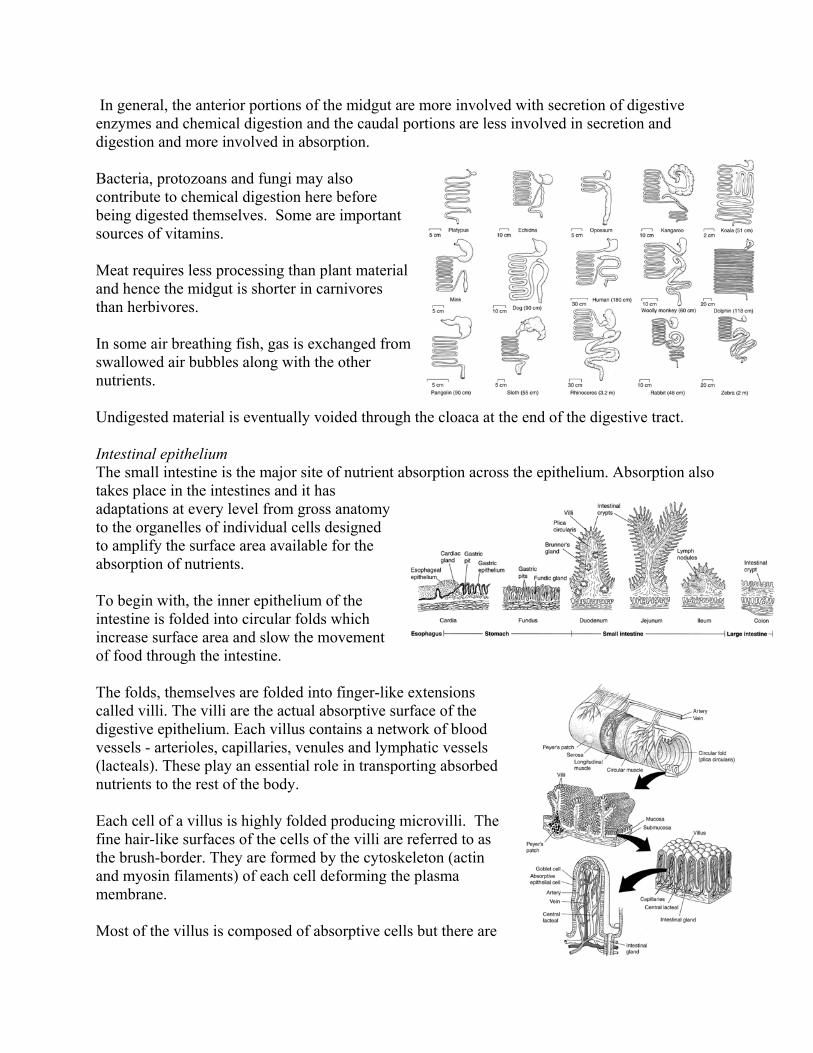
In general, the anterior portions of the midgut are more involved with secretion of digestive enzymes and chemical digestion and the caudal portions are less involved in secretion and digestion and more involved in absorption.
Bacteria, protozoans and fungi may also contribute to chemical digestion here before being digested themselves. Some are important sources of vitamins.
Meat requires less processing than plant material and hence the midgut is shorter in carnivores than herbivores.
In some air breathing fish, gas is exchanged fswallowed air bubbles along with the other nutrients.
rom
Most of the villus is composed of absorptive cells but there are
Undigested material is eventually voided through the cloaca at the end of the digestive tract. Intestinal epithelium The small intestine is the major site of nutrient absorption across the epithelium. Absorption also takes place in the intestines and it has adaptations at every level from gross anatomy to the organelles of individual cells designed to amplify the surface area available for the absorption of nutrients.
To begin with, the inner epithelium of the intestine is folded into circular folds which increase surface area and slow the movement of food through the intestine.
The folds, themselves are folded into finger-like extensions called villi. The villi are the actual absorptive surface of the digestive epithelium. Each villus contains a network of blood vessels - arterioles, capillaries, venules and lymphatic vessels (lacteals). These play an essential role in transporting absorbed nutrients to the rest of the body.
Each cell of a villus is highly folded producing microvilli. The fine hair-like surfaces of the cells of the villi are referred to as the brush-border. They are formed by the cytoskeleton (actin and myosin filaments) of each cell deforming the plasma membrane.
also goblet cells which are responsible for producing mucus to lubricate the digestive epithelium. Adjacent absorptive cells areheld together by desmosomes and tight junctions. The tight junctions act to prevent any material from entering the body between the cells of the epithelium.
Only substances that can enter the absorptive cells can enter the
t
aecum erbivores, a caecum is usually present at the junction
an
e es
arge Intestine (Hindgut): Water and Ion Absorption and
function of the hindgut is water reabsorption. The contents that remain after that
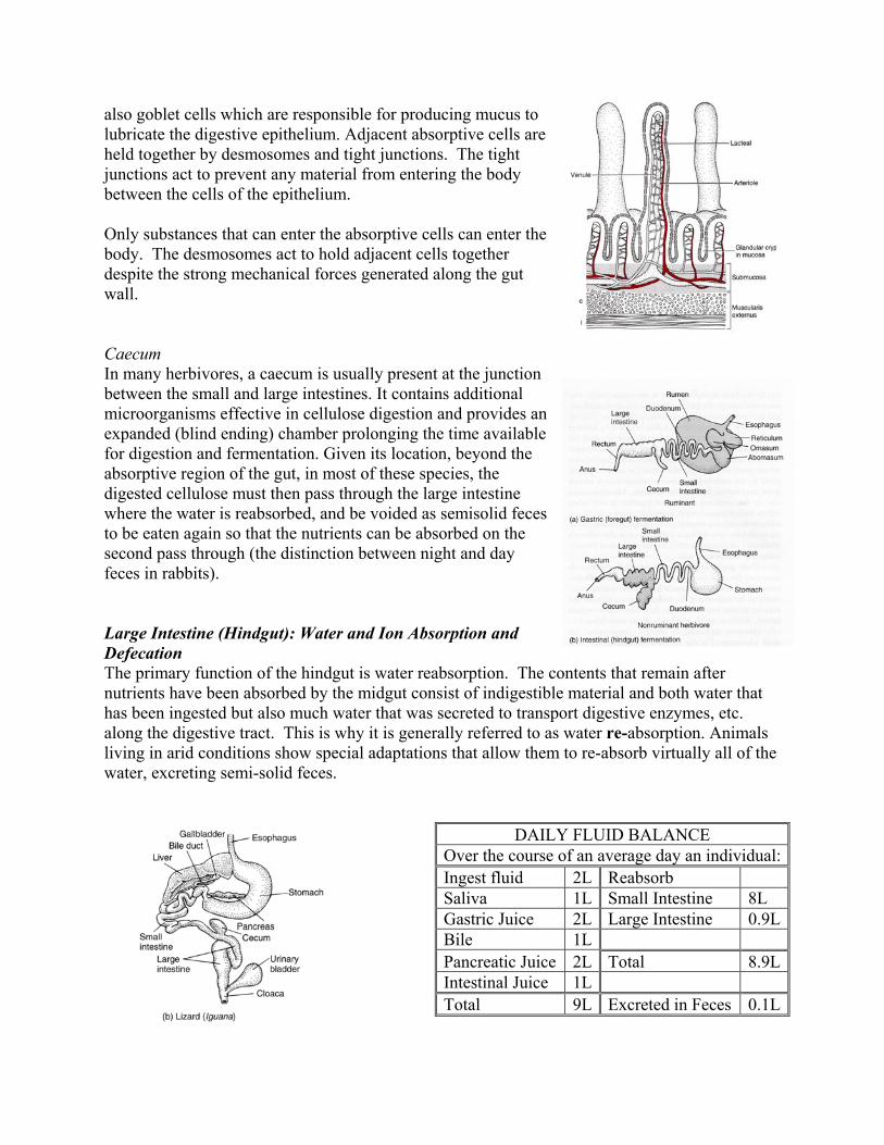
DAILY FLUID BALANCE
body. The desmosomes act to hold adjacent cells together despite the strong mechanical forces generated along the guwall. CIn many hbetween the small and large intestines. It contains additional microorganisms effective in cellulose digestion and provides expanded (blind ending) chamber prolonging the time available for digestion and fermentation. Given its location, beyond the absorptive region of the gut, in most of these species, the digested cellulose must then pass through the large intestinwhere the water is reabsorbed, and be voided as semisolid fecto be eaten again so that the nutrients can be absorbed on the second pass through (the distinction between night and day feces in rabbits). LDefecation The primarynutrients have been absorbed by the midgut consist of indigestible material and both waterhas been ingested but also much water that was secreted to transport digestive enzymes, etc. along the digestive tract. This is why it is generally referred to as water re-absorption. Animals living in arid conditions show special adaptations that allow them to re-absorb virtually all of thewater, excreting semi-solid feces.
Over the course of an average day an individual: Ingest fluid 2L Reabsorb
Saliva 1L Small Intestine L 8Gastric Juice 2L Large Intestine 0.9LBile 1L Pancreatic Juice otal .9L2L T 8Intestinal Juice 1L Total 9L Excreted in Feces .1L0
The hindgut consolidates the undigested material into feces for defecation.
many species from arid environments, the hindgut ends in a cloaca rather than an anus. The
e
ynamics of Gut Structure: Influence of Diet e sloughed off and replaced from the human
d with it, the length of the small
The gut atrophies in hibernating animals and then re-grows in the spring after they come out of
The Burmese python will increase the mass of the small intestine by 40% within hours of eating
Even without increasing the length or mass of the gut, animals can alter the number of transport
These are all reversible changes designed to provide sufficient uptake capacity when needed but
Indistinction here is that the urethra from the bladder terminates in a common chamber with the undigested food from the intestine, rather than through its own independent opening through thexternal genitalia (cloaca is latin for sewer). The advantage of this is that water can then be re-absorbed from the urine also resulting in the formation of a semi-solid paste and a further reduction in water loss. DIt is estimated that up to 2 x 10¹⁰ cells per day arintestine. The entire midgut lining is replaced every few days. This is expensive. In animals that are opportunistic eaters, there are strong selection pressures to reduce the size of the gut during periods of fasting and to increase it following a meal. Cold and exercise have been shown to increase food intake anintestine in house wrens.
hibernation and start to feed again - the mass of the stomach may increase 3-4 times.
a meal after a fast and will double the mass within 2 days. This will increase the surface area for absorption by up to 24 times.
proteins in the plasma membranes of the absorptive cells to match uptake capacity to the level ofthe nutrient intake.
to reduce the costs of maintaining such an expensive structure when it is not needed.