Embed Size (px)
Citation preview
MD0501 1-1
LESSON ASSIGNMENT
LESSON 1 Introduction to Anatomy and Physiology. LESSON ASSIGNMENT Paragraphs 1-1 through 1-14. LESSON OBJECTIVES After completing this lesson, you should be able to: 1-1. Identify basic terminology related to human anatomy and physiology. 1-2. Identify the structure and function of the integumentary system. 1-3. Identify the structure and function of the skeletal system. 1-4. Identify the structure a d function of the muscular system. 1-5. Identify the structure and function of the circulatory system. 1-6. Identify the structure and function of the digestive system. 1-7. Identify the structure and function of the nervous system. 1-8. Identify the structure and function of the urogenital system. 1-9. Identify the structure and function of the endocrine system. SUGGESTION After studying the assignment, complete the exercises at the end of this lesson. These exercises will help you to achieve the lesson objectives.
MD0501 1-2
LESSON 1
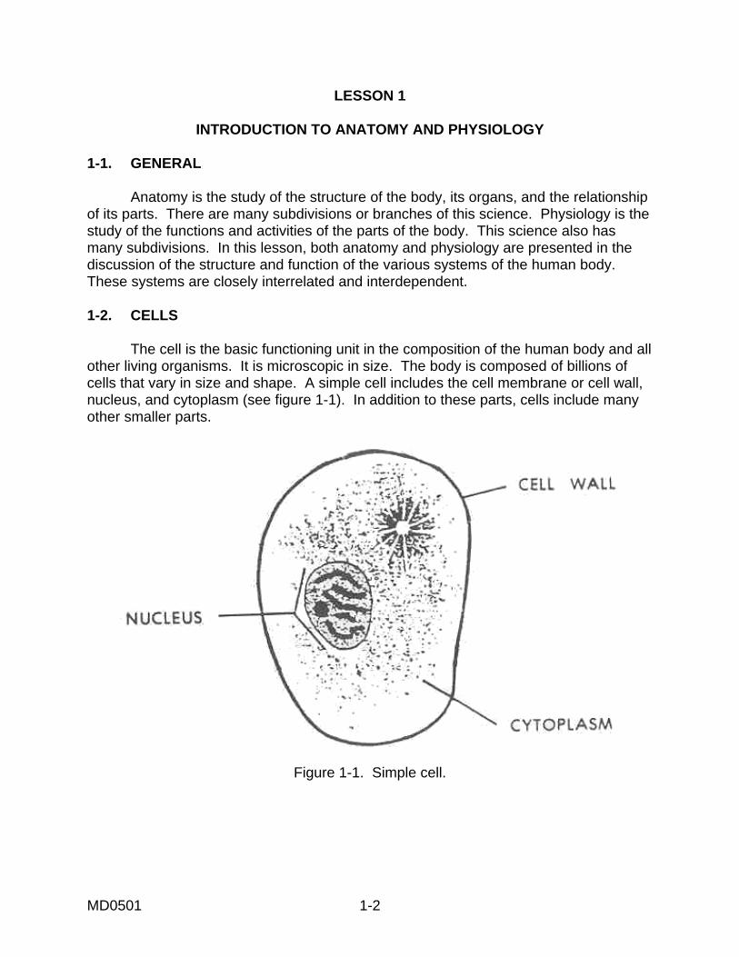
INTRODUCTION TO ANATOMY AND PHYSIOLOGY 1-1. GENERAL Anatomy is the study of the structure of the body, its organs, and the relationship of its parts. There are many subdivisions or branches of this science. Physiology is the study of the functions and activities of the parts of the body. This science also has many subdivisions. In this lesson, both anatomy and physiology are presented in the discussion of the structure and function of the various systems of the human body. These systems are closely interrelated and interdependent. 1-2. CELLS The cell is the basic functioning unit in the composition of the human body and all other living organisms. It is microscopic in size. The body is composed of billions of cells that vary in size and shape. A simple cell includes the cell membrane or cell wall, nucleus, and cytoplasm (see figure 1-1). In addition to these parts, cells include many other smaller parts.
Figure 1-1. Simple cell.

MD0501 1-3
a. Cell Membrane. This membrane surrounds and separates the cell from its environment. It encloses the cytoplasm. The cell membrane also allows certain materials to pass through it as they enter or leave the cell. It is through the cell membrane that all materials essential to metabolism are received. All products of metabolism are disposed of through the cell membrane. The bloodstream and tissue fluid circulate around the cells and transport the materials to and from these cells. b. Nucleus. This controls all activities of the cell, including growth and reproduction. Information is stored in the nucleus and distributed to guide the life processes of the cell. Cells reproduce to replace worn-out cells, to build new tissues, and to bring about the growth of the body. c. Cytoplasm. This is a semifluid material found inside the cell, but outside the nucleus. Cytoplasm is enclosed by the cell membrane. It is responsible for most of the chemical activities of the cell. 1-3. TISSUES A tissue is a group of similarly-specialized cells working together to perform a particular function. The four main types of tissues are epithelial, connective, muscle, and nerve. a. Epithelial. Epithelial tissue covers inner and outer surface of the body. It serves as a lining for vessels and other small cavities inside the body and as skin outside the body. b. Connective. Connective tissues are distributed throughout the body to hold tissues together, to support other tissues, and to fill spaces. c. Muscle. There are three specialized types of muscular tissue found in the body--striated (arms and legs), smooth (blood vessels), and cardiac (heart). Muscular tissue has the ability to contract (shorten), thus producing movement. d. Nerve. Nerve tissue receives and transmits electrical impulses (messages). 1-4. ORGANS An organ is a structure that is a somewhat independent part of the body and is composed of several different tissues. Each organ performs a specialized function or functions. The body has many organs. 1-5. SYSTEMS The organs of the body are arranged into nine major systems. Each system has a specific function to perform. All systems are interdependent.
MD0501 1-4
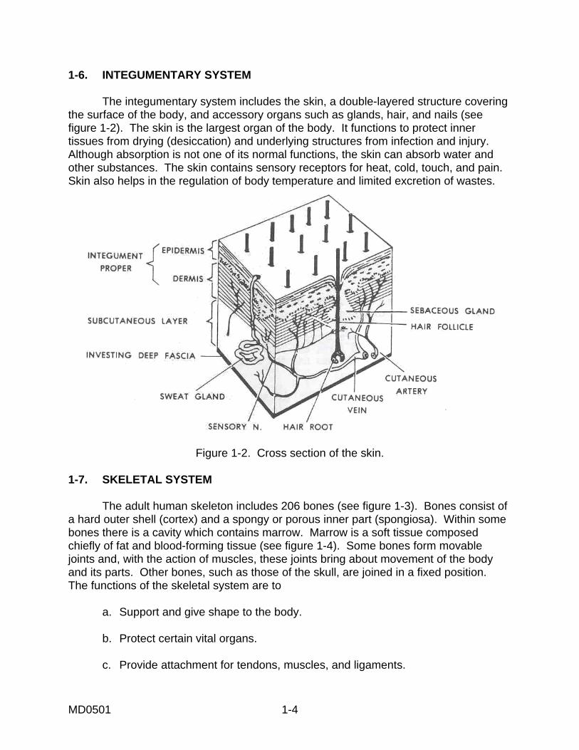
1-6. INTEGUMENTARY SYSTEM The integumentary system includes the skin, a double-layered structure covering the surface of the body, and accessory organs such as glands, hair, and nails (see figure 1-2). The skin is the largest organ of the body. It functions to protect inner tissues from drying (desiccation) and underlying structures from infection and injury. Although absorption is not one of its normal functions, the skin can absorb water and other substances. The skin contains sensory receptors for heat, cold, touch, and pain. Skin also helps in the regulation of body temperature and limited excretion of wastes.
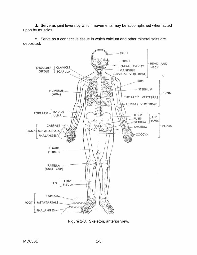
Figure 1-2. Cross section of the skin. 1-7. SKELETAL SYSTEM The adult human skeleton includes 206 bones (see figure 1-3). Bones consist of a hard outer shell (cortex) and a spongy or porous inner part (spongiosa). Within some bones there is a cavity which contains marrow. Marrow is a soft tissue composed chiefly of fat and blood-forming tissue (see figure 1-4). Some bones form movable joints and, with the action of muscles, these joints bring about movement of the body and its parts. Other bones, such as those of the skull, are joined in a fixed position. The functions of the skeletal system are to a. Support and give shape to the body. b. Protect certain vital organs. c. Provide attachment for tendons, muscles, and ligaments.
MD0501 1-5
d. Serve as joint levers by which movements may be accomplished when acted upon by muscles. e. Serve as a connective tissue in which calcium and other mineral salts are deposited.
Figure 1-3. Skeleton, anterior view.

MD0501 1-6
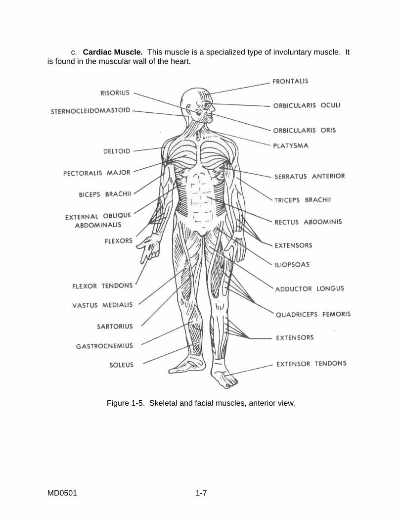
Figure 1-4. Diagram of a bone. 1-8. MUSCULAR SYSTEM. This system includes over 350 muscles. These muscles are made up of muscle tissue. They constitute 40 to 50 percent of the body's weight. The muscular system moves and propels the body and the contents of the hollow organs. It also keeps the body erect and produces body heat. Three types of muscles are found in the body. a. Striated Muscle. This muscle produces bodily movement. Skeletal muscle is also called voluntary muscle since it can be consciously controlled. Figures 1-5 and 1-6 illustrate some skeletal muscles. b. Smooth Muscle. This muscle is found in various visceral organs where continuous automatic functions are necessary. Smooth muscle is also called involuntary muscle since it contracts without conscious direction by the individual. For example, the unconscious contraction of the intestinal muscle moves food through the digestive system.
MD0501 1-7
c. Cardiac Muscle. This muscle is a specialized type of involuntary muscle. It is found in the muscular wall of the heart.
Figure 1-5. Skeletal and facial muscles, anterior view.
MD0501 1-8
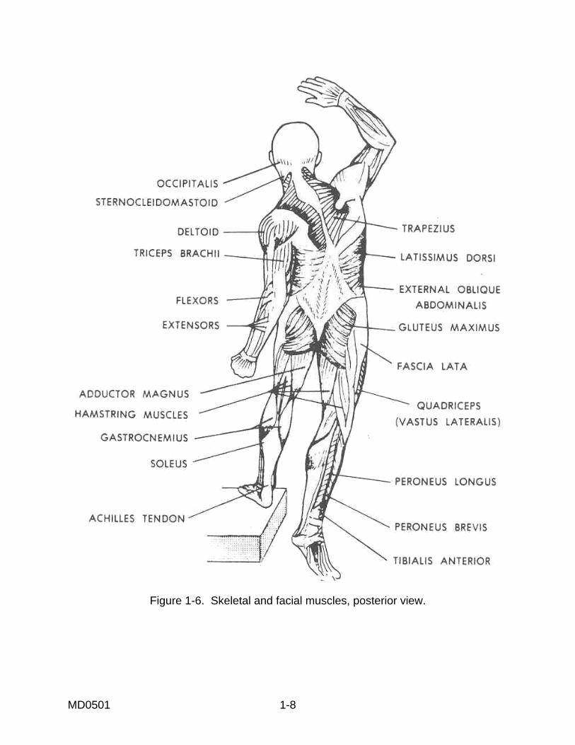
Figure 1-6. Skeletal and facial muscles, posterior view.
MD0501 1-9
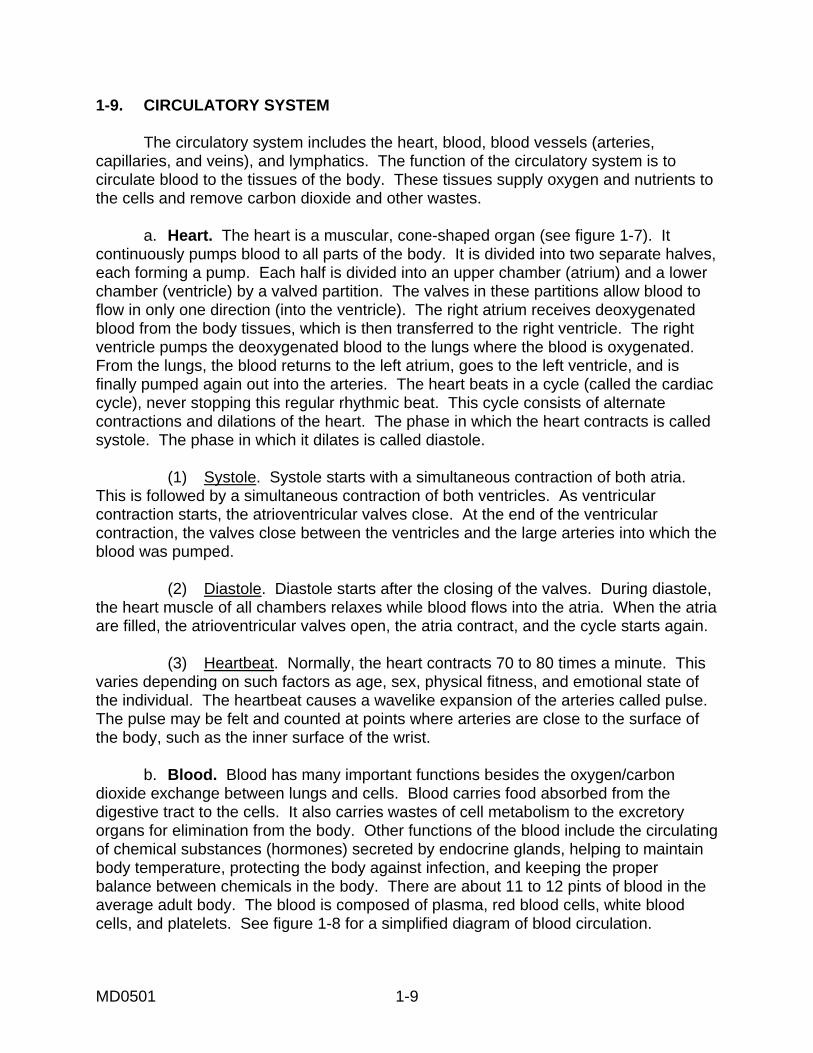
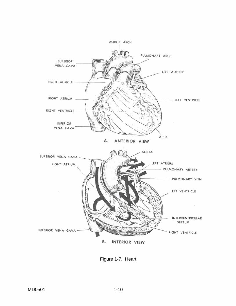
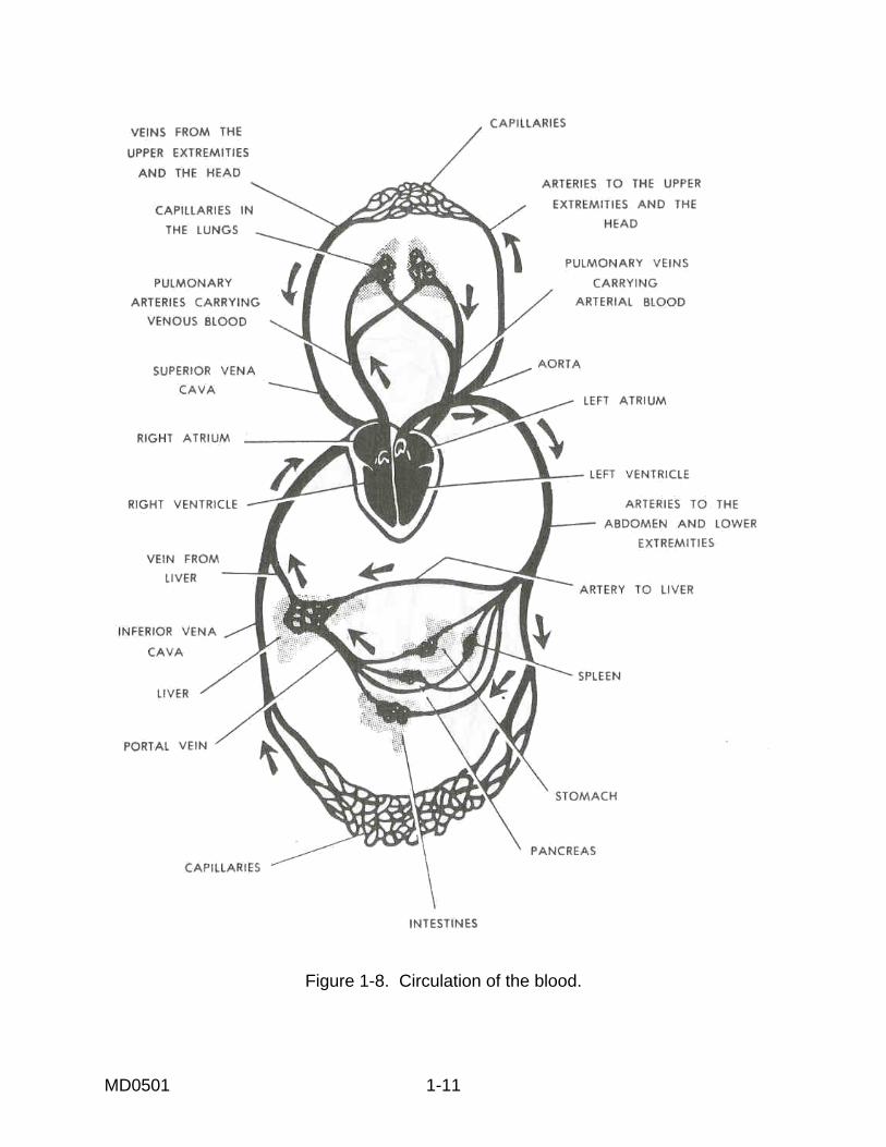
1-9. CIRCULATORY SYSTEM The circulatory system includes the heart, blood, blood vessels (arteries, capillaries, and veins), and lymphatics. The function of the circulatory system is to circulate blood to the tissues of the body. These tissues supply oxygen and nutrients to the cells and remove carbon dioxide and other wastes. a. Heart. The heart is a muscular, cone-shaped organ (see figure 1-7). It continuously pumps blood to all parts of the body. It is divided into two separate halves, each forming a pump. Each half is divided into an upper chamber (atrium) and a lower chamber (ventricle) by a valved partition. The valves in these partitions allow blood to flow in only one direction (into the ventricle). The right atrium receives deoxygenated blood from the body tissues, which is then transferred to the right ventricle. The right ventricle pumps the deoxygenated blood to the lungs where the blood is oxygenated. From the lungs, the blood returns to the left atrium, goes to the left ventricle, and is finally pumped again out into the arteries. The heart beats in a cycle (called the cardiac cycle), never stopping this regular rhythmic beat. This cycle consists of alternate contractions and dilations of the heart. The phase in which the heart contracts is called systole. The phase in which it dilates is called diastole. (1) Systole. Systole starts with a simultaneous contraction of both atria. This is followed by a simultaneous contraction of both ventricles. As ventricular contraction starts, the atrioventricular valves close. At the end of the ventricular contraction, the valves close between the ventricles and the large arteries into which the blood was pumped. (2) Diastole. Diastole starts after the closing of the valves. During diastole, the heart muscle of all chambers relaxes while blood flows into the atria. When the atria are filled, the atrioventricular valves open, the atria contract, and the cycle starts again. (3) Heartbeat. Normally, the heart contracts 70 to 80 times a minute. This varies depending on such factors as age, sex, physical fitness, and emotional state of the individual. The heartbeat causes a wavelike expansion of the arteries called pulse. The pulse may be felt and counted at points where arteries are close to the surface of the body, such as the inner surface of the wrist. b. Blood. Blood has many important functions besides the oxygen/carbon dioxide exchange between lungs and cells. Blood carries food absorbed from the digestive tract to the cells. It also carries wastes of cell metabolism to the excretory organs for elimination from the body. Other functions of the blood include the circulating of chemical substances (hormones) secreted by endocrine glands, helping to maintain body temperature, protecting the body against infection, and keeping the proper balance between chemicals in the body. There are about 11 to 12 pints of blood in the average adult body. The blood is composed of plasma, red blood cells, white blood cells, and platelets. See figure 1-8 for a simplified diagram of blood circulation.
MD0501 1-10
Figure 1-7. Heart
MD0501 1-11
Figure 1-8. Circulation of the blood.
MD0501 1-12
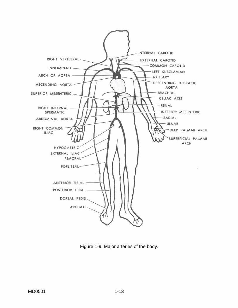
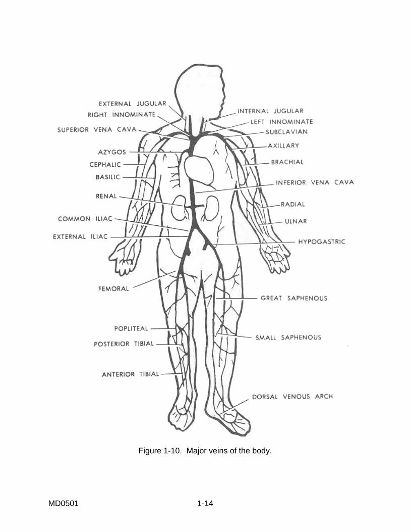
(1) Plasma. Plasma is a protein-containing fluid which constitutes 50 to 60 percent of the blood by weight. Plasma is the liquid medium that carries food to the cells and waste products away from the cells. (2) Other blood components. Red blood cells (erythrocytes), white blood cells (leukocytes), and platelets (thrombocytes) are carried through the circulatory system suspended in plasma. Red blood cells, white blood cells, and platelets constitute 40 to 50 percent of blood by weight. Red blood cells are disk-shaped, cell-like bodies without a nucleus. They contain a substance called hemoglobin. Hemoglobin combines with oxygen to carry it from the lungs to the tissues. Oxygenated hemoglobin is red and gives blood its color. White blood cells are colorless, vary in size and shape, and have a nucleus. They protect the body by destroying foreign substances, such as bacteria, in the blood and tissues. White blood cells move about by a "flowing" motion. They squeeze through capillary walls and move through tissues in pursuit of bacteria. Platelets are smaller than red blood cells. They are round or oval in shape without a nucleus. Platelets aid in the clotting of blood at wound sites. In an average person, each cubic millimeter of blood contains about 5,000,000 red blood cells, 5,000 to 10,000 white blood cells, and 300,000 platelets. c. Blood Vessels. (1) Arteries. Blood pumped by the heart is carried to the tissues through a system of elastic, hollow tubes called arteries (see figure 1-9). This system of arteries is like a tree with a large trunk. The arteries leave the heart and give off branches which repeatedly divide and become smaller and smaller. The arteries have a nerve supply controlled by the autonomic nervous system allowing them to enlarge or constrict. (2) Capillaries. The arteries branch into billions of tiny vessels called capillaries. The capillaries have very thin walls through which food and oxygen pass from the blood to the cells. While food and oxygen are passing through the walls of the capillaries, another process is going on. This process causes the waste materials and carbon dioxide from the cells to pass back into the capillaries. Thus, the capillaries make the necessary exchanges of water, gases, salts, food, and wastes between the blood and the tissues. (3) Veins. As carbon dioxide and waste materials enter the capillaries and as the blood loses oxygen and food, it turns from a bright red to a darker red. Here the venous system begins. The vessels, now called veins, are no longer elastic and muscular. Their walls are thin and collapsible. Veins have paired valves to prevent the backflow of blood. Veins, like arteries, resemble a tree with many branches and eventually form major trunks leading back to the heart (see figure 1-10). The supply of blood to any part of the body may be increased or decreased by a change in the rate of heartbeat or in the size of blood vessels.
MD0501 1-13
Figure 1-9. Major arteries of the body.
MD0501 1-14
Figure 1-10. Major veins of the body.

MD0501 1-15
d. Lymphatics. The lymphatic system consists of lymph, lymphatic vessels, and lymph nodes (see figure 1-11). Structures and glands characterized by the presence of lymphocytic cells--spleen, tonsils, thymus, and lymphatic nodules of the intestine (Peyer's patches)--are also part of the lymphatic system. Lymph drains through its own system of lymphatic vessels (lymphatics) into the jugulo-subclavian venous region of the neck and thus back to the heart. Along the course of the lymphatics are structures called lymph nodes. Lymph nodes are filters removing infectious materials from the lymph stream. They are also the main source of certain white blood cells. Examples of lymph nodes are the axillary nodes and the submaxillary nodes.
Figure 1-11. Lymphatic system.
MD0501 1-16
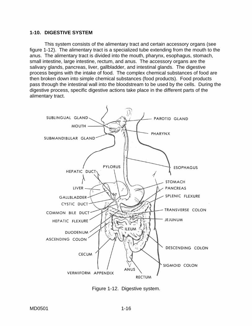
1-10. DIGESTIVE SYSTEM This system consists of the alimentary tract and certain accessory organs (see figure 1-12). The alimentary tract is a specialized tube extending from the mouth to the anus. The alimentary tract is divided into the mouth, pharynx, esophagus, stomach, small intestine, large intestine, rectum, and anus. The accessory organs are the salivary glands, pancreas, liver, gallbladder, and intestinal glands. The digestive process begins with the intake of food. The complex chemical substances of food are then broken down into simple chemical substances (food products). Food products pass through the intestinal wall into the bloodstream to be used by the cells. During the digestive process, specific digestive actions take place in the different parts of the alimentary tract.
Figure 1-12. Digestive system.

MD0501 1-17
a. Mouth. In the mouth, food is ground into small particles by the teeth. Food is moistened and softened by saliva. Saliva is secreted by the salivary glands. This action of the teeth and saliva prepares the food so it can be easily swallowed. An enzyme in the saliva (ptyalin) starts the chemical breakdown of starchy foods. b. Pharynx. The pharynx is a muscular tube forming a passage from the mouth to the esophagus. When swallowing occurs, the tongue pushes the food to the back of the mouth. The food passes through the pharynx to the esophagus. The pharynx connects with both the mouth and nose. Air passes from the nose, through the pharynx, to the larynx, and to the trachea (windpipe). When food is being swallowed, a lid-like structure (the epiglottis) closes off the larynx so food will not enter. c. Esophagus. The esophagus is also a muscular tube. It extends from the pharynx through the thorax to the upper end of the stomach. The chewed food is passed through the esophagus by a wavelike motion of its muscular walls. This motion, called peristalsis, is also found in the intestines. d. Stomach. The stomach is a muscular, bag-like organ. It is located in the upper left part of the abdomen. At its upper end, the stomach connects with the esophagus. At its lower end, the stomach connects with the small intestine. The stomach secretes several enzymes and an acid which aid in the chemical breakdown of carbohydrates, proteins, and fats. The churning action of the stomach's wall, the action of the enzymes, and the action of the acid reduce the food to a semifluid mass called chyme. The stomach does not empty this mass all at once. At intervals, it squirts chyme into the small intestine. By the time chyme leaves the stomach, the food is about half digested. e. Small Intestine. The small intestine is a muscular tube about 21 feet in length and 1 inch in diameter. It has many folds and curves and occupies a large part of the abdomen. Here, the final phase of digestion is completed. In this phase, enzymes secreted from the pancreas, liver, and walls of the intestine itself mix with the food as it enters the small intestine and act upon the food as it passes through the intestine by peristaltic action. When digestion is completed, the digested end products are absorbed through the wall of the small intestine and into the bloodstream. Certain ingredients of food are not digested. They cannot be broken down into an absorbable form by the digestive process. This undigested food passes through the small intestine into the large intestine. f. Large Intestine. The large intestine, continuous with the small intestine, is about 5 feet in length and 2 inches in diameter. It is called the large intestine because its diameter is approximately twice that of the small intestine. The main function of the large intestine is the recovery of water. Undigested food, bacteria in the intestine and secretions from the digestive tract enter the large intestine in a semifluid form. During the passage of this material through the large intestine, water is absorbed through the intestinal wall. The remaining solid material is packed or squeezed into a form called feces. The feces then pass through the rectum.

MD0501 1-18
g. Rectum and Anus. The rectum, continuous with the large intestine, is about 6 inches in length. It receives feces from the large intestine and periodically expels it through the anus. The anus is the external opening at the end of the digestive tract. 1-11. NERVOUS SYSTEM This system includes a group of organs which controls and integrates the intellectual and physical processes of the individual. The nervous system is made up of nerve tissues. The main structure of the conductive nerve tissues is the neuron, a specialized cell designed to conduct electrical impulses. The nervous system is divided into the central nervous system, the peripheral nervous system, and the autonomic nervous system. a. Central Nervous System. The central nervous system includes the brain and the spinal cord (see figure 1-13). The brain, located within the cranium, is the control center of the body. Within the brain, specific areas control specific body functions. For example, there are control centers for speech, sight, hearing, movement, heartbeat, breathing rate, and body temperature. The spinal cord is a reflex control center. It is composed of countless nerve fibers. These nerve fibers go down from and go up to the brain. The spinal cord is enclosed in the spinal column (backbone) for protection. The nerves of the peripheral and autonomic nervous systems have their roots in the central nervous system,
Figure 1-13. Central nervous system.
MD0501 1-19
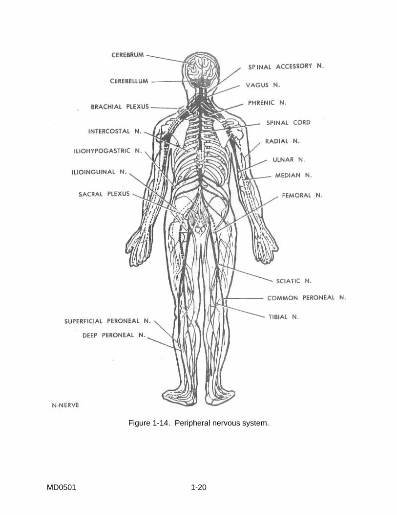
b. Peripheral Nervous System. (1) Structure. The peripheral nervous system consists of the cranial and spinal nerves and their branches (see figure 1-14). Twelve pairs of cranial nerves have their roots in the brain. These nerves give off branches to the structures of the head and face. Thirty-one pairs of spinal nerves have their roots in the spinal cord. They give off branches to the structures of the body from the neck down. Each nerve supplies a specific body area or structure (called innervations). Because many of the various nerve branches join together (anastomose), the result is an overlapping of nerve supply to certain parts. Even though some peripheral nerves are composed entirely of either motor fibers or sensory fibers, most contain both. (2) Function. The peripheral nervous system primarily involves conscious activity of the body. The sensory nerves carry impulses (such as touch, pain, and sight) to the brain. The brain normally evaluates the impulse and sends out, through motor nerves, impulses which cause a bodily response. Normally, all conscious body movements result from interaction of the peripheral and central nervous systems. However, there is an exception called a reflex action. Reflex action results when some sensation or stimulation passes over a reflex arc to a peripheral organ. The organ is thus stimulated to act without the aid of consciousness. For example, jerking your hand away from a hot stove that you accidentally touched. c. Autonomic Nervous System. This system controls the action of cardiac and smooth muscles, sweat glands, digestive glands, some endocrine glands, and dilation and contraction of blood vessels. The control of this system is almost entirely involuntary and subconscious.
MD0501 1-20
Figure 1-14. Peripheral nervous system.

MD0501 1-21
1-12. RESPIRATORY SYSTEM This system includes the lungs and a branched air passage leading into them. This passageway consists of the nose (or the mouth), pharynx, larynx, trachea, and two bronchi (see figure 1-15). The bronchi stem from the trachea (or windpipe). One bronchus passes into each lung. Within the lungs, the bronchi branch into smaller tubes. These tubes branch and rebranch many times to form a system of tiny air tubules. These tubules, called bronchioles, go to all parts of the lungs. They end in tiny air spaces called alveoli. In the alveoli, the exchange of oxygen and carbon dioxide occurs. a. Exchange of Oxygen and Carbon Dioxide. The respiratory system provides the body with oxygen and eliminates carbon dioxide. In the lungs, oxygen enters the blood stream from the air and carbon dioxide passes out of the bloodstream into the air (external respiration). Throughout the tissues of the body, oxygen passes out of the bloodstream to the cells and carbon dioxide passes from the cells into the bloodstream (internal respiration). b. Utilization of Oxygen. The cells of the body require a constant supply of oxygen to carry out the chemical processes necessary for life. Oxygen is taken into the body through the process of respiration (the act of breathing). Breathing may be described as the act of drawing air into the lungs (inhaling) and of forcing air out of the lungs (exhaling). c. The Process of Breathing. Breathing is controlled automatically by a respiratory center in the brain. Breathing can be consciously controlled for short periods of time. The normal rate of breathing is 16 to 20 inhalations and exhalations per minute.

MD0501 1-22
Figure 1-15. Respiratory system.
MD0501 1-23
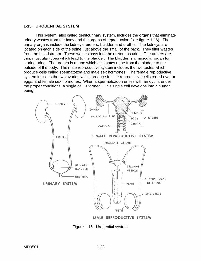
1-13. UROGENITAL SYSTEM This system, also called genitourinary system, includes the organs that eliminate urinary wastes from the body and the organs of reproduction (see figure 1-16). The urinary organs include the kidneys, ureters, bladder, and urethra. The kidneys are located on each side of the spine, just above the small of the back. They filter wastes from the bloodstream. These wastes pass into the ureters as urine. The ureters are thin, muscular tubes which lead to the bladder. The bladder is a muscular organ for storing urine. The urethra is a tube which eliminates urine from the bladder to the outside of the body. The male reproductive system includes the two testes which produce cells called spermatozoa and male sex hormones. The female reproductive system includes the two ovaries which produce female reproductive cells called ova, or eggs, and female sex hormones. When a spermatozoon unites with an ovum, under the proper conditions, a single cell is formed. This single cell develops into a human being.
Figure 1-16. Urogenital system.

MD0501 1-24
1-14. ENDOCRINE SYSTEM This system includes the glands which secrete hormones into the bloodstream (see figure 1-17). Hormones are small in quantity but powerful in effect. Carried throughout the body by the blood, hormones influence growth, blood pressure, fluid balance, ability to use food, and sexual characteristics.
Figure 1-17. Endocrine glands.
Continue with Exercises
Return to Table of Contents
MD0501 1-25
EXERCISES, LESSON 1 INSTRUCTIONS: Answer the following exercises by marking the lettered response that best answers the question or best completes the incomplete statement or by writing the answer in the space provided. After you have completed all the exercises, turn to "Solutions to Exercises" at the end of the lesson and check your answers. For each exercise answered incorrectly, reread the material referenced after the answer. 1. Anatomy is the study of: a. The structure of the body. b. The relation of the parts of the body. c. The body's organs. d. The functions and activities of the parts of the body. e. Items "a," "b," and "c" above. 2. Physiology is the study of: a. The functions and activities of the parts of the body. b. The structure of the body. c. The relation of the parts of the body. d. The systems of the human body. e. Items "a" and "d" above.
MD0501 1-26
SPECIAL INSTRUCTIONS FOR EXERCISES 3 THROUGH 5. Match each term in Column A to its definition in Column B. Mark your answers in the space provided. COLUMN A COLUMN B
____ 3. Nucleus a. Surrounds and separates the cell from its environment. Through this, all the materials essential to metabolism are received and ____ 4. Cytoplasm. disposed of. b. Controls all activities of the cell, including growth ___ 5. Cell membrane. and reproduction. c. Responsible for most of the chemical activities of
the cell. SPECIAL INSTRUCTIONS FOR EXERCISES 6 THROUGH 9. Match each term in Column A to its definition in Column B. Mark your answers in the space provided. COLUMN A COLUMN B ___ 6. Muscle tissue. a. Serves as a lining for vessels and other small cavities inside the body and as skin outside the body. ___ 7. Epithelial tissue. b. Holds tissues together, supports tissues, and fills spaces ___ 8. Nerve tissue. c. Has the ability to contract (shorten). ___ 9. Connective tissue. d. Receives and transmits electrical impulses (messages). SPECIAL INSTRUCTIONS FOR EXERCISES 10 THROUGH 12. Match each term in Column A to its definition in Column B. Mark your answers in the space provided. COLUMN A COLUMN B ___ 10. Tissue. a. The basic functioning unit of the body. ___ 11. Organ. b. A group of similarly-specialized cells that work
together to perform a particular function. ___ 12. Cell. c. A somewhat independent part of the body
which performs a specialized function or functions.

MD0501 1-27
13. The skin functions to protect: a. Inner tissues from _______________________________ . b. Underlying structures from _____________ and ______________. 14. The __________ is the largest organ of the body. Among other functions, the skin regulates body __________________________. 15. The adult human skeleton has ___________ bones. 16. All of the following are found in a cross-section of skin EXCEPT: a. Sweat glands. b. Arteries and veins. c. Sebaceous glands. d. Epidermis. e. Epiphysis. 17. The porous, inner part of a bone is called the: a. Cortex. b. Spongiosa. c. Marrow. d. Periosteum.
MD0501 1-28
18. Which of the following bones are parts of the head or neck? a. Scapula. b. Phalanges. c. Cervical vertebrae. d. Sacrum. e. Ulna. 19. The muscular system includes more than ___________ muscles. Cardiac muscle is found in the muscular wall of the _______________. 20. Involuntary muscles are another name for ___________________ muscle. a. Smooth. b. Cardiac. c. Striated. SPECIAL INSTRUCTIONS FOR EXERCISES 21 THROUGH 23. Match the type of mulsle in Column A to its description in Column B. Mark your answers in the space provided. COLUMN A COLUMN B ___ 21. Striated. a. Found in various visceral organs where
continuous automatic functions are necessary. _ 22. Smooth. b. A specialized type of involuntary muscle. _ 23. Cardiac. c. Skeletal muscles that can be consciously
controlled.
MD0501 1-29
24. Which of the following muscles are situated in the head or face? a. Iliopsoas. b. Deltoid. c. Quadriceps. d. Risorius. e. Trapezius. 25. The circulatory system includes the: a. ___________________________________. b. ___________________________________. c. ___________________________________. d. ___________________________________. 26. Complete the information concerning the human heart. a. The heart is a muscular, _________ - ____________ organ. b. The upper chamber of the heart is the __________________. c. The lower chamber of the heart is the _____________________. d. The phase in the cardiac cycle when the heart contracts is called _________. e. The phase in the cardiac cycle when the heart dilates (relaxes) is called _____________________. f. Normally, the heart contracts _________ to ____________ times per minute.

MD0501 1-30
27. In the average adult human body, there are about ____________ pints of blood. a. 11 to 12. b. 10 to 11. c. 13 to 14. d. 12 to 13. 28. A wavelike expansion of the arteries is called the: a. Systole. b. Diastole. c. Pulse. d. Heartbeat. SPECIAL INSTRUCTIONS FOR EXERCISES 29 THROUGH 32. Match the name of the blood component in Column A to its description in Column B. Mark your answers in the space provided. COLUMN A COLUMN B ___ 29. Plasma. a. Carry oxygen from the lungs to the tissues. ___ 30. Erythrocytes. b. The liquid medium that carries food to the cells and waste products away from the cells. ___ 31. Leukocytes. c. Aid in the clotting of blood. ___ 32. Thrombocytes. d. Destroy foreign substances in the blood and the
tissues.

MD0501 1-31
33. Oxygenated hemoglobin is carried in: a. White blood cells. b. Plasma. c. Red blood cells. d. Platelets. e. Items "c" and "d" above. 34. All of the following have lymphocytic cells EXCEPT: a. The thymus. b. The spleen. c. Tonsils. d. The heart. e. Peyer's patches. 35. List the three kinds of blood vessels. a. _________________________________________. b. _________________________________________. c. _________________________________________.

MD0501 1-32
36. According to figure 1-10, all of the following blood vessels are veins EXCEPT the: a. Right innominate. b. Subclavian. c. Internal jugular. d. Great saphenous. e. External carotid. 37. Select the lymph node that is NOT a part of the lymphatic system of the head and neck. a. Posterior auricular node. b. Axillary node. c. Submental node. d. Occipital node. e Submaxillary node. 38. All of the following are parts of the alimentary tract EXCEPT the: a. Larynx. b. Mouth. c. Pharynx. d. Rectum. e. Esophagus.

MD0501 1-33
39. All of the following are accessory organs of the digestive system EXCEPT the: a. Salivary glands. b. Pancreas c. Intestinal glands. d. Spleen. e. Gallbladder. 40. What is the enzyme in the mouth that starts the chemical breakdown of starchy foods? _____________________________________. 41. When food is swallowed, what structure closes off the larynx? ________________________________.
MD0501 1-34
42. Complete the information related to the digestive system. a. Chewed food is passed from the pharynx through the ____________ by a wavelike motion of the muscular walls, called ___________________. b. The action of several enzymes and an acid aid in the chemical breakdown of food. This occurs in the ________ and the resulting semifluid mass is called _____________________. c. The digestion process is completed in the , which is ______ feet in length and __________ inch in diameter. d. The main function of the large intestine is the recovery of ________. The large intestine is _____ feet in length and _____ inches in diameter. e. The _____________ is continuous with the large intestine and is only about 6 inches long. It receives ___________ (waste products) from the large intestine and periodically expels it through the _____________. SPECIAL INSTRUCTIONS FOR EXERCISES 43 THROUGH 45. Match the term in Column A to its description in Column B. Mark your answers in the space provided. COLUMN A COLUMN B ___ 43. Central a. Controls smooth muscles, the heart, sweat nervous system. glands, digestive glands, the blood vessels, and
some endocrine glands. ___ 44. Peripheral
nervous system. b. Controls specific body functions, such as movement, breathing, heartbeat, body temperature, speech, sight, and hearing.
___ 45. Autonomic c. Controls all conscious body movements and
nervous system reflex action.

MD0501 1-35
46. Complete the information related to the nervous system. a. The central nervous system includes the _________ and the ________ ______________. b. The control center of the body is the ________________, located in the ____________. The spinal cord is enclosed in the _________ ________ and is sometimes called the ___________________ . c. There are ________ pairs of cranial nerves that give off branches to the structures of the head and face. There are ________ pairs of spinal nerves that give off branches to the structures of the body from the neck down. d. Each nerve supplies a specific body area or structure (called __________________________ . When various nerve branches join together, they are said to _______________________. e. Most peripheral nerves contain both _________ fibers and _________fibers. The control of the autonomic nervous system is almost entirely ___________ and subconscious. 47. The action of the cardiac muscle is controlled by the: a. Central nervous system. b. Autonomic nervous system. c. Peripheral nervous system.
MD0501 1-36
48. The specialized cell designed to control electrical impulses generated in the nervous system is the: a. Sacral plexus. b. Bronchi. c. Neuron. d. Pons. e. Vagus nerve. SPECIAL INSTRUCTIONS FOR EXERCISES 49 THROUGH 53. Match the structure of the respiratory system in Column A to its description in Column B. Mark your answers in the space provided. COLUMN A COLUMN B ___ 49. Larynx. a. Two branches of the air passageway, leading to
the lungs. ___ 50. Trachea. b. A system of tiny air tubules within the lungs. ___ 51. Bronchi. c. Tiny air spaces for exchange of oxygen and
carbon dioxide. ___ 52. Bronchioles. d. Between the pharynx and the trachea, containing
the vocal cords. ___ 53. Alveoli. e. Carries air from the larynx to the bronchi. 54. What is the term for the air passage that enters directly into one of the lungs? a. Pleural cavity. b. Trachea. c. Bronchus. d. Larynx. e. Bronchi.

MD0501 1-37
55. The normal rate of breathing is inhalations and exhalations per minute. a. 12 to 16. b. 16 to 20. c. 18 to 22. d. 20 to 24. SPECIAL INSTRUCTIONS FOR EXERCISES 56 THROUGH 61. Match the structure of the urogenital system in Column A to its description in Column B. Mark your answers in the space provided. COLUMN A COLUMN B ___ 56. Kidney. a. Eliminates urine to the outside of the body. ___ 57. Ureter. b. Produces spermatozoa and male sex hormones. ___ 58. Bladder. c. Produces ova and female sex hormones. ___ 59. Urethra. d. Stores urine. ___ 60. Testis. e. Tubes which lead to the bladder. ___ 61. Ovary f. Filters wastes from the bloodstream. 62. Under the proper conditions, a single cell is formed when a __________unites with an ______________ . This single cell develops into a human being. 63. The endocrine system includes various _________ which secrete ___________ into the bloodstream.

MD0501 1-38
64. List the four endocrine glands in the head and neck. a. _________________________________. b. _________________________________. c. _________________________________. d. _________________________________. 65. According to the lesson, all of the following glands are part of the endocrine system EXCEPT the: a. Adrenal gland. b. Pituitary body. c. Testis or ovary. d. Pancreatic islets.
e. Thymus.
Check Your Answers on Next Page
MD0501 1-39
SOLUTIONS TO EXERCISES, LESSON 1 1. e (para 1-1) 2. a (para 1-1) 3. b (para 1-2b) 4. c (para 1-2c) 5. a (para 1-2a) 6. c (para 1-3c) 7. a (para 1-3a) 8. d (para 1-3d) 9. b (para 1-3b) 10. b (para 1-3) 11. c (para 1-4) 12. a (para 1-2) 13. a. drying (dessication) b. infection; injury (para 1-6) 14. skin; temperature (para 1-6) 15. 206 (para 1-7) 16. e (figure 1-2) 17. b (para 1-7) 18. c (figure 1-3) 19. 350; heart (para 1-8) 20. a (para 1-8b)
MD0501 1-40
21. c (para 1-8a) 22. a (para 1-8b) 23. b (para 1-8c) 24. d (figures 1-5 and 1-6) 25. Heart. Blood. Blood vessels. Lymphatics (para 1-9). 26. a. cone-shaped. b. atrium. c. ventricle. d. systole. e. diastole. f. 70; 80. (paras 1-9a, a(1), (2)) 27. a (para 1-9b) 28. c (para 1-9a(3)) 29. b (para 1-9b(1)) 30. a (para 1-9b(2)) 31. d (para 1-9b(2)) 32. c (para 1-9b(2)) 33. c (para 1-9b(2)) 34. d (para 1-9d) 35. Arteries. Capillaries. Veins (para 1-9c). 36. e (figures 1-10, 1-9) 37. b (figure 1-11) 38. a (para 1-10)
MD0501 1-41
39. d (para 1-10) 40. Ptaylin. (para 1-10a) 41. Epiglottis. (para 1-10b) 42. a. esophagus; peristalsis (para 1-10c). b. stomach; chyme (para 1-10d). c. small intestine; 21; 1 (para 1-10e) d. water; 5; 2 (para 1-10f) e. rectum; feces; anus (para 1-10f, g) 43. b (para 1-11a) 44. c (para 1-11b(2)) 45. a (para 1-11c) 46. a. brain; spinal cord (para 1-11a) b. brain; cranium; spinal column; backbone (para 1-11a) c. 12; 31 (para 1-11b(1)) d. innervation; anastomose (para 1-11b(1)) e. motor; sensory; involuntary (paras 1-11b(1), 1-11c) 47. b (para 1-11c) 48. c (para 1-11) 49. d (para 1-12; figure 1-15) 50. e (para 1-12; figure 1-15) 51. a (para 1-12) 52. b (para 1-12) 53. c (para 1-12) 54. c (para 1-12) 55. b (para 1-12c)
MD0501 1-42
56. f (para 1-13) 57. e (para 1-13) 58. d (para 1-13) 59. a (para 1-13) 60. b (para 1-13; figure 1-16) 61. c (para 1-13; figure 1-16) 62. spermatozoon; ovum (para 1-13) 63. glands; hormones (para 1-14) 64. a. Pituitary. b. Pineal. c. Thyroid. d. Parathyroid. (para 1-14; figure 1-17) 65. e (para 1-14; figure 1-17)
Return to Table of Contents