Embed Size (px)
Citation preview
130
J Clin Exp HematopVol. 56, No. 2, December 2016
Letter to the Editor
TO THE EDITORChromosome 17 abnormalities in haematologic malig-
nancies are important because the TP53 tumour suppressor gene is found on 17p 13.1.1 Chromosome 17 abnormalities were reported in 49 (4.3%) of cases in a study of patients with myelodysplastic syndrome (MDS) and acute myeloid leukaemia (AML). In this cohort, 14 patients had mono-somy 17 and 35 patients had unbalanced translocations between chromosome 17p and another chromosome. Most of them also had other cytogenetic abnormalities and 10 were therapy related, 69% also had a TP53 mutation. The pseudo Pelger-Huët like anomaly and vacuolated neutrophils were described in 70% of these patients.2 Chromosome 17 abnor-malities have also been associated with therapy related myeloid neoplasms, unbalanced translocations involving chromosome 17, monosomy 17 and isochromosome 17q or 17p deletion having been reported.3
Isochromosome(17q) occurs when chromosome 17 has two identical arms, i.e. no “p” arm but two “q” arms. This results in a cell having only one copy of 17p and three copies of 17q. It is thought to occur due to breakage of the proxi-mal part of 17p followed by rejoining of the centromere
containing chromatids and inactivation of one centromere.4 Isochromosome(17q) has been reported in a variety of hae-matologic and solid malignancies, it is usually associated with a complex karyotype but has less commonly been reported in isolation.5-9 Myeloid neoplasms with isolated i(17q) have been reported to have features of an MDS/ myeloproliferative neoplasm (MPN) overlap syndrome. Myeloid dysplasia, monocytosis and a high propensity for leukaemic transformation with a short overall survival are features of this condition.10
Myeloid sarcoma (MS) is characterized by extramedul-lary tumour masses composed of myeloblasts or myelomono-cytic cells.11 MS may present de novo or occur concomi-tantly with MDS, MPN or AML. Alternatively MS may represent transformation of a MPN or MDS to AML or occur at relapse.12 Fluorescence in situ hybridization analysis of 49 MS specimens showed clonal abnormalities in 25, specifi-cally, monosomy 7 (10%), trisomy 8 (10%) and MLL gene rearrangement (8.5%). Concordance between fluorescence in situ hybridization on the MS specimen and bone marrow karyotyping was shown in 10 of 14 cases in which both anal-yses were performed.11 Interestingly, t(8;21) has been found in paediatr ic cases of MS and in orbital tumours. 13 Comparative genomic hybridization (CGH) of seven MS cases showed chromosome 8 abnormalities in 3, with other abnormalities including loss of 4q, 6q, 12p and gain of 11q, 13q, 19 and 21.14 Li and co-workers performed next genera-tion sequencing of 21 AML/MDS related genes in 6 cases of MS. They detected mutations in tyrosine kinases (FLT-3 and KIT), the WT1 tumour suppressor gene, the epigenetic regulators EZH2, ASXL1 and TET1 as well as SF3B1, an RNA splicosome protein.15 This study showed a genetic overlap between MS and AML, it is likely however that there are differences between these entities which account for the extramedullary localization of MS. A patient with AML and concomitant testicular MS was found to have i(17)q as part of a near tetraploid karyotype on cytogenetic analysis of the
Isochromosome 17q; A Novel Finding in Myeloid Sarcoma
Sanjay de Mel,1) Joanne Lee,1) Constance Chua,2) Sok Peng Chua,2) Leena Gole,2) Limei Poon,1)
Jenny Li,2) Siok Bian Ng,3) Te Chih Liu,2) Wee Joo Chng,1) and Yen Lin Chee1)
Keywords: isochromosome 17q, myeloid sarcoma, comparative genomic hybridization
Received: May 30, 2016Revised : July 14, 2016Accepted: August, 15, 20161)Department of Haematology, Oncology, National University Cancer Institute, National
University Health System, Singapore2)Department of Laboratory Medicine, National University Health System, Singapore3)Department of Pathology, Yong Loo Lin School of Medicine, National University of
Singapore, Singapore Department of Pathology, National University Hospital, National University Health
System, SingaporeCorresponding author: Dr. Sanjay de Mel, Department of Haematology Oncology,
National University Cancer Institute, National University Health System, 1E Kent Ridge Road, SingaporeE-mail: [email protected]
131
de Mel S, et al.
bone marrow.16 Genomic analysis of the MS tissue was not however performed in this case. It is noteworthy that i(17)q has not been reported previously in a tissue biopsy of MS.
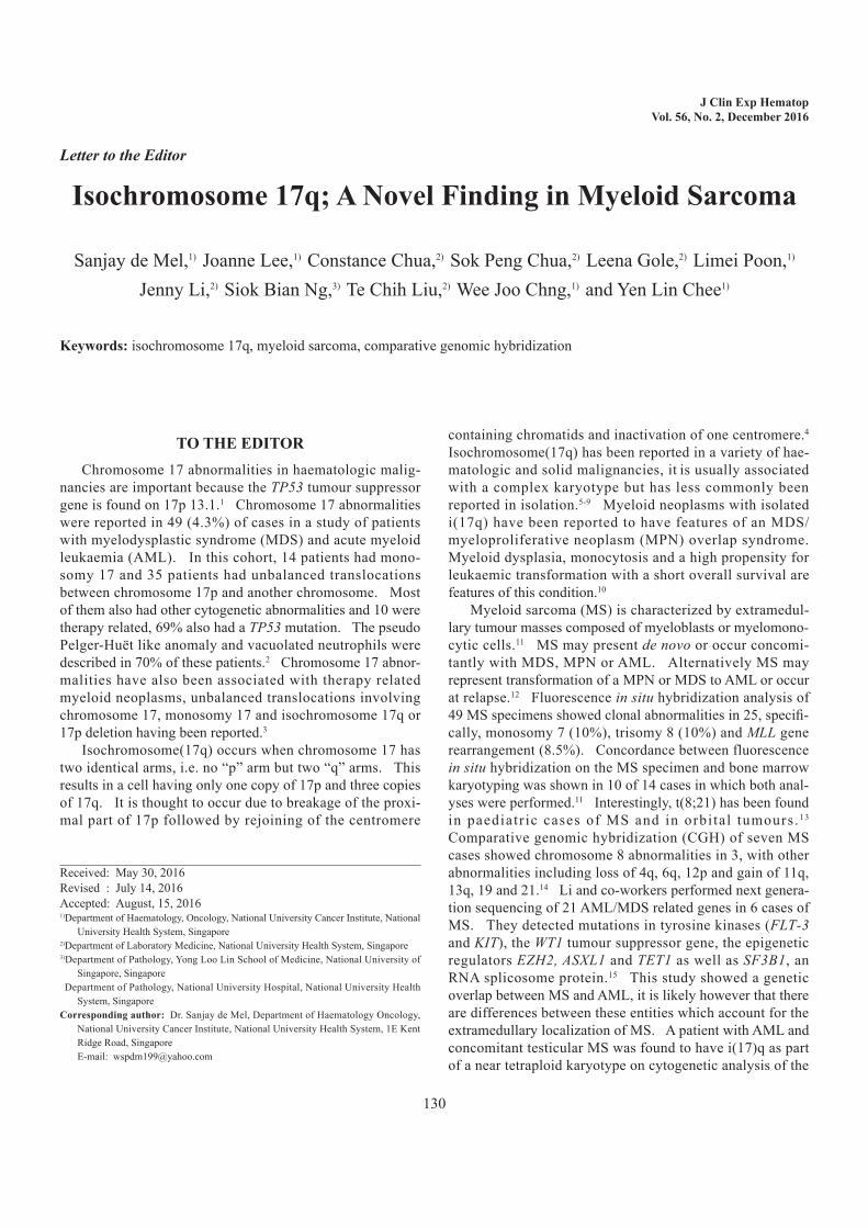
A 79 year-old male presented with a one month history of lethargy and ankle oedema. He had no bleeding symptoms and denied weight loss, night sweats, fever or rash. Physical examination revealed bilateral cervical lymphadenopathy and moderate splenomegaly. Initial investigations revealed; hae-moglobin 11.0 g/dL, white blood cells 17.8 x 10⁹/L, neutro-phils 7.3 x10⁹/L, monocytes 3.2 x 10 9/L, eosinophils 1.42 x 10 9/L, and basophils 1.96 x 10⁹/L. A peripheral blood film showed dysplastic neutrophils and monocytes with platelet anisocytosis. A left shift was seen with blasts comprising 8% of white blood cells (Fig. 1a). His bone marrow aspirate and trephine were hypercellular with dysplastic granulopoie-sis and megakaryopoiesis. Myeloblasts comprised 19% of nucleated cells with eosinophilia (12%) and basophilia (4%) (Fig. 1b & 1c). Multiparameter flow cytometry showed dys-coordinated granulocytic maturation with CD34 positive myeloblasts comprising 13% of events.
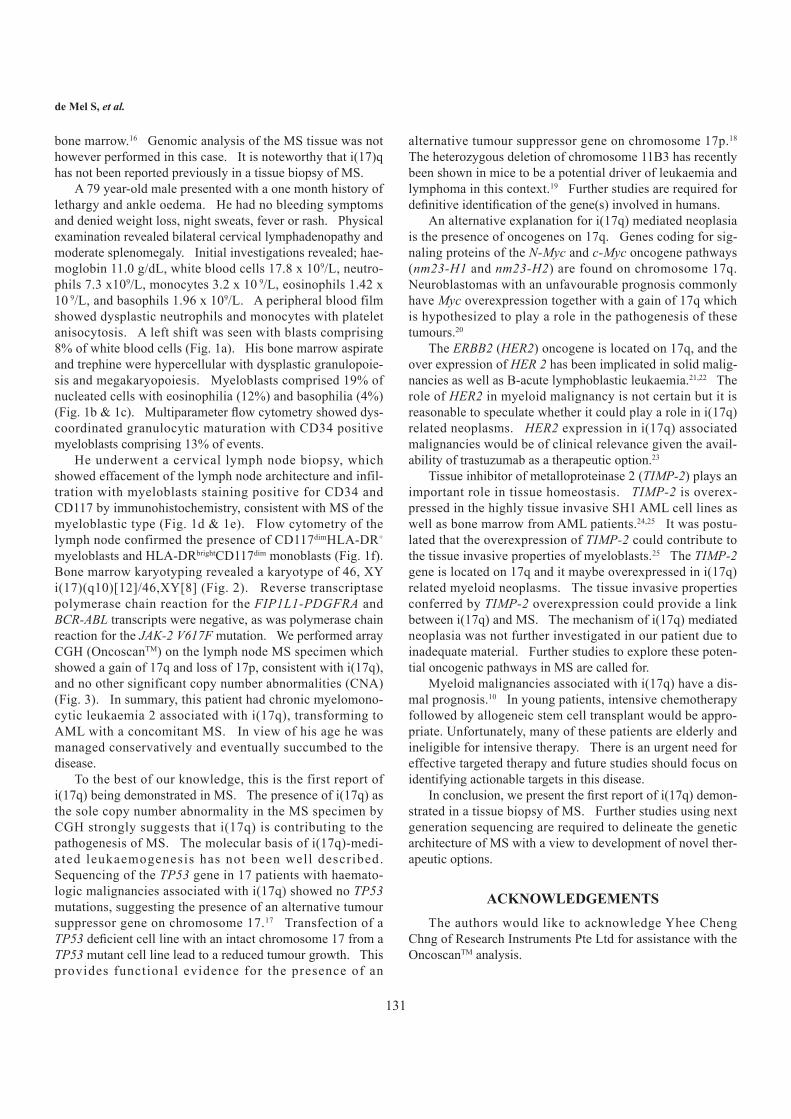
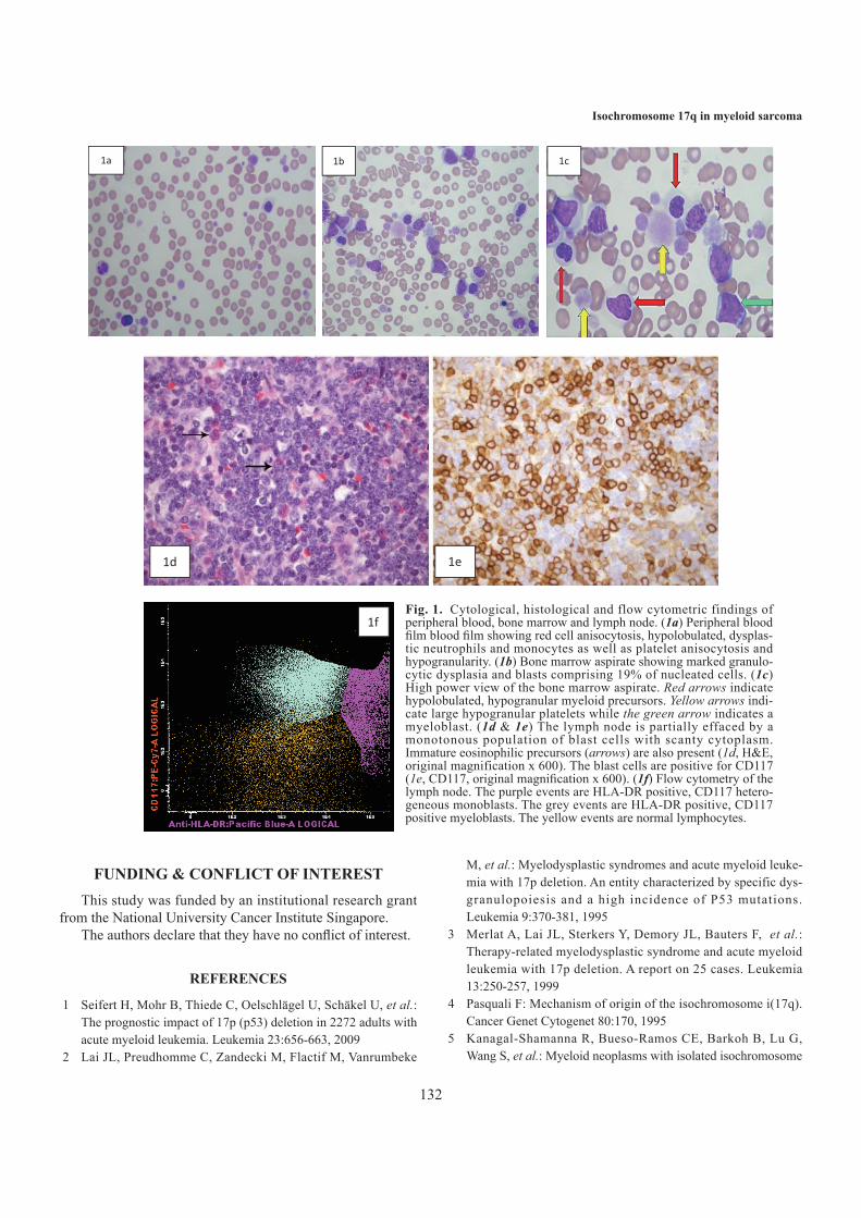
He underwent a cervical lymph node biopsy, which showed effacement of the lymph node architecture and infil-tration with myeloblasts staining positive for CD34 and CD117 by immunohistochemistry, consistent with MS of the myeloblastic type (Fig. 1d & 1e). Flow cytometry of the lymph node confirmed the presence of CD117dimHLA-DR+ myeloblasts and HLA-DRbrightCD117dim monoblasts (Fig. 1f). Bone marrow karyotyping revealed a karyotype of 46, XY i(17)(q10)[12]/46,XY[8] (Fig. 2). Reverse transcriptase polymerase chain reaction for the FIP1L1-PDGFRA and BCR-ABL transcripts were negative, as was polymerase chain reaction for the JAK-2 V617F mutation. We performed array CGH (OncoscanTM) on the lymph node MS specimen which showed a gain of 17q and loss of 17p, consistent with i(17q), and no other significant copy number abnormalities (CNA) (Fig. 3). In summary, this patient had chronic myelomono-cytic leukaemia 2 associated with i(17q), transforming to AML with a concomitant MS. In view of his age he was managed conservatively and eventually succumbed to the disease.
To the best of our knowledge, this is the first report of i(17q) being demonstrated in MS. The presence of i(17q) as the sole copy number abnormality in the MS specimen by CGH strongly suggests that i(17q) is contributing to the pathogenesis of MS. The molecular basis of i(17q)-medi-ated leukaemogenesis has not been well described. Sequencing of the TP53 gene in 17 patients with haemato-logic malignancies associated with i(17q) showed no TP53 mutations, suggesting the presence of an alternative tumour suppressor gene on chromosome 17.17 Transfection of a TP53 deficient cell line with an intact chromosome 17 from a TP53 mutant cell line lead to a reduced tumour growth. This provides functional evidence for the presence of an
alternative tumour suppressor gene on chromosome 17p.18 The heterozygous deletion of chromosome 11B3 has recently been shown in mice to be a potential driver of leukaemia and lymphoma in this context.19 Further studies are required for definitive identification of the gene(s) involved in humans.
An alternative explanation for i(17q) mediated neoplasia is the presence of oncogenes on 17q. Genes coding for sig-naling proteins of the N-Myc and c-Myc oncogene pathways (nm23-H1 and nm23-H2) are found on chromosome 17q. Neuroblastomas with an unfavourable prognosis commonly have Myc overexpression together with a gain of 17q which is hypothesized to play a role in the pathogenesis of these tumours.20
The ERBB2 (HER2) oncogene is located on 17q, and the over expression of HER 2 has been implicated in solid malig-nancies as well as B-acute lymphoblastic leukaemia.21,22 The role of HER2 in myeloid malignancy is not certain but it is reasonable to speculate whether it could play a role in i(17q) related neoplasms. HER2 expression in i(17q) associated malignancies would be of clinical relevance given the avail-ability of trastuzumab as a therapeutic option.23
Tissue inhibitor of metalloproteinase 2 (TIMP-2) plays an important role in tissue homeostasis. TIMP-2 is overex-pressed in the highly tissue invasive SH1 AML cell lines as well as bone marrow from AML patients.24,25 It was postu-lated that the overexpression of TIMP-2 could contribute to the tissue invasive properties of myeloblasts.25 The TIMP-2 gene is located on 17q and it maybe overexpressed in i(17q) related myeloid neoplasms. The tissue invasive properties conferred by TIMP-2 overexpression could provide a link between i(17q) and MS. The mechanism of i(17q) mediated neoplasia was not further investigated in our patient due to inadequate material. Further studies to explore these poten-tial oncogenic pathways in MS are called for.
Myeloid malignancies associated with i(17q) have a dis-mal prognosis.10 In young patients, intensive chemotherapy followed by allogeneic stem cell transplant would be appro-priate. Unfortunately, many of these patients are elderly and ineligible for intensive therapy. There is an urgent need for effective targeted therapy and future studies should focus on identifying actionable targets in this disease.
In conclusion, we present the first report of i(17q) demon-strated in a tissue biopsy of MS. Further studies using next generation sequencing are required to delineate the genetic architecture of MS with a view to development of novel ther-apeutic options.
ACKNOWLEDGEMENTSThe authors would like to acknowledge Yhee Cheng
Chng of Research Instruments Pte Ltd for assistance with the OncoscanTM analysis.
132
Isochromosome 17q in myeloid sarcoma
FUNDING & CONFLICT OF INTERESTThis study was funded by an institutional research grant
from the National University Cancer Institute Singapore.The authors declare that they have no conflict of interest.
REFERENCES
1 Seifert H, Mohr B, Thiede C, Oelschlägel U, Schäkel U, et al.: The prognostic impact of 17p (p53) deletion in 2272 adults with acute myeloid leukemia. Leukemia 23:656-663, 2009
2 Lai JL, Preudhomme C, Zandecki M, Flactif M, Vanrumbeke
M, et al.: Myelodysplastic syndromes and acute myeloid leuke-mia with 17p deletion. An entity characterized by specific dys-granulopoiesis and a high incidence of P53 mutations. Leukemia 9:370-381, 1995
3 Merlat A, Lai JL, Sterkers Y, Demory JL, Bauters F, et al.: Therapy-related myelodysplastic syndrome and acute myeloid leukemia with 17p deletion. A report on 25 cases. Leukemia 13:250-257, 1999
4 Pasquali F: Mechanism of origin of the isochromosome i(17q). Cancer Genet Cytogenet 80:170, 1995
5 Kanagal-Shamanna R, Bueso-Ramos CE, Barkoh B, Lu G, Wang S, et al.: Myeloid neoplasms with isolated isochromosome
Fig. 1. Cytological, histological and flow cytometric findings of peripheral blood, bone marrow and lymph node. (1a) Peripheral blood film blood film showing red cell anisocytosis, hypolobulated, dysplas-tic neutrophils and monocytes as well as platelet anisocytosis and hypogranularity. (1b) Bone marrow aspirate showing marked granulo-cytic dysplasia and blasts comprising 19% of nucleated cells. (1c) High power view of the bone marrow aspirate. Red arrows indicate hypolobulated, hypogranular myeloid precursors. Yellow arrows indi-cate large hypogranular platelets while the green arrow indicates a myeloblast. (1d & 1e) The lymph node is partially effaced by a monotonous population of blast cells with scanty cytoplasm. Immature eosinophilic precursors (arrows) are also present (1d, H&E, original magnification x 600). The blast cells are positive for CD117 (1e, CD117, original magnification x 600). (1f) Flow cytometry of the lymph node. The purple events are HLA-DR positive, CD117 hetero-geneous monoblasts. The grey events are HLA-DR positive, CD117 positive myeloblasts. The yellow events are normal lymphocytes.
133
de Mel S, et al.
Chromosome 2 Mosaic Gain
Chromosome 17 Mosaic Gain and Loss
ChromosomeX and Y and Mosaic Gain
Fig. 2. Bone marrow karyotyping revealed a karyotype of 46,XY,i(17)(q10)[12]/46,XY[8].
Fig. 3. Array comparative genomic hybridisation. A mosaic gain of 17q and loss of 17p is demonstrated. This pattern is consistent with i(17q).
134
Isochromosome 17q in myeloid sarcoma
17q represent a clinicopathologic entity associated with myelo-dysplastic/myeloproliferative features, a high risk of leukemic transformation, and wild-type TP53. Cancer 118:2879-2888, 2012
6 Lazarević V, Djordjević V, Magić Z, Marisavljevic D, Colović M: Refractory anemia with ring sideroblasts associated with i(17q) and mutation of the TP53 gene. Cancer Genet Cytogenet 136:86-89, 2002
7 Pinheiro RF, Chauffaille Mde L, Silva MR: Isochromosome 17q in MDS: a marker of a distinct entity. Cancer Genet Cytogenet 166:189-190, 2006
8 Testa JR, Cohen BC: Dicentric chromosome 17 in patients with leukemia. Cancer Genet Cytogenet 23:47-52, 1986
9 Mertens F, Johansson B, Mitelman F: Isochromosomes in neo-plasia. Genes Chromosomes Cancer 10:221-230, 1994
10 McClure RF, Dewald GW, Hoyer JD, Hanson CA: Isolated iso-chromosome 17q: a distinct type of mixed myeloproliferative disorder/myelodysplastic syndrome with an aggressive clinical course. Br J Haematol 106:445-454, 1999
11 Pileri SA, Ascani S, Cox MC, Campidelli C, Bacci F, et al.: Myeloid sarcoma: clinico-pathologic, phenotypic and cytoge-netic analysis of 92 adult patients. Leukemia 21:340-350, 2007
12 Campidelli C, Agostinelli C, Stitson R, Pileri SA: Myeloid sar-coma: extramedullary manifestation of myeloid disorders. Am J Clin Pathol 132:426-437, 2009
13 Schwyzer R, Sherman GG, Cohn RJ, Poole JE, Willem P: Granulocytic sarcoma in children with acute myeloblastic leu-kemia and t(8;21). Med Pediatr Oncol 31:144-149, 1998
14 Deeb G, Baer MR, Gaile DP, Sait SN, Barcos M, et al.: Genomic profiling of myeloid sarcoma by array comparative genomic hybridization. Genes Chromosomes Cancer 44:373-383, 2005
15 Li Z, Stölzel F, Onel K, Sukhanova M, Mirza MK, et al.: Next-generation sequencing reveals clinically actionable molecular markers in myeloid sarcoma. Leukemia 29:2113-2116, 2015
16 Bettio D, Venci A, Sarina B: Near-tetraploid karyotype with an isochromosome 17q as the sole structural chromosomal rear-rangement in a case of testicular granulocytic sarcoma. Cancer Genet Cytogenet 181:69-71, 2008
17 Fioretos T, Strömbeck B, Sandberg T, Johansson B, Billström R, et al.: Isochromosome 17q in blast crisis of chronic myeloid leukemia and in other hematologic malignancies is the result of clustered breakpoints in 17p11 and is not associated with coding TP53 mutations. Blood 94:225-232, 1999
18 Chen P, Ellmore N, Weissman BE: Functional evidence for a second tumor suppressor gene on human chromosome 17. Mol Cell Biol 14:534-542, 1994
19 Liu Y, Chen C, Xu Z, Scuoppo C, Rillahan CD, et al.: Deletions linked to TP53 loss drive cancer through p53-independent mechanisms. Nature 531:471-475, 2016
20 Godfried MB, Veenstra M, v Sluis P, Boon K, v Asperen R, et al.: The N-myc and c-myc downstream pathways include the chromosome 17q genes nm23-H1 and nm23-H2. Oncogene 21:2097-2101, 2002
21 Gutierrez C, Schiff R: HER2: biology, detection, and clinical implications. Arch Pathol Lab Med 135:55-62, 2011
22 Chevallier P, Robillard N, Wuilleme-Toumi S, Méchinaud F, Harousseau JL, et al.: Overexpression of Her2/neu is observed in one third of adult acute lymphoblastic leukemia patients and is associated with chemoresistance in these pat ients . Haematologica 89:1399-1401, 2004
23 Chevallier P, Robillard N, Charbonnier A, Raffoux E, Maury S, et al.: Trastuzumab for treatment of refractory/relapsed HER2-positive adult B-ALL: results of a phase 2 GRAALL study. Blood 119:2474-2477, 2012
24 Lin LI, Lin DT, Chang CJ, Lee CY, Tang JL, et al.: Marrow matrix metalloproteinases (MMPs) and tissue inhibitors of MMP in acute leukaemia: potential role of MMP-9 as a surro-gate marker to monitor leukaemic status in patients with acute myelogenous leukaemia. Br J Haematol 117:835-841, 2002
25 Wang C, Chen Z, Li Z, Cen J: The essential roles of matrix metalloproteinase-2, membrane type 1 metalloproteinase and tissue inhibitor of metalloproteinase-2 in the invasive capacity of acute monocytic leukemia SHI-1 cells. Leuk Res 34:1083-1090, 2010
![Pallister Killian - Chromosome...has some normal cells and some with a little structure called an isochromosome that consists of the short arm of chromosome 12 [12p] repeated either](https://img.pdfslide.net/doc/110x75/5f48526e276d6f6c223aa7cc/pallister-killian-chromosome-has-some-normal-cells-and-some-with-a-little.jpg)