Embed Size (px)
Citation preview
.
585ISSN 0372-5480Printed in Croatia
VETERINARSKI ARHIV 87 (5), 585-595, 2017
Leucine-enkephalin is co-expressed with substance P, galanin and somatostatin in the ovine pancreas
Aleksandra Górska*, Anna Zacharko-Siembida, and Marcin B. Arciszewski
Department of Animal Anatomy and Histology, Faculty of Veterinary Medicine, University of Life Sciences, Lublin, Poland
________________________________________________________________________________________GÓRSKA, A., A. ZACHARKO-SIEMBIDA, M. B. ARCISZEWSKI: Leucine-enkephalin is co-expressed with substance P, galanin and somatostatin in the ovine pancreas. Vet. arhiv 87, 585-595, 2017.
ABSTRACTLeucine-enkephalin (Leu-Enk) is an endogenous opioid peptide that binds to opioid receptors. Leu-Enk
is widely distributed in the central and peripheral nervous system. The aim of the present immunofluorescence study was to examine the distribution of Leu-Enk-immunoreactive (IR) neuronal elements in the ovine pancreas. Using double immunohistochemical staining, the co-localization of Leu-Enk with galanin, somatostatin and substance P was also studied. In the intrapancreatic ganglia, immunoreactivity to Leu-Enk was found in 64.9 ± 1.7% of neurons. Small arterioles and the ductal system were innervated by numerous Leu-Enk-IR nerve terminals. Moderate Leu-Enk-IR nerve fibres surrounded the islets of Langerhans but none of them penetrated into spaces between endocrine cells. In 66.7 ± 4.3% of Leu-Enk-immunoreactive intrapancreatic neurons, expression of galanin was found. A statistically smaller subpopulation of Leu-Enk-IR intrapancreatic neurons (37.4 ± 6.2%) exhibited immunoreactivity to SP. The expression of somatostatin was detected in the relatively smallest group (21.2 ± 3.8%) of Leu-Enk-positive intrapancreatic neurons. Co-expression of Leu-Enk and SP was detected in nerve terminals encircling the pancreatic small arterioles, connective tissue and ducts. Leu-Enk-positive nerve fibres around the islets of Langerhans were not immunoreactive for SP. None of the Leu-ENK-positive nerve fibres around the islets of Langerhans co-stored somatostatin. In general, there was also no co-localization between Leu-Enk and somatostatin in nerve terminals supplying small arterioles and veins. Co-expression of GAL and Leu-Enk was observed well in nerve fibres encircling the blood vessels, but not in nerve fibres of the connective tissue. We conclude that abundant immunoreactivity to Leu-Enk in the ovine pancreas and the co-localization of Leu-Enk with other regulatory neuropeptides may reflect the possible involvement of Leu-Enk as a regulator of the exocrine and endocrine pancreatic functions, as well as in regulation of pancreatic blood flow.
Key words: opioid peptide, sheep, pancreas, leucine-enkephalin, intrapancreatic ganglia, immunohistochemistry________________________________________________________________________________________
*Corresponding author:Aleksandra Górska, Department of Animal Anatomy and Histology, Faculty of Veterinary Medicine, University of Life Sciences, Akademicka 12, 20-033, Lublin, Poland, Phone and Fax: +48 81 445 6737; E-mail: [email protected]
doi: 10.24099/vet.arhiv.160624
586 Vet. arhiv 87 (5), 585-595, 2017
A. Górska et al.: Leucine-enkephalin is co-expressed with substance P, galanin and somatostatin in the ovine pancreas
Introduction Leucine-enkephalin (Leu-Enk) is a naturally occurring endogenous opioid peptide
neurotransmitter with morphine-like activity. Opioid peptides are involved in regulating nociception in the body, and regulate pain sensitivity (KONERU et al., 2009). In many cases, opioids are a successful long-term care strategy for those with chronic pain and visceral pain (NOBLE et al., 2010; GHELARDINI et al., 2015). Leu-Enk is one of the two forms of enkephalin containing leucine, the other is met-enkephalin containing methionine. Leu-Enk and other opioid compounds have been discovered in the central nervous system (HUGHES et al., 1975), the peripheral nervous system and in the gastrointestinal tract (POLAK et al., 1977; KHAWAJA et al., 1990). It has been postulated that Leu-Enk plays a role in the modulation and regulation of pancreatic hormone secretion (MILLAN and HERZ, 1985). The expression of Leu-Enk has been shown in the endocrine pancreas of various species, such as rats (TIMMERS et al., 1989; KHAWAJA et al., 1990; ADEGHATE and PONERY, 2001), guinea pigs (CETIN, 1990), pigs (ADEGHATE et al., 1996), cats (LARSSON, 1979) and dogs (CHEY et al., 1980), but sheep so far have not attracted researchers’ interest. Therefore, the aim of the present immunofluorescence study was to examine the distribution pattern of neuronal elements containing Leu-Enk in the ovine pancreas. Using double immunohistochemical staining, the possibility that Leu-Enk is co-stored in the ovine pancreas with substance P (SP), galanin (GAL) and somatostatin (SOM) was also studied. The choice of biologically active substances in the co-localization study was justified by their activities in relation to the mammalian pancreas. For example, SP was found to inhibit amylase release and induce pancreatic juice flow in the rat (KIRKWOOD et al., 1999). GAL is known to inhibit amylase and insulin secretion (AHREN et al., 1988; DUNNING et al., 1986), reduce pancreatic vascular perfusion (BROOKE-SMITHet al., 2008) and to increase pancreatic juice secretion rate (BHANDARI et al., 2006). Experimental studies have proved that SOM is able to inhibit pancreatic enzyme secretion (TABORSKY and ENSINCK, 1983).
The use of the sheep as an experimental model is justified because the physiology of the ovine pancreas differs from monogastric animals, and this makes it interesting for a comparative point of view (BONETTI and GHIANI, 1955). According to our best knowledge, this kind of study, describing the presence of Leu-Enk, has not been reported in this species before.
Material and methodsAnimals and tissue specimen collection. All experimental procedures were carried
out in accordance with the Polish law on animal experiments, the guidelines of the local Ethical Committee and in agreement with the Principles of Laboratory Animal Care at the University of Life Sciences, Lublin, Poland. Five healthy (n = 5), one year old Merino
587Vet. arhiv 87 (5), 585-595, 2017
A. Górska et al.: Leucine-enkephalin is co-expressed with substance P, galanin and somatostatin in the ovine pancreas
sheep, of both sexes (3 females and 2 males) were used in this study. The animals were kept and slaughtered in the animal house. After being sedated with xylazine (Rometar, Spofa Prague, Czech Republic, 0.4 mg/kg b.m., i.m.) the sheep were killed by an overdose of sodium pentobarbital (Pentobarbitalnatrium, Apoteket, Sweden; 35 mg/kg b.m., i.v.). The abdomen was immediately opened with a midline incision and the entire pancreas was localized and removed. Removed pancreas samples measuring approximately 1 cm3 were rinsed in 0.01 M phosphate buffered saline (PBS, pH = 7.3) and transferred into paraformaldehyde and picric acid (Stefanini solution). After 3 days of fixation, the specimens were rinsed (4 times, 1 change per day) in 16% sucrose solution (4 °C) containing 0.01% sodium azide as a preservative and kept until they sank to the bottom of the container. Finally, the pancreas samples were embedded in O.C.T. compound and frozen. Every fourth 10-μm section was cut using a cryostat, placed on chrome alum gelatin-coated glass slides and stored at -70 °C for further immunohistochemistry.
Immunohistochemistry. In order to visualize the presence of Leu-Enk in intrapancreatic neurons, immunohistochemical staining, using rabbit anti-leucine-enkephalin antibody (1:800, Biomol, UK, EA1149), was applied. In order to visualize neuronal cells in the ovine pancreas, mouse antibody raised against Hu C/D (1:400, Molecular Probes, USA, A-21271) was used as a pan-neuronal marker. In order to evaluate whether Leu-Enk was co-expressed in the pancreas of the sheep with other biologically active substances, double immunohistochemical staining was also used, according to the procedure described elsewhere (ARCISZEWSKI and ZACHARKO-SIEMBIDA, 2007). Briefly, the tissue was pre-incubated for 15 minutes at room temperature (RT) and washed (3×10 minutes) with 0.1 M phosphate buffered saline (PBS; pH = 7.3) supplemented by 10% normal goat serum, 0.25%, Triton X-100 and 1% bovine serum albumin (Sigma-Aldrich). Next, the sections were incubated in a humid chamber and left for incubation overnight at RT, with a mixture of primary antibodies. Rabbit anti-Leu-Enk sera were mixed with one of the following: rat anti-SP sera (1:200, Enzo Life Sciences, UK; SA1270), guinea pig anti-GAL sera (1:300, Peninsula Laboratories, USA; T-5036) or rat anti-SOM sera (1:300, Biogene, USA, 8330-0009). After pouring off the excess primary antisera, the samples were again washed with three changes of PBS (15 min), and treated with a mixture of species-specific fluorochrome-conjugated secondary antibodies for one hour at RT. For visualization of antigen-antibody complexes, Texas Red-conjugated goat anti-rabbit IgG (1:400; MP Biomedicals, USA) were combined with either FITC-conjugated goat anti-guinea-pig IgG (1:400; MP Biomedicals, USA) or FITC-conjugated goat anti-rat IgG (1:400; MP Biomedicals, USA). After removal of secondary antisera and final washing with PBS (3×15 minutes), the sections were cover-slipped with phosphate-buffered glycerol (pH = 7.4). In order to verify the staining specificity, control procedures were performed. For control staining, the sections were incubated with non-immunoreactive
588 Vet. arhiv 87 (5), 585-595, 2017
A. Górska et al.: Leucine-enkephalin is co-expressed with substance P, galanin and somatostatin in the ovine pancreas
sera instead of primary antibodies. In the control sections no positive immunostaining was observed.
Semi-quantification data analysis. The sections were examined under a spinning disk confocal microscope (BX-DSU Olympus, Nagano, Japan). For detection of FITC, Texas Red MNIBA2 (470-490 nm) and MWIY2 (545-580 nm) interference filters were used (respectively). All images were acquired using a digital color camera (DP-70, Olympus) and Cell^M software (Olympus). No less than 20 sections per animal were analyzed under 10× and 20× objectives. The frequencies of Leu-Enk-positive intrapancreatic neurons was expressed as a percentage of the total numbers of Hu C/D-positive neurons. The density of Leu-Enk-IR nerve fibres in the ovine pancreas was assessed visually according to the following scale: very numerous, numerous, moderate, single, absent. The subpopulations of Leu-Enk-positive intrapancreatic neurons co-expressing SP, SOM or GAL were calculated in relation to the total number of Leu-Enk-IR neurons analyzed. Two independent images were made for a co-localization study. The mean values and standard errors of the mean were calculated from the results. All data were analyzed with the advanced analytic software package, Statistica 5.0 (StatSoft, Inc., USA). In order to test differences between different subpopulations of Leu-Enk-expressing intrapancreatic neurons, the one-way analysis of variance (ANOVA) test was used. Differences were considered significant if P<0.05.
ResultsImmunohistochemical detection of Leu-Enk in the ovine pancreas. Neurons
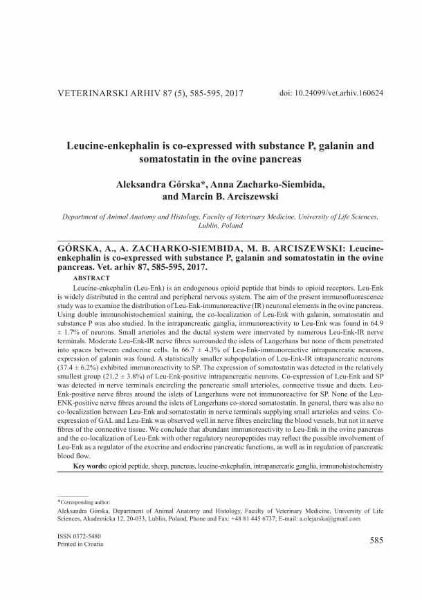
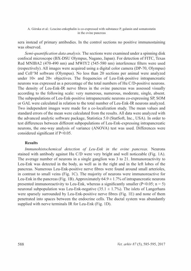
stained with antibody against Hu C/D were very bright and well noticeable (Fig. 1A). The average number of neurons in a single ganglion was 3 to 21. Immunoreactivity to Leu-Enk was detected in the body, as well as in the right and in the left lobes of the pancreas. Numerous Leu-Enk-positive nerve fibres were found around small arterioles, in contrast to small veins (Fig. 1C). The majority of neurons were immunoreactive for Leu-Enk in the pancreas (Fig. 1B). Approximately 64.9 ± 1.7% of intrapancreatic neurons presented immunoreactivity to Leu-Enk, whereas a significantly smaller (P<0.05; n = 5) neuronal subpopulation was Leu-Enk-negative (35.1 ± 1.7%). The islets of Langerhans were sparsely surrounded by Leu-Enk-positive nerve fibres (Fig. 1E) and none of them penetrated into spaces between the endocrine cells. The ductal system was abundantly supplied with nerve terminals IR for Leu-Enk (Fig. 1D).
589Vet. arhiv 87 (5), 585-595, 2017
A. Górska et al.: Leucine-enkephalin is co-expressed with substance P, galanin and somatostatin in the ovine pancreas
Fig. 1. Micrographs showing immunoreactivity to Hu C/D and Leu-Enk in the intrapancreatic ganglia of the ovine pancreas. Neurons stained with an antibody against Hu C/D are presented in Fig. 1A. In the intrapancreatic ganglia (Fig. 1B) the expression of Leu-Enk was observed in numerous intrapancreatic neurons (arrows). The arrowhead marks a neuron lacking Leu-Enk.
Arrows indicate Leu-Enk-positive nerve fibres supplying the small arterioles in the pancreas in Fig. 1C, and ducts in Fig. 1D. Islets-supplying nerve fibres simultaneously displaying Leu-Enk
(arrow Fig. 1E) are present. A-arteriole, V-blood vessel, D-duct, I-islet. Scale bar = 100 μm.
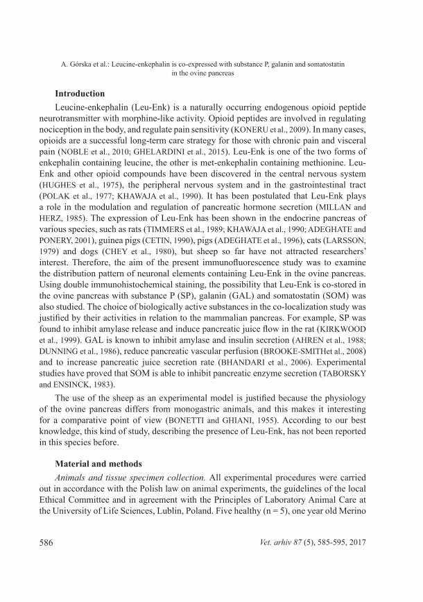
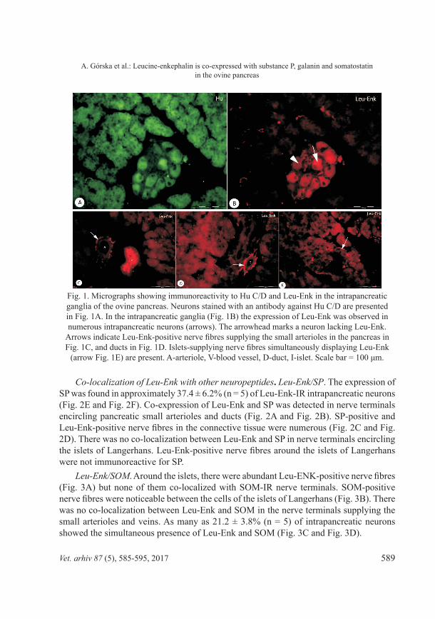
Co-localization of Leu-Enk with other neuropeptides. Leu-Enk/SP. The expression of SP was found in approximately 37.4 ± 6.2% (n = 5) of Leu-Enk-IR intrapancreatic neurons (Fig. 2E and Fig. 2F). Co-expression of Leu-Enk and SP was detected in nerve terminals encircling pancreatic small arterioles and ducts (Fig. 2A and Fig. 2B). SP-positive and Leu-Enk-positive nerve fibres in the connective tissue were numerous (Fig. 2C and Fig. 2D). There was no co-localization between Leu-Enk and SP in nerve terminals encircling the islets of Langerhans. Leu-Enk-positive nerve fibres around the islets of Langerhans were not immunoreactive for SP.
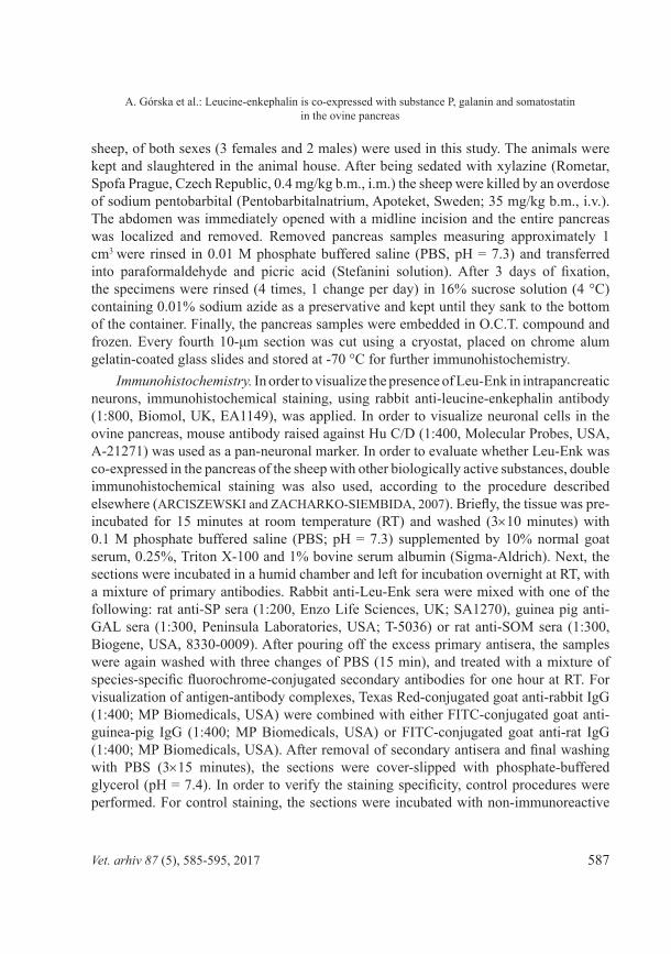
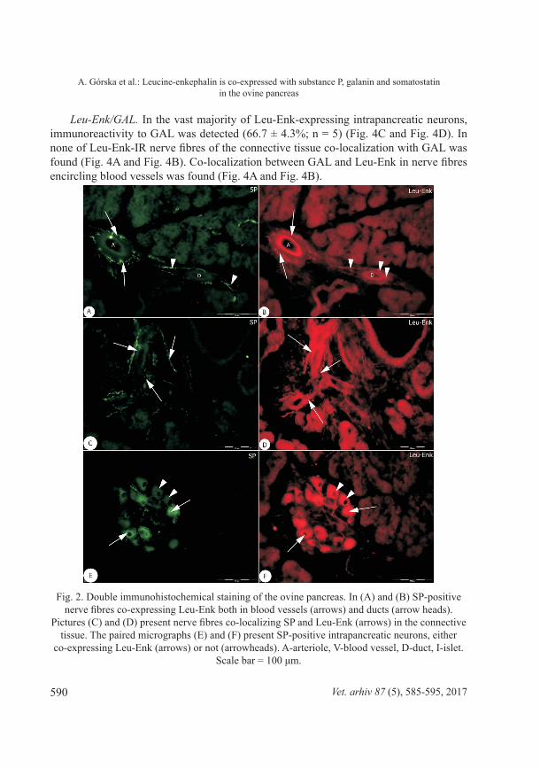
Leu-Enk/SOM. Around the islets, there were abundant Leu-ENK-positive nerve fibres (Fig. 3A) but none of them co-localized with SOM-IR nerve terminals. SOM-positive nerve fibres were noticeable between the cells of the islets of Langerhans (Fig. 3B). There was no co-localization between Leu-Enk and SOM in the nerve terminals supplying the small arterioles and veins. As many as 21.2 ± 3.8% (n = 5) of intrapancreatic neurons showed the simultaneous presence of Leu-Enk and SOM (Fig. 3C and Fig. 3D).
590 Vet. arhiv 87 (5), 585-595, 2017
A. Górska et al.: Leucine-enkephalin is co-expressed with substance P, galanin and somatostatin in the ovine pancreas
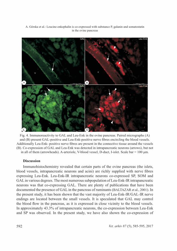
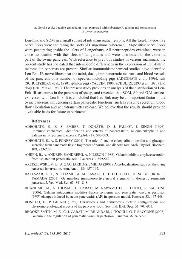
Leu-Enk/GAL. In the vast majority of Leu-Enk-expressing intrapancreatic neurons, immunoreactivity to GAL was detected (66.7 ± 4.3%; n = 5) (Fig. 4C and Fig. 4D). In none of Leu-Enk-IR nerve fibres of the connective tissue co-localization with GAL was found (Fig. 4A and Fig. 4B). Co-localization between GAL and Leu-Enk in nerve fibres encircling blood vessels was found (Fig. 4A and Fig. 4B).
Fig. 2. Double immunohistochemical staining of the ovine pancreas. In (A) and (B) SP-positive nerve fibres co-expressing Leu-Enk both in blood vessels (arrows) and ducts (arrow heads).
Pictures (C) and (D) present nerve fibres co-localizing SP and Leu-Enk (arrows) in the connective tissue. The paired micrographs (E) and (F) present SP-positive intrapancreatic neurons, either
co-expressing Leu-Enk (arrows) or not (arrowheads). A-arteriole, V-blood vessel, D-duct, I-islet. Scale bar = 100 μm.
591Vet. arhiv 87 (5), 585-595, 2017
A. Górska et al.: Leucine-enkephalin is co-expressed with substance P, galanin and somatostatin in the ovine pancreas
Fig. 3. Fluorescent images of the pancreas of sheep, demonstrating immunoreactivities to SOM and Leu-Enk. In micrographs (A) and (B) isle of Langerhans is well observed; all the Leu-Enk-positive nerve fibres are encircling the islet, but the SOM-positive nerve fibres are
penetrating inside isle. Arrow heads in (C) and (D) indicate co-expression of SOM and Leu-Enk in intrapancreatic neurons; arrowheads show SOM-IR neurons, which are not Leu-Enk-positive.
A-arteriole, V-blood vessel, D-duct, I-islet. Scale bar = 100 μm.
592 Vet. arhiv 87 (5), 585-595, 2017
A. Górska et al.: Leucine-enkephalin is co-expressed with substance P, galanin and somatostatin in the ovine pancreas
Fig. 4. Immunoreactivity to GAL and Leu-Enk in the ovine pancreas. Paired micrographs (A) and (B) present GAL-positive and Leu-Enk-positive nerve fibres encircling the blood vessels.
Additionally Leu-Enk- positive nerve fibres are present in the connective tissue around the vessels (B). Co-expression of GAL and Leu-Enk was detected in intrapancreatic neurons (arrows), but not
in all of them (arrowheads). A-arteriole, V-blood vessel, D-duct, I-islet. Scale bar = 100 μm.
DiscussionImmunohistochemistry revealed that certain parts of the ovine pancreas (the islets,
blood vessels, intrapancreatic neurons and acini) are richly supplied with nerve fibres expressing Leu-Enk. Leu-Enk-IR intrapancreatic neurons co-expressed SP, SOM and GAL in various degrees. The most numerous subpopulation of Leu-Enk-IR intrapancreatic neurons was that co-expressing GAL. There are plenty of publications that have been documented the presence of GAL in the pancreas of ruminants (BALTAZAR et al., 2001). In the present study, it has been shown that the vast majority of Leu-Enk-IR/GAL-IR nerve endings are located between the small vessels. It is speculated that GAL may control the blood flow in the pancreas, as it is expressed in close vicinity to the blood vessels. In approximately 43.3% of intrapancreatic neurons, the co-expression between Leu-Enk and SP was observed. In the present study, we have also shown the co-expression of
593Vet. arhiv 87 (5), 585-595, 2017
A. Górska et al.: Leucine-enkephalin is co-expressed with substance P, galanin and somatostatin in the ovine pancreas
Leu-Enk and SOM in a small subset of intrapancreatic neurons. All the Leu-Enk-positive nerve fibres were encircling the islets of Langerhans, whereas SOM-positive nerve fibres were penetrating inside the islets of Langerhans. All neuropeptides examined were in close association with the islets of Langerhans and were distributed in the exocrine part of the ovine pancreas. With reference to previous studies in various mammals, the present study has indicated that interspecific differences in the expression of Leu-Enk in mammalian pancreas are present. Similar immunohistochemical studies have identified Leu-Enk-IR nerve fibres near the acini, ducts, intrapancreatic neurons, and blood vessels of the pancreas of a number of species, including pigs (ADEGHATE et al., 1996), rats (SCHULTZBERG et al., 1980), guinea pigs (YALCIN, 1990; SCHULTZBERG et al., 1980) and dogs (CHEY et al., 1980). The present study provides an analysis of the distribution of Leu-Enk-IR structures in the pancreas of sheep, and revealed that SOM, SP and GAL are co-expressed with Leu-Enk. It is concluded that Leu-Enk may be an important factor in the ovine pancreas, influencing certain pancreatic functions, such as enzyme secretion, blood flow circulation and neurotransmitter release. We believe that the results should provide a valuable basis for future experiments.
ReferencesADEGHATE, E., Z. S. EMBER, T. DONÁTH, D. J. PALLOT, J. SINGH (1996):
Immunohistochemical identification and effects of pancreastatin, leucine-enkephalin and galanin in the porcine pancreas. Peptides 17, 503-509.
ADEGHATE, E., A. S. PONERY (2001): The role of leucine-enkephalin on insulin and glucagon secretion from pancreatic tissue fragments of normal and diabetic rats. Arch. Physiol. Biochem. 109, 223-229.
AHREN, B., A. ANDREN-SANDBERG, A. NILSSON (1988): Galanin inhibits amylase secretion from isolated rat pancreatic acini. Pancreas 3, 559-562.
ARCISZEWSKI, M. B., A. ZACHARKO-SIEMBIDA (2007): A co-localization study on the ovine pancreas innervation. Ann. Anat. 189, 157-167.
BALTAZAR, E. T., N. KITAMURA, M. SASAKI, D. F. COTTRELL, H. M. BOLORON, J. YAMADA (2001): Galanin-like immunoreactive neural elements in domestic ruminants pancreas. J. Vet. Med. Sci. 63, 841-848.
BHANDARI, M., A. THOMAS, C. CARATI, M. KAWAMOTO, J. TOOULI, G. SACCONE (2006): Galanin antagonism modifies hyperenzymemia and pancreatic vascular perfusion (PVP) changes induced by acute pancreatitis (AP) in apossum model. Pancreas 33, 447-448.
BONETTI, D., P. GHIANI (1955): Carnivorous and herbivorous dietetic configurations and physiomorphological aspects of the pancreas. Boll. Soc. Ital. Biol. Sper. 31, 901-903.
BROOKE-SMITH, M. E., C. J. CARATI, M. BHANDARI, J. TOOULI, G. T. SACCONE (2008): Galanin in the regulation of pancreatic vascular perfusion. Pancreas 36, 267-273.
594 Vet. arhiv 87 (5), 585-595, 2017
A. Górska et al.: Leucine-enkephalin is co-expressed with substance P, galanin and somatostatin in the ovine pancreas
CETIN, Y. (1990): Immunohistochemistry of opioid peptides in the guinea pig endocrine pancreas. Cell Tissue Res. 259, 313-319.
CHEY, W. Y., D. H. COY, S. J. KONTUREK, A. V. SCHALLY, J. TASLER (1980): Enkephalin inhibits the release and action of secretin on pancreatic secretion in the dog. J. Physiol. 298, 429-436.
DUNNING, B. E., B. AHREN, R. C. VEITH, G. BOTTCHER, F. SUNDLER, G J. TABORSKY (1986): Galanin: a novel pancreatic neuropeptide. Am. J. Physiol. 251, 127-133.
GHELARDINI, C., L. DI CESARE MANNELLI, E. BIANCHI (2015): The pharmacological basis of opioids. Clin. Cases Miner. Bone Metab. 12, 219-221.
HUGHES, J., T. W. SMITH, H. W. KOSTERLITZ, L. A. FOTHERGILL, B. A. MORGAN, H. R. MORRIS (1975): Identification of two related peptides from the brain with potent opiate agonist activity. Nature, 258, 577-579.
KHAWAJA, X. Z., I. C. GREEN, J. R. THORPE, M. A. TITHERADGE (1990): The occurrence and receptor specificity of endogenous opioid peptides within the pancreas and liver of the rat. Comparison with brain. Biochem. J. 267, 233-240.
KIRKWOOD, K. S., E. H. KIM, X. D. HE, E. Q. CALAUSTRO, C. DOMUSH, S. K. YOSHIMI, E. F. GRADY, J. MAA, N. W. BUNNETT, H. T. DEBAS (1999): Substance P inhibits pancreatic exocrine secretion via a neural mechanism. Am. J. Physiol. 277, 314-320.
KONERU, A, S. SREEMANTULA, R. SHAIK (2009): Endogenous opioids: their physiological role and receptors. Global J. Pharmacol. 3, 149-153.
LARSSON, L. I. (1979): Innervation of the pancreas by substance P, enkephalin, vasoactive intestinal polypeptide and gastrin/CCK immunoractive nerves. J. Histochem. Cytochem. 27, 1283-1284.
MILLAN, M. J., A. HERZ (1985): The endocrinology of the opioids. Int. Rev. Neurobiol. 26, 1-83.NOBLE, M., J. R. TREADWELL, S. J. TREGEAR, V. H. COATES. P. J. WIFFEN, C. AKAFOMO,
K. M. SCHOELLES (2010): Noble, Meredith, ed. “Long-term opioid management for chronic noncancer pain”. Cochrane Database of Systematic Reviews, art no. CD006605
POLAK, J. M., R. S. BLOOM, S. N. SULLIVAN, A. G. E. PEARSE (1977): Enkephalin-like immunoreactivity in the human gastrointestinal tract. Lancet 1, 972-974.
SCHULTZBERG, M., T. HOKFELT, G. NILSSON (1980): Distribution of peptide and catecholamine containing neurons in the gastrointestinal tract of rat and guinea pig: Immunochistohemical studies with antisera to substance P, VIP, encephalin, somatostatin, gastrin/cholecystokinin, neurotensin and dopamine-beta-hydroxylase. Neuroscience 5, 689-744.
TABORSKY, G. J., J. W. ENSINCK (1983): Extraction of somatostatin by the pancreas. Endocrinology 112, 303-307.
TIMMERS, K., N. R. VOYLES, S. WILKINS, O. E. MICHAELIS, S. J. BHATHENA, L. RECANT (1989): Immunoreactive beta-endorphin and met- and leu-enkephalin contents in pancreas and pituitary of corpulent (cp/cp) rats. Int. J. Obes. 13, 337-345.
595Vet. arhiv 87 (5), 585-595, 2017
A. Górska et al.: Leucine-enkephalin is co-expressed with substance P, galanin and somatostatin in the ovine pancreas
YALCIN, C. (1990): Immunohistochemistry of opioid peptides in the guinea pig endocrine pancreas. Cell. Tissue Res. 259, 313-319.
Received: 24 June 2016Accepted: 30 January 2017
________________________________________________________________________________________GÓRSKA, A., A. ZACHARKO-SIEMBIDA, M. B. ARCISZEWSKI: Leucin-enkefalin u pankreasu ovaca ima zajedničku izražajnost s tvari P, galaninom i somatostatinom. Vet. arhiv 87, 585-595, 2017.
SAŽETAKLeucin-enkefalin (Leu-Enk) je endogeni opioidni peptid koji se veže na opioidne receptore. Leu-Enk
je široko rasprostranjen u središnjem i perifernom živčanom sustavu. Cilj ovog istraživanja bio je pomoću imunofluorescencije utvrditi raspodjelu Leu-Enk-imunoreaktivnih (IR) neuronskih elemenata u pankreasu ovaca. Primjenom dvostrukog imunohistokemijskog bojenja, istražena je i zajednička lokalizacija Leu-Enk s galaninom, somatostatinom i tvari P. U intrapankreasnim ganglijima imunoreaktivnost prema Leu-Enku pronađena je u 64,9 ± 1,7% neurona. Male arteriole i kanalni sustav bili su inervirani mnogim Leu-Enk-IR živčanim završetcima. Umjerena Leu-Enk-IR živčana vlakna okruživala su Langerhansove otočiće, ali nijedno od njih nije prodrlo u prostore između endokrinih stanica. U 66,7 ± 4,3% Leu-Enk-imunoreaktivnih intrapankreasnih neurona otkrivena je ekspresija galanina. Statistički niža subpopulacija Leu-Enk-IR intrapankreasnih neurona (37,4 ± 6,2%) pokazala je imunoreaktivnost prema SP. Izražajnost somatostatina otkrivena je u relativno najmanjoj skupini (21,2 ± 3,8%) Leu-Enk-pozitivnih intrapankreasnih neurona. Zajednička izražajnost Leu-Enk i SP otkrivena je u živčanim završetcima koji okružuju male arteriole pankreasa, vezivno tkivo i kanale. Leu-Enk-pozitivna vlakna živaca oko Langerhansovih otočića nisu bila imunoreaktivna na SP. Nijedno od Leu-Enk-pozitivnih živčanih vlakana oko Langerhansovih otočića nije bilo zajednički lokalizirano sa somatostatin-imunoreaktivnim živčanim završetcima. Općenito, nije bilo zajedničke lokalizacije između Leu-Enka i somatostatin živčanih završetaka koji opskrbljuju male arteriole i vene. Zajednička izražajnost GAL i Leu-Enk dobro je vidljiva u živčanim vlaknima koja okružuju krvne žile, ali ne i u živčanim vlaknima vezivnog tkiva. Zaključeno je da obilna imunoreaktivnost Leu-Enk u pankreasu ovaca, te njegova zajednička lokalizacija s drugim regulatornim neuropeptidima, može odražavati moguću uključenost Leu-Enk u regulaciju egzokrine i endokrine funkcije pankreasa odnosno u regulaciju optjecaja krvi u pankreasu.
Ključne riječi: opioidni peptid, ovce, pankreas, leucin-enkefalin, intrapankreasni gangliji, imunohistokemija________________________________________________________________________________________

.