Embed Size (px)
Citation preview
Leukemogenic Mechanisms of MLL Fusion Proteins
by
Jiaying Tan
A dissertation submitted in partial fulfillment of the requirements for the degree of
Doctor of Philosophy (Molecular and Cellular Pathology)
in the University of Michigan 2012
Doctoral Committee: Professor Jay L. Hess, Chair Professor Gregory R. Dressler Professor Tom K. W. Kerppola Assistant Professor Yali Dou Assistant Professor Ivan P. Maillard

© Jiaying Tan 2012
All Rights Reserved

ii
To Mom and Dad

iii
Acknowledgements
This thesis could not have been completed without the ones walking with me
through the past four years. I would like to first thank my advisor, Dr. Jay Hess,
for providing me the opportunity to learn and grow as a young scientist, for
inspiring me to strive for my personal best in research, for encouraging me to
think independently and creatively, and for guiding me to develop and pursue my
scientific interests persistently. I would also like to thank my committee members,
Drs. Yali Dou, Ivan Maillard, Tom Kerppola, and Gregory Dressler for generously
offering their time and invaluable insights during the course.
I am grateful to all the current and previous Hess lab members. It has been such
a pleasure to work with this group and to share the ups and downs along the way.
I wish you all the best! In particular, I would like to thank Dr. Andrew Muntean,
from whom I have learned so much through our productive collaboration and
discussions, and Stephanie Jo, whose incredible diligence and determination has
been a real inspiration. Additionally, a special thank you goes to the Dou lab next
door for their moral and technical support and to the Maillard lab for the delightful
collaboration. Many thanks to our program director Dr. Nicholas Lukacs and his
amazingly enthusiastic assistant Laura Hessler, their strong support and

iv
confidence on me has been tremendously encouraging the entire time. As every
pathology graduate student would have said, Laura, you are beyond super!
I can never thank enough my dear friends for lightening up my life, both the ones
in Michigan and those back in China. Everlasting gratitude goes to Hanyu, Jesse,
Peng, and Haiyan, for hearing me out, for lifting me up, and for walking me
through the rain, never giving up. The most special thank you goes to Jie for
being you and for loving me. It has been an incredible journey for both of us, and
I cannot wait to see where this leads. Finally, no words can adequately express
how deeply grateful and indebted I am to my parents, Xiaoan and Feifei. Your
unwavering support has been with me every step of the way. You encourage me
to follow my heart and to explore my potential. I could have never accomplished
this work without you two. I would never become who I am now without your
unconditional love.
v
Table of Contents
Dedication ......................................................................................................... ii
Acknowledgements ......................................................................................... iii
List of Figures ................................................................................................. vii
List of Abbreviations ....................................................................................... xii
Abstract .......................................................................................................... xiv
Chapter 1 The Role of the PAF Complex in MLL Fusion Protein-Induced
Leukemogenesis ............................................................................ 1
Introduction ..................................................................................... 1
Materials and Methods ................................................................. 13
Results .......................................................................................... 21
Discussion .................................................................................... 52
Chapter 2 The Role of CBX8, a Polycomb Group Protein, in MLL
Fusion Protein-Induced Leukemogenesis .................................... 60
Introduction ................................................................................... 60
Materials and Methods ................................................................. 72
Results .......................................................................................... 80
Discussion .................................................................................. 118
Chapter 3 Concluding Remarks .................................................................. 133
References .................................................................................................. 136
vi
List of Figures
Figure 1.1 Schematic of Wild-Type MLL and MLL-Rearranged
Oncoproteins ................................................................................................... 7
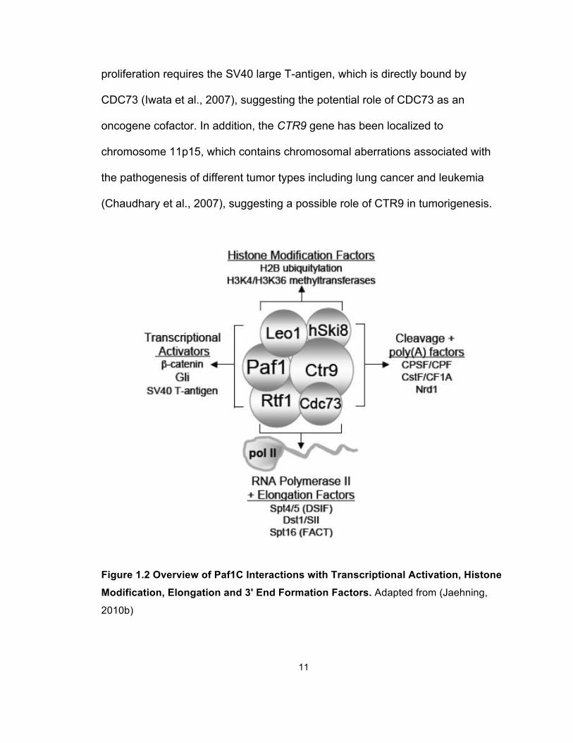
Figure 1.2 Overview of Paf1C Interactions with Transcriptional Activation,
Histone Modification, Elongation and 3' End Formation Factors ................... 11
Figure 1.3 MLL Fragment Interacts with the PAF Complex (PAFc) .............. 22
Figure 1.4 MLL and MLL-AF9 Fragments Interact with PAFc in a
DNA-Independent Manner ............................................................................. 24
Figure 1.5 MLL and MLL Fusion Proteins Interact with PAFc in a
DNA-Independent Manner ............................................................................. 25
Figure 1.6 Schematic Diagram of MLL and Bacterially Purified
CxxC-RD2 and RD2 Regions ........................................................................ 26
Figure 1.7 PAF1 and CTR9 Bind Directly to the CxxC-RD2 Region of MLL . 27
Figure 1.8 Amino Acids within the RD2 Region are Necessary for
MLL-PAFc Interaction .................................................................................... 29
Figure 1.9 Pre-CxxC and RD2 Regions are Both Involved in MLL-PAFc
Interaction ...................................................................................................... 30
Figure 1.10 PAFc Synergizes with MLL and MLL-AF9 to Augment
Transcriptional Activity ................................................................................... 32
Figure 1.11 PAFc Synergizes with MLL-AF9 to Promote Transcription ........ 33
vii
Figure 1.12 PAFc Interaction Region on RD2 Is Necessary for Bone
Marrow Transformation (BMT) by MLL-AF9 .................................................. 35
Figure 1.13 p-Iodonitro Tetrazolium Violet (INT)-Stained Colonies after
Three Rounds of Colony Replating ............................................................... 36
Figure 1.14 Morphology of Primary Bone Marrow Transduced by
MLL-AF9 Fusion Proteins .............................................................................. 37
Figure 1.15 Hoxa9 Expression in Cells Collected after the Third
Round of BMT Assays ................................................................................... 37
Figure 1.16 Schematic Diagram for MLL-AF9 BMT Assay with
shRNA-Mediated Knockdown of Cdc73 and Ctr9 ......................................... 38
Figure 1.17 Knockdown of Cdc73 or Ctr9 Impairs MLL-AF9-Induced Bone
Marrow Transformation.................................................................................. 39
Figure 1.18 RT-PCR for Hoxa9 and Meis1 Expression in MLL-ENL and
E2A-HLF Cell lines ........................................................................................ 40
Figure 1.19 PAFc and MLL-ENL Bind across the Hoxa9 Locus .................... 41
Figure 1.20 PAFc and MLL-ENL Bind at the Meis1 Locus ............................ 42
Figure 1.21 Knockdown of PAFc Reduces MLL-AF9-Induced
Transactivation .............................................................................................. 43
Figure 1.22 Simultaneous siRNA-Mediated Knockdown of PAFc
(siCTR9, siLEO1, siPAF1 and siCDC73)....................................................... 44
Figure 1.23 Knockdown of PAFc Reduces MLL Binding to the
HOXA9 Locus ................................................................................................ 45
Figure 1.24 Differentiation of Hoxa9ER Cell Line Induced by
viii
4-OHT Withdrawal ......................................................................................... 47
Figure 1.25 PAFc Is Downregulated during Differentiation of
Hoxa9ER Cells Induced by 4-OHT Withdrawal ............................................. 48
Figure 1.26 PAFc Is Downregulated during Differentiation of
HL-60 Cells Induced by PMA Treatment ....................................................... 49
Figure 1.27 PAFc Overexpression in THP-1 Cells Leads to
Resistance to Differentiation Induction by PMA Treatment ........................... 50
Figure 1.28 Structure of the CxxC Domain and Flanking Sequences ........... 51
Figure 1.29 Functional Implication of the MLL-PAFc Interaction ................... 53
Figure 1.30 Schematic of a Potential Mechanism of MLL-Rearranged
Leukemogenesis ........................................................................................... 55
Figure 2.1 Diversity of PRC1 and PRC2 Complexes Formed by
Vertebrate PcG Proteins ................................................................................ 61
Figure 2.2 Coordinated Epigenetic Silencing Activity of PcG Complexes ..... 64
Figure 2.3 Established and Putative Connections of Tip60 to
Oncogenesis .................................................................................................. 71
Figure 2.4 CBX8 Specifically Interacts with MLL-AF9 at the
C-terminal Domain (CTD) .............................................................................. 81
Figure 2.5 Specific Disruption of the CBX8 Interaction Does not
Affect the Interaction between DOT1L and MLL-AF9 .................................... 83
Figure 2.6 CBX8/MLL-AF9 Interaction Is Essential for
MLL-AF9 Leukemic Transformation .............................................................. 85
ix
Figure 2.7 Cbx8 Knockdown by shRNA Impairs MLL-AF9 Leukemic
Transformation............................................................................................... 86
Figure 2.8 Strategy of Conditional Knockdown of Cbx8 in Mice .................... 87
Figure 2.9 Experimental Scheme for the BMT Assays with Cbx8
Excision in Primary BM from Cbx8 f/f; Cre+ and Cbx8 f/f; Cre- Mice ................ 88
Figure 2.10 Cbx8 Is Required for Both Initiation and Maintenance of
MLL-AF9 Leukemic Transformation .............................................................. 89
Figure 2.11 Cbx8 Is Required for MLL-AF9-Induced
Leukemogenesis in Vivo ................................................................................ 91
Figure 2.12 Cbx8 Is Required for the Leukemic Transformation by
MLL-ENL, but not for That by E2A-HLF ........................................................ 93
Figure 2.13 Western Blot Showing the Cbx8 Protein Level upon 4-OHT
Treatment in Primary BM Transformed by MLL-AF9, MLL-ENL or
E2A-HLF ........................................................................................................ 94
Figure 2.14 CBX8 Is Crucial for Proliferation and Survival of
MLL-AF9-transformed Leukemic Cells .......................................................... 94
Figure 2.15 Apoptosis Analysis of MLL-AF9 Leukemic Cell in the
Presence or Absence of Cbx8 ....................................................................... 95
Figure 2.16 Cell Cycle Analysis of MLL-AF9 Leukemic Cell in the
Presence or Absence of Cbx8 ....................................................................... 96
Figure 2.17 Cbx8 Is required for Hoxa9 Upregulation in
MLL-AF9-Transformed Primary BM Cells ..................................................... 98
Figure 2.18 Cbx8 Is required for Hoxa9 Upregulation in Human and
x
Murine MLL-AF9-Transformed Leukemic Cell Lines ..................................... 99
Figure 2.19 CBX8 Is Crucial for MLL-AF9-Induced Transcriptional
Activation ..................................................................................................... 101
Figure 2.20 Cbx8 Is not Required for the collective localization of
MLL-AF9 and WT AF9 at the Hoxa9 Promoter ........................................... 103
Figure 2.21 Ring1b Knockdown Does not Recapitulate the Effects of
Cbx8 Knockdown on MLL-AF9 Leukemic Transformation .......................... 105
Figure 2.22 Ring1b Knockdown Does not Recapitulate the Effects of
Cbx8 Knockdown on MLL-AF9-Induced Transactivation of the
Target Promoters ......................................................................................... 106
Figure 2.23 Bmi1 Knockdown Does not Recapitulate the Effects of
Cbx8 Knockdown in MLL-AF9 Leukemic Transformation ........................... 107
Figure 2.24 Bmi1 Knockdown Does not Recapitulate the Effects of
Cbx8 Knockdown on MLL-AF9-Induced Transactivation of the
Target Promoters ......................................................................................... 108
Figure 2.25 CBX8 Interacts with TIP60, Whose Downregulation
Phenocopies the Effects of Cbx8 Knockdown on MLL-AF9 Leukemic
Transformation............................................................................................. 110
Figure 2.26 Tip60 Knockdown Phenocopies the Effects of Cbx8
Knockdown in MLL-AF9 Leukemic Transformation ..................................... 111
Figure 2.27 Cbx8 Affects Tip60 Binding, Whose Downregulation
Phenocopies the Effects of Cbx8 Knockdown on MLL-AF9-Induced
Transactivation of the Target Promoters ..................................................... 112
xi
Figure 2.28 Cbx8-Depleted Mice Shows No Abnormality in the CBC
Analysis of Peripheral Blood ........................................................................ 114
Figure 2.29 Cbx8-Depleted Mice Shows Normality Cellularity of Major
Hematopoietic Organs and Mature Hematopoietic Populations .................. 115
Figure 2.30 Cbx8-Depleted Mice Shows No Abnormality in
Maintaining the Primitive Long-Term Hematopoietic Stem Cell
(LT-HSC) Population ................................................................................... 116
Figure 2.31 Cbx8-Depleted Mice Shows Normal Stem and Progenitor
Cell Function ................................................................................................ 117
Figure 2.32 Schematic Model Illustrating Role of CBX8 in Promoting
MLL-AF9-Induced Leukemogenesis ............................................................ 118
Figure 2.33 Effect of Cbx8 Deletion on Leukemogenic Transformation
Induced by MLL-GAS7 ................................................................................ 120
Figure 2.34 Ink4a/Arf Expression Is not Activated upon Cbx8
Downregulation or Depletion or upon Tip60 Downregulation ...................... 122
Figure 2.35 Comparison of the Key Enzymatic Components between
Wild-Type MLL and Common MLL-Fusion Proteins .................................... 125
Figure 2.36 Knockdown of Tip60 Does not Affect the Transformation of
E2A-HLF Leukemic Cells ............................................................................ 131
xii
List of Abbreviations
AEP, AF4 family/ENL family/P-TEFb;
ALL, acute lymphoblastic leukemia;
AML, acute myeloid leukemia;
ATM, ataxia telangiectasia mutated;
CBP, Creb-binding protein;
CBX, chromobox homolog;
CDK, cyclin-dependent kinase;
C/EBPα, CCAAT-enhancer-binding protein α
ChIP, chromatin immunoprecipitation;
CTD, carboxy-terminal domain;
DOT1L, disruptor of telomeric silencing 1-like;
EAP, Elongation Assisting Proteins or ENL-associated Proteins;
GM-CSF, granulocyte macrophage-colony stimulating factor;
HAT, histone acetyl transferase;
HDAC, histone deacetylase;
HMT, histone methyl transferase;
HOX, homeobox;
IL, interleukin;
LPS, lipopolysaccharide;
xiii
LT-HSC, long term-hematopoietic stem cell;
MLL, Mixed Lineage Leukemia;
MOF, males absent on the first;
MYST, Moz, Ybf2/Sas3, Sas2, Tip60;
NF-κB, nuclear factor kappa-light-chain-enhancer of activated B cells;
NuA4, nucleosome acetyltransferase of histone H4;
PAFc, polymerase associated factor complex;
Pc, polycomb;
PcG, polycomb group;
PCL, polycomblike;
PHD, plant homeodomain;
PMA, phorbol 12-myristate 13-acetate;
PRC, polycomb repressive complex;
PTD, partial tandem duplication;
RB, retinoblastoma;
RBBP, retinoblastoma-binding protein;
RNAP II, RNA polymerase II;
SCF, stem cell factor;
SET, Su(var), Enhancer of zeste, Trithorax;
STAT, signal transducers and activators of transcription
TEL, translocated ETS leukemia;
TIP60, HIV Tat-interacting protein of 60 kDa;
TrxG, trithorax group;
xiv
Abstract
Translocations that generate MLL fusion proteins are common causes of human
acute leukemias. Aberrant target gene activation is the primary driver of MLL-
rearranged leukemogenesis, but the underlying mechanisms remain poorly
understood. In the present study, we identified two partners of the MLL fusion
complex, PAFc and CBX8, which can interact with a common MLL fusion protein,
MLL-AF9, through the N-terminal MLL part and the C-terminal AF9 part,
respectively. Our data demonstrate that both PAFc and CBX8 are required for
MLL-AF9-mediated transcriptional upregulation and leukemic transformation. The
molecular mechanisms for their requirements in the leukemogenic process are
different. By chromatin immunoprecipitation, we show that PAFc binds at the
MLL target gene (e.g., Hoxa9 and Meis1) loci in MLL-AF9 leukemic cells. Its
binding contributes to the recruitment of MLL-AF9 and augments MLL-AF9-
mediated transcriptional activation. In contrast, CBX8 promotes the
transcriptional activation of MLL-AF9 target genes, not by regulating MLL-AF9
recruitment but may through interacting with other cofactors, such as the histone
acetyltransferase TIP60, whose enzymatic activity could potentially facilitate
gene transcription. Furthermore, although CBX8 is essential for MLL-AF9
transformation, it is not required for normal hematopoiesis, as shown by the
xv
normal viability of hematopoiesis in Cbx8-deficient mice. This suggests targeting
CBX8 may be of therapeutic value in treating MLL-rearranged leukemias. In
conclusion, our findings demonstrate that both PAFc and CBX8 play essential
roles in MLL-AF9-mediated transcriptional regulation and leukemogenesis.
1
Chapter 1
The Role of the PAF Complex in MLL Fusion Protein-Induced
Leukemogenesis
Introduction
Mixed Lineage Leukemia
Mixed lineage leukemia is a highly aggressive hematopoietic malignancy that
occurs predominantly in pediatric patients. In contrast to other types of acute
leukemia, mixed lineage leukemia stands out as a particular clinical challenge
because patients with this disease present with an extremely dismal prognosis, in
part due to their poor responses to conventional therapeutic treatment, such as
chemotherapy (Balgobind et al., 2011; Slany, 2009). Mixed lineage leukemia
possesses unique clinical features, which were first described in the early 1980s.
At the time, physicians realized that certain subsets of patients initially diagnosed
with acute myeloid leukemia (AML) or acute lymphoblastic leukemia (ALL) fared
far worse than others, especially some newborn and infant patients with similar
clinical aspects. By fluorescent activated cell sorting (FACS) analysis, it was
found that the leukemic blasts from these specific cases often expressed surface
markers of both the myeloid and lymphoid lineages, although the
immunophenotype may be more consistent with one or the other in a particular
case (Krivtsov and Armstrong, 2007; Slany, 2009). In addition, a complete
2
lineage switch was even observed during treatment, meaning an ALL case could
relapse as AML, therefore delivering the term of mixed lineage leukemia (Stass
et al., 1984).
Although chromosomal translocations causing the rearrangements of the 11q23
locus were recognized as typical characteristics of mixed lineage leukemia soon
after these genetic lesions had been observed, it was not until the early 1990s
that the gene spanning this region was successfully cloned by four individual
groups, which is now known as the mixed lineage leukemia (MLL) gene (Djabali
et al., 1992; Gu et al., 1992; Tkachuk et al., 1992; Ziemin-van der Poel et al.,
1991). MLL rearrangements generate a large variety of oncogenic MLL fusion
proteins. To date, more than 60 different fusion partners have been identified,
among which the most common ones are nuclear proteins with transcriptional
activating activity (Krivtsov and Armstrong, 2007; Monroe et al., 2010; Yokoyama
et al., 2010). In ALL, the most common translocations are t(11;19) and t(4;11),
resulting in the fusion proteins MLL-ENL and MLL-AF4, respectively. In contrast,
the t(9;11) translocation, resulting in the MLL-AF9 fusion protein, is more
frequently found in AML. In addition to the nuclear translocation partners, another
class of MLL fusion partners consists of cytoplasmic proteins that contain
dimerization domains, such as AF6. Dimerization of these MLL fusion proteins
leads to potent transcriptional activation and is essential for their leukemogenic
capacity; however, the detailed leukemogenic mechanism remains elusive
(Martin et al., 2003; So et al., 2003). MLL-related translocations are also
commonly observed in secondary acute leukemias after topoisomerase inhibitor
3
treatment (Felix, 1998). In addition, around 8% of AML patients with normal
cytogenetic features harbor internal tandem duplications of partial MLL N-
terminal sequence, known as MLL-PTD (Figure 1.1). Overall, genetic lesions in
the MLL gene are associated with more than 70% of infant leukemias and
approximately 10% adult leukemias (Krivtsov and Armstrong, 2007).
Wild-Type MLL
The MLL gene encodes a histone methyltransferase containing a C-terminal SET
domain that catalyzes the methylation of histone H3 lysine 4 (H3K4), a histone
modification commonly associated with gene activation (Milne et al., 2002;
Nakamura et al., 2002; Strahl et al., 1999). MLL was found to be part of a large
chromatin-modifying complex that promotes transcription activation through
histone methyltransferase and histone acetyltransferase activities (Nakamura et
al., 2002). During the formation of this macromolecular complex, the MLL protein
is cleaved by an aspartic protease named taspase into an N-terminal fragment
(MLLN) and a C-terminal subunit (MLLC) (Hsieh et al., 2003a; Hsieh et al., 2003b;
Takeda et al., 2006; Yokoyama et al., 2002). On the one hand, the MLLN
fragment contains several functional regions that are considered essential for
correct localization of the MLL complex. On the other hand, the MLLC subunit
associates with at least four proteins, including MOF, WDR5, ASH2L and RBBP5,
to modify chromatin structure, thereby facilitating transcription activation. Among
these critical interacting partners, MOF neutralizes charges on histones by
depositing a site-specific acetylation mark on histone H4 lysine 16, potentially
decondensing chromatin for efficient transcription (Slany, 2009). WDR5, ASH2L
4
and RBBP5 form a common structural platform, which stabilizes the functionally
active configuration of the catalytic MLLC fragment. In particular, WDR5 mediates
MLLC interactions both with this platform and with the histone substrate, which in
turn achieves the full H3K4 methyltransferase activity of the MLL complex (Dou
et al., 2006; Ruthenburg et al., 2006; Schuetz et al., 2006; Southall et al., 2009).
In summary, the MLLN and MLLC fragments, in coordination with the other MLL
complex components, modulate the chromatin structure at MLL target loci,
thereby mediating transcriptional regulation of downstream genes.
Major MLL Targets – HOX Genes
Based on the sequence similarity, MLL is a human homolog of the Drosophila
trithorax (TrxG) protein originally identified in genetic screens as “anti-silencers”
counteracting the action of Polycomb group (PcG) proteins that represses Hox
gene expression (Mills, 2010). Consistent with the role of Drosophila TrxG
proteins, in mice, Mll is required for normal embryonic development through
maintaining the proper Hox gene expression pattern (Yu et al., 1995). Several
studies demonstrated that it also plays a central role in regulating hematopoietic
stem cell self-renewal and progenitor expansion (Jude et al., 2007; McMahon et
al., 2007). Notably, Hox gene expression is dynamically regulated and
functionally important in these processes (He et al., 2011).
Hox genes encode a large family of transcription factors that are evolutionarily
conserved among metazoans. They play crucial roles during development by
regulating a number of important physiological processes, including apoptosis,
receptor signaling, cell motility, angiogenesis and hematopoiesis (Shah and
5
Sukumar, 2010). In human, a total of 39 HOX genes have been identified. They
are linked in as four separate clusters, HOXA, HOXB, HOXC and HOXD, located
on chromosomes 7, 17, 12 and 2, respectively (Ansari and Mandal, 2010). Based
on sequence similarities and their positions within the clusters, they are further
classified into 13 paralogue groups arranging from position 1 to 13 in a 3’-5’
direction (He et al., 2011; Krumlauf, 1994).
This clustered organization is of particular interest because it correlates with the
temporal and spatial expression patterns of Hox genes, meaning that the 3’ Hox
genes are expressed first and are more restricted to the anterior region of the
embryo, whereas the 5’ Hox genes are expressed sequentially later and more
caudally (He et al., 2011). Mutation of single Hox genes does not cause dramatic
alterations in morphogenesis in vertebrates, suggesting functional redundancy
within the Hox family. Nevertheless, the expression of specific Hox genes and
combinations of Hox gene products vary at different stages of development,
indicating that proper regulation and maintenance of Hox gene expression are
essential mechanisms governing developmental processes. For instance, Hox1-6
genes are maximally expressed in hematopoietic stem cells, whereas Hox7-13
genes are present in committed progenitors. During definitive hematopoiesis,
Hox gene expression decreases as differentiation proceeds. In particular,
previous studies have shown that 3’ HoxA and HoxB cluster genes are
preferentially expressed in primitive hematopoietic stem cells (HSCs), relative to
their levels in the HSC-low or HSC-depleted subpopulations (Guo et al., 2003;
Pineault et al., 2002). Moreover, in hematopoietic cells, Hox genes are
6
expressed in a lineage-specific manner, with most HoxB and certain HoxC genes
selectively expressed in cell lines showing erythroid characteristics and certain
HoxA genes predominantly expressed in myeloid-lineage cell lines (Lawrence et
al., 1996). More specifically, populations enriched for myeloid progenitors
preferentially express 5’ Hox genes, such as HoxA9, HoxB9 and HoxA10, and
these genes are coordinately activated in myeloid leukemia cells (Celetti et al.,
1993).
Leukemogenic MLL Fusion Proteins
The most striking property of leukemogenic MLL fusion proteins is the significant
diversity of this family. While the oncogenic mechanisms are likely to differ in
detail, all rearranged forms of MLL upregulate expression of certain HOX genes
and their cofactors, including HOXA9 and MEIS1, which is critical for
leukemogenic transformation (Armstrong et al., 2002; Ayton and Cleary, 2003;
Kumar et al., 2004). Although it is well established that constitutive activation of
these downstream targets, particularly HOXA9, is a key feature of MLL leukemia
pathogenesis, the molecular mechanisms governing the aberrant HOX gene
activation have not been defined (Sitwala et al., 2008; Yokoyama and Cleary,
2008).
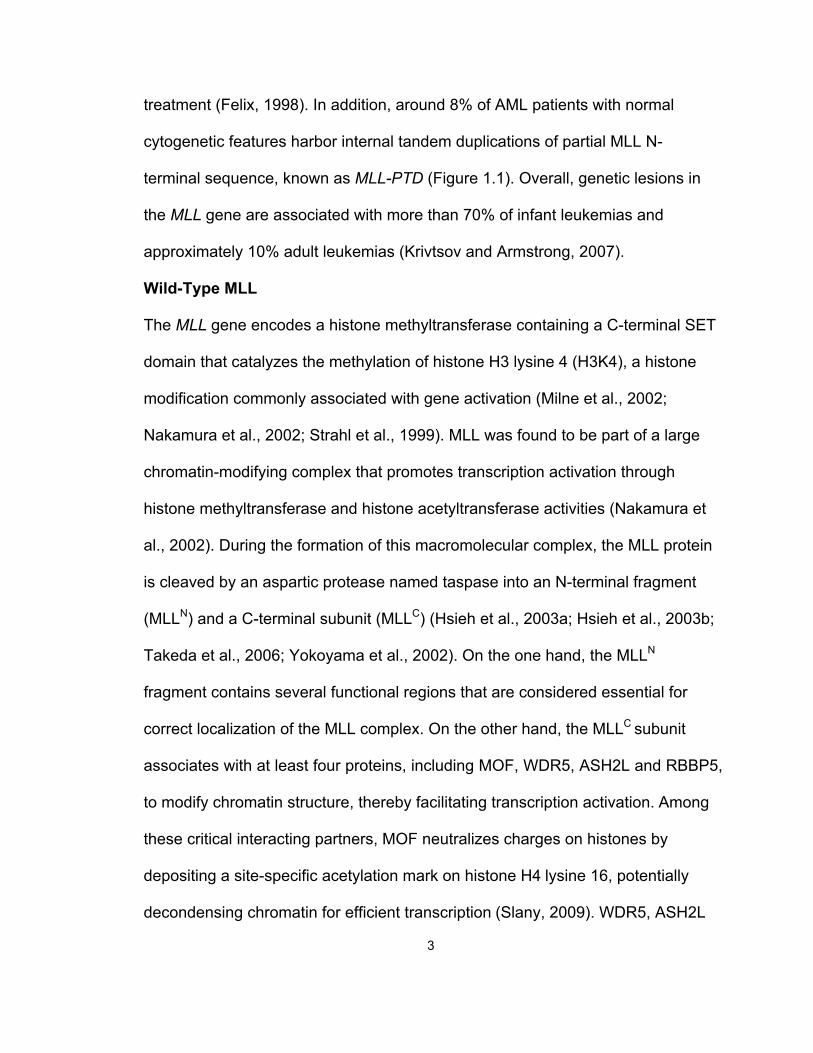
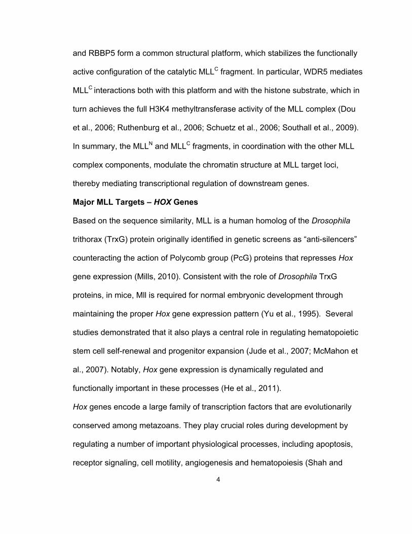
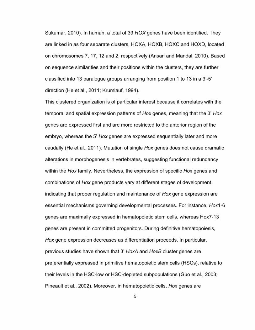
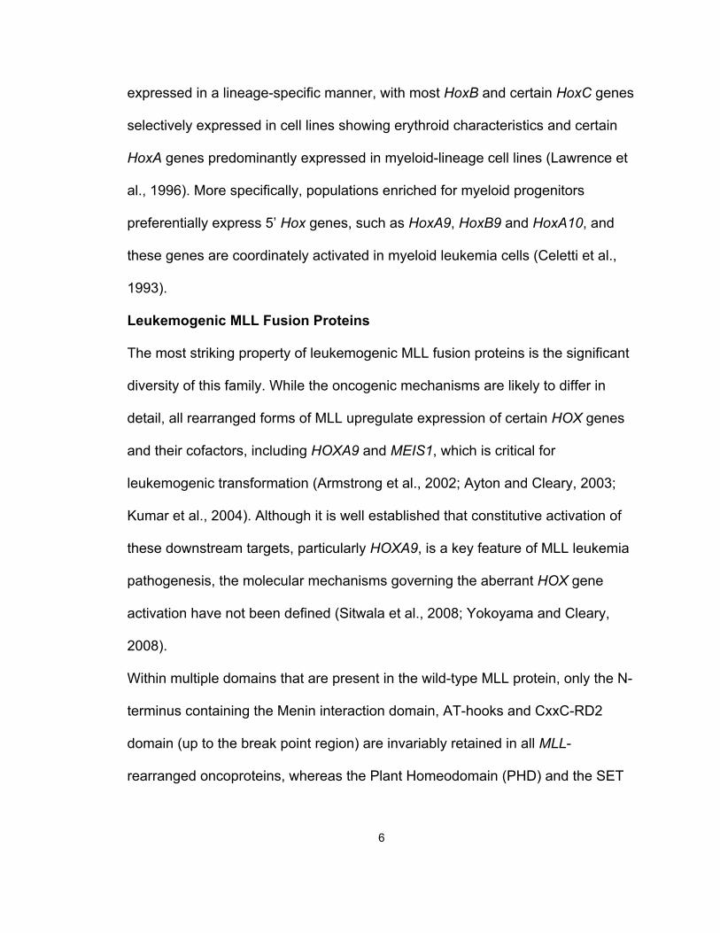
Within multiple domains that are present in the wild-type MLL protein, only the N-
terminus containing the Menin interaction domain, AT-hooks and CxxC-RD2
domain (up to the break point region) are invariably retained in all MLL-
rearranged oncoproteins, whereas the Plant Homeodomain (PHD) and the SET
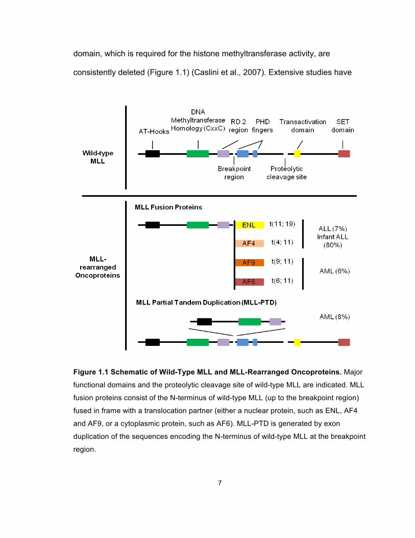
7
domain, which is required for the histone methyltransferase activity, are
consistently deleted (Figure 1.1) (Caslini et al., 2007). Extensive studies have
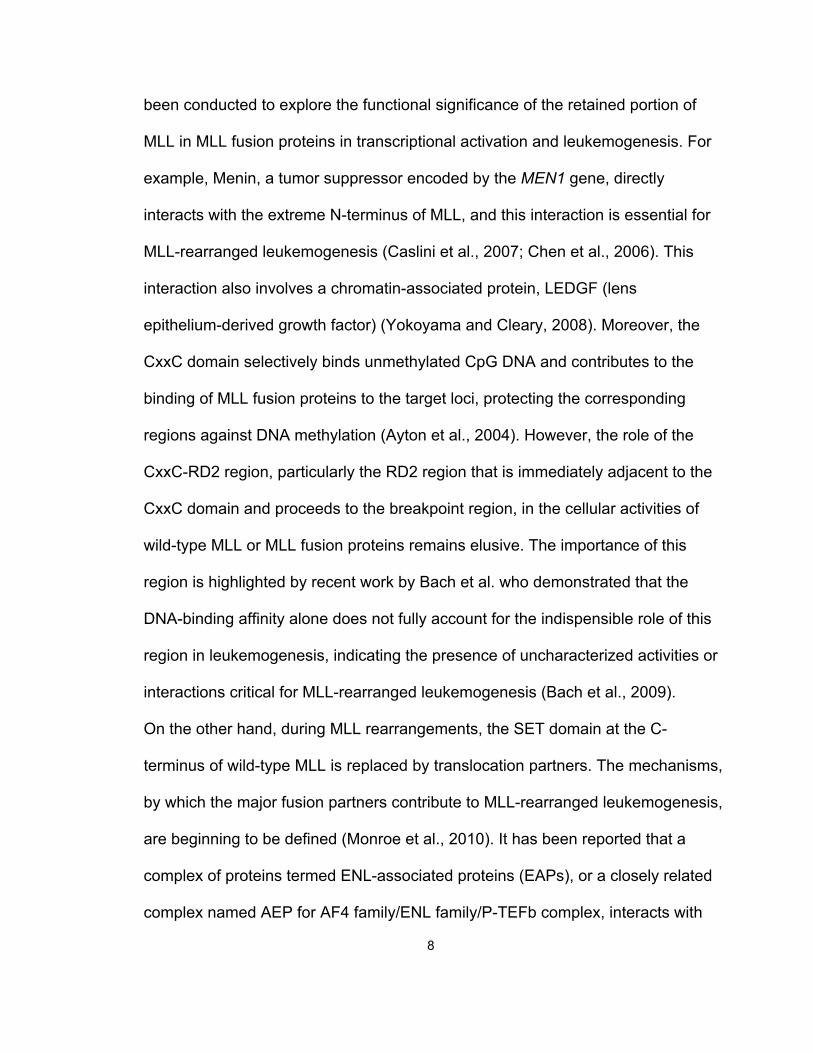
Figure 1.1 Schematic of Wild-Type MLL and MLL-Rearranged Oncoproteins. Major
functional domains and the proteolytic cleavage site of wild-type MLL are indicated. MLL
fusion proteins consist of the N-terminus of wild-type MLL (up to the breakpoint region)
fused in frame with a translocation partner (either a nuclear protein, such as ENL, AF4
and AF9, or a cytoplasmic protein, such as AF6). MLL-PTD is generated by exon
duplication of the sequences encoding the N-terminus of wild-type MLL at the breakpoint
region.
8
been conducted to explore the functional significance of the retained portion of
MLL in MLL fusion proteins in transcriptional activation and leukemogenesis. For
example, Menin, a tumor suppressor encoded by the MEN1 gene, directly
interacts with the extreme N-terminus of MLL, and this interaction is essential for
MLL-rearranged leukemogenesis (Caslini et al., 2007; Chen et al., 2006). This
interaction also involves a chromatin-associated protein, LEDGF (lens
epithelium-derived growth factor) (Yokoyama and Cleary, 2008). Moreover, the
CxxC domain selectively binds unmethylated CpG DNA and contributes to the
binding of MLL fusion proteins to the target loci, protecting the corresponding
regions against DNA methylation (Ayton et al., 2004). However, the role of the
CxxC-RD2 region, particularly the RD2 region that is immediately adjacent to the
CxxC domain and proceeds to the breakpoint region, in the cellular activities of
wild-type MLL or MLL fusion proteins remains elusive. The importance of this
region is highlighted by recent work by Bach et al. who demonstrated that the
DNA-binding affinity alone does not fully account for the indispensible role of this
region in leukemogenesis, indicating the presence of uncharacterized activities or
interactions critical for MLL-rearranged leukemogenesis (Bach et al., 2009).
On the other hand, during MLL rearrangements, the SET domain at the C-
terminus of wild-type MLL is replaced by translocation partners. The mechanisms,
by which the major fusion partners contribute to MLL-rearranged leukemogenesis,
are beginning to be defined (Monroe et al., 2010). It has been reported that a
complex of proteins termed ENL-associated proteins (EAPs), or a closely related
complex named AEP for AF4 family/ENL family/P-TEFb complex, interacts with
9
the major MLL fusion partners AF9, ENL and AF4 (Lin et al., 2010; Muntean et
al., 2010; Yokoyama et al., 2010). The EAP complex includes both the common
MLL fusion partners, such as AF9 and ENL, and also the histone
methyltransferase DOT1L and the P-TEFb complex (consisting of CDK9 and
cyclin T1), both of which positively regulate transcription elongation (Krivtsov et
al., 2008; Mueller et al., 2007). Meanwhile, other investigators have described an
H3K79 methyltransferase complex, DotCom, which contains several common
MLL fusion partners, including AF9, ENL and AF10, that play a positive role in
leukemogenesis (Mohan et al., 2010a). The components of these complexes
partially overlap, suggesting that they may share certain mechanisms that
contribute to MLL-rearranged leukemogenesis (Mohan et al., 2010b; Mueller et
al., 2007). Notably, the interaction partners of MLL fusion proteins are distinctive
from the components of the wild-type MLL complex, suggesting that they may
activate transcription through differential mechanisms.
PAFc
The Polymerase Associated Factor complex (PAFc) is a multi-protein complex,
with the core components of PAF1, LEO1, CDC73, CTR9, WDR61 (also known
as hSki8), and, in some cases, Rtf1 (Jaehning, 2010a; Kim et al., 2010;
Rozenblatt-Rosen et al., 2005; Zhu et al., 2005a). Increasing evidence has
revealed that PAFc plays important roles in a wide range of biological processes,
including H2B monoubiquitination, the initiation, elongation and termination of
gene transcription, cell cycle regulation, and mRNA processing (Figure 1.2)
(Chaudhary et al., 2007; Kim et al., 2009; Kim and Roeder, 2009).
10
In addition, several components of PAFc are known to play important roles in
carcinogenesis. For instance, in a study to identify genes involved in pancreatic
tumor progression, PAF1 was found to be overexpressed as a result of a double
minute amplification involving chromosome 19q13 (Batra et al., 1991). Along this
line, overexpression of PAF1 results in transformation of NIH3T3 cells (Moniaux
et al., 2006). These findings suggest that PAF1 may function as an oncogene
promoting tumorigenesis. In contrast, CDC73 has been implicated to serve as
both a tumor suppressor and an “aider and abettor” of an oncoprotein in a
context-dependent manner. One the one hand, most notably mutations in CDC73,
encoded by HRPT-2 (hereditary hyperparathyroidism type 2), are responsible for
the familial hyperparathyroidism-jaw tumor (HPT-JT) syndrome (Szabo et al.,
1995). HPT-JT is an autosomal dominant disorder associated with
hyperparathyroidism (HPT) and a high incidence of parathyroid adenomas,
hyperplasias and carcinomas as well as renal abnormalities and uterine tumors
(Newey et al., 2009). Some mutations in HRPT-2 are predicted to lead to loss of
function due to premature termination. The chromosome 1q25-q31 region
spanning HRPT-2 frequently undergoes loss of heterozygosity in tumors arising
in HPT-JT patients, suggesting that CDC73 functions as a tumor suppressor
(Newey et al., 2009). Consistent with this role, overexpression of wild-type
CDC73, but not a mutant form found in HPT, blocks cell proliferation and inhibits
the cell cycle regulator cyclin D1 (Woodard et al., 2005). On the other hand,
CDC73 overexpression in 293T and COS7 cells increases S-phase entry and
promotes cellular proliferation. This induction of CDC73-mediated cell-dependent
11
proliferation requires the SV40 large T-antigen, which is directly bound by
CDC73 (Iwata et al., 2007), suggesting the potential role of CDC73 as an
oncogene cofactor. In addition, the CTR9 gene has been localized to
chromosome 11p15, which contains chromosomal aberrations associated with
the pathogenesis of different tumor types including lung cancer and leukemia
(Chaudhary et al., 2007), suggesting a possible role of CTR9 in tumorigenesis.
Figure 1.2 Overview of Paf1C Interactions with Transcriptional Activation, Histone
Modification, Elongation and 3' End Formation Factors. Adapted from (Jaehning,
2010b)
12
Previous studies have demonstrated that the yeast PAF complex is required for
the recruitment of the yeast Set1 methyltransferase complex, termed COMPASS,
to RNA polymerase II. It is also indispensable for both COMPASS-mediated
histone H3K4 and Dot1L-mediated H3K79 methylation (Krogan et al., 2003;
Rozenblatt-Rosen et al., 2005). Given that the MLL complex is a human homolog
of COMPASS, and that the only H3K79 methyltransferase in human, hDOT1L, is
recruited by MLL fusion proteins, it is both mechanistically important and
therapeutically intriguing to investigate whether PAFc is physically and
functionally associated with the MLL complex, as well as the leukemogenic MLL
fusion protein complexes.
13
Materials and Methods
Cell Culture
293 and HeLa cells were cultured in Dulbecco’s modified Eagle’s medium
(DMEM) supplemented with 10%FBS and 1X non-essential amino acids. MLL-
ENL and E2A-HLF cells were cultured in Iscove's modified Dulbecco's medium
(IMDM) supplemented with 15% fetal calf serum (FBS) (Stem Cell Technologies).
Hoxa9-ER cells were cultured in IMDM supplemented with 15% FBS and 0.1%
IL3. Plat-E cells were cultured in DMEM supplemented with 10% FBS. HL-60,
THP-1, KOPN8 and K562 cells were cultured in RPMI-1640 medium
supplemented with 10% FBS. Differentiation of HL-60 and THP-1 cells was
induced by 10 nM PMA treatment.
Luciferase Assay
293 cells were transiently transfected with MSCV MLL-AF9 (and derivatives),
CMV-Renilla, and Hoxa9-LUC (or Myc-E box-LUC) constructs using FuGene 6
(Roche) according to manufacturer’s instructions. Cells were then serum starved
in 0.5% FBS in OPTI-MEM media for 48 hours. Luciferase assays were
performed using the Dual Luciferase assay kit (Promega) according to
manufacturer’s instructions. Emission was detected using a Monolight 3010 (BD
Biosciences).
Vector Construction
The pFMLL-AF9 vector and pMSCV-neo constructs encoding MLL-AF9 have
been described previously (Muntean et al., 2008). The expression vectors for
various MLL-AF9 deletions and CxxC-RD2-AF9 deletions tagged with FLAG/Myc
14
were generated by restriction enzyme digestion and PCR-based mutagenesis.
Expression vectors for CDC73, CTR9, PAF1, LEO1 and WDR61 were purchased
from Origene. pSM2c scrambled, pSM2c shCdc73 (clone ID V2MM_49292) and
pSM2c shCtr9 (clone ID V2MM_46348) retroviral vectors were purchased from
Open Biosystems.
Retrovirus Packaging
pMSCV (for FLAG-MLL-AF9 and deletions) and pSM2c (for shScram, shCdc73
and shCtr9) were transfected using FuGene 6 reagent (Roche) into Plat-E cells
and selected using puromycin (1 µg/ml) and blasticidin (10 µg/ml). Media
containing the recombinant retrovirus was collected for transduction at 48 and 72
hours post transfection.
Bone Marrow transformation assays
Bone marrow transformation assays were performed as described (Muntean et
al., 2008) with the addition of p-iodonitro tetrazolium violet (INT) staining of
tertiary colonies. Briefly, bone marrow was isolated from 6-8 week old C57B6
mice injected with 5-fluorouracil. Cells were collected from the tibia and femur
and cultured in pre-stimulation cocktail that includes SCF, IL3 and IL6. Cells were
transduced by spinoculation twice with MLL-AF9 or MLL-AF9 deletion
retroviruses and plated in MethoCult® M3234 (Stem Cell Technologies) with IL3,
IL6, SCF, GM-CSF and 1mg/ml G418. After three rounds of replating colonies
were stained with 0.1% INT for 30 minutes and scored. Cells harvested from
bone marrow transformation assays were cytospun and stained with Hema 3
Stain Kit (Thermo Fisher Scientific). Images were acquired using a 100× lens and
15
Olympus BX-51 microscope with Olympus DP controller software (Olympus). For
knock down experiments, MLL-AF9 transduced cells were collected after the
second round of plating and transduced twice with either scrambled control
retrovirus, shCdc73 retrovirus or shCtr9 retrovirus by spinoculation and replated
in the MethoCult® medium described above and selected in 2 µg/ml puromycin
at 5 x 10⁴ cells per plate. Colonies of greater than 50 cells were scored after the
final replating. All animal studies were approved by the University of Michigan
Committee on Use and Care of Animals and Unit for Laboratory Medicine.
Immunoprecipitation and Immunoblotting
Preparation of cell lysates, immunoprecipitation, and immunoblotting were
performed as described previously (Muntean et al., 2008). 293 cells were
transiently transfected with FuGene 6 (Roche) according to manufacturer’s
instructions. Cells were lysed in BC-300 buffer (20mM Tris-HCl (pH 7.4), 10%
glycerol, 300 mM KCl, 0.1% NP-40), and immunoprecipitations were performed
overnight with resins described below. IPs were washed 4 times with BC-300
buffer and proteins were eluted by boiling in SDS-loading buffer. Proteins were
visualized by SDS-PAGE and western blotting. Primary antibodies included
rabbit anti-Paf1, anti-Leo1, anti-Parafibromin, anti-Ctr9 (Bethyl Laboratories, Inc.)
and mouse anti-WDR61 (Abcam). Rabbit anti-MLL C was generously provided
by Dr. Yali Dou. Additional primary antibodies included rabbit anti-Hoxa9
(Millipore), goat anti-Myc (Abcam), mouse anti-beta-actin (Sigma), rabbit anti-
cyclin-T1 (H-245) (Santa Cruz) and rabbit anti-HA tag (Abcam). Rabbit anti-FLAG
antibody and agarose affinity beads coupled to mouse anti-FLAG M2 monoclonal
16
antibody were purchased from Sigma. Agarose affinity beads coupled to mouse
anti-Myc monoclonal antibody were purchased from Clontech.
In Vitro Binding
Equal amounts of MLL and individual PAF proteins (1-2 µg) along with 15 µl of
Protein G affinity agarose beads (Roche) were incubated with MLL antibodies
(Bethyl) in Binding Buffer (50 mM Tris-HCl pH7.9, 100 mM KCl, 0.05% NP40,
0.1% BSA) overnight at 4 degrees. Bound material was washed with Wash
Buffer (50 mM Tris-HCl pH 7.9, 500 mM KCl, 0.3% NP40) 4 times and eluted
from beads by boiling in SDS loading buffer. Eluted material was visualized by
western blot or Coomasie staining. Reciprocal IPs were performed by incubating
individual PAF proteins and MLL fragments with Amylose resin (New England
Biolabs) in Binding Buffer overnight at 4 ℃. Bound material was washed with
Wash Buffer 4 times and eluted by boiling in SDS loading buffer. Proteins were
visualized as described above.
Protein Identification by LC-Tandem Mass Spectroscopy
293 cells were transiently transfected with control or FLAG/HA CxxC-RD2
vectors as described above. Cells were lysed and immunoprecipitations were
performed as described above. Bound material was eluted with 40 µg FLAG
peptide followed by 15 rocking at 4 ℃ (Sigma). FLAG elutions were repeated and
protein was concentrated with a Micron YM-30 centrifugal filter column (Millipore).
Proteins were separated by SDS-PAGE on a 4-20% gradient tris-glycine gel and
visualized by silver staining (Sigma). Silver stained (PROTSIL-2, Sigma) gel
lanes corresponding to control and CxxC-RD2 IP were cut into 16 slices each

17
and destained following manufacturer’s protocol. Upon reduction (10 mM DTT)
and alkylation (50 mM iodoacetamide) of the cysteines, proteins were digested
overnight with sequencing grade, modified trypsin (Promega). Resulting
peptides were resolved on a nano-capillary reverse phase column (Picofrit
column, New Objective) using a 1% acetic acid/acetonitrile gradient at 300 nl/min
and directly introduced in to an ion-trap mass spectrometer (LTQ XL,
ThermoFisher). Data-dependent MS/MS spectra on the 5 most intense ion from
each full MS scan were collected (relative CE ~35%). Proteins were identified by
searching the data against Human IPI database (v 3.41, 72,254 entries)
appended with decoy (reverse) sequences using X!Tandem/Trans-Proteomic
Pipeline (TPP) software suite (Keller et al., 2002) (Nesvizhskii et al., 2003). All
proteins with a ProteinProphet probability score of >0.9 (error rate <2%) were
considered positive identifications and manually verified.
Bacterial Expression
CxxC-RD2, RD2, PAF1 and LEO1 were cloned into the pMocr (DelProposto et
al., 2009) bacterial expression vector, which contains a His-MOCR tag. CDC73,
CTR9-N and CTR9-C were generated by cloning into the pMCSG9 (Donnelly et
al., 2006) vector with a His-MBP tag. Expression plasmids were transformed into
BL21(DE3) bacteria containing pRARE-CDF (for additional tRNA expression)
and screened at the University of Michigan High-Throughput Protein Lab.
Bacteria was grown at 37 degrees Celsius in TB at 250 rpms with 50ug/ml
spectinomycin and 100 µg/ml ampicillin to an OD600 of approximately 1 followed
by temperature reduction to 20 degrees Celsius for 1 hour. Protein expression
18
was induced with 200 µM IPTG and continued growth overnight. Proteins were
purified by lysis in CelLytic B buffer (Sigma) supplemented with 0.15 M NaCl, 1
mM PMSF, 1 mM DTT, 0.4 mg/ml lysozyme, 2 mM MgCl2, and 100 units/ml
Benzonase. Proteins were purified over a Ni column (GE Pharmacia) using an
AKTA Purifier liquid chromatography system (GE Pharmacia) and eluted with 20
CV of elution buffer (20 mM Tris-HCl, pH 7.5, 10% glycerol, 1 mM DTT, 0.15 M
NaCl, 20-500 mM imidazole). Secondary purifications were performed using a
Mono-Q column (GE Pharmacia) or amylose column (GE Pharmacia) and
identical elution buffers except 0.15 M-1 M NaCl gradient. Purified proteins were
visualized by Coomasie staining.
Real Time PCR
RNA was extracted from cells using TRIzol reagent (Invitrogen). cDNA was
generated using Superscript III Reverse Transcriptase (Invitrogen) according to
manufacturer’s instructions. Relative quantitation of real time PCR product was
performed using comparative ΔΔCt method (described in ABI Prism 7700
Sequence Detection System User Bulletin No. 2) and TaqMan or SYBR green
fluorescent labeling and ABI 7500 PCR Detection System. FLAG-MLL-AF9 was
detected using the following primers for SYBR green detection: FLAG-F-5’-
ggactacaaggacgacgatga-3’ and MLL-R-5’-acagctgtgcgccatgtt-3’. TaqMan primer
probe sets were purchased from Applied Biosystems for mouse Hoxa9, Meis1,
Paf1, Leo1, Cdc73, Ctr9, Wdr61, and human PAF1, LEO1, CDC73, CTR9,
WDR61 and HOXA9.
THP-1 Electroporation and FACS
19
THP-1 cells were transfected with pTurbo-GFP and either empty MSCV or a
mixture of the five PAFc expression plasmids at a ratio of 1:5. A total of 600 ng of
DNA was transfected into THP-1 cells using the Amaxa Nucleofector II device
(Lonza) using Amaxa Cell Line Nucleofector Kit V for THP-1 cells according to
manufacturer’s instructions. Briefly, 1X10⁶ cells were transfected using either
program U-001 or V-001. Cells were allowed to recover overnight. Cells were
split in half with one half being treated with 10 nM PMA to induce differentiation.
Cells were incubated an additional 24 hours and then washed twice with PBS
followed by staining with PE-mouse anti-human CD11b or isotype control (BD
Pharmingen). After 30-minute incubation cells were washed with Standard Buffer
(1X PBS, 0.1% sodium azide, 1% heat inactivated FBS) twice and resuspended
in 250 µl Standard Buffer. FACS data was collected on an LSRII (BD). CD11b
expression was monitored on both the GFP positive and GFP negative
populations as an internal control.
Chromatin Immunoprecipitation
ChIP was performed as described previously (Milne et al., 2005a) using primary
antibodies specific for MLLC (gift from Dr. Yali Dou), ENL (gift from Dr. Robert
Slany), histone H3, H3K4 dimethylation, H3K4 trimethylation and H3K79 tri-
methylation (Abcam) and Paf1, Leo1, Parafibromin and Ctr9 (Bethyl Laboratories
Inc,. as described above). Quantitative real-time PCR was performed on the
precipitated DNAs with TaqMan fluorescent labeling using the following primers
and qPCR probes:
Human HOXA9 TaqMan primer probe sets
20
Promoter
Forward Primer – TCTAACCTTTCCAAGTCCTCGTAAA
Reverse Primer – GCGGGAAGTCGGAAACG
Probe – FAM-CCACGGCGAGGCAAACGAATCT-TAMRA
Coding
Forward Primer – GGCCCAGGACCGAGATACTT
Reverse Primer – CGCTCACGGACAATCTAGTTGT
Probe – FAM-CGTTCTTCGAAAGCAGTGCAGCCC-TAMRA
Mouse Hoxa9 TaqMan primer probe sets have been described earlier (Milne et
al., 2005a).
Binding was quantitated as follows: ΔCT = CT(input) - CT(Chromatin IP), % total =
2ΔCT.
siRNA Knockdown of PAFc
siRNA smart pools were obtained from Dharmacon for CTR9, CDC73, PAF1 and
LEO1. siRNA transfection of HeLa cells was achieved using Lipofectamine 2000
(Invitrogen) according to manufacturer’s instructions for analysis in luciferase
assays. Oligofectamine (Invitrogen) was used according to manufacturer’s
instructions for siRNA transfection of HeLa cells and PAFc knock down for ChIP
assays.
Accession Numbers
Microarray data has been deposited in the Gene Expression Omnibus (GEO)
repository from the National Center for Biotechnology Information (NCBI) with
accession code GSE21299.
21
Results
PAFc Interacts with the CxxC-RD2 Domain of MLL
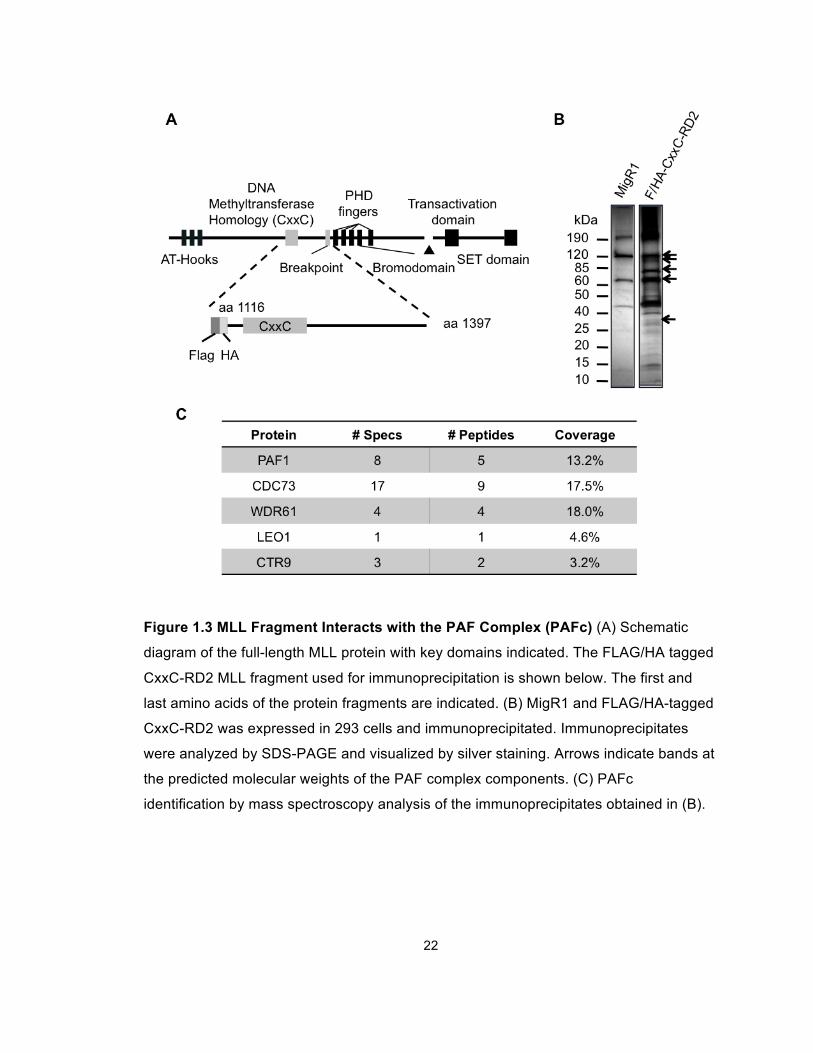
To identify proteins that associate with MLL CxxC-RD2, we transiently expressed
epitope tagged portions of this region in human embryonic kidney 293 cells
(Figure 1.3A). FLAG-tagged CxxC-RD2 including a nuclear localization signal
(NLS) was immunoprecipitated from transiently transfected 293 cells using M2
anti-FLAG agarose beads. An “empty” expression vector with FLAG epitope tag
and NLS was also subjected to immunoprecipitation as a non-specific
immunoprecipitation control. Coeluted proteins were resolved by SDS-PAGE
(Figure 1.3B) and analyzed by mass spectroscopy. Multiple peptides
corresponding to subunits of PAFc were identified with high probability including
CTR9, LEO1, PAF1, CDC73 and WDR61 that correlated with silver stained
bands at 133 kDa, 105 kDa, 75 kDa, 64 kDa and 34 kDa, respectively (Figures
1.3B and 1.3C). Each of the five PAFc subunits identified by mass spectrometry
(PAF1, CDC73, CTR9, LEO1 and WDR61) was confirmed to co-
immunoprecipitate with Myc-tagged CxxC-RD2 by western blotting in 293 cells
(Figure 1.4A).
22
Figure 1.3 MLL Fragment Interacts with the PAF Complex (PAFc) (A) Schematic
diagram of the full-length MLL protein with key domains indicated. The FLAG/HA tagged
CxxC-RD2 MLL fragment used for immunoprecipitation is shown below. The first and
last amino acids of the protein fragments are indicated. (B) MigR1 and FLAG/HA-tagged
CxxC-RD2 was expressed in 293 cells and immunoprecipitated. Immunoprecipitates
were analyzed by SDS-PAGE and visualized by silver staining. Arrows indicate bands at
the predicted molecular weights of the PAF complex components. (C) PAFc
identification by mass spectroscopy analysis of the immunoprecipitates obtained in (B).
23
To exclude the possibility of a DNA-mediated MLL-PAFc interaction and confirm
that the PAFc interaction is preserved in the context of a fusion protein,
immunoprecipitations were repeated with Myc-tagged CxxC-RD2-AF9 in 293
cells following Benzonase treatment. Immunoprecipitation was preserved in the
presence of Benzonase suggesting the interaction is not DNA dependent (Figure
1.4B). Furthermore, PAFc co-immunoprecipitated with Myc-CxxC-RD2-AF9, as
well as with CxxC-RD2 (Figures 1.4B-1.4D).
These experiments were repeated with transfection of an expression vector for
FLAG-tagged full-length MLL-AF9 or wild-type MLL into 293 cells followed by
immunoprecipitation and western blotting (Figures 1.5A and 1.5B). We also
confirmed the PAFc interaction by co-immunoprecipitation of CDC73 with MLL-
ENL in the KOPN8 cell line (Figure 1.5C). Together, these experiments show the
MLL-PAFc interaction is maintained both in the context of full-length (MLLN) and
in the context of leukemogenic MLL fusion proteins (Figure 1.5).
24
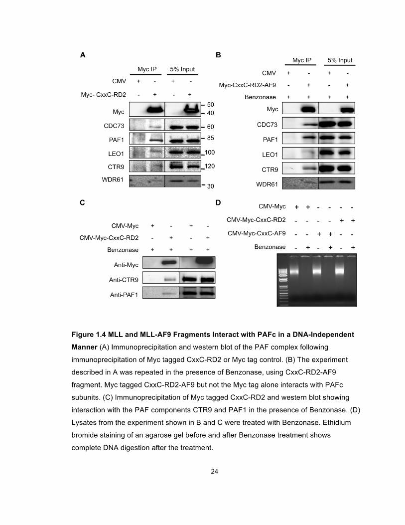
Figure 1.4 MLL and MLL-AF9 Fragments Interact with PAFc in a DNA-Independent
Manner (A) Immunoprecipitation and western blot of the PAF complex following
immunoprecipitation of Myc tagged CxxC-RD2 or Myc tag control. (B) The experiment
described in A was repeated in the presence of Benzonase, using CxxC-RD2-AF9
fragment. Myc tagged CxxC-RD2-AF9 but not the Myc tag alone interacts with PAFc
subunits. (C) Immunoprecipitation of Myc tagged CxxC-RD2 and western blot showing
interaction with the PAF components CTR9 and PAF1 in the presence of Benzonase. (D)
Lysates from the experiment shown in B and C were treated with Benzonase. Ethidium
bromide staining of an agarose gel before and after Benzonase treatment shows
complete DNA digestion after the treatment.
25
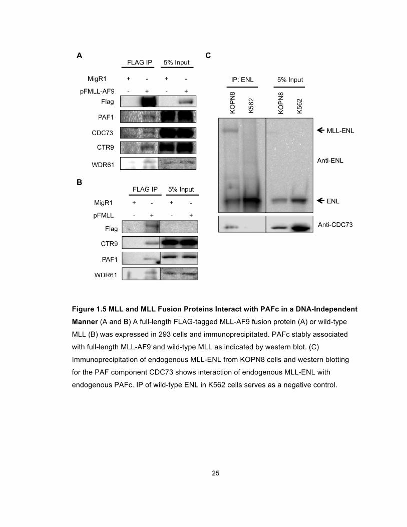
Figure 1.5 MLL and MLL Fusion Proteins Interact with PAFc in a DNA-Independent
Manner (A and B) A full-length FLAG-tagged MLL-AF9 fusion protein (A) or wild-type
MLL (B) was expressed in 293 cells and immunoprecipitated. PAFc stably associated
with full-length MLL-AF9 and wild-type MLL as indicated by western blot. (C)
Immunoprecipitation of endogenous MLL-ENL from KOPN8 cells and western blotting
for the PAF component CDC73 shows interaction of endogenous MLL-ENL with
endogenous PAFc. IP of wild-type ENL in K562 cells serves as a negative control.
26
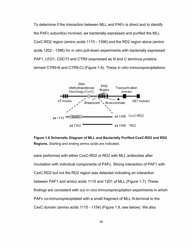
To determine if the interaction between MLL and PAFc is direct and to identify
the PAFc subunit(s) involved, we bacterially expressed and purified the MLL
CxxC-RD2 region (amino acids 1115 - 1396) and the RD2 region alone (amino
acids 1202 - 1396) for in vitro pull-down experiments with bacterially expressed
PAF1, LEO1, CDC73 and CTR9 (expressed as N and C terminus proteins
termed CTR9-N and CTR9-C) (Figure 1.6). These in vitro immunoprecipitations
Figure 1.6 Schematic Diagram of MLL and Bacterially Purified CxxC-RD2 and RD2
Regions. Starting and ending amino acids are indicated.
were performed with either CxxC-RD2 or RD2 with MLL antibodies after
incubation with individual components of PAFc. Strong interaction of PAF1 with
CxxC-RD2 but not the RD2 region was detected indicating an interaction
between PAF1 and amino acids 1115 and 1201 of MLL (Figure 1.7). These
findings are consistent with our in vivo immunoprecipitation experiments in which
PAFc co-immunoprecipitated with a small fragment of MLL N-terminal to the
CxxC domain (amino acids 1115 - 1154) (Figure 1.8, see below). We also

27
detected a second interaction between CTR9-C and both the MLL CxxC-RD2
and RD2 regions (Figure 1.7).
Figure 1.7 PAF1 and CTR9 Bind Directly to the CxxC-RD2 Region of MLL (A)
Coomassie blue staining of bacterially purified His-MOCR tagged CxxC-RD2, RD2,
PAF1, LEO1, and His-MBP tagged CDC73, CTR9-N and CTR9-C. The amino acids of
CTR9-N and CTR9-C are indicated. (C) Immunoprecipitations performed with bacterially
purified recombinant CxxC-RD2 or RD2 and PAF complex components. Individual PAF
components were incubated with either CxxC-RD2 or RD2 and immunoprecipitated with
MLL antibodies. PAF components and immunoprecipitated MLL fragments were
detected with the indicated antibodies by western blot. Asterisk denotes detection of the
IgG heavy chain.
28
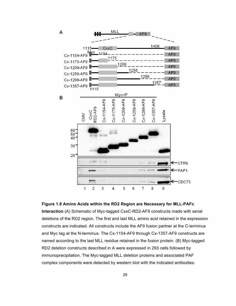
We then performed a series of deletion experiments to further map the MLL
residues that participate in the MLL-PAFc interaction. Expression vectors for
Myc-tagged CxxC-RD2-AF9 deletion mutants spanning the RD2 region of MLL
(Figure 1.8A, Cx-1154-AF9 through Cx-1357-AF9) were transiently transfected
into 293 cells and tested for PAFc interaction. These experiments reveal a sharp
decrease in the MLL-PAFc interaction when C terminal deletions were made past
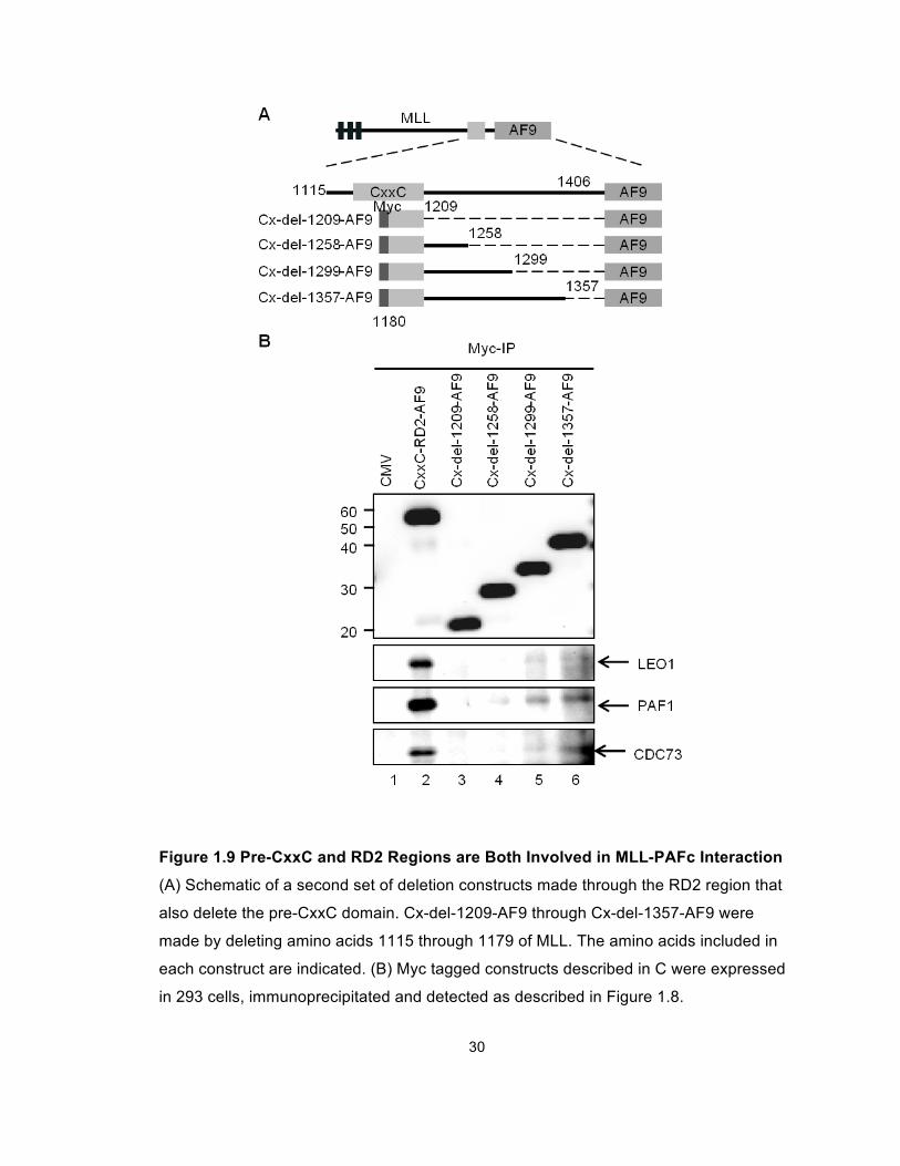
amino acid 1299 (Figure 1.8B, compare lanes 6 and 7). To overcome the
residual low level binding of PAFc with proteins deleted at amino acids 1209 or
amino acids 1258, presumably due to the multiple binding sites between PAFc
and MLL, which were only partially affected by these deletions (Figure 1.7), we
repeated this experiment with a set of deletion constructs that begin with MLL
amino acid 1180 thereby deleting the proximal site of PAFc interaction (Figure
1.9A, Cx-del-1209-AF9 through Cx-del-1357-AF9). These experiments showed
PAFc interaction with MLL is completely eliminated with deletions beyond amino
acid 1299 (Figure 1.9B, compare lanes 4 and 5). Together, our data suggest the
MLL-PAFc interaction is multivalent involving residues of MLL in both the pre-
CxxC domain and the RD2 region. Furthermore, the binding of PAFc by both pre
and post CxxC domains is consistent with the structure of the MLL CxxC domain
determined by multidimensional NMR spectroscopy (Protein Database structure,
2J2S) (Allen et al., 2006), which shows the DNA binding CxxC domain
coordinates two zinc atoms thereby bringing the pre and post CxxC regions into
close opposition (Figure 1.28).
29
Figure 1.8 Amino Acids within the RD2 Region are Necessary for MLL-PAFc
Interaction (A) Schematic of Myc-tagged CxxC-RD2-AF9 constructs made with serial
deletions of the RD2 region. The first and last MLL amino acid retained in the expression
constructs are indicated. All constructs include the AF9 fusion partner at the C-terminus
and Myc tag at the N-terminus. The Cx-1154-AF9 through Cx-1357-AF9 constructs are
named according to the last MLL residue retained in the fusion protein. (B) Myc-tagged
RD2 deletion constructs described in A were expressed in 293 cells followed by
immunoprecipitation. The Myc-tagged MLL deletion proteins and associated PAF
complex components were detected by western blot with the indicated antibodies.
30
Figure 1.9 Pre-CxxC and RD2 Regions are Both Involved in MLL-PAFc Interaction
(A) Schematic of a second set of deletion constructs made through the RD2 region that
also delete the pre-CxxC domain. Cx-del-1209-AF9 through Cx-del-1357-AF9 were
made by deleting amino acids 1115 through 1179 of MLL. The amino acids included in
each construct are indicated. (B) Myc tagged constructs described in C were expressed
in 293 cells, immunoprecipitated and detected as described in Figure 1.8.
31
PAFc Stimulates Transcriptional Activation Induced by MLL and MLL
Fusion Proteins
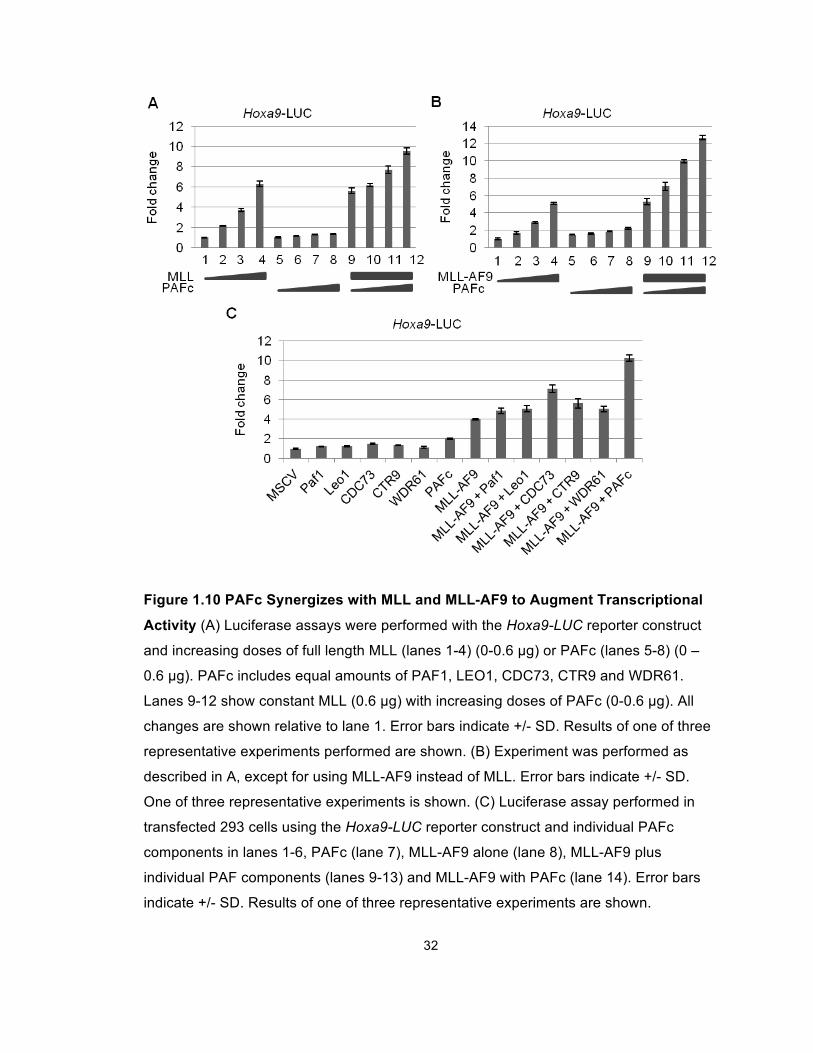
We then tested whether PAFc affects the transcriptional output mediated by MLL
and the MLL-AF9 fusion protein. Dual luciferase assays were performed in 293
cells transfected with a luciferase reporter construct under the transcriptional
control of the murine Hoxa9 promoter (Hoxa9-LUC). These experiments showed
that transcriptional activation by wild type MLL is enhanced by co-expression of
the five PAFc subunits (Figure 1.10A). Consistent with our earlier finding (Milne
et al., 2002), we observed a dose dependent transcriptional activation of the
reporter gene driven by the Hoxa9 promoter by expression of increasing amounts
of MLL-AF9 (Figure 1.10B). Furthermore, we observed a dose-dependent
augmentation of MLL-AF9 dependent transcription when increasing amounts of
PAFc were expressed. Notably, expression of PAFc alone had little effect in our
assay (Figures 1.10A and 1.10B). A similar trend was observed when using an
MLL-AF9 responsive luciferase construct containing a thymidine kinase promoter
and multimerized Myc E-boxes (Figure 1.11). Furthermore, we did not observe
augmented transcription when single PAF components were introduced (Figure
1.10C). Together, these findings show that MLL and MLL-AF9 synergize with
PAFc to augment transcriptional activation on MLL-responsive elements.
32
Figure 1.10 PAFc Synergizes with MLL and MLL-AF9 to Augment Transcriptional
Activity (A) Luciferase assays were performed with the Hoxa9-LUC reporter construct
and increasing doses of full length MLL (lanes 1-4) (0-0.6 µg) or PAFc (lanes 5-8) (0 –
0.6 µg). PAFc includes equal amounts of PAF1, LEO1, CDC73, CTR9 and WDR61.
Lanes 9-12 show constant MLL (0.6 µg) with increasing doses of PAFc (0-0.6 µg). All
changes are shown relative to lane 1. Error bars indicate +/- SD. Results of one of three
representative experiments performed are shown. (B) Experiment was performed as
described in A, except for using MLL-AF9 instead of MLL. Error bars indicate +/- SD.
One of three representative experiments is shown. (C) Luciferase assay performed in
transfected 293 cells using the Hoxa9-LUC reporter construct and individual PAFc
components in lanes 1-6, PAFc (lane 7), MLL-AF9 alone (lane 8), MLL-AF9 plus
individual PAF components (lanes 9-13) and MLL-AF9 with PAFc (lane 14). Error bars
indicate +/- SD. Results of one of three representative experiments are shown.
33
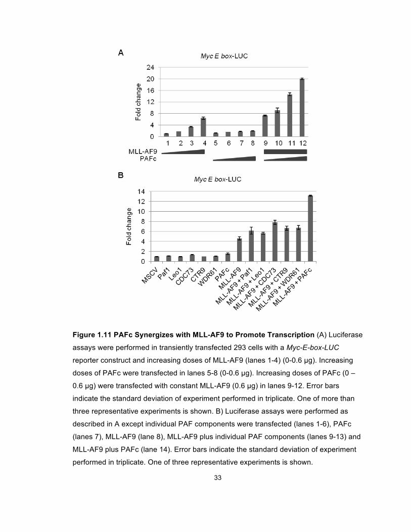
Figure 1.11 PAFc Synergizes with MLL-AF9 to Promote Transcription (A) Luciferase
assays were performed in transiently transfected 293 cells with a Myc-E-box-LUC
reporter construct and increasing doses of MLL-AF9 (lanes 1-4) (0-0.6 µg). Increasing
doses of PAFc were transfected in lanes 5-8 (0-0.6 µg). Increasing doses of PAFc (0 –
0.6 µg) were transfected with constant MLL-AF9 (0.6 µg) in lanes 9-12. Error bars
indicate the standard deviation of experiment performed in triplicate. One of more than
three representative experiments is shown. B) Luciferase assays were performed as
described in A except individual PAF components were transfected (lanes 1-6), PAFc
(lanes 7), MLL-AF9 (lane 8), MLL-AF9 plus individual PAF components (lanes 9-13) and
MLL-AF9 plus PAFc (lane 14). Error bars indicate the standard deviation of experiment
performed in triplicate. One of three representative experiments is shown.
34
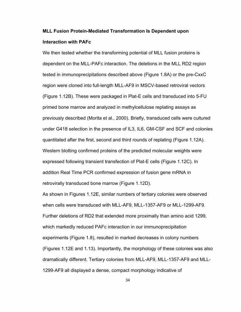
MLL Fusion Protein-Mediated Transformation Is Dependent upon
Interaction with PAFc
We then tested whether the transforming potential of MLL fusion proteins is
dependent on the MLL-PAFc interaction. The deletions in the MLL RD2 region
tested in immunoprecipitations described above (Figure 1.8A) or the pre-CxxC
region were cloned into full-length MLL-AF9 in MSCV-based retroviral vectors
(Figure 1.12B). These were packaged in Plat-E cells and transduced into 5-FU
primed bone marrow and analyzed in methylcellulose replating assays as
previously described (Morita et al., 2000). Briefly, transduced cells were cultured
under G418 selection in the presence of IL3, IL6, GM-CSF and SCF and colonies
quantitated after the first, second and third rounds of replating (Figure 1.12A).
Western blotting confirmed proteins of the predicted molecular weights were
expressed following transient transfection of Plat-E cells (Figure 1.12C). In
addition Real Time PCR confirmed expression of fusion gene mRNA in
retrovirally transduced bone marrow (Figure 1.12D).
As shown in Figures 1.12E, similar numbers of tertiary colonies were observed
when cells were transduced with MLL-AF9, MLL-1357-AF9 or MLL-1299-AF9.
Further deletions of RD2 that extended more proximally than amino acid 1299,
which markedly reduced PAFc interaction in our immunoprecipitation
experiments (Figure 1.8), resulted in marked decreases in colony numbers
(Figures 1.12E and 1.13). Importantly, the morphology of these colonies was also
dramatically different. Tertiary colonies from MLL-AF9, MLL-1357-AF9 and MLL-
1299-AF9 all displayed a dense, compact morphology indicative of
35
Figure 1.12 PAFc Interaction Region on RD2 Is Necessary for Bone Marrow
Transformation (BMT) by MLL-AF9 (A) Schematic diagram for MLL-AF9 and MLL-AF9
deletion BMT assay. (B) Constructs used for the BMT assay. Final amino acids of the
MLL deletions are shown. (C) Protein levels of the FLAG-tagged MLL-AF9 deletions
shown in B. LC: loading control. (D) Relative mRNA levels of the FLAG-tagged MLL-AF9
deletions shown in B. Error bars indicate +/- SD. (E) Primary, secondary and tertiary
colony counts are shown for BMT assays performed with the indicated MLL-AF9 fusion
proteins. Error bars indicate SD from duplicate experiments. One of more than three
representative experiments is shown.
36
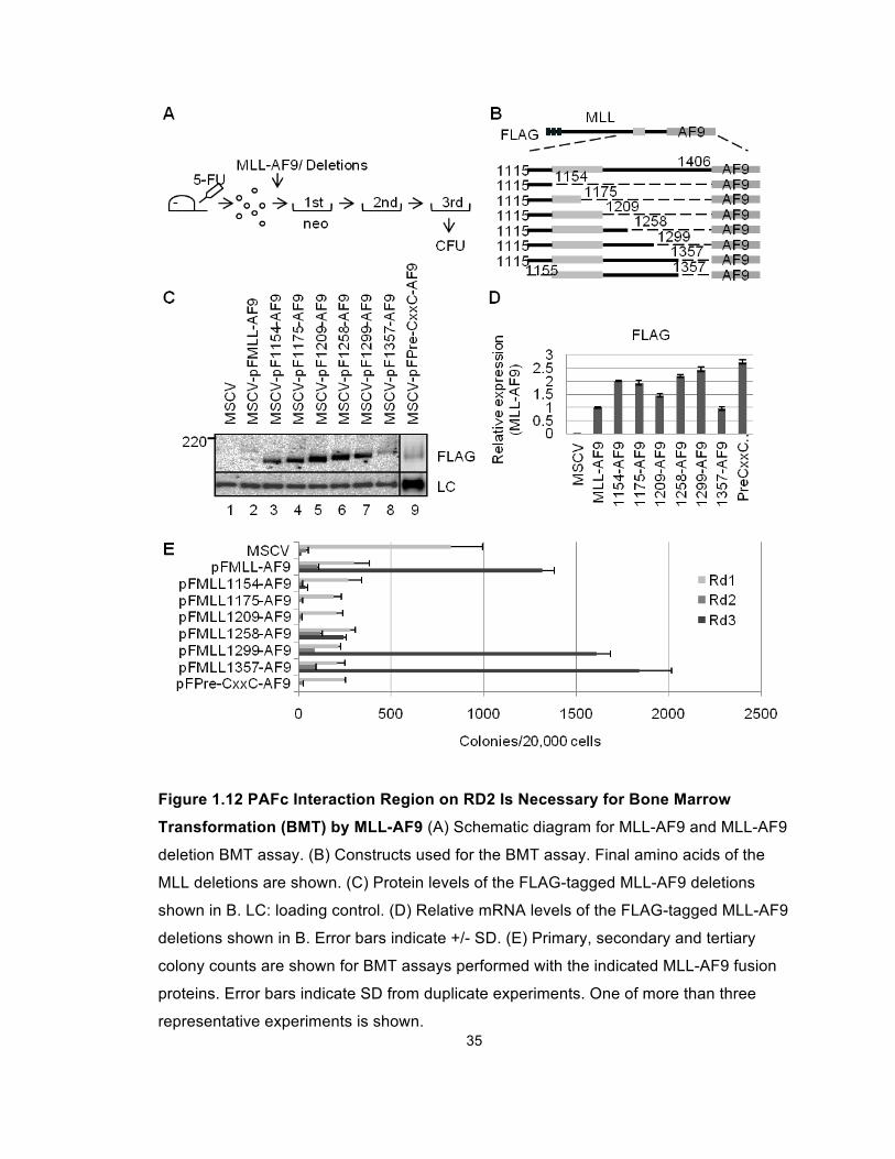
Figure 1.13 p-Iodonitro Tetrazolium Violet (INT)-Stained Colonies after Three
Rounds of Colony Replating. Dense red colonies are visible from MLL-AF9, MLL-
1299-AF9 and MLL-1357-AF9 transduced bone marrow.
transformation (Lavau et al., 1997) (Figure 1.14A). Wright Giemsa-stained
cytospins showed these compact colonies were composed of myeloblasts
(Figure 1.14B). In contrast, transductions of constructs with more extensive
deletions resulted in diffuse colonies composed of differentiating myeloid cells
including monocytes and macrophages (Figures 1.13 and 1.14). Of note, MLL-
1258-AF9 retained a limited capacity to produce dense colonies after tertiary
replating, but colony numbers were significantly reduced compared to MLL-AF9,
MLL-1357-AF9 and MLL-1299-AF9 (Figures 1.13 and 1.14). In keeping with this,
minimal binding of PAFc was observed with the Cx-1258-AF9 construct (Figure
1.8).
37
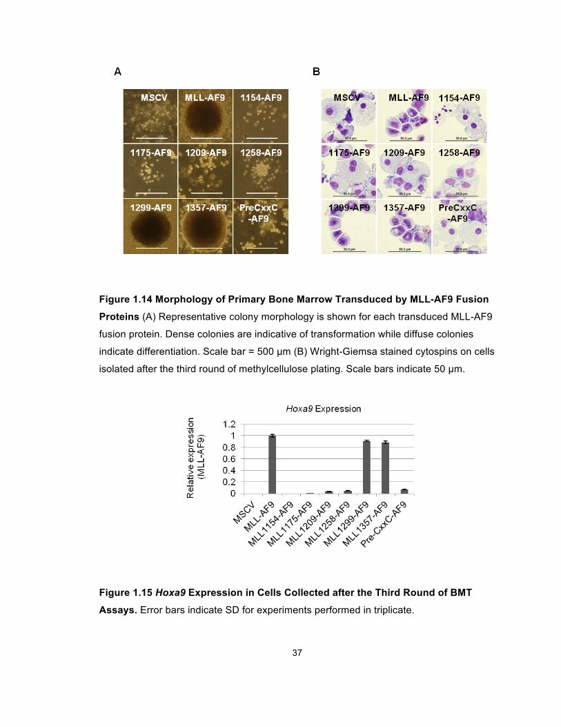
Figure 1.14 Morphology of Primary Bone Marrow Transduced by MLL-AF9 Fusion
Proteins (A) Representative colony morphology is shown for each transduced MLL-AF9
fusion protein. Dense colonies are indicative of transformation while diffuse colonies
indicate differentiation. Scale bar = 500 µm (B) Wright-Giemsa stained cytospins on cells
isolated after the third round of methylcellulose plating. Scale bars indicate 50 µm.
Figure 1.15 Hoxa9 Expression in Cells Collected after the Third Round of BMT
Assays. Error bars indicate SD for experiments performed in triplicate.
38
We then used compared the Hoxa9 expression in cells transduced by wild-type
MLL-AF9 and different deletion constructs. As expected, MLL-AF9, as well as
1357 and 1299 deletions, markedly upregulated Hoxa9 expression, while forms
incapable of PAFc interaction did not (Figure 1.15).
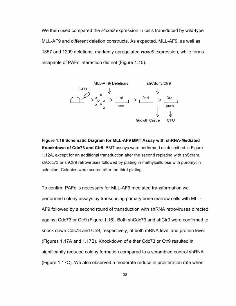
Figure 1.16 Schematic Diagram for MLL-AF9 BMT Assay with shRNA-Mediated
Knockdown of Cdc73 and Ctr9. BMT assays were performed as described in Figure
1.12A, except for an additional transduction after the second replating with shScram,
shCdc73 or shCtr9 retroviruses followed by plating in methylcellulose with puromycin
selection. Colonies were scored after the third plating.
To confirm PAFc is necessary for MLL-AF9 mediated transformation we
performed colony assays by transducing primary bone marrow cells with MLL-
AF9 followed by a second round of transduction with shRNA retroviruses directed
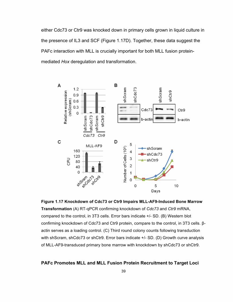
against Cdc73 or Ctr9 (Figure 1.16). Both shCdc73 and shCtr9 were confirmed to
knock down Cdc73 and Ctr9, respectively, at both mRNA level and protein level
(Figures 1.17A and 1.17B). Knockdown of either Cdc73 or Ctr9 resulted in
significantly reduced colony formation compared to a scrambled control shRNA
(Figure 1.17C). We also observed a moderate reduce in proliferation rate when
39
either Cdc73 or Ctr9 was knocked down in primary cells grown in liquid culture in
the presence of IL3 and SCF (Figure 1.17D). Together, these data suggest the
PAFc interaction with MLL is crucially important for both MLL fusion protein-
mediated Hox deregulation and transformation.
Figure 1.17 Knockdown of Cdc73 or Ctr9 Impairs MLL-AF9-Induced Bone Marrow
Transformation (A) RT-qPCR confirming knockdown of Cdc73 and Ctr9 mRNA,
compared to the control, in 3T3 cells. Error bars indicate +/- SD. (B) Western blot
confirming knockdown of Cdc73 and Ctr9 protein, compare to the control, in 3T3 cells. β-
actin serves as a loading control. (C) Third round colony counts following transduction
with shScram, shCdc73 or shCtr9. Error bars indicate +/- SD. (D) Growth curve analysis
of MLL-AF9-transduced primary bone marrow with knockdown by shCdc73 or shCtr9.
PAFc Promotes MLL and MLL Fusion Protein Recruitment to Target Loci
40
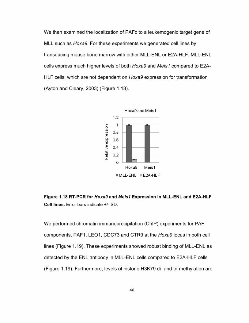
We then examined the localization of PAFc to a leukemogenic target gene of
MLL such as Hoxa9. For these experiments we generated cell lines by
transducing mouse bone marrow with either MLL-ENL or E2A-HLF. MLL-ENL
cells express much higher levels of both Hoxa9 and Meis1 compared to E2A-
HLF cells, which are not dependent on Hoxa9 expression for transformation
(Ayton and Cleary, 2003) (Figure 1.18).
Figure 1.18 RT-PCR for Hoxa9 and Meis1 Expression in MLL-ENL and E2A-HLF
Cell lines. Error bars indicate +/- SD.
We performed chromatin immunoprecipitation (ChIP) experiments for PAF
components, PAF1, LEO1, CDC73 and CTR9 at the Hoxa9 locus in both cell
lines (Figure 1.19). These experiments showed robust binding of MLL-ENL as
detected by the ENL antibody in MLL-ENL cells compared to E2A-HLF cells
(Figure 1.19). Furthermore, levels of histone H3K79 di- and tri-methylation are
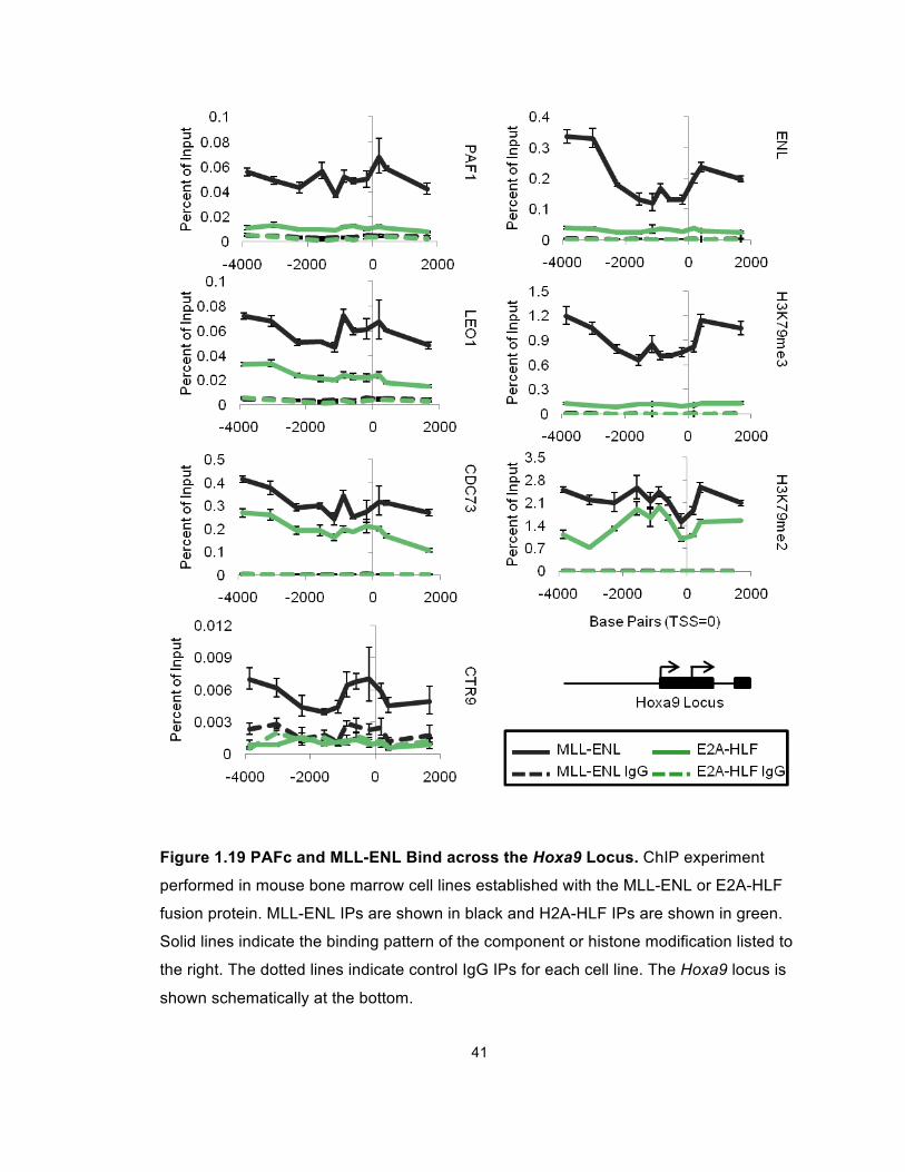
41
Figure 1.19 PAFc and MLL-ENL Bind across the Hoxa9 Locus. ChIP experiment
performed in mouse bone marrow cell lines established with the MLL-ENL or E2A-HLF
fusion protein. MLL-ENL IPs are shown in black and H2A-HLF IPs are shown in green.
Solid lines indicate the binding pattern of the component or histone modification listed to
the right. The dotted lines indicate control IgG IPs for each cell line. The Hoxa9 locus is
shown schematically at the bottom.
42
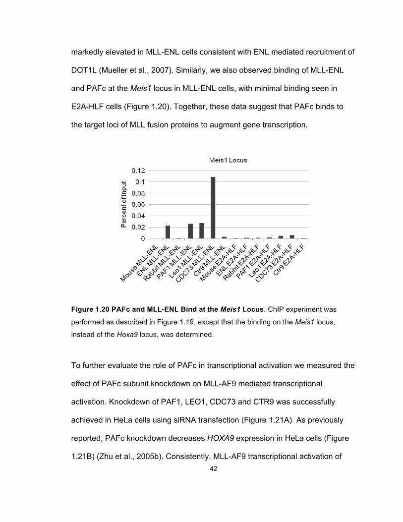
markedly elevated in MLL-ENL cells consistent with ENL mediated recruitment of
DOT1L (Mueller et al., 2007). Similarly, we also observed binding of MLL-ENL
and PAFc at the Meis1 locus in MLL-ENL cells, with minimal binding seen in
E2A-HLF cells (Figure 1.20). Together, these data suggest that PAFc binds to
the target loci of MLL fusion proteins to augment gene transcription.
Figure 1.20 PAFc and MLL-ENL Bind at the Meis1 Locus. ChIP experiment was
performed as described in Figure 1.19, except that the binding on the Meis1 locus,
instead of the Hoxa9 locus, was determined.
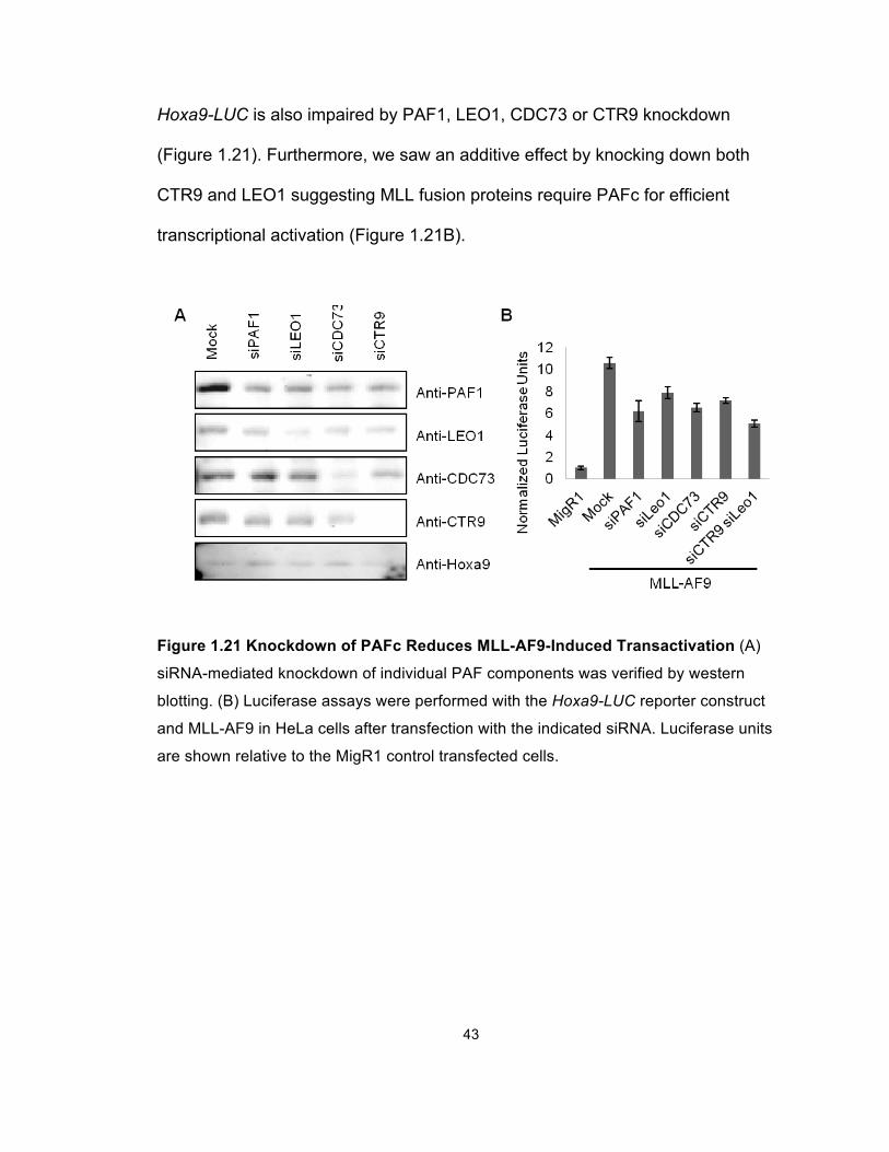
To further evaluate the role of PAFc in transcriptional activation we measured the
effect of PAFc subunit knockdown on MLL-AF9 mediated transcriptional
activation. Knockdown of PAF1, LEO1, CDC73 and CTR9 was successfully
achieved in HeLa cells using siRNA transfection (Figure 1.21A). As previously
reported, PAFc knockdown decreases HOXA9 expression in HeLa cells (Figure
1.21B) (Zhu et al., 2005b). Consistently, MLL-AF9 transcriptional activation of
43
Hoxa9-LUC is also impaired by PAF1, LEO1, CDC73 or CTR9 knockdown
(Figure 1.21). Furthermore, we saw an additive effect by knocking down both
CTR9 and LEO1 suggesting MLL fusion proteins require PAFc for efficient
transcriptional activation (Figure 1.21B).
Figure 1.21 Knockdown of PAFc Reduces MLL-AF9-Induced Transactivation (A)
siRNA-mediated knockdown of individual PAF components was verified by western
blotting. (B) Luciferase assays were performed with the Hoxa9-LUC reporter construct
and MLL-AF9 in HeLa cells after transfection with the indicated siRNA. Luciferase units
are shown relative to the MigR1 control transfected cells.
44
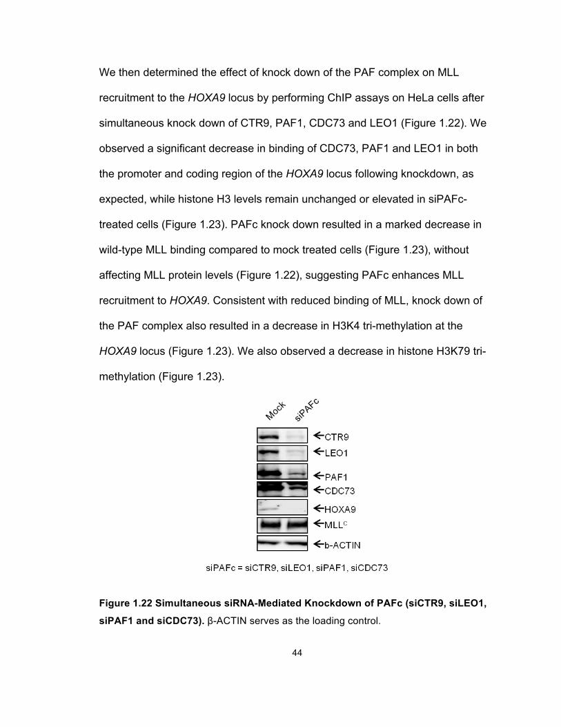
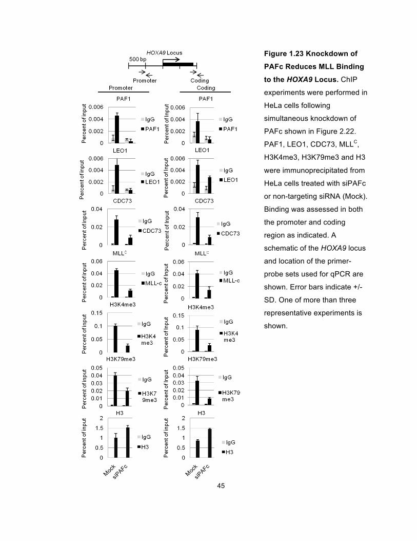
We then determined the effect of knock down of the PAF complex on MLL
recruitment to the HOXA9 locus by performing ChIP assays on HeLa cells after
simultaneous knock down of CTR9, PAF1, CDC73 and LEO1 (Figure 1.22). We
observed a significant decrease in binding of CDC73, PAF1 and LEO1 in both
the promoter and coding region of the HOXA9 locus following knockdown, as
expected, while histone H3 levels remain unchanged or elevated in siPAFc-
treated cells (Figure 1.23). PAFc knock down resulted in a marked decrease in
wild-type MLL binding compared to mock treated cells (Figure 1.23), without
affecting MLL protein levels (Figure 1.22), suggesting PAFc enhances MLL
recruitment to HOXA9. Consistent with reduced binding of MLL, knock down of
the PAF complex also resulted in a decrease in H3K4 tri-methylation at the
HOXA9 locus (Figure 1.23). We also observed a decrease in histone H3K79 tri-
methylation (Figure 1.23).
Figure 1.22 Simultaneous siRNA-Mediated Knockdown of PAFc (siCTR9, siLEO1,
siPAF1 and siCDC73). β-ACTIN serves as the loading control.
45
Figure 1.23 Knockdown of
PAFc Reduces MLL Binding
to the HOXA9 Locus. ChIP
experiments were performed in
HeLa cells following
simultaneous knockdown of
PAFc shown in Figure 2.22.
PAF1, LEO1, CDC73, MLLC,
H3K4me3, H3K79me3 and H3
were immunoprecipitated from
HeLa cells treated with siPAFc
or non-targeting siRNA (Mock).
Binding was assessed in both
the promoter and coding
region as indicated. A
schematic of the HOXA9 locus
and location of the primer-
probe sets used for qPCR are
shown. Error bars indicate +/-
SD. One of more than three
representative experiments is
shown.
46
PAFc expression is coordinately downregulated during myeloid
differentiation
The above results suggest that the PAF complex enhances MLL recruitment to
the HOXA9 locus and that modulating PAFc levels may be an important
mechanism for modulating MLL activity. It is noteworthy in this regard that a
recent unbiased genome-wide siRNA screen identified PAFc subunits Ctr9,
Wdr61 and Rtf1 amongst the 30 top genes regulating Oct4 expression and stem
cell renewal (Ding et al., 2009). In this study PAFc was found to bind to key
pluripotency genes, which is remarkable because MLL fusion protein transformed
cells show, in addition to HOX gene over expression, a distinctive embryonic
stem cell (ESC)-like pluripotency signature (Somervaille et al., 2009).
Furthermore, expression of PAFc subunits is strongly regulated upon
differentiation of ESCs into embryoid bodies (Ding et al., 2009). Collectively,
these findings suggested that PAFc might be positively involved in maintaining
the relatively undifferentiated stage of hematopoietic progenitors.
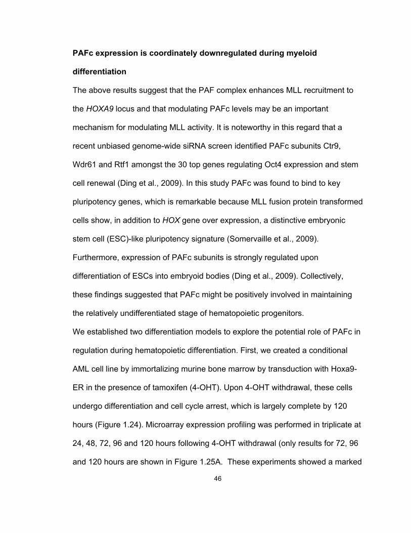
We established two differentiation models to explore the potential role of PAFc in
regulation during hematopoietic differentiation. First, we created a conditional
AML cell line by immortalizing murine bone marrow by transduction with Hoxa9-
ER in the presence of tamoxifen (4-OHT). Upon 4-OHT withdrawal, these cells
undergo differentiation and cell cycle arrest, which is largely complete by 120
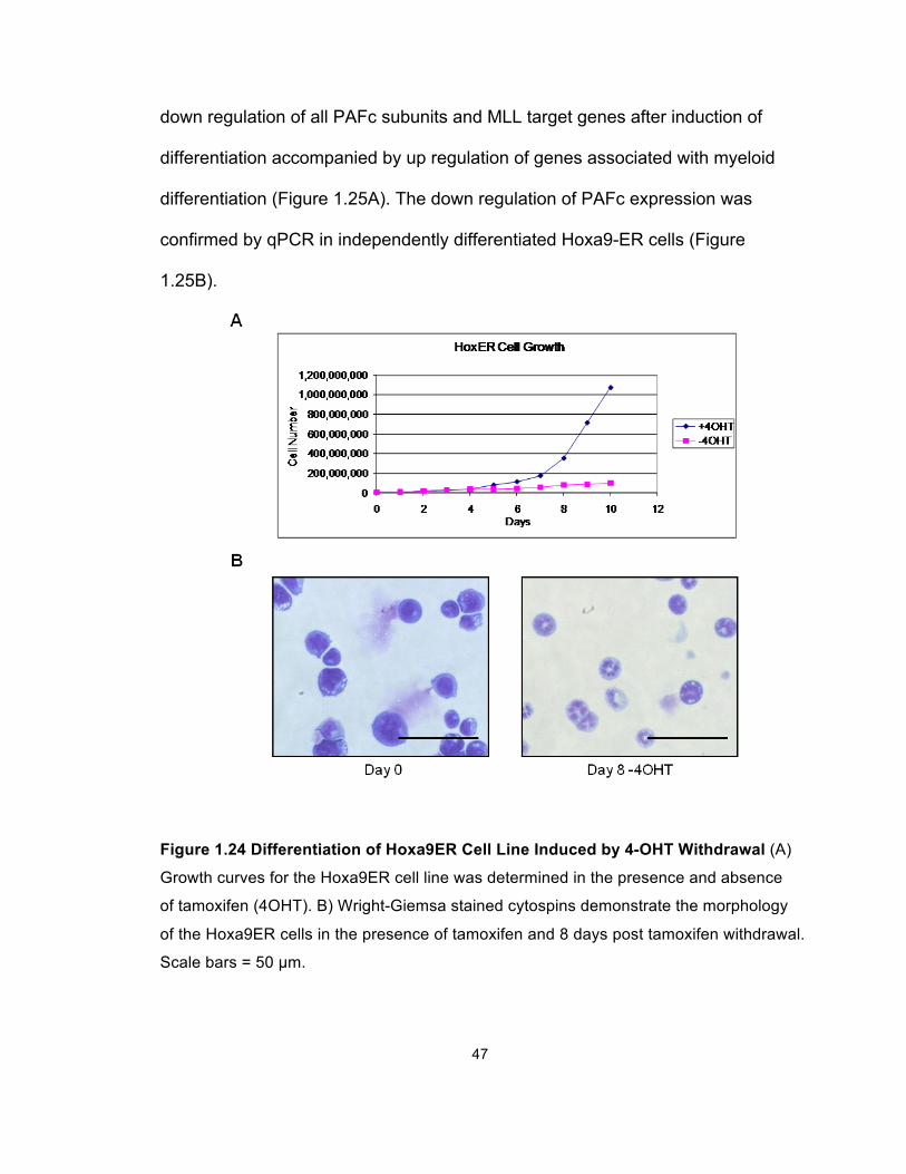
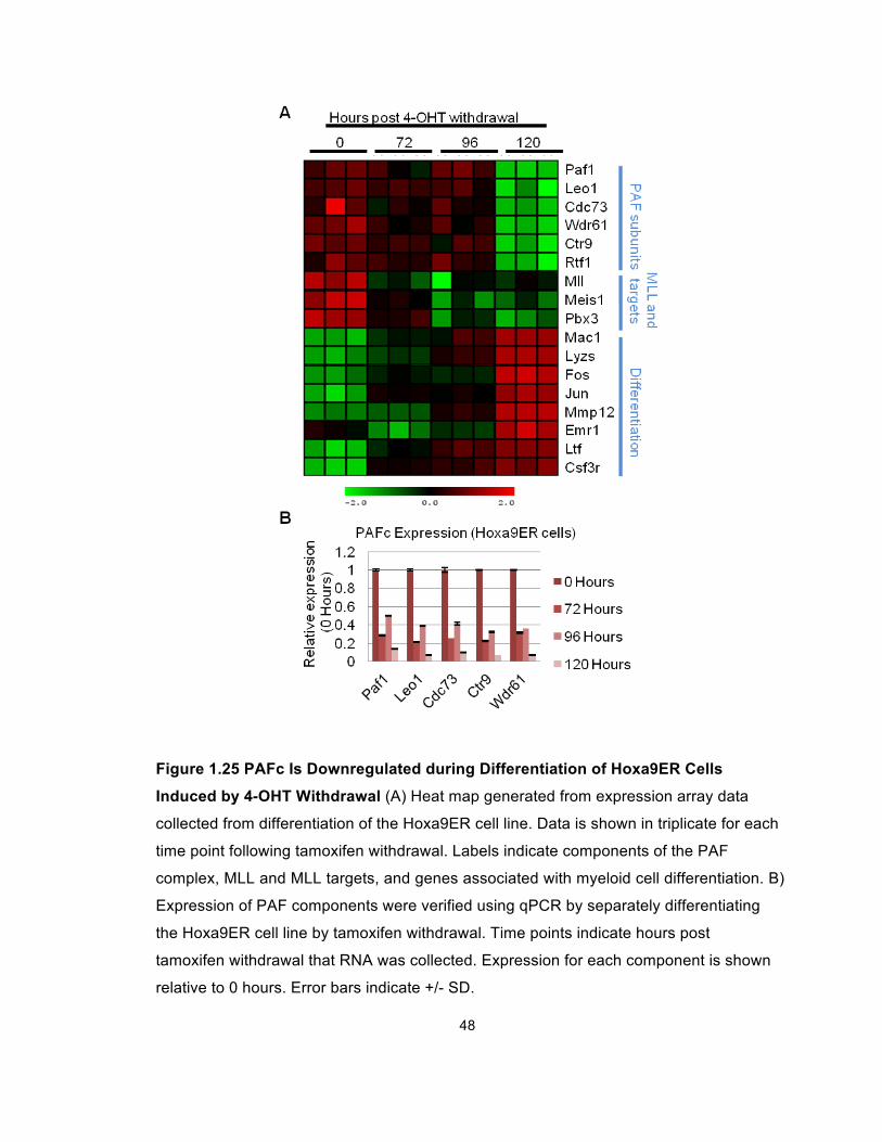
hours (Figure 1.24). Microarray expression profiling was performed in triplicate at
24, 48, 72, 96 and 120 hours following 4-OHT withdrawal (only results for 72, 96
and 120 hours are shown in Figure 1.25A. These experiments showed a marked
47
down regulation of all PAFc subunits and MLL target genes after induction of
differentiation accompanied by up regulation of genes associated with myeloid
differentiation (Figure 1.25A). The down regulation of PAFc expression was
confirmed by qPCR in independently differentiated Hoxa9-ER cells (Figure
1.25B).
Figure 1.24 Differentiation of Hoxa9ER Cell Line Induced by 4-OHT Withdrawal (A)
Growth curves for the Hoxa9ER cell line was determined in the presence and absence
of tamoxifen (4OHT). B) Wright-Giemsa stained cytospins demonstrate the morphology
of the Hoxa9ER cells in the presence of tamoxifen and 8 days post tamoxifen withdrawal.
Scale bars = 50 µm.
48
Figure 1.25 PAFc Is Downregulated during Differentiation of Hoxa9ER Cells
Induced by 4-OHT Withdrawal (A) Heat map generated from expression array data
collected from differentiation of the Hoxa9ER cell line. Data is shown in triplicate for each
time point following tamoxifen withdrawal. Labels indicate components of the PAF
complex, MLL and MLL targets, and genes associated with myeloid cell differentiation. B)
Expression of PAF components were verified using qPCR by separately differentiating
the Hoxa9ER cell line by tamoxifen withdrawal. Time points indicate hours post
tamoxifen withdrawal that RNA was collected. Expression for each component is shown
relative to 0 hours. Error bars indicate +/- SD.
49
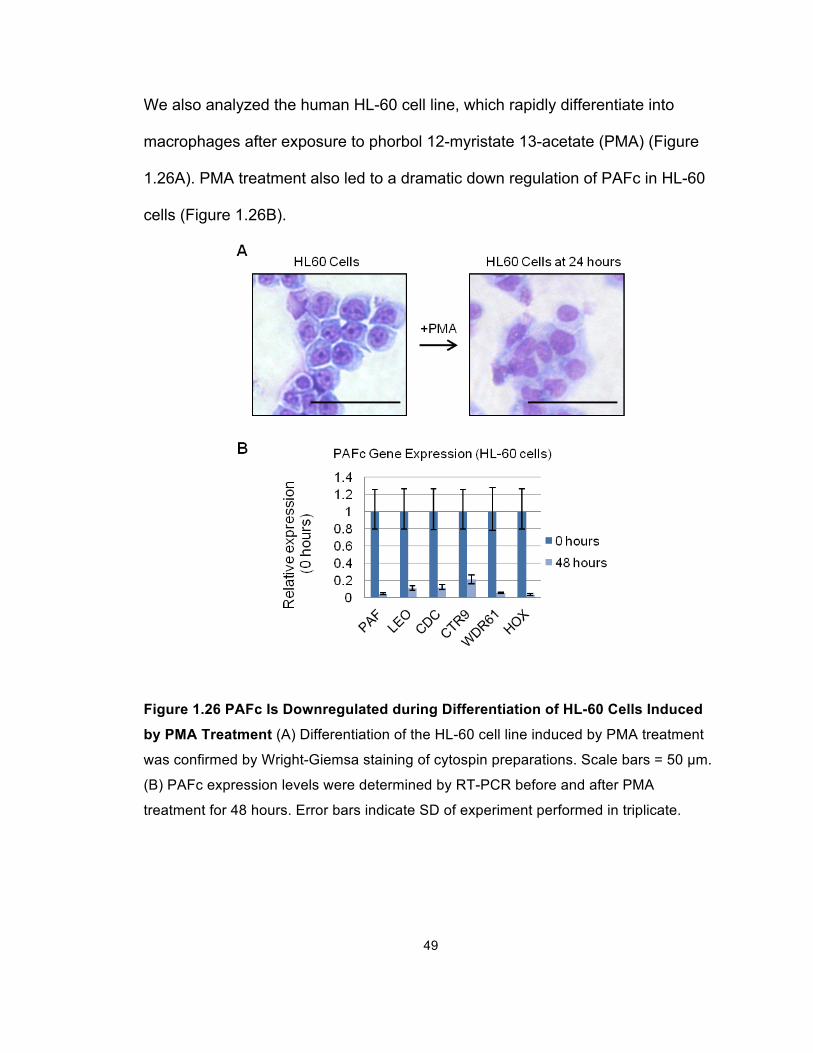
We also analyzed the human HL-60 cell line, which rapidly differentiate into
macrophages after exposure to phorbol 12-myristate 13-acetate (PMA) (Figure
1.26A). PMA treatment also led to a dramatic down regulation of PAFc in HL-60
cells (Figure 1.26B).
Figure 1.26 PAFc Is Downregulated during Differentiation of HL-60 Cells Induced
by PMA Treatment (A) Differentiation of the HL-60 cell line induced by PMA treatment
was confirmed by Wright-Giemsa staining of cytospin preparations. Scale bars = 50 µm.
(B) PAFc expression levels were determined by RT-PCR before and after PMA
treatment for 48 hours. Error bars indicate SD of experiment performed in triplicate.
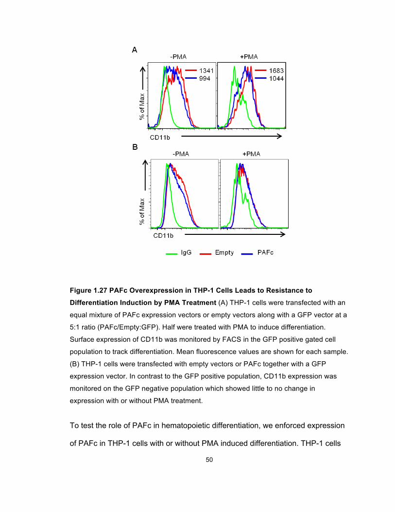
50
Figure 1.27 PAFc Overexpression in THP-1 Cells Leads to Resistance to
Differentiation Induction by PMA Treatment (A) THP-1 cells were transfected with an
equal mixture of PAFc expression vectors or empty vectors along with a GFP vector at a
5:1 ratio (PAFc/Empty:GFP). Half were treated with PMA to induce differentiation.
Surface expression of CD11b was monitored by FACS in the GFP positive gated cell
population to track differentiation. Mean fluorescence values are shown for each sample.
(B) THP-1 cells were transfected with empty vectors or PAFc together with a GFP
expression vector. In contrast to the GFP positive population, CD11b expression was
monitored on the GFP negative population which showed little to no change in
expression with or without PMA treatment.
To test the role of PAFc in hematopoietic differentiation, we enforced expression
of PAFc in THP-1 cells with or without PMA induced differentiation. THP-1 cells
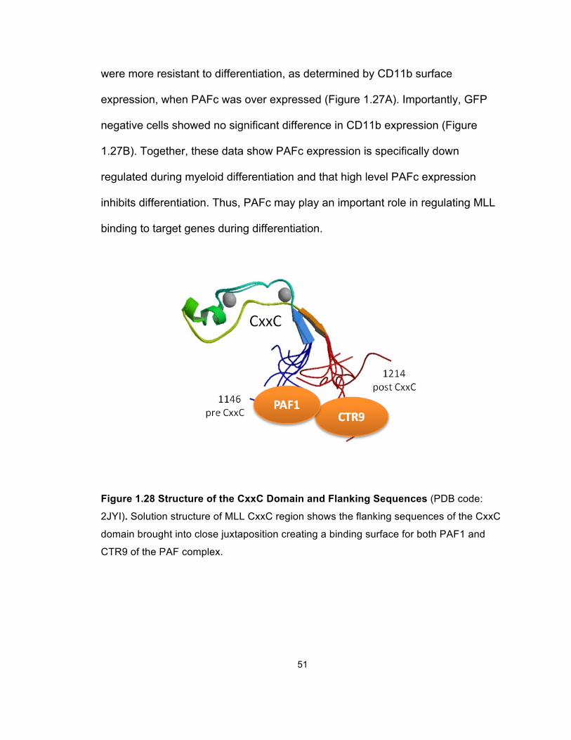
51
were more resistant to differentiation, as determined by CD11b surface
expression, when PAFc was over expressed (Figure 1.27A). Importantly, GFP
negative cells showed no significant difference in CD11b expression (Figure
1.27B). Together, these data show PAFc expression is specifically down
regulated during myeloid differentiation and that high level PAFc expression
inhibits differentiation. Thus, PAFc may play an important role in regulating MLL
binding to target genes during differentiation.
Figure 1.28 Structure of the CxxC Domain and Flanking Sequences (PDB code:
2JYI). Solution structure of MLL CxxC region shows the flanking sequences of the CxxC
domain brought into close juxtaposition creating a binding surface for both PAF1 and
CTR9 of the PAF complex.
52
Discussion
The work presented here establishes PAFc as an important cofactor for both
transcriptional regulation by MLL and for leukemogenesis mediated by MLL
fusion proteins (Figure 1.29). By mass spectrum analysis, we demonstrated that
PAFc interacts with the CxxC-RD2 region of MLL, a region that is always
retained in MLL-rearranged oncoproteins. Detailed mapping revealed two
interaction sites flanking the CxxC domain with two individual components of
PAFc (Figure 1.28). Most importantly, we were able to show that the PAFc-MLL
interaction enhances the transcriptional activation by MLL-AF9 and plays an
indispensible role in MLL-AF9-induced leukemogenic transformation.
Characterization of the PAFc-MLL interaction provides valuable insight into the
mechanisms of MLL-rearranged leukemogenesis. The best-defined target genes
of MLL are the clustered homeobox (Hox) genes, a transcription factor family
important in cell fate determination during development. Among these targets,
Hoxa9 and its cofactor Meis1 have been shown to be crucial for MLL-rearranged
leukemogenesis. Normally, Hoxa9 and Meis1 are only briefly expressed in
hematopoietic stem cell and progenitor cell populations and are then rapidly
down regulated during hematopoietic differentiation (Lawrence et al., 1996; Magli
et al., 1997; Zeisig et al., 2004). However, in the presence of MLL-rearranged
oncoproteins, both remain expressed at high levels, which accounts for their
leukemogenic capacity. Although many interaction partners of MLL-rearranged
proteins have been identified and shown to be important in leukemogenesis, it
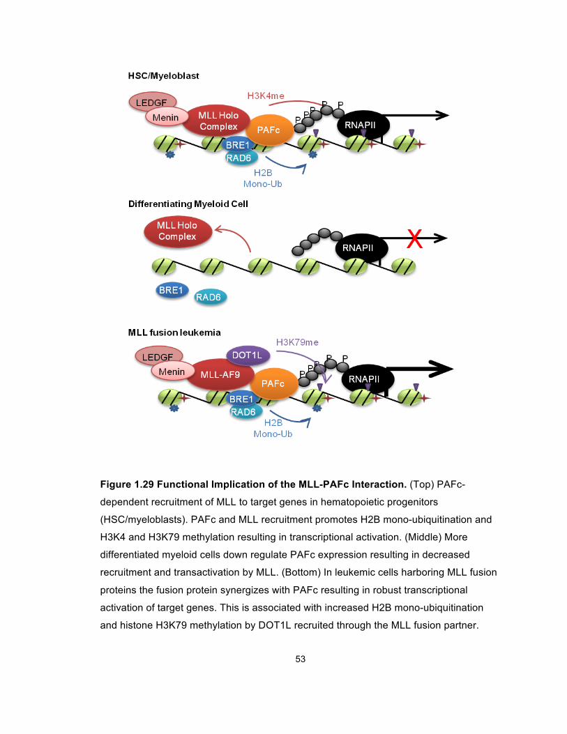
53
Figure 1.29 Functional Implication of the MLL-PAFc Interaction. (Top) PAFc-
dependent recruitment of MLL to target genes in hematopoietic progenitors
(HSC/myeloblasts). PAFc and MLL recruitment promotes H2B mono-ubiquitination and
H3K4 and H3K79 methylation resulting in transcriptional activation. (Middle) More
differentiated myeloid cells down regulate PAFc expression resulting in decreased
recruitment and transactivation by MLL. (Bottom) In leukemic cells harboring MLL fusion
proteins the fusion protein synergizes with PAFc resulting in robust transcriptional
activation of target genes. This is associated with increased H2B mono-ubiquitination
and histone H3K79 methylation by DOT1L recruited through the MLL fusion partner.
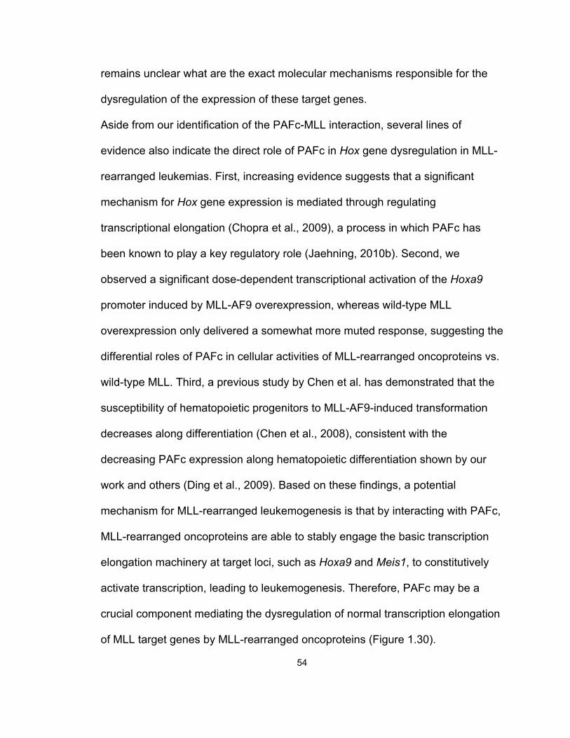
54
remains unclear what are the exact molecular mechanisms responsible for the
dysregulation of the expression of these target genes.
Aside from our identification of the PAFc-MLL interaction, several lines of
evidence also indicate the direct role of PAFc in Hox gene dysregulation in MLL-
rearranged leukemias. First, increasing evidence suggests that a significant
mechanism for Hox gene expression is mediated through regulating
transcriptional elongation (Chopra et al., 2009), a process in which PAFc has
been known to play a key regulatory role (Jaehning, 2010b). Second, we
observed a significant dose-dependent transcriptional activation of the Hoxa9
promoter induced by MLL-AF9 overexpression, whereas wild-type MLL
overexpression only delivered a somewhat more muted response, suggesting the
differential roles of PAFc in cellular activities of MLL-rearranged oncoproteins vs.
wild-type MLL. Third, a previous study by Chen et al. has demonstrated that the
susceptibility of hematopoietic progenitors to MLL-AF9-induced transformation
decreases along differentiation (Chen et al., 2008), consistent with the
decreasing PAFc expression along hematopoietic differentiation shown by our
work and others (Ding et al., 2009). Based on these findings, a potential
mechanism for MLL-rearranged leukemogenesis is that by interacting with PAFc,
MLL-rearranged oncoproteins are able to stably engage the basic transcription
elongation machinery at target loci, such as Hoxa9 and Meis1, to constitutively
activate transcription, leading to leukemogenesis. Therefore, PAFc may be a
crucial component mediating the dysregulation of normal transcription elongation
of MLL target genes by MLL-rearranged oncoproteins (Figure 1.30).
55
Figure 1.30 Schematic of a Potential Mechanism of MLL-Rearranged
Leukemogenesis (top) In the absence of PAFc, RNA polymerase II (RNAP II)
elongation is inhibited by the negative elongation factor (NELF) in collaboration with the
DRB sensitivity-inducing factor DSIF. (middle) DSIF recruits PAFc that directly interacts
with the E1/E2 ubiquitin ligase complex BRE1/RAD6, resulting in histone H2B
monoubiquitination. Recruitment of positive transcription elongation factor b (P-TEFb)
blocks the negative actions of NELF and DSIF by P-TEFb-dependent phosphorylation of
RNAP II CTD and DSIF, priming the target promoter for transcription elongation (bottom)
The interaction between PAFc and the most common MLL-rearranged oncoproteins
(represented by MLL-AF9) recruits the ENL-associated proteins (EAPs) that include
multiple common MLL translocation partners, DOT1L and P-TEFb to the target loci,
promoting H2B monoubiquitination ( ), H3K4 methylation ( ) and H3K79
methylation ( ), resulting in constitutively activated transcription.
56
Under normal conditions, Hoxa9 is expressed in primitive hematopoietic cells,
playing a significant role in early hematopoiesis (Lawrence et al., 2005; Pineault
et al., 2002). During hematopoietic differentiation, Hoxa9 expression is rapidly
silenced, likely by pausing transcription elongation, an important mechanism
regulating hox gene expression in Drosophila (Brookes and Pombo, 2009;
Chopra et al., 2009). In this case, although RNA polymerase II (RNAP II) still
localizes at the promoter region, its C-terminal domain (CTD) is
unphosphorylated, and transcription elongation is inhibited by the negative
elongation factor (NELF) in collaboration with the DRB sensitivity-inducing factor
(DSIF) (Figure 1.30A). DSIF recruits PAFc that directly interacts with the E1/E2
ubiquitin ligase complex BRE1/RAD6, resulting in histone H2B
monoubiquitination. Meanwhile, the recruitment of the positive transcription
elongation factor b (P-TEFb) reverts the negative actions of NELF and DSIF by
P-TEFb-dependent phosphorylation of RNAP II CTD and DSIF (Jaehning,
2010b). Thus, in the presence of PAFc, the target gene promoter region can
progress to the active elongation stage (Figure 1.30B). It is worth noting that this
status is probably a temporary transition stage, dynamically regulated by cell-
specific mechanisms, such as the abundance of PAFc, the binding affinity of
other transcription elongation machinery components determined by the
phosphorylation level of RNAP II CTD, the recruitment of histone
methyltransferases, such as wild-type MLL and DOT1L, exerting H3K4 and
H3K79 methylation, respectively, and the regulation of their enzymatic activities.
For instance, in hematopoietic stem cells and early-stage progenitor cells, PAFc
57
is expressed at a high level; therefore, this temporary transition status is more
likely to progress into a fully active elongation stage, which in turn leads to the
Hox gene expression. In contrast, in the differentiated cells, PAFc
downregulation may revert this transition status back to the inactive transcription
stage, silencing the Hox gene expression. The dynamics of the multiple
regulatory mechanisms is likely to be disrupted by MLL-rearranged oncoproteins,
such as MLL-AF9.
As previously mentioned, the most common MLL-rearranged oncoproteins,
including MLL-AF9, MLL-ENL and MLL-AF4, are known to interact with the EAP
complex or a closely related complex called AEP (Lin et al., 2010; Muntean et al.,
2010; Yokoyama et al., 2010). By interacting with PAFc, these MLL-rearranged
oncoproteins recruits EAP that includes DOT1L, P-TEFb and multiple common
MLL translocation partners to the target loci, promoting H3K79 methylation,
resulting in dysregulated constitutively active gene expression (Figure 1.30C). In
addition, wild-type MLL has recently been shown to synergize with MLL-AF9,
which further increases the H3K4 methylation level and presumably contributes
to target gene transcriptional activation (Thiel et al., 2010). Notably, in the study
by Chen et al., the authors showed that LSK (Lin-Sca1+C-kit+) stem cells, but not
the more differentiated committed granulocyte-monocyte progenitors (GMPs),
can be transformed by MLL-AF9 under endogenous regulatory control,
suggesting that under physiological conditions, additional linage-specific
transcription factor(s) or coactivator(s), other than the MLL-rearranged
oncoproteins, are critical for leukemogenesis (Chen et al., 2008). Given the
58
downregulation of PAFc during hematopoietic differentiation, it is possible that
PAFc might partially accounts for the susceptibility of different progenitor
populations to MLL fusion protein induced leukemogenesis.
A number of questions remain regarding the mechanism of the PAFc-MLL
interaction in MLL-rearranged leukemogenesis. First, apart from the most
common MLL translocations resulting in MLL fusion proteins with a nuclear
translocation partner, MLL fusion proteins with cytoplasmic partners and MLL-
PTD have not been extensively studied. Therefore, the leukemogenic
mechanisms of MLL-PTD and MLL-rearranged oncoproteins with a cytoplasmic
partner, both of which in effect involve duplication of the N-terminus of MLL (up to
the breakpoint region) by either intramolecular partial tandem duplication or
intermolecular dimerization, are unknown (Dou and Hess, 2008). Given the
pivotal role of PAFc in MLL-rearranged leukemogenesis, it will be important to
determine if either of these two types of MLL-rearranged oncoproteins involves
enhanced physical or functional interaction with PAFc. Second, although our
ChIP results suggest that both PAFc and MLL fusion protein bind to the target
gene loci, such as Hoxa9 and Meis1, the specificity of this associated binding
pattern warrants further investigation. For instance, whole genome ChIP-seq
analysis of the binding sites of PAFc and MLL fusion proteins in MLL-rearranged
leukemic cells will be important to determine how significant the overlapped
binding might be, therefore better addressing whether the MLL-PAFc association
only affects selected loci or plays a general role in the majority, if not all, of the
downstream targets of MLL fusion proteins. Third, it is yet to be determined how,
59
and to what extent, PAFc plays differential roles in the cellular activities of MLL-
rearranged oncoproteins vs. wild-type MLL. It will be important to determine if
such a therapeutic window exists for targeting PAFc, for example, through
targeting the MLL-PAFc interaction with small molecule inhibitors, which could be
used as a new therapy for MLL-rearranged leukemias. Last but not least, the
data presented here primarily focused on the role of PAFc in one specific type of
leukemia, MLL-rearranged leukemia. Since PAFc belongs to the basal
transcriptional machinery that plays a general role in transcriptional regulation, it
will be of therapeutic value to further explore whether PAFc is involved in the
function of MLL-unrelated leukemogenic fusion proteins, therefore shedding light
on the specificity of the role of PAFc in leukemogenesis.
* The work presented in Chapter 1 was conducted in collaboration with Dr.
Andrew Muntean. Both parties contributed to generating these experimental data.
60
Chapter 2
The Role of CBX8, a Polycomb Group Protein, in MLL Fusion Protein-
Induced Leukemogenesis
Introduction
PcG Complexes
PcG proteins were originally identified in Drosophila more than 30 years ago as
regulators of anterior-posterior body patterning through the repression of
homeobox (Hox) genes (Sauvageau and Sauvageau, 2010). They have since
been recognized as evolutionarily conserved global epigenetic modifiers that
antagonize the function of TrxG proteins. Generally, PcG proteins establish
histone modifications that repress transcription, whereas TrxG proteins establish
histone modifications that activate transcription (Mills, 2010). A number of studies
have established the key regulatory roles of PcG proteins in multiple
physiological processes, such as embryonic development, adult somatic cell
differentiation, stem cell pluripotency and X chromosome inactivation (Bracken
and Helin, 2009; Cao et al., 2011; Kanhere et al., 2010; Margueron and Reinberg,
2011; Mills, 2010; Sauvageau and Sauvageau, 2010; Sparmann and van
Lohuizen, 2006; Surface et al., 2010).
The PcG family comprises a number of chromatin-associated complexes that are
assembled from a structurally diverse set of proteins. The composition of these
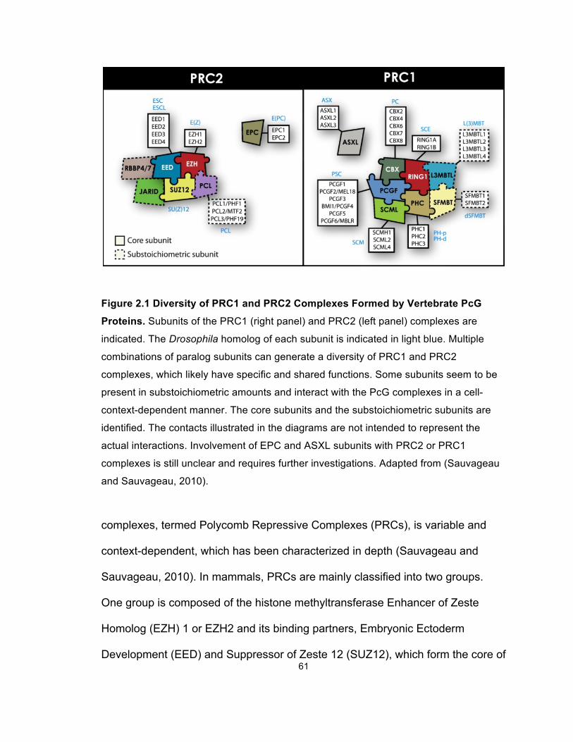
61
Figure 2.1 Diversity of PRC1 and PRC2 Complexes Formed by Vertebrate PcG
Proteins. Subunits of the PRC1 (right panel) and PRC2 (left panel) complexes are
indicated. The Drosophila homolog of each subunit is indicated in light blue. Multiple
combinations of paralog subunits can generate a diversity of PRC1 and PRC2
complexes, which likely have specific and shared functions. Some subunits seem to be
present in substoichiometric amounts and interact with the PcG complexes in a cell-
context-dependent manner. The core subunits and the substoichiometric subunits are
identified. The contacts illustrated in the diagrams are not intended to represent the
actual interactions. Involvement of EPC and ASXL subunits with PRC2 or PRC1
complexes is still unclear and requires further investigations. Adapted from (Sauvageau
and Sauvageau, 2010).
complexes, termed Polycomb Repressive Complexes (PRCs), is variable and
context-dependent, which has been characterized in depth (Sauvageau and
Sauvageau, 2010). In mammals, PRCs are mainly classified into two groups.
One group is composed of the histone methyltransferase Enhancer of Zeste
Homolog (EZH) 1 or EZH2 and its binding partners, Embryonic Ectoderm
Development (EED) and Suppressor of Zeste 12 (SUZ12), which form the core of
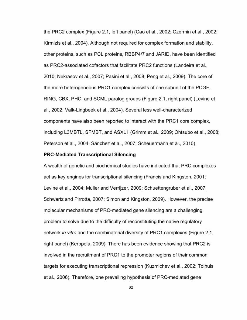
62
the PRC2 complex (Figure 2.1, left panel) (Cao et al., 2002; Czermin et al., 2002;
Kirmizis et al., 2004). Although not required for complex formation and stability,
other proteins, such as PCL proteins, RBBP4/7 and JARID, have been identified
as PRC2-associated cofactors that facilitate PRC2 functions (Landeira et al.,
2010; Nekrasov et al., 2007; Pasini et al., 2008; Peng et al., 2009). The core of
the more heterogeneous PRC1 complex consists of one subunit of the PCGF,
RING, CBX, PHC, and SCML paralog groups (Figure 2.1, right panel) (Levine et
al., 2002; Valk-Lingbeek et al., 2004). Several less well-characterized
components have also been reported to interact with the PRC1 core complex,
including L3MBTL, SFMBT, and ASXL1 (Grimm et al., 2009; Ohtsubo et al., 2008;
Peterson et al., 2004; Sanchez et al., 2007; Scheuermann et al., 2010).
PRC-Mediated Transcriptional Silencing
A wealth of genetic and biochemical studies have indicated that PRC complexes
act as key engines for transcriptional silencing (Francis and Kingston, 2001;
Levine et al., 2004; Muller and Verrijzer, 2009; Schuettengruber et al., 2007;
Schwartz and Pirrotta, 2007; Simon and Kingston, 2009). However, the precise
molecular mechanisms of PRC-mediated gene silencing are a challenging
problem to solve due to the difficulty of reconstituting the native regulatory
network in vitro and the combinatorial diversity of PRC1 complexes (Figure 2.1,
right panel) (Kerppola, 2009). There has been evidence showing that PRC2 is
involved in the recruitment of PRC1 to the promoter regions of their common
targets for executing transcriptional repression (Kuzmichev et al., 2002; Tolhuis
et al., 2006). Therefore, one prevailing hypothesis of PRC-mediated gene
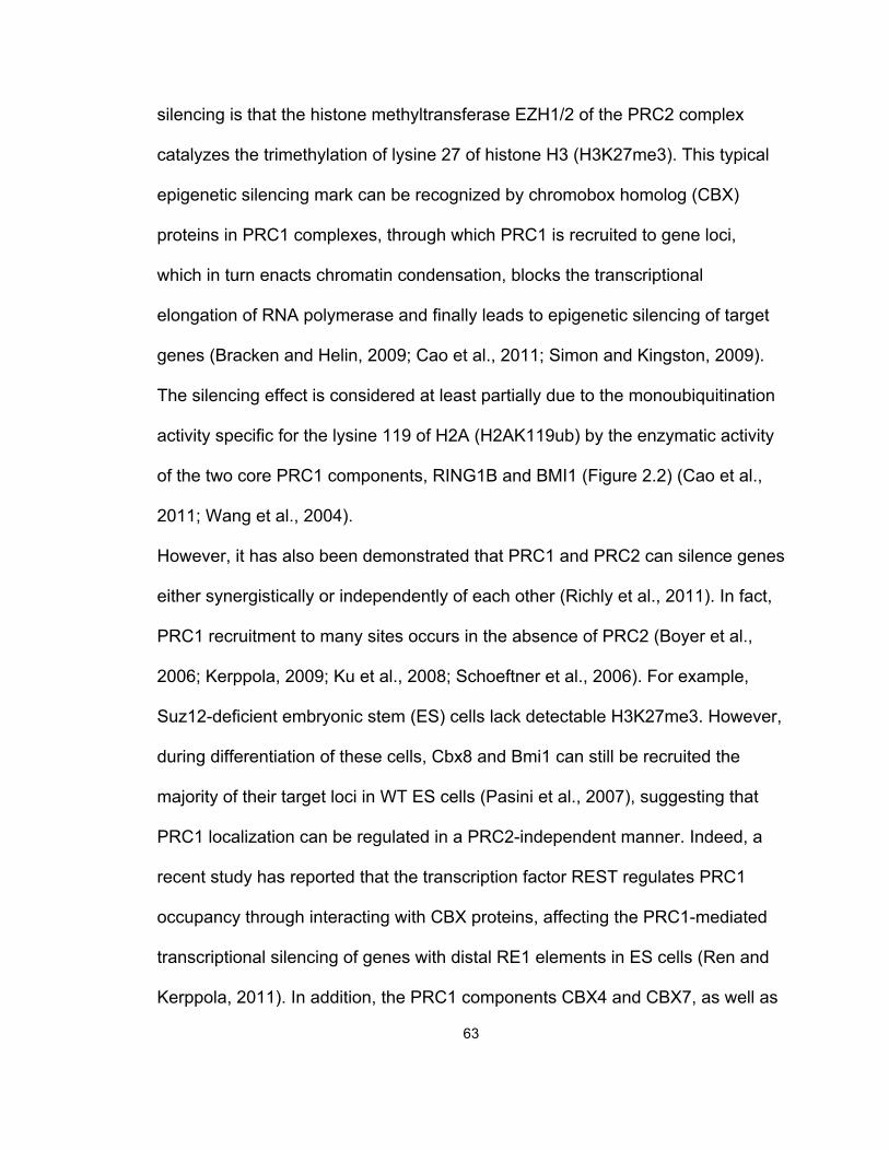
63
silencing is that the histone methyltransferase EZH1/2 of the PRC2 complex
catalyzes the trimethylation of lysine 27 of histone H3 (H3K27me3). This typical
epigenetic silencing mark can be recognized by chromobox homolog (CBX)
proteins in PRC1 complexes, through which PRC1 is recruited to gene loci,
which in turn enacts chromatin condensation, blocks the transcriptional
elongation of RNA polymerase and finally leads to epigenetic silencing of target
genes (Bracken and Helin, 2009; Cao et al., 2011; Simon and Kingston, 2009).
The silencing effect is considered at least partially due to the monoubiquitination
activity specific for the lysine 119 of H2A (H2AK119ub) by the enzymatic activity
of the two core PRC1 components, RING1B and BMI1 (Figure 2.2) (Cao et al.,
2011; Wang et al., 2004).
However, it has also been demonstrated that PRC1 and PRC2 can silence genes
either synergistically or independently of each other (Richly et al., 2011). In fact,
PRC1 recruitment to many sites occurs in the absence of PRC2 (Boyer et al.,
2006; Kerppola, 2009; Ku et al., 2008; Schoeftner et al., 2006). For example,
Suz12-deficient embryonic stem (ES) cells lack detectable H3K27me3. However,
during differentiation of these cells, Cbx8 and Bmi1 can still be recruited the
majority of their target loci in WT ES cells (Pasini et al., 2007), suggesting that
PRC1 localization can be regulated in a PRC2-independent manner. Indeed, a
recent study has reported that the transcription factor REST regulates PRC1
occupancy through interacting with CBX proteins, affecting the PRC1-mediated
transcriptional silencing of genes with distal RE1 elements in ES cells (Ren and
Kerppola, 2011). In addition, the PRC1 components CBX4 and CBX7, as well as
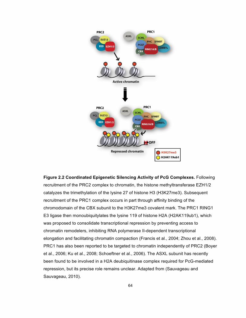
64
Figure 2.2 Coordinated Epigenetic Silencing Activity of PcG Complexes. Following
recruitment of the PRC2 complex to chromatin, the histone methyltransferase EZH1/2
catalyzes the trimethylation of the lysine 27 of histone H3 (H3K27me3). Subsequent
recruitment of the PRC1 complex occurs in part through affinity binding of the
chromodomain of the CBX subunit to the H3K27me3 covalent mark. The PRC1 RING1
E3 ligase then monoubiquitylates the lysine 119 of histone H2A (H2AK119ub1), which
was proposed to consolidate transcriptional repression by preventing access to
chromatin remodelers, inhibiting RNA polymerase II-dependent transcriptional
elongation and facilitating chromatin compaction (Francis et al., 2004; Zhou et al., 2008).
PRC1 has also been reported to be targeted to chromatin independently of PRC2 (Boyer
et al., 2006; Ku et al., 2008; Schoeftner et al., 2006). The ASXL subunit has recently
been found to be involved in a H2A deubiquitinase complex required for PcG-mediated
repression, but its precise role remains unclear. Adapted from (Sauvageau and
Sauvageau, 2010).
65
the PRC2 component EZH2, have been reported to interact with DNA
methyltransferases (DNMTs), suggesting that other than their direct enzymatic
activities that induce transcriptional repression, PRC proteins may also contribute
to gene silencing through recruiting DNMT to catalyze DNA methylation (Kim et
al., 2008; Mohammad et al., 2009; Vire et al., 2006).
PcG and Cancer
The multifaceted regulatory functions of PcG proteins in key physiological
processes during normal development strongly indicate that they are important
players in oncogenesis. Indeed, both PRC1 and PRC2 components have been
found to be overexpressed in a variety of tumors. On the one hand, expression
changes associated with oncogenic transformation have been found for at least
two core PRC1 proteins, BMI1 and RING1B, whose expression levels are
elevated in a large series of tumors including gastrointestinal tumors, pituitary
and parathyroid adenomas, and lymphomas, compared to the expression in
normal cell counterparts (Sanchez-Beato et al., 2006). Especially, BMI1 activity is
crucial for the maintenance of both normal and cancer stem cell because loss of
BMI1 leads to progressive deletion of these cell population (Lessard and
Sauvageau, 2003). On the other hand, the PRC2 proteins are also
overexpressed in multiple human cancers (Bracken and Helin, 2009; Sparmann
and van Lohuizen, 2006). For instance, increased expression of EZH2 directly
correlates to the invasive potential of multiple types of carcinomas, including
bladder cancer, prostate cancer and breast cancer (Bachmann et al., 2006;
Collett et al., 2006; Richly et al., 2011; Weikert et al., 2005). Moreover, EZH2
66
deregulation has been associated with the onset of colorectal cancer and oral
squamous carcinomas (Kidani et al., 2009; Mimori et al., 2005).
Other than transcriptional upregulation, missense mutations and chromosomal
translocations involving PcG components have been identified in different types
of human cancers as well (Sauvageau and Sauvageau, 2010). EZH2 mutations
that alter its methyltransferase activity were found in germinal center diffuse large
B cell lymphomas, follicular lymphomas and myeloid disorders (Ernst et al., 2010;
Morin et al., 2010; Nikoloski et al., 2010). Moreover, Eed mutations in mice
facilitate the formation and progression of lymphoid tumors (Richie et al., 2002),
and a Suz12 point mutation has been shown to alter the repopulation capacity of
the hematopoietic system (Majewski et al., 2008). Together, these studies
emphasize that aberrant PRC2 activity is a frequent abnormality that contributes
to oncogenesis. In addition, several PcG members, including the PRC1-
associated protein ASXL2, SUZ12 and PCL1, are involved in chromosomal
translocations observed in human cancers such as acute leukemias and
endometrial stromal sarcomas (Li et al., 2007; Nakahata et al., 2009).
Aside from the transcriptional regulation and genetic mutations that alter the PRC
activity, RNA-mediated regulatory mechanisms have also been found to gear
PcG proteins towards tumorigenesis. First, the large intervening noncoding RNA
(lincRNA) HOTAIR was reported to recruit the PRC2 complex for executing gene
silencing (Gupta et al., 2010; Rinn et al., 2007). PRC2 relocalization in cancer
cells due to HOTAIR overexpression promotes invasion and metastasis of
epithelial tumors (Gupta et al., 2010). Additionally, the lincRNA ANRIL has been
67
shown to interact with the PRC1 component CBX7, which is required for the
recruitment of PRC1 to the INK4A/ARF loci to induce transcriptional repression
(Yap et al., 2010). These findings suggest that deregulated lincRNA-mediated
PRC localization may contribute to the tumorigenic role of PcG proteins. Second,
post-transcriptional regulation of PcG proteins by miRNAs has been well-
documented as an essential mechanism to modify PRC activities. Accumulated
evidence suggest that increased PRC2 activity due to loss of miR-101 or miR-
26a promotes oncogenesis (Cao et al., 2011). Suppression of mir-183, mir-200c
or mir-213, which leads to BMI1 upregulation, has been reported in several types
of tumors (Sauvageau and Sauvageau, 2010). Furthermore, a recent study
proposed a coordinate regulation of PRC1 and PRC2 activities mediated by
miRNAs, which may play a significant role on cancer progression (Cao et al.,
2011). Taken together, multiple lines of evidence highlight the correlative role of
PcG proteins in tumorigenesis. Abnormal PRC activities due to deregulated
expression, somatic mutations, chromosomal translocations and aberrant
recruitment of PRC1 and PRC2 are associated with cancer development, mainly
because of the broad involvement of PRC complexes in major biological
processes that are essential for both normal physiology and pathogenesis.
CBX8
Chromobox homolog 8 (CBX8), also known as HPC3 (Human Polycomb 3),
belongs to the CBX protein family (including CBX2, 4, 6, 7 and 8) that are
homologs of the Drosophila Polycomb (Pc) protein (Kerppola, 2009). CBX8 was
originally characterized as a transcriptional repressor, interacting with RING1a/b
68
and associating with BMI1 in PRC1 (Bardos et al., 2000). A previous study has
reported that as a PRC1 component, CBX8 represses the INK4a/ARF expression
in fibroblasts (Dietrich et al., 2007). Further studies showed that several distinct
PRC1 complexes colocalize and regulate the INK4a/ARF expression, suggesting
that the INK4a/ARF locus is a general target for PRC1 complexes, rather than a
CBX8-specific downstream target (Maertens et al., 2009). Therefore, the exact
role of CBX8 in transcriptional regulation remains largely undefined. It has been
reported that certain CBX proteins, such as CBX4, can associate with protein
complexes other than PRC1, thereby playing a PRC1-independent role in
transcriptional regulation (Kerppola, 2009). However, it remains unknown
whether CBX8 has a PRC1-independent function as such, and if so, what the
biological implication might be.
Interestingly, despite its well-characterized transcription repressor role, CBX8
has also been shown to be present in complexes recruited by MLL fusion
proteins, which induces leukemogenesis through transcriptional activation
(Monroe et al., 2010; Mueller et al., 2007). More specifically, CBX8 directly
interacts with two of the most common MLL fusion partners, AF9 and ENL, both
of which are transcription factors involved in gene activation (Garcia-Cuellar et al.,
2001; Hemenway et al., 2001; Monroe et al., 2010). However, the functional
significance of this seemingly paradoxical association has not yet been defined,
therefore raising the question that what the role of CBX8 plays in MLL-mediated
leukemogenesis.
HIV Tat interacting protein of 60 kDa
69
HIV Tat interacting protein of 60 kDa (TIP60) is a founding member of the MYST
(Moz, Ybf2/Sas3, Sas2, Tip60) protein family, the largest family of histone
acetyltransferases (HATs) that are highly conserved in all eukaryotes (Voss and
Thomas, 2009). TIP60 is the catalytic subunit of the evolutionarily conserved
NuA4 (nucleosome acetyltransferase of histone H4) complex (Doyon et al., 2004).
Previous studies have shown that TIP60, in the context of the stable multi-protein
complex, selectively acetylates nucleosomal H4 at lysines 5, 8, 12, and 16, as
well as H2A, in vitro (Ikura et al., 2000; Sapountzi et al., 2006). Evidence from
Drosophila indicates that modified histone variants, such as phospho-H2Av, can
also be acetylated by TIP60 (Kusch et al., 2004). Apart from these histone
substrates, TIP60 also has by far the most known non-histone targets. The
majority of these substrates are transcription factors (Sapountzi and Cote, 2011).
For example, one of the extensively studied non-histone substrates of TIP60 is
the tumor suppressor p53. TIP60 can specifically acetylates lysine 120 (K120) of
p53 upon DNA damage, thereby modulating the decision between cell cycle
arrest and apoptosis upon p53 activation and by co-activating p53-driven
transcription (Berns et al., 2004; Doyon et al., 2004; Legube et al., 2004; Sykes
et al., 2006; Tang et al., 2006). Other transcription factors whose activity is
regulated by TIP60-mediated acetylation include c-Myc (Patel et al., 2004), RB
(Leduc et al., 2006), and androgen receptor (Brady et al., 1999; Gaughan et al.,
2001; Gaughan et al., 2002). In addition to transcription factors, it has been
shown that the ATM kinase, a key player in the DNA repair pathway, is also
subjected to TIP60-mediated acetylation (Sun et al., 2005).
70
The broad spectrum of its acetylated substrates indicates that TIP60 plays a role
in multiple biological processes. First, it is critically involved in DNA repair, mainly
through the TIP60-p53 functional interaction, acetylation-mediated ATM
activation, and TIP60-related H2Av modification (Avvakumov and Cote, 2007).
Second, TIP60 has been well-characterized as a transcriptional co-activator in
multiple key signaling pathways, such as c-Myc signaling, Wnt signaling, NF-κB
signaling, and androgen receptor signaling (Baek et al., 2002; Frank et al., 2003;
Sapountzi et al., 2006; Sierra et al., 2006). Although in the majority of cases,
TIP60 has been associated with transcription activation, it has also been reported
to induce gene repression in a context-dependent manner. For instance, TIP60
can interact with HDAC to modulate STAT3 activity (Xiao et al., 2003). The
multiple roles of TIP60 in DNA repair and transcriptional regulation have linked it
to a number of human diseases, including HIV infection and Acquired Immune
Deficiency Syndrome (AIDS), Alzheimer’s disease, and cancer (Avvakumov and
Cote, 2007; Baek et al., 2002; Cao and Sudhof, 2001; Creaven et al., 1999;
Kinoshita et al., 2002). A wealth of studies have suggested the involvement of
TIP60 in oncogenesis, although in many of these cases, the direct mechanistic
evidence is still lacking (Figure 2.3) (Avvakumov and Cote, 2007).
It is worth noting that so far, TIP60 has not been shown to be involved in
chromosomal translocation, which is the most common cause of human
leukemias. However, it can act as a transcriptional regulator when interacting
with TEL, which fuses with AML1 in the majority of childhood leukemias (So and
van der Reijden, 2008). An additional study shows that TIP60 directly interacts
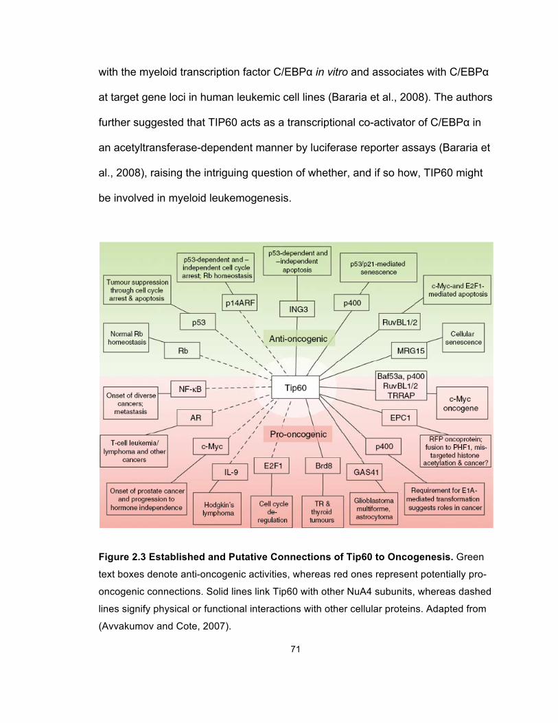
71
with the myeloid transcription factor C/EBPα in vitro and associates with C/EBPα
at target gene loci in human leukemic cell lines (Bararia et al., 2008). The authors
further suggested that TIP60 acts as a transcriptional co-activator of C/EBPα in
an acetyltransferase-dependent manner by luciferase reporter assays (Bararia et
al., 2008), raising the intriguing question of whether, and if so how, TIP60 might
be involved in myeloid leukemogenesis.
Figure 2.3 Established and Putative Connections of Tip60 to Oncogenesis. Green
text boxes denote anti-oncogenic activities, whereas red ones represent potentially pro-
oncogenic connections. Solid lines link Tip60 with other NuA4 subunits, whereas dashed
lines signify physical or functional interactions with other cellular proteins. Adapted from
(Avvakumov and Cote, 2007).
72
Materials and Methods
Cell Culture and Animal Use
HeLa and 293 cells were cultured in Dulbecco’s modified Eagle’s medium
(DMEM) supplemented with 10% fetal bovine serum (FBS) and non-essential
amino acids. MLL-AF9, MLL-ENL and E2A-HLF cells were cultured in Iscove’s
modified Dulbecco’s medium (IMDM) supplemented with 15% fetal calf serum
(FCS, Stem Cell Technologies). THP-1, Mono Mac 6 (MM6), and K562 cells
were cultured in RPMI-1640 medium supplemented with 10% FBS. The Tripz-
RFP-shCBX8 expression was induced by 0.5 µg/ml doxycycline. For conditional
knockout of Cbx8 in mice, two LoxP sites were inserted to flank the exons 2 to 4
of the Cbx8 gene. Cbx8 excision was achieved upon 4-OHT treatment. Full
details of conditional gene targeting of Cbx8 and analysis of Cbx8−/− embryos will
be provided in a subsequent manuscript (H.K., unpublished). All animal
experiments in this study were approved by the University of Michigan
Committee on Use and Care of Animals and Unit for Laboratory Animal Medicine
(ULAM).
In Vivo Leukemogenesis Assays
Lin- BM was isolated from 6 to 8-week-old mice (Cbx8 f/f; Cre+) injected with 5-
fluorouacil (see Supplemental Information). The harvested Lin- BM cells were
retrovirally transduced with MigR1-MLL-AF9 by two rounds of spinoculation in the
presence of either 4-OHT (100 nM) or ethanol as a control. The cells were then
counted and injected intravenously through the tail vein to cohorts of lethally
73
irradiated (900 rads) C57BL/6 mice (3.5 x 10⁴ cells per injection). Recipient mice
were maintained on antibiotics for 2 weeks after transplantation.
Complete Blood Count Analysis
Peripheral blood was harvested in EDTA (ethylenediaminetetraacetic acid)-
containing Microtainer tubes (BD Biosciences) and subjected for analysis
performed by the ULAM laboratory.
Vector Construction
The pfMLL-AF9 vector and pMSCV-neo constructs encoding MLL-AF9 were
previously described (Muntean et al., 2010). The FLAG-CBX8 and HA-DOT1L
constructs were generously provided by Dr. Robert Slany (University of Erlangen,
Germany). Expression vectors for MLL-AF9 mutants and CxxC-AF9 mutants
(T542A and T554A) were generated by restriction enzyme digestion and PCR-
based mutagenesis. The pSM2c scrambled, pSM2c shCbx8 (clone ID
V2MM_66688), pSM2c shRing1b (clone IDs V2MM_197123 and V2MM_83055),
pSM2c shBmi1 (clone ID V2HS_48576) and pSM2c shTip60 (clone ID
V2MM_92042) retroviral vectors and the pTRIPZ-shCBX8 lentiviral vector (clone
ID V2THS_58819) were purchased from Open Biosystems. An additional shRNA
set for Tip60 knockdown based on the pGFP-V-RS retroviral vector system
(Catalog Numbers TG512714 and TG30013) was purchased from OriGene.
Immunoprecipitation and Immunoblotting Assays
Cell lysates preparation, immunoprecipitation and immunoblotting were
performed as previously described (Muntean et al., 2010). Immunoprecipitation
was performed overnight, followed by three times of wash using BC-300 buffer.
74
Proteins were eluted by boiling in 1X SDS-loading buffer and separated by SDS-
PAGE, followed by western blot analysis. Primary antibodies included rabbit anti-
FLAG (Sigma), goat anti-Myc (Abcam), rabbit anti-HA (Abcam), rabbit anti-AF9
(Bethyl Laboratories, Inc.), mouse anti-β-actin (Sigma), rabbit anti-CBX8 (Abcam),
goat anti-CBX8 (Everest Biotech), mouse anti-MLL N (Millipore), mouse anti-
Bmi1 (Millipore), mouse anti-Ring1b (Medical and Biological Laboratories), and
goat anti-TIP60 (Santa Cruz). An agarose affinity beads coupled to mouse anti-
FLAG M2 monoclonal antibody was purchased from Sigma. An agarose affinity
beads coupled to mouse anti-Myc M2 monoclonal antibody was purchased from
Clontech. Protein G agarose beads were purchased from Roche.
Virus Production for Bone Marrow Transformation (BMT) Assays
The pMSCV (for FLAG-MLL-AF9 and the mutants), pSM2c (for shScram, shCbx8,
shRing1b-1, shRing1b-2, shBmi1 and shTip60) and pGFP-V-RS (for shScram-1
and shTip60-1) constructs were transfected using Fugene 6 reagent (Roche) into
Plat-E cells selected in puromycin (1 µg/ml) and blasticidin (10 µg/ml). Media
containing the recombinant retrovirus was collected for transduction at 48 and 72
hours post transfection. Lentivirus production was performed by the University of
Michigan Vector Core. Medium containing virus was collected 48 h post
transfection.
BMT Assays
BMT assays were performed as previously described, with some modifications
(Muntean et al., 2010). Briefly, BM was harvested from 6 to 8-week-old mice
(Cbx8 f/f; Cre+, or Cbx8 f/f; Cre-) injected with 5-fluorouacil. Cells were collected
75
from the tibia and femur. Lin- BM cells were isolated using the EasySep® Mouse
Hematopoietic Progenitor Cell Enrichment Kit (STEMCELL Technologies). The
cells were then transduced with recombinant retrovirus containing MLL-AF9 (WT
or mutants) by spinoculation and plated in methylcellulose medium (M3234,
STEMCELL Technologies), supplemented with IL-3, IL-6, SCF, GM-CSF and 1
mg/ml G418. After three rounds of plating, CFU (with > 50 cells) was scored
under the microscope; colonies were stained using 0.1% INT and scanned. Cells
harvested at the end of the experiment were cytospun and stained with the Hema
3 Stain Kit (Thermo Fisher Scientific). Images were acquired using a 100x lens
under an Olympus BX-51 microscope (Olympus). For secondary transductions,
MLL-AF9-transduced cells harvested after the third round of plating were
transduced with the indicated shRNA retrovirus by spinoculation and plated as
described above, using 2 µg/ml puromycin in methylcellulose medium for
selection. For in vitro Cbx8 excision, cells were treated with 100 nM 4-OHT
(Sigma) either during transduction (pre-transformation excision) or after three
rounds of plating (post-transformation excision).
Colony Formation Assays
For assessing the total hematopoietic progenitor cell activity, BM was harvested
from Cbx8 f/f; Cre+ mice 6 months after in vivo 4-OHT administration or corn oil
control treatment. After red blood cell lysis using ACK lysis buffer (Cambrex),
nucleated BM cells were counted and plated in methylcellulose medium (M3534,
STEMCELL Technologies). The colony number is counted 7 days after plating.
Quantitative RT-PCR
76
Reverse transcription and quantitative PCR were performed as previously
described (Muntean et al., 2010). FLAG-MLL-AF9 was detected using the
following primers for SYBR green detection: FLAG-F-5’-ggactacaaggacgacgatga-
3’ and MLL-R-5’-acagctgtgcgccatgtt-3’. TaqMan primer probe sets were
purchased from Applied Biosystems for mouse Hoxa9, Cbx8, Ring1b, Bmi1,
Tip60, Ink4a/Arf and β-actin, as well as human HOXA9, CBX8, TIP60 and β-
ACTIN. Expression levels were analyzed using the comparative ΔΔCt method as
described in ABI User Bulletin #2.
Luciferase Reporter Assays
293 cells were transiently transfected with MSCV, MLL-AF9 (WT and mutants),
CMV-Renilla, and Hoxa9-LUC (or Myc E box-LUC) constructs using FuGene 6
(Roche) according to the manufacturer’s instructions. The cells then underwent
serum starvation in 0.5% FBS in OPTI-MEM media for 48 h. Luciferase reporter
assays were performed using a Dual Luciferase assay kit (Promega) according
to manufacturer’s instructions. Emission was detected using a Monolight 3010
luminometer (BD Biosciences). For the luciferase assay with siRNA knockdown,
HeLa cells were transiently transfected with the DNA constructs listed above
using Lipofectamine (Invitrogen) and siRNA smart pools for targeting CBX8,
RING1b, BMI1 and TIP60 (Dharmacon) using Oligofectamine (Invitrogen)
according to the manufacturer’s instructions. The firefly luciferase activity
readouts are normalized to Renilla luciferase readouts.
Chromatin Immunoprecipitation
77
ChIP was performed as previously described (Milne et al., 2005b), using primary
antibodies for CBX8 (Abcam), Cbx8 (also named as Mpc3, Santa Cruz), RNAP II
(Covance), TIP60 (Santa Cruz), AF9 (Bethyl Laboratories, Inc.) and histone H3
(Abcam). Quantitative RT-PCR was performed on the precipitated DNAs using
primers and qPCR probes described before (Muntean et al., 2010). Binding was
quantitated as follows: ΔCT = CT(input) − CT(Chromatin IP), % total = 2ΔCT.
Annexin V Staining
For apoptosis analysis, MLL-AF9 leukemic cells from Cbx8 f/f; Cre+ and Cbx8 f/f;
Cre- mice were treated with 4-OHT (100 nM) for 72 h, using ethanol as a control
treatment. The treated cells were washed and resuspended in Annexin V binding
buffer (BD Biosciences). The resuspended cells were stained with Annexin V-
FITC (BioLegend) and 1 µg/ml DAPI. Stained cells were then analyzed by flow
cytometry.
Cell Cycle Analysis
MLL-AF9 leukemic cells from Cbx8 f/f; Cre+ and Cbx8 f/f; Cre- mice were treated
with 4-OHT (100 nM) for 72 h, using ethanol as a control treatment. The treated
cells were washed twice in phosphate buffered saline (PBS). The cells were then
resuspended in ice-cold PBS and fixed in 70% ethanol for at least 24 h. The fixed
cells were washed with standard buffer for flow cytometry, treated with 100 µg/ml
RNase and stained with 10 µg/ml PI. Stained cells were then analyzed by flow
cytometry.
Competitive Transplantation Assays
78
Competitive transplantation assays were performed as previously described, with
some modifications (Maillard et al., 2009). Briefly, BM was harvested from Cbx8
f/f; Cre+ mice 6 months after in vivo 4-OHT administration or corn oil control
treatment. After red blood cell lysis using ACK lysis buffer (Cambrex), nucleated
BM cells were counted and resuspended in ice-cold phosphate-buffered saline
(PBS). These cells (CD45.2+) were mixed at specific ratios with 5 x
105 CD45.1+ competitor BM cells from B6-SJL mice (1:1, 3:1 and 9:1). This
mixture was injected intravenously through the tail vein to cohorts of lethally
irradiated (900 rads) B6-SJL (B6-CD45.1) mice. Recipient mice were maintained
on antibiotics for 2 weeks after transplantation, and their peripheral blood was
assessed by flow cytometry.
Flow Cytometry and Cell Sorting
After blocking non-specific binding with unlabeled rat plus mouse IgG (Sigma-
Aldrich), cells were stained on ice in PBS plus 4% fetal calf serum (FCS) and
sorted on FACSAria (BD Biosciences). Analysis was performed on LSR II,
FACSCanto, or FACSAria (BD Biosciences). Files were analyzed in FlowJo
(TreeStar).
Statistical Analysis
Statistical significance was determined by Student’s t test using the Excel
software (Microsoft 2007); p< 0.05 was considered statistically significant.
Antibodies for Flow Cytometry Analysis
The antibodies used for flow cytometry analysis were obtained from BioLegend
and listed in Table 2.1.
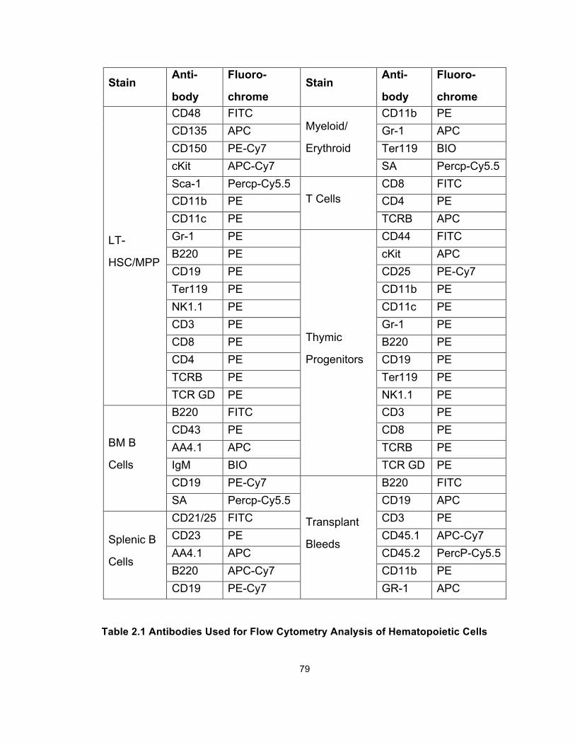
79
Stain Anti-
body
Fluoro-
chrome Stain
Anti-
body
Fluoro-
chrome
LT-
HSC/MPP
CD48 FITC Myeloid/
Erythroid
CD11b PE CD135 APC Gr-1 APC CD150 PE-Cy7 Ter119 BIO cKit APC-Cy7 SA Percp-Cy5.5 Sca-1 Percp-Cy5.5
T Cells CD8 FITC
CD11b PE CD4 PE CD11c PE TCRB APC Gr-1 PE
Thymic
Progenitors
CD44 FITC B220 PE cKit APC CD19 PE CD25 PE-Cy7 Ter119 PE CD11b PE NK1.1 PE CD11c PE CD3 PE Gr-1 PE CD8 PE B220 PE CD4 PE CD19 PE TCRB PE Ter119 PE TCR GD PE NK1.1 PE
BM B
Cells
B220 FITC CD3 PE CD43 PE CD8 PE AA4.1 APC TCRB PE IgM BIO TCR GD PE CD19 PE-Cy7
Transplant
Bleeds
B220 FITC SA Percp-Cy5.5 CD19 APC
Splenic B
Cells
CD21/25 FITC CD3 PE CD23 PE CD45.1 APC-Cy7 AA4.1 APC CD45.2 PercP-Cy5.5 B220 APC-Cy7 CD11b PE CD19 PE-Cy7 GR-1 APC
Table 2.1 Antibodies Used for Flow Cytometry Analysis of Hematopoietic Cells
80
Results
CBX8 Specifically Interacts with MLL-AF9 at the C-Terminal Domain (CTD)
Previous studies have reported that the MLL fusion partner AF9 directly interacts
with CBX8 through the evolutionarily conserved CTD (Figure 2.4A) (Garcia-
Cuellar et al., 2001; Hemenway et al., 2001; Monroe et al., 2010). However,
whether this interaction is retained in MLL-AF9 fusion protein has not been
defined. To address this question, we transiently co-expressed epitope-tagged
MLL-AF9 and CBX8 in human embryonic kidney 293 cells, using a FLAG-tagged
“empty” vector as a negative control. Specific interaction between CBX8 and
MLL-AF9 was detected by immunoprecipitation (IP) experiments. When using
AF9-conjugated agarose beads to pull down the full-length fusion protein, we
consistently observed that CBX8 coprecipitated with MLL-AF9 (Figure 2.4B). To
further characterize this interaction, we performed IP experiments in the
presence of Benzonase. Using anti-FLAG antibody to pull down FLAG-tagged
MLL-AF9, we detected endogenous CBX8 coprecipitating with the fusion protein,
suggesting that CBX8 interacts with MLL-AF9 in a DNA-independent manner
(Figures 2.4C). Next, we characterized the critical CBX8 interaction sites on MLL-
AF9, by generating 15 point mutants within the CTD through single amino acid
substitution. By Co-IP experiments, we identified two point mutants (T542A and
T554A) that specifically disrupt the CBX8 interaction (Figures 2.4A and 2.4D).
This observation was further supported by reciprocal Co-IP experiments, using
anti-FLAG or anti-Myc antibodies to pull down CBX8 or CxxC-AF9, respectively
(Figures 2.4E and 2.4F), in which case CxxC-AF9, a previous characterized
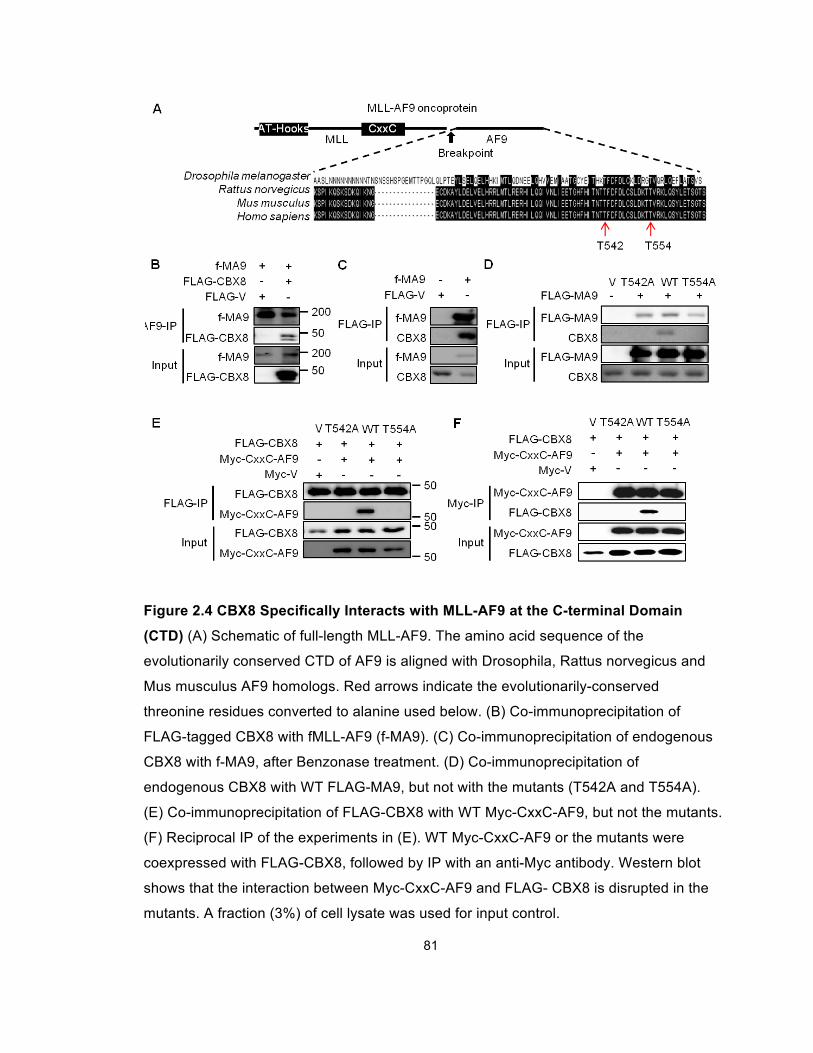
81
Figure 2.4 CBX8 Specifically Interacts with MLL-AF9 at the C-terminal Domain
(CTD) (A) Schematic of full-length MLL-AF9. The amino acid sequence of the
evolutionarily conserved CTD of AF9 is aligned with Drosophila, Rattus norvegicus and
Mus musculus AF9 homologs. Red arrows indicate the evolutionarily-conserved
threonine residues converted to alanine used below. (B) Co-immunoprecipitation of
FLAG-tagged CBX8 with fMLL-AF9 (f-MA9). (C) Co-immunoprecipitation of endogenous
CBX8 with f-MA9, after Benzonase treatment. (D) Co-immunoprecipitation of
endogenous CBX8 with WT FLAG-MA9, but not with the mutants (T542A and T554A).
(E) Co-immunoprecipitation of FLAG-CBX8 with WT Myc-CxxC-AF9, but not the mutants.
(F) Reciprocal IP of the experiments in (E). WT Myc-CxxC-AF9 or the mutants were
coexpressed with FLAG-CBX8, followed by IP with an anti-Myc antibody. Western blot
shows that the interaction between Myc-CxxC-AF9 and FLAG- CBX8 is disrupted in the
mutants. A fraction (3%) of cell lysate was used for input control.
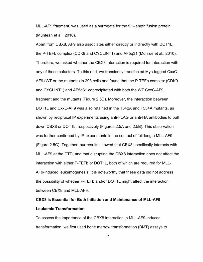
82
MLL-AF9 fragment, was used as a surrogate for the full-length fusion protein
(Muntean et al., 2010).
Apart from CBX8, AF9 also associates either directly or indirectly with DOT1L,
the P-TEFb complex (CDK9 and CYCLINT1) and AF5q31 (Monroe et al., 2010).
Therefore, we asked whether the CBX8 interaction is required for interaction with
any of these cofactors. To this end, we transiently transfected Myc-tagged CxxC-
AF9 (WT or the mutants) in 293 cells and found that the P-TEFb complex (CDK9
and CYCLINT1) and AF5q31 coprecipitated with both the WT CxxC-AF9
fragment and the mutants (Figure 2.5D). Moreover, the interaction between
DOT1L and CxxC-AF9 was also retained in the T542A and T554A mutants, as
shown by reciprocal IP experiments using anti-FLAG or anti-HA antibodies to pull
down CBX8 or DOT1L, respectively (Figures 2.5A and 2.5B). This observation
was further confirmed by IP experiments in the context of full-length MLL-AF9
(Figure 2.5C). Together, our results showed that CBX8 specifically interacts with
MLL-AF9 at the CTD, and that disrupting the CBX8 interaction does not affect the
interaction with either P-TEFb or DOT1L, both of which are required for MLL-
AF9-induced leukemogenesis. It is noteworthy that these data did not address
the possibility of whether P-TEFb and/or DOT1L might affect the interaction
between CBX8 and MLL-AF9.
CBX8 Is Essential for Both Initiation and Maintenance of MLL-AF9
Leukemic Transformation
To assess the importance of the CBX8 interaction in MLL-AF9-induced
transformation, we first used bone marrow transformation (BMT) assays to
83
examine the transformation ability of the MLL-AF9 mutants (T542A and T554A),
which lack the CBX8 interaction. Briefly, Lin- hematopoietic cells derived from
Figure 2.5 Specific Disruption of the CBX8 Interaction Does not Affect the
Interaction between DOT1L and MLL-AF9 (A) Co-immunoprecipitation of both WT
Myc-CxxC-AF9 and the mutants with HA-DOT1L. (B) WT Myc-CxxC-AF9 or the mutants
were coexpressed with HA- DOT1L, followed by IP with an anti-Myc antibody. Western
blot shows HA- DOT1L coprecipitating with both WT Myc-CxxC-AF9 and the mutants. (C)
WT FLAG-MA9 or the FLAG-MA9 mutants were coexpressed with HA-DOT1L, followed
by IP with an anti-HA antibody. Western blot shows HA- DOT1L coprecipitating with both
WT FLAG-MA9 and the mutants. (D) Co-immunoprecipitation of endogenous CDK9,
CYCLIN T1 and AF5q31 with both WT Myc-CxxC-AF9 and the mutants. All of the Co-IP
experiments included an epitope-tagged empty vector (FLAG-V or Myc-V) as a control
and were performed in 293 cells. A fraction (3%) of cell lysate was used for input control.
84
primary murine bone marrow (BM) were retrovirally transduced with either WT
MLL-AF9 or the mutants, followed by three consecutive rounds of plating (Figure
2.6A). Despite the comparable expression of the fusion transcripts, as confirmed
by real-time quantitative polymerase chain reaction (RT-PCR), the T542A and
T554A mutations completely abolished myeloid transformation, whereas the WT
control potently transformed primary hematopoietic cells, forming a large number
of colonies (Figures 2.6B and 2.6C). The tertiary colonies formed by WT MLL-
AF9-transduced cells displayed a dense, compact morphology, indicative of
immortalization. Wright Giemsa staining shows that these colonies are composed
of myeloblasts (Figure 2.6D). In contrast, the MLL-AF9 mutant-transduced cells
failed to form colonies in the second round of selection, and the residual living
cells were composed primarily of monocytes and macrophages (Figure 2.6D).
To further confirm that Cbx8 is required for MLL-AF9-induced transformation, we
transduced the MLL-AF9-transformed BM cells with either the control shRNA or a
shRNA directed against Cbx8 after the third round plating, followed by puromycin
selection (Figure 2.7A). Cbx8 expression, as measured by RT-qPCR, was
effectively downregulated (Figure 2.7B), whereas the MLL-AF9 expression level
was not significantly affected (p>0.05, Figure 2.7C). As expected, knockdown of
Cbx8 significantly reduced the colony formation ability of MLL-AF9-transduced
cells, compared to the control (p<0.01, Figures 2.7D and 2.7E). Together, these
results suggest that the CBX8/MLL-AF9 interaction is required for MLL-AF9-
mediated immortalization.
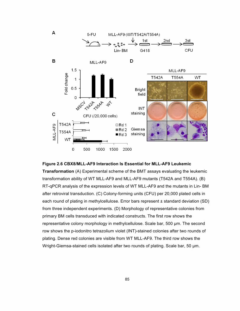
85
Figure 2.6 CBX8/MLL-AF9 Interaction Is Essential for MLL-AF9 Leukemic
Transformation (A) Experimental scheme of the BMT assays evaluating the leukemic
transformation ability of WT MLL-AF9 and MLL-AF9 mutants (T542A and T554A). (B)
RT-qPCR analysis of the expression levels of WT MLL-AF9 and the mutants in Lin- BM
after retroviral transduction. (C) Colony-forming units (CFU) per 20,000 plated cells in
each round of plating in methylcellulose. Error bars represent ± standard deviation (SD)
from three independent experiments. (D) Morphology of representative colonies from
primary BM cells transduced with indicated constructs. The first row shows the
representative colony morphology in methylcellulose. Scale bar, 500 µm. The second
row shows the p-iodonitro tetrazolium violet (INT)-stained colonies after two rounds of
plating. Dense red colonies are visible from WT MLL-AF9. The third row shows the
Wright-Giemsa-stained cells isolated after two rounds of plating. Scale bar, 50 µm.
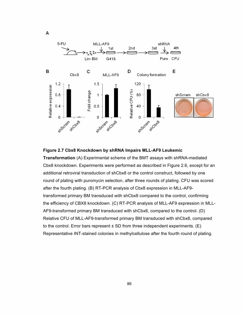
86
Figure 2.7 Cbx8 Knockdown by shRNA Impairs MLL-AF9 Leukemic
Transformation (A) Experimental scheme of the BMT assays with shRNA-mediated
Cbx8 knockdown. Experiments were performed as described in Figure 2.6, except for an
additional retroviral transduction of shCbx8 or the control construct, followed by one
round of plating with puromycin selection, after three rounds of plating. CFU was scored
after the fourth plating. (B) RT-PCR analysis of Cbx8 expression in MLL-AF9-
transformed primary BM transduced with shCbx8 compared to the control, confirming
the efficiency of CBX8 knockdown. (C) RT-PCR analysis of MLL-AF9 expression in MLL-
AF9-transformed primary BM transduced with shCbx8, compared to the control. (D)
Relative CFU of MLL-AF9-transformed primary BM transduced with shCbx8, compared
to the control. Error bars represent ± SD from three independent experiments. (E)
Representative INT-stained colonies in methylcellulose after the fourth round of plating.
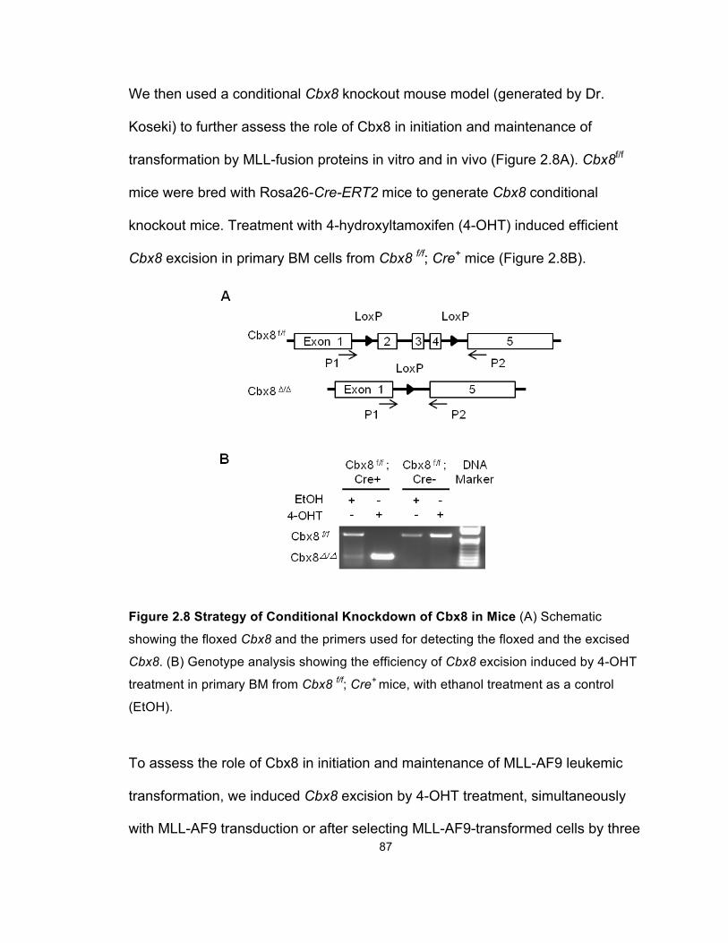
87
We then used a conditional Cbx8 knockout mouse model (generated by Dr.
Koseki) to further assess the role of Cbx8 in initiation and maintenance of
transformation by MLL-fusion proteins in vitro and in vivo (Figure 2.8A). Cbx8f/f
mice were bred with Rosa26-Cre-ERT2 mice to generate Cbx8 conditional
knockout mice. Treatment with 4-hydroxyltamoxifen (4-OHT) induced efficient
Cbx8 excision in primary BM cells from Cbx8 f/f; Cre+ mice (Figure 2.8B).
Figure 2.8 Strategy of Conditional Knockdown of Cbx8 in Mice (A) Schematic
showing the floxed Cbx8 and the primers used for detecting the floxed and the excised
Cbx8. (B) Genotype analysis showing the efficiency of Cbx8 excision induced by 4-OHT
treatment in primary BM from Cbx8 f/f; Cre+ mice, with ethanol treatment as a control
(EtOH).
To assess the role of Cbx8 in initiation and maintenance of MLL-AF9 leukemic
transformation, we induced Cbx8 excision by 4-OHT treatment, simultaneously
with MLL-AF9 transduction or after selecting MLL-AF9-transformed cells by three
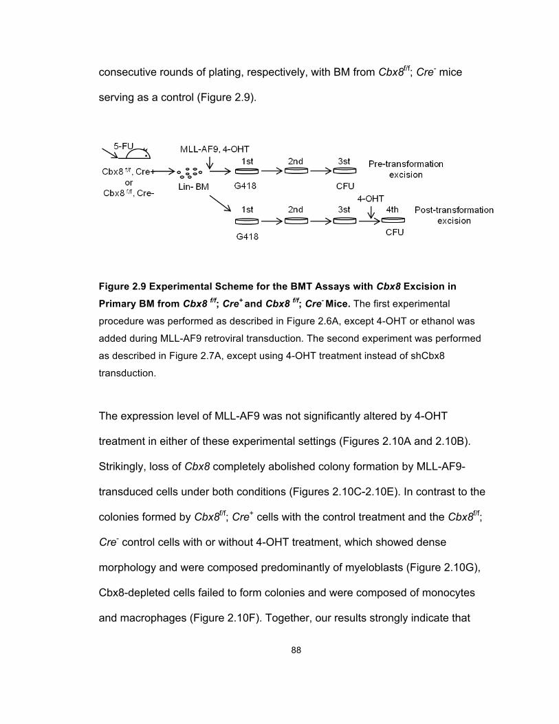
88
consecutive rounds of plating, respectively, with BM from Cbx8f/f; Cre- mice
serving as a control (Figure 2.9).
Figure 2.9 Experimental Scheme for the BMT Assays with Cbx8 Excision in
Primary BM from Cbx8 f/f; Cre+ and Cbx8 f/f; Cre- Mice. The first experimental
procedure was performed as described in Figure 2.6A, except 4-OHT or ethanol was
added during MLL-AF9 retroviral transduction. The second experiment was performed
as described in Figure 2.7A, except using 4-OHT treatment instead of shCbx8
transduction.
The expression level of MLL-AF9 was not significantly altered by 4-OHT
treatment in either of these experimental settings (Figures 2.10A and 2.10B).
Strikingly, loss of Cbx8 completely abolished colony formation by MLL-AF9-
transduced cells under both conditions (Figures 2.10C-2.10E). In contrast to the
colonies formed by Cbx8f/f; Cre+ cells with the control treatment and the Cbx8f/f;
Cre- control cells with or without 4-OHT treatment, which showed dense
morphology and were composed predominantly of myeloblasts (Figure 2.10G),
Cbx8-depleted cells failed to form colonies and were composed of monocytes
and macrophages (Figure 2.10F). Together, our results strongly indicate that
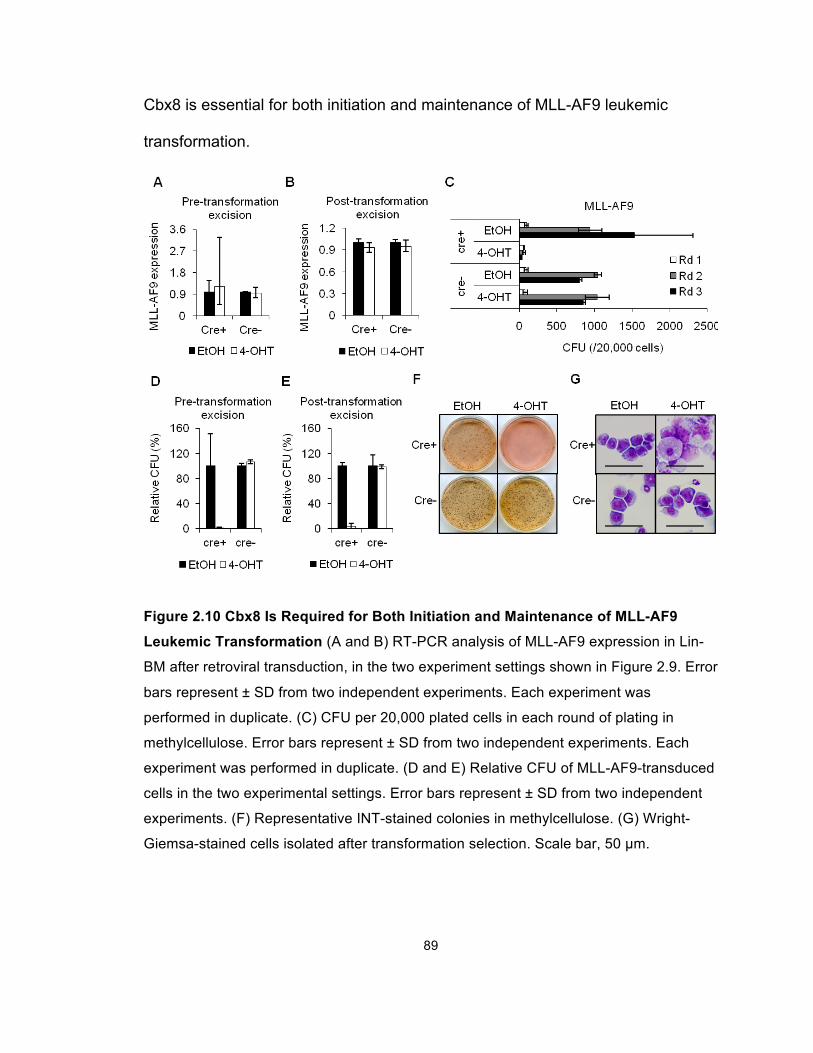
89
Cbx8 is essential for both initiation and maintenance of MLL-AF9 leukemic
transformation.
Figure 2.10 Cbx8 Is Required for Both Initiation and Maintenance of MLL-AF9
Leukemic Transformation (A and B) RT-PCR analysis of MLL-AF9 expression in Lin-
BM after retroviral transduction, in the two experiment settings shown in Figure 2.9. Error
bars represent ± SD from two independent experiments. Each experiment was
performed in duplicate. (C) CFU per 20,000 plated cells in each round of plating in
methylcellulose. Error bars represent ± SD from two independent experiments. Each
experiment was performed in duplicate. (D and E) Relative CFU of MLL-AF9-transduced
cells in the two experimental settings. Error bars represent ± SD from two independent
experiments. (F) Representative INT-stained colonies in methylcellulose. (G) Wright-
Giemsa-stained cells isolated after transformation selection. Scale bar, 50 µm.
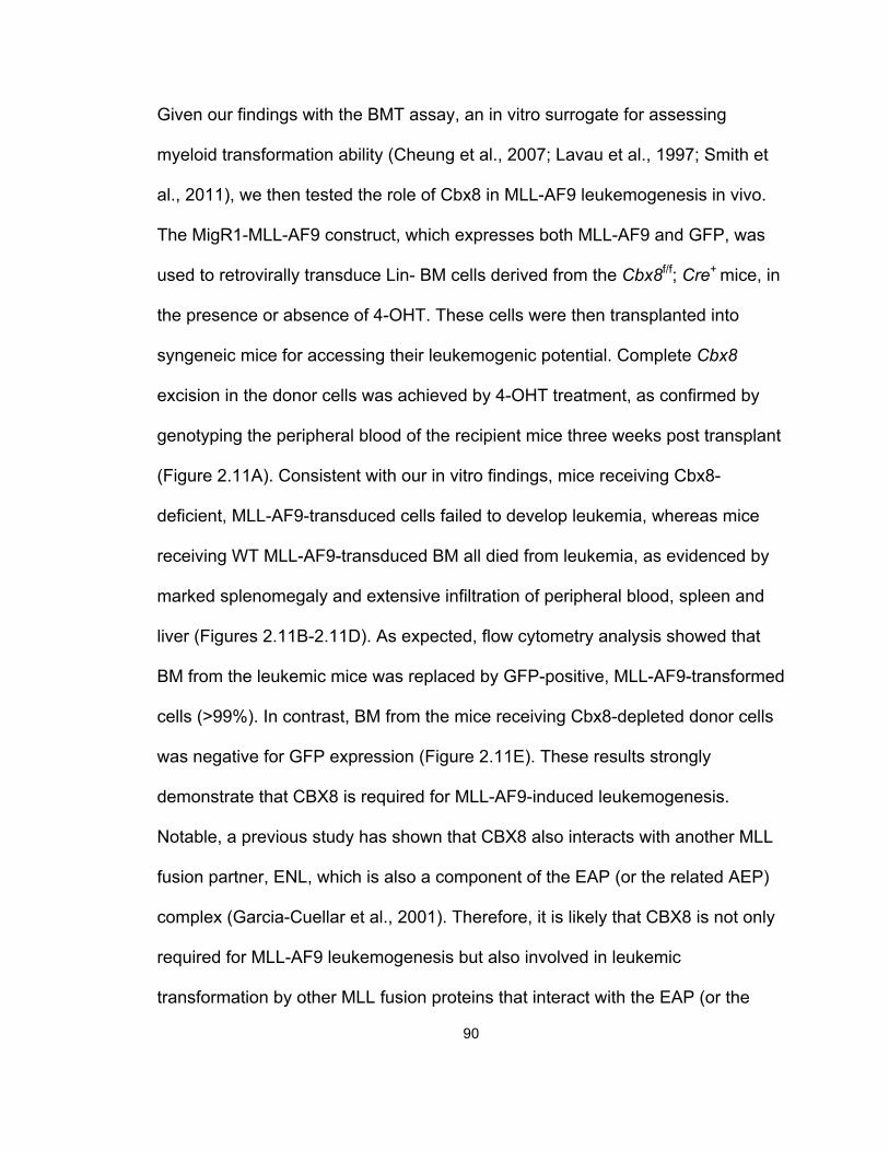
90
Given our findings with the BMT assay, an in vitro surrogate for assessing
myeloid transformation ability (Cheung et al., 2007; Lavau et al., 1997; Smith et
al., 2011), we then tested the role of Cbx8 in MLL-AF9 leukemogenesis in vivo.
The MigR1-MLL-AF9 construct, which expresses both MLL-AF9 and GFP, was
used to retrovirally transduce Lin- BM cells derived from the Cbx8f/f; Cre+ mice, in
the presence or absence of 4-OHT. These cells were then transplanted into
syngeneic mice for accessing their leukemogenic potential. Complete Cbx8
excision in the donor cells was achieved by 4-OHT treatment, as confirmed by
genotyping the peripheral blood of the recipient mice three weeks post transplant
(Figure 2.11A). Consistent with our in vitro findings, mice receiving Cbx8-
deficient, MLL-AF9-transduced cells failed to develop leukemia, whereas mice
receiving WT MLL-AF9-transduced BM all died from leukemia, as evidenced by
marked splenomegaly and extensive infiltration of peripheral blood, spleen and
liver (Figures 2.11B-2.11D). As expected, flow cytometry analysis showed that
BM from the leukemic mice was replaced by GFP-positive, MLL-AF9-transformed
cells (>99%). In contrast, BM from the mice receiving Cbx8-depleted donor cells
was negative for GFP expression (Figure 2.11E). These results strongly
demonstrate that CBX8 is required for MLL-AF9-induced leukemogenesis.
Notable, a previous study has shown that CBX8 also interacts with another MLL
fusion partner, ENL, which is also a component of the EAP (or the related AEP)
complex (Garcia-Cuellar et al., 2001). Therefore, it is likely that CBX8 is not only
required for MLL-AF9 leukemogenesis but also involved in leukemic
transformation by other MLL fusion proteins that interact with the EAP (or the
91
Figure 2.11 Cbx8 Is Required for MLL-AF9-Induced Leukemogenesis in Vivo. (A)
Genotype analysis of peripheral blood from recipient mice transplanted with Cbx8 f/f or
Cbx8 ∆/∆ donor BM three weeks post transplant. (B) Representative pictures of spleens
harvested from recipient mice transplanted with Cbx8 f/f or Cbx8 ∆/∆ donor BM. (C) Wright-
Giemsa staining of peripheral blood (PB) smear and BM and histology of liver and
spleen from the transplanted mice. (D) Kaplan-Meier survival analysis of mice
transplanted with Cbx8 ∆/∆ (n=5) or Cbx8 f/f (n=5) donor BM. (E) GFP expression of BM
from the transplanted mice, assessed by flow cytometry.
92
related AEP and the Dotcom) complex, such as MLL-ENL. Indeed, similar to
MLL-AF9, Cbx8 is essential for initiation and maintenance of leukemic
transformation induced by MLL-ENL, as shown by BMT assays (Figures 2.12A-
2.12D). This finding suggests that the dependence on CBX8 of leukemic
transformation is not restricted to MLL-AF9 but may apply to other MLL fusion
proteins as well.
CBX8 Is Crucial for Proliferation and Survival of MLL-AF9-transformed
Leukemic Cells and for MLL-AF9-Induced Transcriptional Activation
To explore the underlying mechanisms of Cbx8-dependent oncogenic
transformation, we first investigated whether the Cbx8 dependence is specific for
certain MLL-rearranged transformation or for leukemic transformation in general.
Using the conditional Cbx8 knockout mice, we assessed the impact of Cbx8
deletion on leukemic transformation by E2A-HLF, a leukemogenic fusion protein
that transforms through Hox-independent pathways (Ayton and Cleary, 2003).
Despite the complete depletion of the Cbx8 protein achieved 4-OHT treatment,
neither the initiation nor the maintenance of E2A-HLF-induced leukemic
transformation was affected, suggesting the specificity of Cbx8-dependent
transformation (Figures 2.12E-2.12H and 2.13). Together, these findings suggest
that Cbx8 plays a specific role in leukemic transformation by certain MLL fusion
proteins, such as MLL-AF9.
We then examined whether Cbx8 is important in regulating the proliferation of
MLL-AF9 leukemic cells and found that the Cbx8 shRNA, but not the scrambled
control, decreased the growth rate of MLL-AF9 leukemic cells (Figure 2.14A).
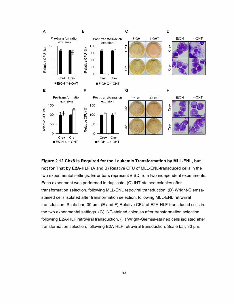
93
Figure 2.12 Cbx8 Is Required for the Leukemic Transformation by MLL-ENL, but
not for That by E2A-HLF (A and B) Relative CFU of MLL-ENL-transduced cells in the
two experimental settings. Error bars represent ± SD from two independent experiments.
Each experiment was performed in duplicate. (C) INT-stained colonies after
transformation selection, following MLL-ENL retroviral transduction. (D) Wright-Giemsa-
stained cells isolated after transformation selection, following MLL-ENL retroviral
transduction. Scale bar, 30 µm. (E and F) Relative CFU of E2A-HLF-transduced cells in
the two experimental settings. (G) INT-stained colonies after transformation selection,
following E2A-HLF retroviral transduction. (H) Wright-Giemsa-stained cells isolated after
transformation selection, following E2A-HLF retroviral transduction. Scale bar, 30 µm.
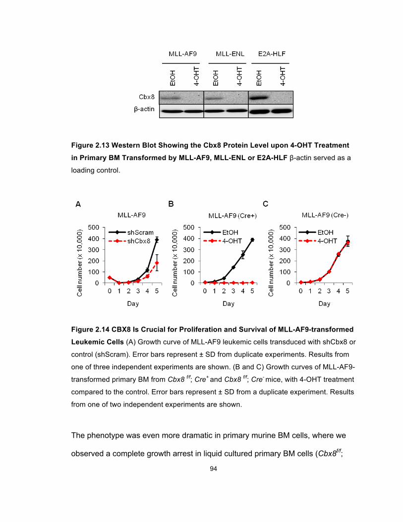
94
Figure 2.13 Western Blot Showing the Cbx8 Protein Level upon 4-OHT Treatment
in Primary BM Transformed by MLL-AF9, MLL-ENL or E2A-HLF β-actin served as a
loading control.
Figure 2.14 CBX8 Is Crucial for Proliferation and Survival of MLL-AF9-transformed
Leukemic Cells (A) Growth curve of MLL-AF9 leukemic cells transduced with shCbx8 or
control (shScram). Error bars represent ± SD from duplicate experiments. Results from
one of three independent experiments are shown. (B and C) Growth curves of MLL-AF9-
transformed primary BM from Cbx8 f/f; Cre+ and Cbx8 f/f; Cre- mice, with 4-OHT treatment
compared to the control. Error bars represent ± SD from a duplicate experiment. Results
from one of two independent experiments are shown.
The phenotype was even more dramatic in primary murine BM cells, where we
observed a complete growth arrest in liquid cultured primary BM cells (Cbx8f/f;
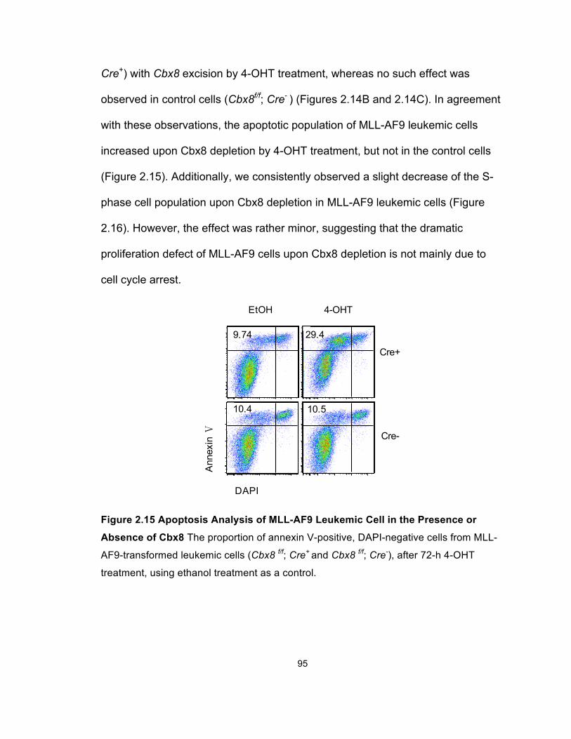
95
Cre+) with Cbx8 excision by 4-OHT treatment, whereas no such effect was
observed in control cells (Cbx8f/f; Cre- ) (Figures 2.14B and 2.14C). In agreement
with these observations, the apoptotic population of MLL-AF9 leukemic cells
increased upon Cbx8 depletion by 4-OHT treatment, but not in the control cells
(Figure 2.15). Additionally, we consistently observed a slight decrease of the S-
phase cell population upon Cbx8 depletion in MLL-AF9 leukemic cells (Figure
2.16). However, the effect was rather minor, suggesting that the dramatic
proliferation defect of MLL-AF9 cells upon Cbx8 depletion is not mainly due to
cell cycle arrest.
Figure 2.15 Apoptosis Analysis of MLL-AF9 Leukemic Cell in the Presence or
Absence of Cbx8 The proportion of annexin V-positive, DAPI-negative cells from MLL-
AF9-transformed leukemic cells (Cbx8 f/f; Cre+ and Cbx8 f/f; Cre-), after 72-h 4-OHT
treatment, using ethanol treatment as a control.
Ann
exin
Ⅴ
9.74 29.4
10.4 10.5
DAPI
Cre+
Cre-
EtOH 4-OHT
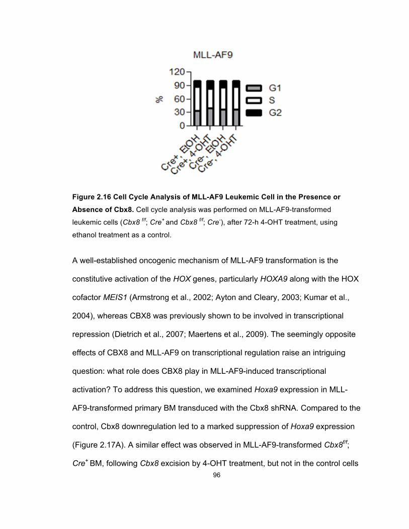
96
Figure 2.16 Cell Cycle Analysis of MLL-AF9 Leukemic Cell in the Presence or
Absence of Cbx8. Cell cycle analysis was performed on MLL-AF9-transformed
leukemic cells (Cbx8 f/f; Cre+ and Cbx8 f/f; Cre-), after 72-h 4-OHT treatment, using
ethanol treatment as a control.
A well-established oncogenic mechanism of MLL-AF9 transformation is the
constitutive activation of the HOX genes, particularly HOXA9 along with the HOX
cofactor MEIS1 (Armstrong et al., 2002; Ayton and Cleary, 2003; Kumar et al.,
2004), whereas CBX8 was previously shown to be involved in transcriptional
repression (Dietrich et al., 2007; Maertens et al., 2009). The seemingly opposite
effects of CBX8 and MLL-AF9 on transcriptional regulation raise an intriguing
question: what role does CBX8 play in MLL-AF9-induced transcriptional
activation? To address this question, we examined Hoxa9 expression in MLL-
AF9-transformed primary BM transduced with the Cbx8 shRNA. Compared to the
control, Cbx8 downregulation led to a marked suppression of Hoxa9 expression
(Figure 2.17A). A similar effect was observed in MLL-AF9-transformed Cbx8f/f;
Cre+ BM, following Cbx8 excision by 4-OHT treatment, but not in the control cells
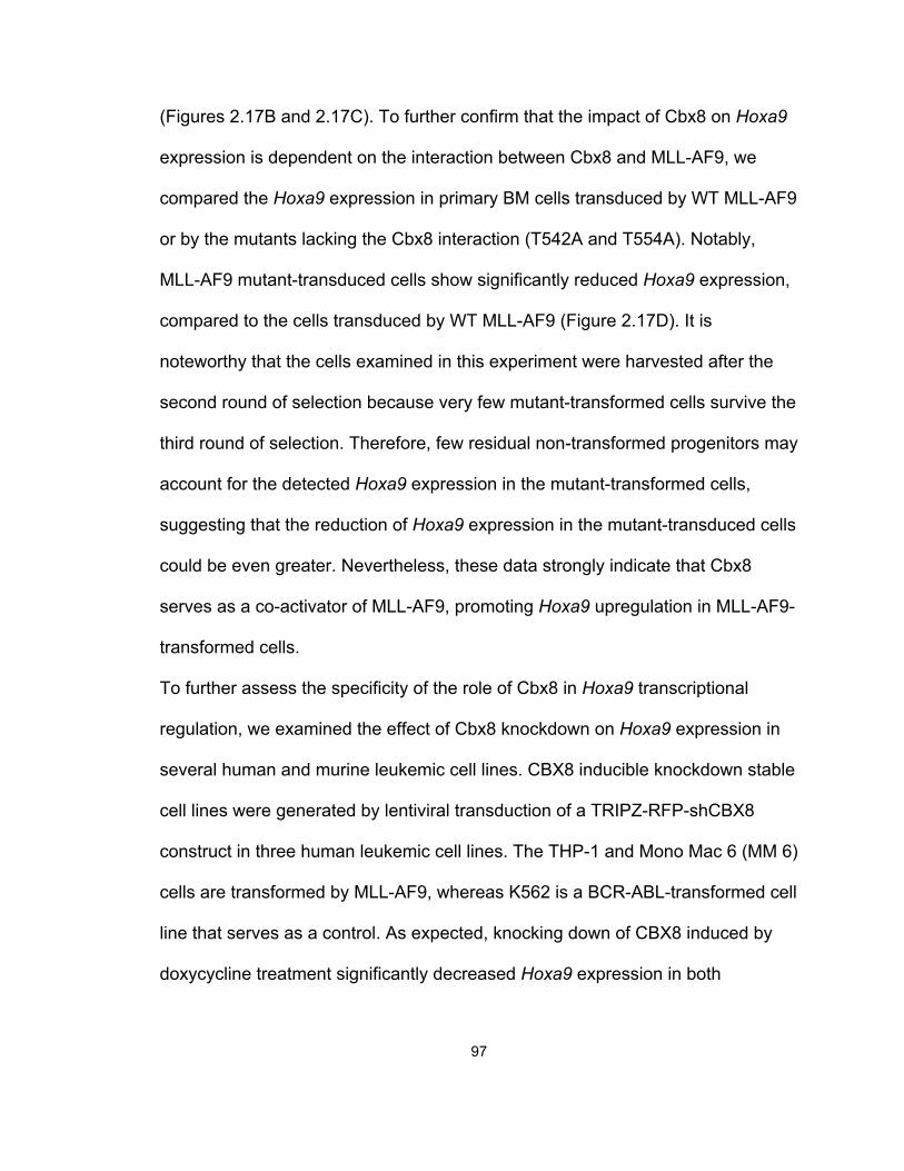
97
(Figures 2.17B and 2.17C). To further confirm that the impact of Cbx8 on Hoxa9
expression is dependent on the interaction between Cbx8 and MLL-AF9, we
compared the Hoxa9 expression in primary BM cells transduced by WT MLL-AF9
or by the mutants lacking the Cbx8 interaction (T542A and T554A). Notably,
MLL-AF9 mutant-transduced cells show significantly reduced Hoxa9 expression,
compared to the cells transduced by WT MLL-AF9 (Figure 2.17D). It is
noteworthy that the cells examined in this experiment were harvested after the
second round of selection because very few mutant-transformed cells survive the
third round of selection. Therefore, few residual non-transformed progenitors may
account for the detected Hoxa9 expression in the mutant-transformed cells,
suggesting that the reduction of Hoxa9 expression in the mutant-transduced cells
could be even greater. Nevertheless, these data strongly indicate that Cbx8
serves as a co-activator of MLL-AF9, promoting Hoxa9 upregulation in MLL-AF9-
transformed cells.
To further assess the specificity of the role of Cbx8 in Hoxa9 transcriptional
regulation, we examined the effect of Cbx8 knockdown on Hoxa9 expression in
several human and murine leukemic cell lines. CBX8 inducible knockdown stable
cell lines were generated by lentiviral transduction of a TRIPZ-RFP-shCBX8
construct in three human leukemic cell lines. The THP-1 and Mono Mac 6 (MM 6)
cells are transformed by MLL-AF9, whereas K562 is a BCR-ABL-transformed cell
line that serves as a control. As expected, knocking down of CBX8 induced by
doxycycline treatment significantly decreased Hoxa9 expression in both
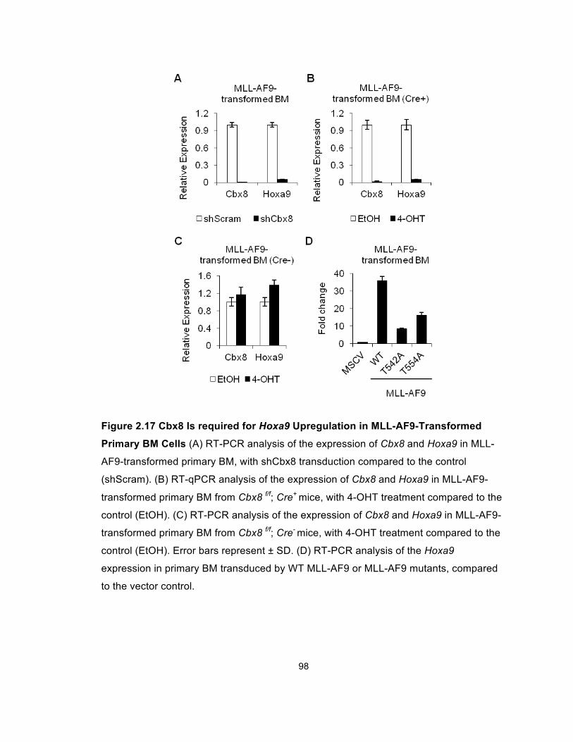
98
Figure 2.17 Cbx8 Is required for Hoxa9 Upregulation in MLL-AF9-Transformed
Primary BM Cells (A) RT-PCR analysis of the expression of Cbx8 and Hoxa9 in MLL-
AF9-transformed primary BM, with shCbx8 transduction compared to the control
(shScram). (B) RT-qPCR analysis of the expression of Cbx8 and Hoxa9 in MLL-AF9-
transformed primary BM from Cbx8 f/f; Cre+ mice, with 4-OHT treatment compared to the
control (EtOH). (C) RT-PCR analysis of the expression of Cbx8 and Hoxa9 in MLL-AF9-
transformed primary BM from Cbx8 f/f; Cre- mice, with 4-OHT treatment compared to the
control (EtOH). Error bars represent ± SD. (D) RT-PCR analysis of the Hoxa9
expression in primary BM transduced by WT MLL-AF9 or MLL-AF9 mutants, compared
to the vector control.
99
Figure 2.18 Cbx8 Is required for Hoxa9 Upregulation in Human and Murine MLL-
AF9-Transformed Leukemic Cell Lines (A) RT-PCR analysis of the expression of
Cbx8 and Hoxa9 in two human MLL-AF9 leukemic cell lines (THP-1 and MM 6) and one
MLL-independent human leukemic cell line (K562), with shRNA-mediated CBX8
knockdown. These three cell lines were integrated with a TRIPZ-RFP-shCBX8 construct
by lentiviral transduction, whose expression can be induced by doxycycline treatment.
Error bars represent ± SD. (B) RT-PCR analysis of the expression of Cbx8 and Hoxa9 in
a MLL-AF9 murine leukemic cell line and an E2A-HLF murine leukemic cell line, with
shCbx8 transduction compared to the control (shScram). Error bars represent ± SD.
100
MLL- AF9-transformed cell lines (MM 6 and THP-1), but not in the control cell line
(Figure 2.18A). Consistent with this observation, Cbx8 knockdown by shRNA led
to a marked decrease of Hoxa9 expression in a murine MLL-AF9 cell line, but not
in the Hoxa9-independent E2A-HLF cell line (Figure 2.18B). These findings
suggest that Cbx8 specifically contributes to MLL-AF9-induced Hoxa9
transcriptional activation.
In order to mechanistically understand how Cbx8 facilitates MLL-AF9-induced
Hoxa9 upregulation, we investigated the effect of Cbx8 on Hoxa9 promoter
activity in the presence of MLL-AF9. We first performed dual luciferase assays in
293 cells transfected with a MLL-AF9 responsive luciferase construct, under the
control of the murine Hoxa9 promoter (Hoxa9-LUC). Our data show that
disrupting the CBX8 interaction by the point mutation of T542A or T554A
significantly decreased the activation of the Hoxa9 promoter by MLL-AF9 (T542A:
p<0.01, T554A: p<0.01; Figure 2.19A). Consistent with this result, knocking down
the CBX8 expression by siRNAs reduced the MLL-AF9 induced transcriptional
activation by around 50% (p<0.01, Figures 2.19B and 2.19G). A similar response
was observed using another MLL-AF9 responsive luciferase reporter containing
the thymidine kinase promoter and multimerized Myc E box, further supporting
the importance of Cbx8 in MLL-AF9-induced transcriptional activation (Figures
2.19C and 2.19D). Notably, neither the point mutations nor CBX8 knockdown
significantly affected MLL-AF9 expression, indicating that the reduction in Hoxa9
promoter activity was not due to a general decrease in the MLL-AF9 level
(Figures 2.19E and 2.19F).
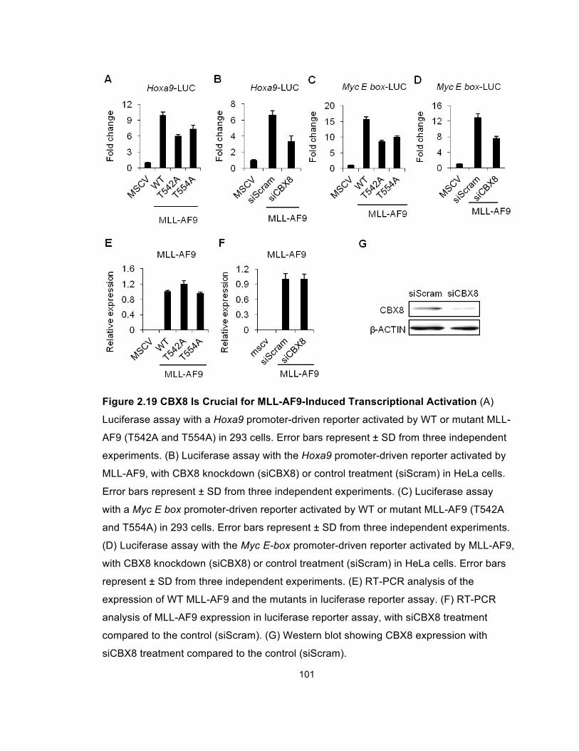
101
Figure 2.19 CBX8 Is Crucial for MLL-AF9-Induced Transcriptional Activation (A)
Luciferase assay with a Hoxa9 promoter-driven reporter activated by WT or mutant MLL-
AF9 (T542A and T554A) in 293 cells. Error bars represent ± SD from three independent
experiments. (B) Luciferase assay with the Hoxa9 promoter-driven reporter activated by
MLL-AF9, with CBX8 knockdown (siCBX8) or control treatment (siScram) in HeLa cells.
Error bars represent ± SD from three independent experiments. (C) Luciferase assay
with a Myc E box promoter-driven reporter activated by WT or mutant MLL-AF9 (T542A
and T554A) in 293 cells. Error bars represent ± SD from three independent experiments.
(D) Luciferase assay with the Myc E-box promoter-driven reporter activated by MLL-AF9,
with CBX8 knockdown (siCBX8) or control treatment (siScram) in HeLa cells. Error bars
represent ± SD from three independent experiments. (E) RT-PCR analysis of the
expression of WT MLL-AF9 and the mutants in luciferase reporter assay. (F) RT-PCR
analysis of MLL-AF9 expression in luciferase reporter assay, with siCBX8 treatment
compared to the control (siScram). (G) Western blot showing CBX8 expression with
siCBX8 treatment compared to the control (siScram).
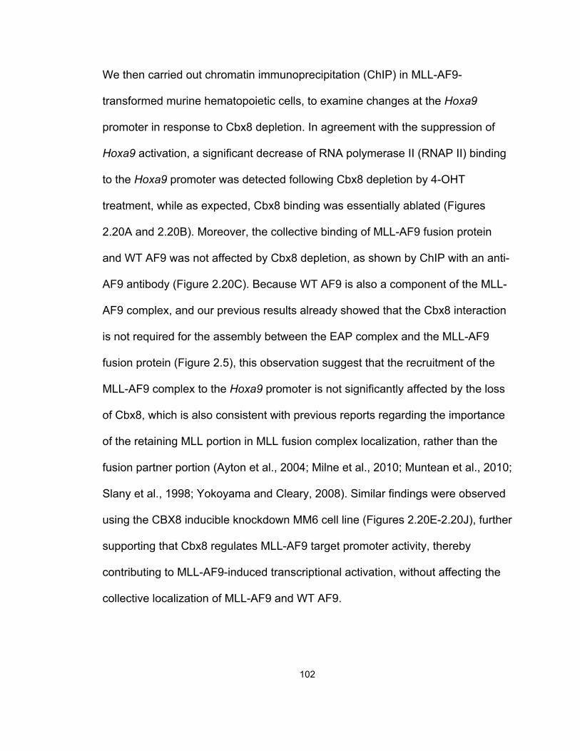
102
We then carried out chromatin immunoprecipitation (ChIP) in MLL-AF9-
transformed murine hematopoietic cells, to examine changes at the Hoxa9
promoter in response to Cbx8 depletion. In agreement with the suppression of
Hoxa9 activation, a significant decrease of RNA polymerase II (RNAP II) binding
to the Hoxa9 promoter was detected following Cbx8 depletion by 4-OHT
treatment, while as expected, Cbx8 binding was essentially ablated (Figures
2.20A and 2.20B). Moreover, the collective binding of MLL-AF9 fusion protein
and WT AF9 was not affected by Cbx8 depletion, as shown by ChIP with an anti-
AF9 antibody (Figure 2.20C). Because WT AF9 is also a component of the MLL-
AF9 complex, and our previous results already showed that the Cbx8 interaction
is not required for the assembly between the EAP complex and the MLL-AF9
fusion protein (Figure 2.5), this observation suggest that the recruitment of the
MLL-AF9 complex to the Hoxa9 promoter is not significantly affected by the loss
of Cbx8, which is also consistent with previous reports regarding the importance
of the retaining MLL portion in MLL fusion complex localization, rather than the
fusion partner portion (Ayton et al., 2004; Milne et al., 2010; Muntean et al., 2010;
Slany et al., 1998; Yokoyama and Cleary, 2008). Similar findings were observed
using the CBX8 inducible knockdown MM6 cell line (Figures 2.20E-2.20J), further
supporting that Cbx8 regulates MLL-AF9 target promoter activity, thereby
contributing to MLL-AF9-induced transcriptional activation, without affecting the
collective localization of MLL-AF9 and WT AF9.
103
Figure 2.20 Cbx8 Is not Required for the collective localization of MLL-AF9 and WT
AF9 at the Hoxa9 Promoter (A-D) Relative binding of Cbx8, RNAP II and MLL-AF9
together with WT AF9, as well as H3 binding, to the Hoxa9 promoter in MLL-AF9-
transformed cells from Cbx8 f/f; Cre+ mice, with 4-OHT treatment compared to the control
(EtOH). (E-H) Relative binding of CBX8, RNAP II and MLL-AF9 together with WT AF9,
as well as H3 binding, to the HOXA9 promoter in TRIPZ-RFP-shCBX8-containing MM6
cells, with doxycycline treatment compared to the control. Error bars represent ± SD
from three independent experiments. (I) RFP expression induced by doxycycline
treatment, indicating shRNA induction. Scale bars = 100 µm. (J) Western blot showing
the CBX8 protein level in TRIPZ-RFP-shCBX8-containing MM 6 cells after doxycycline
treatment, using β-ACTIN as a loading control.
104
Role of CBX8 in MLL-AF9 Leukemic Transformation and Transcriptional
Activation Is Independent of PRC1
To date, the only reported functional characterization of Cbx8 is its role as a
transcriptional repressor in PRC1, whereas our data indicate that Cbx8 serves as
a transcriptional coactivator in the presence of MLL-AF9. These opposing
transcriptional regulatory roles suggest that Cbx8 functions in a PRC1-
independent manner in MLL-AF9 leukemic transformation. It has been shown
that Ring1b, another PRC1 component, is required for the stability of PRC1
complexes (Leeb and Wutz, 2007; van der Stoop et al., 2008). In addition,
previous studies have indicated that Ring1b also interacts with AF9 (Monroe et
al., 2010), which we confirmed by IP experiments showing that endogenous
RING1b consistently coprecipitates with the MLL-AF9 fragment, CxxC-AF9
(Figure 2.21A). Therefore, to test our hypothesis of the potential PRC1-
independent function of Cbx8, we first assessed the impact of Ring1b on MLL-
AF9 leukemic transformation by BMT assays. Two individual shRNA molecules
specifically targeting Ring1b were used to effectively knock down Ring1b
expression in MLL-AF9-transformed leukemic cells. Reduction in Ring1b
expression in these experiments did not impair the transformation ability of MLL-
AF9 (Figures 2.21B, 2.21C and 2.21F). Knocking down Ring1b did not
significantly affect the growth rate or Hoxa9 expression in MLL-AF9 cells either
(Figures 2.21D and 2.21E). We also performed dual luciferase assays to
examine the impact of knocking down RING1b by siRNA on the MLL-AF9 target
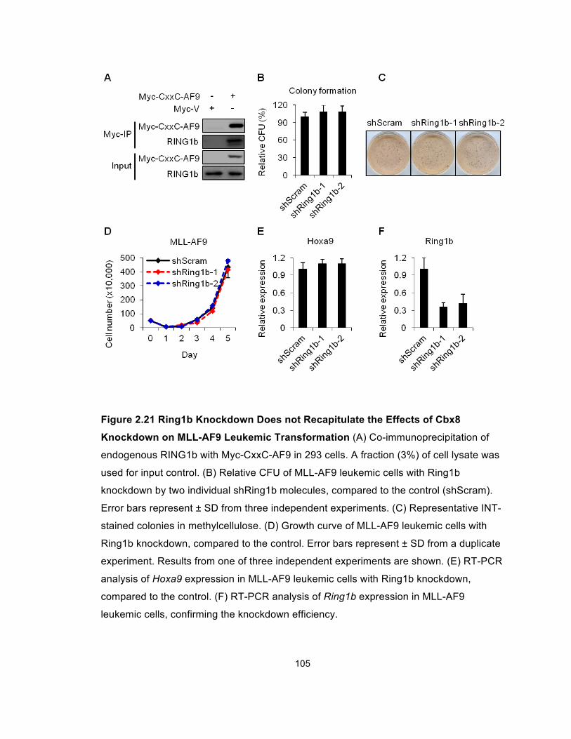
105
Figure 2.21 Ring1b Knockdown Does not Recapitulate the Effects of Cbx8
Knockdown on MLL-AF9 Leukemic Transformation (A) Co-immunoprecipitation of
endogenous RING1b with Myc-CxxC-AF9 in 293 cells. A fraction (3%) of cell lysate was
used for input control. (B) Relative CFU of MLL-AF9 leukemic cells with Ring1b
knockdown by two individual shRing1b molecules, compared to the control (shScram).
Error bars represent ± SD from three independent experiments. (C) Representative INT-
stained colonies in methylcellulose. (D) Growth curve of MLL-AF9 leukemic cells with
Ring1b knockdown, compared to the control. Error bars represent ± SD from a duplicate
experiment. Results from one of three independent experiments are shown. (E) RT-PCR
analysis of Hoxa9 expression in MLL-AF9 leukemic cells with Ring1b knockdown,
compared to the control. (F) RT-PCR analysis of Ring1b expression in MLL-AF9
leukemic cells, confirming the knockdown efficiency.
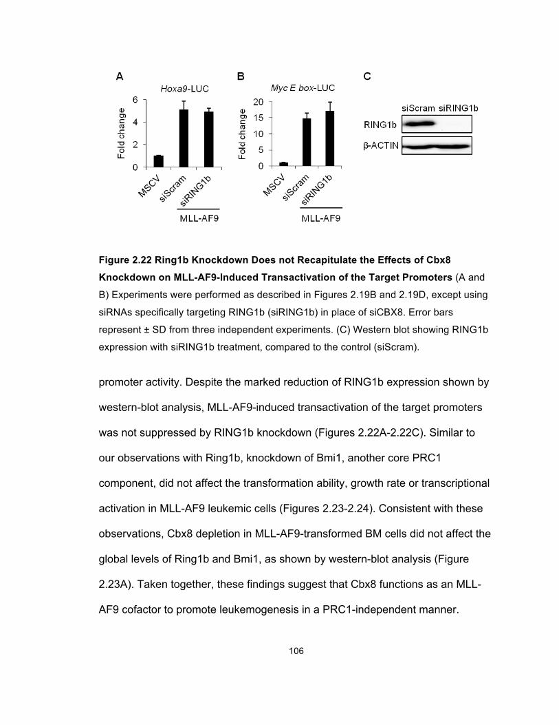
106
Figure 2.22 Ring1b Knockdown Does not Recapitulate the Effects of Cbx8
Knockdown on MLL-AF9-Induced Transactivation of the Target Promoters (A and
B) Experiments were performed as described in Figures 2.19B and 2.19D, except using
siRNAs specifically targeting RING1b (siRING1b) in place of siCBX8. Error bars
represent ± SD from three independent experiments. (C) Western blot showing RING1b
expression with siRING1b treatment, compared to the control (siScram).
promoter activity. Despite the marked reduction of RING1b expression shown by
western-blot analysis, MLL-AF9-induced transactivation of the target promoters
was not suppressed by RING1b knockdown (Figures 2.22A-2.22C). Similar to
our observations with Ring1b, knockdown of Bmi1, another core PRC1
component, did not affect the transformation ability, growth rate or transcriptional
activation in MLL-AF9 leukemic cells (Figures 2.23-2.24). Consistent with these
observations, Cbx8 depletion in MLL-AF9-transformed BM cells did not affect the
global levels of Ring1b and Bmi1, as shown by western-blot analysis (Figure
2.23A). Taken together, these findings suggest that Cbx8 functions as an MLL-
AF9 cofactor to promote leukemogenesis in a PRC1-independent manner.
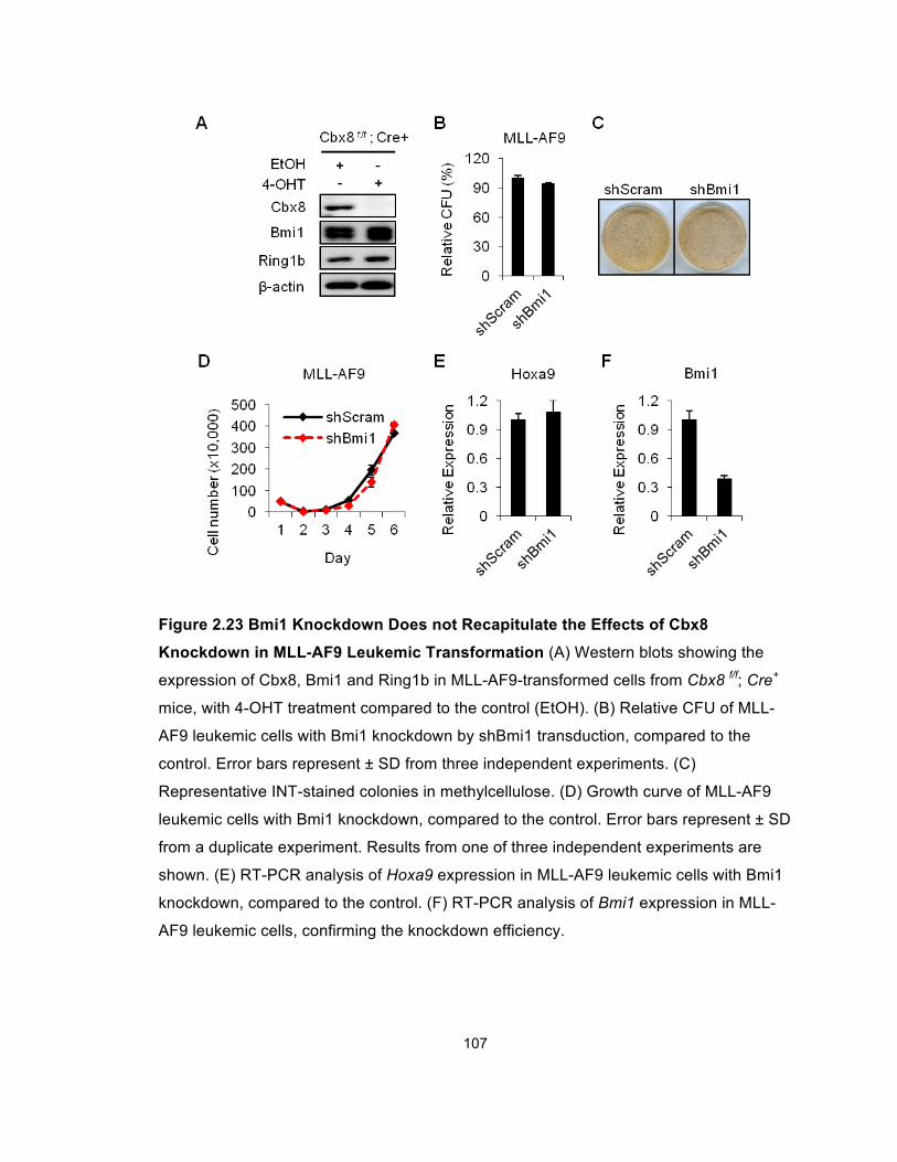
107
Figure 2.23 Bmi1 Knockdown Does not Recapitulate the Effects of Cbx8
Knockdown in MLL-AF9 Leukemic Transformation (A) Western blots showing the
expression of Cbx8, Bmi1 and Ring1b in MLL-AF9-transformed cells from Cbx8 f/f; Cre+
mice, with 4-OHT treatment compared to the control (EtOH). (B) Relative CFU of MLL-
AF9 leukemic cells with Bmi1 knockdown by shBmi1 transduction, compared to the
control. Error bars represent ± SD from three independent experiments. (C)
Representative INT-stained colonies in methylcellulose. (D) Growth curve of MLL-AF9
leukemic cells with Bmi1 knockdown, compared to the control. Error bars represent ± SD
from a duplicate experiment. Results from one of three independent experiments are
shown. (E) RT-PCR analysis of Hoxa9 expression in MLL-AF9 leukemic cells with Bmi1
knockdown, compared to the control. (F) RT-PCR analysis of Bmi1 expression in MLL-
AF9 leukemic cells, confirming the knockdown efficiency.
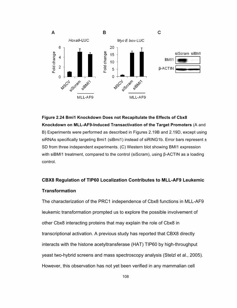
108
Figure 2.24 Bmi1 Knockdown Does not Recapitulate the Effects of Cbx8
Knockdown on MLL-AF9-Induced Transactivation of the Target Promoters (A and
B) Experiments were performed as described in Figures 2.19B and 2.19D, except using
siRNAs specifically targeting Bmi1 (siBmi1) instead of siRING1b. Error bars represent ±
SD from three independent experiments. (C) Western blot showing BMI1 expression
with siBMI1 treatment, compared to the control (siScram), using β-ACTIN as a loading
control.
CBX8 Regulation of TIP60 Localization Contributes to MLL-AF9 Leukemic
Transformation
The characterization of the PRC1 independence of Cbx8 functions in MLL-AF9
leukemic transformation prompted us to explore the possible involvement of
other Cbx8 interacting proteins that may explain the role of Cbx8 in
transcriptional activation. A previous study has reported that CBX8 directly
interacts with the histone acetyltransferase (HAT) TIP60 by high-throughput
yeast two-hybrid screens and mass spectroscopy analysis (Stelzl et al., 2005).
However, this observation has not yet been verified in any mammalian cell
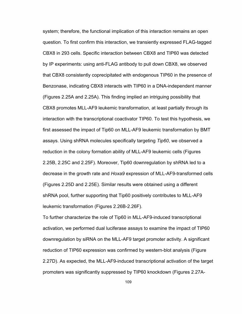
109
system; therefore, the functional implication of this interaction remains an open
question. To first confirm this interaction, we transiently expressed FLAG-tagged
CBX8 in 293 cells. Specific interaction between CBX8 and TIP60 was detected
by IP experiments: using anti-FLAG antibody to pull down CBX8, we observed
that CBX8 consistently coprecipitated with endogenous TIP60 in the presence of
Benzonase, indicating CBX8 interacts with TIP60 in a DNA-independent manner
(Figures 2.25A and 2.25A). This finding implied an intriguing possibility that
CBX8 promotes MLL-AF9 leukemic transformation, at least partially through its
interaction with the transcriptional coactivator TIP60. To test this hypothesis, we
first assessed the impact of Tip60 on MLL-AF9 leukemic transformation by BMT
assays. Using shRNA molecules specifically targeting Tip60, we observed a
reduction in the colony formation ability of MLL-AF9 leukemic cells (Figures
2.25B, 2.25C and 2.25F). Moreover, Tip60 downregulation by shRNA led to a
decrease in the growth rate and Hoxa9 expression of MLL-AF9-transformed cells
(Figures 2.25D and 2.25E). Similar results were obtained using a different
shRNA pool, further supporting that Tip60 positively contributes to MLL-AF9
leukemic transformation (Figures 2.26B-2.26F).
To further characterize the role of Tip60 in MLL-AF9-induced transcriptional
activation, we performed dual luciferase assays to examine the impact of TIP60
downregulation by siRNA on the MLL-AF9 target promoter activity. A significant
reduction of TIP60 expression was confirmed by western-blot analysis (Figure
2.27D). As expected, the MLL-AF9-induced transcriptional activation of the target
promoters was significantly suppressed by TIP60 knockdown (Figures 2.27A-
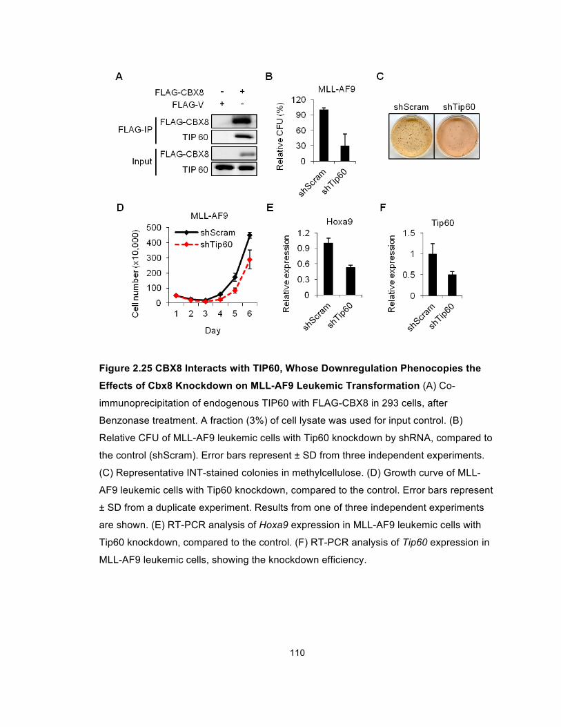
110
Figure 2.25 CBX8 Interacts with TIP60, Whose Downregulation Phenocopies the
Effects of Cbx8 Knockdown on MLL-AF9 Leukemic Transformation (A) Co-
immunoprecipitation of endogenous TIP60 with FLAG-CBX8 in 293 cells, after
Benzonase treatment. A fraction (3%) of cell lysate was used for input control. (B)
Relative CFU of MLL-AF9 leukemic cells with Tip60 knockdown by shRNA, compared to
the control (shScram). Error bars represent ± SD from three independent experiments.
(C) Representative INT-stained colonies in methylcellulose. (D) Growth curve of MLL-
AF9 leukemic cells with Tip60 knockdown, compared to the control. Error bars represent
± SD from a duplicate experiment. Results from one of three independent experiments
are shown. (E) RT-PCR analysis of Hoxa9 expression in MLL-AF9 leukemic cells with
Tip60 knockdown, compared to the control. (F) RT-PCR analysis of Tip60 expression in
MLL-AF9 leukemic cells, showing the knockdown efficiency.
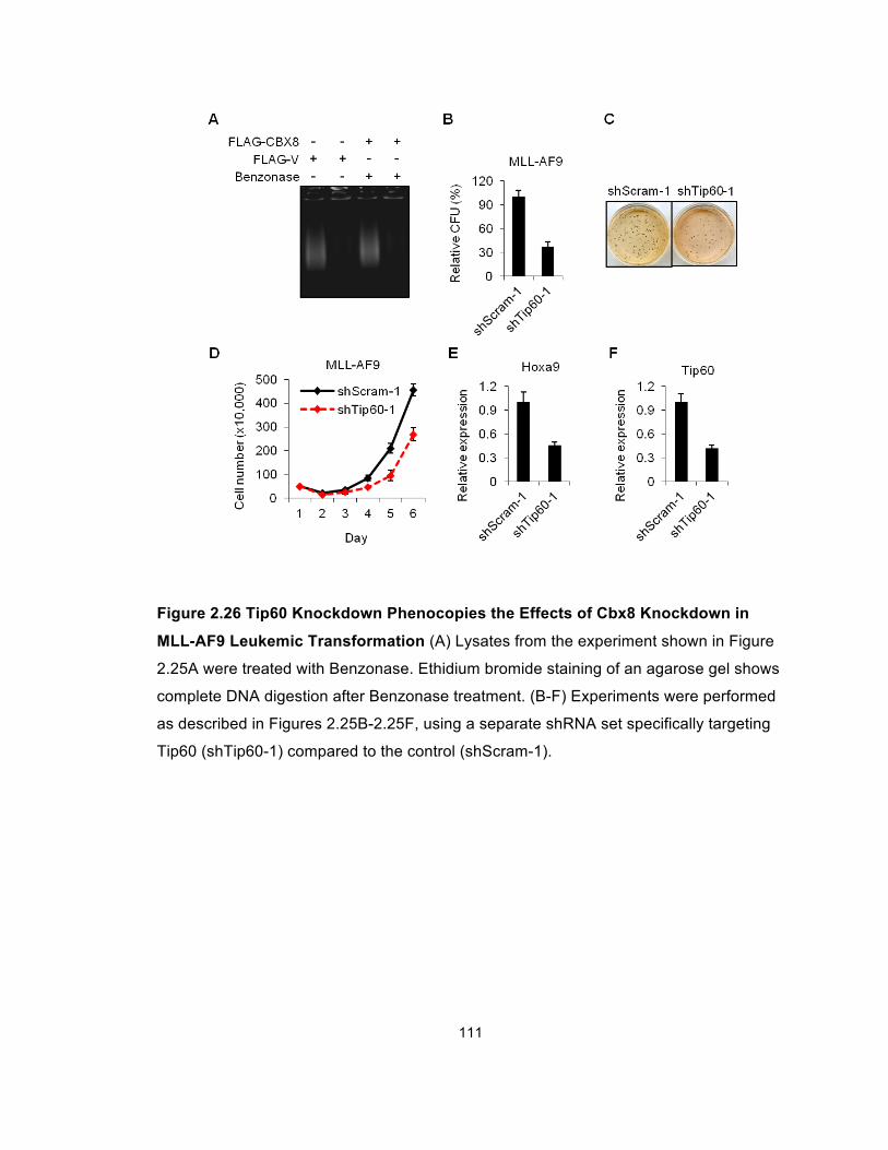
111
Figure 2.26 Tip60 Knockdown Phenocopies the Effects of Cbx8 Knockdown in
MLL-AF9 Leukemic Transformation (A) Lysates from the experiment shown in Figure
2.25A were treated with Benzonase. Ethidium bromide staining of an agarose gel shows
complete DNA digestion after Benzonase treatment. (B-F) Experiments were performed
as described in Figures 2.25B-2.25F, using a separate shRNA set specifically targeting
Tip60 (shTip60-1) compared to the control (shScram-1).
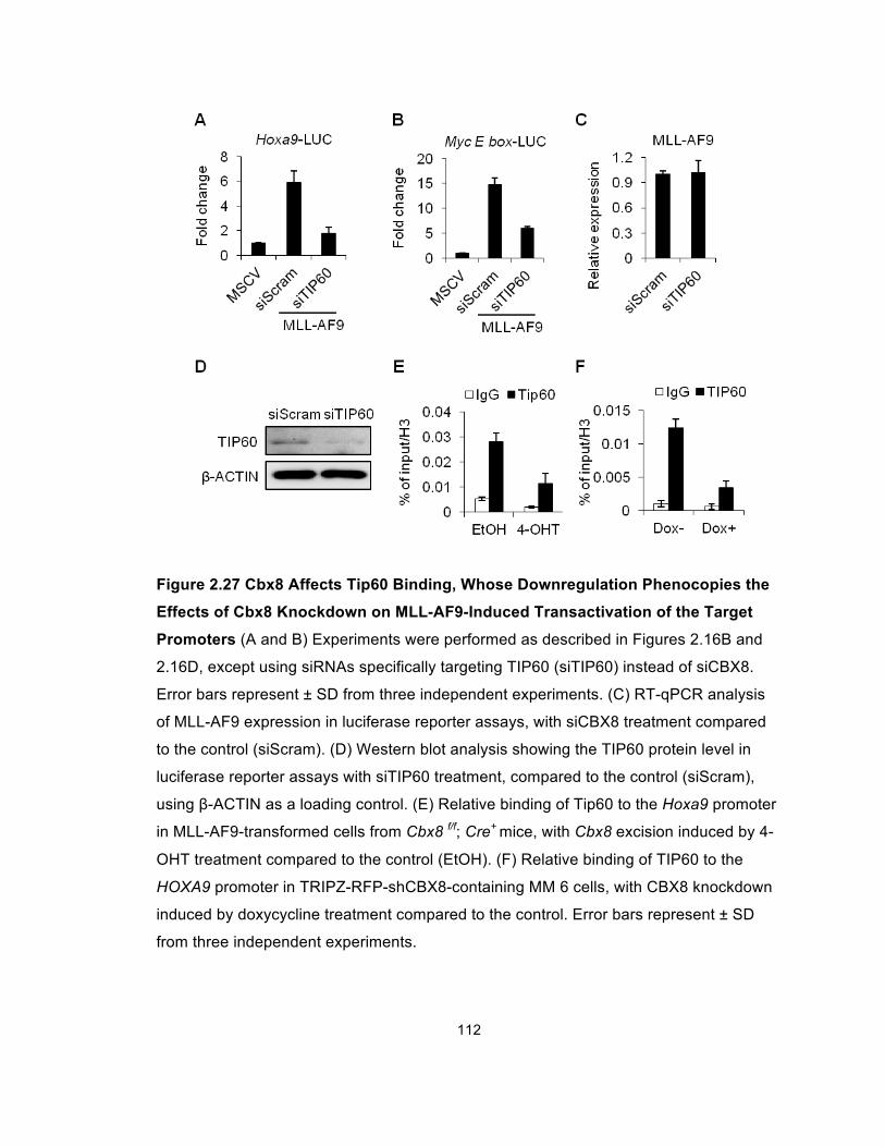
112
Figure 2.27 Cbx8 Affects Tip60 Binding, Whose Downregulation Phenocopies the
Effects of Cbx8 Knockdown on MLL-AF9-Induced Transactivation of the Target
Promoters (A and B) Experiments were performed as described in Figures 2.16B and
2.16D, except using siRNAs specifically targeting TIP60 (siTIP60) instead of siCBX8.
Error bars represent ± SD from three independent experiments. (C) RT-qPCR analysis
of MLL-AF9 expression in luciferase reporter assays, with siCBX8 treatment compared
to the control (siScram). (D) Western blot analysis showing the TIP60 protein level in
luciferase reporter assays with siTIP60 treatment, compared to the control (siScram),
using β-ACTIN as a loading control. (E) Relative binding of Tip60 to the Hoxa9 promoter
in MLL-AF9-transformed cells from Cbx8 f/f; Cre+ mice, with Cbx8 excision induced by 4-
OHT treatment compared to the control (EtOH). (F) Relative binding of TIP60 to the
HOXA9 promoter in TRIPZ-RFP-shCBX8-containing MM 6 cells, with CBX8 knockdown
induced by doxycycline treatment compared to the control. Error bars represent ± SD
from three independent experiments.
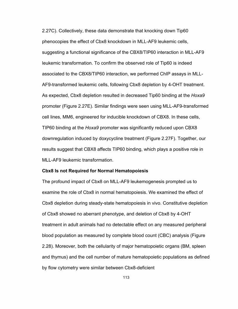
113
2.27C). Collectively, these data demonstrate that knocking down Tip60
phenocopies the effect of Cbx8 knockdown in MLL-AF9 leukemic cells,
suggesting a functional significance of the CBX8/TIP60 interaction in MLL-AF9
leukemic transformation. To confirm the observed role of Tip60 is indeed
associated to the CBX8/TIP60 interaction, we performed ChIP assays in MLL-
AF9-transformed leukemic cells, following Cbx8 depletion by 4-OHT treatment.
As expected, Cbx8 depletion resulted in decreased Tip60 binding at the Hoxa9
promoter (Figure 2.27E). Similar findings were seen using MLL-AF9-transformed
cell lines, MM6, engineered for inducible knockdown of CBX8. In these cells,
TIP60 binding at the Hoxa9 promoter was significantly reduced upon CBX8
downregulation induced by doxycycline treatment (Figure 2.27F). Together, our
results suggest that CBX8 affects TIP60 binding, which plays a positive role in
MLL-AF9 leukemic transformation.
Cbx8 Is not Required for Normal Hematopoiesis
The profound impact of Cbx8 on MLL-AF9 leukemogenesis prompted us to
examine the role of Cbx8 in normal hematopoiesis. We examined the effect of
Cbx8 depletion during steady-state hematopoiesis in vivo. Constitutive depletion
of Cbx8 showed no aberrant phenotype, and deletion of Cbx8 by 4-OHT
treatment in adult animals had no detectable effect on any measured peripheral
blood population as measured by complete blood count (CBC) analysis (Figure
2.28). Moreover, both the cellularity of major hematopoietic organs (BM, spleen
and thymus) and the cell number of mature hematopoietic populations as defined
by flow cytometry were similar between Cbx8-deficient
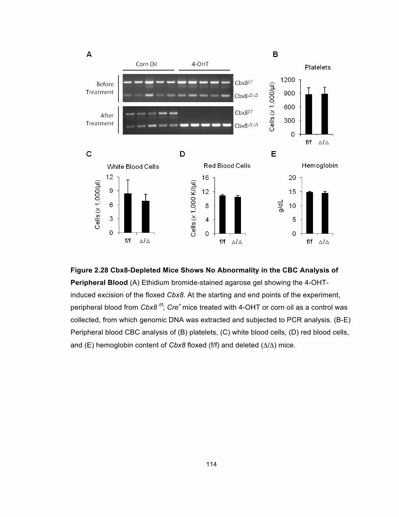
114
Figure 2.28 Cbx8-Depleted Mice Shows No Abnormality in the CBC Analysis of
Peripheral Blood (A) Ethidium bromide-stained agarose gel showing the 4-OHT-
induced excision of the floxed Cbx8. At the starting and end points of the experiment,
peripheral blood from Cbx8 f/f; Cre+ mice treated with 4-OHT or corn oil as a control was
collected, from which genomic DNA was extracted and subjected to PCR analysis. (B-E)
Peripheral blood CBC analysis of (B) platelets, (C) white blood cells, (D) red blood cells,
and (E) hemoglobin content of Cbx8 floxed (f/f) and deleted (Δ/Δ) mice.
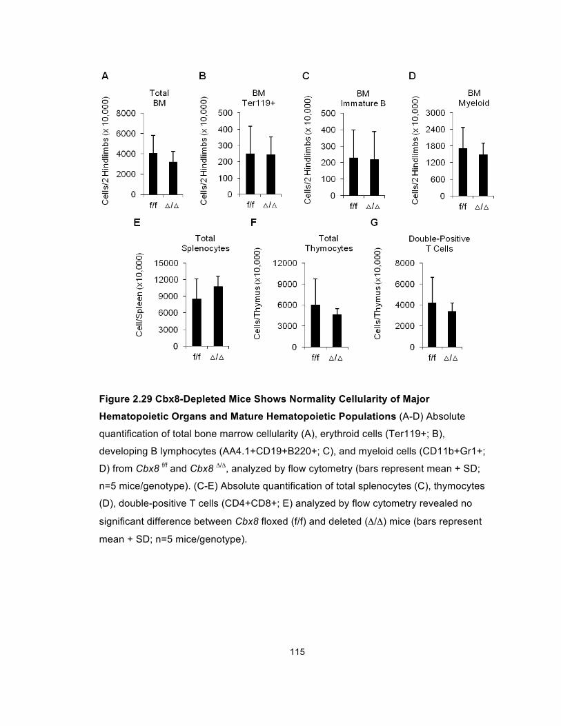
115
Figure 2.29 Cbx8-Depleted Mice Shows Normality Cellularity of Major
Hematopoietic Organs and Mature Hematopoietic Populations (A-D) Absolute
quantification of total bone marrow cellularity (A), erythroid cells (Ter119+; B),
developing B lymphocytes (AA4.1+CD19+B220+; C), and myeloid cells (CD11b+Gr1+;
D) from Cbx8 f/f and Cbx8 ∆/∆, analyzed by flow cytometry (bars represent mean + SD;
n=5 mice/genotype). (C-E) Absolute quantification of total splenocytes (C), thymocytes
(D), double-positive T cells (CD4+CD8+; E) analyzed by flow cytometry revealed no
significant difference between Cbx8 floxed (f/f) and deleted (Δ/Δ) mice (bars represent
mean + SD; n=5 mice/genotype).
116
mice and controls (Figure 2.29). To address potential effects of Cbx8 deletion on
primitive long-term hematopoietic stem cells (LT-HSCs), we combined flow
cytometry for characterization of progenitor populations and competitive BM
transplantation assays. These analyses revealed no detectable differences in LT-
HSC numbers (Figure 2.30) or hematopoietic reconstitution ability of Cbx8 WT or
deficient BM in lethally irradiated recipients (Figure 2.31A). In addition, the total
progenitor output from the BM of Cbx8-deficient animals was similar to controls,
as measure by colony forming unit assays (Figures 2.31B and 2.31C). Together,
these findings indicate that Cbx8 is not required for steady state hematopoiesis,
LT-HSC maintenance, or stem and progenitor cell function.
Figure 2.30 Cbx8-Depleted Mice Shows No Abnormality in Maintaining the
Primitive Long-Term Hematopoietic Stem Cell (LT-HSC) Population. Flow
cytometric analysis of LT-HSCs (CD150+CD48-LSK) from Cbx8 f/f and Cbx8 ∆/∆ mice
(bars represent mean + SD; n=5 mice/genotype).
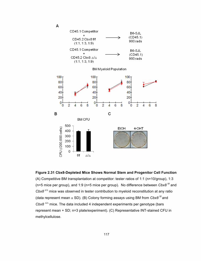
117
Figure 2.31 Cbx8-Depleted Mice Shows Normal Stem and Progenitor Cell Function
(A) Competitive BM transplantation at competitor: tester ratios of 1:1 (n=10/group), 1:3
(n=5 mice per group), and 1:9 (n=5 mice per group). No difference between Cbx8 f/f and
Cbx8 ∆/∆ mice was observed in tester contribution to myeloid reconstitution at any ratio
(data represent mean ± SD). (B) Colony forming assays using BM from Cbx8 f/f and
Cbx8 ∆/∆ mice. The data included 4 independent experiments per genotype (bars
represent mean + SD; n=3 plate/experiment). (C) Representative INT-stained CFU in
methylcellulose.
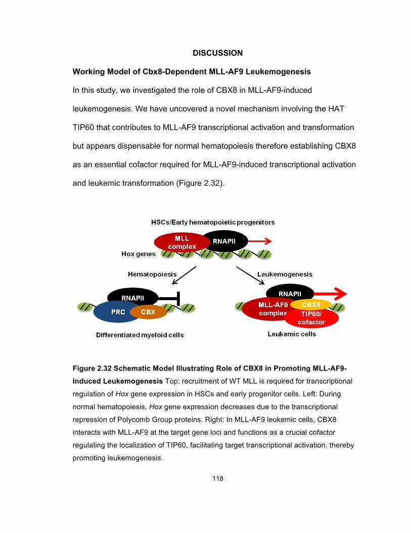
118
DISCUSSION
Working Model of Cbx8-Dependent MLL-AF9 Leukemogenesis
In this study, we investigated the role of CBX8 in MLL-AF9-induced
leukemogenesis. We have uncovered a novel mechanism involving the HAT
TIP60 that contributes to MLL-AF9 transcriptional activation and transformation
but appears dispensable for normal hematopoiesis therefore establishing CBX8
as an essential cofactor required for MLL-AF9-induced transcriptional activation
and leukemic transformation (Figure 2.32).
Figure 2.32 Schematic Model Illustrating Role of CBX8 in Promoting MLL-AF9-
Induced Leukemogenesis Top: recruitment of WT MLL is required for transcriptional
regulation of Hox gene expression in HSCs and early progenitor cells. Left: During
normal hematopoiesis, Hox gene expression decreases due to the transcriptional
repression of Polycomb Group proteins. Right: In MLL-AF9 leukemic cells, CBX8
interacts with MLL-AF9 at the target gene loci and functions as a crucial cofactor
regulating the localization of TIP60, facilitating target transcriptional activation, thereby
promoting leukemogenesis.
119
Potential Broad Role of CBX8 in MLL-Rearranged Leukemogenesis
CBX8 is one of the five human homologs of the Drosophila Pc protein. Although
all CBX proteins share highly conserved chromodomains and Pc boxes, their
different sizes and the presence of other motifs suggest potentially different
functions (Whitcomb et al., 2007). Indeed, previous studies have reported that
mice deficient for different PRC1 components show only a partial overlap in
phenotype, raising the possibility of PRC1-independent functions of these
components that may be context dependent and involve other protein complexes
(de Napoles et al., 2004; Katoh-Fukui et al., 1998; Leeb and Wutz, 2007; Suzuki
et al., 2002; Voncken et al., 2003). Our study has uncovered such a PRC1-
independent function of CBX8 in MLL-AF9-induced transcriptional regulation.
Interestingly, contrary to its role as a transcriptional repressor in PRC1, CBX8
serves as a transcriptional coactivator in the MLL-AF9 complex. Furthermore,
CBX8 is present in the EAP (or the related AEP and the Dotcom) transcriptional
activation complex (Mohan et al., 2010b; Monroe et al., 2010; Yokoyama et al.,
2010), and our data show that it is also required for the leukemic transformation
induced by MLL-ENL, another EAP-interacting MLL fusion protein, suggesting
that the CBX8-dependent leukemogenic mechanism may apply to not only MLL-
AF9 but also other MLL fusion proteins whose fusion partners are present in the
EAP complex.
The drastic effect of Cbx8 deletion on leukemogenic capacity of MLL-AF9 and
MLL-ENL prompted us to ask whether this “Cbx8 addition” is further adopted by
the MLL fusion proteins with translocation partners absent in the EAP complex,
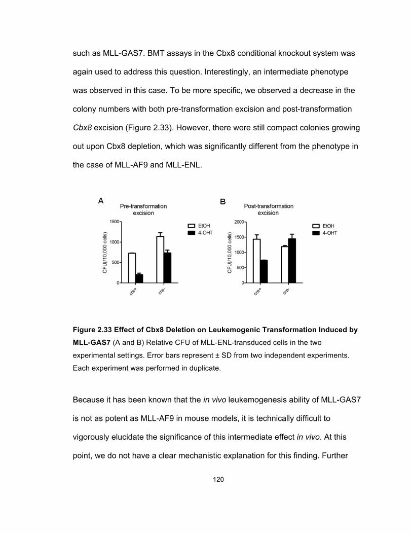
120
such as MLL-GAS7. BMT assays in the Cbx8 conditional knockout system was
again used to address this question. Interestingly, an intermediate phenotype
was observed in this case. To be more specific, we observed a decrease in the
colony numbers with both pre-transformation excision and post-transformation
Cbx8 excision (Figure 2.33). However, there were still compact colonies growing
out upon Cbx8 depletion, which was significantly different from the phenotype in
the case of MLL-AF9 and MLL-ENL.
Figure 2.33 Effect of Cbx8 Deletion on Leukemogenic Transformation Induced by
MLL-GAS7 (A and B) Relative CFU of MLL-ENL-transduced cells in the two
experimental settings. Error bars represent ± SD from two independent experiments.
Each experiment was performed in duplicate.
Because it has been known that the in vivo leukemogenesis ability of MLL-GAS7
is not as potent as MLL-AF9 in mouse models, it is technically difficult to
vigorously elucidate the significance of this intermediate effect in vivo. At this
point, we do not have a clear mechanistic explanation for this finding. Further
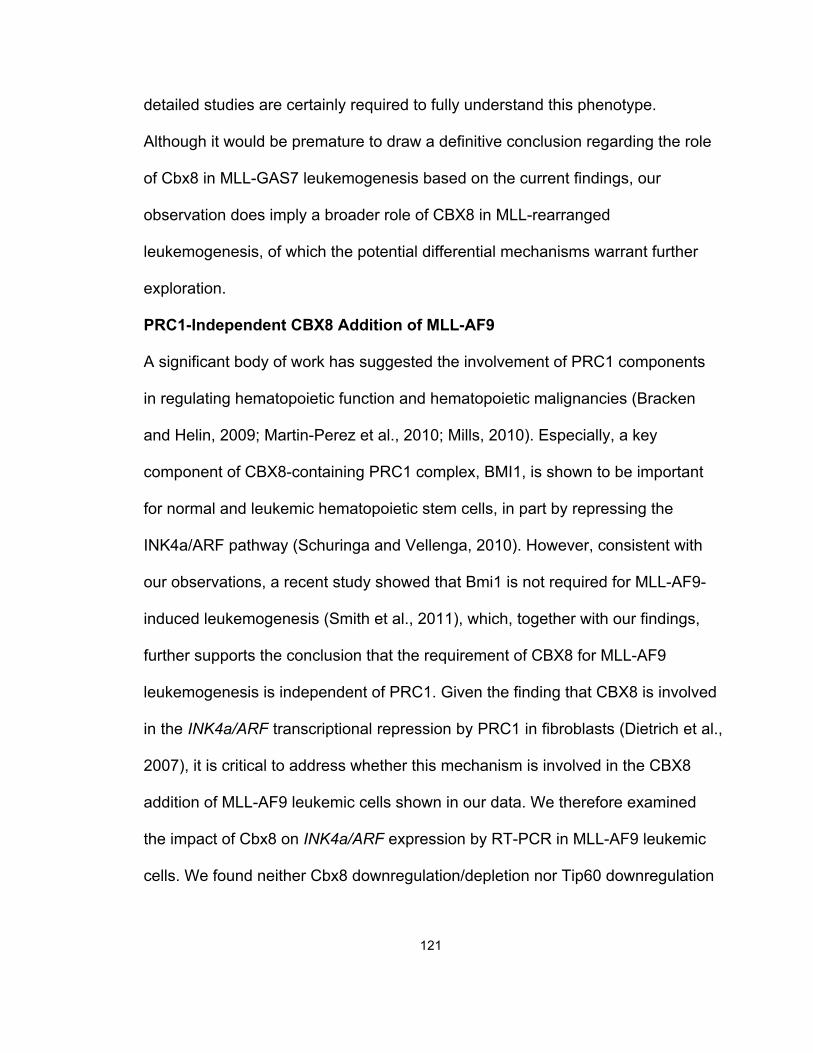
121
detailed studies are certainly required to fully understand this phenotype.
Although it would be premature to draw a definitive conclusion regarding the role
of Cbx8 in MLL-GAS7 leukemogenesis based on the current findings, our
observation does imply a broader role of CBX8 in MLL-rearranged
leukemogenesis, of which the potential differential mechanisms warrant further
exploration.
PRC1-Independent CBX8 Addition of MLL-AF9
A significant body of work has suggested the involvement of PRC1 components
in regulating hematopoietic function and hematopoietic malignancies (Bracken
and Helin, 2009; Martin-Perez et al., 2010; Mills, 2010). Especially, a key
component of CBX8-containing PRC1 complex, BMI1, is shown to be important
for normal and leukemic hematopoietic stem cells, in part by repressing the
INK4a/ARF pathway (Schuringa and Vellenga, 2010). However, consistent with
our observations, a recent study showed that Bmi1 is not required for MLL-AF9-
induced leukemogenesis (Smith et al., 2011), which, together with our findings,
further supports the conclusion that the requirement of CBX8 for MLL-AF9
leukemogenesis is independent of PRC1. Given the finding that CBX8 is involved
in the INK4a/ARF transcriptional repression by PRC1 in fibroblasts (Dietrich et al.,
2007), it is critical to address whether this mechanism is involved in the CBX8
addition of MLL-AF9 leukemic cells shown in our data. We therefore examined
the impact of Cbx8 on INK4a/ARF expression by RT-PCR in MLL-AF9 leukemic
cells. We found neither Cbx8 downregulation/depletion nor Tip60 downregulation
122
led to INK4a/ARF activation (Figure 2.34), confirming that the CBX8-dependent
MLL-AF9 transformation is not due to INK4a/ARF repression.
Figure 2.34 Ink4a/Arf Expression Is not Activated upon Cbx8 Downregulation or
Depletion or upon Tip60 Downregulation (A) RT-PCR analysis of Ink4a/Arf
expression in MLL-AF9-transformed leukemic cells with shCbx8 transduction compared
to the control (shScram). (B) RT-PCR analysis of Ink4a/Arf expression in MLL-AF9-
transformed leukemic cells from Cbx8 f/f; Cre+ and Cbx8 f/f; Cre- mice, with 4-OHT
treatment compared to the control (EtOH). (C and D) RT-PCR analysis of Ink4a/Arf
expression in MLL-AF9-transformed leukemic cells with Tip60 knockdown by two
separate shRNA sets (described in Figures 2.25 and 2.26), compared to the respective
control.
123
TIP60 as the First HAT Associated with the MLL-AF9 Complex
In addition to the MLL fusion partners, CBX8 has previously been shown to
directly interact with the HAT TIP60, a core member of the MYST family (Stelzl et
al., 2005). Several TrxG complexes are known to recruit HATs during normal
development. For example, another core member of the MYST family, MOF
(MYST1), has been purified in WT MLL complex, and the HAT CREB-binding
protein (CBP) is known to interacts with both MLL and another TrxG protein
ASH1 (Bantignies et al., 2000; Dou et al., 2005; Ernst et al., 2001; Petruk et al.,
2001). Under normal physiological conditions, HATs function as a coactivator to
facilitate TrxG-induced transcriptional activation, antagonizing the transcriptional
repressive effect of the PcG complex (Mills, 2010; Pasini et al., 2010). This
mechanism contributes to the active HOXA9 expression in hematopoietic stem
cells and early progenitors (Figure 2.32). In addition, another two MYST proteins,
MOZ (MYST3) and MORF (MYST4), are directly involved in chromosomal
translocations in AML to form chimeric fusion proteins with CBP and/or p300 that
lead to leukemogenesis (Katsumoto et al., 2008; So and van der Reijden, 2008).
Moreover, MLL is fused to CBP or P300 in a subset of acute leukemias (Wang et
al., 2005). Together, multiple lines of evidence have established the critical
cooperation between MLL physiological function and HAT activity and the pro-
oncogenic role of certain MYST family members in human myeloid leukemias.
However, previous studies have not reported HATs as a component of the EAP
complex, which is recruited by the most common MLL fusion partners. Therefore,
whether HATs contribute to transcriptional activation induced by common MLL-
124
rearranged oncoproteins remains unknown. Our finding of the CBX8-dependent
TIP60 localization at the HOXA9 promoter indicates that this transcriptional
activation mechanism is likely to be adopted by common MLL fusion proteins to
activate target gene expression, such as HOXA9. Interestingly, a previous RNAi
screening study in mouse embryonic stem cells (ESCs) has reported that Tip60
is required for pluripotency, while MLL myeloid leukemia stem cells have been
shown to share the transcriptional program with ESCs, rather than adult stem
cells (Fazzio et al., 2008; Somervaille et al., 2009). Together, these observations
suggest a possible functional association between the TIP60-regulated signaling
network and the transcriptional program in MLL-rearranged leukemic cells,
therefore further supporting our model that CBX8 is essential for MLL-AF9-
induced oncogenic transformation, likely through its interaction with TIP60 at the
target gene loci to help establish the transcriptional program required for
leukemogenesis (Figure 2.32). Furthermore, this study, together with previous
findings, reveals an intriguing mechanistic similarity between WT MLL and
oncogenic MLL fusion proteins. Specifically, the synergistic activity between HMT
(H3K4me3) and HAT in transcriptional activation by WT MLL is dependent on the
C-terminal SET domain, which is absent in MLL fusion proteins as a result of
translocations. However, oncogenic MLL fusion proteins regain this classic
combination of enzymatic activities by recruiting another HMT that facilitates
gene transcription, DOT1L (Bernt et al., 2011; Jo et al., 2011; Nguyen et al.,
2011), and an HAT with a high structural similarity of MOF, TIP60 (Figure 2.35),
thereby nicely reinforcing the concept of cancer cells hijacking the regulatory
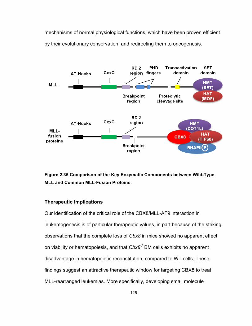
125
mechanisms of normal physiological functions, which have been proven efficient
by their evolutionary conservation, and redirecting them to oncogenesis.
Figure 2.35 Comparison of the Key Enzymatic Components between Wild-Type
MLL and Common MLL-Fusion Proteins.
Therapeutic Implications
Our identification of the critical role of the CBX8/MLL-AF9 interaction in
leukemogenesis is of particular therapeutic values, in part because of the striking
observations that the complete loss of Cbx8 in mice showed no apparent effect
on viability or hematopoiesis, and that Cbx8-/- BM cells exhibits no apparent
disadvantage in hematopoietic reconstitution, compared to WT cells. These
findings suggest an attractive therapeutic window for targeting CBX8 to treat
MLL-rearranged leukemias. More specifically, developing small molecule
126
inhibitors disrupting the CBX8/AF9 interaction may be a particular promising
approach. Moreover, the characterization of the CBX8/TIP60 interaction and its
contribution in MLL-AF9 leukemogenesis suggests that it may be worthwhile to
explore the therapeutic potential of targeting this interaction. Notably, although
development and reproduction of Tip60 wild-type and heterozygous mice were
normal, homozygous ablation of the Tip60 gene caused embryonic lethality near
the blastocyst stage of development in mice (Hu et al., 2009). Given the broad
involvement of TIP60 in multiple important biological processes, directly targeting
TIP60 may raise the issue of toxicity, in which case careful assessment is
certainly required to evaluate the possible side effects. Nonetheless, the
essentially null phenotype of Cbx8 knockout mice suggests that this interaction is
unlikely to be critical for the major function of TIP60. Therefore, specific
disruption of the CBX8/TIP60 interaction might still be an interesting angle for
tackling MLL-rearranged leukemias.
Remaining Questions
Several important questions remain to be addressed for both better
understanding the biology of MLL-rearranged leukemias and further exploring the
clinical application potential of our current findings. First and foremost, it is not
yet known which domains or regions in CBX8 are responsible for its role in MLL-
AF9-induced leukemogenesis. To address this question, two directions are worth
exploring. On the one hand, mapping the critical domains, or amino acids, of
CBX8 that are important for CBX8/AF9 and CBX8/TIP60 interactions is of prior
importance. A previous study has indicated that Cbx8 binds AF9 within a region
127
of approximately 130 amino acids that is not conserved in other Polycomb
proteins (Hemenway et al., 2001). However, this region is obviously too broad for
therapeutic targeting. Generating CBX8 mutants containing small fragment
deletions and point mutations in this region may narrow down the interest to a
limited region that is suitable for the binding of small molecule inhibitors.
Additionally, single amino acid substitution analyses may reveal the key amino
acids required for the CBX8/AF9 interaction. Small peptides containing the
corresponding sequences may be used to design dominant negative inhibitors.
Similar strategies can also be applied to identify the key element of CBX8 for the
CBX8/TIP60 interaction. On the other hand, characterizing the role of the CBX8
chromodomain in this particular case is of significant value. Chromodomains are
present in many chromatin regulatory proteins and are thought to mediate
interaction with methylated histone lysines or RNA molecules (Akhtar et al., 2000;
Sapountzi et al., 2006; Utley and Cote, 2003). The chromodomains of Pc
proteins are thought to, at least in part, localize the proteins and their respective
complexes to appropriately marked sites of the epigenome through
reorganization of either H3K9me3 or H3K27me3, both of which indicate
transcriptional repression (Kaustov et al., 2011). However, despite the high
degree of conservation among the Pc protein chromodomains, previous studies
have shown that they display significant differences in histone peptide binding
preferences (Bernstein et al., 2006; Kaustov et al., 2011). Specifically, the CBX8
chromodomain has been shown to display essentially no binding preference to
either H3K9me3 or H3K27me3, and alternative non-histone sequences are
128
suggested as potential binding targets for this chromodomain (Bernstein et al.,
2006; Kaustov et al., 2011). Therefore, it is intriguing to test whether the CBX8
chromodomain represents a diverged evolutionary direction of Pc
chromodomains, which is opposite of their traditional transcription repression role.
Most importantly, the structure of the CBX8 chromodomain has been well-
characterized, including the potential peptide binding pocket (Kaustov et al.,
2011), which has laid a solid foundation for potential small molecule inhibitor
development.
Second, based on our findings so far, it is not yet clear what are the exact
molecular events responsible for the contribution of TIP60 in MLL-AF9-induced
leukemogenesis. On one hand, the specificity of the CBX8-affected TIP60
binding in MLL-AF9 leukemic cells warrants further investigation. For instance,
ChIP-seq analysis of the binding pattern of Tip60 in the genome of MLL-AF9
leukemic cells, particularly at MLL-AF9 target loci, in the presence or absence of
Cbx8, will be helpful to better understand that, to what extent, Cbx8 may affect
the binding of Tip60. This information will be important for further investigation on
whether TIP60 may contribute to MLL-AF9 leukemogenesis through both CBX8-
dependent and CBX8-independent mechanisms.
On the other hand, it will be useful to identify the critical region of TIP60 required
for the CBX8/TIP60 interaction using the mutagenesis approach previously
mentioned. Interestingly, TIP60 also contains a chromodomain, and it may have
yet unidentified functions other than the traditionally characterized interaction
with histone or RNA molecules (Akhtar et al., 2000; Sapountzi et al., 2006; Utley
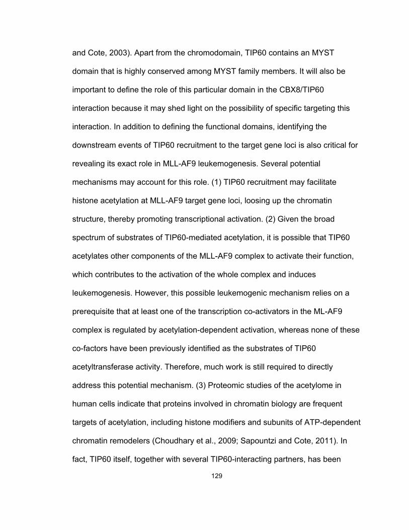
129
and Cote, 2003). Apart from the chromodomain, TIP60 contains an MYST
domain that is highly conserved among MYST family members. It will also be
important to define the role of this particular domain in the CBX8/TIP60
interaction because it may shed light on the possibility of specific targeting this
interaction. In addition to defining the functional domains, identifying the
downstream events of TIP60 recruitment to the target gene loci is also critical for
revealing its exact role in MLL-AF9 leukemogenesis. Several potential
mechanisms may account for this role. (1) TIP60 recruitment may facilitate
histone acetylation at MLL-AF9 target gene loci, loosing up the chromatin
structure, thereby promoting transcriptional activation. (2) Given the broad
spectrum of substrates of TIP60-mediated acetylation, it is possible that TIP60
acetylates other components of the MLL-AF9 complex to activate their function,
which contributes to the activation of the whole complex and induces
leukemogenesis. However, this possible leukemogenic mechanism relies on a
prerequisite that at least one of the transcription co-activators in the ML-AF9
complex is regulated by acetylation-dependent activation, whereas none of these
co-factors have been previously identified as the substrates of TIP60
acetyltransferase activity. Therefore, much work is still required to directly
address this potential mechanism. (3) Proteomic studies of the acetylome in
human cells indicate that proteins involved in chromatin biology are frequent
targets of acetylation, including histone modifiers and subunits of ATP-dependent
chromatin remodelers (Choudhary et al., 2009; Sapountzi and Cote, 2011). In
fact, TIP60 itself, together with several TIP60-interacting partners, has been
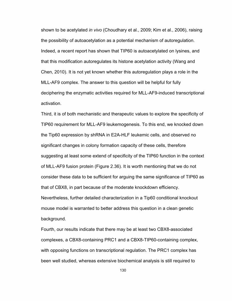
130
shown to be acetylated in vivo (Choudhary et al., 2009; Kim et al., 2006), raising
the possibility of autoacetylation as a potential mechanism of autoregulation.
Indeed, a recent report has shown that TIP60 is autoacetylated on lysines, and
that this modification autoregulates its histone acetylation activity (Wang and
Chen, 2010). It is not yet known whether this autoregulation plays a role in the
MLL-AF9 complex. The answer to this question will be helpful for fully
deciphering the enzymatic activities required for MLL-AF9-induced transcriptional
activation.
Third, it is of both mechanistic and therapeutic values to explore the specificity of
TIP60 requirement for MLL-AF9 leukemogenesis. To this end, we knocked down
the Tip60 expression by shRNA in E2A-HLF leukemic cells, and observed no
significant changes in colony formation capacity of these cells, therefore
suggesting at least some extend of specificity of the TIP60 function in the context
of MLL-AF9 fusion protein (Figure 2.36). It is worth mentioning that we do not
consider these data to be sufficient for arguing the same significance of TIP60 as
that of CBX8, in part because of the moderate knockdown efficiency.
Nevertheless, further detailed characterization in a Tip60 conditional knockout
mouse model is warranted to better address this question in a clean genetic
background.
Fourth, our results indicate that there may be at least two CBX8-associated
complexes, a CBX8-containing PRC1 and a CBX8-TIP60-containing complex,
with opposing functions on transcriptional regulation. The PRC1 complex has
been well studied, whereas extensive biochemical analysis is still required to
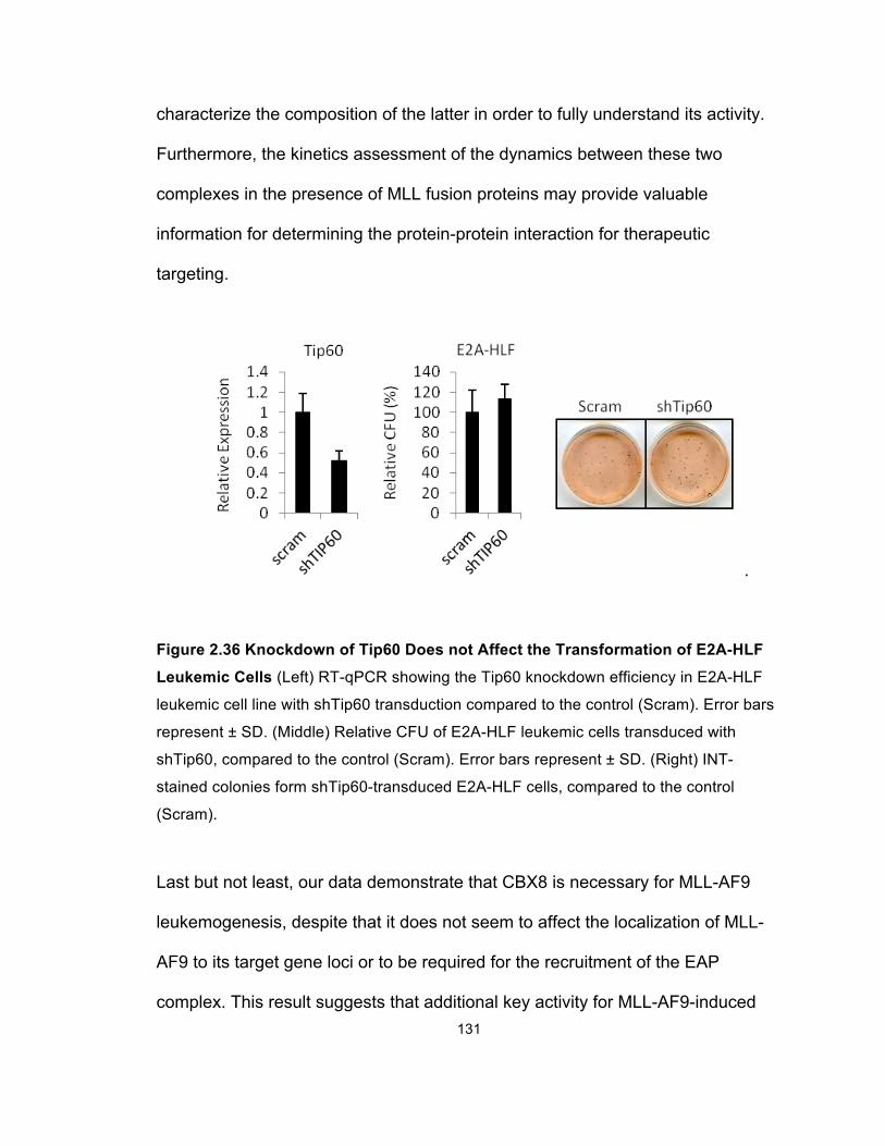
131
characterize the composition of the latter in order to fully understand its activity.
Furthermore, the kinetics assessment of the dynamics between these two
complexes in the presence of MLL fusion proteins may provide valuable
information for determining the protein-protein interaction for therapeutic
targeting.
.
Figure 2.36 Knockdown of Tip60 Does not Affect the Transformation of E2A-HLF
Leukemic Cells (Left) RT-qPCR showing the Tip60 knockdown efficiency in E2A-HLF
leukemic cell line with shTip60 transduction compared to the control (Scram). Error bars
represent ± SD. (Middle) Relative CFU of E2A-HLF leukemic cells transduced with
shTip60, compared to the control (Scram). Error bars represent ± SD. (Right) INT-
stained colonies form shTip60-transduced E2A-HLF cells, compared to the control
(Scram).
Last but not least, our data demonstrate that CBX8 is necessary for MLL-AF9
leukemogenesis, despite that it does not seem to affect the localization of MLL-
AF9 to its target gene loci or to be required for the recruitment of the EAP
complex. This result suggests that additional key activity for MLL-AF9-induced
132
transcriptional activation is yet to be identified. Although our finding regarding
TIP60 suggests that acetylation activity may be part of the answer, it does not
rule out the possibility that in addition to TIP60, other co-factors may be involved
in the dependence of MLL-AF9 leukemogenesis on CBX8 as well. Thus, future
unbiased analysis is of importance to compare the global changes in gene
expression and protein-protein interactions in the presence or absence of CBX8
to fully decipher the leukemogenic mechanisms of MLL-AF9.
133
Chapter 3
Concluding Remarks
Chromosome translocations at the MLL locus that generate chimeric MLL fusion
proteins are one of the major genetic lesions leading to human acute leukemias.
Although aberrant target gene activation is known as the primary drive of MLL-
rearranged leukemogenesis, the underlying mechanisms remain poorly
understood. Multiple lines of evidence have indicated that both the N-terminal
MLL portion and the C-terminal translocation partners of MLL fusion proteins
bear functional significance. The work described in this thesis explored molecular
mechanisms that can help decipher this significance and identified novel co-
factors of both the N terminus and the C terminus of MLL fusion proteins, which
are critical for leukemogenesis. The key findings are summarized as following:
For the N-terminal MLL portion:
• PAFc interacts with the pre-CxxC and RD2 domains of MLL
• Transcriptional activity of MLL and MLL fusion proteins is stimulated by
PAFc
• MLL fusion protein mediated transformation is dependent upon interaction
with PAFc
• PAFc expression is coordinately downregulated during myeloid
differentiation
134
For the C-terminal translocation partners (AF9/ENL):
• CBX8 is essential for both initiation and maintenance of MLL-AF9
transformation
• CBX8 is crucial for MLL-AF9-induced transcriptional activation
• Role of CBX8 in MLL-AF9 leukemogenesis is Independent of PRC1
• CBX8 regulation of TIP60 localization contributes to MLL-AF9
transformation
These observations were primarily obtained using the common fusion proteins
MLL-AF9 and MLL-ENL. Because of the diversity of MLL fusion partners and the
elusive leukemogenic mechanisms for many of the less frequent MLL fusion
proteins (Martin et al., 2003; So et al., 2003), it is yet to be evaluated that to what
extend our findings can apply to these other fusion proteins. Nevertheless, this
study has helped reveal a sophisticated oncogenic system orchestrated by MLL
fusion proteins: the oncoproteins promote constitutive transcriptional activation
by both hijacking the basal transcriptional machinery (e.g., PAFc) and recruiting
the co-factor whose functional significance seems rather context-dependent (e.g.,
CBX8, evidenced by its dispensable role in normal hematopoiesis). Moreover, it
well exemplifies how the oncogenic variants (MLL fusion proteins) of a wild-type
protein (MLL) rewire the evolutionarily conserved functional collaboration of
enzymatic activities (HMT-HAT for transcriptional activation: SET and MOF for
MLL vs. DOT1L and TIP60 for MLL-AF9) for oncogenic purposes.
Taken together, our observations implicate two potential directions for
therapeutic application. On the one hand, for the co-factors with critical
135
physiological functions in normal cells, such as PAFc, it is worthwhile to define
the specificity of their importance in the context of MLL-rearranged leukemias,
therefore exploring the therapeutic window for preferentially eliminating leukemia
cells by targeting these factors. On the other hand, for the co-factors whose roles
are highly context-dependent, such as CBX8, it is of great value to identify their
differential interaction partners in specific pathogenic processes and to
characterize the interaction surface and binding kinetics, which in turn lays a
critical foundation for designing small molecular inhibitors to specifically target
protein-protein interactions. In summary, this work has presented valuable
substrates for employing these approaches to further explore novel therapeutic
strategies for treating MLL-rearranged leukemias.
136
References
Akhtar, A., Zink, D., and Becker, P. B. (2000). Chromodomains are protein-RNA
interaction modules. Nature 407, 405-409.
Allen, M. D., Grummitt, C. G., Hilcenko, C., Min, S. Y., Tonkin, L. M., Johnson, C.
M., Freund, S. M., Bycroft, M., and Warren, A. J. (2006). Solution structure of the
nonmethyl-CpG-binding CXXC domain of the leukaemia-associated MLL histone
methyltransferase. EMBO J 25, 4503-4512.
Ansari, K. I., and Mandal, S. S. (2010). Mixed lineage leukemia: roles in gene
expression, hormone signaling and mRNA processing. FEBS J 277, 1790-1804.
Armstrong, S. A., Staunton, J. E., Silverman, L. B., Pieters, R., den Boer, M. L.,
Minden, M. D., Sallan, S. E., Lander, E. S., Golub, T. R., and Korsmeyer, S. J.
(2002). MLL translocations specify a distinct gene expression profile that
distinguishes a unique leukemia. Nat Genet 30, 41-47.
Avvakumov, N., and Cote, J. (2007). The MYST family of histone
acetyltransferases and their intimate links to cancer. Oncogene 26, 5395-5407.
Ayton, P. M., Chen, E. H., and Cleary, M. L. (2004). Binding to nonmethylated
CpG DNA is essential for target recognition, transactivation, and myeloid
transformation by an MLL oncoprotein. Mol Cell Biol 24, 10470-10478.
Ayton, P. M., and Cleary, M. L. (2003). Transformation of myeloid progenitors by
MLL oncoproteins is dependent on Hoxa7 and Hoxa9. Genes Dev 17, 2298-2307.
Bach, C., Mueller, D., Buhl, S., Garcia-Cuellar, M. P., and Slany, R. K. (2009).
Alterations of the CxxC domain preclude oncogenic activation of mixed-lineage
leukemia 2. Oncogene 28, 815-823.
137
Bachmann, I. M., Halvorsen, O. J., Collett, K., Stefansson, I. M., Straume, O.,
Haukaas, S. A., Salvesen, H. B., Otte, A. P., and Akslen, L. A. (2006). EZH2
expression is associated with high proliferation rate and aggressive tumor
subgroups in cutaneous melanoma and cancers of the endometrium, prostate,
and breast. J Clin Oncol 24, 268-273.
Baek, S. H., Ohgi, K. A., Rose, D. W., Koo, E. H., Glass, C. K., and Rosenfeld, M.
G. (2002). Exchange of N-CoR corepressor and Tip60 coactivator complexes
links gene expression by NF-kappaB and beta-amyloid precursor protein. Cell
110, 55-67.
Balgobind, B. V., Zwaan, C. M., Pieters, R., and Van den Heuvel-Eibrink, M. M.
(2011). The heterogeneity of pediatric MLL-rearranged acute myeloid leukemia.
Leukemia 25, 1239-1248.
Bantignies, F., Goodman, R. H., and Smolik, S. M. (2000). Functional interaction
between the coactivator Drosophila CREB-binding protein and ASH1, a member
of the trithorax group of chromatin modifiers. Mol Cell Biol 20, 9317-9330.
Bararia, D., Trivedi, A. K., Zada, A. A., Greif, P. A., Mulaw, M. A., Christopeit, M.,
Hiddemann, W., Bohlander, S. K., and Behre, G. (2008). Proteomic identification
of the MYST domain histone acetyltransferase TIP60 (HTATIP) as a co-activator
of the myeloid transcription factor C/EBPalpha. Leukemia 22, 800-807.
Bardos, J. I., Saurin, A. J., Tissot, C., Duprez, E., and Freemont, P. S. (2000).
HPC3 is a new human polycomb orthologue that interacts and associates with
RING1 and Bmi1 and has transcriptional repression properties. J Biol Chem 275,
28785-28792.
138
Batra, S. K., Metzgar, R. S., and Hollingsworth, M. A. (1991). Isolation and
characterization of a complementary DNA (PD-1) differentially expressed by
human pancreatic ductal cell tumors. Cell Growth Differ 2, 385-390.
Berns, K., Hijmans, E. M., Mullenders, J., Brummelkamp, T. R., Velds, A.,
Heimerikx, M., Kerkhoven, R. M., Madiredjo, M., Nijkamp, W., Weigelt, B., et al.
(2004). A large-scale RNAi screen in human cells identifies new components of
the p53 pathway. Nature 428, 431-437.
Bernstein, E., Duncan, E. M., Masui, O., Gil, J., Heard, E., and Allis, C. D. (2006).
Mouse polycomb proteins bind differentially to methylated histone H3 and RNA
and are enriched in facultative heterochromatin. Mol Cell Biol 26, 2560-2569.
Bernt, K. M., Zhu, N., Sinha, A. U., Vempati, S., Faber, J., Krivtsov, A. V., Feng,
Z., Punt, N., Daigle, A., Bullinger, L., et al. (2011). MLL-Rearranged Leukemia Is
Dependent on Aberrant H3K79 Methylation by DOT1L. Cancer Cell 20, 66-78.
Boyer, L. A., Plath, K., Zeitlinger, J., Brambrink, T., Medeiros, L. A., Lee, T. I.,
Levine, S. S., Wernig, M., Tajonar, A., Ray, M. K., et al. (2006). Polycomb
complexes repress developmental regulators in murine embryonic stem cells.
Nature 441, 349-353.
Bracken, A. P., and Helin, K. (2009). Polycomb group proteins: navigators of
lineage pathways led astray in cancer. Nat Rev Cancer 9, 773-784.
Brady, M. E., Ozanne, D. M., Gaughan, L., Waite, I., Cook, S., Neal, D. E., and
Robson, C. N. (1999). Tip60 is a nuclear hormone receptor coactivator. J Biol
Chem 274, 17599-17604.
139
Brookes, E., and Pombo, A. (2009). Modifications of RNA polymerase II are
pivotal in regulating gene expression states. EMBO Rep 10, 1213-1219.
Cao, Q., Mani, R. S., Ateeq, B., Dhanasekaran, S. M., Asangani, I. A., Prensner,
J. R., Kim, J. H., Brenner, J. C., Jing, X., Cao, X., et al. (2011). Coordinated
Regulation of Polycomb Group Complexes through microRNAs in Cancer.
Cancer Cell 20, 187-199.
Cao, R., Wang, L., Wang, H., Xia, L., Erdjument-Bromage, H., Tempst, P., Jones,
R. S., and Zhang, Y. (2002). Role of histone H3 lysine 27 methylation in
Polycomb-group silencing. Science 298, 1039-1043.
Cao, X., and Sudhof, T. C. (2001). A transcriptionally [correction of transcriptively]
active complex of APP with Fe65 and histone acetyltransferase Tip60. Science
293, 115-120.
Caslini, C., Yang, Z., El-Osta, M., Milne, T. A., Slany, R. K., and Hess, J. L.
(2007). Interaction of MLL amino terminal sequences with menin is required for
transformation. Cancer Res 67, 7275-7283.
Celetti, A., Barba, P., Cillo, C., Rotoli, B., Boncinelli, E., and Magli, M. C. (1993).
Characteristic patterns of HOX gene expression in different types of human
leukemia. Int J Cancer 53, 237-244.
Chaudhary, K., Deb, S., Moniaux, N., Ponnusamy, M. P., and Batra, S. K. (2007).
Human RNA polymerase II-associated factor complex: dysregulation in cancer.
Oncogene 26, 7499-7507.
140
Chen, W., Kumar, A. R., Hudson, W. A., Li, Q., Wu, B., Staggs, R. A., Lund, E. A.,
Sam, T. N., and Kersey, J. H. (2008). Malignant transformation initiated by Mll-
AF9: gene dosage and critical target cells. Cancer Cell 13, 432-440.
Chen, Y. X., Yan, J., Keeshan, K., Tubbs, A. T., Wang, H., Silva, A., Brown, E. J.,
Hess, J. L., Pear, W. S., and Hua, X. (2006). The tumor suppressor menin
regulates hematopoiesis and myeloid transformation by influencing Hox gene
expression. Proc Natl Acad Sci U S A 103, 1018-1023.
Cheung, N., Chan, L. C., Thompson, A., Cleary, M. L., and So, C. W. (2007).
Protein arginine-methyltransferase-dependent oncogenesis. Nat Cell Biol 9,
1208-1215.
Chopra, V. S., Hong, J. W., and Levine, M. (2009). Regulation of Hox gene
activity by transcriptional elongation in Drosophila. Curr Biol 19, 688-693.
Choudhary, C., Kumar, C., Gnad, F., Nielsen, M. L., Rehman, M., Walther, T. C.,
Olsen, J. V., and Mann, M. (2009). Lysine acetylation targets protein complexes
and co-regulates major cellular functions. Science 325, 834-840.
Collett, K., Eide, G. E., Arnes, J., Stefansson, I. M., Eide, J., Braaten, A., Aas, T.,
Otte, A. P., and Akslen, L. A. (2006). Expression of enhancer of zeste homologue
2 is significantly associated with increased tumor cell proliferation and is a
marker of aggressive breast cancer. Clin Cancer Res 12, 1168-1174.
Creaven, M., Hans, F., Mutskov, V., Col, E., Caron, C., Dimitrov, S., and
Khochbin, S. (1999). Control of the histone-acetyltransferase activity of Tip60 by
the HIV-1 transactivator protein, Tat. Biochemistry 38, 8826-8830.
141
Czermin, B., Melfi, R., McCabe, D., Seitz, V., Imhof, A., and Pirrotta, V. (2002).
Drosophila enhancer of Zeste/ESC complexes have a histone H3
methyltransferase activity that marks chromosomal Polycomb sites. Cell 111,
185-196.
de Napoles, M., Mermoud, J. E., Wakao, R., Tang, Y. A., Endoh, M., Appanah,
R., Nesterova, T. B., Silva, J., Otte, A. P., Vidal, M., et al. (2004). Polycomb
group proteins Ring1A/B link ubiquitylation of histone H2A to heritable gene
silencing and X inactivation. Dev Cell 7, 663-676.
DelProposto, J., Majmudar, C. Y., Smith, J. L., and Brown, W. C. (2009). Mocr: a
novel fusion tag for enhancing solubility that is compatible with structural biology
applications. Protein Expr Purif 63, 40-49.
Dietrich, N., Bracken, A. P., Trinh, E., Schjerling, C. K., Koseki, H., Rappsilber, J.,
Helin, K., and Hansen, K. H. (2007). Bypass of senescence by the polycomb
group protein CBX8 through direct binding to the INK4A-ARF locus. EMBO J 26,
1637-1648.
Ding, L., Paszkowski-Rogacz, M., Nitzsche, A., Slabicki, M. M., Heninger, A. K.,
de Vries, I., Kittler, R., Junqueira, M., Shevchenko, A., Schulz, H., et al. (2009). A
genome-scale RNAi screen for Oct4 modulators defines a role of the Paf1
complex for embryonic stem cell identity. Cell Stem Cell 4, 403-415.
Djabali, M., Selleri, L., Parry, P., Bower, M., Young, B. D., and Evans, G. A.
(1992). A trithorax-like gene is interrupted by chromosome 11q23 translocations
in acute leukaemias. Nat Genet 2, 113-118.
142
Donnelly, M. I., Zhou, M., Millard, C. S., Clancy, S., Stols, L., Eschenfeldt, W. H.,
Collart, F. R., and Joachimiak, A. (2006). An expression vector tailored for large-
scale, high-throughput purification of recombinant proteins. Protein Expr Purif 47,
446-454.
Dou, Y., and Hess, J. L. (2008). Mechanisms of transcriptional regulation by MLL
and its disruption in acute leukemia. Int J Hematol 87, 10-18.
Dou, Y., Milne, T. A., Ruthenburg, A. J., Lee, S., Lee, J. W., Verdine, G. L., Allis,
C. D., and Roeder, R. G. (2006). Regulation of MLL1 H3K4 methyltransferase
activity by its core components. Nat Struct Mol Biol 13, 713-719.
Dou, Y., Milne, T. A., Tackett, A. J., Smith, E. R., Fukuda, A., Wysocka, J., Allis,
C. D., Chait, B. T., Hess, J. L., and Roeder, R. G. (2005). Physical association
and coordinate function of the H3 K4 methyltransferase MLL1 and the H4 K16
acetyltransferase MOF. Cell 121, 873-885.
Doyon, Y., Selleck, W., Lane, W. S., Tan, S., and Cote, J. (2004). Structural and
functional conservation of the NuA4 histone acetyltransferase complex from
yeast to humans. Mol Cell Biol 24, 1884-1896.
Ernst, P., Wang, J., Huang, M., Goodman, R. H., and Korsmeyer, S. J. (2001).
MLL and CREB bind cooperatively to the nuclear coactivator CREB-binding
protein. Mol Cell Biol 21, 2249-2258.
Ernst, T., Chase, A. J., Score, J., Hidalgo-Curtis, C. E., Bryant, C., Jones, A. V.,
Waghorn, K., Zoi, K., Ross, F. M., Reiter, A., et al. (2010). Inactivating mutations
of the histone methyltransferase gene EZH2 in myeloid disorders. Nat Genet 42,
722-726.
143
Fazzio, T. G., Huff, J. T., and Panning, B. (2008). An RNAi screen of chromatin
proteins identifies Tip60-p400 as a regulator of embryonic stem cell identity. Cell
134, 162-174.
Felix, C. A. (1998). Secondary leukemias induced by topoisomerase-targeted
drugs. Biochim Biophys Acta 1400, 233-255.
Francis, N. J., and Kingston, R. E. (2001). Mechanisms of transcriptional memory.
Nat Rev Mol Cell Biol 2, 409-421.
Francis, N. J., Kingston, R. E., and Woodcock, C. L. (2004). Chromatin
compaction by a polycomb group protein complex. Science 306, 1574-1577.
Frank, S. R., Parisi, T., Taubert, S., Fernandez, P., Fuchs, M., Chan, H. M.,
Livingston, D. M., and Amati, B. (2003). MYC recruits the TIP60 histone
acetyltransferase complex to chromatin. EMBO Rep 4, 575-580.
Garcia-Cuellar, M. P., Zilles, O., Schreiner, S. A., Birke, M., Winkler, T. H., and
Slany, R. K. (2001). The ENL moiety of the childhood leukemia-associated MLL-
ENL oncoprotein recruits human Polycomb 3. Oncogene 20, 411-419.
Gaughan, L., Brady, M. E., Cook, S., Neal, D. E., and Robson, C. N. (2001).
Tip60 is a co-activator specific for class I nuclear hormone receptors. J Biol
Chem 276, 46841-46848.
Gaughan, L., Logan, I. R., Cook, S., Neal, D. E., and Robson, C. N. (2002).
Tip60 and histone deacetylase 1 regulate androgen receptor activity through
changes to the acetylation status of the receptor. J Biol Chem 277, 25904-25913.
144
Grimm, C., Matos, R., Ly-Hartig, N., Steuerwald, U., Lindner, D., Rybin, V.,
Muller, J., and Muller, C. W. (2009). Molecular recognition of histone lysine
methylation by the Polycomb group repressor dSfmbt. EMBO J 28, 1965-1977.
Gu, Y., Nakamura, T., Alder, H., Prasad, R., Canaani, O., Cimino, G., Croce, C.
M., and Canaani, E. (1992). The t(4;11) chromosome translocation of human
acute leukemias fuses the ALL-1 gene, related to Drosophila trithorax, to the AF-
4 gene. Cell 71, 701-708.
Guo, Y., Lubbert, M., and Engelhardt, M. (2003). CD34- hematopoietic stem cells:
current concepts and controversies. Stem Cells 21, 15-20.
Gupta, R. A., Shah, N., Wang, K. C., Kim, J., Horlings, H. M., Wong, D. J., Tsai,
M. C., Hung, T., Argani, P., Rinn, J. L., et al. (2010). Long non-coding RNA
HOTAIR reprograms chromatin state to promote cancer metastasis. Nature 464,
1071-1076.
He, H., Hua, X., and Yan, J. (2011). Epigenetic regulations in hematopoietic Hox
code. Oncogene 30, 379-388.
Hemenway, C. S., de Erkenez, A. C., and Gould, G. C. (2001). The polycomb
protein MPc3 interacts with AF9, an MLL fusion partner in t(9;11)(p22;q23) acute
leukemias. Oncogene 20, 3798-3805.
Hsieh, J. J., Cheng, E. H., and Korsmeyer, S. J. (2003a). Taspase1: a threonine
aspartase required for cleavage of MLL and proper HOX gene expression. Cell
115, 293-303.
Hsieh, J. J., Ernst, P., Erdjument-Bromage, H., Tempst, P., and Korsmeyer, S. J.
(2003b). Proteolytic cleavage of MLL generates a complex of N- and C-terminal
145
fragments that confers protein stability and subnuclear localization. Mol Cell Biol
23, 186-194.
Hu, Y., Fisher, J. B., Koprowski, S., McAllister, D., Kim, M. S., and Lough, J.
(2009). Homozygous disruption of the Tip60 gene causes early embryonic
lethality. Dev Dyn 238, 2912-2921.
Ikura, T., Ogryzko, V. V., Grigoriev, M., Groisman, R., Wang, J., Horikoshi, M.,
Scully, R., Qin, J., and Nakatani, Y. (2000). Involvement of the TIP60 histone
acetylase complex in DNA repair and apoptosis. Cell 102, 463-473.
Iwata, T., Mizusawa, N., Taketani, Y., Itakura, M., and Yoshimoto, K. (2007).
Parafibromin tumor suppressor enhances cell growth in the cells expressing
SV40 large T antigen. Oncogene 26, 6176-6183.
Jaehning, J. A. (2010a). The Paf1 complex: platform or player in RNA
polymerase II transcription? Biochim Biophys Acta 1799, 379-388.
Jaehning, J. A. (2010b). The Paf1 complex: Platform or player in RNA
polymerase II transcription? Biochimica Et Biophysica Acta-Gene Regulatory
Mechanisms 1799, 379-388.
Jo, S. Y., Granowicz, E. M., Maillard, I., Thomas, D., and Hess, J. L. (2011).
Requirement for Dot1l in murine postnatal hematopoiesis and leukemogenesis
by MLL translocation. Blood 117, 4759-4768.
Jude, C. D., Climer, L., Xu, D., Artinger, E., Fisher, J. K., and Ernst, P. (2007).
Unique and independent roles for MLL in adult hematopoietic stem cells and
progenitors. Cell Stem Cell 1, 324-337.
146
Kanhere, A., Viiri, K., Araujo, C. C., Rasaiyaah, J., Bouwman, R. D., Whyte, W.
A., Pereira, C. F., Brookes, E., Walker, K., Bell, G. W., et al. (2010). Short RNAs
are transcribed from repressed polycomb target genes and interact with
polycomb repressive complex-2. Mol Cell 38, 675-688.
Katoh-Fukui, Y., Tsuchiya, R., Shiroishi, T., Nakahara, Y., Hashimoto, N.,
Noguchi, K., and Higashinakagawa, T. (1998). Male-to-female sex reversal in
M33 mutant mice. Nature 393, 688-692.
Katsumoto, T., Yoshida, N., and Kitabayashi, I. (2008). Roles of the histone
acetyltransferase monocytic leukemia zinc finger protein in normal and malignant
hematopoiesis. Cancer Sci 99, 1523-1527.
Kaustov, L., Ouyang, H., Amaya, M., Lemak, A., Nady, N., Duan, S., Wasney, G.
A., Li, Z., Vedadi, M., Schapira, M., et al. (2011). Recognition and specificity
determinants of the human cbx chromodomains. J Biol Chem 286, 521-529.
Keller, A., Nesvizhskii, A. I., Kolker, E., and Aebersold, R. (2002). Empirical
statistical model to estimate the accuracy of peptide identifications made by
MS/MS and database search. Anal Chem 74, 5383-5392.
Kerppola, T. K. (2009). Polycomb group complexes--many combinations, many
functions. Trends Cell Biol 19, 692-704.
Kidani, K., Osaki, M., Tamura, T., Yamaga, K., Shomori, K., Ryoke, K., and Ito, H.
(2009). High expression of EZH2 is associated with tumor proliferation and
prognosis in human oral squamous cell carcinomas. Oral Oncol 45, 39-46.
Kim, J., Guermah, M., McGinty, R. K., Lee, J. S., Tang, Z., Milne, T. A.,
Shilatifard, A., Muir, T. W., and Roeder, R. G. (2009). RAD6-Mediated
147
transcription-coupled H2B ubiquitylation directly stimulates H3K4 methylation in
human cells. Cell 137, 459-471.
Kim, J., Guermah, M., and Roeder, R. G. (2010). The Human PAF1 Complex
Acts in Chromatin Transcription Elongation Both Independently and
Cooperatively with SII/TFIIS. Cell 140, 491-503.
Kim, J., and Roeder, R. G. (2009). Direct Bre1-Paf1 complex interactions and
RING finger-independent Bre1-Rad6 interactions mediate histone H2B
ubiquitylation in yeast. J Biol Chem 284, 20582-20592.
Kim, S. C., Sprung, R., Chen, Y., Xu, Y., Ball, H., Pei, J., Cheng, T., Kho, Y.,
Xiao, H., Xiao, L., et al. (2006). Substrate and functional diversity of lysine
acetylation revealed by a proteomics survey. Mol Cell 23, 607-618.
Kim, S. H., Park, J., Choi, M. C., Park, J. H., Kim, H. P., Lee, J. H., Oh, D. Y., Im,
S. A., Bang, Y. J., and Kim, T. Y. (2008). DNA methyltransferase 3B acts as a
co-repressor of the human polycomb protein hPc2 to repress fibroblast growth
factor receptor 3 transcription. Int J Biochem Cell Biol 40, 2462-2471.
Kinoshita, A., Whelan, C. M., Berezovska, O., and Hyman, B. T. (2002). The
gamma secretase-generated carboxyl-terminal domain of the amyloid precursor
protein induces apoptosis via Tip60 in H4 cells. J Biol Chem 277, 28530-28536.
Kirmizis, A., Bartley, S. M., Kuzmichev, A., Margueron, R., Reinberg, D., Green,
R., and Farnham, P. J. (2004). Silencing of human polycomb target genes is
associated with methylation of histone H3 Lys 27. Genes Dev 18, 1592-1605.
Krivtsov, A. V., and Armstrong, S. A. (2007). MLL translocations, histone
modifications and leukaemia stem-cell development. Nat Rev Cancer 7, 823-833.
148
Krivtsov, A. V., Feng, Z., Lemieux, M. E., Faber, J., Vempati, S., Sinha, A. U., Xia,
X., Jesneck, J., Bracken, A. P., Silverman, L. B., et al. (2008). H3K79 methylation
profiles define murine and human MLL-AF4 leukemias. Cancer Cell 14, 355-368.
Krogan, N. J., Dover, J., Wood, A., Schneider, J., Heidt, J., Boateng, M. A., Dean,
K., Ryan, O. W., Golshani, A., Johnston, M., et al. (2003). The Paf1 complex is
required for histone H3 methylation by COMPASS and Dot1p: linking
transcriptional elongation to histone methylation. Mol Cell 11, 721-729.
Krumlauf, R. (1994). Hox genes in vertebrate development. Cell 78, 191-201.
Ku, M., Koche, R. P., Rheinbay, E., Mendenhall, E. M., Endoh, M., Mikkelsen, T.
S., Presser, A., Nusbaum, C., Xie, X., Chi, A. S., et al. (2008). Genomewide
analysis of PRC1 and PRC2 occupancy identifies two classes of bivalent
domains. PLoS Genet 4, e1000242.
Kumar, A. R., Hudson, W. A., Chen, W., Nishiuchi, R., Yao, Q., and Kersey, J. H.
(2004). Hoxa9 influences the phenotype but not the incidence of Mll-AF9 fusion
gene leukemia. Blood 103, 1823-1828.
Kusch, T., Florens, L., Macdonald, W. H., Swanson, S. K., Glaser, R. L., Yates, J.
R., 3rd, Abmayr, S. M., Washburn, M. P., and Workman, J. L. (2004). Acetylation
by Tip60 is required for selective histone variant exchange at DNA lesions.
Science 306, 2084-2087.
Kuzmichev, A., Nishioka, K., Erdjument-Bromage, H., Tempst, P., and Reinberg,
D. (2002). Histone methyltransferase activity associated with a human
multiprotein complex containing the Enhancer of Zeste protein. Genes Dev 16,
2893-2905.
149
Landeira, D., Sauer, S., Poot, R., Dvorkina, M., Mazzarella, L., Jorgensen, H. F.,
Pereira, C. F., Leleu, M., Piccolo, F. M., Spivakov, M., et al. (2010). Jarid2 is a
PRC2 component in embryonic stem cells required for multi-lineage
differentiation and recruitment of PRC1 and RNA Polymerase II to developmental
regulators. Nat Cell Biol 12, 618-624.
Lavau, C., Szilvassy, S. J., Slany, R., and Cleary, M. L. (1997). Immortalization
and leukemic transformation of a myelomonocytic precursor by retrovirally
transduced HRX-ENL. EMBO J 16, 4226-4237.
Lawrence, H. J., Christensen, J., Fong, S., Hu, Y. L., Weissman, I., Sauvageau,
G., Humphries, R. K., and Largman, C. (2005). Loss of expression of the Hoxa-9
homeobox gene impairs the proliferation and repopulating ability of
hematopoietic stem cells. Blood 106, 3988-3994.
Lawrence, H. J., Sauvageau, G., Humphries, R. K., and Largman, C. (1996). The
role of HOX homeobox genes in normal and leukemic hematopoiesis. Stem Cells
14, 281-291.
Leduc, C., Claverie, P., Eymin, B., Col, E., Khochbin, S., Brambilla, E., and
Gazzeri, S. (2006). p14ARF promotes RB accumulation through inhibition of its
Tip60-dependent acetylation. Oncogene 25, 4147-4154.
Leeb, M., and Wutz, A. (2007). Ring1B is crucial for the regulation of
developmental control genes and PRC1 proteins but not X inactivation in
embryonic cells. J Cell Biol 178, 219-229.
150
Legube, G., Linares, L. K., Tyteca, S., Caron, C., Scheffner, M., Chevillard-Briet,
M., and Trouche, D. (2004). Role of the histone acetyl transferase Tip60 in the
p53 pathway. J Biol Chem 279, 44825-44833.
Lessard, J., and Sauvageau, G. (2003). Polycomb group genes as epigenetic
regulators of normal and leukemic hemopoiesis. Exp Hematol 31, 567-585.
Levine, S. S., King, I. F., and Kingston, R. E. (2004). Division of labor in
polycomb group repression. Trends Biochem Sci 29, 478-485.
Levine, S. S., Weiss, A., Erdjument-Bromage, H., Shao, Z., Tempst, P., and
Kingston, R. E. (2002). The core of the polycomb repressive complex is
compositionally and functionally conserved in flies and humans. Mol Cell Biol 22,
6070-6078.
Li, H., Ma, X., Wang, J., Koontz, J., Nucci, M., and Sklar, J. (2007). Effects of
rearrangement and allelic exclusion of JJAZ1/SUZ12 on cell proliferation and
survival. Proc Natl Acad Sci U S A 104, 20001-20006.
Lin, C., Smith, E. R., Takahashi, H., Lai, K. C., Martin-Brown, S., Florens, L.,
Washburn, M. P., Conaway, J. W., Conaway, R. C., and Shilatifard, A. (2010).
AFF4, a component of the ELL/P-TEFb elongation complex and a shared subunit
of MLL chimeras, can link transcription elongation to leukemia. Mol Cell 37, 429-
437.
Maertens, G. N., El Messaoudi-Aubert, S., Racek, T., Stock, J. K., Nicholls, J.,
Rodriguez-Niedenfuhr, M., Gil, J., and Peters, G. (2009). Several distinct
polycomb complexes regulate and co-localize on the INK4a tumor suppressor
locus. PLoS One 4, e6380.
151
Magli, M. C., Largman, C., and Lawrence, H. J. (1997). Effects of HOX
homeobox genes in blood cell differentiation. J Cell Physiol 173, 168-177.
Maillard, I., Chen, Y. X., Friedman, A., Yang, Y., Tubbs, A. T., Shestova, O., Pear,
W. S., and Hua, X. (2009). Menin regulates the function of hematopoietic stem
cells and lymphoid progenitors. Blood 113, 1661-1669.
Majewski, I. J., Blewitt, M. E., de Graaf, C. A., McManus, E. J., Bahlo, M., Hilton,
A. A., Hyland, C. D., Smyth, G. K., Corbin, J. E., Metcalf, D., et al. (2008).
Polycomb repressive complex 2 (PRC2) restricts hematopoietic stem cell activity.
PLoS Biol 6, e93.
Margueron, R., and Reinberg, D. (2011). The Polycomb complex PRC2 and its
mark in life. Nature 469, 343-349.
Martin, M. E., Milne, T. A., Bloyer, S., Galoian, K., Shen, W., Gibbs, D., Brock, H.
W., Slany, R., and Hess, J. L. (2003). Dimerization of MLL fusion proteins
immortalizes hematopoietic cells. Cancer Cell 4, 197-207.
Martin-Perez, D., Piris, M. A., and Sanchez-Beato, M. (2010). Polycomb proteins
in hematologic malignancies. Blood 116, 5465-5475.
McMahon, K. A., Hiew, S. Y., Hadjur, S., Veiga-Fernandes, H., Menzel, U., Price,
A. J., Kioussis, D., Williams, O., and Brady, H. J. (2007). Mll has a critical role in
fetal and adult hematopoietic stem cell self-renewal. Cell Stem Cell 1, 338-345.
Mills, A. A. (2010). Throwing the cancer switch: reciprocal roles of polycomb and
trithorax proteins. Nat Rev Cancer 10, 669-682.

152
Milne, T. A., Briggs, S. D., Brock, H. W., Martin, M. E., Gibbs, D., Allis, C. D., and
Hess, J. L. (2002). MLL targets SET domain methyltransferase activity to Hox
gene promoters. Mol Cell 10, 1107-1117.
Milne, T. A., Dou, Y., Martin, M. E., Brock, H. W., Roeder, R. G., and Hess, J. L.
(2005a). MLL associates specifically with a subset of transcriptionally active
target genes. Proceedings of the National Academy of Sciences of the United
States of America 102, 14765-14770.
Milne, T. A., Hughes, C. M., Lloyd, R., Yang, Z., Rozenblatt-Rosen, O., Dou, Y.,
Schnepp, R. W., Krankel, C., Livolsi, V. A., Gibbs, D., et al. (2005b). Menin and
MLL cooperatively regulate expression of cyclin-dependent kinase inhibitors.
Proc Natl Acad Sci U S A 102, 749-754.
Milne, T. A., Kim, J., Wang, G. G., Stadler, S. C., Basrur, V., Whitcomb, S. J.,
Wang, Z., Ruthenburg, A. J., Elenitoba-Johnson, K. S., Roeder, R. G., and Allis,
C. D. (2010). Multiple interactions recruit MLL1 and MLL1 fusion proteins to the
HOXA9 locus in leukemogenesis. Mol Cell 38, 853-863.
Mimori, K., Ogawa, K., Okamoto, M., Sudo, T., Inoue, H., and Mori, M. (2005).
Clinical significance of enhancer of zeste homolog 2 expression in colorectal
cancer cases. Eur J Surg Oncol 31, 376-380.
Mohammad, H. P., Cai, Y., McGarvey, K. M., Easwaran, H., Van Neste, L., Ohm,
J. E., O'Hagan, H. M., and Baylin, S. B. (2009). Polycomb CBX7 promotes
initiation of heritable repression of genes frequently silenced with cancer-specific
DNA hypermethylation. Cancer Res 69, 6322-6330.
153
Mohan, M., Herz, H. M., Takahashi, Y. H., Lin, C., Lai, K. C., Zhang, Y.,
Washburn, M. P., Florens, L., and Shilatifard, A. (2010a). Linking H3K79
trimethylation to Wnt signaling through a novel Dot1-containing complex
(DotCom). Genes Dev 24, 574-589.
Mohan, M., Lin, C., Guest, E., and Shilatifard, A. (2010b). Licensed to elongate: a
molecular mechanism for MLL-based leukaemogenesis. Nat Rev Cancer 10,
721-728.
Moniaux, N., Nemos, C., Schmied, B. M., Chauhan, S. C., Deb, S., Morikane, K.,
Choudhury, A., Vanlith, M., Sutherlin, M., Sikela, J. M., et al. (2006). The human
homologue of the RNA polymerase II-associated factor 1 (hPaf1), localized on
the 19q13 amplicon, is associated with tumorigenesis. Oncogene 25, 3247-3257.
Monroe, S. C., Jo, S. Y., Sanders, D. S., Basrur, V., Elenitoba-Johnson, K. S.,
Slany, R. K., and Hess, J. L. (2010). MLL-AF9 and MLL-ENL alter the dynamic
association of transcriptional regulators with genes critical for leukemia. Exp
Hematol.
Morin, R. D., Johnson, N. A., Severson, T. M., Mungall, A. J., An, J., Goya, R.,
Paul, J. E., Boyle, M., Woolcock, B. W., Kuchenbauer, F., et al. (2010). Somatic
mutations altering EZH2 (Tyr641) in follicular and diffuse large B-cell lymphomas
of germinal-center origin. Nat Genet 42, 181-185.
Morita, S., Kojima, T., and Kitamura, T. (2000). Plat-E: an efficient and stable
system for transient packaging of retroviruses. Gene Ther 7, 1063-1066.
Mueller, D., Bach, C., Zeisig, D., Garcia-Cuellar, M. P., Monroe, S., Sreekumar,
A., Zhou, R., Nesvizhskii, A., Chinnaiyan, A., Hess, J. L., and Slany, R. K. (2007).
154
A role for the MLL fusion partner ENL in transcriptional elongation and chromatin
modification. Blood 110, 4445-4454.
Muller, J., and Verrijzer, P. (2009). Biochemical mechanisms of gene regulation
by polycomb group protein complexes. Curr Opin Genet Dev 19, 150-158.
Muntean, A. G., Giannola, D., Udager, A. M., and Hess, J. L. (2008). The PHD
fingers of MLL block MLL fusion protein-mediated transformation. Blood 112,
4690-4693.
Muntean, A. G., Tan, J., Sitwala, K., Huang, Y., Bronstein, J., Connelly, J. A.,
Basrur, V., Elenitoba-Johnson, K. S., and Hess, J. L. (2010). The PAF complex
synergizes with MLL fusion proteins at HOX loci to promote leukemogenesis.
Cancer Cell 17, 609-621.
Nakahata, S., Saito, Y., Hamasaki, M., Hidaka, T., Arai, Y., Taki, T., Taniwaki, M.,
and Morishita, K. (2009). Alteration of enhancer of polycomb 1 at 10p11.2 is one
of the genetic events leading to development of adult T-cell leukemia/lymphoma.
Genes Chromosomes Cancer 48, 768-776.
Nakamura, T., Mori, T., Tada, S., Krajewski, W., Rozovskaia, T., Wassell, R.,
Dubois, G., Mazo, A., Croce, C. M., and Canaani, E. (2002). ALL-1 is a histone
methyltransferase that assembles a supercomplex of proteins involved in
transcriptional regulation. Mol Cell 10, 1119-1128.
Nekrasov, M., Klymenko, T., Fraterman, S., Papp, B., Oktaba, K., Kocher, T.,
Cohen, A., Stunnenberg, H. G., Wilm, M., and Muller, J. (2007). Pcl-PRC2 is
needed to generate high levels of H3-K27 trimethylation at Polycomb target
genes. EMBO J 26, 4078-4088.
155
Nesvizhskii, A. I., Keller, A., Kolker, E., and Aebersold, R. (2003). A statistical
model for identifying proteins by tandem mass spectrometry. Anal Chem 75,
4646-4658.
Newey, P. J., Bowl, M. R., and Thakker, R. V. (2009). Parafibromin--functional
insights. J Intern Med 266, 84-98.
Nguyen, A. T., Taranova, O., He, J., and Zhang, Y. (2011). DOT1L, the H3K79
methyltransferase, is required for MLL-AF9-mediated leukemogenesis. Blood
117, 6912-6922.
Nikoloski, G., Langemeijer, S. M., Kuiper, R. P., Knops, R., Massop, M.,
Tonnissen, E. R., van der Heijden, A., Scheele, T. N., Vandenberghe, P., de
Witte, T., et al. (2010). Somatic mutations of the histone methyltransferase gene
EZH2 in myelodysplastic syndromes. Nat Genet 42, 665-667.
Ohtsubo, M., Yasunaga, S., Ohno, Y., Tsumura, M., Okada, S., Ishikawa, N.,
Shirao, K., Kikuchi, A., Nishitani, H., Kobayashi, M., and Takihara, Y. (2008).
Polycomb-group complex 1 acts as an E3 ubiquitin ligase for Geminin to sustain
hematopoietic stem cell activity. Proc Natl Acad Sci U S A 105, 10396-10401.
Pasini, D., Bracken, A. P., Hansen, J. B., Capillo, M., and Helin, K. (2007). The
polycomb group protein Suz12 is required for embryonic stem cell differentiation.
Mol Cell Biol 27, 3769-3779.
Pasini, D., Hansen, K. H., Christensen, J., Agger, K., Cloos, P. A., and Helin, K.
(2008). Coordinated regulation of transcriptional repression by the RBP2 H3K4
demethylase and Polycomb-Repressive Complex 2. Genes Dev 22, 1345-1355.
156
Pasini, D., Malatesta, M., Jung, H. R., Walfridsson, J., Willer, A., Olsson, L.,
Skotte, J., Wutz, A., Porse, B., Jensen, O. N., and Helin, K. (2010).
Characterization of an antagonistic switch between histone H3 lysine 27
methylation and acetylation in the transcriptional regulation of Polycomb group
target genes. Nucleic Acids Res 38, 4958-4969.
Patel, J. H., Du, Y., Ard, P. G., Phillips, C., Carella, B., Chen, C. J., Rakowski, C.,
Chatterjee, C., Lieberman, P. M., Lane, W. S., et al. (2004). The c-MYC
oncoprotein is a substrate of the acetyltransferases hGCN5/PCAF and TIP60.
Mol Cell Biol 24, 10826-10834.
Peng, J. C., Valouev, A., Swigut, T., Zhang, J., Zhao, Y., Sidow, A., and
Wysocka, J. (2009). Jarid2/Jumonji coordinates control of PRC2 enzymatic
activity and target gene occupancy in pluripotent cells. Cell 139, 1290-1302.
Peterson, A. J., Mallin, D. R., Francis, N. J., Ketel, C. S., Stamm, J., Voeller, R.
K., Kingston, R. E., and Simon, J. A. (2004). Requirement for sex comb on
midleg protein interactions in Drosophila polycomb group repression. Genetics
167, 1225-1239.
Petruk, S., Sedkov, Y., Smith, S., Tillib, S., Kraevski, V., Nakamura, T., Canaani,
E., Croce, C. M., and Mazo, A. (2001). Trithorax and dCBP acting in a complex
to maintain expression of a homeotic gene. Science 294, 1331-1334.
Pineault, N., Helgason, C. D., Lawrence, H. J., and Humphries, R. K. (2002).
Differential expression of Hox, Meis1, and Pbx1 genes in primitive cells
throughout murine hematopoietic ontogeny. Exp Hematol 30, 49-57.
157
Ren, X., and Kerppola, T. K. (2011). REST interacts with Cbx proteins and
regulates polycomb repressive complex 1 occupancy at RE1 elements. Mol Cell
Biol 31, 2100-2110.
Richie, E. R., Schumacher, A., Angel, J. M., Holloway, M., Rinchik, E. M., and
Magnuson, T. (2002). The Polycomb-group gene eed regulates thymocyte
differentiation and suppresses the development of carcinogen-induced T-cell
lymphomas. Oncogene 21, 299-306.
Richly, H., Aloia, L., and Di Croce, L. (2011). Roles of the Polycomb group
proteins in stem cells and cancer. Cell Death Dis 2, e204.
Rinn, J. L., Kertesz, M., Wang, J. K., Squazzo, S. L., Xu, X., Brugmann, S. A.,
Goodnough, L. H., Helms, J. A., Farnham, P. J., Segal, E., and Chang, H. Y.
(2007). Functional demarcation of active and silent chromatin domains in human
HOX loci by noncoding RNAs. Cell 129, 1311-1323.
Rozenblatt-Rosen, O., Hughes, C. M., Nannepaga, S. J., Shanmugam, K. S.,
Copeland, T. D., Guszczynski, T., Resau, J. H., and Meyerson, M. (2005). The
parafibromin tumor suppressor protein is part of a human Paf1 complex. Mol Cell
Biol 25, 612-620.
Ruthenburg, A. J., Wang, W., Graybosch, D. M., Li, H., Allis, C. D., Patel, D. J.,
and Verdine, G. L. (2006). Histone H3 recognition and presentation by the WDR5
module of the MLL1 complex. Nat Struct Mol Biol 13, 704-712.
Sanchez, C., Sanchez, I., Demmers, J. A., Rodriguez, P., Strouboulis, J., and
Vidal, M. (2007). Proteomics analysis of Ring1B/Rnf2 interactors identifies a
158
novel complex with the Fbxl10/Jhdm1B histone demethylase and the Bcl6
interacting corepressor. Mol Cell Proteomics 6, 820-834.
Sanchez-Beato, M., Sanchez, E., Gonzalez-Carrero, J., Morente, M., Diez, A.,
Sanchez-Verde, L., Martin, M. C., Cigudosa, J. C., Vidal, M., and Piris, M. A.
(2006). Variability in the expression of polycomb proteins in different normal and
tumoral tissues. A pilot study using tissue microarrays. Mod Pathol 19, 684-694.
Sapountzi, V., and Cote, J. (2011). MYST-family histone acetyltransferases:
beyond chromatin. Cell Mol Life Sci 68, 1147-1156.
Sapountzi, V., Logan, I. R., and Robson, C. N. (2006). Cellular functions of TIP60.
Int J Biochem Cell Biol 38, 1496-1509.
Sauvageau, M., and Sauvageau, G. (2010). Polycomb group proteins: multi-
faceted regulators of somatic stem cells and cancer. Cell Stem Cell 7, 299-313.
Scheuermann, J. C., de Ayala Alonso, A. G., Oktaba, K., Ly-Hartig, N., McGinty,
R. K., Fraterman, S., Wilm, M., Muir, T. W., and Muller, J. (2010). Histone H2A
deubiquitinase activity of the Polycomb repressive complex PR-DUB. Nature 465,
243-247.
Schoeftner, S., Sengupta, A. K., Kubicek, S., Mechtler, K., Spahn, L., Koseki, H.,
Jenuwein, T., and Wutz, A. (2006). Recruitment of PRC1 function at the initiation
of X inactivation independent of PRC2 and silencing. EMBO J 25, 3110-3122.
Schuettengruber, B., Chourrout, D., Vervoort, M., Leblanc, B., and Cavalli, G.
(2007). Genome regulation by polycomb and trithorax proteins. Cell 128, 735-745.
Schuetz, A., Allali-Hassani, A., Martin, F., Loppnau, P., Vedadi, M., Bochkarev,
A., Plotnikov, A. N., Arrowsmith, C. H., and Min, J. (2006). Structural basis for
159
molecular recognition and presentation of histone H3 by WDR5. EMBO J 25,
4245-4252.
Schuringa, J. J., and Vellenga, E. (2010). Role of the polycomb group gene BMI1
in normal and leukemic hematopoietic stem and progenitor cells. Curr Opin
Hematol 17, 294-299.
Schwartz, Y. B., and Pirrotta, V. (2007). Polycomb silencing mechanisms and the
management of genomic programmes. Nat Rev Genet 8, 9-22.
Shah, N., and Sukumar, S. (2010). The Hox genes and their roles in oncogenesis.
Nat Rev Cancer 10, 361-371.
Sierra, J., Yoshida, T., Joazeiro, C. A., and Jones, K. A. (2006). The APC tumor
suppressor counteracts beta-catenin activation and H3K4 methylation at Wnt
target genes. Genes Dev 20, 586-600.
Simon, J. A., and Kingston, R. E. (2009). Mechanisms of polycomb gene
silencing: knowns and unknowns. Nat Rev Mol Cell Biol 10, 697-708.
Sitwala, K. V., Dandekar, M. N., and Hess, J. L. (2008). HOX proteins and
leukemia. Int J Clin Exp Pathol 1, 461-474.
Slany, R. K. (2009). The molecular biology of mixed lineage leukemia.
Haematologica 94, 984-993.
Slany, R. K., Lavau, C., and Cleary, M. L. (1998). The oncogenic capacity of
HRX-ENL requires the transcriptional transactivation activity of ENL and the DNA
binding motifs of HRX. Mol Cell Biol 18, 122-129.
Smith, L. L., Yeung, J., Zeisig, B. B., Popov, N., Huijbers, I., Barnes, J., Wilson, A.
J., Taskesen, E., Delwel, R., Gil, J., et al. (2011). Functional Crosstalk between
160
Bmi1 and MLL/Hoxa9 Axis in Establishment of Normal Hematopoietic and
Leukemic Stem Cells. Cell Stem Cell 8, 649-662.
So, C. W., Lin, M., Ayton, P. M., Chen, E. H., and Cleary, M. L. (2003).
Dimerization contributes to oncogenic activation of MLL chimeras in acute
leukemias. Cancer Cell 4, 99-110.
So, C. W., and van der Reijden, B. A. (2008). C/EBPalpha, do not forget your
TIP60. Leukemia 22, 676-677.
Somervaille, T. C., Matheny, C. J., Spencer, G. J., Iwasaki, M., Rinn, J. L., Witten,
D. M., Chang, H. Y., Shurtleff, S. A., Downing, J. R., and Cleary, M. L. (2009).
Hierarchical maintenance of MLL myeloid leukemia stem cells employs a
transcriptional program shared with embryonic rather than adult stem cells. Cell
Stem Cell 4, 129-140.
Southall, S. M., Wong, P. S., Odho, Z., Roe, S. M., and Wilson, J. R. (2009).
Structural basis for the requirement of additional factors for MLL1 SET domain
activity and recognition of epigenetic marks. Mol Cell 33, 181-191.
Sparmann, A., and van Lohuizen, M. (2006). Polycomb silencers control cell fate,
development and cancer. Nat Rev Cancer 6, 846-856.
Stass, S., Mirro, J., Melvin, S., Pui, C. H., Murphy, S. B., and Williams, D. (1984).
Lineage switch in acute leukemia. Blood 64, 701-706.
Stelzl, U., Worm, U., Lalowski, M., Haenig, C., Brembeck, F. H., Goehler, H.,
Stroedicke, M., Zenkner, M., Schoenherr, A., Koeppen, S., et al. (2005). A
human protein-protein interaction network: a resource for annotating the
proteome. Cell 122, 957-968.
161
Strahl, B. D., Ohba, R., Cook, R. G., and Allis, C. D. (1999). Methylation of
histone H3 at lysine 4 is highly conserved and correlates with transcriptionally
active nuclei in Tetrahymena. Proc Natl Acad Sci U S A 96, 14967-14972.
Sun, Y., Jiang, X., Chen, S., Fernandes, N., and Price, B. D. (2005). A role for
the Tip60 histone acetyltransferase in the acetylation and activation of ATM. Proc
Natl Acad Sci U S A 102, 13182-13187.
Surface, L. E., Thornton, S. R., and Boyer, L. A. (2010). Polycomb group proteins
set the stage for early lineage commitment. Cell Stem Cell 7, 288-298.
Suzuki, M., Mizutani-Koseki, Y., Fujimura, Y., Miyagishima, H., Kaneko, T.,
Takada, Y., Akasaka, T., Tanzawa, H., Takihara, Y., Nakano, M., et al. (2002).
Involvement of the Polycomb-group gene Ring1B in the specification of the
anterior-posterior axis in mice. Development 129, 4171-4183.
Sykes, S. M., Mellert, H. S., Holbert, M. A., Li, K., Marmorstein, R., Lane, W. S.,
and McMahon, S. B. (2006). Acetylation of the p53 DNA-binding domain
regulates apoptosis induction. Mol Cell 24, 841-851.
Szabo, J., Heath, B., Hill, V. M., Jackson, C. E., Zarbo, R. J., Mallette, L. E.,
Chew, S. L., Besser, G. M., Thakker, R. V., Huff, V., and et al. (1995). Hereditary
hyperparathyroidism-jaw tumor syndrome: the endocrine tumor gene HRPT2
maps to chromosome 1q21-q31. Am J Hum Genet 56, 944-950.
Takeda, S., Chen, D. Y., Westergard, T. D., Fisher, J. K., Rubens, J. A.,
Sasagawa, S., Kan, J. T., Korsmeyer, S. J., Cheng, E. H., and Hsieh, J. J. (2006).
Proteolysis of MLL family proteins is essential for taspase1-orchestrated cell
cycle progression. Genes Dev 20, 2397-2409.
162
Tang, Y., Luo, J., Zhang, W., and Gu, W. (2006). Tip60-dependent acetylation of
p53 modulates the decision between cell-cycle arrest and apoptosis. Mol Cell 24,
827-839.
Thiel, A. T., Blessington, P., Zou, T., Feather, D., Wu, X., Yan, J., Zhang, H., Liu,
Z., Ernst, P., Koretzky, G. A., and Hua, X. (2010). MLL-AF9-induced
leukemogenesis requires coexpression of the wild-type Mll allele. Cancer Cell 17,
148-159.
Tkachuk, D. C., Kohler, S., and Cleary, M. L. (1992). Involvement of a homolog
of Drosophila trithorax by 11q23 chromosomal translocations in acute leukemias.
Cell 71, 691-700.
Tolhuis, B., de Wit, E., Muijrers, I., Teunissen, H., Talhout, W., van Steensel, B.,
and van Lohuizen, M. (2006). Genome-wide profiling of PRC1 and PRC2
Polycomb chromatin binding in Drosophila melanogaster. Nat Genet 38, 694-699.
Utley, R. T., and Cote, J. (2003). The MYST family of histone acetyltransferases.
Curr Top Microbiol Immunol 274, 203-236.
Valk-Lingbeek, M. E., Bruggeman, S. W., and van Lohuizen, M. (2004). Stem
cells and cancer; the polycomb connection. Cell 118, 409-418.
van der Stoop, P., Boutsma, E. A., Hulsman, D., Noback, S., Heimerikx, M.,
Kerkhoven, R. M., Voncken, J. W., Wessels, L. F., and van Lohuizen, M. (2008).
Ubiquitin E3 ligase Ring1b/Rnf2 of polycomb repressive complex 1 contributes to
stable maintenance of mouse embryonic stem cells. PLoS One 3, e2235.
163
Vire, E., Brenner, C., Deplus, R., Blanchon, L., Fraga, M., Didelot, C., Morey, L.,
Van Eynde, A., Bernard, D., Vanderwinden, J. M., et al. (2006). The Polycomb
group protein EZH2 directly controls DNA methylation. Nature 439, 871-874.
Voncken, J. W., Roelen, B. A., Roefs, M., de Vries, S., Verhoeven, E., Marino, S.,
Deschamps, J., and van Lohuizen, M. (2003). Rnf2 (Ring1b) deficiency causes
gastrulation arrest and cell cycle inhibition. Proc Natl Acad Sci U S A 100, 2468-
2473.
Voss, A. K., and Thomas, T. (2009). MYST family histone acetyltransferases take
center stage in stem cells and development. Bioessays 31, 1050-1061.
Wang, H., Wang, L., Erdjument-Bromage, H., Vidal, M., Tempst, P., Jones, R. S.,
and Zhang, Y. (2004). Role of histone H2A ubiquitination in Polycomb silencing.
Nature 431, 873-878.
Wang, J., and Chen, J. (2010). SIRT1 regulates autoacetylation and histone
acetyltransferase activity of TIP60. J Biol Chem 285, 11458-11464.
Wang, J., Iwasaki, H., Krivtsov, A., Febbo, P. G., Thorner, A. R., Ernst, P.,
Anastasiadou, E., Kutok, J. L., Kogan, S. C., Zinkel, S. S., et al. (2005).
Conditional MLL-CBP targets GMP and models therapy-related
myeloproliferative disease. EMBO J 24, 368-381.
Weikert, S., Christoph, F., Kollermann, J., Muller, M., Schrader, M., Miller, K.,
and Krause, H. (2005). Expression levels of the EZH2 polycomb transcriptional
repressor correlate with aggressiveness and invasive potential of bladder
carcinomas. Int J Mol Med 16, 349-353.
164
Whitcomb, S. J., Basu, A., Allis, C. D., and Bernstein, E. (2007). Polycomb Group
proteins: an evolutionary perspective. Trends Genet 23, 494-502.
Woodard, G. E., Lin, L., Zhang, J. H., Agarwal, S. K., Marx, S. J., and Simonds,
W. F. (2005). Parafibromin, product of the hyperparathyroidism-jaw tumor
syndrome gene HRPT2, regulates cyclin D1/PRAD1 expression. Oncogene 24,
1272-1276.
Xiao, H., Chung, J., Kao, H. Y., and Yang, Y. C. (2003). Tip60 is a co-repressor
for STAT3. J Biol Chem 278, 11197-11204.
Yap, K. L., Li, S., Munoz-Cabello, A. M., Raguz, S., Zeng, L., Mujtaba, S., Gil, J.,
Walsh, M. J., and Zhou, M. M. (2010). Molecular interplay of the noncoding RNA
ANRIL and methylated histone H3 lysine 27 by polycomb CBX7 in transcriptional
silencing of INK4a. Mol Cell 38, 662-674.
Yokoyama, A., and Cleary, M. L. (2008). Menin critically links MLL proteins with
LEDGF on cancer-associated target genes. Cancer Cell 14, 36-46.
Yokoyama, A., Kitabayashi, I., Ayton, P. M., Cleary, M. L., and Ohki, M. (2002).
Leukemia proto-oncoprotein MLL is proteolytically processed into 2 fragments
with opposite transcriptional properties. Blood 100, 3710-3718.
Yokoyama, A., Lin, M., Naresh, A., Kitabayashi, I., and Cleary, M. L. (2010). A
higher-order complex containing AF4 and ENL family proteins with P-TEFb
facilitates oncogenic and physiologic MLL-dependent transcription. Cancer Cell
17, 198-212.
165
Yu, B. D., Hess, J. L., Horning, S. E., Brown, G. A., and Korsmeyer, S. J. (1995).
Altered Hox expression and segmental identity in Mll-mutant mice. Nature 378,
505-508.
Zeisig, B. B., Milne, T., Garcia-Cuellar, M. P., Schreiner, S., Martin, M. E., Fuchs,
U., Borkhardt, A., Chanda, S. K., Walker, J., Soden, R., et al. (2004). Hoxa9 and
Meis1 are key targets for MLL-ENL-mediated cellular immortalization. Mol Cell
Biol 24, 617-628.
Zhou, W., Zhu, P., Wang, J., Pascual, G., Ohgi, K. A., Lozach, J., Glass, C. K.,
and Rosenfeld, M. G. (2008). Histone H2A monoubiquitination represses
transcription by inhibiting RNA polymerase II transcriptional elongation. Mol Cell
29, 69-80.
Zhu, B., Mandal, S. S., Pham, A. D., Zheng, Y., Erdjument-Bromage, H., Batra, S.
K., Tempst, P., and Reinberg, D. (2005a). The human PAF complex coordinates
transcription with events downstream of RNA synthesis. Genes Dev 19, 1668-
1673.
Zhu, B., Zheng, Y., Pham, A. D., Mandal, S. S., Erdjument-Bromage, H., Tempst,
P., and Reinberg, D. (2005b). Monoubiquitination of human histone H2B: the
factors involved and their roles in HOX gene regulation. Mol Cell 20, 601-611.
Ziemin-van der Poel, S., McCabe, N. R., Gill, H. J., Espinosa, R., 3rd, Patel, Y.,
Harden, A., Rubinelli, P., Smith, S. D., LeBeau, M. M., Rowley, J. D., and et al.
(1991). Identification of a gene, MLL, that spans the breakpoint in 11q23
translocations associated with human leukemias. Proc Natl Acad Sci U S A 88,
10735-10739.