Embed Size (px)
Citation preview
Bioorganic & Medicinal Chemistry 16 (2008) 6924–6935
Contents lists available at ScienceDirect
Bioorganic & Medicinal Chemistry
journal homepage: www.elsevier .com/locate /bmc
Ligands to the (IRAP)/AT4 receptor encompassinga 4-hydroxydiphenylmethane scaffold replacing Tyr2
Hanna Andersson a, Heidi Demaegdt b, Georges Vauquelin b, Gunnar Lindeberg a,Anders Karlén a, Mathias Hallberg c,*
a Department of Medicinal Chemistry, Uppsala University, Box 574, SE-751 23 Uppsala, Swedenb Department of Molecular and Biochemical Pharmacology, Vrije Universiteit Brussel, Pleinlaan 2, 1050 Brussels, Belgiumc Department of Pharmaceutical Biosciences, Division of Biological Research on Drug Dependence, Uppsala University, PO Box 591, SE-751 24 Uppsala, Sweden
a r t i c l e i n f o a b s t r a c t
1 2 3 4 5 6
Article history:Received 22 January 2008Revised 9 May 2008Accepted 22 May 2008Available online 27 May 2008Keywords:Angiotensin IVInsulin-regulated aminopeptidase (IRAP)Cystinyl aminopeptidase (CAP)Aminopeptidase N (AP-N)Structure–activity relationshipPeptide synthesisPeptide mimetic4-HydroxydiphenylmethaneTyrosine mimetic
0968-0896/$ - see front matter � 2008 Elsevier Ltd. Adoi:10.1016/j.bmc.2008.05.046
* Corresponding author. Tel.: +46 18 471 4141; faxE-mail address: [email protected] (
Analogues of the hexapeptide angiotensin IV (Ang IV, Val -Tyr -Ile -His -Pro -Phe ) encompassing a4-hydroxydiphenylmethane scaffold replacing Tyr2 and a phenylacetic or benzoic acid moiety replacingHis4-Pro5-Phe6 have been synthesized and evaluated in biological assays. The analogues inhibited theproteolytic activity of cystinyl aminopeptidase (CAP), frequently referred to as the insulin-regulated ami-nopeptidase (IRAP), and were found less efficient as inhibitors of aminopeptidase N (AP-N). The best AngIV mimetics in the series were approximately 20 times less potent than Ang IV as IRAP inhibitors. Fur-thermore, it was found that the ligands at best exhibited a 140 times lower binding affinity to the mem-brane-bound IRAP/AT4 receptor than Ang IV. Although the best compounds still exert lower activitiesthan Ang IV, it is notable that these compounds comprise only two amino acid residues and are consi-derably less peptidic in character than the majority of the Ang IV analogues previously reported as IRAPinhibitors in the literature.
� 2008 Elsevier Ltd. All rights reserved.
1. Introduction
Proteolytic degradation of neuropeptides often provides prod-ucts that are essentially inactive. However, there are alsonumerous examples disclosed where the fragments formeddemonstrate pronounced biological effects and outcomes thatfrequently differ considerably from those exerted by the parentpeptide.1,2 Angiotensin IV (Ang IV, Val1-Tyr2-Ile3-His4-Pro5-Phe6),a component in the renin–angiotensin system, provides a goodexample of a degradation product that at least partly is exhibitingdifferent effects than its precursors.3–5 The angiotensin IV receptor,first described as the specific high-affinity binding site for Ang IV,6–8
has been identified as a membrane bound enzyme of the M1metallopeptidase family denoted cystinyl aminopeptidase (CAP),9 apeptidase that is more often referred to as the insulin-regulatedmembrane aminopeptidase (IRAP; EC 3.4.11.3).9,10
The ability of Ang IV to facilitate learning and memory inbehavioural models has attracted a particular interest in recentyears.11–18 As deduced from autoradiographic studies, the high-affinity Ang IV binding site but not the AT1 receptor is abundant
ll rights reserved.
: +46 18 501920.M. Hallberg).
in neocortex, cerebellum, dentate gyrus and CA1–CA3 subfieldsof the hippocampus.19–22 These structures are all representingimportant brain areas believed to be involved with cognitiveprocesses. A precise mechanism explaining the observation thatAng IV acts as a potent cognitive enhancer has not yet been pre-sented, but some hypotheses have emerged. The fact that IRAP isabundantly occurring in vesicles containing the insulin-sensitiveglucose transporter GLUT4,10,23 led to the hypothesis that Ang IVis able to enhance memory and learning by modulating thetranslocation of GLUT4 to the cell surface and consequently in-crease glucose uptake in neurons.3 Alternatively, since IRAP/AT4 receptor ligands have been shown to act by binding to IRAPand thereby inhibiting the catalytic activity of the enzyme,24,25 ithas been proposed that Ang IV at least partly exerts its action byprolonging the half-life of one or more of its neuropeptide sub-strates, such as somatostatin or vasopressin that are recognizedfor their ability to facilitate learning and memory.4,10,26 It hasalso been suggested that alternative targets of Ang IV exist andthat IRAP may act as a classical receptor.4,25,27 AminopeptidaseN (AP-N) constitutes one potential target candidate because thecatalytic activity was found to be sensitive to Ang IV.28 Notably,it was recently shown that Ang IV was able to bind with highaffinity to the CAP apo-enzyme, but neither to the native CAP,
H. Andersson et al. / Bioorg. Med. Chem. 16 (2008) 6924–6935 6925
the native AP-N nor to the AP-N apo-enzyme.29 A receptor sub-type that has been demonstrated to be involved at least partiallyin the mediation of the observed cognitive effects of Ang IV isthe D2 dopamine receptor. It was clearly shown that D2 receptorblockade abolished all cognitive effects mediated by Ang IV.30
Hence, it was proposed that Ang IV promotes release of dopa-mine and possibly also acetylcholine in vivo31 and that the po-tential liberation of these transmitter substances might play afundamental role downstream from IRAP/AT4 for the enhance-ment of the cognitive effects observed.30 However, the link be-tween Ang IV analogues and the IRAP/AT4 receptor in relationto dopamine and the D2 receptor is still not clear, but the closelocation of the IRAP/AT4 and D2 receptors in brain areas associ-ated with learning and memory should be favourable for interac-tions.32–34 Furthermore, a recent study showed that the effect ofAng IV on dopamine release was not a result of inhibition of theIRAP and AP-N enzyme activity, hence it was hypothesized thatthe effect is a result of activation of IRAP and/or AP-N acting asreceptors.35
Regardless of the detailed underlying mechanism, the IRAP/AT4receptor has recently evolved into a new target for pharmaceuticalsaimed for treatment of various cognitive disorders. We are con-vinced that non-peptidic metabolically stable drug-like Ang IV ana-logues that efficiently can cross the blood–brain barrier would beattractive as research tools and enable extensive studies of the im-pact of Ang IV on the in vivo physiology in complex animal models.Our long-term goal is to design and synthesize such ligands. In addi-tion, non-peptidic ligands could possibly serve as potential leadstructures for further optimization in drug discovery programmes.
We herein report that incorporation of a 4-hydroxydiphenyl-methane scaffold as a substitute for Tyr2, in combination with aphenylacetic acid or benzoic acid scaffold as substitute for theHis4-Pro5-Phe6 amino acid residues in Ang IV deliver more drug-like IRAP/AT4 receptor ligands, although with lower potency thanAng IV both with regard to receptor binding and IRAP inhibition(1–11, Table 1). Further truncations and/or internal deletions ren-der compounds that are essentially inactive (12–15, Table 1). Acomparison of the Ki values from the receptor binding and IRAPinhibition assays indicates some differences in the structure–activ-ity relationships (SAR).
2. Results and discussion
2.1. Synthesis of Ang IV analogues
The scaffolds utilized in the synthesis of the target analogues(1–15, Table 1) were prepared via modifications of previously re-ported procedures.36–38 The diphenylmethane scaffold 2036 wassynthesized according to the route outlined in Scheme 1. The firststep was conducted by a Negishi coupling39,40 using the acid chlo-ride of the commercially available benzoic acid and the organozincreagent of p-bromoanisole. Conversion of p-bromoanisole to thecorresponding Grignard reagent followed by transmetallationafforded the organozinc reagent which was used immediately. Asolid supported scavenger, 3-(1-thioureido)propyl-functionalizedsilica gel,41 was used prior to the reduction of ketone 1636 to avoidpalladium-catalyzed side reactions. Triethylsilane and triflic acid inTFA/CH2Cl2 were employed for the quantitative reduction provid-ing 17.36 The hydrolysis of the ester was performed with LiOH inTHF/H2O/MeOH affording compound 1836 in a quantitative yield.Compound 1936 was obtained by hydrogenation of the nitro groupusing Pd/C in absolute ethanol followed by Fmoc protection of theresulting aniline. Demethylation of the p-methoxy group to pro-duce the corresponding phenol 2036 was accomplished with borontrifluoride-methyl sulfide complex in CH2Cl2.
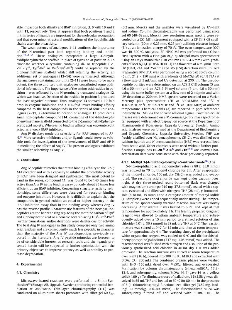
Building blocks 21a42 and 21b37,38 (Scheme 2) were preparedessentially according to a reported three-step procedure.37,38 Ini-tially, a solvent-free protocol43 was used to obtain the brominatedintermediates ethyl 2-(bromomethyl)benzoate and ethyl 2-(bro-momethyl)phenylacetate, but the reaction was found to be slug-gish and gave complex reaction mixtures. Therefore, a protocolemploying methyl acetate as a solvent and microwave-assistedheating was modified and applied.44 The commercially availableethyl 2-methylbenzoate and ethyl 2-methylphenylacetate were re-acted with N-bromosuccinimide (NBS) and 2,20-azobis(2-methyl-propionitrile) (AIBN) in MeCN at 90 �C for 15 min to obtain ethyl2-(bromomethyl)benzoate and ethyl 2-(bromomethyl)phenylace-tate. The transformations of the bromomethyl groups into the cor-responding azidomethyl groups were achieved by treatment withNaN3 in DMF. Finally, the esters were hydrolyzed with LiOH inTHF/MeOH/H2O to obtain the corresponding carboxylic acids 21aand 21b.
The target angiotensin IV analogues 2–15 were prepared bymanual solid-phase synthesis techniques using Fmoc protectionas described previously (Scheme 2).45 Initial coupling of 21a and21b to 2-chlorotrityl chloride resin furnished 22a and 22b,46 whichwere further elongated with L-proline (23a and 23b), L-leucine(24a and 24b), L-isoleucine (25a and 25b) and 20 (26b). The elon-gation was performed by a slightly modified literature procedure37
involving addition of tributylphosphine to a mixture of resin boundazide and activated amino acid in CH2Cl2 or 20 in CH2Cl2/MeCN.Prolonged reaction times were used for the subsequent couplingsof 20, L-valine and L-norleucine as these reactions were expectedto proceed slowly. Analogues 1 and 15 were prepared in a similarfashion by consecutive coupling of 20 and L-valine to 22c and 22d,respectively.
The Fmoc-protected scaffold 20 was used in the pseudopeptidesto replace the Tyr2, Val1-Tyr2, Tyr2-Ile3 or Val1-Tyr2-Ile3 residues inAng IV. The building blocks 21a and 21b were utilized in com-pounds 2–14 as c-turn mimicing scaffolds to replace His4-Pro5-Phe6 in Ang IV.
2.2. Biochemical evaluation
The Ang IV analogues were tested and compared for their bind-ing affinity to IRAP natively expressed in CHO-K1 cells as well astheir ability to inhibit the catalytic activity of recombinant humanIRAP and aminopeptidase N transiently transfected in HEK293cells. The chemical structures, the binding affinities and proteaseinhibition data of the compounds are presented in Table 1.
Our long-term objective is to develop drug-like Ang IV mimeticsthat exhibit a high metabolic stability and oral bioavailability. Weforesee that such compounds, characterized by a considerablylonger duration of action than the parent peptide Ang IV, wouldserve as attractive research tools, in particular when experimentsin complex animal models are performed. One approach to peptidemimetics is to induce constrains by cyclizations and in those caseswhere potent cyclized peptides are identified, proceed by conduct-ing a conformational analysis to determine the bioactive confor-mation of the target peptide. Subsequent displacement of thepeptide backbone with proper organic scaffolds, which mimic thesecondary structure of the bioactive conformation, followed bystructural optimization should eventually provide drug-like pep-tide mimetics.47,48
Recently, we demonstrated that 1 to 3 cyclization of the N-ter-minal of Ang IV, for example, oxidative cyclization of Cys1 withCys3 where Val1 and Ile3 in Ang IV had been displaced, delivereda peptide with an approximately 1000-fold lower binding affinitythan Ang IV itself.49 Since Cys1/Cys3 cyclized peptides preferen-tially adopt c-turns, it was concluded that it is not likely that ac-turn is present in the N-terminal of Ang IV when the peptide is
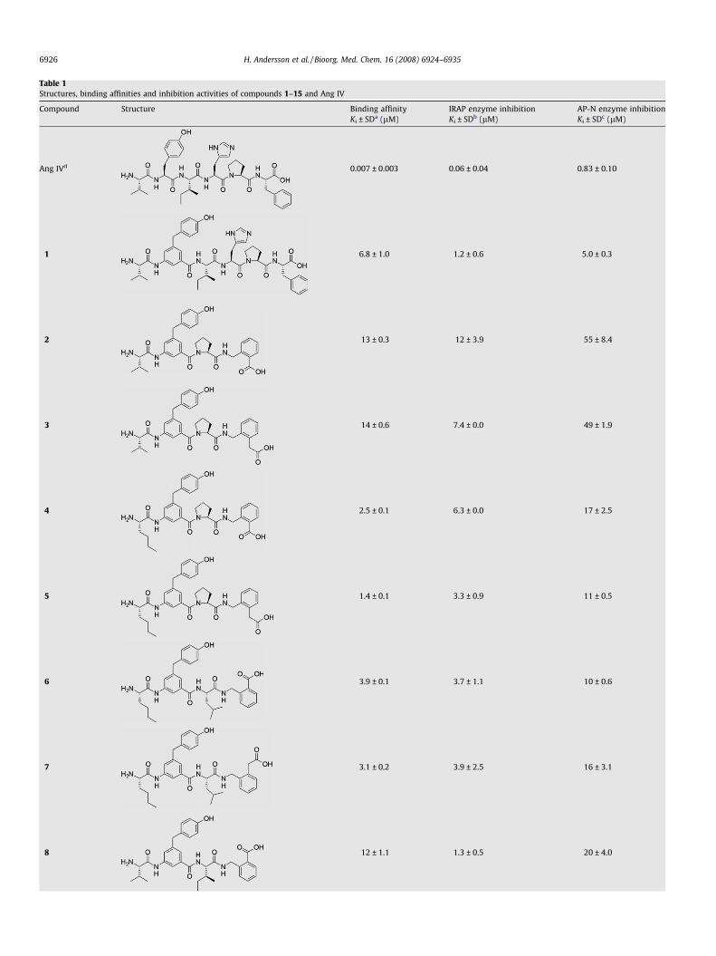
Table 1Structures, binding affinities and inhibition activities of compounds 1–15 and Ang IV
Compound Structure Binding affinityKi ± SDa (lM)
IRAP enzyme inhibitionKi ± SDb (lM)
AP-N enzyme inhibitionKi ± SDc (lM)
Ang IVd 0.007 ± 0.003 0.06 ± 0.04 0.83 ± 0.10
1 6.8 ± 1.0 1.2 ± 0.6 5.0 ± 0.3
2 13 ± 0.3 12 ± 3.9 55 ± 8.4
3 14 ± 0.6 7.4 ± 0.0 49 ± 1.9
4 2.5 ± 0.1 6.3 ± 0.0 17 ± 2.5
5 1.4 ± 0.1 3.3 ± 0.9 11 ± 0.5
6 3.9 ± 0.1 3.7 ± 1.1 10 ± 0.6
7 3.1 ± 0.2 3.9 ± 2.5 16 ± 3.1
8 12 ± 1.1 1.3 ± 0.5 20 ± 4.0
6926 H. Andersson et al. / Bioorg. Med. Chem. 16 (2008) 6924–6935
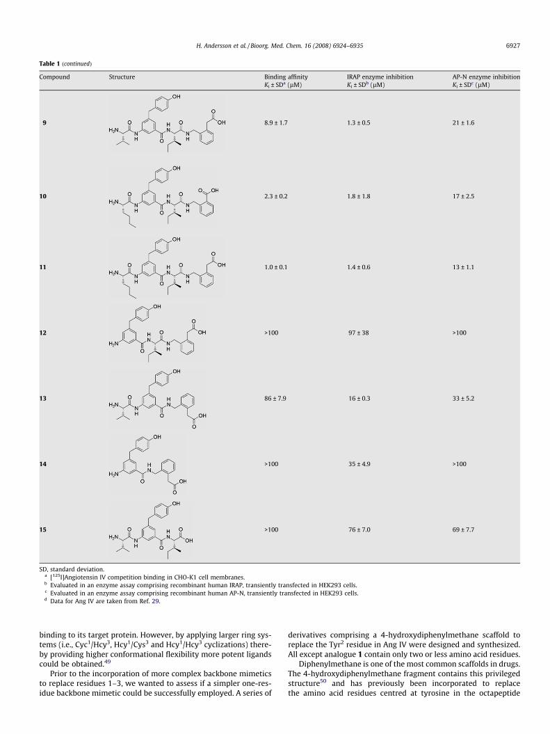
Table 1 (continued)
Compound Structure Binding affinityKi ± SDa (lM)
IRAP enzyme inhibitionKi ± SDb (lM)
AP-N enzyme inhibitionKi ± SDc (lM)
9 8.9 ± 1.7 1.3 ± 0.5 21 ± 1.6
10 2.3 ± 0.2 1.8 ± 1.8 17 ± 2.5
11 1.0 ± 0.1 1.4 ± 0.6 13 ± 1.1
12 >100 97 ± 38 >100
13 86 ± 7.9 16 ± 0.3 33 ± 5.2
14 >100 35 ± 4.9 >100
15 >100 76 ± 7.0 69 ± 7.7
SD, standard deviation.a [125I]Angiotensin IV competition binding in CHO-K1 cell membranes.b Evaluated in an enzyme assay comprising recombinant human IRAP, transiently transfected in HEK293 cells.c Evaluated in an enzyme assay comprising recombinant human AP-N, transiently transfected in HEK293 cells.d Data for Ang IV are taken from Ref. 29.
H. Andersson et al. / Bioorg. Med. Chem. 16 (2008) 6924–6935 6927
binding to its target protein. However, by applying larger ring sys-tems (i.e., Cyc1/Hcy3, Hcy1/Cys3 and Hcy1/Hcy3 cyclizations) there-by providing higher conformational flexibility more potent ligandscould be obtained.49
Prior to the incorporation of more complex backbone mimeticsto replace residues 1–3, we wanted to assess if a simpler one-res-idue backbone mimetic could be successfully employed. A series of
derivatives comprising a 4-hydroxydiphenylmethane scaffold toreplace the Tyr2 residue in Ang IV were designed and synthesized.All except analogue 1 contain only two or less amino acid residues.
Diphenylmethane is one of the most common scaffolds in drugs.The 4-hydroxydiphenylmethane fragment contains this privilegedstructure50 and has previously been incorporated to replacethe amino acid residues centred at tyrosine in the octapeptide
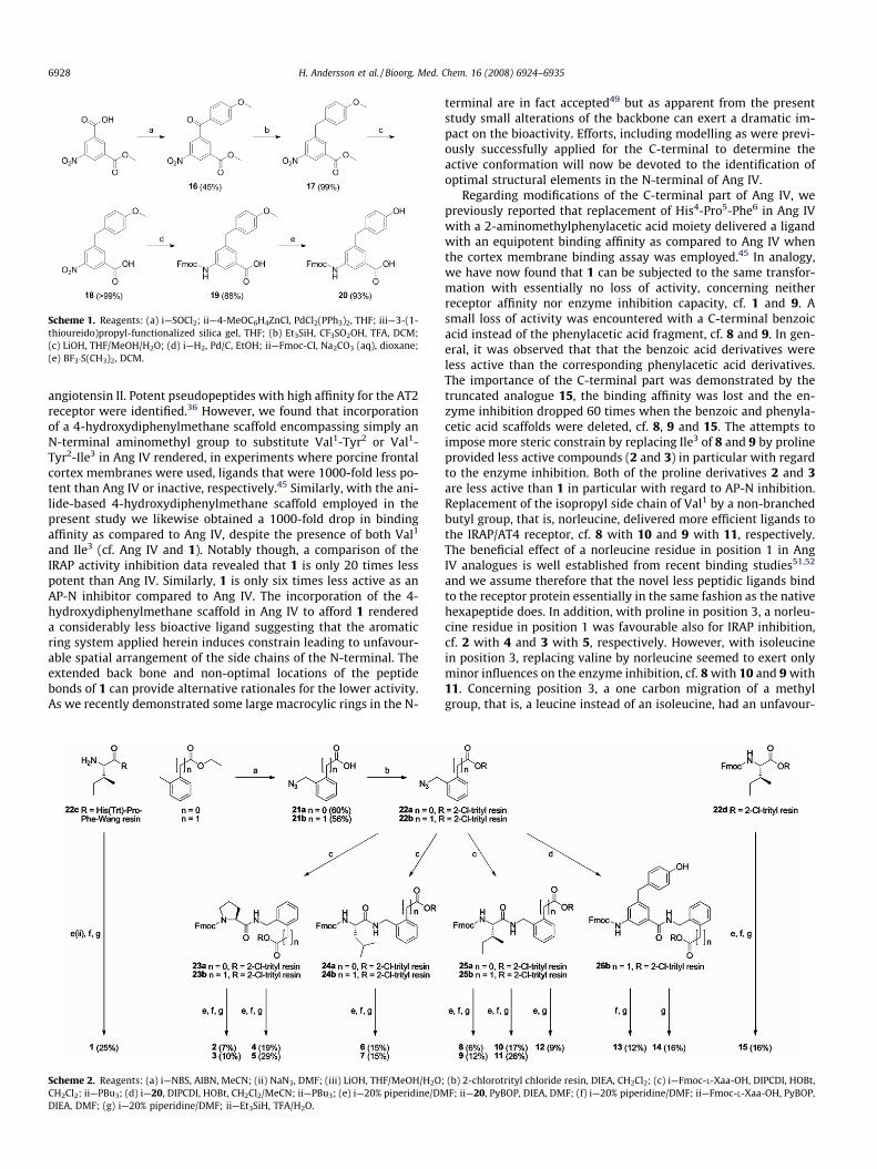
Scheme 1. Reagents: (a) i—SOCl2; ii—4-MeOC6H4ZnCl, PdCl2(PPh3)2, THF; iii—3-(1-thioureido)propyl-functionalized silica gel, THF; (b) Et3SiH, CF3SO2OH, TFA, DCM;(c) LiOH, THF/MeOH/H2O; (d) i—H2, Pd/C, EtOH; ii—Fmoc-Cl, Na2CO3 (aq), dioxane;(e) BF3�S(CH3)2, DCM.
6928 H. Andersson et al. / Bioorg. Med. Chem. 16 (2008) 6924–6935
angiotensin II. Potent pseudopeptides with high affinity for the AT2receptor were identified.36 However, we found that incorporationof a 4-hydroxydiphenylmethane scaffold encompassing simply anN-terminal aminomethyl group to substitute Val1-Tyr2 or Val1-Tyr2-Ile3 in Ang IV rendered, in experiments where porcine frontalcortex membranes were used, ligands that were 1000-fold less po-tent than Ang IV or inactive, respectively.45 Similarly, with the ani-lide-based 4-hydroxydiphenylmethane scaffold employed in thepresent study we likewise obtained a 1000-fold drop in bindingaffinity as compared to Ang IV, despite the presence of both Val1
and Ile3 (cf. Ang IV and 1). Notably though, a comparison of theIRAP activity inhibition data revealed that 1 is only 20 times lesspotent than Ang IV. Similarly, 1 is only six times less active as anAP-N inhibitor compared to Ang IV. The incorporation of the 4-hydroxydiphenylmethane scaffold in Ang IV to afford 1 rendereda considerably less bioactive ligand suggesting that the aromaticring system applied herein induces constrain leading to unfavour-able spatial arrangement of the side chains of the N-terminal. Theextended back bone and non-optimal locations of the peptidebonds of 1 can provide alternative rationales for the lower activity.As we recently demonstrated some large macrocylic rings in the N-
Scheme 2. Reagents: (a) i—NBS, AIBN, MeCN; (ii) NaN3, DMF; (iii) LiOH, THF/MeOH/H2OCH2Cl2; ii—PBu3; (d) i—20, DIPCDI, HOBt, CH2Cl2/MeCN; ii—PBu3; (e) i—20% piperidine/DMDIEA, DMF; (g) i—20% piperidine/DMF; ii—Et3SiH, TFA/H2O.
terminal are in fact accepted49 but as apparent from the presentstudy small alterations of the backbone can exert a dramatic im-pact on the bioactivity. Efforts, including modelling as were previ-ously successfully applied for the C-terminal to determine theactive conformation will now be devoted to the identification ofoptimal structural elements in the N-terminal of Ang IV.
Regarding modifications of the C-terminal part of Ang IV, wepreviously reported that replacement of His4-Pro5-Phe6 in Ang IVwith a 2-aminomethylphenylacetic acid moiety delivered a ligandwith an equipotent binding affinity as compared to Ang IV whenthe cortex membrane binding assay was employed.45 In analogy,we have now found that 1 can be subjected to the same transfor-mation with essentially no loss of activity, concerning neitherreceptor affinity nor enzyme inhibition capacity, cf. 1 and 9. Asmall loss of activity was encountered with a C-terminal benzoicacid instead of the phenylacetic acid fragment, cf. 8 and 9. In gen-eral, it was observed that that the benzoic acid derivatives wereless active than the corresponding phenylacetic acid derivatives.The importance of the C-terminal part was demonstrated by thetruncated analogue 15, the binding affinity was lost and the en-zyme inhibition dropped 60 times when the benzoic and phenyla-cetic acid scaffolds were deleted, cf. 8, 9 and 15. The attempts toimpose more steric constrain by replacing Ile3 of 8 and 9 by prolineprovided less active compounds (2 and 3) in particular with regardto the enzyme inhibition. Both of the proline derivatives 2 and 3are less active than 1 in particular with regard to AP-N inhibition.Replacement of the isopropyl side chain of Val1 by a non-branchedbutyl group, that is, norleucine, delivered more efficient ligands tothe IRAP/AT4 receptor, cf. 8 with 10 and 9 with 11, respectively.The beneficial effect of a norleucine residue in position 1 in AngIV analogues is well established from recent binding studies51,52
and we assume therefore that the novel less peptidic ligands bindto the receptor protein essentially in the same fashion as the nativehexapeptide does. In addition, with proline in position 3, a norleu-cine residue in position 1 was favourable also for IRAP inhibition,cf. 2 with 4 and 3 with 5, respectively. However, with isoleucinein position 3, replacing valine by norleucine seemed to exert onlyminor influences on the enzyme inhibition, cf. 8 with 10 and 9 with11. Concerning position 3, a one carbon migration of a methylgroup, that is, a leucine instead of an isoleucine, had an unfavour-
; (b) 2-chlorotrityl chloride resin, DIEA, CH2Cl2; (c) i—Fmoc-L-Xaa-OH, DIPCDI, HOBt,F; ii—20, PyBOP, DIEA, DMF; (f) i—20% piperidine/DMF; ii—Fmoc-L-Xaa-OH, PyBOP,
H. Andersson et al. / Bioorg. Med. Chem. 16 (2008) 6924–6935 6929
able impact on both affinity and IRAP inhibition, cf. 6 with 10 and 7with 11, respectively. Thus, it appears that both positions 1 and 3in this series of ligands are important for the molecular recognitionand that even minor structural modifications of the lipophilic sidechains alter the bioactivity.
The weak potency of analogues 1–11 confirms the importanceof the N-terminal part both regarding binding and inhibi-tion.49,51–54 These analogues incorporate a large 4-dihydr-oxidiphenylmethane scaffold in place of tyrosine at position 2. Toelucidate whether a tyrosine containing di- or tripeptide (i.e.,Val1-Tyr2, Tyr2-Ile3 or Val1-Tyr2-Ile3) could be replaced by thediphenylmethane scaffold whilst still retaining the activity, anadditional set of analogues (12–14) were synthesized. Althoughthe analogues containing four units (2–11) were found to be morepotent, the three and two unit analogues contributed some addi-tional information. The importance of the amino acid residue in po-sition 1 was reflected by the N-terminally truncated analogue 12,which was inactive. Deletion of the residue in position 3 producedthe least negative outcome. Thus, analogue 13 showed a 10-folddrop in enzyme inhibition and a 100-fold lower binding affinitycompared to the best compounds in the series of four unit ana-logues (2–11). C-Terminal truncation of analogue 13 produced asmall non-peptidic compound (14) consisting of the 4-hydroxydi-phenylmethane scaffold connected to the 2-(aminomethyl)phenyl-acetic acid moiety. Whereas no binding affinity was encountered itacted as a weak IRAP inhibitor.
Ang IV displays moderate selectivity for IRAP compared to AP-N.29 More selective inhibitors and/or ligands could serve as valu-able tools for investigation of the involvement of IRAP and AP-Nin mediating the effects of Ang IV. The present analogues exhibitedthe similar selectivity as Ang IV.
3. Conclusions
Ang IV peptide mimetics that retain binding affinity to the IRAP/AT4 receptor and with a capacity to inhibit the proteolytic activityof IRAP have been designed and synthesized. The most potent li-gand in the series, compound 11, was found to be 140 times lessactive than Ang IV in the binding assay but only about 25 times lessefficient as an IRAP inhibitor. Concerning structure–activity rela-tionships, some differences were observed for receptor bindingand enzyme inhibition. However, it is difficult to explain that thecompounds in general exhibit an equal or higher potency in theIRAP inhibition assay than in the binding assay whereas Ang IVhas the reverse profile. Characteristic features of the new pseudo-peptides are the benzene ring replacing the methine carbon of Tyr2
and a phenylacetic acid or a benzoic acid replacing His4-Pro5-Phe6.Further truncations and/or deletions were deleterious for activity.The best Ang IV analogues in this study comprise only two aminoacid residues and are consequently much less peptidic in characterthan the majority of the Ang IV pseudopeptides previously re-ported in the literature. Ang IV peptide mimetics are foreseen tobe of considerable interest as research tools and the ligands pre-sented herein will be subjected to further optimization with theprimary objectives to improve potency as well as stability to pro-tease degradation.
4. Experimental
4.1. Chemistry
Microwave-heated reactions were performed in a Smith Syn-thesizerTM (Biotage AB, Uppsala, Sweden) producing controlled irra-diation at 2450 MHz. Thin-layer chromatography (TLC) wasconducted on aluminium sheets precoated with silica gel 60 F254
(0.2 mm, Merck) and the analytes were visualized by UV-lightand iodine. Column chromatography was performed using silicagel 60 (40–63 lm, Merck). Low resolution mass spectra were re-corded on a GC–MS instrument equipped with a CP-Sil 8 CB capil-lary column (30 m � 0.25 mm, 0.25 lm) utilizing electron impact(EI) at an ionization energy of 70 eV. The oven temperature (GC)was 40–300 �C. Analytical RP-HPLC-MS was performed on a GilsonHPLC system with a Finnigan AQA quadrupol mass spectrometerusing an Onyx monolithic C18 column (50 � 4.6 mm) with gradi-ents of MeCN/H2O (0.05% HCOOH) at a flow rate of 4 mL/min. BothUV (DAD, 214 and 254 nm) and MS (ESI) detection were utilized.Preparative RP-HPLC was performed using a Zorbax SB-C8 column(5 lm, 21.2 � 150 mm) with gradients of MeCN/H2O (0.1% TFA) ata flow rate of 5 mL/min and UV detection at 230 nm. The pseudo-peptide purities were determined on an ACE 5 C18 column (5 lm,4.6 � 50 mm) and an ACE 5 Phenyl column (5 lm, 4.6 � 50 mm)using the same buffer system at a flow rate of 2 mL/min and withUV detection at 220 nm. NMR spectra were recorded on a VarianMercury plus spectrometer (1H at 399.8 MHz and 13C at100.5 MHz or 1H at 399.9 MHz and 13C at 100.6 MHz) at ambienttemperature. Chemical shifts (d) are reported in ppm referencedindirectly to TMS via the solvent residual signal. Exact molecularmasses were determined on a Micromass Q-Tof2 mass spectrome-ter equipped with an electrospray ion source at the Department ofPharmaceutical Biosciences, Uppsala University, Sweden. Aminoacid analyses were performed at the Department of Biochemistryand Organic Chemistry, Uppsala University, Sweden. THF wasfreshly distilled over Na/benzophenone, CH2Cl2 was distilled overcalcium hydride and N-bromosuccinimide (NBS) was recrystallizedfrom acetic acid. Other chemicals were used without further puri-fication. Compounds 16–20,36 21a42 and 21b37,38 are known. Char-acterization data were consistent with those previously reported.
4.1.1. Methyl 3-(4-methoxy-benzoyl)-5-nitrobenzoate36 (16)5-Nitroisophthalic acid monomethyl ester (7.88 g, 35.0 mmol)
was refluxed in 70 mL thionyl chloride for 2 h. After evaporationof the thionyl chloride, 100 mL dry CH2Cl2 was added and evapo-rated. The resulting acid chloride was kept under vacuum over-night. A dry three-necked round-bottomed flask was chargedwith magnesium turnings (919 mg, 37.8 mmol), sealed with a sep-tum, evacuated and filled with nitrogen. THF (28 mL), p-bromoani-sole (4.38 mL, 35 mmol) and a catalytic amount of methyl iodide(10 droplets) were added sequentially under stirring. The temper-ature of the spontaneously warmed reaction mixture was slowlydecreasing. After 40 min it was heated to 60 �C and kept at thattemperature for approximately 3 h. The freshly prepared Grignardreagent was allowed to attain ambient temperature and subse-quently added over a 15 min period to a stirred solution of zincchloride (5.01 g, 36.8 mmol) in 28 mL dry THF at 0 �C. The reactionmixture was stirred at 0 �C for 15 min and then at room tempera-ture for approximately 4 h. The resulting slurry of the precipitatedwhite organozinc reagent was cooled to 0 �C and dichlorobis(tri-phenylphosphine)palladium (737 mg, 1.05 mmol) was added. Thereaction vessel was flushed with nitrogen and a solution of the pre-viously synthesized acid chloride in 40 mL dry THF was addeddropwise. The reaction mixture was stirred at room temperatureover night (16 h), poured into 300 mL 0.5 M HCl and extracted withEtOAc (3� 200 mL). The combined organic phases were washedwith H2O (150 mL), dried over MgSO4, filtered and evaporated.Purification by column chromatography (i-hexane/EtOAc 17:3–13:4, and subsequently, toluene/EtOAc 96:4) gave 16 as a yellowsolid (4.99 g). To eliminate traces of palladium, 16 (3.58 g) was dis-solved in 55 mL THF and heated to 60 �C for 80 min in the presenceof 3-(1-thioureido)propyl-functionalized silica gel (3.82 mg, load-ing: 1.1 mmol/g, 200–400 mesh). The functionalized silica wassubsequently filtered off and washed with 50 mL THF. The
6930 H. Andersson et al. / Bioorg. Med. Chem. 16 (2008) 6924–6935
combined filtrates were evaporated to give 1636 as a pale yellowsolid (3.55 g, 45% overall).
4.1.2. Methyl 3-(4-methoxybenzyl)-5-nitrobenzoate36 (17)Trifluoroacetic acid (TFA) (8.45 mL, 110 mmol) and triflic acid
(0.10 mL, 1.1 mmol) were added to a stirred solution of 16 (3.48 g,11 mmol) in 25 mL CH2Cl2 at 0 �C. After dropwise addition of trieth-ylsilane (5.59 mL, 35 mmol), the reaction mixture was allowed to at-tain ambient temperature. It was stirred for 3 h and then slowlypoured into a cold saturated solution of NaHCO3 (100 mL). The aque-ous phase was extracted with CH2Cl2 (2� 50 mL) and the combinedorganic phases were washed with H2O (2� 75 mL), dried overMgSO4, filtered and evaporated. Excessive triethylsilane was re-moved by repeated addition and evaporation of toluene. Compound1736 was collected as a pale yellow solid (3.29 g, 99%).
4.1.3. 3-(4-Methoxybenzyl)-5-nitrobenzoic acid36 (18)LiOH (1.30 g, 54.2 mmol) was added to a solution of 17 (3.25 g,
10.8 mmol) in THF (130 mL), MeOH (43 mL) and H2O (43 mL). Afterstirring over night the reaction mixture was concentrated, dilutedwith H2O and acidified with 1 M HCl. EtOAc (250 mL) was addedand the two phases were separated. The aqueous phase was ex-tracted with EtOAc (50 mL) and the combined organic phases werewashed with brine (2� 100 mL) and H2O (100 mL), dried overMgSO4, filtered and evaporated to give 1836 as a solid (3.09 g, quant.).
4.1.4. 3-(9H-Fluoren-9-ylmethoxycarbonylamino)-5-(4-methoxybenzyl)benzoic acid36 (19)
A mixture of compound 18 (3.07 g, 10.7 mmol) and 10% Pd/C(1.14 g, 1.07 mmol) in 150 mL absolute ethanol was stirred underhydrogen at atmospheric pressure and room temperature forapproximately 5 h. The crude product obtained after filtrationand evaporation, when analyzed by LC–MS, was considered to beof sufficient purity to be used in the next step without further puri-fication. An aqueous solution of Na2CO3 (0.3 M, 125 mL) wasslowly added to a solution of the aniline in 100 mL dioxane at0 �C. 9-Fluorenylmethyl chloroformate (Fmoc-Cl) (3.36 g,11.8 mmol) in 100 mL dioxane was added dropwise, whereafterthe reaction mixture was allowed to reach ambient temperatureand left under continuous stirring for approximately 44 h. Thereaction mixture was slowly acidified with 1 M HCl and EtOAc(200 mL) was added. The aqueous phase was extracted with EtOAc(2� 100 mL) and the combined organic phases were washed withbrine (2� 100 mL) and H2O (2� 100 mL), dried over MgSO4,filtered and evaporated. Purification by column chromatography(i-hexane/EtOAc/HCOOH 74:25:1) and an additional extractionusing EtOAc and H2O gave 1936 as a solid (4.52 g, 88%).
4.1.5. 3-(9H-Fluoren-9-ylmethoxycarbonylamino)-5-(4-hydroxybenzyl)benzoic acid36 (20)
Boron trifluoride-methyl sulfide complex (10.2 mL, 97 mmol)was added dropwise to 19 (4.65 g, 9.70 mmol) in 145 mL dry CH2Cl2
at 0 �C under nitrogen. The reaction mixture was allowed to reachambient temperature and left under continuous stirring for 26 h.The reaction was quenched by very slow addition of the mixture tocold 0.5 M HCl under stirring. Subsequent extraction with EtOAcwas followed by washing with NaHCO3, brine and H2O, drying overMgSO4, filtration and evaporation. Purification by column chroma-tography (i-hexane/EtOAc/HCOOH 65:34:1 followed by EtOAc toelute the product) gave 2036 as a solid (4.18 g, 93%).
4.1.6. 2-(Azidomethyl)benzoic acid42 (21a)Ethyl 2-methylbenzoate (1.91 mL, 12.0 mmol), NBS (2.35 g,
13.2 mmol), AIBN (98.5 mg, 0.60 mmol) and MeCN (18 mL) wereadded to a 20 mL Smith Process VialTM containing a stirring bar.After capping and evacuation of the vial, the reaction mixture
was heated to 90 �C by microwave irradiation for 15 min, cooledto room temperature, transferred to a round-bottomed flask andconcentrated under reduced pressure. Subsequent precipitationof succinimide by i-hexane/CH2Cl2, filtration, evaporation and puri-fication by column chromatography (i-hexane/ether 98:2) gaveethyl 2-(bromomethyl)benzoate as a pale yellow oil (1.97 g, 67%).1H NMR (CDCl3) d 7.96 (dd, 7.8, 1.4 Hz, 1H), 7.50–7.43 (m, 2H),7.36 (ddd, J = 7.8, 6.8, 2.0 Hz, 1H), 4.95 (s, 2H), 4.40 (q, J = 7.1 Hz,2H), 1.42 (t, J = 7.1 Hz, 3H). 13C NMR (CDCl3) d 166.7, 139.1,132.5, 131.7, 131.3, 129.6, 128.6, 61.4, 31.7, 14.3. EI+-MS m/z 243(M+). The residual compound obtained above (1.94 g, 7.99 mmol)was reacted with NaN3 (0.62 g, 9.58 mmol) in 10 mL DMF at roomtemperature for 22 h. The reaction was quenched with H2O and ex-tracted with EtOAc. The combined organic layers were washed withbrine and H2O, dried over Na2SO4, filtered and evaporated. Purifica-tion by column chromatography (i-hexane/EtOAc 95:5) gave ethyl2-(azidomethyl)benzoate as a colourless oil (1.56 g, 95%). 1H NMR(CDCl3) d 8.02 (dd, J = 7.8, 1.5 Hz, 1H), 7.55 (ddd, J = 7.5, 7.5, 1.5 Hz,1H), 7.49 (ddd, J = 7.8, 1.5, 0.5 Hz, 1H), 7.40 (ddd, J = 7.5, 7.5,1.5 Hz, 1H), 4.82 (s, 2H), 4.39 (q, J = 7.1 Hz, 2H), 1.41 (t, J = 7.1 Hz,3H). 13C NMR (CDCl3) d 166.9, 137.3, 132.7, 131.2, 129.9, 129.3,128.2, 61.4, 53.2, 14.4. LiOH (0.89 g, 37.0 mmol) was added to a solu-tion of the residual compound obtained above (1.52 g, 7.41 mmol) inTHF (14 mL), MeOH (2 mL) and H2O (4 mL). After stirring for 17 h, thereaction mixture was diluted with CH2Cl2 and H2O, and extractedwith CH2Cl2. The aqueous phase was acidified with 3 M HCl and ex-tracted with CH2Cl2. The combined organic phases were washedwith brine and H2O, dried over Na2SO4, filtered and evaporated togive 21a42 as a white solid (1.24 g, 95%). 1H NMR (acetone-d6) d8.08 (dd, J = 7.8, 1.5 Hz, 1H), 7.64 (ddd, J = 7.5, 7.5, 1.5 Hz, 2H), 7.56(dd, J = 7.8, 1.5 Hz, 1H), 7.50 (ddd, J = 7.5, 7.5, 1.5 Hz, 2H), 4.87 (s,2H). 13C NMR (acetone-d6) d 168.2, 138.3, 133.5, 132.1, 131.2,130.4, 129.1, 53.4. ESI�-MS m/z 176.0 (M�H)�, 222.0(M+HCOOH�H)�, 353.1 (2M�H)�, 572.3 (3M�H)�.
4.1.7. 2-(Azidomethyl)phenylacetic acid37,38 (21b)2-(Azidomethyl)phenylacetic acid was prepared according to the
procedure described above. Ethyl 2-methylphenylacetate (2.14 mL,12.0 mmol), NBS (2.35 g, 13.2 mmol), AIBN (98.5 mg, 0.60 mmol)and MeCN (18 mL) were used in the first step. Ethyl 2-(bromo-methyl)phenylacetate was obtained as a pale yellow oil (2.39 g,77%). 1H NMR (CDCl3) d 7.37 (m, 1H), 7.30–7.24 (m, 3H), 4.60 (s,2H), 4.16 (q, J = 7.2 Hz, 2H), 3.79 (s, 2H), 1.26 (t, J = 7.2 Hz, 3H). 13CNMR (CDCl3) d 171.2, 136.5, 133.6, 131.4, 130.8, 129.3, 128.1, 61.3,38.5, 31.9, 14.3. EI+-MS m/z 257 (M+). The residual compound ob-tained above (2.19 g, 8.52 mmol), NaN3 (0.66 g, 10.2 mmol) andDMF (10 mL) were used in the second step. Ethyl 2-(azido-methyl)phenylacetate was obtained as a pale yellow oil (1.85 g).1H NMR (CDCl3) d 7.34–7.29 (m, 4H), 4.42 (s, 2H), 4.15 (q,J = 7.2 Hz, 2H), 3.71 (s, 2H), 1.25 (t, J = 7.2 Hz, 3H). 13C NMR (CDCl3)d 171.2, 134.1, 133.3, 131.3, 130.0, 129.0, 127.9, 61.2, 52.9, 38.7,14.3. The ethyl 2-(azidomethyl)phenylacetate obtained above(1.85 g), LiOH (1.01 g, 42.2 mmol), THF (14 mL), MeOH (2 mL) andH2O (4 mL) were used in the last step. The crude product was purifiedby filtration trough a pad of silica followed by recrystallization (tol-uene/ether 1:3) and column chromatography (i-hexane/EtOAc/HCOOH 60:40:1) to afford 21b37,38 as a white solid (1.20 g, 73% overtwo steps). 1H NMR (acetone-d6) d 7.43–7.29 (m, 4H), 4.54 (s, 2H),3.77 (s, 2H). 13C NMR (acetone-d6) d 172.4, 135.4, 135.0, 132.1,130.5, 129.4, 128.2, 53.1, 38.4. ESI�-MS m/z 190.0 (M�H)�, 236.1(M+HCOOH�H)�, 381.2 (2M�H)�, 572.3 (3M�H)�.
4.1.8. 2-(Azidomethyl)benzoyl-2-chlorotrityl resin (22a)2-(Azidomethyl)benzoic acid 21a (101 mg, 0.57 mmol) was re-
acted with 2-chlorotrityl chloride resin (584 mg, 0.86 mmol) inCH2Cl2 (6 mL) in the presence of N,N-diisopropylethylamine (DIEA)
H. Andersson et al. / Bioorg. Med. Chem. 16 (2008) 6924–6935 6931
(0.39 mL, 2.24 mmol). After 4 h, MeOH (0.5 mL) was added and themixture was stirred for another 25 min. The resin was washed withseveral portions of, in turn, CH2Cl2, DMF and CH2Cl2, and dried in va-cuo overnight. Two batches were made and the resulting substitu-tion degree was determined to 0.86 and 0.89 mmol/g based on theweight increase.
4.1.9. 2-(Azidomethyl)phenylacetyl-2-chlorotrityl resin (22b)2-(Azidomethyl)phenylacetic acid 21b (101 mg, 0.53 mmol)
was reacted with 2-chlorotrityl chloride resin (556 mg, 0.82 mmol)in CH2Cl2 (6 mL) in the presence of DIEA (0.37 mL, 2.12 mmol) fol-lowing the procedure described above. Two batches were madeand the resulting substitution degree was determined to 0.84and 0.85 mmol/g from the weight increase. Another batch wasmade using 2-(azidomethyl)phenylacetic acid 21b (104 mg,0.54 mmol), 2-chlorotrityl chloride resin (534 mg, 0.79 mmol)and DIEA (0.37 mL, 2.1 mmol). Substitution degree: 0.88 mmol/g.
4.1.10. Ile-His(Trt)-Pro-Phe-Wang resin (22c)The Ile-His(Trt)-Pro-Phe-Wang resin was synthesized with a
Symphony instrument using Fmoc protection. The starting poly-mer was Fmoc-Phe-Wang resin (297 mg, 0.25 mmol). Removal ofthe Fmoc group was achieved by reaction with 20% piperidine inDMF (2� 2.5 mL, 5 + 10 min). The amino acids (125 lmol) werecoupled in DMF using 2-(1H-benzotriazole-1-yl)-1,1,3,3-tetram-ethyluronium hexafluorophosphate (HBTU) (125 lmol) in thepresence of N-methylmorpholine (NMM) (500 lmol). Double cou-plings (2 � 30 min) were used for all amino acids. At the end ofeach coupling cycle, the remaining amino groups were capped byaddition of 20% acetic anhydride in DMF (1.25 mL) to the reactionmixture and allowing the reaction to proceed for 5 min. After com-pletion of the synthesis the Fmoc group was removed and the par-tially protected peptide resin was washed with several portions ofDMF and CH2Cl2 and then dried in a stream of nitrogen and in va-cuo. The resulting substitution degree was determined to0.55 mmol/g based on the weight increase.
4.1.11. Fmoc-Ile-2-chlorotrityl resin (22d)Fmoc-Ile-OH (142 mg, 0.40 mmol) was reacted with 2-chloro-
trityl chloride resin (341 mg, 0.50 mmol) in CH2Cl2 (6 mL) in thepresence of DIEA (0.28 mL, 1.61 mmol) following the procedure de-scribed for 22a. Substitution degree: 0.93 mmol/g.
4.2. General procedure for the preparation of resins 23a, 23b,24a, 24b, 25a, 25b and 26b
A round-bottomed flask was charged with the appropriate ami-no acid or 20, 1-hydroxybenzotriazole (HOBt) and N,N0-diisopro-pylcarbodiimide (DIPCDI) in dry CH2Cl2 or CH2Cl2/MeCN. Afterpreactivation under nitrogen for 10 min, 22a or 22b was added,followed by tributylphosphine after another 10 min. The reactionmixture was stirred under nitrogen for 4–6 h. The resin was fil-tered off and washed with several portions of, in turn, CH2Cl2,DMF, MeOH and CH2Cl2, and finally dried in in vacuo overnight.
4.2.1. Fmoc-Pro-2-(aminomethyl)benzoyl-2-chlorotrityl resin (23a)Resin 23a was prepared according to the general procedure
using Fmoc-Pro-OH (263 mg, 779 lmol), HOBt (105 mg,779 lmol), DIPCDI (122 lL, 779 lmol), CH2Cl2 (15 mL), 22a(303 mg, 262 lmol) and tributylphosphine (150 lL, 520 lmol).Reaction time: 6 h. Yield: 373 mg.
4.2.2. Fmoc-Pro-2-(aminomethyl)phenylacetyl-2-chlorotritylresin (23b)
Resin 23b was prepared according to the general procedureusing Fmoc-Pro-OH (260 mg, 771 lmol), HOBt (104 mg,
771 lmol), DIPCDI (121 lL, 771 lmol), CH2Cl2 (15 mL), 22b(283 mg, 242 lmol) and tributylphosphine (149 lL, 514 lmol).Reaction time: 6 h. Yield: 354 mg.
4.2.3. Fmoc-Leu-2-(aminomethyl)benzoyl-2-chlorotrityl resin(24a)
Resin 24a was prepared according to the general procedureusing Fmoc-Leu-OH (379 mg, 1.07 mmol), HOBt (145 mg,1.07 lmol), DIPCDI (168 lL, 1.07 mmol), CH2Cl2 (20 mL), 22a(315 mg, 281 lmol) and tributylphosphine (206 lL, 710 lmol).Reaction time: 4 h. Yield: 379 mg.
4.2.4. Fmoc-Leu-2-(aminomethyl)phenylacetyl-2-chlorotritylresin (24b)
Resin 24b was prepared according to the general procedureusing Fmoc-Leu-OH (269 mg, 760 lmol), HOBt (105 mg,774 lmol), DIPCDI (119 lL, 761 lmol), CH2Cl2 (20 mL), 22b(305 mg, 258 lmol) and tributylphosphine (147 lL, 510 lmol).Reaction time: 4 h. Yield: 373 mg.
4.2.5. Fmoc-Ile-2-(aminomethyl)benzoyl-2-chlorotrityl resin(25a)
Resin 25a was prepared according to the general procedureusing Fmoc-Ile-OH (275 mg, 779 lmol), HOBt (105 mg, 779 lmol),DIPCDI (122 lL, 779 lmol), CH2Cl2 (15 mL), 22a (301 mg,260 lmol) and tributylphosphine (150 lL, 520 lmol). Reactiontime: 6 h. Yield: 378 mg.
4.2.6. Fmoc-Ile-2-(aminomethyl)phenylacetyl-2-chlorotritylresin (25b)
Resin 25b was prepared according to the general procedureusing Fmoc-Ile-OH (273 mg, 771 lmol), HOBt (104 mg, 771 lmol),DIPCDI (121 lL, 771 lmol), CH2Cl2 (15 mL), 22b (301 mg,257 lmol) and tributylphosphine (149 lL, 514 lmol). Reactiontime: 6 h. Yield 381 mg.
4.2.7. 3-(9H-Fluoren-9-ylmethoxycarbonylamino)-5-(4-hydroxybenzyl)benzoyl-2-(aminomethyl)phenylacetyl-2-chlorotrityl resin (26b)
Resin 26b was prepared according to the general procedureusing 20 (246 mg, 528 lmol), HOBt (77.7 mg, 575 lmol), DIPCDI(82.2 lL, 525 lmol), CH2Cl2 (30 mL), MeCN (5 mL), 22b (399 mg,351 lmol) and tributylphosphine (203 lL, 700 lmol). Reactiontime: 5 h. Yield: 517 mg.
4.3. General synthesis of angiotensin IV analogues 1–15
Resin 22c, 22d, 23a, 23b, 24a, 24b, 25a, 25b or 26b was weighedinto a 2 mL disposable syringe fitted with porous polyethylene fil-ter. The Fmoc group was removed by treatment with 20% piperi-dine in DMF (2� 1.5 mL, 10 + 15 min) and the polymer waswashed with DMF (5� 1.5 mL, 5 � 1 min). Coupling of 20 and/orthe appropriate amino acid was performed by agitation over nightin DMF (1.5 mL) using benzotriazole-1-yl-oxy-tris-pyrrolidino-phosphonium hexafluorophosphate (PyBOP) in the presence ofDIEA. The resin was washed with DMF (5� 1.5 mL, 5 � 1 min)and subsequently deprotected and washed as described above.After completion of the coupling cycle, the resin was also washedwith several portions of, in turn, CH2Cl2, MeOH and CH2Cl2 beforeit was dried in in vacuo. The pseudopeptide was cleaved by treat-ment with TFA/H2O/triethylsilane (90:5:5 ca. 1 mL/100 mg resin)for 2 h. The resin was filtered off and washed with TFA (4� 0.3–0.5 mL). The filtrate was collected in a centrifuge tube and concen-trated in a stream of nitrogen. Cold diethyl ether (ca. 13 mL) wasused to precipitate the product, which was collected by centrifuga-tion, washed with cold diethyl ether (63� 7 mL) and dried. The
6932 H. Andersson et al. / Bioorg. Med. Chem. 16 (2008) 6924–6935
crude pseudopeptide was dissolved in MeCN/0.1% aqueous TFA, fil-tered through a 0.45 lm nylon membrane and purified in 1–2 runsby RP-HPLC. Selected fractions were analyzed by RP-HPLC and RP-HPLC-MS, and those containing pure product were pooled andlyophilized.
4.3.1. Analogue 1Except for the initial Fmoc deprotection, the analogue was pre-
pared according to the general procedure using 22c (251 mg,138 lmol), 20 (80.2 mg, 172 lmol), PyBOP (89.6 mg, 172 lmol)and DIEA (59.9 lL, 344 lmol) in the first coupling followed byFmoc-Val-OH (142 mg, 418 lmol), PyBOP (217 mg, 417 lmol)and DIEA (144 lL, 829 lmol) in the second. Reactiontime: 18 hfor both couplings. Purification by preparative RP-HPLC gave 1 asa white solid (11.3 mg, 25%). HPLC purity: C18 column 99.0%, Phe-nyl column 99.1%. HRMS (M+H+): 837.4277, C45H57N8O8 requires837.4299. Amino acid analysis: Val 1.05; Ile 0.99; His 0.98; Pro0.98; Phe 1.00.
4.3.2. Analogue 2The analogue was prepared according to the general procedure
using 23a (170 mg, 120 lmol), 20 (85.7 mg, 184 lmol), PyBOP(95.8 mg, 184 lmol) and DIEA (64.1 lL, 368 lmol) in the first cou-pling followed by Fmoc-Val-OH (100 mg, 294 lmol), PyBOP(153 mg, 294 lmol) and DIEA (103 lL, 589 lmol) in the second.Reaction time: 17 h and 18 h, respectively. Purification by prepara-tive RP-HPLC gave 2 as a white solid (5.1 mg, 7%). 1H NMR (CD3OD)d (3:1 mixture of rotamers, major rotamer reported) 7.98 (dd,J = 7.8, 1.4 Hz, 1H), 7.73 (m, 1H), 7.58 (m, 1H), 7.52 (m, 1H), 7.46(m, 1H), 7.35 (m, 1H), 7.25 (m, 1H), 7.03 (m, 2H), 6.70 (m, 2H),4.78 (app. s, 2H), 4.60 (dd, J = 8.2, 5.8 Hz, 1H), 3.92 (app. s, 2H),3.73 (d, J = 5.9 Hz, 1H), 3.68 (m, 1H), 3.56 (m, 1H), 2.35–2.17 (m,2H), 2.06–1.97 (m, 2H), 1.87 (m, 1H), 1.11 (d, J = 6.9 Hz, 3H), 1.08(d, J = 6.9 Hz, 3H). HPLC purity: C18 column >99.9%, Phenyl column98.3%. HRMS (M+H+): 573.2711, C32H37N4O6 requires 573.2713.Amino acid analysis: Val, 1.13; Pro, 0.87 (72% peptide).
4.3.3. Analogue 3The analogue was prepared according to the general procedure
using 23b (148 mg, 101 lmol), 20 (84.8 mg, 182 lmol), PyBOP(92.5 mg, 178 lmol) and DIEA (63.5 lL, 364 lmol) in the first cou-pling followed by Fmoc-Val-OH (99.4 mg, 293 lmol), PyBOP(153 mg, 293 lmol) and DIEA (102 lL, 586 lmol) in the second.Reaction time: 16 h for both couplings. Purification by preparativeRP-HPLC gave 3 as a white solid (5.9 mg, 10%). 1H NMR (CD3OD) d(3:1 mixture of rotamers, major rotamer reported) 7.73 (m, 1H),7.46 (m, 1H), 7.39 (m, 1H), 7.29–7.20 (m, 4H), 7.02 (m, 2H), 6.70(m, 2H), 4.54 (dd, J = 8.1, 5.8 Hz, 1H), 4.50 (d, J = 15.0 Hz, 1H),4.46 (d, J = 15.0 Hz, 1H), 3.91 (app. s, 2H), 3.75 (app. s, 2H), 3.73(d, J = 5.9 Hz, 1H), 3.63 (m, 1H), 3.52 (m, 1H), 2.35–2.17 (m, 2H),2.05–1.95 (m, 2H), 1.86 (m, 1H), 1.11 (d, J = 6.9 Hz, 3H), 1.08 (d,J = 6.9 Hz, 3H). 13C NMR (CD3OD) d (major rotamer reported)175.4, 174.4, 171.6, 168.0, 157.0, 145.2, 139.1, 138.2, 138.0,134.4, 132.5, 131.8, 131.0, 129.8, 128.6, 124.9, 123.1, 117.7,116.4, 62.2, 60.5, 51.8, 42.1, 41.7, 39.1, 31.7, 31.2, 26.4, 19.0,17.8. One carbon signal in the aromatic area is missing due to over-lapping signals. HPLC purity: C18 column 99.4%, Phenyl column99.4%. HRMS (M+H+): 587.2854, C33H39N4O6 requires 587.2870.Amino acid analysis: Val, 1.04; Pro, 0.96 (77% peptide).
4.3.4. Analogue 4The analogue was prepared according to the general procedure
using 23a (170 mg, 120 lmol), 20 (85.7 mg, 184 lmol), PyBOP(95.8 mg, 184 lmol) and DIEA (64.1 lL, 368 lmol) in the first cou-pling followed by Fmoc-Nle-OH (104 mg, 294 lmol), PyBOP(153 mg, 294 lmol) and DIEA (103 lL, 589 lmol) in the second.
Reaction time: 17 h and 18 h, respectively. Purification by prepara-tive RP-HPLC gave 4 as a white solid (13.6 mg, 19%). 1H NMR(CD3OD) d (3:1 mixture of rotamers, major rotamer reported)7.96 (dd, J = 7.8, 1.4 Hz, 1H), 7.73 (m, 1H), 7.57 (m, 1H), 7.51 (m,1H), 7.47 (m, 1H), 7.34 (m, 1H), 7.24 (m, 1H), 7.02 (m, 2H), 6.70(m, 2H), 4.77 (app. s, 2H), 4.60 (dd, J = 8.1, 5.8 Hz, 1H), 3.96–3.91(m, 3H), 3.62 (m, 1H), 3.51 (m, 1H), 2.32 (m, 1H), 2.05–1.81 (m,5H), 1.44–1.35 (m, 4H), 0.95 (t, J = 7.2 Hz, 3H). 13C NMR (CD3OD)d (major rotamer reported) 174.5, 171.7, 170.9, 168.7, 157.0,145.2, 140.9, 139.2, 138.2, 133.3, 132.5, 132.0, 131.0, 129.8,128.1, 124.8, 123.1, 117.7, 116.4, 62.2, 55.2, 51.7, 43.1, 41.7, 32.5,31.1, 28.0, 26.3, 23.4, 14.1. One carbon signal in the aromatic areais missing. HPLC purity: C18 column 99.5%, Phenyl column 99.7%.HRMS (M+H+): 587.2873, C33H39N4O6 requires 587.2870. Aminoacid analysis: Nle, 1.01; Pro, 0.99 (77% peptide).
4.3.5. Analogue 5The analogue was prepared according to the general procedure
using 23b (148 mg, 101 lmol), 20 (84.8 mg, 182 lmol), PyBOP(92.5 mg, 178 lmol) and DIEA (63.5 lL, 364 lmol) in the first cou-pling followed by Fmoc-Nle-OH (91.4 mg, 253 lmol), PyBOP(132 mg, 253 lmol) and DIEA (88.2 lL, 506 lmol) in the second.Reaction time: 16 h for both couplings. Purification by preparativeRP-HPLC gave 5 as a white solid (17.4 mg, 29%). 1H NMR (CD3OD) d(3:1 mixture of rotamers, major rotamer reported) 7.73 (dd, J = 2.1,1.6 Hz, 1H), 7.46 (dd, J = 2.1, 1.6 Hz, 1H), 7.40 (m, 1H), 7.28–7.21(m, 4H), 7.02 (m, 2H), 6.70 (m, 2H), 4.54 (dd, J = 8.1, 5.8 Hz, 1H),4.50 (d, J = 15.0 Hz, 1H), 4.46 (d, J = 15.0 Hz, 1H), 3.94–3.91 (m,3H), 3.77 (d, J = 16.0 Hz, 1H), 3.73 (d, J = 16.0 Hz, 1H), 3.62 (m,1H), 3.51 (m, 1H), 2.31 (m, 1H), 2.03–1.81 (m, 5H), 1.44–1.35 (m,4H), 0.95 (t, J = 7.2 Hz, 3H). 13C NMR (CD3OD) d (major rotamer re-ported) 175.4, 174.4, 171.6, 168.7, 157.0, 145.2, 139.2, 138.2, 138.1,134.4, 132.5, 131.8, 131.0, 129.8, 128.6, 124.9, 123.1, 117.6, 116.4,62.2, 55.2, 51.8, 42.0, 41.7, 39.1, 32.5, 31.2, 28.0, 26.4, 23.4, 14.1.One carbon signal in the aromatic area is missing due to overlap-ping signals. HPLC purity: C18 column 98.5%, Phenyl column99.5%. HRMS (M+H+): 601.3038, C34H41N4O6 requires 601.3026.Amino acid analysis: Nle, 1.01; Pro, 0.99 (59% peptide).
4.3.6. Analogue 6The analogue was prepared according to the general procedure
using 24a (100 mg, 74.2 lmol), 20 (115 mg, 247 lmol), PyBOP(130 mg, 249 lmol) and DIEA (86.8 lL, 498 lmol) in the first cou-pling followed by Fmoc-Nle-OH (96.5 mg, 272 lmol), PyBOP(130 mg, 249 lmol) and DIEA (86.8 lL, 498 lmol) in the second.Reaction time: 15 h and 17 h, respectively. Purification by prepara-tive RP-HPLC gave 6 as a white solid (6.6 mg, 15%). 1H NMR(CD3OD) d (9:1 mixture of rotamers, major rotamer reported)7.98 (m, 1H), 7.91 (dd, J = 2.1, 1.6 Hz, 1H), 7.52–7.48 (m, 4H),7.35 (m, 1H), 7.04 (m, 2H), 6.71 (m, 2H), 4.73 (m, 2H), 4.64 (m,1H), 3.95–3.91 (m, 3H), 1.99–1.84 (m, 2H), 1.78–1.67 (m, 3H),1.47–1.36 (m, 4H), 1.00–0.93 (m, 9H). 13C NMR (CD3OD) d (majorrotamer reported) 174.8, 170.6, 170.2, 168.8, 157.0, 145.2, 141.0,139.3, 136.5, 133.5, 132.5, 132.2, 131.0, 130.1, 128.3, 125.1,124.6, 118.2, 116.4, 55.2, 54.1, 43.2, 41.7, 32.5, 28.0, 26.2, 23.4,22.0, 14.1. One carbon signal in the aromatic area is missing andtwo carbon signals in the aliphatic area are missing due to overlap-ping signals. HPLC purity: C18 column >99.9%, Phenyl column99.5%. HRMS (M+H+): 603.3172, C34H43N4O6 requires 603.3183.Amino acid analysis: Nle, 1.01; Leu, 0.99 (89% peptide).
4.3.7. Analogue 7The analogue was prepared according to the general procedure
using 24b (151 mg, 101 lmol), 20 (174 mg, 374 lmol), PyBOP(96.7 mg, 187 lmol) and DIEA (130 lL, 74.8 lmol) in the first cou-pling followed by Fmoc-Nle-OH (133 mg, 374 lmol), PyBOP
H. Andersson et al. / Bioorg. Med. Chem. 16 (2008) 6924–6935 6933
(196 mg, 374 lmol) and DIEA (130 lL, 748 lmol) in the second.Reaction time: 16 h and 6 h, respectively. Purification by prepara-tive RP-HPLC gave 7 as a white solid (9.8 mg, 15%). 1H NMR(CD3OD) d (9:1 mixture of rotamers, major rotamer reported)7.91 (m, 1H), 7.52 (m, 1H), 7.50 (m, 1H), 7.32 (m, 1H), 7.26–7.20(m, 3H), 7.03 (m, 2H), 6.71 (m, 2H), 4.63 (m, 1H), 4.47 (d,J = 15.1 Hz, 1H), 4.43 (d, J = 15.1 Hz, 1H), 3.95–3.91 (m, 3H), 3.73(app. s, 2H), 2.00–1.84 (m, 2H), 1.80–1.63 (m, 3H), 1.47–1.36 (m,4H), 0.98 (d, J = 6.4 Hz, 3H), 0.96 (d, J = 6.4 Hz, 3H), 0.95 (t,J = 7.2 Hz, 3H). 13C NMR (CD3OD) d (major rotamer reported)175.4, 174.7, 170.1, 168.8, 157.0, 145.2, 139.3, 138.1, 136.5,134.5, 132.5, 131.8, 131.0, 129.8, 128.64, 128.56, 125.1, 124.6,118.2, 116.4, 55.2, 54.0, 42.0, 41.9, 41.7, 39.2, 32.5, 28.0, 26.2,23.41, 23.40, 22.0, 14.1. HPLC purity: C18 column >99.9%, Phenylcolumn 99.2%. HRMS (M+H+): 617.3350, C35H45N4O6 requires617.3339. Amino acid analysis: Nle, 1.01; Leu, 0.99 (81% peptide).
4.3.8. Analogue 8The analogue was prepared according to the general procedure
using 25a (170 mg, 117 lmol), 20 (85.7 mg, 184 lmol), PyBOP(95.8 mg, 184 lmol) and DIEA (64.1 lL, 368 lmol) in the first cou-pling followed by Fmoc-Val-OH (99.9 mg, 294 lmol), PyBOP(153 mg, 294 lmol) and DIEA (103 lL, 589 lmol) in the second.Reaction time: 19 h and 18 h, respectively. Purification by prepara-tive RP-HPLC gave 8 as a white solid (4.0 mg, 6%). 1H NMR (CD3OD)d (17:3 mixture of rotamers, major rotamer reported) 7.94 (m, 1H),7.90 (m, 1H), 7.52 (m, 1H), 7.48–7.42 (m, 3H), 7.33 (m, 1H), 7.03(m, 2H), 6.71 (m, 2H), 4.76 (d, J = 15.4 Hz, 1H), 4.70 (d,J = 15.3 Hz, 1H), 4.42 (d, J = 8.2 Hz, 1H), 3.93 (app. s, 2H), 3.74 (d,J = 5.9 Hz, 1H), 2.28 (m, 1H), 1.96 (m, 1H), 1.59 (m, 1H), 1.23 (m,1H), 1.12 (d, J = 6.9 Hz, 3H), 1.09 (d, J = 6.9 Hz, 3H), 0.96–0.88 (m,6H). HPLC purity: C18 column 99.1%, Phenyl column 99.3%. HRMS(M+H+): 589.3029, C33H41N4O6 requires 589.3026. Amino acidanalysis: Val, 1.02; Ile, 0.98 (70% peptide).
4.3.9. Analogue 9The analogue was prepared according to the general procedure
using 25b (170 mg, 115 lmol), 20 (84.8 mg, 182 lmol), PyBOP(94.8 mg, 183 lmol) and DIEA (63.5 lL, 364 lmol) in the first cou-pling cycle followed by Fmoc-Val-OH (100 mg, 292 lmol), PyBOP(152 mg, 292 lmol) and DIEA (102 lL, 584 lmol) in the second cy-cle. Reaction time: 18 h and 16 h, respectively. Purification by pre-parative RP-HPLC gave 9 as a white solid (8.3 mg, 12%). 1H NMR(CD3OD) d (9:1 mixture of rotamers, major rotamer reported)7.91 (dd, J = 2.2, 1.6 Hz, 1H), 7.53 (m, 1H), 7.47 (m, 1H), 7.34 (m,1H), 7.25–7.20 (m, 3H), 7.03 (m, 2H), 6.71 (m, 2H), 4.48 (d,J = 15.3 Hz, 1H), 4.44 (d, J = 15.3 Hz, 1H), 4.40 (d, J = 8.6 Hz, 1H),3.93 (app. s, 2H), 3.78–3.70 (m, 3H), 2.28 (m, 1H), 1.97 (m, 1H),1.60 (m, 1H), 1.23 (m, 1H), 1.11 (d, J = 6.9 Hz, 3H), 1.08 (d,J = 6.9 Hz, 3H), 0.95–0.90 (m, 6H). 13C NMR (CD3OD) d (major rot-amer reported) 175.4, 173.7, 170.1, 168.2, 157.0, 145.3, 139.2,138.0, 136.6, 134.5, 132.4, 131.8, 131.0, 130.0, 128.7, 128.6,125.1, 124.5, 118.1, 116.4, 60.5, 60.0, 41.9, 41.7, 39.2, 37.9, 31.7,26.3, 19.0, 17.8, 16.0, 11.2. HPLC purity: C18 column 99.0%, Phenylcolumn 99.5%. HRMS (M+H+): 603.3181, C34H43N4O6 requires603.3183. Amino acid analysis: Val, 1.05; Ile, 0.95 (79% peptide).
4.3.10. Analogue 10The analogue was prepared according to the general procedure
using 25a (170 mg, 121 lmol), 20 (84.9 mg, 182 lmol), PyBOP(94.9 mg, 182 lmol) and DIEA (63.5 lL, 365 lmol) in the first cou-pling followed by Fmoc-Nle-OH (103 mg, 292 lmol), PyBOP(152 mg, 292 lmol) and DIEA (102 lL, 583 lmol) in the second.Reaction time: 18 h and 16 h, respectively. Purification by prepara-tive RP-HPLC gave 10 as a white solid (12.5 mg, 17%). 1H NMR(CD3OD) d (19:1 mixture of rotamers, major rotamer reported)
7.99 (m, 1H), 7.90 (dd, J = 2.1, 1.6 Hz, 1H), 7.52 (dd, J = 2.1, 1.5 Hz,1H), 7.49–7.45 (m, 3H), 7.35 (m, 1H), 7.03 (m, 2H), 6.71 (m, 2H),4.78 (d, J = 15.5 Hz, 1H), 4.73 (d, J = 15.5 Hz, 1H), 4.42 (d,J = 8.5 Hz, 2H), 3.95–3.92 (m, 3H), 2.00–1.84 (m, 3H), 1.59 (ddq,J = 13.6, 3.5, 7.5 Hz, 1H), 1.47–1.36 (m, 4H), 1.24 (ddq, J = 13.6,9.0, 7.4 Hz, 1H), 0.97–0.90 (m, 9H). 13C NMR (CD3OD) d (major rot-amer reported) 173.8, 170.4, 170.2, 168.8, 157.0, 145.3, 141.0,139.3, 136.6, 133.5, 132.4, 132.2, 131.0, 130.3, 128.3, 125.0,124.5, 118.0, 116.4, 60.1, 55.2, 43.1, 41.7, 37.8, 32.5, 28.0, 26.3,23.4, 16.0, 14.1, 11.2. One carbon signal in the aromatic area ismissing. HPLC purity: C18 column >99.9%, Phenyl column 99.5%.HRMS (M+H+): 603.3199, C34H43N4O6 requires 603.3183. Aminoacid analysis: Nle, 0.99; Ile, 1.01 (84% peptide).
4.3.11. Analogue 11The analogue was prepared according to the general procedure
using 25b (170 mg, 115 lmol), 20 (84.8 mg, 182 lmol), PyBOP(95.0 mg, 183 lmol) and DIEA (63.5 lL, 364 lmol) in the first cou-pling followed by Fmoc-Nle-OH (106 mg, 299 lmol), PyBOP(154 mg, 295 lmol) and DIEA (102 lL, 584 lmol) in the second.Reaction time: 18 h and 16 h, respectively. Purification by prepara-tive RP-HPLC gave 11 as a white solid (18.5 mg, 26%). 1H NMR(CD3OD) d (19:1 mixture of rotamers, major rotamer reported)7.91 (m, 1H), 7.52 (m, 1H),7.46 (m, 1H), 7.34 (m, 1H), 7.24–7.20(m, 3H), 7.03 (m, 2H), 6.71 (m, 2H), 4.48 (d, J = 15.2 Hz, 1H), 4.44(d, J = 15.2 Hz, 1H), 4.40 (d, J = 8.6 Hz, 1H), 3.95–3.92 (m, 3H),3.76 (d, J = 16.3 Hz, 1H), 3.72 (d, J = 16.3 Hz, 1H), 2.02–1.84 (m,3H), 1.60 (ddq,J = 13.6, 3.4, 7.6 Hz, 1H), 1.47–1.35 (m, 4H), 1.24(ddq, J = 13.6, 9.0, 7.4 Hz, 1H), 0.97–0.90 (m, 9H). 13C NMR (CD3OD)d (major rotamer reported) 175.4, 173.7, 170.1, 168.8, 157.0, 145.3,139.3, 138.0, 136.6, 134.5, 132.5, 131.8, 131.0, 123.0, 128.7, 128.6,125.0, 124.5, 118.0, 116.4, 60.1, 55.2, 41.9, 41.7, 39.2, 37.9, 32.5,28.0, 26.3, 23.4, 16.0, 14.1, 11.2. HPLC purity: C18 column>99.9%, Phenyl column 99.2%. HRMS (M+H+): 617.3327,C35H45N4O6 requires 617.3339. Amino acid analysis: Nle, 1.03;Ile, 0.97 (83% peptide).
4.3.12. Analogue 12The analogue was prepared according to the general procedure
using 25b (200 mg, 148 lmol), 20 (102 mg, 220 lmol), PyBOP(114 mg, 219 lmol) and DIEA (76.0 lL, 436 lmol). Reaction time:18 h. Purification by preparative RP-HPLC gave 12 as a white solid(6.4 mg, 9%). 1H NMR (CD3OD) d (17:3 mixture of rotamers, majorrotamer reported) 7.40 (m, 1H), 7.34 (m, 1H), 7.29 (m, 1H), 7.25–7.20 (m, 3H), 7.03 (m, 2H), 7.01 (m, 1H), 6.71 (m, 2H), 4.46 (m,2H), 4.39 (d, J = 8.5 Hz, 1H), 3.91 (app. s, 2H), 3.76 (d, J = 16.3 Hz,1H), 3.72 (d, J = 16.3 Hz, 1H), 1.96 (m, 1H), 1.59 (ddq, J = 13.6, 3.4,7.6 Hz, 1H), 1.23 (m, 1H), 0.96–0.90 (m, 6H). HPLC purity: C18 col-umn >99.9%, Phenyl column >99.9%. HRMS (M+H+): 504.2509,C29H34N3O5 requires 504.2498.
4.3.13. Analogue 13The analogue was prepared according to the general procedure
using 26b (263 mg, 179 lmol), Fmoc-Val-OH (183 mg, 540 lmol),PyBOP (277 mg, 533 lmol) and DIEA (186 lL, 1.07 mmol). Reactiontime: 19 h. Purification by preparative RP-HPLC gave 13 as a whitesolid (10.8 mg, 12%). 1H NMR (CD3OD) d 7.93 (dd, J = 2.1, 1.6 Hz,1H), 7.51 (dd, J = 2.1, 1.5 Hz, 1H), 7.49 (m, 1H), 7.37 (m, 1H),7.29–7.23 (m, 3H), 7.02 (m, 2H), 6.70 (m, 2H), 4.62 (app. s, 2H),3.92 (app. s, 2H), 3.80 (app. s, 2H), 3.73 (d, J = 5.9 Hz, 1H), 2.26(dqq, J = 5.9, 6.9, 6.9 Hz, 1H), 1.10 (d, J = 6.9 Hz, 3H), 1.07 (d,J = 6.9 Hz, 3H). 13C NMR (CD3OD) d 175.7, 169.6, 168.1, 157.0,145.2, 139.2, 138.3, 136.7, 134.7, 132.5, 131.9, 131.0, 130.0,128.7, 128.5, 124.9, 124.5, 118.2, 116.4, 60.5, 42.6, 41.8, 39.3,31.7, 19.0, 17.8. HPLC purity: C18 column 98.2%, Phenyl column98.1%. HRMS (M+H+): 490.2331, C28H32N3O5 requires 490.2342.
6934 H. Andersson et al. / Bioorg. Med. Chem. 16 (2008) 6924–6935
4.3.14. Analogue 14Resin 26b (222 mg, 150 lmol) was Fmoc deprotected and
cleaved from the resin according to the general procedure. Purifica-tion by preparative RP-HPLC gave 14 as a white solid (9.4 mg, 16%).1H NMR (CD3OD) d 7.56 (m, 1H), 7.44 (dd, J = 2.2, 1.6 Hz, 1H), 7.36(m, 1H), 7.29–7.23 (m, 3H), 7.09 (dd, J = 2.2, 1.5 Hz, 1H), 7.03 (m,2H), 6.71 (m, 2H), 4.62 (s, 2H), 3.93 (s, 2H), 3.80 (s, 2H). 13C NMR(CD3OD) d 175.6, 168.9, 157.1, 146.3, 138.2, 137.4, 134.6, 132.2,132.0, 131.0, 130.1, 128.8, 128.6, 125.5, 124.8, 118.4, 116.4, 42.6,41.5, 39.1. One carbon signal in the aromatic area is missing. HPLCpurity: C18 column 99.5%, Phenyl column >99.9%. HRMS (M+H+):391.1652, C23H23N2O4 requires 391.1658.
4.3.15. Analogue 15The analogue was prepared according to the general procedure
using 22d (381 mg, 354 lmol), 20 (191 mg, 411 lmol), PyBOP(224 mg, 430 lmol) and DIEA (150 lL, 858 lmol) in the first cou-pling followed by Fmoc-Val-OH (334 mg, 982 lmol), PyBOP(512 mg, 981 lmol) and DIEA (342 lL, 1.96 mmol) in the second.Reaction time: 18 h and 19 h, respectively. Purification by prepara-tive RP-HPLC gave 15 as a white solid (25.3 mg, 16%). 1H NMR(CD3OD) d 7.92 (dd, J = 2.1, 1.6 Hz, 1H), 7.52 (dd, J = 2.1, 1.5 Hz,1H), 7.48 (m, 1H), 7.03 (m, 2H), 6.71 (m, 2H), 4.53 (d, J = 6.2 Hz,1H), 3.92 (app. s, 2H), 3.77 (d, J = 6.0 Hz, 1H), 2.27 (dqq, J = 6.0, 6.9,6.9 Hz, 1H), 2.02 (m, 1H), 1.61 (ddq, J = 13.6, 4.3, 7.5 Hz, 1H), 1.31(ddq, J = 13.6, 9.0, 7.4 Hz, 1H), 1.11 (d, J = 6.9 Hz, 3H), 1.08 (d,J = 6.9 Hz, 3H), 1.02 (d, J = 6.8 Hz, 3H), 0.96 (m, 3H). 13C NMR (CD3OD)d 170.2, 168.2, 164.1, 157.0, 145.2, 139.2, 136.7, 132.5, 131.0, 125.1,124.5, 118.1, 116.4, 60.5, 59.1, 41.7, 38.3, 31.7, 26.6, 19.0, 17.8, 16.1,11.7. HPLC purity: C18 column 99.0%, Phenyl column 98.7%. HRMS(M+H+): 456.2505, C25H34N3O5 requires 456.2498.
4.4. Biochemical evaluation
L-Leucine-p-nitroanilide (L-Leu-pNA) was obtained from Sigma–Aldrich (Bornem, Belgium). Tyr2 of Ang IV was iodinated as de-scribed by Lahoutte et al.55 using the Iodogen iodination reagentfrom Pierce (Erembodegem, Belgium).55 125I was obtained fromMP Biomedicals (Asse, Belgium). Monoiodinated Ang IV was iso-lated on a GraceVydac C18 monomeric 120A reverse-phase HPLCcolumn and stored at �20 �C in 10 mM KH2PO4, pH 6.5, containing45% ethanol. All other reagents were of the highest grade commer-cially available. CHO-K1 cells were kindly obtained from the Pas-teur Institute (Brussels, Belgium).
4.4.1. Cell culture, transient transfection and membranepreparation
CHO-K1 and HEK293 cell lines were cultured in 75 and 500 cm2
culture flasks in Dulbecco’s modified essential medium (DMEM)supplemented with L-glutamine (2 mM), 2% (v/v) of a stock solu-tion containing 5000 IU/mL penicillin and 5000 lg/mL streptomy-cin (Invitrogen, Merelbeke, Belgium), 1% (v/v) of a stock solutioncontaining non-essential amino acids, 1 mM sodium pyruvateand 10% (v/v) foetal bovine serum (Invitrogen, Merelbeke, Bel-gium). The cells were grown in 5% CO2 at 37 �C until confluent.
HEK293 cells were transiently transfected with plasmid DNA,pCIneo containing the gene of human IRAP (kindly obtained fromProf. M. Tsujimoto, Laboratoty of Cellular Biochemistry, Saitama,Japan) or pTEJ456 carrying the complete human aminopeptidaseN cDNA.57 The transient transfection was performed as describedpreviously with 8 lL/mL LipofectAMINE (Invitrogen, Merelbeke,Belgium) and 1 lg/mL plasmid DNA.58 After transfection, the cellswere cultured for 2 more days. IRAP and AP-N transfected HEK293cells displayed a 10 and 8 times higher enzyme activity than non-transfected cells.
CHO-K1 cell and transfected HEK293 cell membranes were pre-pared as described previously.59 In short, the cells were harvestedwith 0.2% EDTA (w/v) (in PBS, pH 7.4) and centrifuged for 5 min at500g at room temperature. After resuspending in PBS, the numberof cells were counted and washed. The cells were then homoge-nized in 50 mM Tris–HCl (at pH 7.4) using a Polytron (10 s at max-imum speed) and Potter homogenizer (30 strokes at 1000 rpm) andthen centrifuged for 30 min (30,000g at 4 �C). The pellet was resus-pended in 50 mM Tris–HCl, centrifuged (30 min 30,000g at 4 �C)and the supernatant was removed. The resulting pellets werestored at �20 �C until use.
4.4.2. Enzyme assayDetermination of the aminopeptidase catalytic activity was
based on the cleavage of the substrate L-Leu-pNA59 into L-leucineand p-nitroaniline. This latter compound displays a characteristiclight absorption maximum at 405 nm. Pellets, prepared as de-scribed above, were thawed and resuspended using a Polytronhomogenizer in enzyme assay buffer containing 50 mM Tris–HCl(pH 7.4), 140 mM NaCl, 0.1% (w/v) bovine serum albumin (BSA)and 100 lM phenylmethylsulfonyl fluoride. The incubation mix-ture comprised 50 lL membrane homogenate, 200 lL L-Leu-pNA(1.5 mM) and 50 lL enzyme assay buffer alone or with test com-pound. The amount of membrane homogenate corresponded to4 � 105 CHO-K1 cells and 1.5 � 105 transfected HEK293 cells ineach well. Assays were carried out at 37 �C in 96-well plates (Med-isch Labo Service, Menen, Belgium) and the formation of p-nitroan-iline was followed by measuring the absorption at 405 nm every5 min between 10 and 50 min in a Bio-Whittaker ELISA reader.The enzymatic activities were calculated by linear regression anal-ysis of the time-wise increase of the absorption.
4.4.3. Radioligand binding assaysThe binding affinities of the compounds were assessed using
CHO-K1 cell membranes. Membrane pellets were thawed andresuspended using a Polytron homogenizer in 50 mM Tris–HCl(pH 7.4) enzyme assay buffer, including 30 mM EDTA/600 lM1,10-phenantroline (1,10-Phe). The assays were carried out inpolyethylene 24-well plates (Elscolab, Kruibeke, Belgium). Pre-incubations were carried out for 40 min at 37 �C in a finale volumeof 250 lL containing 150 lL membrane homogenate, 50 lL en-zyme assay buffer (for total binding), 50 lL enzyme assay bufferwith the compounds (for competition binding assays) or 60 lMunlabelled Ang IV (for non-specific binding). The binding assaywas initiated by adding 50 lL enzyme assay buffer containing[125I]Ang IV and the mixture was further incubated for 30 min at37 �C. Final membrane concentrations were the same as for the en-zyme assays, final chelator concentrations were 5 mM EDTA and100 lM 1,10-Phe, the final [125I]Ang IV concentration was 1 nM.After incubation, the mixture was vacuum filtered using an Inotech24-well cell-harvester through GF/B glass fibre filters (Whatman)pre-soaked in 1% (w/v) BSA. After drying, the radioactivity retainedin the filters was measured using a Perkin-Elmer c-counter.
4.4.4. Data analysisAll experiments were performed two times with duplicate
determinations each. The calculation of IC50 values from competi-tion binding (or enzyme inhibition) experiments was performed bynonlinear regression analysis using GraphPad Prism 4.0. The equi-librium dissociation constants (Ki values) of the tested compoundsin the binding and enzyme assays were calculated using the equa-tion Ki = {IC50/(1+[L]/K)} in which [L] is the concentration of freeradioligand (binding) or free substrate concentration (enzyme as-say) and K the equilibrium dissociation constant (KD) of [125I]AngIV (from saturation binding experiments) or the Michaelis–Mentenconstant (Km) for substrate cleavage.60
H. Andersson et al. / Bioorg. Med. Chem. 16 (2008) 6924–6935 6935
Acknowledgments
We thank The Swedish Research Council, the Research Councilof the ‘Vrije Universiteit Brussel’ (GOA-2002), the ‘GeneeskundigeStichting Koningin Elisabeth’ and the Institute for the Promotionof Innovation through Science and Technology in Flanders(I.W.T.-Vlaanderen) for economic support. We are also obliged toMatthias Bauwens and Ken Kersemans (Medical Imaging and Phys-ical Sciences, Vrije Universiteit Brussel) for the preparation ofradiolabelled Ang IV and Anders Johnsson for preparative work.
Supplementary data
HPLC purity for analogues 1–15. Supplementary data associatedwith this article can be found, in the online version, at doi:10.1016/j.bmc.2008.05.046.
References and notes
1. Hallberg, M.; Le Greves, P.; Nyberg, F. Neuropeptide processing. In Proteases inbiology Biology and disease. Disease. Proteases in the brainBrain; SpringerScience: New York, 2005; pp 203–234.
2. Hallberg, M.; Nyberg, F. Curr. Protein Pept. Sci. 2003, 4, 31–44.3. Chai, S. Y.; Fernando, R.; Peck, G.; Ye, S. Y.; Mendelsohn, F. A.; Jenkins, T. A.;
Albiston, A. L. Cell. Mol. Life Sci. 2004, 61, 2728–2737.4. Vauquelin, G.; Michotte, Y.; Smolders, I.; Sarre, S.; Ebinger, G.; Dupont, A.;
Vanderheyden, P. J. Renin Angiotensin Aldosterone Syst. 2002, 3, 195–204.5. von Bohlen und Halbach, O. Cell Tissue Res. 2003, 311, 1–9.6. de Gasparo, M.; Husain, A.; Alexander, W.; Catt, K. J.; Chiu, A. T.; Drew, M.;
Goodfriend, T.; Harding, J. W.; Inagami, T.; Timmermans, P. B. M. W. M.Hypertension 1995, 25, 924–927.
7. de Gasparo, M.; Catt, K. J.; Inagami, T.; Wright, J. W.; Unger, T. Pharmacol. Rev.2000, 52, 415–472.
8. Swanson, G. N.; Hanesworth, J. M.; Sardinia, M. F.; Coleman, J. K.; Wright, J. W.;Hall, K. L.; Miller-Wing, A. V.; Stobb, J. W.; Cook, V. I.; Harding, E. C., et al Regul.Pept. 1992, 40, 409–419.
9. Rogi, T.; Tsujimoto, M.; Nakazato, H.; Mizutani, S.; Tomoda, Y. J. Biol. Chem.1996, 271, 56–61.
10. Albiston, A. L.; McDowall, S. G.; Matsacos, D.; Sim, P.; Clune, E.; Mustafa, T.; Lee,J.; Mendelsohn, F. A.; Simpson, R. J.; Connolly, L. M.; Chai, S. Y. J. Biol. Chem.2001, 276, 48623–48626.
11. Braszko, J. J.; Kupryszewski, G.; Witczuk, B.; Wisniewski, K. Neuroscience 1988,27, 777–783.
12. Delorenzi, A.; Locatelli, F.; Romano, A.; Nahmod, V.; Maldonado, H. Neurosci.Lett. 1997, 226, 143–146.
13. Delorenzi, A.; Maldonado, H. Neurosci. Lett. 1999, 266, 1–4.14. Lee, J.; Albiston, A. L.; Allen, A. M.; Mendelsohn, F. A.; Ping, S. E.; Barrett, G. L.;
Murphy, M.; Morris, M. J.; McDowall, S. G.; Chai, S. Y. Neuroscience 2004, 124,341–349.
15. Olson, M. L.; Olson, E. A.; Qualls, J. H.; Stratton, J. J.; Harding, J. W.; Wright, J. W.Peptides 2004, 25, 233–241.
16. Pederson, E. S.; Harding, J. W.; Wright, J. W. Regul. Pept. 1998, 74, 97–103.17. Wright, J. W.; Miller-Wing, A. V.; Shaffer, M. J.; Higginson, C.; Wright, D. E.;
Hanesworth, J. M.; Harding, J. W. Brain Res. Bull. 1993, 32, 497–502.18. Wright, J. W.; Stubley, L.; Pederson, E. S.; Kramar, E. A.; Hanesworth, J. M.;
Harding, J. W. J. Neurosci. 1999, 19, 3952–3961.19. Miller-Wing, A. V.; Hanesworth, J. M.; Sardinia, M. F.; Hall, K. L.; Wright, J. W.;
Speth, R. C.; Grove, K. L.; Harding, J. W. J. Pharmacol. Exp. Ther. 1993, 266, 1718–1726.
20. Roberts, K. A.; Krebs, L. T.; Kramar, E. A.; Shaffer, M. J.; Harding, J. W.; Wright, J.W. Brain Res. 1995, 682, 13–21.
21. Wright, J. W.; Krebs, L. T.; Stobb, J. W.; Harding, J. W. Front. Neuroendocrinol.1995, 16, 23–52.
22. Fernando, R. N.; Larm, J.; Albiston, A. L.; Chai, S. Y. J. Comp. Neurol. 2005, 487,372–390.
23. Keller, S. R.; Scott, H. M.; Mastick, C. C.; Aebersold, R.; Lienhard, G. E. J. Biol.Chem. 1995, 270, 23612–23618.
24. Lew, R. A.; Mustafa, T.; Ye, S.; McDowall, S. G.; Chai, S. Y.; Albiston, A. L. J.Neurochem. 2003, 86, 344–350.
25. Albiston, A. L.; Mustafa, T.; McDowall, S. G.; Mendelsohn, F. A.; Lee, J.; Chai, S. Y.Trends Endocrinol. Metab. 2003, 14, 72–77.
26. Wallis, M. G.; Lankford, M. F.; Keller, S. R. Am. J. Physiol. Endocrinol. Metab.2007.
27. Wright, J. W.; Harding, J. W. Prog. Neurobiol. 2004, 72, 263–293.28. Garreau, I.; Chansel, D.; Vandermeersch, S.; Fruitier, I.; Piot, J. M.; Ardaillou, R.
Peptides 1998, 19, 1339–1348.29. Demaegdt, H.; Lenaerts, P. J.; Swales, J.; De Backer, J. P.; Laeremans, H.; Le, M.
T.; Kersemans, K.; Vogel, L. K.; Michotte, Y.; Vanderheyden, P.; Vauquelin, G.Eur. J. Pharmacol. 2006, 546, 19–27.
30. Braszko, J. J. Physiol. Behav. 2006, 88, 152–159.31. Lee, J.; Chai, S. Y.; Mendelsohn, F. A.; Morris, M. J.; Allen, A. M.
Neuropharmacology 2001, 40, 618–623.32. Ariano, M. A.; Fisher, R. S.; Smyk-Randall, E.; Sibley, D. R.; Levine, M. S. Brain
Res. 1993, 609, 71–80.33. von Bohlen und Halbach, O.; Albrecht, D. Neuroendocrinology 2000, 72, 218–223.34. Wright, J. W.; Harding, J. W. Brain Res. Brain Res. Rev. 1997, 25, 96–124.35. Stragier, B.; Demaegdt, H.; De Bundel, D.; Smolders, I.; Sarre, S.; Vauquelin, G.;
Ebinger, G.; Michotte, Y.; Vanderheyden, P. Brain Res. 2007, 1131, 97–105.36. Georgsson, J.; Skold, C.; Plouffe, B.; Lindeberg, G.; Botros, M.; Larhed, M.;
Nyberg, F.; Gallo-Payet, N.; Gogoll, A.; Karlen, A.; Hallberg, A. J. Med. Chem.2005, 48, 6620–6631.
37. Tang, Z. L.; Pelletier, J. C. Tetrahedron Lett. 1998, 39, 4773–4776.38. Xu, J.; Guo, Z. Carbohydr. Res. 2002, 337, 87–91.39. Negishi, E.-i.; Bagheri, V.; Chatterjee, S.; Luo, F.-T.; Miller, J. A.; Stoll, A. T.
Tetrahedron Lett. 1983, 24, 5181–5184.40. Handbook of Organopalladium Chemistry for Organic Synthesis; Negishi, E.-i., Ed.;
Wiley-Interscience: New York, 2002; Vol. 1,.41. Chaumel, F.; Beland, F. Abstr. Papers Am. Chem. Soc. 2002, 224, U184.42. Wada, T.; Ohkubo, A.; Mochizuki, A.; Sekine, M. Tetrahedron Lett. 2001, 42,
1069–1072.43. Togo, H.; Hirai, T. Synlett 2003, 702–704.44. Amijs, C. H. M.; van Klink, G. P. M.; van Koten, G. Green Chem. 2003, 5, 470–474.45. Axén, A.; Andersson, H.; Lindeberg, G.; Ronnholm, H.; Kortesmaa, J.; Demaegdt,
H.; Vauquelin, G.; Karlen, A.; Hallberg, M. J. Pept. Sci. 2007, 13, 434–444.46. Barlos, K.; Gatos, D.; Kallitsis, J.; Papaphotiu, G.; Sotiriu, P.; Yao, W. Q.; Schafer,
W. Tetrahedron Lett. 1989, 30, 3943–3946.47. Hruby, V. J. Nat. Rev. Drug Discov. 2002, 1, 847–858.48. Hruby, V. J.; Balse, P. M. Curr. Med. Chem. 2000, 7, 945–970.49. Axén, A.; Lindeberg, G.; Demaegdt, H.; Vauquelin, G.; Karlén, A.; Hallberg, M.
J. Pept. Sci. 2006, 12, 705–713.50. Patchett, A. A.; Nargund, R. P. Annu. Rep. Med. Chem. 2000, 35, 289–298.51. Sardinia, M. F.; Hanesworth, J. M.; Krishnan, F.; Harding, J. W. Peptides 1994, 15,
1399–1406.52. Kobori, T.; Goda, K.; Sugimoto, K.; Ota, T.; Tomisawa, K. Preparation of amino
acid derivatives as angiotensin IV receptor agonists. WO 98/05624 A1, 1998.53. Sardinia, M. F.; Hanesworth, J. M.; Krebs, L. T.; Harding, J. W. Peptides 1993, 14,
949–954.54. Krishnan, R.; Hanesworth, J. M.; Wright, J. W.; Harding, J. W. Peptides 1999, 20,
915–920.55. Lahoutte, T.; Mertens, J.; Caveliers, V.; Franken, P. R.; Everaert, H. J. Nucl. Med.
2003, 44, 1489–1494.56. Johansen, T. E.; Scholler, M. S.; Tolstoy, S.; Schwartz, T. W. FEBS Lett. 1990, 267,
289–294.57. Olsen, J.; Cowell, G. M.; Konigshofer, E.; Danielsen, E. M.; Moller, J.; Laustsen, L.;
Hansen, O. C.; Welinder, K. G.; Engberg, J.; Hunziker, W.; Spiess, M.; Sjostrom,H.; Noren, O. FEBS Lett. 1988, 238, 307–314.
58. Le, M. T.; De Backer, J. P.; Hunyady, L.; Vanderheyden, P. M.; Vauquelin, G. Eur.J. Pharmacol. 2005, 513, 35–45.
59. Demaegdt, H.; Vanderheyden, P.; De Backer, J. P.; Mosselmans, S.; Laeremans,H.; Le, M. T.; Kersemans, V.; Michotte, Y.; Vauquelin, G. Biochem. Pharmacol.2004, 68, 885–892.
60. Cheng, Y.; Prusoff, W. H. Biochem. Pharmacol. 1973, 22, 3099–3108.