Embed Size (px)
Citation preview
8/8/2019 Light Chain Disorder
http://slidepdf.com/reader/full/light-chain-disorder 1/23
emedicine.medscape.com
eMedicine Specialties > Nephrology > The Kidney in Systemic Diseases
Light Chain-Associated Renal DisordersMalvinder S Parmar, MB, MS, FRCP(C), FACP, FASN, Assistant Professor (VPT), Faculty of Medicine, University of Ottawa;
Associate Professor, Department of Internal Medicine, Northern Ontario School of Medicine; Consulting Physician, Timmins andDistrict Hospital, Ontario, Canada
Updated: Aug 21, 2008
Introduction
Background
Thomas Alexander McBean died on New Year's Day in 1846. The cause of death was certified as "atrophy from
albuminuria." Thomas Watson, MD, a general practitioner, had observed some unusual properties in this patient's urine
and described them to Henry Bence Jones, MD, in his letter dated Saturday, November 1, 1845, as follows:
Dear Dr. Jones,
...The tube contains urine of very high specific gravity. When boiled, it becomes slightly opaque. On addition of nitric
acid, it effervesces, assumes a reddish hue, and becomes quite clear, but as it cools, assumes the consistency and
appearance which you see. Heat reliquifies it. What is it?
The clinical features were summarized as follows:
When I saw the patient, there was excessive emaciation, yellowish skin, clear conjunctivae, lips dry, tongue
fissured, moist, furred at the back, pulse 85, small, skin moist, bowels - tendency to diarrhoea, motions reported
not unhealthy, urine not passed in large quantities, no urgency - no frequency, pinkish urates deposited, much
mucous rales in the chest, over-strong pulsation of heart, complained of pain in the left shoulder and side, was
obliged to be moved most gently in bed on account of the pain.
At autopsy, the remarkable feature was the "softness of bones," which gave rise to the original name to the condition,
mollities ossium .
Since the initial report, the term Bence Jones protein has been used to designate a urinary protein that leaves solution
at approximately 56°C under certain conditions of pH and ionic strength and returns to the solution upon further heating
to 100°C. The Bence Jones protein represents a homogeneous population of immunoglobulin light chains of either
kappa type or lambda type and is the product of a presumed single clone of plasma cells. The presence of light-chain
proteins in the urine is associated with a number of systemic diseases (see Causes).
Smithline et al first used the term light-chain nephropathy in 1976 to describe a case of renal tubular dysfunction with
light-chain proteinuria.[1 ]The term has been associated with various glomerular abnormalities that are caused by the
deposition of these monoclonal immunoglobulins (or their heavy-chain [HC] or light-chain [LC] subunits) and are
broadly classified into 2 categories depending on the pattern of deposition, as follows:
Organized deposits
Fibrillar (amyloidosis)
Microtubular (cryoglobulinemia, immunotactoid glomerulonephritis)
Nonorganized, granular deposits
Monoclonal immunoglobulin deposition disease (MIDD)
Page 1 of 23Light Chain-Associated Renal Disorders: [Print] - eMedicine Nephrology
2/10/2010http://emedicine.medscape.com/article/244082-print

8/8/2019 Light Chain Disorder
http://slidepdf.com/reader/full/light-chain-disorder 2/23
Light-chain, heavy-chain, and light- and heavy-chain deposition disease
Pathophysiology
Normal (renal) handling of light-chain proteins
Light chains (molecular weight 22,000 d) are polypeptides synthesized by plasma cells and assembled with heavy
chains to form the various classes of immunoglobulins, for example, immunoglobulin G (IgG), immunoglobulin M (IgM),
and immunoglobulin A (IgA). Plasma cells normally produce a slight excess of light chains that are either excreted or
catabolized by the kidney.
Light chains are divided into 2 major classes based on the amino acid sequence in the constant portion of the
polypeptide chain and are designated as kappa and lambda. These are further divided into at least 10 subtypes (4
kappa and 6 lambda) based on the amino acid sequence in the variable region of the polypeptide chain. Individual
immunoglobulins have either kappa or lambda light chains, but not both.
Kappa light chains usually exist as monomers (22,000 d) and are therefore small enough to be filtered through the
glomerulus, but they may exist as dimers. Lambda light chains usually exist as dimers (44,000 d) and, therefore, are
less likely to be filtered and appear in urine. At times, light chains of either kappa or lambda type may form tetramers
(88,000 d), which are not filtered, and a patient may have light-chain proteinemia without light-chain proteinuria.
The kidney is the major site of metabolism of light-chain proteins. The filtered light-chain proteins, reabsorbed by the
proximal tubular cells via the tandem megalin/cubilin receptors, are catabolized by lysosomal enzymes. This process is
exceedingly efficient, and only a minute amount of light-chain protein normally appears in the urine. Metabolism
(catabolism) of these filtered light-chain proteins depends on normal proximal tubular cell function, and damage to
these cells can result in increased excretion of light-chain proteins in the urine. Hence, light-chain proteins appear in
urine in high concentration either when the production of light-chain proteins is markedly increased or when the ability
of the proximal tubules to reabsorb all the filtered protein is exceeded.
Glomerulopathic light-chains (G-LC) interact with mesangial cells and alter the mesangial homeostasis in 2 different
ways, depending on whether G-LC is from a patient with LCCDD or amyloidosis. In contrast, the tubulopathic lightchains (T-LC) from patients with myeloma cast nephropathy do not significantly interact with mesangial cells and do not
alter mesangial homeostasis. Some of these light chains are toxic to proximal tubule cells and induce
inflammatory/proinflammatory cytokines that may contribute to kidney disease in myeloma.
Light-chain proteins may manifest in the urine because of the following: (1) asymptomatic light-chain proteinuria, (2)
proximal tubular dysfunction (ie, Fanconi syndrome), (3) chronic renal failure or acute renal failure, (4) light chain
deposition disease (ie, nodular glomerulosclerosis or, rarely, glomerulonephritis), (5) cast nephropathy, or (6)
amyloidosis.
The isoelectric point (pI) of the light chain may be an important determinant of its potential for inducing renal damage.
Proteins with a relatively high pI (>5.8-6) appear to be more likely to be associated with renal failure. These light chains
have a cationic charge at acidic urine pH in the distal nephron. This allows them to interact with anionic Tamm-Horsfall
mucoprotein, thereby forming obstructing casts. However, some investigators have been unable to confirm the
correlation between nephrotoxicity and pI of the light-chain proteins.
Fanconi syndrome (proximal tubular dysfunction)
Fanconi syndrome is a generalized dysfunction of the proximal tubule resulting in variable degrees of phosphate,
glucose, amino acid, and bicarbonate wasting by the proximal tubule. This may occur as a hereditary disorder (in
children) or as an acquired form. Acquired forms in adults are usually associated with paraproteinemias. Light-chain
proteins are catabolized in the proximal tubules, and their clearance varies inversely with the clearance of creatinine.
Page 2 of 23Light Chain-Associated Renal Disorders: [Print] - eMedicine Nephrology
2/10/2010http://emedicine.medscape.com/article/244082-print
8/8/2019 Light Chain Disorder
http://slidepdf.com/reader/full/light-chain-disorder 3/23
Increased concentration of light chains exerts a toxic effect on renal tubular function, resulting in Fanconi syndrome
(proximal tubular dysfunction), distal renal tubular acidosis, or nephrogenic diabetes insipidus, depending on the site of
action.
Light chain deposition disease
Light chain deposition disease (LCDD) is a systemic disease caused by the overproduction and extracellular deposition
of monoclonal light chains.
Deposition does not mean pathogenicity. Deposition of light-chains similar to LCDD by IF but with no or only scanty
granular electron dense deposits in the tubular basement membrane with no glomerular lesions or tubular basement
membrane thickening has been described by Lin and Gallo. Hence, the IF staining of LC alone should not be
considered a sufficient criteria for diagnosis of MIDD that is associated with local fibrosis. In approximately 80% of
cases, these deposits are composed of kappa, rather than lambda, light chains. The deposits are granular and do not
form fibrils or beta-pleated sheets and are negative for Congo red stain. These deposits are on the constant region of
the immunoglobulin light chain, in contrast to the deposits associated with amyloidosis.
The pathogenesis of glomerulosclerosis in LCDD is not entirely clear, but pathogenic Ig chains stimulate mesangial
cells to secrete extracellular matrix [ECM] components through growth factors, especially transforming growth factor-
beta, that act as an autocoid and promote cells to produce matrix proteins, such as type IV collagen, laminin,
fibronectin, and tenascin.
Myeloma kidney (cast nephropathy)
More than 50% of patients with multiple myeloma die from renal failure, and a large number of these deaths are
erroneously attributed to so-called myeloma kidney. However, myeloma kidney is only one of the several causes of
renal dysfunction in patients with multiple myeloma, in which specifically proteinaceous casts are observed obstructing
the distal tubules and collecting ducts.
Factors that might contribute to myeloma cast nephropathy include the following: (1) the direct toxicity of Bence Jones
proteins to tubular cells, (2) protein complex formation in the distal nephron, (3) tubular fluid pH, (4) a reduction in renal
plasma flow and glomerular filtration rate (ie, decreased urine flow), and (5) systemic electrolyte abnormalities (eg,
hypercalcemia and dehydration).
Amyloidosis
Adams probably recognized the association of amyloidosis and multiple myeloma in 1872, but Magnus-Levy suggested
a relationship between Bence Jones proteinuria (BJP) and amyloidosis in 1931. [2 ]
In 1971, Glenner et al demonstrated that amyloid fibrils from a patient with primary amyloidosis had an amino acid
sequence almost identical to the variable portion of monoclonal light chains (ie, Bence Jones proteins) and that amyloid
fibrils could be created from Bence Jones proteins, establishing a definite link between immunoglobulin light chains and
one type of amyloid.[3 ]
Amyloid is not a single substance, but a family of complex glycoproteins of variable composition. Amyloids have a
common characteristic ultrastructure (nonbranching fibrils 7.5-10 nm wide and of indefinite length) and tinctorial
properties (green birefringence when stained with Congo red). These characteristics are related to the beta-pleated
sheet configuration that all types of amyloid are found to have when examined using x-ray diffraction. Two major types
of amyloid fibrils have been identified.
The first is AA amyloid. The major component of AA amyloid is a protein consisting of 76 amino acids with a molecular
weight of 8500 d that is unrelated to immunoglobulins. This type is found in patients with secondary amyloidosis
Page 3 of 23Light Chain-Associated Renal Disorders: [Print] - eMedicine Nephrology
2/10/2010http://emedicine.medscape.com/article/244082-print
8/8/2019 Light Chain Disorder
http://slidepdf.com/reader/full/light-chain-disorder 4/23
associated with rheumatoid arthritis, syphilis, chronic osteomyelitis, and familial Mediterranean fever.
The second is AL amyloid. Immunoglobulin light chains are the major constituent of AL amyloid, which is found in
patients with primary amyloidosis and multiple myeloma. Of patients with multiple myeloma, 6-24% develop
amyloidosis. Conversely, among patients presenting with primary (AL) amyloidosis, a substantial proportion have, or
eventually develop, a plasma cell dyscrasia with plasmacytosis in the bone marrow, immunoglobulin light chains in the
serum, and BJP.
The mechanism of amyloid fibril formation remains unknown. Studies on animal models and in vitro studies of
secondary (AA) amyloidosis suggest that in response to chronic injury, monocytes are activated and release
interleukin-1, which acts on the liver to induce synthesis of a precursor protein designated as serum amyloid (SAA).
SAA is then degraded by macrophages under the influence of certain enhancing factors to form amyloid fibrils.
Although no such model exists for AL amyloidosis, it appears that the final event, the production of the fibrils by
macrophages, is similar for all types of amyloid.
Frequency
United States
The occurrence of light-chain proteinuria (ie, BJP) depends on the underlying condition.
The incidence of monoclonal gammopathies increases with age. They occur in 1-5% of persons older than 65
years.
Overall, BJP occurs in 47-70% of persons with multiple myeloma, with the specific rate depending on the type of
myeloma. With IgG myeloma, the rate is 60%. With IgA myeloma, the rate is 71%. With immunoglobulin D (IgD)
myeloma, the rate is 100%.
Of patients with Waldenström macroglobulinemia, 30-40% have BJP.
Of patients with primary amyloidosis, 92% have BJP.
Mortality/ Morbidity
Overall, the prognosis depends on the type and extent of the underlying condition. Renal failure is much more
prevalent in patients with light-chain proteinuria, and the severity of the renal failure correlates with the light-chain
protein excretion rate. Acute renal failure is observed less frequently (8-30%), while chronic renal failure is quite
common (30-60%).
Benign monoclonal gammopathy: Clinical renal disease is uncommon in persons with true benign monoclonal
gammopathy. Only 1-2% have mild renal insufficiency, and some have mild proteinuria or hematuria.
Light-chain deposition disease: Prognosis for patients with LCDD is generally poor, and death is often attributed
to cardiac disease, heart failure, or infectious complications. The survival rate is 90% at 1 year and 70 % at 5years, with renal survival in 67% and 37% at 1 and 5 years, respectively, after chemotherapy (ie, with melphalan
and prednisone).
Multiple myeloma: Infections and renal failure are the major causes of death in patients with multiple myeloma.
Renal failure represents the most important factor influencing survival in patients with multiple myeloma. Despite
aggressive therapy, patients with renal failure and myeloma have a considerably worse prognosis compared to
those with myeloma who do not have renal insufficiency. The prevalence rates for renal failure are also related
to the type of myeloma, ie, 14% of patients with IgG myeloma, 33% of those with IgA myeloma, and 60% of
Page 4 of 23Light Chain-Associated Renal Disorders: [Print] - eMedicine Nephrology
2/10/2010http://emedicine.medscape.com/article/244082-print
8/8/2019 Light Chain Disorder
http://slidepdf.com/reader/full/light-chain-disorder 5/23
individuals with IgD myeloma have renal failure.
Amyloidosis: The prognosis for patients with AL amyloidosis is poor, with a median survival of less than 2 years
in most series. In a review of patients treated with melphalan and prednisone, the overall median survival was
89.4 months (78% 5-y survival rate) in responders versus 14.7 months (7% 5-y survival rate) in nonresponders.
Race
No racial predilection is recognized for this condition.
Sex
Light chain–associated renal syndromes are common in men.
BJP is common in men.
In one study, the incidence of light-chain nephropathy was 10 times higher in men compared to women.
In AL amyloidosis, men are affected twice as often as women.
Age
BJP usually manifests in the fifth to seventh decade of life (age 40-66 y).
AL amyloidosis occurs in patients older than 50 years (median age 59-63 y).
Multiple myeloma reaches a peak in the eighth decade in men, and fewer than 1% of cases are diagnosed in
patients younger than 40 years.
Clinical
History
Patients with light-chain nephropathy may present with symptoms of underlying systemic disease and/or with
symptoms of associated renal syndrome(s).
Symptoms of underlying systemic disease
Weakness or lethargy
Weight loss, anorexia
Bone pain occurs in 80% of patients with myeloma.
Symptoms of peripheral neuropathy include numbness and burning pain in the lower extremities.
Symptoms of compression fracture
Symptoms secondary to associated renal syndromes
Symptoms of acute renal failure (5-30%) or chronic renal failure (30-60%) may include peripheral edema
and dyspnea.
Symptoms of Fanconi syndrome (proximal tubular dysfunction): Fanconi syndrome occurs in up to 30%
of patients with light-chain proteinuria. Varying degrees of glucosuria, aminoaciduria, phosphaturia,
lysozymuria, and proximal tubular acidosis can occur in these patients. Fanconi syndrome is associated
Page 5 of 23Light Chain-Associated Renal Disorders: [Print] - eMedicine Nephrology
2/10/2010http://emedicine.medscape.com/article/244082-print
8/8/2019 Light Chain Disorder
http://slidepdf.com/reader/full/light-chain-disorder 6/23
almost exclusively with kappa light-chain proteinuria, with the exception of 2 patients reported with
lambda light-chain proteinuria.
Asymptomatic: Normal renal function is observed in 10-40% of patients with light-chain proteinuria. Many
patients with multiple myeloma have no demonstrable renal dysfunction despite persistent light-chain
proteinuria. The amount, type, or duration of light-chain proteinuria does not correlate with the level of
renal dysfunction.
Nephrotic syndrome: Characterized by edema, hypoalbuminemia, and nephrotic range proteinuria (>3 gof urine protein per d), this may occur in 30% of patients.
Polyuria and polydipsia
History of or symptoms related to recurrent infections
Physical
Patients may have physical signs of underlying systemic illness and/or associated renal syndromes.
Pallor
Cachexia
Dehydration
Hypertension
Edema
Causes
The following diseases are associated with light-chain proteinuria.
Frequent associations
Multiple myeloma (47-70%): The frequency of light-chain proteinuria depends on the type of myeloma.
IgG myeloma - Occurs in 60% (kappa light chain)
IgA myeloma - Occurs in 71% (kappa light chain)
IgD myeloma - Occurs in 100% (lambda light chain)
Waldenström macroglobulinemia (30-40%): This is usually with IgM paraproteins. IgM is a pentamer and
leads to hyperviscosity syndrome.
Amyloidosis (92%): This is usually the lambda type.
Less frequent associations
Lymphoma: The associated type is malignant lymphoma.
Leukemia: Types include chronic lymphocytic leukemia and plasma cell leukemia.
Rare associations
Nonreticular neoplasms: These may include angioimmunoblastic lymphadenopathy, adenocarcinoma of
the pancreas, and medullary carcinoma of thyroid.
Light-chain deposition disease
Idiopathic BJP: This is the least common cause of light-chain proteinuria.
Page 6 of 23Light Chain-Associated Renal Disorders: [Print] - eMedicine Nephrology
2/10/2010http://emedicine.medscape.com/article/244082-print

8/8/2019 Light Chain Disorder
http://slidepdf.com/reader/full/light-chain-disorder 7/23
Benign monoclonal gammopathy of unknown significance: A few of these patients may have detectable
light-chain proteinuria, but the amount of protein is usually negligible.
Drug-induced light-chain proteinuria: Rifampin is the implicated drug.
Differential Diagnoses
Workup
Laboratory Studies
Urinalysis
Urinalysis results may indicate low-grade proteinuria. A discrepancy between the results of a urine
dipstick test for protein and the findings from a test for 24-hour urine protein excretion should suggest the
possibility of light-chain proteinuria. Light-chain proteins in the urine cannot be detected using Albustix or
other dipstick methods.
Perform the Putnam heat test or the sulfosalicylic acid (SSA) test (with Exton reagent) to help detect
urinary light-chain proteins. The results from either test are insensitive. The Putnam heat test can help
detect urinary light chains only when the concentration exceeds 150 mg/dL. False-negative results are
common with the SSA test if the specific gravity of urine is less than 1.01.
If a patient has a negative result from the Albustix test (which detects albumin) and a positive result from
the SSA test, consider the possibility of light-chain proteinuria.
Urine immunoelectrophoresis: Light-chain proteins are best detected and identified usingimmunoelectrophoresis with monospecific antikappa and antilambda sera.
Complete blood cell count: Anemia may be present in patients with multiple myeloma.
Serum electrolytes and serum bicarbonate with anion gap calculation: Because of the cationic charge of
paraproteins, the level of this serum chloride is slightly elevated and the anion gap is lower than normal.
Peripheral smear: Rouleaux formation is observed in patients with multiple myeloma and Waldenström
macroglobulinemia.
Serum calcium: Hypercalcemia may be present in patients with multiple myeloma.
Serum protein electrophoresis and immunoelectrophoresis: These can be used to evaluate and quantitate theabnormal monoclonal spike.
Serum electrolytes, including serum bicarbonate: Patients with tubular dysfunction may present with low or
normal anion gap metabolic acidosis.
Urine for glucose: Glucosuria may be observed in the absence of hyperglycemia.
Nephrotic-range proteinuria: This may be present in patients with AL amyloidosis.
Serum albumin: Hypoalbuminemia and a reversal of the albumin-globulin ratio may be present.
Amyloidosis, AA (Inflammatory) Diabetic Nephropathy
Amyloidosis, Beta2M (Dialysis-Related) Light-Chain Deposition Disease
Amyloidosis, Familial Renal Multiple Myeloma
Amyloidosis, Immunoglobulin-Related Waldenstrom Hypergammaglobulinemia
Page 7 of 23Light Chain-Associated Renal Disorders: [Print] - eMedicine Nephrology
2/10/2010http://emedicine.medscape.com/article/244082-print
8/8/2019 Light Chain Disorder
http://slidepdf.com/reader/full/light-chain-disorder 8/23
Erythrocyte sedimentation rate: This is often significantly elevated in patients with myeloma.
Hepatic profile: A moderate degree of liver dysfunction may be observed because of the deposition of light
chains in the liver or other organs.
Free light chains (FLC, quantitative assay) have been shown to be sensitive and specific for various light chain
associated disorders. In 110 patients with amyloidosis, the FLC kappa/lambda ratio was positive in 91% of
patients (compared with 69% of patients) for serum immunofixation (IFE) and in 83% of patients for urine IFE.[4 ]
The combination of serum IFE and serum FLC detected an abnormal result in 99% of patients.
Imaging Studies
Renal ultrasound: Renal ultrasound images can help assess renal echogenicity and renal size in patients
presenting with renal failure. Findings can also help to rule out renal calcification or stones. One third of the
patients may have enlarged kidneys.
Skeletal survey: Results may show lytic bone lesions, osteoporosis, or compression fracture(s) in patients with
possible multiple myeloma.
Procedures
Findings from bone marrow aspiration and biopsy can be used to assess plasma cell infiltration.
Kidney biopsy is not mandatory, but it is useful when causes of renal failure other than myeloma are under
consideration.
When AL amyloidosis is suggested, consider performing a biopsy on the affected tissue. The diagnostic yield of
various tissue biopsies is as follows:
Kidney or liver biopsy - Greater than 90%
Abdominal fat pad biopsy - 85%
Rectal biopsy - 50-80%
Gingival biopsy - 60%
Skin biopsy - 58%
Histologic Findings
Light chain–induced tubular dysfunction (Fanconi syndrome)
Needlelike crystals may be seen in renal tubular epithelial cells of some patients with light-chain proteinuria and
Fanconi syndrome.
Classic myeloma kidney (cast nephropathy)
This condition is characterized by eosinophilic, dense, homogeneous casts that are often fractured or laminated and
are partially surrounded by multinucleated foreign body–type giant cells. Congo red–positive casts have been reported
in a few cases. Intratubular light chains apparently may undergo alteration in situ, resulting in amyloid formation.
Light-chain deposition disease
Page 8 of 23Light Chain-Associated Renal Disorders: [Print] - eMedicine Nephrology
2/10/2010http://emedicine.medscape.com/article/244082-print

8/8/2019 Light Chain Disorder
http://slidepdf.com/reader/full/light-chain-disorder 9/23
The most characteristic histologic lesion of LCDD is nodular glomerulosclerosis (see Media file 1) that is virtually
indistinguishable from diabetic glomerulosclerosis when using light microscopy. Routine immunofluorescence findings
are negative because the antibodies used to identify the immunoglobulins are directed at the heavy chains of
immunoglobulins. Therefore, as the name suggests, special stains for light chains must be used to identify this (see
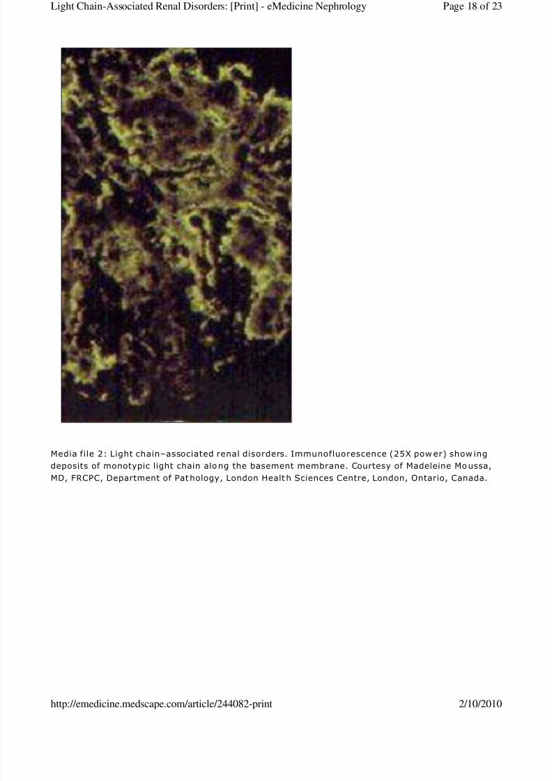
Media file 2) using electron microscopy.
Dense granular deposits on the endothelial side of the glomerular basement membrane (see Media files 2-4), on the
outer aspect of the tubular basement membrane, or on both may be seen with electron microscopy in persons with
LCDD. Classic ultrastructure examination findings include amorphous, noncongophilic, and nonfibrillar deposits. Most
of these deposits are of kappa light chains (see Media file 5). At times, the histologic changes are minimal, and
occasionally glomeruli may have mesangial deposits. Rarely, glomerular crescents can also be seen in patients with
LCDD.
Because many patients with LCDD do not have overt myeloma or any other evidence of monoclonal plasma cell
proliferation, they may present with renal disease manifesting with proteinuria, renal insufficiency, or renal failure.
Therefore, the renal biopsy findings may provide the first clues to the diagnosis of a monoclonal gammopathy.
Immunoglobulin light-chain amyloidosis (AL)
Renal involvement is common in AL amyloidosis. In contrast to LCDD, in which the deposits are usually kappa light
chains, the light chains involved in the formation of amyloidosis are usually of the lambda type. The histologic
appearance of amyloid is usually quite distinctive and is confirmed easily using Congo red stains or by the
ultrastructural demonstration of characteristic fibrils. AA amyloid loses its Congo red positivity when briefly exposed to
potassium permanganate, while non-AA amyloid resists this treatment. In the kidney, a diagnosis of non-AA
amyloidosis strongly suggests light-chain amyloidosis (AL).
Kappa light-chains are more likely to produce tubular dysfunction (Fanconi syndrome) and nodular nonamyloidotic
glomerulosclerosis, while lambda light chains are more likely to be involved in the development of AL amyloidosis.
Treatment
Medical Care
The goals of treatment are to prolong survival and to maintain quality of life.
Management of light-chain nephropathy depends on the underlying disease process. Take steps to limit further cast
precipitation, and implement effective prevention and management of its complications.
Light-chain associated renal syndrome
Asymptomatic light-chain proteinuria
Idiopathic - No therapy
Multiple myeloma - Chemotherapy
Waldenström macroglobulinemia - No therapy
Proximal tubular dysfunction (Fanconi syndrome)
Associated with myeloma - Chemotherapy
Not associated with myeloma - No therapy
Metabolic acidosis - Sodium bicarbonate therapy
Hypophosphatemia - Phosphate supplementation
Distal tubular dysfunction
Distal renal tubular acidosis - Sodium bicarbonate therapy
Nephrogenic diabetes insipidus - Thiazide therapy (if no hypercalcemia is present) or correct
Page 9 of 23Light Chain-Associated Renal Disorders: [Print] - eMedicine Nephrology
2/10/2010http://emedicine.medscape.com/article/244082-print

8/8/2019 Light Chain Disorder
http://slidepdf.com/reader/full/light-chain-disorder 10/23
calcium level (if hypercalcemia is present)
Amyloidosis - Chemotherapy
Acute renal failure
Multiple myeloma - Chemotherapy; avoidance of dehydration and contrast agents; treatment of
hypercalcemia, infection, and hyperuricemia; correction of obstruction; implementation of
plasmapheresis and dialysis
Waldenström macroglobulinemia - Plasmapheresis for hyperviscosity
Treatment of primary (underlying) disease
Chemotherapy: Any of the following regimens can be used in patients with multiple myeloma:
Melphalan and corticosteroids
Combination chemotherapy - VAD (ie, vincristine, Adriamycin, dexamethasone), ABCM (ie,
Adriamycin, carmustine [BiCNU], cyclophosphamide, melphalan), or MEVP (ie, melphalan,
cyclophosphamide, vincristine, prednisone)
Interferon alfa 2b
Autologous stem cell transplantation, allogenic or autologous: Myeloablative high-dose chemotherapy
and autologous stem cell transplantation induce hematologic remission in a high proportion of patients
who are eligible for such treatment, and early results from a limited number of patients suggest that thedeterioration of renal function may be arrested and possibly reversed. [5 ]
Steps to limit further cast precipitation
Rehydration
Cessation of nonsteroidal anti-inflammatory drugs (NSAIDs)
Treatment of infections
Reversal of hypercalcemia
Common precipitating factors
Dehydration: This is important in the precipitation of acute renal failure in a significant number (up to
95%) of patients. Dehydration and aciduria favors precipitation of light chains. Ensure adequate
hydration of patients, especially before initiating chemotherapy.
Hypercalcemia: Excessive calcium is an important cause of acute renal failure in patients with myeloma
and may be present in up to 30% of patients. Hypercalcemia impairs renal concentrating ability, thus
leading to dehydration and promoting precipitation of light-chain proteins in renal tubules. Nausea,
vomiting, and altered mental state associated with hypercalcemia further increase the likelihood of
dehydration. Hypercalciuria also exerts a direct nephrotoxic effect and thus causes tubular degeneration
and necrosis. Implement aggressive treatment of hypercalcemia, with saline diuresis, steroids, calcitonin,
and diphosphonate.
Contrast-induced renal failure: Perform contrast studies judiciously in patients with multiple myeloma
because of the possibility of contrast-induced renal failure. However, McCarthy and Becker reviewed 7retrospective studies of patients with myeloma who were receiving contrast media and noted that the
incidence rate of acute renal failure was only 0.6-1.25%, compared to 0.15% in the general population. [6 ]
Less common precipitating factors
Nephrotoxic agents: Avoid nephrotoxic agents (eg, NSAIDs, nephrotoxic antibiotics).
Infection: Ensure effective treatment of infection (5-20% of patients). Intravenous immunoglobulin has
been found to be safe when used as prophylaxis against infection in the so-called plateau phase.
Page 10 of 23Light Chain-Associated Renal Disorders: [Print] - eMedicine Nephrology
2/10/2010http://emedicine.medscape.com/article/244082-print

8/8/2019 Light Chain Disorder
http://slidepdf.com/reader/full/light-chain-disorder 11/23
Rare causes of acute renal failure
Tumor lysis syndrome: Uric acid released following chemotherapy may precipitate in the tubules and
may precipitate acute renal failure. Hence, pretreating patients undergoing chemotherapy with allopurinol
and diuresis is important.
Nephrolithiasis and urinary tract infections: Treat nephrolithiasis and urinary tract infections promptly.
Hyperviscosity: This can be treated with plasmapheresis.
Myeloma cell infiltration: Treat the underlying process.
Treatment of complications
Renal failure
Dialysis: Institute dialysis early to avoid uremia compounding the complications of underlying
disease. Approximately 20% of patients die within the first month, but predicting which patients
will die is not possible. Because 50% of the survivors live longer than 1 year and because
recovery of renal function is often delayed for several months, a policy of offering dialysis to most
patients is justified. All patients with myeloma who present with acute renal failure should receive
dialysis.
Long-term dialysis (hemodialysis or peritoneal dialysis) should be considered for patients with
chronic renal failure and myeloma who are responsive to chemotherapy. Before instituting long-
term dialysis therapy, consider the extent of other systemic disease. Patients who have
progressive myeloma and do not respond to chemotherapy may not be candidates for long-term
dialysis because their prognosis is very poor.
Plasmapheresis: The strong association between light-chain excretion and renal failure suggests
that light chains play a primary pathogenetic role in producing kidney damage. Plasma exchange
appears to be the most efficient way to rapidly remove large amounts of light chains and has been
advocated by many over the last 15 years, but its efficacy has not been established convincingly.
Zucchelli et al reported significantly higher survival rates in patients treated with plasma exchange
(66% vs 28%, P <.001).[7 ]In another randomized prospective trial, Johnson et al found no
significant difference in the number of patients whose renal function improved with this therapy;
however, they noted that patients with severe renal failure improved with plasma exchange. [8 ] As yet, no firm recommendation can be made on the effectiveness of plasma exchange, but data
justify its use in patients with rapidly rising plasma creatinine levels and high concentrations of
paraprotein.
Transplantation: Transplantation has been performed successfully in patients with end-stage
renal disease secondary to multiple myeloma. Consider renal transplantation only in patients who
have achieved hematologic remission and who have no other major complications of their
monoclonal gammopathy. Recurrent disease is common in patients with LCDD following
transplantation, and renal transplantation should not be done until optimal measures have
ensured substantial reduction in light chain production.
Infection: Treat infection early and effectively with nonnephrotoxic antibiotics. Intravenous
immunoglobulin infusions have been used as prophylaxis against infection in the plateau phase ofthe disease.
Hyperviscosity: This may manifest as confusion and neurological symptoms. Measure plasma
viscosity in these patients, and implement urgent plasmapheresis.
Renal tubular acidosis: Provide sodium bicarbonate supplementation to effectively control
acidosis.
Surgical Care
Patients with renal failure should have early, permanent vascular access because of the high risk of infection
Page 11 of 23Light Chain-Associated Renal Disorders: [Print] - eMedicine Nephrology
2/10/2010http://emedicine.medscape.com/article/244082-print

8/8/2019 Light Chain Disorder
http://slidepdf.com/reader/full/light-chain-disorder 12/23
associated with temporary catheters. Refer the patient to a vascular surgeon for the placement of a permanent
vascular access device.
Consultations
A multidisciplinary approach is important in the treatment of these patients, and consultations with following specialists
are useful:
Hematologist/oncologist - Treatment of the underlying disease process
Nephrologist - Prevention and treatment of acute and chronic renal failure
Infectious disease specialist - Treatment of sepsis in immunocompromised patients
Blood purification specialist - Plasmapheresis
Vascular surgeon - Placement of a permanent vascular access device for dialysis
Diet
No special restrictions are required unless the patient has chronic renal failure.
Optimize nutritional intake.
High fluid intake (2-3 L/d) is important to avoid dehydration and to minimize further cast formation.
Increased fluid intake is important during periods of volume depletion (eg, fever, diarrhea, vomiting).
Activity
Activity should be as tolerated by the individual.
Medication
No standard treatment has been established for light-chain nephropathy.
Antineoplastic agents
The mainstay of treatment is effective control of the underlying (primary) disease. A combination of an alkylating agent
(eg, melphalan) and prednisolone, administered for 4-7 d q4-6wk, is the standard first-line approach and induces
remission in approximately 40% of patients. This combination does not act rapidly, and the dose of melphalan often
must be modified because the drug is excreted via the kidney.
Combination chemotherapy regimens such as VAD, MEVP, or ABCM are generally used in younger patients with
multiple myeloma but do not appear to offer a significant survival advantage over treatment with melphalan.
Vincristine and doxorubicin act quickly and are metabolized in the liver, making them simpler to use in patients with
renal failure.
Melphalan (Alkeran)
Inhibits mitosis by cross-linking DNA strands. Effective against both resting and rapidly dividing tumor cells.
Page 12 of 23Light Chain-Associated Renal Disorders: [Print] - eMedicine Nephrology
2/10/2010http://emedicine.medscape.com/article/244082-print
8/8/2019 Light Chain Disorder
http://slidepdf.com/reader/full/light-chain-disorder 13/23
Dosing
Adult
8 mg/m2 /d PO
Pediatric
Not established
Interactions
Concurrent administration with cyclosporine increases nephrotoxicity; cimetidine and H2 antagonists increase gastric
pH, decreasing effects
Contraindications
Documented hypersensitivity; severe bone marrow depression
Precautions
Pregnancy
C - Fetal risk revealed in studies in animals but not established or not studied in humans; may use if benefits outweighrisk to fetus
Precautions
Amenorrhea may occur; caution in previously diagnosed myelosuppression; consider dose reduction in patients with
bone marrow suppression and renal insufficiency
Vincristine (Vincasar PFS, Oncovin)
Inhibits cellular mitosis by inhibiting intracellular tubulin function and binding to microtubules and spindle proteins in the
S phase.
Dosing
Adult
0.4 mg/d IV for 4 d (part of VAD regimen)
Pediatric
Not established
Interactions
Acute pulmonary reaction may occur when taken concurrently with mitomycin-C
Contraindications
Documented hypersensitivity
Precautions
Pregnancy
D - Fetal risk shown in humans; use only if benefits outweigh risk to fetus
Page 13 of 23Light Chain-Associated Renal Disorders: [Print] - eMedicine Nephrology
2/10/2010http://emedicine.medscape.com/article/244082-print
8/8/2019 Light Chain Disorder
http://slidepdf.com/reader/full/light-chain-disorder 14/23
Precautions
Caution with severe cardiopulmonary or hepatic impairment; caution with preexisting neuromuscular disease
Doxorubicin (Adriamycin, Rubex)
Inhibits topoisomerase II and produces free radicals, which may cause the destruction of DNA. The combination of
these events can, in turn, inhibit the growth of neoplastic cells.
Dosing
Adult
9 mg/m2 IV on day 1 of VAD regimen and repeated every 28 d
Pediatric
Not established
Interactions
May decrease phenytoin and digoxin plasma levels; phenobarbital may decrease plasma levels; cyclosporine mayinduce coma or seizures; mercaptopurine increases toxicity; cyclophosphamide increases cardiac toxicity
Contraindications
Documented hypersensitivity; severe heart failure; cardiomyopathy; impaired cardiac function; preexisting
myelosuppression
Precautions
Pregnancy
D - Fetal risk shown in humans; use only if benefits outweigh risk to fetus
Precautions
Irreversible cardiac toxicity and myelosuppression may occur; extravasation may result in severe local tissue necrosis;
reduce dose in patients with impaired hepatic function
Corticosteroids
Have anti-inflammatory properties and cause profound and varied metabolic effects. Corticosteroids modify the body's
immune response to diverse stimuli.
Prednisone (Deltasone, Meticorten, Orasone)
Immunosuppressant for treatment of autoimmune disorders; may decrease inflammation by reversing increasedcapillary permeability and suppressing PMN activity. Stabilizes lysosomal membranes and also suppresses
lymphocytes and antibody production. Reduced to its pharmacologically active form, prednisolone.
Dosing
Adult
25-60 mg/m2 /d PO
40 mg/d on a 4-d on/4-d off cycle (part of VAD regimen with vincristine and doxorubicin)
Page 14 of 23Light Chain-Associated Renal Disorders: [Print] - eMedicine Nephrology
2/10/2010http://emedicine.medscape.com/article/244082-print
8/8/2019 Light Chain Disorder
http://slidepdf.com/reader/full/light-chain-disorder 15/23
Pediatric
Not established
Interactions
Coadministration with estrogens may decrease clearance; concurrent use with digoxin may cause digitalis toxicity
secondary to hypokalemia; phenobarbital, phenytoin, and rifampin may increase metabolism of glucocorticoids
(consider increasing maintenance dose); monitor for hypokalemia with coadministration of diuretics
Contraindications
Documented hypersensitivity; viral infection; peptic ulcer disease; hepatic dysfunction; connective tissue infections;
fungal or tubercular skin infections; GI disease
Precautions
Pregnancy
B - Fetal risk not confirmed in studies in humans but has been shown in some studies in animals
Precautions
Abrupt discontinuation of glucocorticoids may cause adrenal crisis; hyperglycemia, edema, osteonecrosis, myopathy,
peptic ulcer disease, hypokalemia, osteoporosis, euphoria, psychosis, myasthenia gravis, growth suppression, and
infections may occur with glucocorticoid use
Follow-up
Transfer
Patients may require transfer to another center for the following reasons:
Dialysis: Ensure early institution of dialysis.
Plasmapheresis: Currently, the use of plasmapheresis is justified in patients with rapidly rising plasmacreatinine levels and high concentrations of paraprotein.
Deterrence/ Prevention
Maintain adequate fluid intake (2-3 L/d) to avoid dehydration. Dehydration is a common factor that precipitates
acute renal failure.
Avoid nephrotoxic agents. NSAIDs, often used to relieve bone pain, are the most prominent offenders.
Ensure early and effective treatment of infections with nonnephrotoxic antibiotics.
Early recognition and treatment of hypercalcemia are important.
Pretreat patients undergoing chemotherapy with allopurinol.
Complications
Renal failure: Renal insufficiency may be indolent, chronic and progressive, or rapidly progressive. Renal
insufficiency, a common manifestation of multiple myeloma, is present in more than 50% of patients. In one
Page 15 of 23Light Chain-Associated Renal Disorders: [Print] - eMedicine Nephrology
2/10/2010http://emedicine.medscape.com/article/244082-print

8/8/2019 Light Chain Disorder
http://slidepdf.com/reader/full/light-chain-disorder 16/23
study, the risk of renal failure was 7% in patients with daily light-chain excretion of less than 0.005 g, 17%, with
excretion of 0.005-2 g, and 39% with excretion of more than 2 g.
Infections: Infections of the upper and lower urinary tract are common in patients with multiple myeloma
because of suppression of humoral immunity.
Hyperviscosity syndromes: Measure plasma viscosity in patients with clinical features of hyperviscosity. Urgent
plasmapheresis is required in such patients.
Renal tubular acidosis
Prognosis
Factors associated with a poor prognosis in patients with multiple myeloma include the following:
High tumor mass (burden)
Presence of renal failure (BUN >30 mg/dL, serum creatinine >1.5 mg/dL): Once renal insufficiency is
present, the relationship between the degree of renal impairment and the duration of survival is dramatic.
With a serum creatinine level of less than 120 µmol/L (1.4 mg/dL), median survival is 44 months.
With a serum creatinine level of 120-180 µmol/L (1.4-2 mg/dL), median survival is 18 months.
With a serum creatinine level greater than 180 µmol/L (>2 mg/dL), median survival is 4.3 months.
Data from the fifth Medical Research Council trial on the treatment of myeloma show that only
50% of patients with a plasma creatinine value of greater than 200 µmol/L (2.3 mg/dL) at
presentation were alive after 1 year, compared with nearly 80% of patients with a serum
creatinine value of less than 130 µmol/L (1.5 mg/dL).
Presence of hypercalcemia (serum calcium >12 mg/dL).
Presence of interstitial fibrosis and tubular atrophy based on kidney biopsy findings
Pancytopenia: This is indicated by a WBC count of less than 1000/µL, a hematocrit value of less than
30%, and a platelet count less than 50,000/µL.
Plasma cell leukemia
Previous treatment failure(s)
Lambda light-chain disease compared to kappa light-chain disease
High beta2-microglobulin level: Levels higher than 6 mcg/mL suggest a worse prognosis (survival of
approximately 26 mo) compared to levels of less than 6 mcg/mL (survival of approximately 52 mo).
Patient Education
Instruct patients to maintain adequate hydration, with a daily oral fluid intake of 2-3 liters, unless fluid restriction
is needed because of advanced renal failure.
Warn patients to avoid anti-inflammatory agents because these aggravate renal dysfunction and may precipitate
acute renal failure.
Miscellaneous
Medicolegal Pitfalls
Page 16 of 23Light Chain-Associated Renal Disorders: [Print] - eMedicine Nephrology
2/10/2010http://emedicine.medscape.com/article/244082-print

8/8/2019 Light Chain Disorder
http://slidepdf.com/reader/full/light-chain-disorder 17/23
Failure to consider the diagnosis in a patient presenting with bone pain, anemia, and renal insufficiency
Despite the low risk of contrast agent nephrotoxicity, failure to warn patients of this risk and failure to use
contrast agents cautiously
Special Concerns
Diagnosis is often delayed. In approximately 35% of patients, the interval between the onset of symptoms and
diagnosis is longer than 3 months, and, in 15% of patients, it is longer than 6 months. This delay is particularly
important to the problem of myeloma kidney, which can sometimes be prevented if treatment is instituted early,
before catastrophic and irreversible injury has occurred.
Multimedia
Media file 1: Light chain–associated renal disorders. Light microscopy (hematoxylin and eosin stain
at 25X power) showing nodular glomerulosclerosis (arrow) and thickening of the basement
membrane. Courtesy of Madeleine Moussa, MD, FRCPC, Department of Patho logy, London Health
Sciences Centre, London, Ontario, Canada.
Page 17 of 23Light Chain-Associated Renal Disorders: [Print] - eMedicine Nephrology
2/10/2010http://emedicine.medscape.com/article/244082-print
8/8/2019 Light Chain Disorder
http://slidepdf.com/reader/full/light-chain-disorder 18/23
Media file 2: Light chain–associated renal disorders. Immunofluorescence (25X pow er) show ing
deposits of monotypic light chain along the basement membrane. Courtesy of Madeleine Moussa,
MD, FRCPC, Department of Pathology, London Health Sciences Centre, London, Ontario, Canada.
Page 18 of 23Light Chain-Associated Renal Disorders: [Print] - eMedicine Nephrology
2/10/2010http://emedicine.medscape.com/article/244082-print

8/8/2019 Light Chain Disorder
http://slidepdf.com/reader/full/light-chain-disorder 19/23
Media file 3: Light chain–associated renal disorders. Ultrastructure (electron microscopy at
29,000X power) showing deposition of nonfibrillar electron-dense material in the mesangial
nodule (arrow ). Courtesy of Madeleine Moussa, MD, FRCPC, Department of Pathology, London
Health Sciences Centre, London, Ontario, Canada.
Page 19 of 23Light Chain-Associated Renal Disorders: [Print] - eMedicine Nephrology
2/10/2010http://emedicine.medscape.com/article/244082-print

8/8/2019 Light Chain Disorder
http://slidepdf.com/reader/full/light-chain-disorder 20/23
Media file 4: Light chain–associated renal disorders. Ultrastructure (electron microscopy at
29,000X power) showing deposition of nonfibrillar electron-dense material along the basement
membrane (arrow s). Courtesy of Madeleine Moussa, MD, FRCPC, Department of Pathology, London
Health Sciences Centre, London, Ontario, Canada.
Media file 5: Light chain–associated renal disorders. Immunoelectron microscopy (immunogold at
29,000X power) showing kappa light-chain deposition. Courtesy of Madeleine Moussa, MD, FRCPC,
Department of Pathology, London Health Sciences Centre, London, Ontario, Canada.
References
1. Smithline N, Kassirer JP, Cohen JJ. Light-chain nephropathy. Renal tubular dysfunction associated with light-
chain proteinuria. N Engl J Med . Jan 8 1976;294(2):71-4. [Medline].
2. Adams W. Mollitis ossium. Trans Pathol Soc London . 1872;23:186.
3. Glenner GG, Terry W, Harada M, et al. Amyloid fibril proteins: proof of homology with immunoglobulin light
chains by sequence analyses. Science . Jun 11 1971;172(988):1150-1. [Medline].
4. Katzmann JA, Abraham RS, Dispenzieri A, et al. Diagnostic performance of quantitative kappa and lambda freelight chain assays in clinical practice. Clin Chem . May 2005;51(5):878-81. [Medline].
5. Firkin F, Hill PA, Dwyer K, et al. Reversal of dialysis-dependent renal failure in light-chain deposition disease by
autologous peripheral blood stem cell transplantation. Am J Kidney Dis . Sep 2004;44(3):551-5. [Medline].
6. McCarthy CS, Becker JA. Multiple myeloma and contrast media. Radiology . May 1992;183(2):519-21. [Medline].
7. Zucchelli P, Pasquali S, Cagnoli L, et al. Controlled plasma exchange trial in acute renal failure due to multiple
myeloma. Kidney Int . Jun 1988;33(6):1175-80. [Medline].
Page 20 of 23Light Chain-Associated Renal Disorders: [Print] - eMedicine Nephrology
2/10/2010http://emedicine.medscape.com/article/244082-print
8/8/2019 Light Chain Disorder
http://slidepdf.com/reader/full/light-chain-disorder 21/23
8. Johnson WJ, Kyle RA, Pineda AA, et al. Treatment of renal failure associated with multiple myeloma.
Plasmapheresis, hemodialysis, and chemotherapy. Arch Intern Med . Apr 1990;150(4):863-9. [Medline].
9. Beaufils M, Morel-Maroger L. Pathogenesis of renal disease in monoclonal gammopathies: current
concepts. Nephron . 1978;20(3):125-31. [Medline].
10. Coward RA, Mallick NP, Delamore IW. Should patients with renal failure associated with myeloma be
dialysed?. Br Med J (Clin Res Ed). Nov 26 1983;287(6405):1575-8. [Medline].
11. DeFronzo RA, Cooke CR, Wright JR, et al. Renal function in patients with multiple myeloma. Medicine
(Baltimore). Mar 1978;57(2):151-66. [Medline].
12. Fang LS. Light-chain nephropathy. Kidney Int . Mar 1985;27(3):582-92. [Medline].
13. Firkin F, Hill PA, Dwyer K, et al. Reversal of dialysis-dependent renal failure in light-chain deposition disease by
autologous peripheral blood stem cell transplantation. Am J Kidney Dis . Sep 2004;44(3):551-5. [Medline].
14. Ganeval D, Neël LH, Preud'homme JL, et al. Light-chain deposition disease: its relation with AL-type
amyloidosis. Kidney Int . Jul 1984;26(1):1-9. [Medline].
15. Gertz MA, Kyle RA, Greipp PR. Response rates and survival in primary systemic amyloidosis. Blood . Jan
15 1991;77(2):257-62. [Medline].
16. Herrera GA, Joseph L, Gu X, et al. Renal pathologic spectrum in an autopsy series of patients with plasma cell
dyscrasia. Arch Pathol Lab Med . Aug 2004;128(8):875-9. [Medline].
17. Jones HB. Papers on chemical pathology: Prefaced by the Gulstonian lectures, read at the Royal College of
Physicians, 1846. Lancet . 1847;2:88-92.
18. Klassen RB, Allen PL, Batuman V, et al. Light chains are a ligand for megalin. J Appl Physiol . Jan 2005;98
(1):257-63. [Medline].
19. Leung N, Lager DJ, Gertz MA, et al. Long-term outcome of renal transplantation in light-chain deposition
disease. Am J Kidney Dis . Jan 2004;43(1):147-53. [Medline].
20. Noel LH, Droz D, Ganeval D, et al. Renal granular monoclonal light chain deposits: morphological aspects in 11
cases. Clin Nephrol . May 1984;21(5):263-9. [Medline].
21. Pozzi C, D'Amico M, Fogazzi GB, et al. Light chain deposition disease with renal involvement: clinical
characteristics and prognostic factors. Am J Kidney Dis . Dec 2003;42(6):1154-63. [Medline].
22. Pozzi C, Fogazzi GB, Banfi G, et al. Renal disease and patient survival in light chain deposition disease. Clin
Nephrol . May 1995;43(5):281-7. [Medline].
23. Preud'homme JL, Aucouturier P, Touchard G, et al. Monoclonal immunoglobulin deposition disease (Randall
type). Relationship with structural abnormalities of immunoglobulin chains. Kidney Int . Oct 1994;46(4):965-
72. [Medline].
24. Ronco PM, Alyanakian MA, Mougenot B, et al. Light chain deposition disease: a model of glomerulosclerosis
defined at the molecular level. J Am Soc Nephrol . Jul 2001;12(7):1558-65. [Medline].
25. Ronco PM, Mougenot B, Touchard G, et al. Renal involvement in hematological disorders: monoclonal
immunoglobulins and nephropathy. Curr Opin Nephrol Hypertens . Mar 1995;4(2):130-8. [Medline].
Page 21 of 23Light Chain-Associated Renal Disorders: [Print] - eMedicine Nephrology
2/10/2010http://emedicine.medscape.com/article/244082-print

8/8/2019 Light Chain Disorder
http://slidepdf.com/reader/full/light-chain-disorder 22/23
26. Royer B, Arnulf B, Martinez F, et al. High dose chemotherapy in light chain or light and heavy chain deposition
disease. Kidney Int . Feb 2004;65(2):642-8. [Medline].
27. Salant DJ, Sanchorawala V, D'Agati VD. A case of atypical light chain deposition disease--diagnosis and
treatment. Clin J Am Soc Nephrol . Jul 2007;2(4):858-67. [Medline].
28. Sengul S, Zwizinski C, Batuman V. Role of MAPK pathways in light chain-induced cytokine production in human
proximal tubule cells. Am J Physiol Renal Physiol . Jun 2003;284(6):F1245-54. [Medline].
29. Sengul S, Zwizinski C, Simon EE, et al. Endocytosis of light chains induces cytokines through activation of NF-
kappaB in human proximal tubule cells. Kidney Int . Dec 2002;62(6):1977-88. [Medline].
30. Solomon A, Weiss DT, Kattine AA. Nephrotoxic potential of Bence Jones proteins. N Engl J Med . Jun
27 1991;324(26):1845-51. [Medline].
31. Sturgill BC, Tucker FL, Bolton WK. Immunoglobulin light chain nephropathies. Pathol Annu . 1987;22 Pt 2:133-
50. [Medline].
32. Tubbs RR, Gephardt GN, McMahon JT, et al. Light chain nephropathy. Am J Med . Aug 1981;71(2):263-
9. [Medline].
33. Winearls CG. Acute myeloma kidney. Kidney Int . Oct 1995;48(4):1347-61. [Medline].
34. Zhu L, Herrera GA, Murphy-Ullrich JE, et al. Pathogenesis of glomerulosclerosis in light chain deposition
disease. Role for transforming growth factor-beta. Am J Pathol . Aug 1995;147(2):375-85. [Medline].
Keywords
light chain-associated renal disorders, light chain associated renal disorders, light-chain deposition disease, light chain
deposition disease, light-chain nephropathy, light chain nephropathy, LCDD, renal disease, kidney disease, monoclonal
immunoglobulin deposition disease, MIDD, myeloma kidney, cast nephropathy, AL amyloidosis, primary amyloidosis,
Fanconi's syndrome, Fanconi syndrome, proximal tubular dysfunction, Bence Jones proteinuria, BJP, light-chain
proteinuria, light-chain glomerulopathy, , multiple myeloma
Contributor Information and Disclosures
Author
Malvinder S Parmar, MB, MS, FRCP(C), FACP, FASN, Assistant Professor (VPT), Faculty of Medicine, University of
Ottawa; Associate Professor, Department of Internal Medicine, Northern Ontario School of Medicine; Consulting
Physician, Timmins and District Hospital, Ontario, Canada
Malvinder S Parmar, MB, MS, FRCP(C), FACP, FASN is a member of the following medical societies: American
College of Physicians, American Society of Nephrology, Canadian Medical Association, Ontario Medical Association,
and Royal College of Physicians and Surgeons of Canada
Disclosure: Nothing to disclose.
Medical Editor
Frank C Brosius III, MD, Nephrology Program Director, Professor of Internal Medicine and Physiology, Department of
Internal Medicine, Division of Nephrology, University of Michigan School of Medicine
Frank C Brosius III, MD is a member of the following medical societies: Alpha Omega Alpha, American Diabetes
Association, American Society of Nephrology, and Phi Beta Kappa
Disclosure: Nothing to disclose.
Page 22 of 23Light Chain-Associated Renal Disorders: [Print] - eMedicine Nephrology
2/10/2010http://emedicine.medscape.com/article/244082-print
8/8/2019 Light Chain Disorder
http://slidepdf.com/reader/full/light-chain-disorder 23/23
Pharmacy Editor
Francisco Talavera, PharmD, PhD, Senior Pharmacy Editor, eMedicine
Disclosure: eMedicine Salary Employment
Managing Editor
Christie P Thomas, MBBS, FRCP, FASN, FAHA, Professor, Department of Internal Medicine, Division of Nephrology;
Medical Director, Kidney and Kidney/Pancreas Transplant Program, University of Iowa Hospitals and ClinicsChristie P Thomas, MBBS, FRCP, FASN, FAHA is a member of the following medical societies: American College of
Physicians, American Federation for Medical Research, American Heart Association, American Society of Nephrology,
American Society of Transplantation, American Thoracic Society, International Society of Nephrology, and Royal
College of Physicians
Disclosure: Genzyme Grant/research funds Other
CME Editor
Rebecca J Schmidt, DO, FACP, FASN, Professor of Medicine, Section Chief, Department of Medicine, Section of
Nephrology, West Virginia University School of Medicine
Rebecca J Schmidt, DO, FACP, FASN is a member of the following medical societies: American College of
Osteopathic Internists, American College of Physicians, American Medical Association, American Society ofNephrology, International Society of Nephrology, National Kidney Foundation, Renal Physicians Association, and West
Virginia State Medical Association
Disclosure: Abbott Grant/research funds Speaking and
teaching; Genzyme Honoraria Consulting; Amgen Honoraria Speaking and teaching; Ortho
Biotech Honoraria Speaking and teaching
Chief Editor
Vecihi Batuman, MD, FACP, FASN, Professor of Medicine, Section of Nephrology-Hypertension, Tulane University
School of Medicine; Chief, Medicine Service, Southeast Louisiana Veterans Health Care System
Vecihi Batuman, MD, FACP, FASN is a member of the following medical societies: American College of Physicians,
American Society of Hypertension, American Society of Nephrology, and International Society of NephrologyDisclosure: Nothing to disclose.
Further Reading © 1994- 2010 by Medscape.All Rights Reserved(http://www.medscape.com/public/copyright)
Page 23 of 23Light Chain-Associated Renal Disorders: [Print] - eMedicine Nephrology