Embed Size (px)
Citation preview
Lighting up cells with lanthanide self-assembled helicates
by Jean-Claude G. Bünzli
Interface FocusVolume 3(5):20130032
October 6, 2013
©2013 by The Royal Society
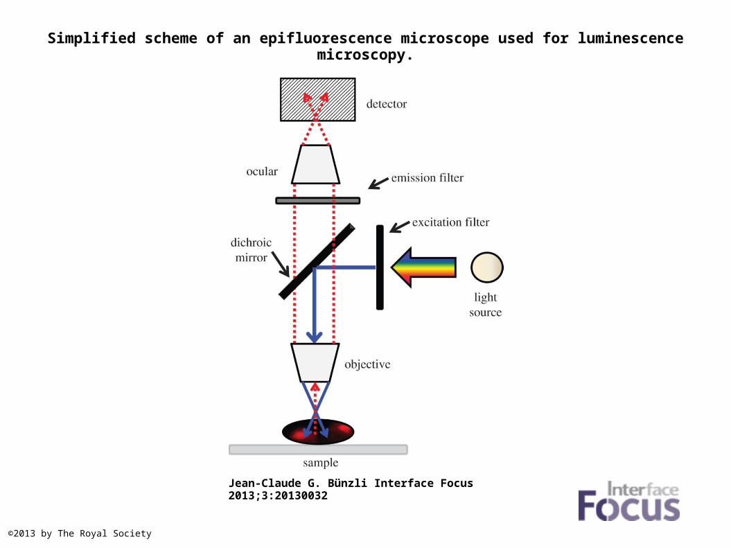
Simplified scheme of an epifluorescence microscope used for luminescence microscopy.
Jean-Claude G. Bünzli Interface Focus 2013;3:20130032
©2013 by The Royal Society
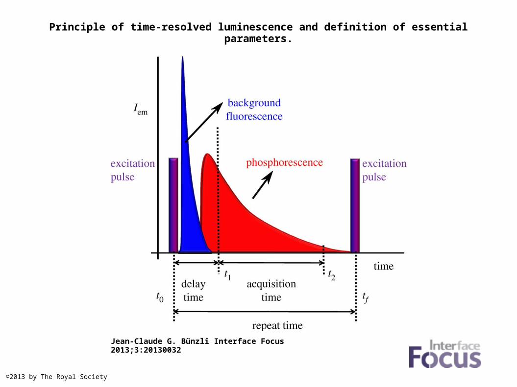
Principle of time-resolved luminescence and definition of essential parameters.
Jean-Claude G. Bünzli Interface Focus 2013;3:20130032
©2013 by The Royal Society
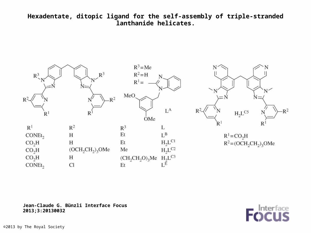
Hexadentate, ditopic ligand for the self-assembly of triple-stranded lanthanide helicates.
Jean-Claude G. Bünzli Interface Focus 2013;3:20130032
©2013 by The Royal Society
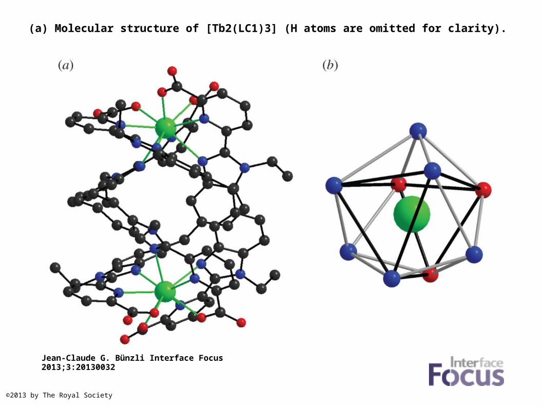
(a) Molecular structure of [Tb2(LC1)3] (H atoms are omitted for clarity).
Jean-Claude G. Bünzli Interface Focus 2013;3:20130032
©2013 by The Royal Society
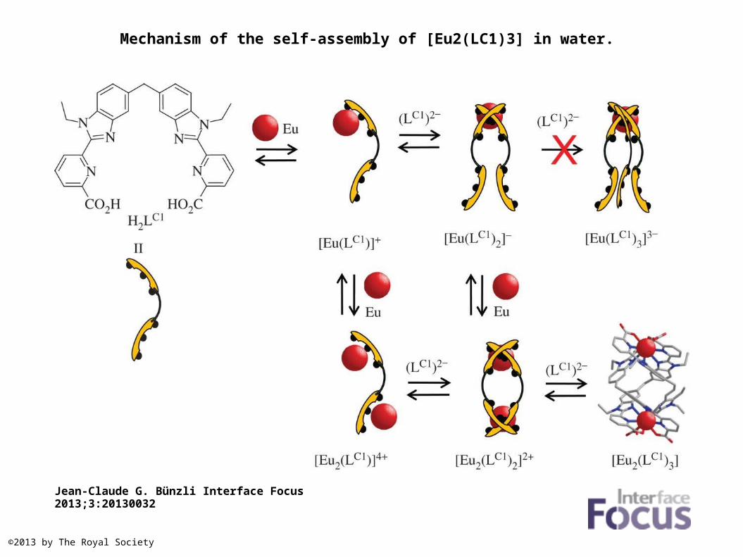
Mechanism of the self-assembly of [Eu2(LC1)3] in water.
Jean-Claude G. Bünzli Interface Focus 2013;3:20130032
©2013 by The Royal Society
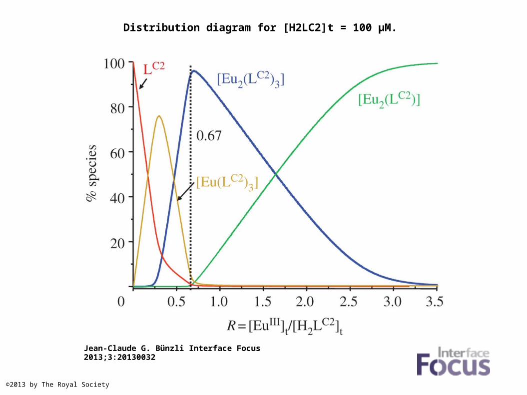
Distribution diagram for [H2LC2]t = 100 µM.
Jean-Claude G. Bünzli Interface Focus 2013;3:20130032
©2013 by The Royal Society
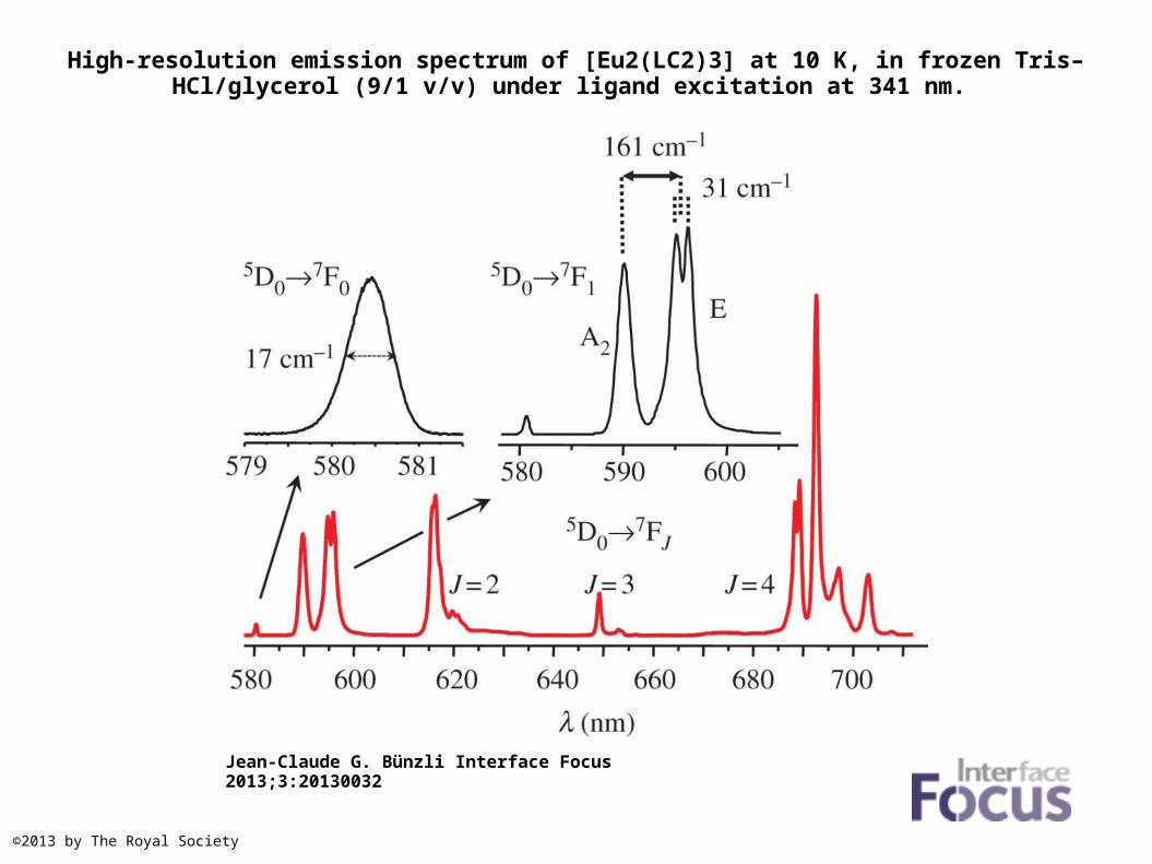
High-resolution emission spectrum of [Eu2(LC2)3] at 10 K, in frozen Tris–HCl/glycerol (9/1 v/v) under ligand excitation at 341 nm.
Jean-Claude G. Bünzli Interface Focus 2013;3:20130032
©2013 by The Royal Society
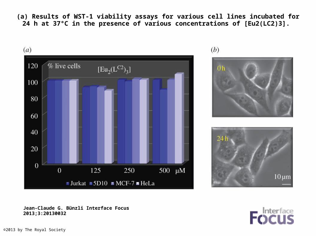
(a) Results of WST-1 viability assays for various cell lines incubated for 24 h at 37°C in the presence of various concentrations of [Eu2(LC2)3].
Jean-Claude G. Bünzli Interface Focus 2013;3:20130032
©2013 by The Royal Society
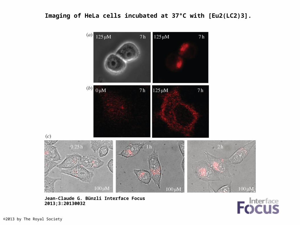
Imaging of HeLa cells incubated at 37°C with [Eu2(LC2)3].
Jean-Claude G. Bünzli Interface Focus 2013;3:20130032
©2013 by The Royal Society
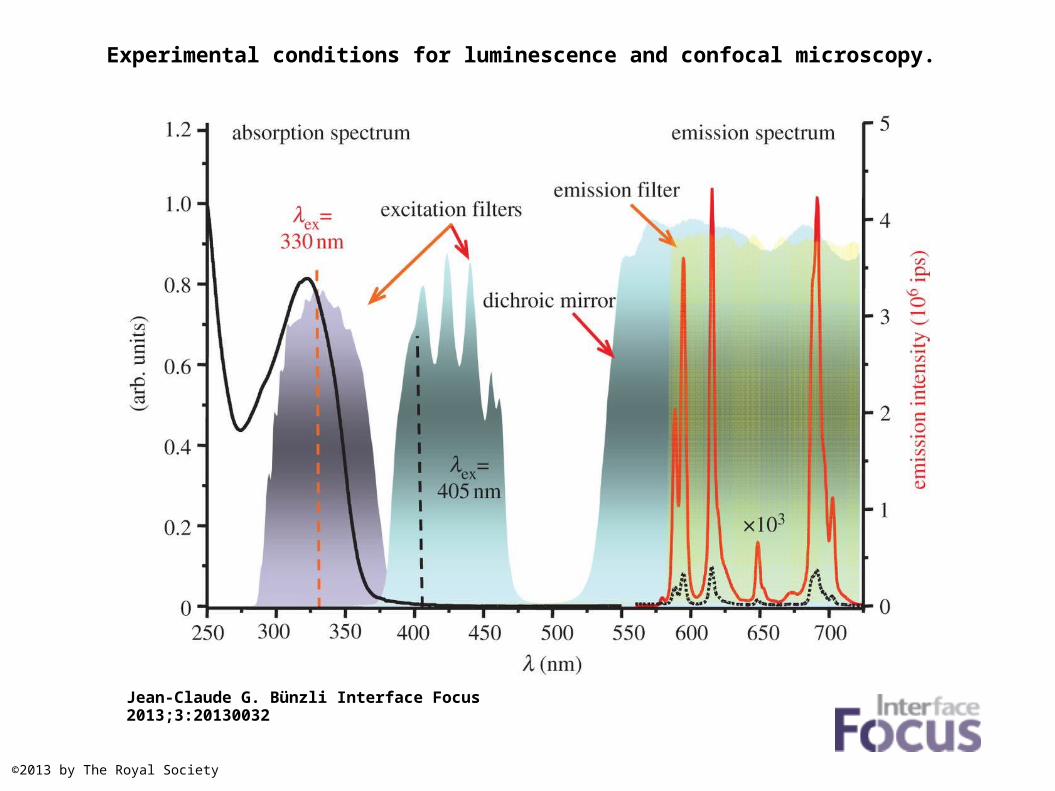
Experimental conditions for luminescence and confocal microscopy.
Jean-Claude G. Bünzli Interface Focus 2013;3:20130032
©2013 by The Royal Society
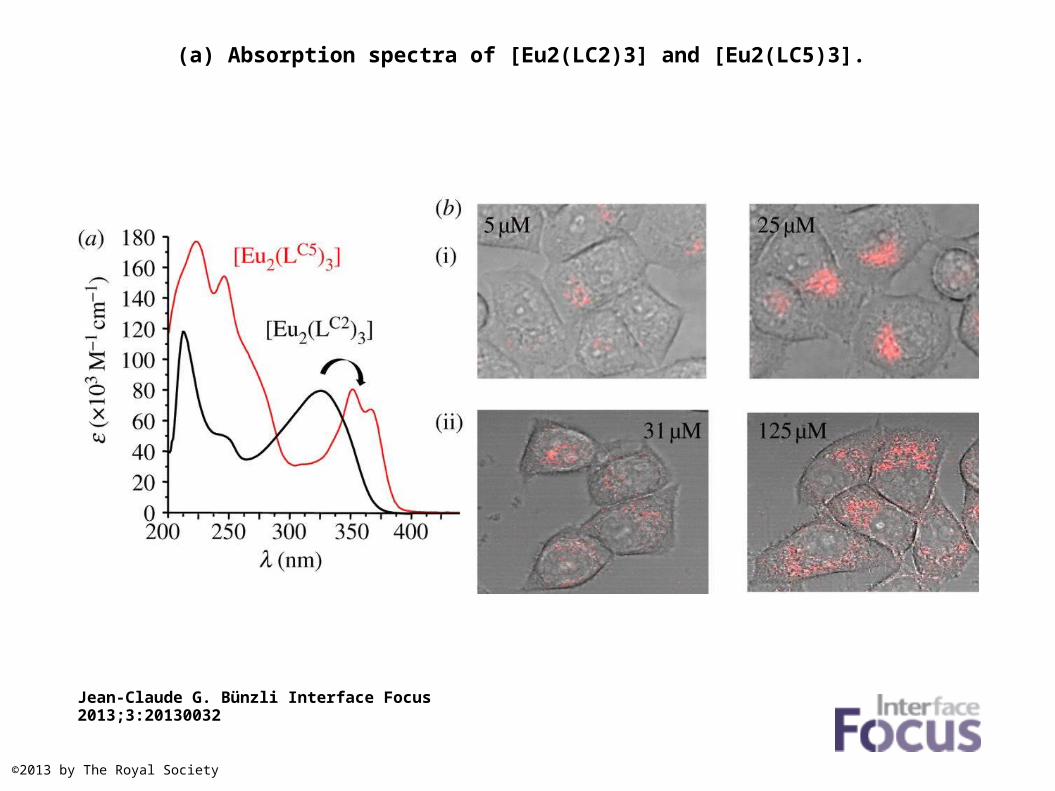
(a) Absorption spectra of [Eu2(LC2)3] and [Eu2(LC5)3].
Jean-Claude G. Bünzli Interface Focus 2013;3:20130032
©2013 by The Royal Society
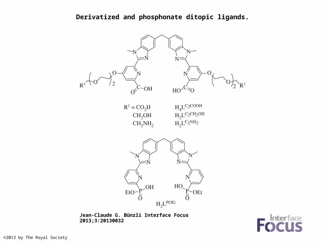
Derivatized and phosphonate ditopic ligands.
Jean-Claude G. Bünzli Interface Focus 2013;3:20130032
©2013 by The Royal Society
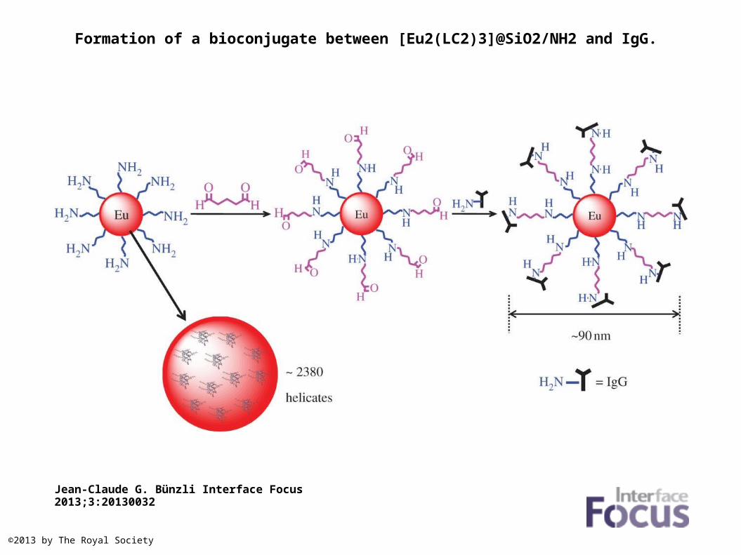
Formation of a bioconjugate between [Eu2(LC2)3]@SiO2/NH2 and IgG.
Jean-Claude G. Bünzli Interface Focus 2013;3:20130032
©2013 by The Royal Society
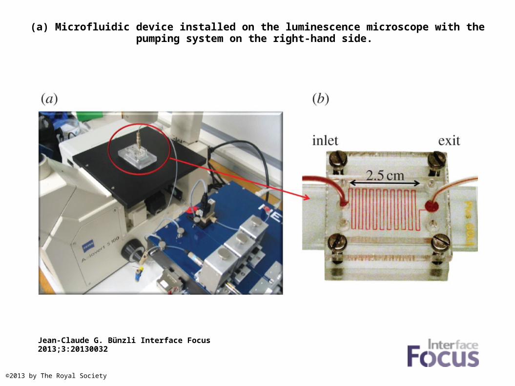
(a) Microfluidic device installed on the luminescence microscope with the pumping system on the right-hand side.
Jean-Claude G. Bünzli Interface Focus 2013;3:20130032
©2013 by The Royal Society
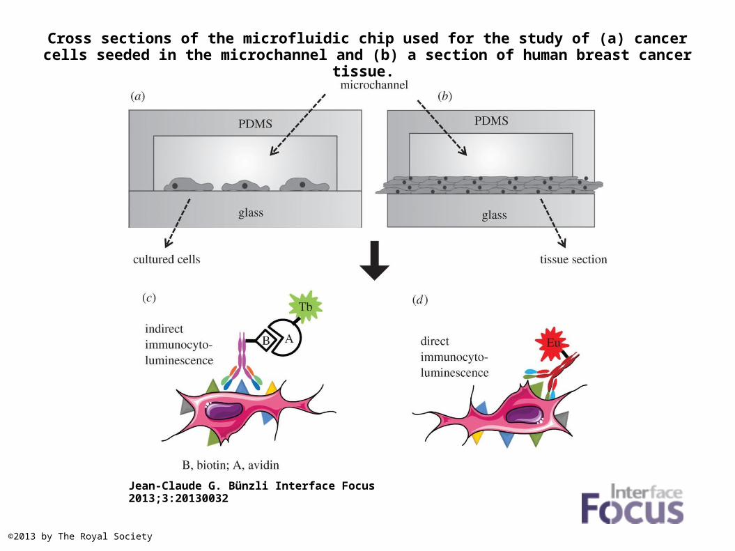
Cross sections of the microfluidic chip used for the study of (a) cancer cells seeded in the microchannel and (b) a section of human breast cancer tissue.
Jean-Claude G. Bünzli Interface Focus 2013;3:20130032
©2013 by The Royal Society
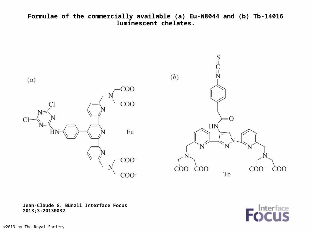
Formulae of the commercially available (a) Eu-W8044 and (b) Tb-14016 luminescent chelates.
Jean-Claude G. Bünzli Interface Focus 2013;3:20130032
©2013 by The Royal Society
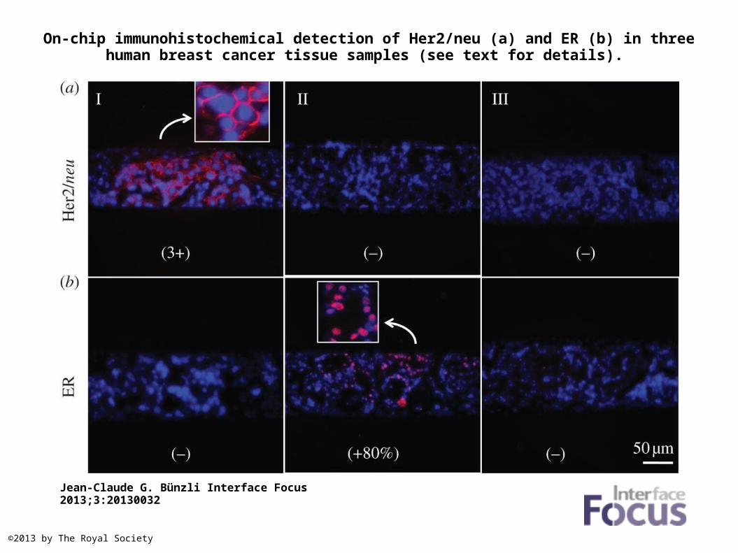
On-chip immunohistochemical detection of Her2/neu (a) and ER (b) in three human breast cancer tissue samples (see text for details).
Jean-Claude G. Bünzli Interface Focus 2013;3:20130032
©2013 by The Royal Society
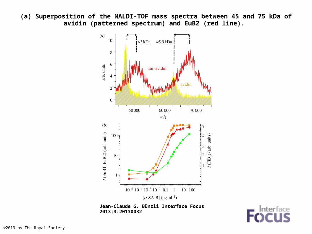
(a) Superposition of the MALDI-TOF mass spectra between 45 and 75 kDa of avidin (patterned spectrum) and EuB2 (red line).
Jean-Claude G. Bünzli Interface Focus 2013;3:20130032
©2013 by The Royal Society
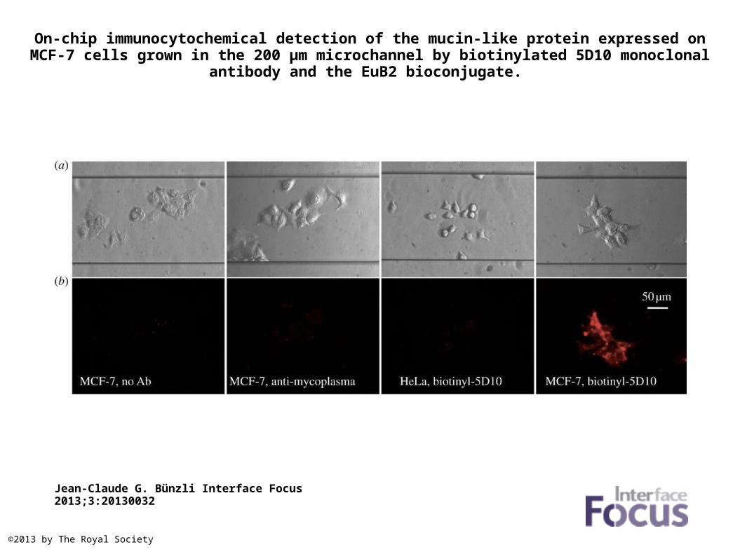
On-chip immunocytochemical detection of the mucin-like protein expressed on MCF-7 cells grown in the 200 µm microchannel by biotinylated 5D10 monoclonal antibody and the EuB2
bioconjugate.
Jean-Claude G. Bünzli Interface Focus 2013;3:20130032
©2013 by The Royal Society

(a) Principle of the two indirect assays for the detection of the Her2/neu (left) and ER (right) receptors expressed by human breast cancer cells.
Jean-Claude G. Bünzli Interface Focus 2013;3:20130032
©2013 by The Royal Society
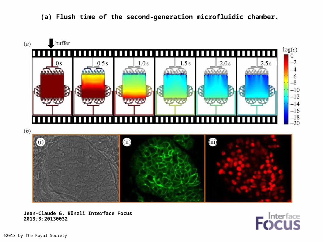
(a) Flush time of the second-generation microfluidic chamber.
Jean-Claude G. Bünzli Interface Focus 2013;3:20130032
©2013 by The Royal Society
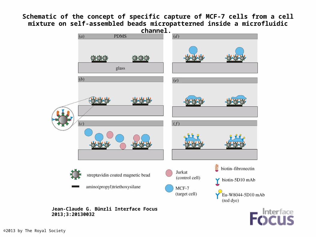
Schematic of the concept of specific capture of MCF-7 cells from a cell mixture on self-assembled beads micropatterned inside a microfluidic channel.
Jean-Claude G. Bünzli Interface Focus 2013;3:20130032
©2013 by The Royal Society
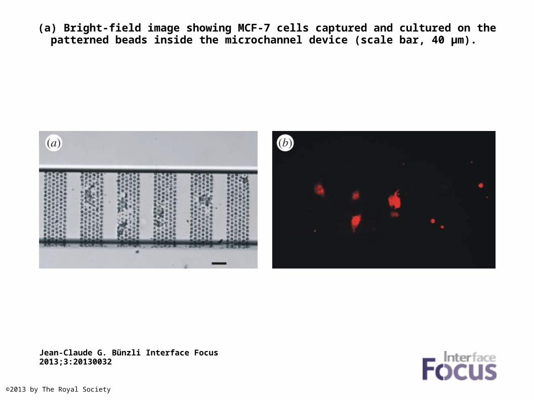
(a) Bright-field image showing MCF-7 cells captured and cultured on the patterned beads inside the microchannel device (scale bar, 40 µm).
Jean-Claude G. Bünzli Interface Focus 2013;3:20130032
©2013 by The Royal Society