Embed Size (px)
Citation preview
RESEARCH ARTICLE Open Access
Lignin metabolism involves Botrytis cinereaBcGs1- induced defense response intomatoChenyu Yang, Yingbo Liang, Dewen Qiu, Hongmei Zeng, Jingjing Yuan and Xiufen Yang*
Abstract
Background: BcGs1, a cell wall-degrading enzyme (CWDE), was originally derived from Botrytis cinerea. Our previousstudy revealed that BcGs1 could trigger defense responses and protect plants against various pathogens. We researchedthe defense response mechanism underlying this BcGs1 elicitation in tomato.
Results: We revealed that the two domains were required for BcGs1’s full necrosis activity. According to analysis andquantitative real-time PCR of the up-regulated proteins and genes filtered by iTRAQ-based quantitative proteomeapproach, oxidative metabolism and phenylpropanoid metabolism were speculated to be involved in BcGs1-triggereddefense response in tomato. Furthermore, experimental evidence showed that BcGs1 triggered reactive oxygen species(ROS) burst and increased the level of phenylalanine-ammonia lyase (PAL) and peroxidase (POD) enzyme activity, as wellas lignin accumulation. Moreover, histochemical analysis revealed that infiltration of BcGs1 in tomato leaves exhibited cellwall thickening compared with untreated plants.
Conclusions: The results suggested that BcGs1 activated the basal defense response included lignin metabolismcontributed to BcGs1-induced resistance to Botrytis. cinerea infection in tomato.
Keywords: Fungal protein elicitor, Defense response, iTRAQ, Phenylpropanoid, Lignin, Botrytis cinerea
BackgroundPlants intimately interact with various microbial pathogensin the complex nature environments. To protect them-selves against microbial colonization, plants have evolved avariety of defense mechanisms, including constitutive andinducible resistance strategies. Most potential pathogensare prevented by preformed physical and chemical barriersor after the induction of a complex array of defenseresponses. In order to Start the relevant resistance systemin plants a specific receptor to identify the pathogen isrequired, at the same time, the plant cell wall-derivedmolecules may be involved [1]. Following pathogen recog-nition, which is identified by pathogen-associated molecularpatterns (PAMP), microbe-associated molecular pattern(MAMP)-triggered immunity (PTI) or effector-triggered
immunity (ETI) through immune recognition systems [2–4], the protein kinase would be activated and ROS wouldbe accumulated [5, 6]. As a chain reaction, downstream sig-nal including the defense-related genes and PR proteinswould be activated [7, 8]. Plants rely on the basal defenseresponse by using a specific recognition system to preventpenetration and restrict the growth of pathogens, such ascell death, oxidative burst, defense genes, PR protein ex-pression, phytoalexins, callose deposition, lignification andcell wall thickness [2].To overcome the barrier of the plant cell wall, phyto-
pathogenic fungi secrete various CWDEs, such ascellulases, pectinase, hemicellulases, cutinase and prote-ase. Most of these enzymes not only degrade cell wallcomponents to get carbon sources for pathogen growthbut can also trigger multiple plant defense responses.During infection, necrotrophic pathogens first kill host
cells and/or feed on dead tissue. The necrotrophic patho-gen Botrytis cinerea often secretes non-host-selectivetoxins, CWDEs and proteinases to facilitate host cell death[9]. Although plant cell death that resulted from biotrophic
* Correspondence: [email protected] State Key Laboratory for Biology of Plant Disease and Insect Pests/ KeyLaboratory of Control of Biological Hazard Factors (Plant Origin) forAgri-product Quality and Safety, Ministry of Agriculture Institute of Plantprotection, Chinese Academy of Agricultural science, No. 12 Zhong-guan-cunSouth Street, Beijing 100081, China
© The Author(s). 2018 Open Access This article is distributed under the terms of the Creative Commons Attribution 4.0International License (http://creativecommons.org/licenses/by/4.0/), which permits unrestricted use, distribution, andreproduction in any medium, provided you give appropriate credit to the original author(s) and the source, provide a link tothe Creative Commons license, and indicate if changes were made. The Creative Commons Public Domain Dedication waiver(http://creativecommons.org/publicdomain/zero/1.0/) applies to the data made available in this article, unless otherwise stated.
Yang et al. BMC Plant Biology (2018) 18:103 https://doi.org/10.1186/s12870-018-1319-0
and necrotrophic pathogen infection plays a central role inmultiple defense responses, it has markedly different rolesin plant responses to necrotrophs and biotrophs that aredependent on plant-pathogen interaction. The cell deathinduced by necrotrophic pathogens is more complex thanthat of biotrophic pathogens in plant immunity becausenecrotrophic fungi have subtler pathogenic tactics [10].Variation in multiple basal defense mechanisms is thoughtto underlie differences in host susceptibility to necro-trophic pathogens. Although the genome of fungus waswell-known and Botrytis cinerea biology has been exten-sively studied [11, 12], the understanding about the bio-logical processes of plants response to Botrytis cinerea isalso very limited. Two elicitors isolated from Botrytiscinerea, namely botrycin and cinerein, caused the forma-tion of necrotic lesions, rapid transcriptional activation ofgenes encoding enzymes of the phenylpropanoid pathwayand distinct mitogen-activated protein kinases (MAPKs) ingrapevines (Vitis vinifera L) [13]. However, the mecha-nisms of most secretory elicitors from Botrytis cinerea thattrigger the plant defense response are unclear.CWDEs stimulate multiple immune responses in
plants and play a special role in pathogen-host plantinteractions, but the mechanism is very complex. SomeCWDEs triggering defense responses are associated withtheir degrading enzyme activity, but some are not relatedto enzyme activity [14]. Botrytis cinerea-produced poly-galacturonases (PGs) and fungal xylanase (Xyn11A) caninduce phytoalexin, ethylene and pathogenesis-relatedprotein synthesis and show an immune resistanceagainst Botrytis cinerea [14–17]. Present evidence sug-gests that these fungal CWDEs trigger typical PTI directlyor indirectly through the perception of damage-associatedmolecular patterns (DAMPs) and through plant cell wallfragments generated by the CWDE degradation [10]. PTIactivates a basal defense response, such as the biosynthesisof antimicrobial secondary metabolites (e.g., phytoalexins)and the expression of defense-related proteins, includingpathogenesis-related proteins (PR), cell wall lignifications,protease inhibitor expression, and hormone biosynthesis[18, 19]. Ultimately, plants exhibited broad-spectrum re-sistance to fungi, bacteria and viruses [16].Botrytis cinerea, which is one of the most destructive
diseases worldwide, is a typical necrotrophic pathogenand causes gray mold disease in tomatoes, strawberries,grapes, cucumbers, soybeans and sunflowers [20]. Resist-ance breeding efforts have not met with success forbotrytis diseases, although genetic resistance is effectiveand sustainable to protect plants. Elicitors could improveplant resistance to pathogens and are an alternativestrategy to reduce plant disease [21]. Understanding theinteraction of necrotrophic pathogens and host plantswill provide an insight for elicitor application. However,plant basal defense response triggered by many single
purified protein elicitors, including BcGs1, has not beenclear. In this study, we first described the differential-displaydefense protein by the iTRAQ method. Based on globaldifferential protein, phenylpropanoid metabolism was im-plied to be involved in BcGs1-induced tomato defenseresponses to Botrytis cinerea. Previous studies have shownPhenylpropanoid metabolism played an important role incotton induced resistance to V. dahlia [22]. Meanwhile, theobvious differences including protein expression, ligninmetabolism level and cell wall thickening, betweenBcGs1-induced plants and control were verified in lateexperiment. This study will help to understand the inter-action between necrotrophic fungal pathogens and hostplants and provide a theoretical basis for gray mold diseasemanagement through elicitor-activating plant immunity.
MethodsThe fungal pathogen cultures and plant cultivatesBotrytis cinerea strain BC-98 was originally isolated fromdiseased tomato tissues at the Beijing Region, PR China.The fungus was maintained on potato dextrose agar(PDA) medium and cultured in Czapek-Dox liquidmedium on a rotary shaker at 25 °C. Tomato seedlings(Zhong za 9, Purchased in Vegetable Flower ResearchInstitute, Chinese Academy of Agricultural Sciences)were grown in a greenhouse at 24–28 °C with 70–80%relative humidity, and dark/light ratio was 10/14 h.
Necrosis activity of BcGs1Protein BcGs1 was obtained by the method described byZhang et al. [23]. The necrosis-inducing activity of BcGs1was observed at 12 h post injection of tomato, tobacco, cu-cumber and pea leaves with 1 μM BcGs1 by a 1 ml syringe,while Tris-HCl buffer (50 mM) was used as a control.
Bioassay for BcGs1-induced disease resistance in tomatoSix to eight tomato plant leaves were injected with 250 nMBcGs1 protein solution (10 μL) and Tris-HCl buffer(50 mM). The same growing leaves were disinfected with al-cohol and then rinsed with distilled water at 48, 72, 96, 120and 168 h post BcGs1 injection, respectively. The two leaveswere placed in a Petri dish with wet filter paper and petiolewere moisturize with wet cotton wool. Botrytis cinerea discwas placed on the leaves and incubated for 48 h under con-tinuous light and 100% humidity at 25 °C in a chamber,then lesion diameter was measured using vernier caliper.The induced disease resistance was calculated using the for-mula: Disease reduction (%) = [(size of lesions on controlleaves-size of lesions on elicitor treated leaves)/size of le-sions on control leaves] × 100 and the results were analyzedby statistical analysis software. Fifteen tomato plants wereused in each treatment and control and three leaves weretaken from per plant. Three times were repeated.
Yang et al. BMC Plant Biology (2018) 18:103 Page 2 of 15
Identification of functional domain of BcGs1Biological information analysis showed that BcGs1contained two domains of Glyco-hydro 15 (GH15) andCBM20_glucoamylase (CBM20). To identify its activestructure, the transient expression vector pYBA1152,which contains a fluorescent protein and could fuse to myprotein/domains of the protein, was used to express pro-tein BcGs1, GH15 and CBM20 in Nicotiana benthamianathrough an Agrobacterium-mediated approach. Fluores-cence confocal microscopy and western blot analysis wereperformed to detect the expression at 48 h after injectionof the recombinant Agrobacterium. At the same time, thenecrosis activity was observed with the naked eye.
Western blotThe sample leaves of Nicotiana benthamiana were col-lected after injected the Agrobacterium carrying recom-binant gene of pYBA1152-BcGs1/GH15/CBM20 48 h.BcGs1, GH15 and CBM20 gene sequences were con-structed to carry a His-tag, respectively. The tree pro-teins were extracted from the sample, separately, usingplant total protein extraction reagent (Purchased fromBiotechnology Company). The total proteins were sub-jected to SDS-PAGE in three lanes after concentrationdetermination and boiling. The PEVD membrane wasused to carry the protein glue after SDS-PAGE, the elec-trotransfer instrument was applied for 1 h to transferthe proteins to PEVD membrane. The PEVD membranewas incubated with blocking solution for another 2 hafter washing with TBST buffer. Anti-His Tag MouseMonoclonal Antibody was added and used to bind toHis-tag and incubated for 2 h, after that HRP
Conjugated Anti-His Tag Monoclonal Antibody was ap-plied to bind to Anti-His Tag Mouse Monoclonal Anti-body for 1 h. The BCIP/NBT was mixed well on PEVDmembrane for color. At last, the membrane was put intothe instrument to exposure and take pictures.
Differential display protein analysisSix to eight tomato plant leaves were injected with 1 μMBcGs1 protein solution (10 μL), and Tris-HCl buffer(50 mM) was used as a control. Tomato leaves werecollected 24 h after treatment and used for proteomicsdetection by iTRAQ (Bangfei Biotechnology, Beijing,China). Each sample was repeated three times. A proteinratio > 1.3 or < 0.77 and P-value < 0.05 was regarded asbeing differentially expressed. Gene ontology (GO) analysiswas applied to predict the protein function and calculatethe functional category distribution frequency. KEGGanalysis was conducted to analyze the functional proteinannotation.
Quantitative real-time PCRThe tomato leaves were injected with BcGs1 andTris-HCl buffer. Total RNA was extracted at 0, 6, 12, 24,48, 72, 96 h post treatment according to the protocol ofthe plant RNA Kit (Tiangen Biotech, Beijing, China).First-strand cDNA was synthesized from the total RNAusing SuperMix for qPCR (TransGen Biotech, Beijing,China). The Applied Biosystems7500 Real Time PCRSystem was used to perform amplification with SYBRGreen SuperMix. The primers (Table 1) were designed byBeacon Designer 8.1. Actin, an internal reference, wasused to normalize the amount of cDNA in each
Table 1 Primers for quantitative validation of differentially expressed proteins induced by BcGs1
Names Forward primers(5 - > 3) Reverse primers(5 - > 3)
PR1 ATCATTTGTTTCCTTACCTTTG ACTCCAACTTGTCTACGA
PR10 TTACAAGACAACAACTGAGTAT AGCGTAGACAGAAGGATT
PR-Leaf 4 GACTATCTTGCGGTTCAC GCTCTTGAGTTGGCATAG
NP24 TTGTTCTCTTCTTCCTTCTT GGTGTATGGACAGTTGTT
PRSTH-2 TGTGTTGAAGGATGAAGAA TAAGCGTAGACAGAAGGA
PRSTH-2-like CTCCACCATCTCCTTGTA ACACCAATTCGTTTATTTAAGG
Endo chitinase EP3 TGTTGGTTCTACTGATGAT GGTAATCTGTGTTGTTCTC
Glucan endo-1,3-β-glucosidase B ATGCTATGTTGGATTCTGTT TTCTCGGACTACCTTCTTTA
Peroxidase1 ACTTCTCGTGCTAATAACAAT CAGTAGTTGAGTCTCTTCTTC
Peroxidase 12 GGCTTACTTCGTCTTCATT GACACAACTTGACCACAT
Peroxidase 21 TGTTATTACCTCTACTTCTTCAC ATGTTGCCACTTGTTCTT
Peroxidase −2 like GATGTTGTTCGGACCTATA ATTACTATTCACCTTGCTACA
Peroxidase71 TGTCCTAATGTTGAATCCA CTCCTGCCAATGATAGAT
ACC1 GTAATGGACACAGTAGAGA GAGATATTAGAAGTAGGAAGATG
Auxin repressed/dormancy associated protein GATGATGTTATGGCTGGT GGTACTTGCTAGATCCTTC
Actin GGTGTGATGGTGGGTATGG GCTGACAATTCCGTGCTC
Yang et al. BMC Plant Biology (2018) 18:103 Page 3 of 15
reaction. All the qPCR was repeated three times tocalculate the average values for quantification. Eachreaction melt curve was analyzed, and a negative con-trol without cDNA template was run with each reac-tion to evaluate the primer specificity. The relativegene expression levels were calculated from the aver-age values by ΔΔCt method.
H2O2 accumulation and formation in tomato leavesH2O2 production was examined in 6–8 tomato leaves at 2,4, and 6 h after treatment with 1 μM BcGs1 or Tris-HClbuffer. Leaves were excised and placed in water with0.01% Triton-X-100 and 1 mg/mL nitro 3, 3′-diaminoben-zidine (DAB); then, the solution was infiltrated with lowvacuum pressure for 5 min, and the leaves were incubatedovernight at room temperature. After that, the leavescleared in alcoholic lactophenol (95% ethanol:lactic acid:-phenol, 2:1:1) at room temperature until the leaves didnot contain chlorophyll, and they were then rinsed withwater. H2O2 can be visualized by a brownish-red precipi-tate that formed by polymerization with DAB.H2O2 content was detected in the leaves using the
H2O2 Test Kit (Jiancheng Biotechnology, Nanjing,China) at 0, 2, 4, 6, 12, and 24 h post-treatment. Samplesof 150 mg were ground using a grinding machine andhomogenized in extraction buffer (0.05 mM Phosphatebuffer, pH 7.2). The supernatant was collected for H2O2
content detection after centrifugation at 10000 g for10 min at 4 °C. The reagent provided by the kit wasincubated with the initial solution following the manu-facturer’s instructions. The H2O2 content was detectedat 405 nm wavelength. Each experiment was repeatedthree times.
Measurement of enzyme activitiesThe leaves treated with 1 μM BcGs1 and Tris-HCl buf-fer (as a control) were collected at 0, 12, 24, 48, 72, and96 h, frozen in liquid nitrogen immediately and thenstored at − 80 °C. One hundred to two hundred mg leafsamples were ground and homogenized in extractionbuffer (1.0 mM Phosphate buffer, pH 7.4). The super-natant was collected for defense enzyme determinationfollowing the extraction kit. PAL activity was detectedusing a PAL Extraction Kit (Jiancheng Biotechnology,Nanjing, China) following the kit’s protocol. The super-natant applied to detect POD activity was collected at3500 g for 10 min. POD activity was detected at 420 nmafter mixture incubation.
Lignin content detection in tomato leavesThe thioglycolic acid (TGA) method, a previously de-scribed method with modifications [24], was used toevaluate the lignin content in three biological replicates.Tomato leaves treated with 1 μM BcGs1 or Tris-HCl
buffer were collected at 12, 24, 48, 72, 96, 120 and 144 hpost BcGs1 treatment. Samples were homogenized andwashed with 20 ml phosphate buffer (pH 7.8) and centri-fuged at 5000 g for 10 min and the process was repeatedthree times. The pellet was dried at 80 °C for 24 h. Oneto two milligram of residue was weighed in tubes, mixedwith 1.5 ml 2 N HCl and 0.3 ml TGA, and then incu-bated at 95 °C for 4 h after mixed well. The mixture wasrapidly cooled on ice and centrifuged at 10,000 g for10 min. The pellet was washed three times with 1 mldistilled water. Pellets were re-suspended with 1 ml0.5 N NaOH and shaken at 200 rpm for 18 h at roomtemperature and centrifuged at 15,000 g for 10 min. Thesuspension was then transferred into a new tube, andthe pellet was washed again with 0.5 ml 0.5 N NaOH.After centrifugation, the two supernatants were com-bined and mixed with 0.3 ml concentrated HCl. Themixture was incubated at 4 °C for 4 h to precipitate thelignothioglycolate derivates. After centrifugation, the pel-let was solubilized in 1 ml 0.5 N NaOH. Absorbance ofthe resulting solution was measured at 280 nm.
Change of cell wall morphologyLeaf samples from 6 to 8 leaf tomato plants were em-bedded using the previously described methods withsome modifications [25]. Parts of the leaves around theBcGs1-induced necrotic spots were harvested at 3 d and5 d post induction. Tris-HCl buffer was used as a con-trol. The samples were then fixed in 1.0 ml phosphatebuffer(pH 7.2) containing 2% glutaraldehyde for 48 h atroom temperature and dehydrated in a graded series ofaqueous ethanol solutions (30, 50, 70, 80, 90, 95 and100% ethanol) for 15 min each. The sections were driedovernight at room temperature, mounted on aluminumstubs and sputter-coated with a gold-palladium alloyunder a vacuum for 2 min. At last, the sections wereobserved using a transmission electron microscopy(Hitachi H-7500).
Statistical analysisAll data provided in this study were from at least threeindependent replicates. Significant differences betweentreatments and controls were determined with an ana-lysis of variance using SAS 8.1 software. The means werecompared using Tukey’s HSD test.
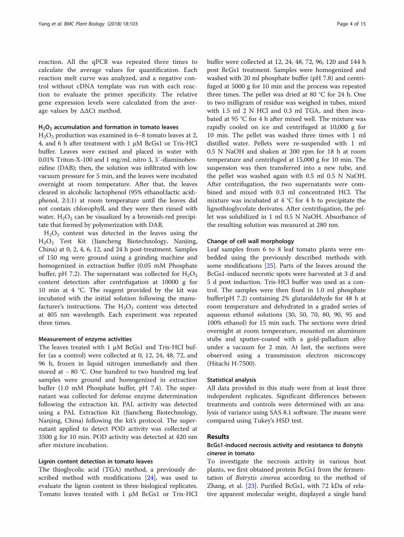
ResultsBcGs1-induced necrosis activity and resistance to Botrytiscinerea in tomatoTo investigate the necrosis activity in various hostplants, we first obtained protein BcGs1 from the fermen-tation of Botrytis cinerea according to the method ofZhang, et al. [23]. Purified BcGs1, with 72 kDa of rela-tive apparent molecular weight, displayed a single band
Yang et al. BMC Plant Biology (2018) 18:103 Page 4 of 15
in the SDS-PAGE (Additional file 1: Figure S1A). TheBcGs1 could induce necrosis activity in the tomato, to-bacco, cucumber and pea leaves 12 h post BcGs1 infil-tration (Additional file 1: Figure S1B), indicating thatBcGs1 has rapid necrosis activity in host plants. Toanalyze the appropriate induction time for disease resist-ance against Botrytis cinerea in tomato, fully mature4-week-old tomato leaves were infiltrated with 0.25 μMBcGs1 solution at opposite sides of the central vein, andTris-HCl buffer was used as control. Botrytis cinereadisc were inoculated on the detached leaves at 48, 72,96, 120 and 168 h after BcGs1 induction. The lesionareas at different induction times were measured usingthe cross method. BcGs1-treated tomato leaves showeda significant reduction in the lesion area caused by Bo-trytis cinerea compared to the Tris-HCl buffer control.The smallest lesion area appeared at 72 h, and the lesionarea was reduced 23.3% compared to the control. Theresult showed that 72 h was the most suitable inductiontime of BcGs1 (Fig. 1a, b). Therefore, our researchshowed that BcGs1 induced significant resistance to Bo-trytis cinerea in tomatoes with obvious necrotic activity.
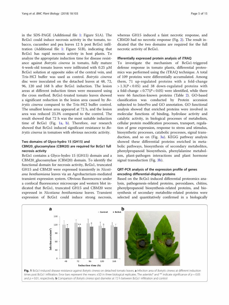
Two domains of Glyco-hydro 15 (GH15) andCBM20_glucoamylase (CBM20) are required for BcGs1 fullnecrosis activityBcGs1 contains a Glyco-hydro 15 (GH15) domain and aCBM20_glucoamylase (CBM20) domain. To identify thefunctional domain for necrosis activity, BcGs1, truncatedGH15 and CBM20 were expressed transiently in Nicoti-ana benthamiana leaves via an Agrobacterium-mediatedtransient expression system. Obvious fluorescence undera confocal fluorescence microscope and western blot in-dicated that BcGs1, truncated GH15 and CBM20 wereexpressed in Nicotiana benthamiana leaves. Transientexpression of BcGs1 could induce strong necrosis,
whereas GH15 induced a faint necrotic response, andCBM20 had no necrotic response (Fig. 2). The result in-dicated that the two domains are required for the fullnecrotic activity of BcGs1.
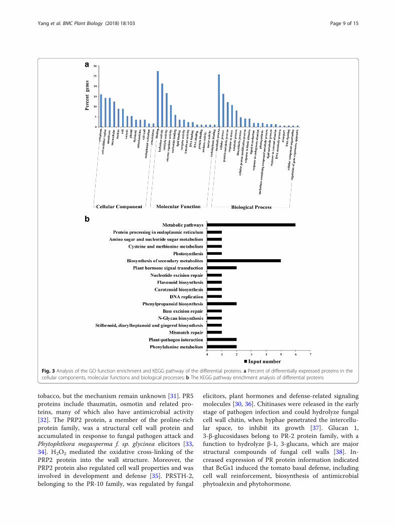
Ifferentially expressed protein analysis of iTRAQTo investigate the mechanism of BcGs1-triggereddefense response in tomato plants, differential proteo-mics was performed using the iTRAQ technique. A totalof 109 proteins were differentially accumulated. Amongthem, 71 up-regulated proteins with a fold-change> 1.3(P < 0.05) and 38 down-regulated proteins witha fold-change < 0.77(P < 0.05) were identified, while therewere 66 function-known proteins (Table 2). GO-basedclassification was conducted by Protein accessionsubjected to InterPro and GO annotation. GO functionalanalysis showed that enriched proteins were involved inmolecular functions of binding, hydrolase activity andcatalytic activity, in biological processes of metabolism,cellular protein modification processes, transport, regula-tion of gene expression, response to stress and stimulus,biosynthetic processes, catabolic processes, signal trans-duction, and so on (Fig. 3a). KEGG pathway analysisshowed these differential proteins enriched in meta-bolic pathways, biosynthesis of secondary metabolites,phenylpropanoid biosynthesis, phenylalanine metabol-ism, plant-pathogen interactions and plant hormonesignal transduction (Fig. 3b).
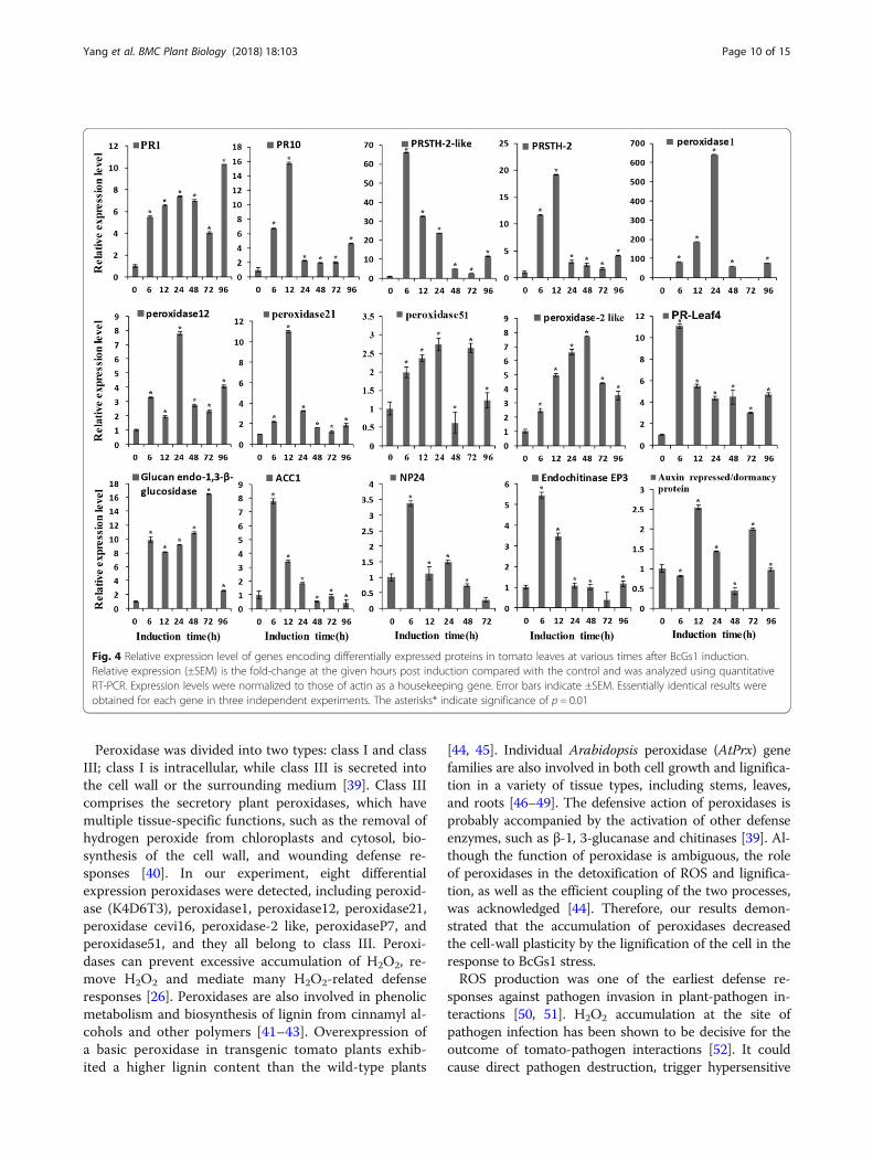
QRT-PCR analysis of the expression profile of genesencoding differential-display proteinsBased on the BcGs1-induced differential proteomics ana-lysis, pathogenesis-related proteins, peroxidases, chitins,phenylpropanoid biosynthesis-related proteins, and bio-synthesis of secondary metabolite-related proteins wereselected and quantitatively confirmed in a biologically
Fig. 1 BcGs1-induced disease resistance against Botrytis cinerea on detached tomato leaves. a Infection area of Botrytis cinerea at different inductiontimes post BcGs1 infiltration. Error bars represent the means ±SD in three biological replicates. The asterisks* and ** indicate significance of p = 0.05and p = 0.01, respectively. b Comparison of Botrytis cinerea spot diameter at 72 h between BcGs1 infiltration and control
Yang et al. BMC Plant Biology (2018) 18:103 Page 5 of 15
independent experiment using qPCR. The relative expres-sion levels of the fifteen genes are shown in Fig. 4. Thegenes PR1 and PR10 were up-regulated 5–8-fold at6–12 h. The PRSTH-2-like protein was up-regulated 60~70-fold at 6 h, and PRSTH-2 was up-regulated ~ 20-foldat 12 h. The peroxidases we tested had an up-regulation4–10-fold between 6 and 48 h, especially peroxidase1(K4D1W6), which was up-regulated 600-fold at 24 h. The1, 3-β-glucanases and chitinases were up-regulated ~ 10and ~ 5-fold at 6 h, respectively. The auxin repressed genewas up-regulated 2.5-fold. The antifungal protein NP24was up-regulated 3.5-fold at 6 h. Ethylene synthesis-relatedprotein ACC1 was up-regulated 8-fold at 6 h. These resultssuggested that most genes encoding defense responseproteins were up-regulated, which was consistent with theproteomics data. Up-regulation of these proteins andgenes indicated that BcGs1 activated the basal defenseresponse in tomato and induced the phenylpropane meta-bolic pathway.
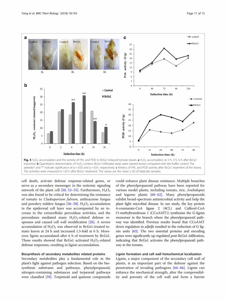
BcGs1 activates the phenylpropanoid metabolite pathwayThe ROS production followed pathogen attacks as anessential component in the signal transduction cas-cade and mediate plant defense response [26]. Wedetected H2O2 accumulation at the inoculation siteusing DAB staining. Brown precipitates were formedin the tomato leaves at 2, 4 and 6 h post BcGs1 infil-tration (Fig. 5a). The quantitative analysis showed theH2O2 accumulation in BcGs1-induced tomato leaveswas significantly higher than that of the control at 2,4, 6, 12 and 24 h post treatment and reached amaximum at 6 h with a 1.5-fold increase (Fig. 5b).According to this data, we suggested that BcGs1enhanced tomato intracellular production of H2O2,
increased the extracellular peroxidase activities, andfurther generate monolignol phenoxy radicals thatcouple spontaneously to form lignin polymers.The phenylpropanoid metabolite pathway is an im-
portant indicator of plant basal defense. The activities ofthe enzymes PAL and POD are involved in the phenyl-propanoid pathway leading to the synthesis of lignin.Accordingly, PAL and POD were measured after BcGs1infiltration (Fig. 5c). The results showed that activities ofPAL and POD increased 1.5 and 2-fold compared to theuntreated leaves, respectively. The data indicated thatthe phenylpropanoid pathway might be activated andplay a defensive function in tomato.
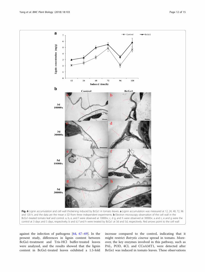
BcGs1 enhances secondary synthesis of lignin andreinforces the cell wallLignin played a critical role in the plant response topathogen infection. The synthesis and deposition of lig-nins were assumed to be physical barriers that made thecell walls more resistant to mechanical pressure duringfungal penetration [25, 27]. BcGs1 elicitor-treated plantsexhibited 1.5-fold increases in lignin deposition com-pared to the control at 48 h after elicitor treatment(Fig. 6a).Cell wall strengthening played an important role in
plant disease resistance [25]. Our results showed thatthe cell wall in the BcGs1-treated leaves was obviouslythickened compared with the Tris-HCl control at 3 daysand 5 days (Fig. 6b). High levels of lignin and cell wallthickness could enhance the toughness and mechanicalstrength of the cell wall, protect the differentiated cells,and reduce cells gap, leading to resistance to pathogeninfection [22, 27].
Fig. 2 Transient expression and Western Blot detection of BcGs1, GH15 and CBM20. The pYBA1152 vector was used to transiently express theseproteins via Agrobacterium-mediated inoculation for 48 h in Nicotiana benthamiana. a a, c, e and g represent the transient expression of thepYBA1152 empty vector, BcGs1, GH15 and CBM20; b, d, f and h represent the necrotic response induced by pYBA1152 empty vector, BcGs1,GH15 and CBM20 protein, respectively. b Western Blot verification of the expression of protein BcGs1-GFP, GH15-GFP and CBM20-GFP by His-tag
Yang et al. BMC Plant Biology (2018) 18:103 Page 6 of 15
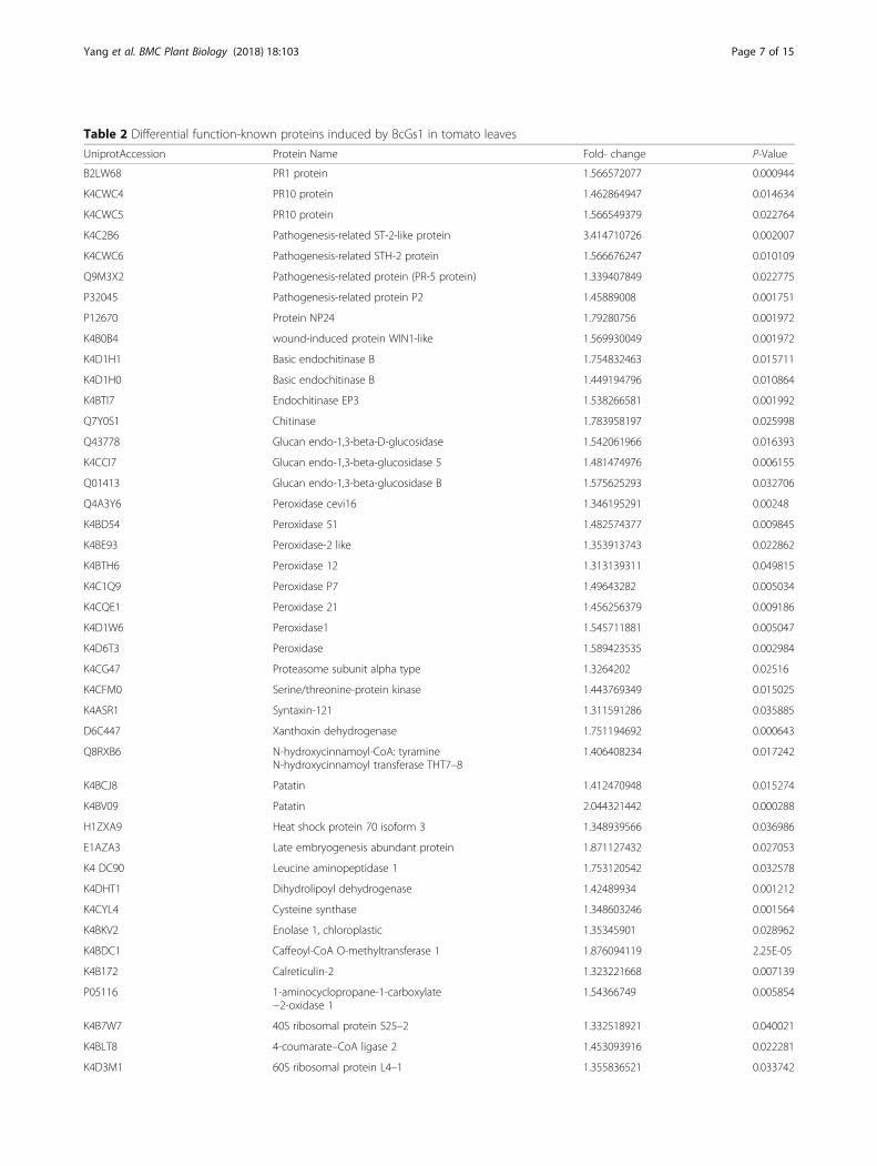
Table 2 Differential function-known proteins induced by BcGs1 in tomato leaves
UniprotAccession Protein Name Fold- change P-Value
B2LW68 PR1 protein 1.566572077 0.000944
K4CWC4 PR10 protein 1.462864947 0.014634
K4CWC5 PR10 protein 1.566549379 0.022764
K4C2B6 Pathogenesis-related ST-2-like protein 3.414710726 0.002007
K4CWC6 Pathogenesis-related STH-2 protein 1.566676247 0.010109
Q9M3X2 Pathogenesis-related protein (PR-5 protein) 1.339407849 0.022775
P32045 Pathogenesis-related protein P2 1.45889008 0.001751
P12670 Protein NP24 1.79280756 0.001972
K4B0B4 wound-induced protein WIN1-like 1.569930049 0.001972
K4D1H1 Basic endochitinase B 1.754832463 0.015711
K4D1H0 Basic endochitinase B 1.449194796 0.010864
K4BTI7 Endochitinase EP3 1.538266581 0.001992
Q7Y0S1 Chitinase 1.783958197 0.025998
Q43778 Glucan endo-1,3-beta-D-glucosidase 1.542061966 0.016393
K4CCI7 Glucan endo-1,3-beta-glucosidase 5 1.481474976 0.006155
Q01413 Glucan endo-1,3-beta-glucosidase B 1.575625293 0.032706
Q4A3Y6 Peroxidase cevi16 1.346195291 0.00248
K4BD54 Peroxidase 51 1.482574377 0.009845
K4BE93 Peroxidase-2 like 1.353913743 0.022862
K4BTH6 Peroxidase 12 1.313139311 0.049815
K4C1Q9 Peroxidase P7 1.49643282 0.005034
K4CQE1 Peroxidase 21 1.456256379 0.009186
K4D1W6 Peroxidase1 1.545711881 0.005047
K4D6T3 Peroxidase 1.589423535 0.002984
K4CG47 Proteasome subunit alpha type 1.3264202 0.02516
K4CFM0 Serine/threonine-protein kinase 1.443769349 0.015025
K4ASR1 Syntaxin-121 1.311591286 0.035885
D6C447 Xanthoxin dehydrogenase 1.751194692 0.000643
Q8RXB6 N-hydroxycinnamoyl-CoA: tyramineN-hydroxycinnamoyl transferase THT7–8
1.406408234 0.017242
K4BCJ8 Patatin 1.412470948 0.015274
K4BV09 Patatin 2.044321442 0.000288
H1ZXA9 Heat shock protein 70 isoform 3 1.348939566 0.036986
E1AZA3 Late embryogenesis abundant protein 1.871127432 0.027053
K4 DC90 Leucine aminopeptidase 1 1.753120542 0.032578
K4DHT1 Dihydrolipoyl dehydrogenase 1.42489934 0.001212
K4CYL4 Cysteine synthase 1.348603246 0.001564
K4BKV2 Enolase 1, chloroplastic 1.35345901 0.028962
K4BDC1 Caffeoyl-CoA O-methyltransferase 1 1.876094119 2.25E-05
K4B172 Calreticulin-2 1.323221668 0.007139
P05116 1-aminocyclopropane-1-carboxylate−2-oxidase 1
1.54366749 0.005854
K4B7W7 40S ribosomal protein S25–2 1.332518921 0.040021
K4BLT8 4-coumarate–CoA ligase 2 1.453093916 0.022281
K4D3M1 60S ribosomal protein L4–1 1.355836521 0.033742
Yang et al. BMC Plant Biology (2018) 18:103 Page 7 of 15
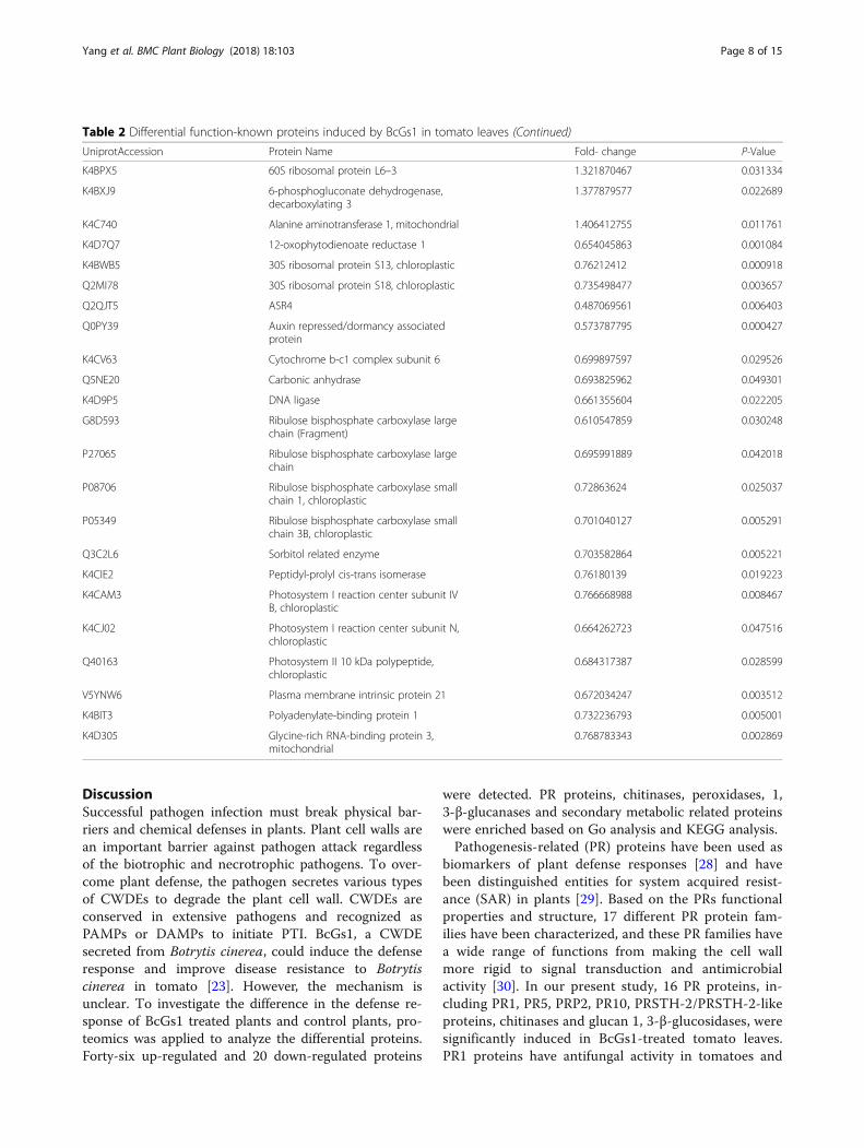
DiscussionSuccessful pathogen infection must break physical bar-riers and chemical defenses in plants. Plant cell walls arean important barrier against pathogen attack regardlessof the biotrophic and necrotrophic pathogens. To over-come plant defense, the pathogen secretes various typesof CWDEs to degrade the plant cell wall. CWDEs areconserved in extensive pathogens and recognized asPAMPs or DAMPs to initiate PTI. BcGs1, a CWDEsecreted from Botrytis cinerea, could induce the defenseresponse and improve disease resistance to Botrytiscinerea in tomato [23]. However, the mechanism isunclear. To investigate the difference in the defense re-sponse of BcGs1 treated plants and control plants, pro-teomics was applied to analyze the differential proteins.Forty-six up-regulated and 20 down-regulated proteins
were detected. PR proteins, chitinases, peroxidases, 1,3-β-glucanases and secondary metabolic related proteinswere enriched based on Go analysis and KEGG analysis.Pathogenesis-related (PR) proteins have been used as
biomarkers of plant defense responses [28] and havebeen distinguished entities for system acquired resist-ance (SAR) in plants [29]. Based on the PRs functionalproperties and structure, 17 different PR protein fam-ilies have been characterized, and these PR families havea wide range of functions from making the cell wallmore rigid to signal transduction and antimicrobialactivity [30]. In our present study, 16 PR proteins, in-cluding PR1, PR5, PRP2, PR10, PRSTH-2/PRSTH-2-likeproteins, chitinases and glucan 1, 3-β-glucosidases, weresignificantly induced in BcGs1-treated tomato leaves.PR1 proteins have antifungal activity in tomatoes and
Table 2 Differential function-known proteins induced by BcGs1 in tomato leaves (Continued)
UniprotAccession Protein Name Fold- change P-Value
K4BPX5 60S ribosomal protein L6–3 1.321870467 0.031334
K4BXJ9 6-phosphogluconate dehydrogenase,decarboxylating 3
1.377879577 0.022689
K4C740 Alanine aminotransferase 1, mitochondrial 1.406412755 0.011761
K4D7Q7 12-oxophytodienoate reductase 1 0.654045863 0.001084
K4BWB5 30S ribosomal protein S13, chloroplastic 0.76212412 0.000918
Q2MI78 30S ribosomal protein S18, chloroplastic 0.735498477 0.003657
Q2QJT5 ASR4 0.487069561 0.006403
Q0PY39 Auxin repressed/dormancy associatedprotein
0.573787795 0.000427
K4CV63 Cytochrome b-c1 complex subunit 6 0.699897597 0.029526
Q5NE20 Carbonic anhydrase 0.693825962 0.049301
K4D9P5 DNA ligase 0.661355604 0.022205
G8D593 Ribulose bisphosphate carboxylase largechain (Fragment)
0.610547859 0.030248
P27065 Ribulose bisphosphate carboxylase largechain
0.695991889 0.042018
P08706 Ribulose bisphosphate carboxylase smallchain 1, chloroplastic
0.72863624 0.025037
P05349 Ribulose bisphosphate carboxylase smallchain 3B, chloroplastic
0.701040127 0.005291
Q3C2L6 Sorbitol related enzyme 0.703582864 0.005221
K4CIE2 Peptidyl-prolyl cis-trans isomerase 0.76180139 0.019223
K4CAM3 Photosystem I reaction center subunit IVB, chloroplastic
0.766668988 0.008467
K4CJ02 Photosystem I reaction center subunit N,chloroplastic
0.664262723 0.047516
Q40163 Photosystem II 10 kDa polypeptide,chloroplastic
0.684317387 0.028599
V5YNW6 Plasma membrane intrinsic protein 21 0.672034247 0.003512
K4BIT3 Polyadenylate-binding protein 1 0.732236793 0.005001
K4D305 Glycine-rich RNA-binding protein 3,mitochondrial
0.768783343 0.002869
Yang et al. BMC Plant Biology (2018) 18:103 Page 8 of 15
tobacco, but the mechanism remain unknown [31]. PR5proteins include thaumatin, osmotin and related pro-teins, many of which also have antimicrobial activity[32]. The PRP2 protein, a member of the proline-richprotein family, was a structural cell wall protein andaccumulated in response to fungal pathogen attack andPhytophthora megasperma f. sp. glycinea elicitors [33,34]. H2O2 mediated the oxidative cross-linking of thePRP2 protein into the wall structure. Moreover, thePRP2 protein also regulated cell wall properties and wasinvolved in development and defense [35]. PRSTH-2,belonging to the PR-10 family, was regulated by fungal
elicitors, plant hormones and defense-related signalingmolecules [30, 36]. Chitinases were released in the earlystage of pathogen infection and could hydrolyze fungalcell wall chitin, when hyphae penetrated the intercellu-lar space, to inhibit its growth [37]. Glucan 1,3-β-glucosidases belong to PR-2 protein family, with afunction to hydrolyze β-1, 3-glucans, which are majorstructural compounds of fungal cell walls [38]. In-creased expression of PR protein information indicatedthat BcGs1 induced the tomato basal defense, includingcell wall reinforcement, biosynthesis of antimicrobialphytoalexin and phytohormone.
Fig. 3 Analysis of the GO function enrichment and KEGG pathway of the differential proteins. a Percent of differentially expressed proteins in thecellular components, molecular functions and biological processes; b The KEGG pathway enrichment analysis of differential proteins
Yang et al. BMC Plant Biology (2018) 18:103 Page 9 of 15
Peroxidase was divided into two types: class I and classIII; class I is intracellular, while class III is secreted intothe cell wall or the surrounding medium [39]. Class IIIcomprises the secretory plant peroxidases, which havemultiple tissue-specific functions, such as the removal ofhydrogen peroxide from chloroplasts and cytosol, bio-synthesis of the cell wall, and wounding defense re-sponses [40]. In our experiment, eight differentialexpression peroxidases were detected, including peroxid-ase (K4D6T3), peroxidase1, peroxidase12, peroxidase21,peroxidase cevi16, peroxidase-2 like, peroxidaseP7, andperoxidase51, and they all belong to class III. Peroxi-dases can prevent excessive accumulation of H2O2, re-move H2O2 and mediate many H2O2-related defenseresponses [26]. Peroxidases are also involved in phenolicmetabolism and biosynthesis of lignin from cinnamyl al-cohols and other polymers [41–43]. Overexpression ofa basic peroxidase in transgenic tomato plants exhib-ited a higher lignin content than the wild-type plants
[44, 45]. Individual Arabidopsis peroxidase (AtPrx) genefamilies are also involved in both cell growth and lignifica-tion in a variety of tissue types, including stems, leaves,and roots [46–49]. The defensive action of peroxidases isprobably accompanied by the activation of other defenseenzymes, such as β-1, 3-glucanase and chitinases [39]. Al-though the function of peroxidase is ambiguous, the roleof peroxidases in the detoxification of ROS and lignifica-tion, as well as the efficient coupling of the two processes,was acknowledged [44]. Therefore, our results demon-strated that the accumulation of peroxidases decreasedthe cell-wall plasticity by the lignification of the cell in theresponse to BcGs1 stress.ROS production was one of the earliest defense re-
sponses against pathogen invasion in plant-pathogen in-teractions [50, 51]. H2O2 accumulation at the site ofpathogen infection has been shown to be decisive for theoutcome of tomato-pathogen interactions [52]. It couldcause direct pathogen destruction, trigger hypersensitive
Fig. 4 Relative expression level of genes encoding differentially expressed proteins in tomato leaves at various times after BcGs1 induction.Relative expression (±SEM) is the fold-change at the given hours post induction compared with the control and was analyzed using quantitativeRT-PCR. Expression levels were normalized to those of actin as a housekeeping gene. Error bars indicate ±SEM. Essentially identical results wereobtained for each gene in three independent experiments. The asterisks* indicate significance of p = 0.01
Yang et al. BMC Plant Biology (2018) 18:103 Page 10 of 15
cell death, activate defense response-related genes, orserve as a secondary messenger in the systemic signalingnetwork of the plant cell [50, 53–55]. Furthermore, H2O2
was also found to be critical for determining the resistanceof tomato to Cladosporium fulvum, anthracnose fungusand powdery mildew fungus [56–58]. H2O2 accumulationin the epidermal cell layer was accompanied by an in-crease in the extracellular peroxidase activities, and theperoxidases mediated many H2O2-related defense re-sponses and caused cell wall modification [26]. A severeaccumulation of H2O2 was observed in BcGs1-treated to-mato leaves at 24 h and increased 1.5-fold at 6 h. More-over, lignin accumulated after 6 h of treatment by BcGs1.These results showed that BcGs1 activated H2O2-relateddefense responses, resulting in lignin accumulation.
Biosynthesis of secondary metabolites related proteinsSecondary metabolites play a fundamental role in theplant’s fight against pathogen infection. Based on the bio-synthesis substrates and pathways, phenylpropanoid,nitrogen-containing substances and terpenoid pathwayswere classified [59]. Terpenoid and quinone compounds
could enhance plant disease resistance. Multiple branchesof the phenylpropanoid pathway have been reported forvarious model plants, including tomato, rice, Arabidopsisand legume plants [60–62]. Many phenylpropanoidsexhibit broad-spectrum antimicrobial activity and help theplant fight microbial disease. In our study, the key protein4-coumarate-CoA ligase 2 (4CL) and Caffeoyl-CoAO-methyltransferase 1 (CCoAMT1) synthesize the G-ligninmonomer in the branch where the phenylpropanoid path-way was identified. Previous results found that CCoAMTdown regulation in alfalfa resulted in the reduction of G lig-nin units [63]. The two essential proteins and encodinggenes were significantly up-regulated post-BcGs1 infiltration,indicating that BcGs1 activates the phenylpropanoid path-way in the tomato.
Lignin formation and cell wall histochemical localizationLignin, a major component of the secondary cell wall ofplants, is an important part of the defense against thepenetration of invading pathogens [64–66]. Lignin canenhance the mechanical strength, alter the compressibil-ity and porosity of the cell wall and form a barrier
Fig. 5 H2O2 accumulation and the activity of PAL and POD in BcGs1-induced tomato leaves. a H2O2 accumulation at 2 h, 4 h, 6 h after BcGs1induction; b Quantitative determination of H2O2 content. BcGs1-infiltrated areas were stained brown compared with the buffer control. Theasterisks* and ** indicate significance of p = 0.05 and p = 0.01, respectively. c Kinetics of PAL and POD activity after BcGs1 treatment of the leaves.The activities were measured 0–120 h after BcGs1 treatment. The values are the mean ± SD of triplicate samples
Yang et al. BMC Plant Biology (2018) 18:103 Page 11 of 15
against the infection of pathogens [64, 67–69]. In thepresent study, differences in lignin content betweenBcGs1-treatment and Tris-HCl buffer-treated leaveswere analyzed, and the results showed that the lignincontent in BcGs1-treated leaves exhibited a 1.5-fold
increase compared to the control, indicating that itmight restrict Botrytis cinerea spread in tomato. More-over, the key enzymes involved in this pathway, such asPAL, POD, 4CL and CCoAMT1, were detected afterBcGs1 was induced in tomato leaves. These observations
Fig. 6 Lignin accumulation and cell wall thickening induced by BcGs1 in tomato leaves. a Lignin accumulation was measured at 12, 24, 48, 72, 96and 120 h, and the data are the mean ± SD from three independent experiments. b Electron microscopy observation of the cell wall in theBcGs1-treated tomato leaf and control. a, b, e, and f were observed at 10000×; c, d g, and h were observed at 30000×. a and c, e and g were thecontrol at 3 days and 5 days, respectively; b and d, f and h were treated by BcGs1 at 3d and 5d, respectively. Red arrows point to the cell wall
Yang et al. BMC Plant Biology (2018) 18:103 Page 12 of 15
were confirmed via the transcriptionally up-regulated ex-pression of the PAL, POD 4CL and CCoAMT1 genescompared to the control. Furthermore, histochemicallocalization showed that the cell wall of BcGs1-treatedleaves was significantly thickened post BcGs1 treatmentcompared to the control. Cell wall thickening enhancedthe mechanical strength and improved disease resistanceto Botrytis cinerea infection.
ConclusionsBcGs1 could significantly activate disease resistanceagainst Botrytis cinerea at 72 h post-induction. Twodomains are required for BcGs1 full necrosis activity.Differential expression proteins were identified intomato plants using a proteomics approach. PR proteins,peroxidases, Glucan endo-1, 3-β-glucosidase, chitinases,ethylene synthesis-related proteins, and biosynthesis ofsecondary metabolites were involved in BcGs1-inducedresistance. BcGs1-infiltrated tomato plants exhibitedH2O2 accumulation, increased levels of regulation of thekey PAL and POD enzymes, lignin end-product accumu-lation in the phenylpropanoid pathway, and cell wallthickening. According to BcGs1-induced PR differentialexpression proteins, peroxidase and the biosynthesisof secondary metabolites, the mechanisms by whichBcGs1 triggers disease resistance against Botrytiscinerea were summarized as follows. BcGs1 inducedH2O2 production, which not only increased peroxid-ase activity but also caused cell wall strengthening andlignin accumulation. Meanwhile, many defense-relatedproteins were up-regulated, including PRs, Glucanendo-1, 3-β-glucosidase and chitinase. By combiningbiochemical and histochemical data, we suggested thatBcGs1 triggers up-regulation of the phenylpropanoidpathway-related genes and protein, enzyme activities, highlignin levels and cell wall thickness, and ultimately gener-ates tomato disease resistance. Overall, lignin metabolismplayed a critical role and was involved in BcGs1-induceddefense response in the tomato.
Additional file
Additional file 1: Figure S1. Purification and necrosis activity of theprotein BcGs1. A: SDS-PAGE analysis of purified BcGs1. M, Protein marker. 1,Purified BcGs1. B: Necrosis activity of BcGs1 in tomato, tobacco, cucumberand pea leaves. (DOCX 673 kb)
Abbreviations4CL: 4-coumarate-CoA ligase 2; CCoAMT1: Caffeoyl-CoA O-methyltransferase1; CWDE: cell wall-degrading enzyme; DAB: nitro 3, 3′-diaminobenzidine;PAL: phenylalanine-ammonia lyase; PAMP: pathogen-associated molecularpatterns; POD: peroxidase; PR: pathogenesis-related proteins; PTI: microbe-associated molecular pattern (MAMP)-triggered immunity; ROS: reactiveoxygen species; TGA: thioglycolic acid
FundingThis research was supported by the National Key Research and DevelopmentProgram of China (2017YFD0201100). The funding body did not play anyroles in the design of the study and collection, analysis, and interpretation ofdata and in writing the manuscript.
Availability of data and materialsAll data generated or analysed during this study are included in this publishedarticle and its supplementary information files.
Authors’ contributionsXY, DQ, HZ and JY designed and guidanced the experiments. CY and YLanalyzed the date of Proteomics. YL collected the samples. CY and XYperformed the experiments and drafted the manuscript with the contributionof all authors. All authors read and approved the final manuscript.
Ethics approval and consent to participateTomato (Solanum lycopersicum L.) seeds “zhongza9hao”, obtained the permissionof Ministry of Agriculture of the People’s Republic of China, were purchased fromInstitute of Vegetables and Flowers, Chinese Academy of Agricultural Sciences.The phytosanitary certificate number is 620,900,200,000,857.
Competing interestsThe authors declare that they have no competing interests.
Publisher’s NoteSpringer Nature remains neutral with regard to jurisdictional claims in publishedmaps and institutional affiliations.
Received: 30 September 2017 Accepted: 24 May 2018
References1. Eder J, Cosio EG. Elicitors of plant defense responses. Int Rev Cytol. 1994;
148:1–36.2. Jones JD, Dangl JL. The plant immune system. Nature. 2006;444(7117):323.3. Pieterse CM, Schaller A, Mauch-Mani B, Conrath U. Signaling in plant
resistance responses: divergence and cross-talk of defense pathways. In:Multigenic and induced systemic resistance in plants. Boston: Springer;2006;166–96.
4. Zipfel C. Early molecular events in PAMP-triggered immunity. Curr OpinPlant Biol. 2009;12(4):414–20.
5. Blumwald E, Aharon GS, Lam BC. Early signal transduction pathways inplant–pathogen interactions. Trends Plant Sci. 1998;3(9):342–6.
6. Nürnberger T, Scheel D. Signal transmission in the plant immune response.Trends Plant Sci. 2001;6(8):372–9.
7. Fritig B, Heitz T, Legrand M. Antimicrobial proteins in induced plant defense.Curr Opin Immunol. 1998;10(1):16–22.
8. Somssich IE, Hahlbrock K. Pathogen defence in plants—a paradigm ofbiological complexity. Trends Plant Sci. 1998;3(3):86–90.
9. Williamson B, Tudzynski B, Tudzynski P, van Kan JA. Botrytis cinerea: thecause of grey mould disease. Mol Plant Pathol. 2007;8(5):561–80.
10. Mengiste T. Plant immunity to necrotrophs. Annu Rev Phytopathol.2012;50:267–94.
11. Elad Y, Williamson B, Tudzynski P, Delen N. Botrytis spp. and diseases theycause in agriculturalsystems–an introduction. Botrytis: Biology, pathologyand control. Dordrecht: Springer; 2007:1–8.
12. van Kan JA. Licensed to kill: the lifestyle of a necrotrophic plant pathogen.Trends Plant Sci. 2006;11(5):247–53.
13. Repka V. Early defence responses induced by two distinct elicitors derivedfrom a Botrytis cinerea in grapevine leaves and cell suspensions. Biol Plant.2006;50(1):94–106.
14. Poinssot B, Vandelle E, Bentéjac M, Adrian M, Levis C, Brygoo Y, Garin J,Sicilia F, Coutos-Thévenot P, Pugin A. The endopolygalacturonase 1 fromBotrytis cinerea activates grapevine defense reactions unrelated to itsenzymatic activity. Mol Plant-Microbe Interact. 2003;16(6):553–64.
15. Brutus A, Sicilia F, Macone A, Cervone F, De Lorenzo G. A domain swapapproach reveals a role of the plant wall-associated kinase 1 (WAK1) as areceptor of oligogalacturonides. Proc Natl Acad Sci. 2010;107(20):9452–7.
16. Ferrari S, Galletti R, Denoux C, De Lorenzo G, Ausubel FM, Dewdney J.Resistance to Botrytis cinerea induced in Arabidopsis by elicitors is
Yang et al. BMC Plant Biology (2018) 18:103 Page 13 of 15
independent of salicylic acid, ethylene, or jasmonate signaling but requiresPHYTOALEXIN DEFICIENT3. Plant Physiol. 2007;144(1):367–79.
17. Noda J, Brito N, González C. The Botrytis cinerea xylanase Xyn11Acontributes to virulence with its necrotizing activity, not with its catalyticactivity. BMC Plant Biol. 2010;10(1):38.
18. Denoux C, Galletti R, Mammarella N, Gopalan S, Werck D, De Lorenzo G, FerrariS, Ausubel FM, Dewdney J. Activation of defense response pathways by OGsand Flg22 elicitors in Arabidopsis seedlings. Mol Plant. 2008;1(3):423–45.
19. Galletti R, Denoux C, Gambetta S, Dewdney J, Ausubel FM, De Lorenzo G,Ferrari S. The AtrbohD-mediated oxidative burst elicited byoligogalacturonides in Arabidopsis is dispensable for the activation ofdefense responses effective against Botrytis cinerea. Plant Physiol. 2008;148(3):1695–706.
20. Mengiste T, Laluk K, AbuQamar S. Mechanisms of induced resistance againstB. Cinerea. In: Prusky D, Gullino ML, editors. Postharvest pathology.Dordrecht: Springer Netherlands; 2010. p. 13–30.
21. Khan NU, Liu M, Yang X, Qiu D. Fungal elicitor MoHrip2 inducesdisease resistance in Rice leaves, triggering stress-related pathways.PLoS One. 2016;11(6):e0158112.
22. Xu L, Zhu L, Tu L, Liu L, Yuan D, Jin L, Long L, Zhang X. Lignin metabolismhas a central role in the resistance of cotton to the wilt fungus Verticilliumdahliae as revealed by RNA-Seq-dependent transcriptional analysis andhistochemistry. J Exp Bot. 2011;62(15):5607–21.
23. Zhang Y, Zhang Y, Qiu D, Zeng H, Guo L, Yang X. BcGs1, a glycoprotein fromBotrytis cinerea, elicits defence response and improves disease resistance inhost plants. Biochem Biophys Res Commun. 2015;457(4):627–34.
24. Eudes A, Pollet B, Sibout R, Do C-T, Séguin A, Lapierre C, Jouanin L.Evidence for a role of AtCAD 1 in lignification of elongating stems ofArabidopsis thaliana. Planta. 2006;225(1):23–39.
25. Bu B, Qiu D, Zeng H, Guo L, Yuan J, Yang X. A fungal protein elicitor PevD1induces Verticillium wilt resistance in cotton. Plant Cell Rep. 2014;33(3):461–70.
26. Asselbergh B, Curvers K, França SC, Audenaert K, Vuylsteke M, VanBreusegem F, Höfte M. Resistance to Botrytis cinerea in sitiens, anabscisic acid-deficient tomato mutant, involves timely production ofhydrogen peroxide and cell wall modifications in the epidermis. PlantPhysiol. 2007;144(4):1863–77.
27. Smit F, Dubery IA. Cell wall reinforcement in cotton hypocotyls in responseto a Verticillium dahliae elicitor. Phytochemistry. 1997;44(5):811–5.
28. Mitsuhara I, Iwai T, Seo S, Yanagawa Y, Kawahigasi H, Hirose S, Ohkawa Y,Ohashi Y. Characteristic expression of twelve rice PR1 family genes inresponse to pathogen infection, wounding, and defense-related signalcompounds (121/180). Mol Gen Genomics. 2008;279(4):415–27.
29. Prins TW, Tudzynski P, von Tiedemann A, Tudzynski B, Ten Have A, Hansen ME,Tenberge K, van KanJA. Infection strategies of Botrytis cinerea and relatednecrotrophic pathogens. Fungal Pathology. Netherlands: Springer; 2000:33–64.
30. Liu J-J, Ekramoddoullah AK. The family 10 of plant pathogenesis-relatedproteins: their structure, regulation, and function in response to biotic andabiotic stresses. Physiol Mol Plant Pathol. 2006;68(1):3–13.
31. Lu S, Friesen TL, Faris JD. Molecular characterization and genomic mappingof the pathogenesis-related protein 1 (PR-1) gene family in hexaploid wheat(Triticum aestivum L.). Mol Gen Genomics. 2011;285(6):485.
32. Shatters RG, Boykin LM, Lapointe SL, Hunter WB, Weathersbee A.Phylogenetic and structural relationships of the PR5 gene family reveal anancient multigene family conserved in plants and select animal taxa. J MolEvol. 2006;63(1):12–29.
33. Bradley DJ, Kjellbom P, Lamb CJ. Elicitor-and wound-induced oxidativecross-linking of a proline-rich plant cell wall protein: a novel, rapid defenseresponse. Cell. 1992;70(1):21–30.
34. McKenzie C, Shatters RG, Doostdar H, Lee S, Inbar M, Mayer RT. Effect ofgeminivirus infection and Bemisia infestation on accumulation of pathogenesis-related proteins in tomato. Arch Insect Biochem Physiol. 2002;49(4):203–14.
35. Keller B. Structural cell wall proteins. Plant Physiol. 1993;101(4):1127.36. Van Loon L, Van Strien E. The families of pathogenesis-related proteins, their
activities, and comparative analysis of PR-1 type proteins. Physiol Mol PlantPathol. 1999;55(2):85–97.
37. Kasprzewska A. Plant chitinases-regulation and function. Cell Mol Biol Lett.2003;8(3):809–24.
38. Floerl S, Majcherczyk A, Possienke M, Feussner K, Tappe H, Gatz C, FeussnerI, Kües U, Polle A. Verticillium longisporum infection affects the leafapoplastic proteome, metabolome, and cell wall properties in Arabidopsisthaliana. PLoS One. 2012;7(2):e31435.
39. Passardi F, Penel C, Dunand C. Performing the paradoxical: how plantperoxidases modify the cell wall. Trends Plant Sci. 2004;9(11):534–40.
40. Hiraga S, Sasaki K, Ito H, Ohashi Y, Matsui H. A large family of class III plantperoxidases. Plant Cell Physiol. 2001;42(5):462–8.
41. Barceló AR. Lignification in plant cell walls. Int Rev Cytol. 1997;176:87–132.42. Blee KA, Choi JW, O'Connell AP, Schuch W, Lewis NG, Bolwell GP. A lignin-
specific peroxidase in tobacco whose antisense suppression leads tovascular tissue modification. Phytochemistry. 2003;64(1):163–76.
43. Li Y, Kajita S, Kawai S, Katayama Y, Morohoshi N. Down-regulation of ananionic peroxidase in transgenic aspen and its effect on lignincharacteristics. J Plant Res. 2003;116(3):175–82.
44. Díaz J, Bernal A, Pomar F, Merino F. Induction of shikimate dehydrogenaseand peroxidase in pepper (Capsicum annuum L.) seedlings in response tocopper stress and its relation to lignification. Plant Sci. 2001;161(1):179–88.
45. El Mansouri I, Mercado JA, Santiago-Dómenech N, Pliego-Alfaro F, ValpuestaV, Quesada MA. Biochemical and phenotypical characterization oftransgenic tomato plants overexpressing a basic peroxidase. Physiol Plant.1999;106(4):355–62.
46. Fraser CM, Chapple C. The phenylpropanoid pathway in Arabidopsis. TheArabidopsis Book. 2011;9(e0152):e0152.
47. Tognolli M, Penel C, Greppin H, Simon P. Analysis and expression of theclass III peroxidase large gene family in Arabidopsis thaliana. Gene. 2002;288(1):129–38.
48. Valerio P, Pereira MM, Goes AM, Leite MF. The effect of ionic products frombioactive glass dissolution on osteoblast proliferation and collagenproduction. Biomaterials. 2004;25(15):2941–8.
49. Welinder KG, Justesen AF, Kjærsgård IV, Jensen RB, Rasmussen SK, JespersenHM, Duroux L. Structural diversity and transcription of class III peroxidasesfrom Arabidopsis thaliana. FEBS J. 2002;269(24):6063–81.
50. Apel K, Hirt H. Reactive oxygen species: metabolism, oxidative stress, andsignal transduction. Annu Rev Plant Biol. 2004;55:373–99.
51. Lamb C, Dixon RA. The oxidative burst in plant disease resistance. Annu RevPlant Biol. 1997;48(1):251–75.
52. Khatun S, Cakilcioglu U, Chakrabarti M, Ojha S, Chatterjee NC. Biochemicaldefense against die-back disease of a traditional medicinal plant Mimusopselengi Linn. Eur J Med Plant. 2011;2(2).
53. Bestwick CS, Brown IR, Mansfield JW. Localized changes in peroxidase activityaccompany hydrogen peroxide generation during the development of anonhost hypersensitive reaction in lettuce. Plant Physiol. 1998;118(3):1067–78.
54. Levine A, Tenhaken R, Dixon R, Lamb C. H 2 O 2 from the oxidativeburst orchestrates the plant hypersensitive disease resistance response.Cell. 1994;79(4):583–93.
55. Shetty NP, Kristensen B, Newman M-A, Møller K, Gregersen PL, JørgensenHL. Association of hydrogen peroxide with restriction of Septoria tritici inresistant wheat. Physiol Mol Plant Pathol. 2003;62(6):333–46.
56. Borden S, Higgins VJ. Hydrogen peroxide plays a critical role in thedefence response of tomato to Cladosporium fulvum. Physiol Mol PlantPathol. 2002;61(4):227–36.
57. Mellersh DG, Foulds IV, Higgins VJ, Heath MC. H2O2 plays different roles indetermining penetration failure in three diverse plant–fungal interactions.Plant J. 2002;29(3):257–68.
58. Mlíčková K, Luhová L, Lebeda A, Mieslerová B, Peč P. Reactive oxygen speciesgeneration and peroxidase activity during Oidium neolycopersici infection onLycopersicon species. Plant Physiol Biochem. 2004;42(10):753–61.
59. Makkar H, Francis G, Becker K. Bioactivity of phytochemicals in some lesser-known plants and their effects and potential applications in livestock andaquaculture production systems. animal. 2007;1(9):1371–91.
60. Azaiez A, Boyle B, Levée V, Séguin A. Transcriptome profiling in hybridpoplar following interactions with Melampsora rust fungi. Mol Plant-Microbe Interact. 2009;22(2):190–200.
61. Uppalapati SR, Marek SM, Lee H-K, Nakashima J, Tang Y, Sledge MK, DixonRA, Mysore KS. Global gene expression profiling during Medicagotruncatula–Phymatotrichopsis omnivora interaction reveals a role forjasmonic acid, ethylene, and the flavonoid pathway in diseasedevelopment. Mol Plant-Microbe Interact. 2009;22(1):7–17.
62. Zabala G, Zou J, Tuteja J, Gonzalez DO, Clough SJ, Vodkin LO. Transcriptomechanges in the phenylpropanoid pathway of Glycine max in response toPseudomonas syringae infection. BMC Plant Biol. 2006;6(1):26.
63. Robbins ML, Roy A, Wang P-H, Gaffoor I, Sekhon RS, Marcia MO, Rohila JS, ChopraS. Comparative proteomics analysis by DIGE and iTRAQ provides insight into theregulation of phenylpropanoids in maize. J Proteome. 2013;93:254–75.
Yang et al. BMC Plant Biology (2018) 18:103 Page 14 of 15
64. Ji H, Kyndt T, He W, Vanholme B, Gheysen G. β-Aminobutyric acid–inducedresistance against root-knot nematodes in rice is based on increased basaldefense. Mol Plant-Microbe Interact. 2015;28(5):519–33.
65. Lewis NG, Yamamoto E. Lignin: occurrence, biogenesis and biodegradation.Annu Rev Plant Biol. 1990;41(1):455–96.
66. Nicholson RL, Hammerschmidt R. Phenolic compounds and their role indisease resistance. Annu Rev Phytopathol. 1992;30(1):369–89.
67. Bonello P, Storer AJ, Gordon TR, Wood DL, Heller W. Systemic effects ofHeterobasidion annosum on ferulic acid glucoside and lignin ofpresymptomatic ponderosa pine phloem, and potential effects on bark-beetle-associated fungi. J Chem Ecol. 2003;29(5):1167–82.
68. Gheysen G, Jones J. Molecular aspects of plant-nematode interactions. Plantnematol. 2006:234–54.
69. Miedes E, Vanholme R, Boerjan W, Molina A. The role of the secondary cellwall in plant resistance to pathogens. Front Plant Sci. 2014;5:358
Yang et al. BMC Plant Biology (2018) 18:103 Page 15 of 15