Embed Size (px)
Citation preview

GENERAL OVERVIEW AND CHAPTER LAYOUT
The entire peripheral nervous system (PNS) of vertebrates isderived from two transient embryonic cell populations: the neuralcrest (Hall, 1999; Le Douarin and Kalcheim, 1999) and cranialectodermal placodes (Webb and Noden, 1993; Baker andBronner-Fraser, 2001; Begbie and Graham, 2001a). Both origi-nate from ectoderm at the border between the prospective neuralplate and epidermis. Neural crest cells delaminate in a rostrocau-dal wave and migrate through the embryo along specific migra-tion pathways. They give rise to all peripheral glia, all peripheralautonomic neurons (postganglionic sympathetic and parasympa-thetic neurons; enteric neurons), all sensory neurons in the trunk,and some cranial sensory neurons, together with many non-neural derivatives such as pigment cells, endocrine cells, facialcartilage and bone, teeth, and smooth muscle. Cranial ectodermalplacodes are paired, discrete regions of thickened cranial ecto-derm that give rise to the paired peripheral sense organs (olfac-tory epithelium, inner ear, anamniote lateral line system plus thelens of the eye), most cranial sensory neurons, and the adenohy-pophysis (anterior pituitary gland). Neural crest, cranial ectoder-mal placodes, and their derivatives comprise many of the keydefining characteristics of the craniates (vertebrates plus hag-fish) within the chordate phylum (Gans and Northcutt, 1983;Northcutt and Gans, 1983; Maisey, 1986; Baker and Bronner-Fraser, 1997).
The neural crest and cranial ectodermal placodes sharemany similarities. Both arise from ectoderm at the neural plateborder. Both give rise to multiple neuronal and non-neuronal celltypes, including some overlapping derivatives, such as cutaneoussensory neurons in the trigeminal ganglion. Like cells in the cen-tral nervous system (CNS) (see Chapter 9), both placode-derivedand neural crest cells have considerable migratory ability,although unlike CNS cells, they migrate in the periphery. Thereare also important differences between the neural crest and cra-nial ectodermal placodes. Neural crest cells form along the entirelength of the neuraxis, except the rostral forebrain, while placodeformation is restricted to the head. Neural crest cells give rise tovarious derivatives not formed by placodes, such as autonomicneurons, melanocytes, cartilage, and smooth muscle. Conversely,unlike neural crest cells, placodes form sensory ciliary receptor
cells (sensory cells with a single modified cilium, e.g., olfactoryreceptor neurons, inner ear hair cells).
The neural crest and cranial ectodermal placodes were dis-covered independently toward the end of the 19th century; neuralcrest cells in chick embryos (His, 1868) and placodes in sharkembryos (van Wijhe, 1883). They have been studied continuouslyever since. What mechanisms and molecules control their forma-tion in the embryo, their adoption of specific migration path-ways, and their diversification into so many different cell types?This chapter summarizes our current understanding of theseprocesses in both the neural crest and placodes.
After a brief description of the derivatives of the neuralcrest (section Neural Crest Derivatives), the chapter follows theorder of neural crest cell development in vivo. The embryonicorigin of neural crest cells at the border between the neural plateand epidermis is described, together with our current knowledgeof the molecular nature of neural crest induction (sectionsEmbryonic Origin of the Neural Crest; Neural Crest Induction).Neural crest cell migration pathways through the embryo arethen outlined, including developments in our understanding of the molecular cues that guide migrating neural crest cells (section Neural Crest Migration). Finally, an overview is given of current hypotheses on how the diversity of neural crest cellderivatives is achieved (section Neural Crest Lineage Diversi-fication), with particular emphasis on the formation of differentcell types in the PNS (section Control of Neural Crest CellDifferentiation in the PNS).
The chapter then introduces the cranial ectodermal pla-codes (section Overview of Cranial Ectodermal Placodes). Theevidence for a common “preplacodal field” at the anterior neuralplate border is described (section A Preplacodal Field at theAnterior Neural Plate Border). Our current knowledge of themechanisms of induction and neurogenesis within each individ-ual placode is then discussed (sections Sense Organ Placodes;Trigeminal and Epibranchial Placodes). For the purposes of thispart of the chapter, the placodes are divided into two groups:those that contribute to the paired sense organs (olfactory, lateralline, otic and lens placodes) (section Sense Organ Placodes), andthose that only (or mainly) form sensory neurons (trigeminal andepibranchial placodes) (section Trigeminal and EpibranchialPlacodes). The hypophyseal placode, which forms the endocrine
4
Neural Crest and Cranial Ectodermal Placodes
Clare Baker
Clare Baker • Department of Anatomy, University of Cambridge, Cambridge, CB2 3DY, United Kingdom.
Developmental Neurobiology, 4th ed., edited by Mahendra S. Rao and Marcus Jacobson. Kluwer Academic / Plenum Publishers, New York, 2005. 67

68 Chapter 4 • Clare Baker
cells of the adenohypophysis, falls outside the scope of this chap-ter and is not discussed.
NEURAL CREST DERIVATIVES
Neural crest cells form a startling array of different celltypes, including cartilage and bone in the head, teeth, endocrinecells, peripheral sensory neurons, all peripheral autonomic neu-rons (enteric, postganglionic sympathetic, and parasympatheticneurons), all peripheral glia, and all epidermal pigment cells(Fig. 1). The neural crest origin of these cells has been deter-mined by a variety of ablation and cell-labeling experiments,some of which are described in detail in the section onExperimental Approaches. Neural crest cells emigrating at dif-ferent rostrocaudal levels along the neuraxis give rise to differentbut overlapping sets of derivatives (see Table 1). There are tradi-tionally four rostrocaudal divisions of the neural crest along theneuraxis based on these differences: cranial (posterior dien-cephalon to rhombomere 6); vagal (axial level of somites 1–7);trunk (axial level of somites 8–28); and lumbosacral (axial levelposterior to somite 28).
Cranial neural crest cells form a large amount of “mesec-toderm,” that is, ectodermal derivatives that are mesodermal incharacter, such as cartilage, bone, teeth, smooth muscle, andother connective tissues. Most of the vertebrate skull is derivedfrom cranial neural crest cells (Fig. 1B). Cranial neural crest cellsalso form melanocytes (Fig. 1A), Schwann cells, all the satellite
glia of the cranial ganglia, parasympathetic neurons, sensoryneurons in some cranial sensory ganglia (see Fig. 11), andendocrine cells. Vagal and lumbosacral neural crest cells togetherprovide all the neurons and glia of the enteric nervous system,plus sensory ganglia, parasympathetic ganglia, melanocytes, andendocrine cells (see Table 1). Trunk neural crest cells form theneurons and satellite glia of the sympathetic and dorsal root gan-glia, together with Schwann cells, melanocytes, and endocrinecells in the adrenal medulla (Table 1; Figs. 1C and 5).
Most of the vagal neural crest is technically a subdivisionof the cranial neural crest, since the boundary between the hind-brain and spinal cord falls at the level of somite 5 (Lumsden,1990; Cambronero and Puelles, 2000). Vagal neural crest clearlyalso forms mesectoderm, including musculoconnective elementsof the major arteries (Le Lièvre and Le Douarin, 1975; Etcheverset al., 2001) and the aorticopulmonary septum of the heart (Kirbyet al., 1983). Although in birds, mesectoderm is only formeddown to the level of the fifth somite (Le Lièvre and Le Douarin,1975), corresponding precisely to the caudal boundary of thehindbrain, mesectoderm production cannot be used as a dividingline between cranial and trunk neural crest cells in all vertebrates.Trunk neural crest cells give rise to dorsal fin mesenchyme infish and amphibians (Raven, 1931; DuShane, 1935; Collazoet al., 1993; Smith et al., 1994). They may contribute dermalbone to the fin rays of bony fish during normal development,although fish neural crest cells have not yet been followed lateenough in development to prove this (Smith et al., 1994). Whenexperimentally challenged with inducing tissues in culture, trunkneural crest cells from the level of the thoracic somites can form
FIGURE 1. Diversity of neural crest derivatives. (A) Melanocytes, seen here as darkly pigmented feathers on the head of a quail–chick chimera. This 11 day-old chick embryo received a unilateral isotopic graft of migrating quail mesencephalic neural crest cells at the 10-somite stage. (B) Schematic to showthat most of the vertebrate cranium derives from the neural crest. Redrawn from Couly et al. (1993). (C) Transverse section through the trunk of a 4 day-oldchick embryo, stained with an anti-neurofilament antibody (dark staining), to show the location of trunk neural crest derivatives (boxes). These include allneurons and satellite cells of the dorsal root ganglia (DRG) and sympathetic ganglia (SG), Schwann cells along the ventral root (VR), and melanocytes in theepidermis (epid).

Neural Crest and Cranial Ectodermal Placodes • Chapter 4 69
TABLE 1. Derivatives of the Neural Crest at Different Axial Levels
Axial level Cell type Tissues
Cranial Mesectoderm Most bones and cartilages of the neurocranium (brain capsule) and splanchnocranium (caudal (facial and pharyngeal)diencephalon to Tooth papilla; odontoblasts; dentine matrixrhombomere 6) Meninges of the brain
Corneal “endothelium”Dermis of head and neckTendonsNon-endothelial components (pericytes, connective, and smooth muscle) of aortic arch-derived arteriesSmooth muscle (feather arrector muscles in birds; in head blood vessels and aortic arch arteries)Connective component of striated muscles (facial and ocular)Subcutaneous adipose tissueMesenchymal component of pituitary, salivary, thyroid and parathyroid glands, and the thymus
Melanocytes Epidermal pigment cellsNeurons
Sensory Proximal region of trigeminal (V) ganglionProximal ganglia of cranial nerves VII, IX, and XMesencephalic nucleus of the trigeminal nerve (inside brain)
Parasympathetic Postganglionic neurons in ciliary, ethmoidal (dorsal pterygopalatine), sphenopalatine (ventral pterygopalatine), submandibular, otic ganglia
Glia Schwann cellsSatellite cells in cranial ganglia
Endocrine Calcitonin-producing cells of the ultimobranchial body (in mammals, parafollicular cells in the thyroid gland)Carotid body
Vagal Mesectoderm Aorticopulmonary septum of the heart(post-otic hindbrain: Non-endothelial components (pericytes, connective, and smooth muscle) of aortic arch-derived arteriessomite levels 1–7) Melanocytes Epidermal pigment cells
NeuronsSensory Proximal ganglia of cranial nerves IX and X
Dorsal root ganglia (somite levels 6–7 only)Parasympathetic Postganglionic neurons of parasympathetic nerves innervating thoracic and abdominal
visceral organs, including cardiac gangliaSympathetic Postganglionic neurons in superior cervical ganglion (somite levels 1–4 in the mouse)Enteric (sensory, Enteric gangliamotor, and interneurons)
Glia Schwann cellsSatellite cells in peripheral ganglia (including enteric)
Endocrine Calcitonin-producing cells of the ultimobranchial body (in mammals, parafollicular cells in the thyroid gland)Carotid body and groups of carotid cells in walls of large arteries arising from heart
Trunk Mesectoderm Fin mesenchyme in fish and amphibians(somite levels 8–28) Melanocytes Epidermal pigment cells
NeuronsSensory Dorsal root gangliaSympathetic Postganglionic neurons in sympathetic ganglia
Glia Schwann cellsSatellite cells in peripheral ganglia
Endocrine Adrenal chromaffin cells (somite levels 18–24)
Melanocytes Epidermal pigment cells(caudal to somite 28) Neurons
Sensory Dorsal root gangliaParasympathetic Remak’s ganglion (birds); postganglionic neurons of pelvic splanchnic nervesSympathetic Postganglionic neurons in sympathetic gangliaEnteric (sensory, Enteric ganglic in post-umbilical gutmotor, and interneurons)

70 Chapter 4 • Clare Baker
teeth and bone (Lumsden, 1988; Graveson et al., 1997). Trunkneural crest cells can also form smooth muscle in vitro (e.g.,Shah et al., 1996). Results from both amphibian and chickembryos suggest that under the right circumstances, trunk neuralcrest cells can even form cartilage (Epperlein et al., 2000;McGonnell and Graham, 2002; Abzhanov et al., 2003). Theseexperiments are examples of many such showing that restrictionsin the fate of neural crest cell populations, at a given axial level(i.e., what they form during normal development), do not seem toresult from restrictions in potential (the range of possible deriva-tives), at least at the population level. This will be discussed morefully in the section on Axial Fate-Restriction.
One proposed derivative of the neural crest has arousedcontroversy: The large sensory neurons that make up the mesen-cephalic nucleus of the trigeminal nerve (mesV) within the midbrain. These neurons were fate-mapped in the chick to mes-encephalic neural crest cells that reenter into the brain immedi-ately after delamination (Narayanan and Narayanan, 1978).Certainly, mesV precursors are not present in the migrating mes-encephalic neural crest cell population that has moved away fromthe brain beneath the adjacent surface ectoderm (Baker et al.,1997). The neural crest origin of mesV neurons has been chal-lenged by a study of molecular marker expression (Hunter et al.,2001), but the question will only be settled by combining a fate-mapping study with molecular markers. Similar large sensoryneurons (Rohon-Beard neurons) in the dorsal neural tube in thetrunk of fish and amphibian embryos were originally proposed tobe a neural crest derivative (Du Shane, 1938; Chibon, 1966).Studies of different zebrafish mutants have shown that Rohon-Beard neurons share a lineage with neural crest cells (Artingeret al., 1999; Cornell and Eisen, 2000, 2002). However, if neuralcrest cells are defined as cells that have delaminated from theneuroepithelium (section Neural Crest Induction), then Rohon-Beard neurons cannot be described as derivatives of the neuralcrest.
EMBRYONIC ORIGIN OF THE NEURAL CREST
Neural crest cells were first recognized in the neurula-stage chick embryo as a strip of cells lying between the pre-sumptive epidermis and the neural tube (His, 1868). This area isalready distinct at the open neural plate stage in amphibians(Brachet, 1907; Raven, 1931; Knouff, 1935; Baker and Graves,
1939) (see Fig. 13A). The prospective neural crest of urodeleamphibians was fate-mapped in early gastrula stages, using vital dyes, to a narrow band of ectoderm between the presump-tive neural plate and epidermis (Vogt, 1929). The prospective neural crest was also fate-mapped in the chick gastrula to aregion between the prospective neural plate and epidermis, using isotopic grafts of tritiated-thymidine labeled epiblast tissue(Rosenquist, 1981). During neurulation, the neural plate borderregion forms the neural folds, which rise up and move togetheruntil they fuse to form the neural tube (Fig. 2). The prospectiveneural crest is thus brought from the lateral edges of the openneural plate to the dorsal midline, that is, the “crest” of the neuraltube (although cranial neural crest cells are not always incorpo-rated into the neural tube; see section Epithelial–MesenchymalTransition). In fish, and in the tail region of tetrapods, the neuraltube forms by secondary neurulation, in which the ectodermthickens ventrally and the lumen of the neural tube forms by cav-itation. However, the morphogenetic movements of secondaryneurulation also involve infolding of the neural plate (Schmitzet al., 1993; Papan and Campos-Ortega, 1994; Catala et al.,1996). In the zebrafish, two bilaterally symmetrical thickeningsform on either side of a medial thickening: These fuse to form theneural keel (Schmitz et al., 1993). Prospective neural crest cells(as well as prospective neural and epidermal cells) are containedwithin the lateral thickenings; they subsequently converge towardthe dorsal midline (Schmitz et al., 1993; Thisse et al., 1995).Neural crest cells, therefore, originate from the border betweenthe neural plate and epidermis in all vertebrates.
Presumptive neural crest cells do not form a segregatedpopulation in the neural plate border region. When single cells inthis region of open neural plate stage chick embryos were labeledand their progeny examined, it was found that individual cellswithin this field could form epidermis, neural crest and neuraltube derivatives in the trunk (Selleck and Bronner-Fraser, 1995).Similarly, when small groups of cells were labeled at the cranialneural plate border, neural crest precursors were found to beintermingled with epidermal, placodal, and neural tube precur-sors (Streit, 2002). The epidermal lineage only segregates fromthe CNS and neural crest cell lineages when the neural tubecloses (Selleck and Bronner-Fraser, 1995). Neural crest and CNScell lineages do not seem to segregate at any stage within theneural tube: Single cells within the dorsal neural tube can formboth neural tube and neural crest cell derivatives (Bronner-Fraserand Fraser, 1989). Dorsal root ganglion neurons and glia, and
TABLE 1. (Continued )
Axial level Cell type Tissues
Lumbosacral Glia Schwann cells(cont’d) Satellite cells in peripheral ganglia (including post-umbilical enteric ganglia)
Pygostyle Melanocytes Epidermal pigment cells(birds only: somite Glia Schwann cellslevels 47–53)
Source: Le Douarin and Kalcheim (1999); Etchevers et al. (2001); Durbec et al. (2001); Durbec et al. (1996); Smith et al. (1994); Collazo et al. (1993);Lim et al. (1987); Catala et al. (2000).

Neural Crest and Cranial Ectodermal Placodes • Chapter 4 71
melanocytes, are generated by the dorsal neural tube as late asembryonic day 5 (E5) in the chick, several days after “classical”neural crest cell emigration has ceased (Sharma et al., 1995).Furthermore, ventral neural tube cells grafted into neural crestcell migration pathways are able to form neural crest cell deriva-tives, although they eventually lose the potential to form neurons(Korade and Frank, 1996). Hence, neural crest cells do not con-stitute a separate population from the CNS until they delaminatefrom the neuroepithelium. Delamination, therefore, is a crucialdefining characteristic of neural crest cells (section Neural CrestInduction).
As will be seen in the section on Evidence for Non-NeuralEcoderm Involvement, neural crest cells can be generated exper-imentally not only from the neural plate, but also from non-neural ectoderm (prospective epidermis), when these tissues areexposed to appropriate signals. Therefore, all ectodermal cellshave the potential to form neural crest cells, at least during earlystages of development. However, during normal development,neural crest cells only arise at the border between neural plate
and epidermis, which is underlain by nonaxial mesoderm. Whatmechanisms and molecules underlie the induction of neural crestcells in this region?
NEURAL CREST INDUCTION
Neural crest cells form at the border between prospectiveneural plate and prospective epidermis, above nonaxial (paraxialand lateral plate) mesoderm. The neural plate border itself is arecognizable domain, characterized by expression of variousgenes, including those encoding transcription factors such asPax3, Zic, and Snail family members. Many of these genes aremaintained in neural crest cells (see Table 1; LaBonne andBronner-Fraser, 1999; Gammill and Bronner-Fraser, 2003).However, induction of the neural plate border is not equivalent toinduction of the neural crest. The most rostral part of the neuralplate border (prospective rostral forebrain) fails to produceneural crest (Adelmann, 1925; Knouff, 1935; Jacobson, 1959;Chibon, 1967a; Couly and Le Douarin, 1985; Sadaghiani andThiébaud, 1987), except possibly for a few in the mouse(Nichols, 1981; Osumi-Yamashita et al., 1994). In the head, theneural plate border also gives rise to cranial ectodermal placodes(section A Preplacodal Field at the Anterior Neural Plate Border).Furthermore, neural plate border markers and morphology canbe induced experimentally without inducing neural crest cells(McLarren et al., 2003).
The available evidence (reviewed in Kalcheim, 2000;Mayor and Aybar, 2001; Knecht and Bronner-Fraser, 2002;Gammill and Bronner-Fraser, 2003) suggests that neural crestinduction can be divided into three main steps: (1) establishmentof the neural plate border, which is initially anterior in character, via intermediate levels of bone morphogenetic protein (BMP)activity and Dlx transcription factor activity; (2) posteriorizationof the neural plate border, and induction of neural crest cell pre-cursors within it, by Wnt and/or FGF signaling; (3) epithelial–mesenchymal transition. Until a cell delaminates from the neuroepithelium into the periphery, it is not a bona fide neuralcrest cell. Indeed, failure to emigrate can lead to neural differen-tiation of neural crest precursors within the neuroepithelium(Borchers et al., 2001). Hence, induction of delamination can beconsidered as the final step in neural crest induction.
Selected molecular markers of neural crest cells, many ofwhich are used in assays for neural crest cell induction, are listedin Table 2 (also see Gammill and Bronner-Fraser, 2003). InXenopus, induction of the genes encoding the zinc finger tran-scription factors, Slug and Twist (section Snail/Slug and FoxD3Are Required for Neural Crest Precursor Formation), is com-monly used as a proxy for neural crest cell induction. The HNK-1 epitope, a carbohydrate expressed on migrating neural crestcells, among other cell types, is frequently used in the chick toidentify neural crest cells (see Table 2). The winged-helix tran-scription factor FoxD3 (sections Snail/Slug and FoxD3 AreRequired for Neural Crest Precursor Formation; FoxD3 PromotesNeural Crest Cell Delimitation at All Axial Levels) and theHMG-box transcription factors Sox9 and Sox10 (section Sox10
FIGURE 2. Schematic of neurulation in the trunk region of the vertebrateembryo, showing the location of prospective neural crest cells at the lateral bor-ders of the neural plate. As the neural folds rise up and approximate to form theneural tube, prospective neural crest cells are brought dorsally to the “crest” ofthe neural tube. Cranial neural crest cells, however, are not always incorporatedinto the neural tube (section Embryonic Origin of the Neural Crest).
72 Chapter 4 • Clare Baker
Is Essential for Formation of the Glial Lineage), which areexpressed in neural crest precursors and migrating neural crestcells, are more recently identified neural crest cell markers.
Step 1: Establishment of the Neural Plate Border
Molecular signals involved in neural plate induction arediscussed at length in Chapter 1 and will not be reviewed here.The classical “default” model for neural plate induction, wherebyhigh levels of bone morphogenetic proteins (BMPs) specify epidermis, and low levels specify neural plate (see Chapter 1),led to the suggestion that intermediate levels of BMP activityspecify the border between the two tissues (reviewed in Mayorand Aybar, 2001). Indeed, intermediate BMP activity levels aresufficient to induce some anterior neural plate border genes in Xenopus ectoderm in vitro (Wilson and Hemmati-Brivanlou,1995; Knecht and Harland, 1997; Villanueva et al., 2002).Importantly, however, no concentration of BMP antagonist is suf-
ficient to induce neural crest cells alone, that is, in the absence ofneural and epidermal markers (Wilson et al., 1997; LaBonne andBronner-Fraser, 1998). This is consistent with the fact that theanterior neural plate border does not produce neural crest cells,and with the hypothesis that additional signals are required toinduce neural crest cell precursors within the neural plate borderregion (see next section).
In Xenopus, overexpression of BMP antagonists in vivoleads to lateral expansion of neural crest markers, contiguouswith their normal domain, at the expense of epidermal ecto-derm (Mayor et al., 1995; LaBonne and Bronner-Fraser, 1998).Conversely, overexpression of BMP4 has little effect on neuralcrest markers, but shifts the border medially at the expense of theneural plate (LaBonne and Bronner-Fraser, 1998). Zebrafishembryos carrying mutations in the BMP signaling pathway alsoshow reduced or expanded domains of neural crest cell precur-sors, depending on the effect of the mutation on BMP activitylevels (Nguyen et al., 1998). In the chick, the balance between
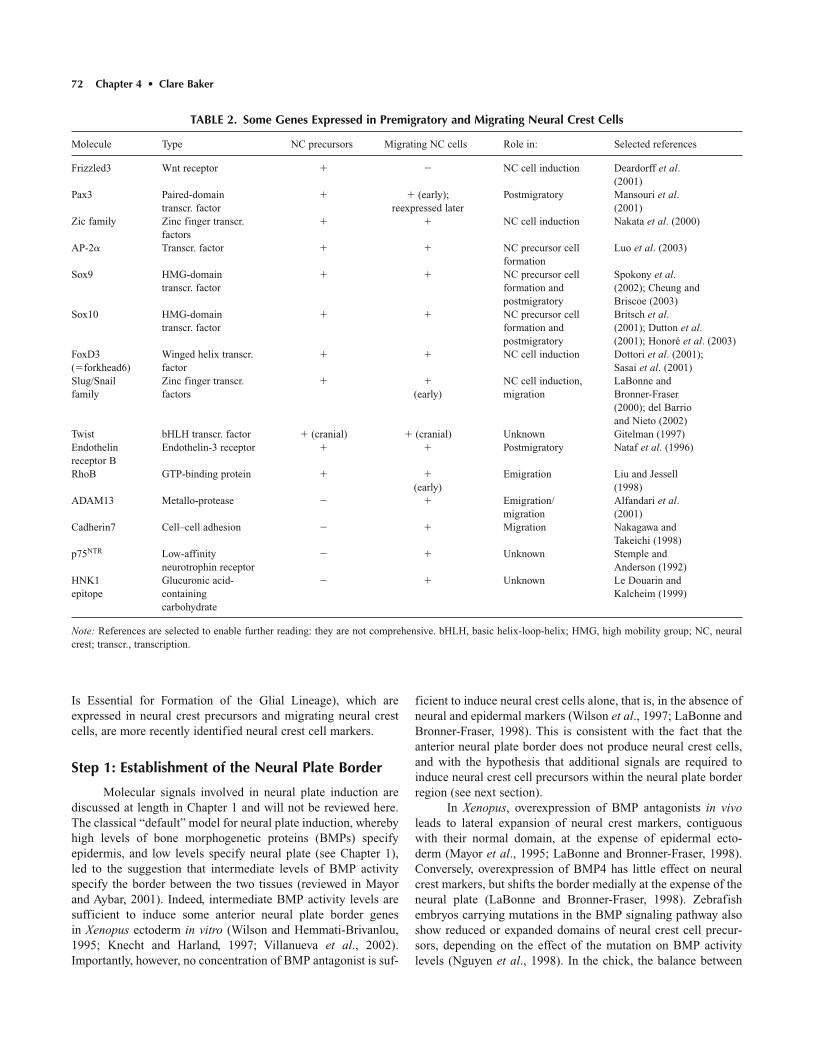
TABLE 2. Some Genes Expressed in Premigratory and Migrating Neural Crest Cells
Molecule Type NC precursors Migrating NC cells Role in: Selected references
Frizzled3 Wnt receptor � � NC cell induction Deardorff et al.(2001)
Pax3 Paired-domain � � (early); Postmigratory Mansouri et al.transcr. factor reexpressed later (2001)
Zic family Zinc finger transcr. � � NC cell induction Nakata et al. (2000)factors
AP-2� Transcr. factor � � NC precursor cell Luo et al. (2003)formation
Sox9 HMG-domain � � NC precursor cell Spokony et al.transcr. factor formation and (2002); Cheung and
postmigratory Briscoe (2003)Sox10 HMG-domain � � NC precursor cell Britsch et al.
transcr. factor formation and (2001); Dutton et al.postmigratory (2001); Honoré et al. (2003)
FoxD3 Winged helix transcr. � � NC cell induction Dottori et al. (2001); (�forkhead6) factor Sasai et al. (2001)Slug/Snail Zinc finger transcr. � � NC cell induction, LaBonne andfamily factors (early) migration Bronner-Fraser
(2000); del Barrioand Nieto (2002)
Twist bHLH transcr. factor � (cranial) � (cranial) Unknown Gitelman (1997)Endothelin Endothelin-3 receptor � � Postmigratory Nataf et al. (1996)receptor BRhoB GTP-binding protein � � Emigration Liu and Jessell
(early) (1998)ADAM13 Metallo-protease � � Emigration/ Alfandari et al.
migration (2001)Cadherin7 Cell–cell adhesion � � Migration Nakagawa and
Takeichi (1998)p75NTR Low-affinity � � Unknown Stemple and
neurotrophin receptor Anderson (1992)HNK1 Glucuronic acid- � � Unknown Le Douarin andepitope containing Kalcheim (1999)
carbohydrate
Note: References are selected to enable further reading: they are not comprehensive. bHLH, basic helix-loop-helix; HMG, high mobility group; NC, neuralcrest; transcr., transcription.

Neural Crest and Cranial Ectodermal Placodes • Chapter 4 73
BMP4 and its antagonists is important for establishing and/ormaintaining the prospective neural plate border: This region,which itself expresses BMP4, is the only region responsive tochanges in the level of BMP signaling at neural plate stages(Streit and Stern, 1999).
These results suggest that BMP signaling is required forneural plate border formation and maintenance, and that changesin BMP activity levels can affect neural crest cell formation,although they are not sufficient to induce neural crest cells.
Members of the Dlx family of transcription factors play animportant role in positioning the neural plate border during gastrulation (McLarren et al., 2003; Woda et al., 2003). In thechick, gain-of-function experiments have shown that Dlx5, itself amarker of the neural plate border, represses neural fates withoutinducing epidermis (McLarren et al., 2003). Furthermore, Dlx5acts non-cell autonomously (presumably by activating downstreamsignaling pathways) to promote the expression of other neuralplate border markers in adjacent cells, such as the transcriptionfactor Msx1, and BMP4 itself (McLarren et al., 2003). However,Dlx5 activity is not sufficient to induce either neural crest cells orplacodes (McLarren et al., 2003). In Xenopus, gain-of-functionand loss-of-function experiments have shown that Dlx3 and Dlx5activity positions the neural plate border, and that Dlx proteinfunction in non-neural ectoderm is required for the subsequentinduction of both neural crest and placodes (Woda et al., 2003).
In summary, the activity of BMP signaling molecules andDlx transcription factors appears to specify the neural plate bor-der region. However, the activity of these molecules is insuffi-cient to specify neural crest cells (or placode cells). IntermediateBMP activity levels induce neural plate border that is anterior incharacter. Hence, additional signals are required to posteriorizethe neural plate border and induce neural crest precursor cellswithin it.
Step 2: Induction of Neural Crest Precursors
It is becoming increasingly evident that Wnt and/or FGFfamily members are involved both in posteriorizing the neuralplate border and inducing neural crest precursor cells within it. These do seem to be separable processes, however, as neuralcrest induction can be experimentally uncoupled from the anterior–posterior patterning of the neural plate (e.g., Chang andHemmati-Brivanlou, 1998; Monsoro-Burq et al., 2003).
Posteriorizing Signals (Wnts and FGFs)
A posteriorizing signal derived from the paraxial meso-derm enables rostral neural plate tissue to form neural crest cellsin the chick (Muhr et al., 1997) and establishes Pax3 expressionat the neural plate border in both chick and Xenopus embryos(Bang et al., 1997, 1999). In the chick, this posteriorizing activity is mediated by Wnt family members, in particular Wnt8cand Wnt11, in conjunction with permissive FGF signaling(Nordström et al., 2002). Paraxial mesoderm produces severalother factors, including FGFs and retinoic acid, that are able toposteriorize the neural plate to induce posterior cell fates. In the
chick, though, FGFs and retinoic acid are insufficient to inducecaudal character in neural cells in vitro: This requires Wnt activityfrom the caudal paraxial mesoderm (Muhr et al., 1997, 1999;Nordström et al., 2002).
Induction of Neural Crest Precursors (Wnts and FGFs)
In both Xenopus and chick embryos, Wnt family membersare both sufficient to induce neural crest cells from neuralizedectoderm in vitro, and necessary for neural crest induction in vivo(reviewed in Wu et al., 2003). Wnts can induce neural crest mark-ers in conjunction with BMP inhibitors in ectodermal explantsin vitro (Saint-Jeannet et al., 1997; Chang and Hemmati-Brivanlou, 1998; LaBonne and Bronner-Fraser, 1998).Conversely, inhibiting Wnt function in vivo by overexpressing adominant negative Wnt ligand prevents early neural crest cellmarker expression (LaBonne and Bronner-Fraser, 1998).Morpholino oligonucleotide-mediated blockage of the transla-tion of the Wnt receptor Frizzled3, or its proposed adaptor pro-tein Kermit, both reduce Slug expression in Xenopus (Deardorffet al., 2001; Tan et al., 2001), again showing a requirement forWnt signaling in neural crest cell formation. Furthermore, theXenopus Slug promoter contains a functional binding site for adownstream effector of Wnt signaling (LEF/�-catenin) that isrequired to drive its expression in neural crest precursors, show-ing that the requirement for Wnt is direct (Vallin et al., 2001).
Wnt activity is also necessary and sufficient for neuralcrest cell induction in the chick (García-Castro et al., 2002).Overexpression of a dominant negative Wnt ligand inhibits Slugexpression in vivo: This can be rescued by application of exoge-nous Wnt (García-Castro et al., 2002). Conversely, DrosophilaWingless (a Wnt1 homologue that triggers the Wnt signalingpathway in vertebrates) can induce neural crest cells from neuralplate in a chemically defined medium that lacks any other growthfactors and hormones (García-Castro et al., 2002). Importantly,BMP4, which was previously shown to induce neural crest cellsfrom neural plate in vitro, in the presence of various additives(Liem et al., 1995), is unable to induce neural crest cells from the neural plate in their absence (García-Castro et al., 2002).Synergism with other factors present in the medium may alsounderlie the induction of neural crest cells by BMP2/4 from dis-sociated rat neural tube cells (Lo et al., 2002) or neuroepithelialstem cells (Mujtaba et al., 1998).
Wnt signaling seems to control the domain of expressionof Iro1 and Iro7, homeodomain transcription factors homologousto the Iroquois family of factors that, in Drosophila, regulate theexpression of proneural genes (section Proneural Genes: AnIntroduction) (Itoh et al., 2002). Functional knockdown of bothIro1 and Iro7 using morpholino antisense oligonucleotides leadsto loss of FoxD3 expression (Itoh et al., 2002). This not only suggests that these transcription factors are upstream of FoxD3,but also provides indirect evidence that Wnt signaling regulatesneural crest induction (Itoh et al., 2002). Furthermore, Wnt signaling is required for the induction of c-Myc, a basic helix-loop-helix zipper transcription factor whose expression is

74 Chapter 4 • Clare Baker
required for Slug and FoxD3 expression and neural crest cell for-mation in Xenopus (Bellmeyer et al., 2003).
The above results clearly show that Wnts are both neces-sary and sufficient to mediate neural crest cell induction fromneuralized ectoderm. Several different models of neural crestinduction have been proposed over the years, variously stressingthe importance of nonaxial mesoderm and neural plate–epidermal interactions. (Some of the data supporting a role forboth paraxial mesoderm and neural plate–epidermal interactionsin neural crest induction are described in the following sections.)However, since both paraxial mesoderm and epidermis expressWnt family members, it is likely that both tissues are involved in vivo. Wnt8 is expressed in the paraxial mesoderm, and Wnt6and Wnt7b are expressed in non-neural ectoderm (Chang andHemmati-Brivanlou, 1998; García-Castro et al., 2002).
Nonetheless, Wnts may not be the whole story. Work inXenopus has suggested that not only Wnt8, but also retinoic acidand FGFs, are able to induce Slug expression, both in the anteriorneural plate border, and in tissue transformed into anterior neuralplate border by intermediate levels of BMP activity (Villanuevaet al., 2002; Monsoro-Burq et al., 2003). Furthermore, FGFsignaling is required for induction of neural crest markers byparaxial mesoderm in Xenopus (Monsoro-Burq et al., 2003).Hence, although most of the evidence so far favors Wnts as theprimary signals that induce neural crest cell precursors within theneural plate border (see Wu et al., 2003), FGF involvementcannot be ruled out.
Evidence for Paraxial Mesoderm Involvement inNeural Crest Induction
Several lines of evidence have suggested a role for non-axial mesoderm in neural crest cell induction. In 1945, Ravenand Kloos showed in an amphibian model that fragments of lateral archenteron roof (prospective paraxial and lateral platemesoderm) can induce neural crest cells from overlying ecto-derm, in the absence of neural tissue, when grafted into the blastocoel (Raven and Kloos, 1945). Over fifty years later,prospective paraxial mesoderm was shown to induce neural crestmarker expression and melanocytes from competent ectoderm inXenopus explant cocultures (Bonstein et al., 1998; Marchantet al., 1998; Monsoro-Burq et al., 2003). In the chick, paraxialmesoderm can induce neural plate explants to form melanocytes(though not neurons) (Selleck and Bronner-Fraser, 1995). Hence,paraxial mesoderm is sufficient to induce at least some neuralcrest cell markers and derivatives in vitro, both from non-neuralectoderm and neural plate. Importantly, removing prospectiveparaxial mesoderm at the start of gastrulation in Xenopus leadsto a reduction in Slug expression and melanocyte formation in vivo (Bonstein et al., 1998; Marchant et al., 1998). This sug-gests that paraxial mesoderm is not only sufficient to induceneural crest cells in vitro, but also necessary for neural crest cellinduction in vivo.
The molecular model of neural crest induction describedthus far (i.e., intermediate BMP activity plus Wnt/FGF signaling)can explain the induction of neural crest cells by paraxial
mesoderm. Paraxial mesoderm expresses both BMP inhibitors,such as Noggin and Follistatin (e.g., Hirsinger et al., 1997;Marcelle et al., 1997; Liem et al., 2000), and Wnt and FGF fam-ily members (see previous section). The BMP inhibitors mayinduce intermediate levels of BMP activity in non-neural ecto-derm, while the Wnt/FGF signals may subsequently induceneural crest cells from this neuralized ectoderm. However, thismodel has not been tested directly.
Evidence for Non-Neural Ectoderm Involvementin Neural Crest Induction
A role for non-neural ectoderm in neural crest cell induction was first proposed in the late 1970s and early 1980s.Rollhäuser-ter Horst used interspecific grafts between differentspecies of urodele amphibians to follow the fate of gastrulaectoderm juxtaposed to different tissues (Rollhäuser-ter Horst,1979, 1980). The ectoderm failed to form neural crest cellsin vitro either when cultured alone, or when cocultured withneural-inducing tissue, but did form neural crest cells when bothtissues were grafted to the belly of host embryos (Rollhäuser-terHorst, 1979). This suggested a requirement for the host epider-mis as well as neural-inducing tissue. When the gastrula ecto-derm was grafted in place of the host neural folds, it also formedneural crest cells (Rollhäuser-ter Horst, 1980), again suggestinga role for interactions between neural and non-neural ectoderm inneural crest induction.
Moury and Jacobson similarly used pigmented and albinoaxolotl embryos as donors and hosts, respectively, to show thatboth neural folds and neural crest cells form at any newly createdboundary between neural plate and epidermis (Moury andJacobson, 1989, 1990). Under these circumstances, both epider-mis and neural plate form neural crest cells. Interestingly, theneural plate forms melanocytes while the epidermis forms sen-sory neurons (Moury and Jacobson, 1990). In Xenopus, labeledneural plate grafted into epidermis in vivo leads to Slug upregu-lation in both donor and host tissues, at the interface betweenthem (Mancilla and Mayor, 1996). Likewise, when quail neuralplate is grafted into chick epidermis in vivo, both quail and chicktissue generate migratory HNK-1-positive cells (Selleck andBronner-Fraser, 1995). Slug is also induced after similar experi-ments using unlabeled chick tissue (although in which tissues isunclear) (Dickinson et al., 1995).
Although these in vivo experiments suggested a role forinteractions between neural plate and epidermis in neural crestcell induction, all the grafted tissues were also exposed to signalsfrom the underlying mesoderm. However, in vitro cocultures ofneural plate and epidermis, in the absence of mesoderm, are suf-ficient to induce Slug expression in Xenopus (Mancilla andMayor, 1996) and neural crest cells in the chick (Slug expression;formation of melanocytes and catecholaminergic neurons)(Dickinson et al., 1995; Selleck and Bronner-Fraser, 1995).Hence, a local interaction between neural and non-neural ecto-derm is sufficient to induce neural crest cells in vitro. This finding has been exploited in a subtractive hybridization screenof a macroarrayed chick cDNA library, in order to provide the

Neural Crest and Cranial Ectodermal Placodes • Chapter 4 75
first gene expression profile of newly induced neural crest cells(Gammill and Bronner-Fraser, 2002).
The interaction between neural and non-neural ectodermseems to recapitulate all of the steps of neural crest inductionseen in vivo, including induction of the neural plate border, sinceneural folds form at all experimentally generated neural/epider-mal interfaces (Moury and Jacobson, 1989). Both epidermal andneural plate cells may contribute to the new neural plate borderregion, perhaps explaining why both tissues form neural crestcells after such interactions.
In summary, there is substantial evidence to implicate bothparaxial mesoderm and non-neural epidermis in neural crest cellinduction in vivo. Their involvement is probably due to their expres-sion of Wnt (and/or FGF) family members, which can induceneural crest cell precursors within the neural plate border region.
AP2� and SoxE Transcription Factors AreInvolved in the Earliest Steps of NeuralCrest Precursor Formation
The transcription factor AP2� is expressed during earlystages of neural crest development in all vertebrates, as well as inother tissues, such as the epidermis (see Luo et al., 2003). InXenopus, AP2� expression, which covers a broader territory thanother early neural crest precursor markers such as Sox9 (see nextparagraph) and Slug, is upregulated by BMP and Wnt signaling(Luo et al., 2003). Morpholino-mediated functional knockdownof AP2� results in failure of neural fold formation and the loss ofSox9 and Slug expression (Luo et al., 2003). These resultssuggest an important role for AP2� in the earliest stages ofneural crest precursor formation. However, the broad expressionpattern of AP2�, in particular in epidermis, implies that otherfactors must be involved in restricting neural crest precursorformation to the correct region.
Sox9 and Sox10 are members of the E subgroup of high-mobility-group (HMG) domain Sox transcription factors. Sox9 isone of the earliest markers of premigratory neural crest cell pre-cursors within the neural plate border; its expression is main-tained during early stages of neural crest migration (Spokonyet al., 2002; Cheung and Briscoe, 2003). Morpholino-mediatedfunctional knockdown of either Sox9 or Sox10 in Xenopusblocks neural fold formation, as well as blocking expression ofneural plate border markers and neural crest precursor markers,including Slug (Spokony et al., 2002; Honoré et al., 2003).Unlike Dlx activity (see section Establishment of the NeuralPlate Border), Sox9 activity is sufficient to induce neural crestprecursor markers, including Slug and FoxD3, in both dorsal andventral regions of the chick neural tube (Cheung and Briscoe,2003). However, Sox9-induced ectopic neural crest precursorsrarely delaminate except in the most dorsal regions of the neuraltube (Cheung and Briscoe, 2003). This suggests that additionalsignals are required for neural crest cell delamination, and thatthese signals are only present dorsally (see section Epithelial–Mesenchymal Transition).
Importantly, Sox9-mediated induction of neural crest pre-cursor markers in the chick does not induce BMP or Wnt family
members, nor require BMP activity, suggesting that, like AP2�,Sox9 lies downstream of these signaling pathways (Cheung andBriscoe, 2003). Blocking either FGF signaling or Wnt signalingin Xenopus also blocks Sox10 expression at the neural plateborder (Honoré et al., 2003), again suggesting that the SoxE tran-scription factors lie downstream of identifed neural crest precur-sor inducing signals.
Although morpholino-mediated functional knockdown ofSox9a in zebrafish does not affect neural crest precursors, it ispossible that Sox9b may instead play this role in zebrafish (Yanet al., 2002). Neural crest-specific knockout of Sox9 in mice doesnot cause neural crest precursor defects (Mori-Akayama et al.,2003), but it is possible that overlapping expression of the otherSoxE subgroup members, Sox8 and Sox10, may compensate forthe loss of Sox9.
In summary, it seems likely that AP2�, Sox9, and Sox10may be crucial downstream target of BMP and Wnt/FGF signalsin the formation of neural crest precursors. AP2� seems to lieupstream of Sox9, whose activity in turn induces the expressionof multiple other markers of neural crest cell precursors, includ-ing Slug and FoxD3 (see next section). However, delaminationfrom the neuroepithelium (i.e., neural crest cell formation)requires additional signals that, at least in the chick, may only bepresent in the dorsal neural tube.
Snail/Slug and FoxD3 Are Required for Neural Crest Precursor Formation
The Snail superfamily of zinc finger transcriptional repres-sors contains two major families: Snail and Scratch (Nieto,2002). In vertebrates, the Snail family is further subdivided intoSnail and Slug subfamilies, both of which are essential duringtwo stages of neural crest formation: (1) The formation of neural crest cell precursors within the neuroepithelium, and (2) delamination of cranial neural crest cells (section Snail FamilyMembers Promote Cranial Neural Crest Cell Delamination). InXenopus, Slug is first expressed at late gastrula stages, longbefore neural crest delamination occurs (Mayor et al., 1995).Slug acts as a transcriptional repressor (LaBonne and Bronner-Fraser, 2000; Mayor et al., 2000). Slug overexpression inXenopus leads to an expansion of the neural crest domain at theexpense of epidermis, and to overproduction of at least someneural crest derivatives (LaBonne and Bronner-Fraser, 1998).Conversely, other early neural crest precursor markers are lostafter expression of a dominant negative Slug construct or anti-sense Slug RNA, showing that Slug function is necessary for theformation of neural crest precursors (Carl et al., 1999; LaBonneand Bronner-Fraser, 2000). However, not all Slug-expressingcells delaminate to form neural crest cells (Linker et al., 2000).
The winged-helix transcription factor FoxD3 (Forkhead6)is also important in early stages of neural crest cell formation(Dottori et al., 2001; Kos et al., 2001; Pohl and Knöchel, 2001;Sasai et al., 2001). Like Slug, FoxD3 is a transcriptional repres-sor (Pohl and Knöchel, 2001; Sasai et al., 2001) and is expressedboth in premigratory neural crest cell precursors and migratingneural crest cells. In Xenopus, inhibiting FoxD3 function in vivo

76 Chapter 4 • Clare Baker
using a dominant negative FoxD3 construct represses the expression of early neural crest precursor markers, includingSlug, and leads to a corresponding expansion of the neural plate(Sasai et al., 2001). Hence, like Slug, FoxD3 function is requiredfor the formation of neural crest precursors. However, overex-pression of FoxD3 in the chick neural tube does not upregulateSlug, suggesting that Slug is not an obligate downstream target ofFoxD3 (Dottori et al., 2001). Instead, the two genes seem to actin concert, in partially overlapping pathways, to promote neuralcrest cell formation (Sasai et al., 2001).
In addition to their importance for the formation of neuralcrest cell precursors, both FoxD3 and Slug can promote neural crest cell delamination (sections FoxD3 Promotes NeuralCrest Cell Delamination at All Axial Levels; Snail FamilyMembers Promote Cranial Neural Crest Cell Delamination).
Step 3: Epithelial–Mesenchymal Transition
The final step in neural crest induction is the activation ofthe epithelial–mesenchymal transition that leads to delaminationfrom the neuroepithelium into the periphery. As described at thebeginning of the section on Neural Crest Induction, a cell cannotbe described as a bona fide neural crest cell until it emigratesfrom the neuroepithelium. Hence, induction of delamination isthe final step in the induction of the neural crest.
In all vertebrates, neural crest cell precursors delaminate ina rostrocaudal wave along the neuraxis. Whether or not neuralcrest cell precursors are initially incorporated into the neural tubedepends on the timing of neural crest cell delamination relativeto the timing of fusion of the neural folds. This varies fromspecies to species and on the axial level within the embryo.Cranial neural crest cells, in particular, which are the first todelaminate, may not be incorporated into the neural tube. In themouse, cranial neural crest delamination begins in the midbrain/rostral hindbrain well before neural tube closure, when the neural folds are approaching one another in the cervical region(Nichols, 1981). In frogs, cranial neural crest cells form largemasses that segregate from the neural tube prior to its closure;these masses do not take part in the morphogenetic movements ofneurulation (Schroeder, 1970; Olsson and Hanken, 1996). In thechick, however, cranial neural crest cells delaminate as the neuralfolds meet or during early apposition, beginning at midbrain levels(Tosney, 1982). Trunk neural crest cells in the chick only emigrateafter the epidermis and neural tube have separated (Tosney, 1978).
In the chick, the first sign of imminent neural crest celldelamination at cranial levels is that the neural crest cell precur-sor cell population becomes less tightly packed, and the cellsextend long cellular processes into the intercellular spaces withinthe population (Tosney, 1982). As emigration starts, the basallamina over the neural crest cells becomes fragmented, and thecells extend long processes into the adjacent cell-free space(Tosney, 1982). Clearly, major changes in cytoskeletal architec-ture, cell–cell and cell–matrix interactions occur during thisepithelial–mesenchymal transition. Recent molecular evidencehas given us a more detailed insight into the genes and signalingpathways controlling these processes.
The Basal Lamina Must Be Degraded beforeDelamination Can Occur
Neural crest cells do not seem to be able to penetrate anintact basal lamina (Erickson, 1987). The basal lamina clearlybreaks down over neural crest cell precursors before they delam-inate from the neuroepithelium (e.g., Tosney, 1982; Raible et al.,1992) and this may be due to neural crest secretion of proteases,although it remains to be demonstrated. Neural crest cell precur-sors produce various proteolytic enzymes, including the serineprotease plasminogen activator (Valinsky and Le Douarin, 1985;Agrawal and Brauer, 1996), BMP1/Tolloid metalloproteases(Martí, 2000), and members of the metalloprotease/disintegrinfamily (Alfandari et al., 1997; Cai et al., 1998). Some of theseproteases are only found in cranial neural crest cell precursorsand migrating cranial neural crest cells, for example, the metal-loprotease/disintegrin ADAM13 in Xenopus (Alfandari et al.,1997, 2001). However, a role for these proteases in neural crestcell delamination has not yet been shown.
Inhibiting Protein Kinase C Signaling Promotes Delamination
If avian neural tube explants are treated with protein kinaseC inhibitors, cells immediately, and precociously, delaminate andmigrate away from the neural tube (Newgreen and Minichiello,1995, 1996). This occurs on both dorsal and ventral sides of theneural tube (although ventral cells are less sensitive than dorsalcells) (Newgreen and Minichiello, 1995, 1996). This stimulatoryeffect of protein kinase C inhibitors does not require protein syn-thesis (Newgreen and Minichiello, 1995). Similarly, proteinkinase C inhibition triggers delamination, migration, and expres-sion of the neural crest marker Sox10, in neuroectoderm cellsproduced from mouse embryonic stem cells in culture (Rathjenet al., 2002). These results suggest that delamination can beinduced by signals that modulate protein kinase C activity.
Delamination is Associated with Downregulationof Cadherin6B
Calcium-dependent cell–cell adhesions are required toprevent precocious emigration of neural crest cells (Newgreenand Gooday, 1985). In the chick, most neural tube cells expressthe calcium-dependent cell–cell adhesion molecule N-cadherin,while epidermal cells express E-cadherin; however, the dorsalneural tube, which contains neural crest cell precursors,expresses neither N-cadherin nor E-cadherin (Akitaya andBronner-Fraser, 1992). In accordance with this, N-cadherin itselfdoes not seem to be required for neural crest cell formation ormigration, as pigmentation and cranial cartilages are normal in N-cadherin mutant zebrafish (Lele et al., 2002). Instead,neural crest cell precursors within the neuroepithelium expresscadherin6B; this expression is lost in emigrating neural crestcells (Nakagawa and Takeichi, 1995, 1998). Type II (atypical)cadherins are then upregulated in subpopulations of migratingneural crest cells, for example cadherin7 and cadherin 11; these

Neural Crest and Cranial Ectodermal Placodes • Chapter 4 77
may be involved in controlling the rate of neural crest cell migra-tion and/or in some aspects of fate specification (Nakagawa andTakeichi, 1995; Borchers et al., 2001).
FoxD3 Promotes Neural Crest Cell Delaminationat All Axial Levels
FoxD3 is essential for the formation of neural crest cell pre-cursors (section Snail/Slug and FoxD3 Are Required for NeuralCrest Precursor Formation), and it may also play a role in neuralcrest cell delamination. Ectopic expression of FoxD3 in the chickneural tube promotes neural crest cell delamination at all axiallevels (Dottori et al., 2001). This is achieved without upregulatingSlug or, apparently, RhoB (section BMP4 Induces RhoB, Which IsEssential for Neutral Crest Cell Delamination), suggesting thatFoxD3 and Slug function independently in regulating neural crestcell delamination (Dottori et al., 2001). The precise mechanism ofaction of FoxD3 in promoting delamination remains unclear.
Snail Family Members Promote Cranial NeuralCrest Cell Delamination
Snail family transcription factors are required for the formation of neural crest cell precursors (section Snail/Slug andFoxD3 Are Required for Neural Crest Precursor Formation).Several different lines of evidence also support a role for Snailfamily genes in epithelial–mesenchymal transitions. Over-expression of mouse Slug in bladder carcinoma cells leads todesmosome dissociation at sites of cell–cell contact, a necessaryprerequisite for epithelial–mesenchymal transition (Savagner et al.,1997). Overexpression of mouse Snail in epithelial cells repressestranscription of the cell–cell adhesion molecule E-cadherin, andleads to epithelial–mesenchymal transition and migratory and invasive cell behaviors (Batlle et al., 2000; Cano et al.,2000). Since Snail and/or Slug genes are expressed in premigra-tory neural crest cell precursors in all vertebrates, a role in neuralcrest cell delamination from the neuroepithelium seems likely.
Early antisense experiments in chick embryos suggested arole for Slug in cranial neural crest cell migration (Nieto et al.,1994). Cranial neural crest cell migration is inhibited in Xenopusin the presence of antisense Slug RNA or a dominant negativeSlug construct (Carl et al., 1999; LaBonne and Bronner-Fraser,2000). Overexpression of Slug in the chick neural tube leads to anincrease in the number of migrating cranial neural crest cells,although not of trunk neural crest cells (del Barrio and Nieto,2002). Other experiments have also shown that, unlike FoxD3,increased Slug activity alone does not cause trunk neural crest celldelamination in the trunk (Sela-Donenfeld and Kalcheim, 1999).The basis of this difference between head and trunk is unknown.
BMP Signaling is Required for Delamination
In the trunk of the chick embryo, neural crest cells onlybegin to delaminate in areas adjacent to the epithelial somites:They do not emigrate at the level of the segmental plate mesoderm (Teillet et al., 1987). The timing of neural crest cellemigration in the trunk can be correlated with expression of the
BMP2/4 antagonist Noggin (Sela-Donenfeld and Kalcheim,1999). Noggin is strongly expressed in the dorsal neural tubeopposite the segmental plate mesoderm, more weakly expressedopposite newly epithelial somites, and absent opposite fully disso-ciated somites, while BMP4 is expressed in the dorsal neural tubeat all levels (Sela-Donenfeld and Kalcheim, 1999). Noggin over-expression (i.e., inhibition of BMP activity) inhibits neural crestcell delamination both in vivo and in vitro, and this can be rescuedin vitro by BMP4 (Sela-Donenfeld and Kalcheim, 1999). Thissuggests that a balance between BMP4 and its antagonists plays arole in the onset of neural crest cell delamination in the trunk(Sela-Donenfeld and Kalcheim, 1999). This balance is now knownto be controlled by the paraxial mesoderm itself: The dorsomedialregion of developing somites produces a signal that downregulatesnoggin transcription in the dorsal neural tube (Sela-Donenfeldand Kalcheim, 2000). This enables the coordination of neuralcrest cell emigration with the formation of a suitable mesodermalsubstrate for migration (section Migration Pathways of TrunkNeural Crest Cells) (Sela-Donenfeld and Kalcheim, 2000).
BMP signaling is also essential for cranial neural crest cellmigration in the mouse (Kanzler et al., 2000). When noggin isexpressed in transgenic embryos under the control of a Hox2aenhancer, leading to noggin overexpression in the hindbrain,hindbrain-level neural crest cells fail to emigrate (Kanzler et al.,2000). Although Bmp4 is not expressed in the dorsal hindbrain inthe mouse, Bmp2 is expressed there, and hindbrain neural crestcells fail to migrate in Bmp2 mutant embryos. Hence, it seemsthat BMP2 activity is necessary for cranial neural crest cellemigration in the mouse (Kanzler et al., 2000).
These results show that BMP signaling is essential not justto establish the neural plate border, but also at a later stage, topromote neural crest cell delamination.
BMP4 Induces RhoB, Which Is Essential for Neural Crest Cell Delamination
The small GTP-binding protein RhoB is expressed inneural crest precursors within the neuroepithelium and is down-regulated shortly after delamination (Liu and Jessell, 1998). Rhoproteins have been implicated in the assembly of the actincytoskeleton required for motility (see Frame and Brunton,2002). Treatment of chick neural tube explants with a Rho-specific inhibitor has shown that Rho function is essential forneural crest cell delamination, and that the actin cytoskeleton inneural crest cell precursors is perturbed (Liu and Jessell, 1998).RhoB also seems to be a downstream target of Slug activity,though whether direct or indirect is unknown (del Barrio andNieto, 2002). It is not, however, detectably induced by FoxD3(Dottori et al., 2001). Nor, interestingly, is RhoB detectablyinduced by Sox9, which induces neural crest precursor formationbut is not sufficient to promote efficient delamination, except at the dorsalmost region of the neural tube (Cheung and Briscoe,2003; section AP2� and SoxE Transcription Factors Are Involvedin the Earliest Steps of Neural Crest Precursor Formation).However, RhoB is induced by BMP4: Indeed, it was originallyidentified in a PCR-based screen for genes induced by BMP4 in

78 Chapter 4 • Clare Baker
neural plate cells (Liu and Jessell, 1998). Since BMP4 is essen-tial for delamination of neural crest cell precursors and inducesRhoB, it seems that BMP4 activity is the most likely candidatefor the dorsally located signal that induces neural crest cell for-mation from premigratory neural crest cell precursors. It will beimportant to establish whether all RhoB-expressing neural crestcell precursor cells do, in fact, emigrate from the neural tube.
Transition from G1 to S Phase of the Cell Cycle IsRequired for Neural Crest Cell Delamination
In the chick, most trunk neural crest cells emigrate fromthe neural tube in the S phase of the cell cycle, when their nucleiare located at or near the basal margin of the neuroepithelium(Burstyn-Cohen and Kalcheim, 2002). Blocking the cell cycletransition from G1 to S phase blocks neural crest delamination,both in vivo and in explants (Burstyn-Cohen and Kalcheim,2002). Thus, the cell cycle status of neural crest cell precursors isan essential prerequisite for the epithelial–mesenchymal transi-tion that forms neural crest cells. It is possible that BMP signal-ing in the dorsal neural tube induces a cascade of signals thatinfluence G1/S transition, perhaps by upregulating cyclin D1.Alternatively, independent pathways downstream of BMP signal-ing and the cell cycle may converge on common downstream targets to initiate delamination.
Summary of Neural Crest Induction
Neural crest induction is a multistep, multisignal processthat can be divided into three distinct phases. Firstly, the neuralplate border is induced during gastrulation, probably by interme-diate levels of BMP activity, and with the involvement of Dlxtranscription factors. Secondly, Wnt and/or FGF signals fromsurrounding tissues (paraxial mesoderm and non-neural ecto-derm) posteriorize the neural plate border and induce neural crestcell precursors within it. Finally, BMP activity in the dorsalneural tube induces RhoB in a subset of neural crest cell precur-sors. After G1/S transition, these cells undergo an epithelial–mesenchymal transition, delaminate from the neuroepithelium asneural crest cells, and migrate into the periphery.
As neural crest cells delaminate from the neuroepithelium,they are faced with very different mesodermal environmentsdepending on their axial level. In the head, they encounter theapparently disorganized cranial paraxial mesenchyme, while in thetrunk, the paraxial mesoderm is segmented into repeating blocks,the somites. In both head and trunk, however, neural crest cells fol-low ordered pathways to their target sites, where they differentiateinto an impressive array of different derivatives. The mechanismsunderlying this migration are discussed in the following section.
NEURAL CREST MIGRATION
Experimental Approaches
Two main experimental approaches have been used to mapthe migration pathways and, concurrently, define the derivatives
of the neural crest. First, ablation studies have been performed, todetermine what cell types and tissues are lacking as a result.Although such experiments yielded a wealth of information,particularly from fish and amphibians, drawbacks included thepossibility of regulation to restore the missing cells, and indirecteffects on other tissues. The second approach has been to labelthe neural folds, including premigratory neural crest cell precur-sors: Labeled neural crest cells delaminating into the peripherycan be distinguished from surrounding unlabeled cells. Earlystudies in amphibian embryos employed vital dyes to label donorembryos, from which neural folds were explanted and graftedinto unlabeled host embryos (e.g., Detwiler, 1937). Hetero-specific grafts were also used extensively in amphibians as dif-ferences in pigmentation and/or cell size enabled donor and hosttissues to be distinguished. Such grafts have also been combinedwith staining techniques that reveal differences in nuclearmorphology (e.g., Sadaghiani and Thiébaud, 1987; Krotoskiet al., 1988).
Tritiated thymidine labeling of the nuclei of donorembryos, followed by grafting of labeled neural folds into unla-beled hosts, was introduced in the 1960s for the chick (Weston,1963) and immediately applied in amphibians (Chibon, 1964).This method was used in avian embryos for about 12 years (e.g., Johnston, 1966; Noden, 1975). It was superseded, however,by Le Douarin’s discovery that the quail nucleolus is associatedwith a large mass of heterochromatin, enabling it to be distin-guished clearly from chick nuclei after appropriate staining (Le Douarin, 1969, 1973). Hence, quail neural folds could begrafted into chick hosts, and the fate of the donor quail cells followed throughout development, up to and including hatching(although graft rejection occurs eventually). This technique wasused in a series of elegant fate-mapping studies to define all thederivatives of the neural crest in the avian embryo along thelength of the neuraxis (e.g., Le Douarin and Teillet, 1973, 1974;Teillet, 1978; Noden, 1978a, b) (reviewed in Le Douarin andKalcheim, 1999). Today, a quail-specific antibody enables easieridentification of grafted quail cells within the chick host, and thequail–chick chimera technique is still commonly used to studyneural crest cell fate, migration, and potential (e.g., Baker et al.,1997; Catala et al., 2000; Etchevers et al., 2001).
Migrating neural crest cells have also been followed usingmonoclonal antibodies, such as the HNK1 antibody in chick andrat embryos (e.g., Rickmann et al., 1985; Bronner-Fraser, 1986;Erickson et al., 1989). Modern, nontoxic vital dyes have beenextensively used to map neural crest cell migration pathways andderivatives in situ, avoiding any risk of artifacts introduced byinvasive surgery or differences in behavior between donor and host cells. The lipophilic dye DiI can be injected into thelumen of the neural tube to label all neural tube cells, includingpremigratory neural crest cells, which can subsequently be fol-lowed as they migrate through the periphery (e.g., Serbedzijaet al., 1989, 1990; Collazo et al., 1993). Time-lapse in ovo con-focal microscopy, combined with DiI labeling, has also enabledmigrating hindbrain neural crest cells to be followed in vivo athigh resolution (e.g., Kulesa and Fraser, 2000). Membrane-impermeant dyes, such as lysinated rhodamine dextran, can be

Neural Crest and Cranial Ectodermal Placodes • Chapter 4 79
injected into individual neural crest cell precursors and migratingneural crest cells in vivo, allowing the progeny of single cells tobe followed during development (Bronner-Fraser and Fraser,1988, 1989; Fraser and Bronner-Fraser, 1991). Retroviral-mediated gene transfer has also enabled the clonal analysis of theprogeny of single neural crest cells in vivo (Frank and Sanes,1991). In mice, the fate of migrating cranial neural crest cells hasbeen followed by using Cre–Lox transgenic technology to acti-vate constitutive �-galactosidase expression under the control ofthe Wnt1 promoter (Chai et al., 2000).
Together, these different cell-labeling approaches haveenabled a detailed picture to be drawn of the migration pathwaysfollowed by neural crest cells through the periphery.
Migration Pathways of Cranial Neural Crest Cells
Cranial neural crest cells migrate beneath the surfaceectoderm, above the paraxial cephalic mesoderm (see Figs. 3 and4B), although a few cells penetrate the paraxial mesoderm.
FIGURE 3. Schematic lateral views of a generalized 20–30 somite-stage amniote embryo with the surface ectoderm removed (except to show the positionsof the cranial ectodermal placodes). Each tissue type from the embryo at the top is shown separately below, illustrating the relative positions of the migratingneural crest, placodes (filled black circles), axial structures, paraxial mesoderm, arteries, and pharyngeal endoderm. The olfactory placodes cannot be seen inthis view. The vertical lines indicate which regions are in register with each pharyngeal arch. Redrawn from Noden (1991). art., artery; fb, forebrain; gen,geniculate; ln, lens; mb, midbrain; mmV, maxillomandibular trigeminal; nod, nodose; opV, ophthalmic trigeminal; pet, petrosal.

80 Chapter 4 • Clare Baker
They migrate as coherent populations; indeed, at the hindbrainlevel, migrating neural crest cells are connected in chains byfilopodia (Kulesa and Fraser, 1998, 2000). They populate theentire embryonic head and form much of the neurocranium(brain capsule) and all of the splanchnocranium (viscerocraniumor visceral skeleton), that is, the skeleton of the face and pharyn-geal arches. They also form neurons and satellite glia in cranialsensory and parasympathetic ganglia, Schwann cells, endocrinecells, and epidermal pigment cells (see Table 1).
Pharyngeal Arches and Neural Crest Streams
The patterning of cranial neural crest cell migration is inti-mately bound up with the segmental nature of both the hindbrain(rhombomeres; see Chapter 3) and the periphery (pharyngealarches). Pharyngeal arches are also known as branchial arches,from the Latin branchia (“gill”), because in aquatic vertebrates the more caudal arches are associated with gills.However, “pharyngeal” is the more appropriate term, because allarches form in the pharynx, but not all arches support gills.Pharyngeal arches form between the pharyngeal pouches, whichare outpocketings of the pharyngeal (fore-gut) endoderm thatfuse with the overlying ectoderm to form slits in the embryo (seeFig. 3). The pharyngeal slits form the gill slits in aquatic verte-brates; the first pharyngeal slit in tetrapods forms the middle earcavity. Paraxial mesoderm in the core of the pharyngeal arches(Figs. 4B, C) gives rise to striated muscles. Cranial neural crestcells migrate subectodermally to populate the space around themesodermal core (Figs. 4B, C), where they give rise to all skele-tal elements of the arches, and the connective component of thestriated muscles.
The first pharyngeal arch is the mandibular, which formsthe mandible (lower jaw). The second arch is the hyoid, whichforms jaw suspension elements in fish but middle ear bones intetrapods, together with parts of the hyoid apparatus/bone (sup-porting elements for the tongue and roof of the mouth). Varyingnumbers of arches follow more caudally. The third and fourth
arches also contribute to the hyoid apparatus and to laryngeal car-tilages in tetrapods; in mammals, the fourth arch forms thyroidcartilages. More caudal arches in fish and aquatic amphibianssupport gills and form laryngeal cartilages in tetrapods.Importantly, pharyngeal arch formation per se, and the regional-ization of gene expression patterns within them (excluding thoseof neural crest-derived structures) are both independent of neuralcrest cell migration (Veitch et al., 1999; Gavalas et al., 2001).
Cranial neural crest cells migrate in characteristic streamsassociated with the pharyngeal arches (Figs. 3 and 4A). There arethree or more major migration streams in all vertebrates. Thefirst stream, from the midbrain and rhombomeres 1 and 2 (r1,2),populates the first (mandibular) arch; the second stream, fromr3–5, populates the second (hyoid) arch, and the third, from r5–7,populates the third arch (Fig. 4). In fish and amphibians, addi-tional caudal streams populate the remaining arches: The axolotl,for example, has four branchial (gill) arches caudal to themandibular and hyoid arches (Fig. 4A). How is the migratingneural crest cell population sculpted to achieve these differentstreams?
Separation of the First, Second, and Third NeuralCrest Streams (Amniotes)
In chick and mouse embryos, there are neural crest cell-free zones adjacent to r3 and r5 (Fig. 3). It was suggested thatneural crest cells at r3 and r5 die by apoptosis to generate adja-cent neural crest-free zones (Graham et al., 1993). However, bothr3 and r5 give rise to neural crest cells during normal develop-ment in both chick and mouse, though r3 generates fewer neuralcrest cells than other rhombomeres (Sechrist et al., 1993;Köntges and Lumsden, 1996; Kulesa and Fraser, 1998; Trainoret al., 2002b). Neural crest cells from r3 and r5 migrate rostrallyand caudally along the neural tube to join the adjacent neuralcrest streams; that is, r3-derived neural crest joins the r1,2 (firstarch) and r4 (second arch) streams, while r5-derived neural crestjoins the r4 (second arch) and r6,7 (third arch) streams (Sechrist
FIGURE 4. Cranial neural crest migration streams in the axolotl visualized by in situ hybridization for the AP-2 gene. (A) Stage 29 (16-somite stage) axolotlembryo showing six AP-2� neural crest migration streams in the head (mandibular, hyoid, and four branchial streams). Premigratory trunk neural crest cellprecursors can be seen as a dark line at the dorsal midline of the embryo. (B) Transverse section through a stage 26 (10–11 somite stage) axolotl embryo show-ing AP-2� neural crest cells (NC) moving out from the neural tube (nt) and down to surround the mesodermal core of the mandibular arch. (C) Horizontalsection through the pharynx of a stage 34 (24–25 somite stage) axolotl embryo showing AP-2� neural crest cells (NC) around the mesodermal cores of eachpharyngeal arch. e, eye; mb, midbrain; mes., mesodermal; NC, neural crest; nt, neural tube; ov, otic vesicle; ph, pharynx. Staging follows Bordzilovskaya et al.(1989). All photographs courtesy of Daniel Meulemans, California Institute of Technology, United States of America.

Neural Crest and Cranial Ectodermal Placodes • Chapter 4 81
et al., 1993; Köntges and Lumsden, 1996; Kulesa and Fraser,1998; Trainor et al., 2002b). This deviation of the r3 and r5neural crest generates the neural crest-free zones adjacent to r3and r5, forming the three characteristic streams in birds and mice(Fig. 3). Hence, the first arch is populated by neural crest cellsfrom the midbrain and r1–3, the second arch by neural crest cellsfrom r3–5, and the third arch by neural crest cells from r5–7.
Neural crest cells leaving r5 are confronted by the oticvesicle (Fig. 3), which provides an obvious mechanical obstacleto migration. No such obstacle exists at r3; instead, paraxialmesoderm at the r3 level is inhibitory for neural crest cell migra-tion, at least in amniotes (Farlie et al., 1999). This inhibition islost in mice lacking ErbB4, a high-affinity receptor for thegrowth factor Neuregulin1 (NRG1) (Golding et al., 1999, 2000).ErbB4 is expressed in the r3 neuroepithelium, while NRG1 isexpressed in r2; ErbB4 activation by NRG1 may somehow signalthe production of inhibitory molecules in r3-level paraxial meso-derm (Golding et al., 2000). A few hours after removing either r3itself, or the surface ectoderm at the r3 level, r4 neural crest cellsmove aberrantly into the mesenchyme adjacent to r3, suggestingthat both r3 itself and r3-level surface ectoderm are necessary toinhibit neural crest cell migration (Trainor et al., 2002b).
Separation of the Third and Fourth Streams(Anamniotes)
Fish and amphibians also have additional cranial neuralcrest streams that populate the more caudal pharyngeal arches. Inamphibians, at least, neural crest cells destined for differentarches do not separate into different streams adjacent to theneural tube; instead, separation occurs at or just before entry intothe arches (Robinson et al., 1997). Another difference inXenopus, in which the otic vesicle is adjacent to r4 rather than r5,is that all r5-derived neural crest cells seem to migrate into thethird arch (Robinson et al., 1997).
In Xenopus, migrating neural crest cells in the third andfourth cranial neural crest streams are separated by repulsivemigration cues. These are mediated by the ephrin family of ligands, acting on their cognate Eph-receptor tyrosine kinases(Smith et al., 1997; Helbling et al., 1998; reviewed in Robinsonet al., 1997; for a general review of ephrins and Eph family mem-bers, see Kullander and Klein, 2002). The transmembrane ligandephrinB2 is expressed in second arch neural crest cells and meso-derm. One ephrinB2 receptor, EphA4, is expressed in third archneural crest cells and mesoderm, while a second ephrinB2 receptor, EphB1, is expressed in both third and fourth arch neuralcrest cells and mesoderm (Smith et al., 1997). Inhibition ofEphA4/EphB1 function using truncated receptors results in theaberrant migration of third arch neural crest cells into the secondand fourth arches. Conversely, ectopic activation of EphA4/EphB1(by overexpressing ephrinB2) results in the scattering of third archneural crest cells into adjacent territories (Smith et al., 1997).Hence, the complementary expression of ephrinB2 and its recep-tors in the second and third arches, respectively, is required to pre-vent mingling of second and third arch neural crest cells beforethey enter the arches. Since ephrinB2 is also expressed in second
arch mesoderm, it is also required to target third arch neural crestcells correctly away from the second arch and into the third arch.EphrinB2-null mice also show defects in cranial neural crest cellmigration, particularly of second arch neural crest cells, whichscatter and do not invade the second arch (Adams et al., 2001).
Migrating Xenopus cranial neural crest cells also expressEphA2; overexpression of a dominant negative (kinase-deficient)EphA2 receptor similarly leads to the failure of the third andfourth neural crest streams to separate, as neural crest cells fromthe third stream migrate posteriorly (Helbling et al., 1998).
Neural Crest Streams and Cranial Skeleto-Muscular Patterning
Cranial neural crest cells form not only many of the skeletalelements of the head, but also the connective component of thestriatal muscles that are attached to them (see Table 1). When thelong-term fate of neural crest cells arising from the midbrain andeach rhombomere was mapped using quail-chick chimeras, itwas found that each rhombomeric population forms the connec-tive components of specific muscles, together with their respec-tive attachment sites on the neurocranium and splanchnocranium(Köntges and Lumsden, 1996). Cranial muscle connective tissuesarising from a given rhombomere attach to skeletal elements aris-ing from the same initial neural crest population, explaining howevolutionary changes in craniofacial skeletal morphology can beaccommodated by the attached muscles (Köntges and Lumsden,1996). Similar results have also been obtained in frog embryos,where connective tissue components of individual muscles ofeither of the first two arches originate from the neural crestmigratory stream associated with that arch (Olsson et al., 2001).Hence, the streaming of cranial neural crest cells into the differentpharyngeal arches is important for patterning not only skeletalelements, but also their associated musculature.
Migration Pathways of Trunk Neural Crest Cells
The migration pathways of trunk neural crest cells havebeen most extensively studied in avian embryos (e.g., Weston,1963; Rickmann et al., 1985; Bronner-Fraser, 1986; Teillet et al.,1987). As described in this section, neural crest cells only leavethe neural tube opposite newly epithelial somites (Fig. 5A) (forreviews of somite formation and maturation, see Stockdale et al.,2000; Pourquié, 2001). Here, they enter a cell-free space that isrich in extracellular matrix. They only migrate into the somites at a level approximately 5–9 somites rostral to the last-formedsomite, where the somites first become subdivided into differentdorsoventral compartments, the sclerotome and dermomyotome(Fig. 5B) (Guillory and Bronner-Fraser, 1986). The sclerotome is formed when the ventral portion of the epithelial somite undergoes an epithelial–mesenchymal transition to form loosemesenchyme. This mesenchyme will eventually form the cartilage and bone of the ribs and axial skeleton. The dorsalsomitic compartment, the dermomyotome, remains epithelial,and will eventually form dermis, skeletal muscle, and vascularderivatives.

82 Chapter 4 • Clare Baker
There are two main neural crest cell migration pathways inthe avian trunk (Fig. 5C): (1) a ventral pathway between the neuraltube and somites, followed by neural crest cells that eventuallygive rise to dorsal root ganglia, Schwann cells, sympathetic gan-glia, and (at somite levels 18–24 in birds) adrenal chromaffincells, and (2) a dorsolateral pathway between the somite and theoverlying ectoderm, followed by neural crest cells that eventuallyform melanocytes.
Ventral Migration Pathway
In the chick, neural crest cells that delaminate oppositeepithelial somites initially migrate ventrally between the somites.Once the sclerotome forms, they migrate exclusively through therostral half of each sclerotome, leading to a segmental pattern of
migration (Rickmann et al., 1985; Bronner-Fraser, 1986). Thispathway is almost identical to that followed by motor axons asthey grow out from the neural tube, shortly after neural crest cellsbegin their migration (Rickmann et al., 1985). Mouse neuralcrest cells are similarly restricted to the rostral sclerotome(Serbedzija et al., 1990).
Neural crest cells that remain within the rostral sclerotomeaggregate to form the dorsal root ganglia (primary sensoryneurons and satellite glial cells), while those that move furtherventrally form postganglionic sympathetic neurons (Fig. 8;section The Autonomic Nervous System: An Introduction) andadrenal chromaffin cells (Fig. 5C). The restriction of neural crestcells to the rostral half of each somite therefore leads to the seg-mental distribution of dorsal root ganglia; as will be seen in thesection on Molecular Guidance Cues for Trunk Neural Crest CellMigration, it results from the presence of repulsive migrationcues in the caudal sclerotome.
Neural crest cells that delaminate opposite the caudal halfof a somite migrate longitudinally along the neural tube in bothdirections. Once they reach the rostral half either of their ownsomite, or of the adjacent (immediately caudal) somite, theyenter the sclerotome (Teillet et al., 1987). Hence, each dorsal rootganglion is derived from neural crest cells emigrating at the same somite level and from one somite anterior to that level. Incontrast, each sympathetic ganglion is derived from neural crestcells originating from up to six somite-levels of the neuraxis:This is approximately equal to the numbers of spinal cord seg-ments contributing to the preganglionic sympathetic neurons thatinnervate each ganglion (see Fig. 8) (Yip, 1986).
There are some differences in the ventral neural crest migra-tion pathway between different vertebrates. In fish and amphib-ians, the somites are mostly myotome, with very little sclerotome.In these animals, the ventral migration pathway is essentially amedial migration pathway, between the somites and the neuraltube/notochord. In Xenopus, neural crest cells following this pathway give rise to dorsal root ganglia, sympathetic ganglia,adrenomedullary cells, and also pigment cells (Krotoski et al.,1988; Collazo et al., 1993). This is also a segmental migration,but in this case, the neural crest cells migrate between the neuraltube and the caudal half of each somite (Krotoski et al., 1988;Collazo et al., 1993). The ventral pathway is the main pathwayfollowed by pigment cell precursors in Xenopus; only a few pig-ment cells follow the dorsolateral pathway beneath the ectoderm(Krotoski et al., 1988; Collazo et al., 1993). In zebrafish, neuralcrest cells enter the medial pathway at any rostrocaudal location;however, they subsequently converge toward the middle of thesomite so that their ventral migration is restricted to the regionhalfway between adjacent somite boundaries (Raible et al.,1992). Rostral sclerotome precursors and motor axons also follow this pathway toward the center of the somite. However,rostral sclerotome cells are not required for this convergence of neural crest cells and motor axons, suggesting that unlike thesituation in avian embryos (section Molecular Guidance Cues forTrunk Neural Crest Cell Migration), neural crest and motor axon guidance cues are not derived from the sclerotome (Morin-Kensicki and Eisen, 1997).
FIGURE 5. Schematic showing trunk neural crest cell migration pathways andderivatives (also see Fig. 1C). Neural crest cells migrate ventrally through thesclerotome to form neurons and satellite glia in the dorsal root ganglia andsympathetic ganglia, chromaffin cells in the adrenal gland (and Schwann cellson the ventral root; not shown). Neural crest cells also migrate dorsolaterallybeneath the epidermis to form melanocytes. nc, notochord; nt, neural tube.
Neural Crest and Cranial Ectodermal Placodes • Chapter 4 83
Dorsolateral Migration Pathway
Neural crest cells that migrate along the dorsolateral path-way, between the somites and the ectoderm, give rise to epidermalmelanocytes in all vertebrates. In chick embryos, melanocytesonly differentiate after they have invaded the ectoderm, while inamphibians, melanocytes often differentiate during migration(see, e.g., Keller and Spieth, 1984). In Xenopus, the subectoder-mal pathway is only a minor pathway for pigment cells, as mostpigment cell precursors follow the ventral pathway (Krotoskiet al., 1988; Collazo et al., 1993). However, in most amphibians,such as the axolotl, the dorsolateral pathway is a major pathwayfor pigment cell precursors (see, e.g., Keller and Spieth, 1984).
By injecting DiI into the lumen of the neural tube atprogressively later stages, the fate of later-migrating neural crestcells can be specifically examined (Serbedzija et al., 1989,1990). The earliest injection labels all neural crest cells, whilesubsequent injections label neural crest cells leaving the neuraltube at progressively later times. These experiments showed thatneural crest cell derivatives are “filled” in a ventral–dorsal order,since the label is progressively lost first from sympathetic gan-glia, and then from dorsal root ganglia, in both mouse and chickembryos (Serbedzija et al., 1989, 1990). The last cells to leavethe neural tube exclusively migrate along the dorsolateral pathway. (The same ventral–dorsal filling of derivatives is alsoseen in the head, where early-migrating mesencephalic neuralcrest cells form both dorsal and ventral derivatives, while late-migrating cells exclusively form dorsal derivatives; Bakeret al., 1997.)
Entry onto the dorsolateral pathway is delayed relative toentry onto the ventral pathway in the chick and zebrafish. In thechick, trunk neural crest cells only begin migrating dorsolaterally24 hr after migration has begun on the ventral pathway (Ericksonet al., 1992; Kitamura et al., 1992). This is concomitant with thedissociation of the epithelial dermomyotome to form a mes-enchymal dermis. (In the vagal region of chick embryos, however,neural crest cells immediately follow the dorsolateral pathway, viawhich they reach the pharyngeal arches; Tucker et al., 1986;Kuratani and Kirby, 1991; Reedy et al., 1998.) In the zebrafish,there is also a delay of several hours before neural crest cellsfollow the dorsolateral pathway (Raible et al., 1992; Jesuthasan,1996). In contrast, neural crest cells follow both dorsolateral andventral pathways simultaneously in the mouse (Serbedzija et al.,1990), while in the axolotl, the dorsolateral pathway is followedbefore the ventral pathway (Löfberg et al., 1980).
In the zebrafish, the lateral somite surface triggers collapseand retraction of neural crest cell protrusions but not Rohon-Beard growth cones, suggesting that the delay in entry onto the dorsolateral pathway is mediated by a repulsive cue on thedermomyotome that acts specifically on neural crest cells(Jesuthasan, 1996). In the chick trunk, inhibitory glycoconju-gates, including peanut agglutinin-binding molecules and chon-droitin-6-sulfate proteoglycans, are expressed on the dorsolateralpathway during the period of exclusion of neural crest cells; their expression decreases concomitant with neural crest cellentry (Oakley et al., 1994). Dermomyotome ablation abolishes
expression of these molecules and accelerates neural crest cellentry onto the dorsolateral pathway (Oakley et al., 1994).Chondroitin-sulfate proteoglycans and the hyaluronan-bindingproteoglycan aggrecan are also found in the perinotochordalspace, which similarly excludes neural crest cells (see, e.g.,Bronner-Fraser, 1986; Pettway et al., 1996; Perissinotto et al.,2000). It has also been suggested that, at least in the chick, onlymelanocyte precursors are able to enter the dorsolateral pathway(Erickson and Goins, 1995). However, this cannot be an absoluterestriction, since multipotent neural crest cells (able to form notonly melanocytes, but also sensory and autonomic neurons) havebeen isolated from the trunk epidermis of quail embryos(Richardson and Sieber-Blum, 1993).
Other Migration Pathways in the Trunk
In amphibians, neural crest cells also migrate dorsally topopulate the dorsal fin (Löfberg et al., 1980; Krotoski et al.,1988; Collazo et al., 1993). In Xenopus, DiI-labeling showed theexistence of two migration pathways toward the ventral fin(Collazo et al., 1993). One pathway leads along the neural tubeand through the dorsal fin around the tip of the tail, while theother leads ventrally toward the anus and directly down the pre-sumptive enteric region to the ventral fin (Collazo et al., 1993).
Molecular Guidance Cues for Trunk NeuralCrest Cell Migration
Various extracellular matrix molecules that are permissivefor neural crest migration are prominent along neural crestmigration pathways, including fibronectin, laminin, and collagentypes I, IV, and VI (reviewed in Perris, 1997; Perris andPerissinotto, 2000). Function-blocking antibodies and antisenseoligonucleotide experiments targeted against the integrin recep-tors for these molecules perturb neural crest cell migration(reviewed in Perris and Perissinotto, 2000). PG-M/versicans(major hyaluronan-binding proteoglycans) are expressed by tis-sues lining neural crest cell migration pathways and may be con-ducive to neural crest cell migration (Perissinotto et al., 2000).
The most important guidance cues for neural crest cellsseem to be repulsive. As discussed in the section on DorsolateralMigration Pathway inhibitory extracellular matrix moleculessuch as chondroitin-sulfate proteoglycans and aggrecan areexpressed in regions that do not permit neural crest cell entry,such as the perinotochordal space. Most molecular information isavailable about guidance cues that act to restrict neural crest cellmigration to the rostral sclerotome in chick and mouse embryos(reviewed in Kalcheim, 2000; Krull, 2001). Microsurgical rota-tion of the neural tube or segmental plate mesoderm showed thatthe guidance cues responsible for the rostral restriction of neuralcrest cell migration, and also sensory and motor axon growth,reside in the mesoderm, not in the neural tube (Keynes and Stern,1984; Bronner-Fraser and Stern, 1991). Similarly, when com-pound somites made up only of rostral somite-halves are surgi-cally created, giant fused dorsal root ganglia form, while verysmall, irregular dorsal root ganglia form when only caudal halves

84 Chapter 4 • Clare Baker
are used (Kalcheim and Teillet, 1989). This also demonstrates theimportance of the mesoderm in segmenting trunk neural crestcell migration. The presence of alternating rostral–caudal somitehalves is also important for the correct formation of the sympa-thetic ganglionic chains (Goldstein and Kalcheim, 1991).
Many different molecules that are localized to the caudalsclerotome have been proposed as candidate repulsive cues forneural crest cells (see Krull, 2001). It is probable that multiplecues are present and act redundantly. Peanut agglutinin-bindingmolecules seem to be important, since application of peanutagglutinin leads to chick neural crest cell migration through bothrostral and caudal half-sclerotomes; however, their identity isunknown (Krull et al., 1995). F-spondin, an extracellular matrixmolecule originally isolated in the floor-plate, is also involved:Overexpression of F-spondin in the chick inhibits neural crestcell migration into the somite, while anti-F-spondin antibodytreatment enables neural crest cell migration into previouslyinhibitory domains, including the caudal sclerotome (Debby-Brafman et al., 1999). Semaphorin 3A (Sema3A; collapsin1), asecreted member of the semaphorin family of proteins that act as(primarily) repulsive guidance cues for axon growth cones(reviewed in Yu and Bargmann, 2001), is also expressed in thecaudal sclerotome (Eickholt et al., 1999). Migrating neural crestcells express the Sema3A receptor, Neuropilin1, and selectivelyavoid Sema3A-coated substrates in vitro (Eickholt et al., 1999).Mice mutant for either sema3A or neuropilin1 show normalneural crest migration through the caudal sclerotome (Kawasakiet al., 2002), but it is possible that other related molecules com-pensate for their loss.
Finally, as in the cranial neural crest (section MigrationPathways of Cranial Neural Crest Cells), ephrin–Eph interac-tions are also important (reviewed in Robinson et al., 1997;Krull, 2001). In the chick, trunk neural crest cells express thereceptor EphB3, while its transmembrane ligand, ephrinB1, islocalized to the caudal sclerotome (Krull et al., 1997). Neuralcrest cells enter both rostral and caudal sclerotomes in explantstreated with soluble ephrinB1 (Krull et al., 1997). Similarephrin–Eph interactions are also important in restricting ratneural crest cells to the rostral somite: Both ephrinB1 andephrinB2 are expressed in the caudal somite, while neural crestcells express the receptor EphB2 and are repelled by both lig-ands (Wang and Anderson, 1997). Ephrin B ligands are alsoexpressed in the dermomyotome in the chick: these seem torepel EphB-expressing neural crest cells from the dorsolateralpathway at early stages of migration, but promote entry onto thedorsolateral pathway at later stages, particularly of melanoblasts(Santiago and Erickson, 2002).
Importantly, ephrins do not simply block migration, but actas a directional cue. Eph� neural crest cells will migrate over auniform ephrin� substrate, but when given a choice betweenephrin� and ephrin-negative substrates, they preferentially migrateon the latter (Krull et al., 1997; Wang and Anderson, 1997).
Migration Arrest at Target Sites
Surprisingly little is known about the signals that controlthe arrest of neural crest cells at specific target sites.
FGF2 and FGF8 have been shown to promote chemotaxisof mesencephalic neural crest cells in vitro; both of these molecules are expressed in tissues in the pharyngeal arches,although an in vivo role has not been demonstrated (Kubota andIto, 2000). Sonic hedgehog (Shh) in the ventral midline seems toact as a migration arrest signal for mesencephalic neural crest-derived trigeminal ganglion cells (Fedtsova et al., 2003). A localsource of Shh blocks migration of these cells in chick embryos,while in Shh knockout mice, trigeminal precursors migratetoward the midline and condense to form a single fused ganglion(Fedtsova et al., 2003). Shh has also been shown to inhibit dis-persal of avian trunk neural crest cells in vitro (Testaz et al.,2001), so it is possible that Shh may be a general migration arrestsignal for neural crest cells.
Glial cell line-derived neurotrophic factor (GDNF), a ligandfor the receptor tyrosine kinase Ret, has chemoattractive activityfor Ret-expressing enteric neural crest cell precursors in the gut(Young et al., 2001). GDNF is expressed throughout the gut mes-enchyme; it may promote neural crest cell migration through thegut and prevent neural crest cells leaving the gut to colonize othertissues, although this has not been proven (Young et al., 2001).
Sema3A, described in the last section as a potential repul-sive guidance cue for neural crest cells migrating through thesclerotome (Eickholt et al., 1999), is required for the accumula-tion of sympathetic neuron precursors around the dorsal aorta(Kawasaki et al., 2002). In mice mutant either for sema3A or thegene encoding its receptor, neuropilin1, neural crest cells migratenormally through the caudal sclerotome, but sympathetic neuronprecursors are widely dispersed, for example in the forelimb,where sema3A is normally expressed (Kawasaki et al., 2002).Sema3A also promotes the aggregation of sympathetic neuronsin culture, suggesting a potential role for Sema3A in clusteringsympathetic neuron precursors at the aorta (Kawasaki et al.,2002). Since sema3A is expressed in the somites (in the der-momyotome as well as in the caudal sclerotome) and in the fore-limb, it is possible that secreted Sema3A forms a dorsoventralgradient, trapping sympathetic neuron precursors by the aorta, atthe ventral point of the gradient (Kawasaki et al., 2002).
Summary of Neural Crest Migration
Neural crest cell migration pathways in the head and trunkare generally conserved across all vertebrates. Distinct streams ofmigrating cranial neural crest cells populate different pharyngealarches. These streams are formed at least partly via the action ofrepulsive guidance cues from the mesoderm, including anunidentified ErbB4-regulated inhibitory cue in r3-level meso-derm in amniotes, and repulsive ephrin–Eph interactions betweenneural crest cells and pharyngeal arch mesoderm in amphibians.In the amniote trunk, the restriction of neural crest cell migrationto the rostral sclerotome is mediated by multiple repulsive cuesfrom the caudal sclerotome, including ephrins. This restriction isessential for the segmentation of the PNS in the trunk. Althoughrelatively little is known about how migration arrest is controlled,a few potential molecular cues have been identified. Theseinclude Sema3A, which is required for the accumulation ofsympathetic neuron precursors at the dorsal aorta.
Neural Crest and Cranial Ectodermal Placodes • Chapter 4 85
NEURAL CREST LINEAGE DIVERSIFICATION
The astonishing diversity of neural crest cell derivativeshas always been a source of fascination, and much effort has beendevoted to understanding how neural crest lineage diversificationis achieved (reviewed in Le Douarin and Kalcheim, 1999;Anderson, 2000; Sieber-Blum, 2000; Dorsky et al., 2000a;Sommer, 2001). The formation of different cell types in differentlocations within the embryo raises two distinct developmentalquestions (Anderson, 2000). First, how are different neural crestcell derivatives generated at distinct rostrocaudal axial levels?During normal development, for example, only cranial neuralcrest cells give rise to cartilage, bone, and teeth; only vagal andlumbosacral neural crest cells form enteric ganglia; and only asubset of trunk neural crest cells form adrenal chromaffin cells(see Table 1). Are these axial differences in neural crest cell fatedetermined by environmental differences or by intrinsic differ-ences in the neural crest cells generated at different axial levels?Second, how are multiple different neural crest cell derivativesgenerated at the same axial level? For example, vagal neural crestcells form mesectodermal derivatives, melanocytes, endocrinecells, sensory neurons, and all three autonomic neuron subtypes(parasympathetic, sympathetic, and enteric). How is this line-age diversification achieved? These two questions will be examined in turn.
Axial Fate-Restriction Does Not GenerallyReflect Restrictions in Potential
The restricted fate of different neural crest cell precursorpopulations along the neuraxis (see Table 1) has been extensivelytested in avian embryos using the quail-chick chimera technique.Neural fold fragments from one axial level of quail donorembryos were grafted into different axial levels of chick hostembryos (reviewed in Le Douarin and Kalcheim, 1999). Theseexperiments revealed that, in general, neural crest cell precursorsfrom all axial levels are plastic, as a population; that is, a premi-gratory population from one axial level can form the neural crestcell derivatives characteristic of any other axial level. For exam-ple, caudal diencephalic neural crest precursors, which do notnormally form neurons or glia, will contribute appropriately tothe parasympathetic ciliary ganglion and proximal cranial sen-sory ganglia after grafts to the mesencephalon or hindbrain(Noden, 1975, 1978b). Trunk neural crest precursors, which donot normally form enteric neurons, will colonize the gut andform enteric neurons, expressing appropriate neurotransmitters,when they are grafted into the vagal region (Le Douarin andTeillet, 1974; Le Douarin et al., 1975; Fontaine-Pérus et al.,1982; Rothman et al., 1986). Cranial and vagal neural crest cells,which do not normally form catecholaminergic derivatives, canform adrenergic cells both in sympathetic ganglia and the adrenalglands, when grafted to the “adrenomedullary level” (somites18–24) of the trunk (Le Douarin and Teillet, 1974). These resultssuggest that axial differences in neural crest fate reflect axial differences in the environment, not intrinsic differences in theneural crest cells themselves, at least at the population level.
There are some exceptions to this general rule, however.For example, the most caudal neural crest cells in the chickembryo (those derived from the level of somites 47–53), onlyform melanocytes and Schwann cells during normal develop-ment (Catala et al., 2000). Furthermore, when tested both byin vitro culture and heterotopic grafting, they seem to lack thepotential to form neurons (Catala et al., 2000).
Until very recently, it was accepted that trunk neural crestcells are intrinsically different from cranial neural crest cells inthat they lack the potential to form cartilage. Trunk neural crestcells do not form cartilage when trunk neural folds are graftedin place of cranial neural folds in either amphibian or avianembryos (Raven, 1931, 1936; Chibon, 1967b; Nakamura andAyer-Le Lièvre, 1982). One study suggested that trunk neuralcrest cells do not migrate into the pharyngeal arches after suchgrafts in the axolotl (Graveson et al., 1995) and hence are notexposed to cartilage-inducing signals from the pharyngeal endo-derm. Even when trunk neural crest cells are cocultured in vitrowith pharyngeal endoderm, however, under the same conditionsthat elicit cartilage from cranial neural crest cells, they do notform cartilage (Graveson and Armstrong, 1987; Graveson et al.,1995). Nonetheless, a study in the axolotl using DiI-labeledtrunk neural folds found some aberrant migration by trunkneural crest cells in the head, and incorporation of a few trunkneural crest cells into cartilaginous skeletal elements (Epperleinet al., 2000).
Cervical and thoracic trunk neural crest cells isolated fromavian embryos will eventually form both bone and cartilage whencultured for many days in a medium commonly used for growingthese tissues (McGonnell and Graham, 2002; Abzhanov et al.,2003). Interestingly, this late differentiation in vitro correlatestemporally with a downregulation of Hox gene expression in asubset of trunk neural crest cells in long-term culture (Abzhanovet al., 2003). This alteration in Hox expression may enable trunkneural crest cells to respond to chondrogenic signals (sectionCranial Neural Crest Cells Are Not Prepatterned). Furthermore,when implanted as loosely packed aggregates directly into themandibular and maxillary primordia, trunk neural crest cellswere found scattered in multiple cartilaginous elements, includ-ing Meckel’s cartilage and the sclera of the eyes (McGonnell andGraham, 2002). Hence, it appears that trunk neural crest cells dohave the potential to form cartilage, although this is onlyexpressed under particular experimental conditions. Notably, theformation of cartilage in vivo is only observed when the cells arescattered among host neural crest cells, rather than when they are present as a coherent mass (McGonnell and Graham, 2002). It is possible that these scattered cells alter their Hox gene expres-sion pattern to accord with the surrounding host neural crestcells, enabling them to respond to chondrogenic signals (sectionCranial Neural Crest Cells Are Not Prepatterned).
When trunk neural crest cell precursors are substituted for the rostral vagal region of the neural tube (somite levels 1–3),they are unable to supply connective tissue to the heart to formthe aorticopulmonary septum (Kirby, 1989). It is possible that,were they implanted as loose aggregates of cells in the heartregion in the same manner as for the cartilage induction experi-ments (McGonnell and Graham, 2002), they would be able to

86 Chapter 4 • Clare Baker
contribute to the aorticopulmonary septum; however, thisremains to be tested.
Most current evidence, therefore, supports the idea thatneural crest cells are largely plastic, at least at the populationlevel. This plasticity was, until very recently, hard to reconcilewith the classical “prepatterning” model of cranial neural crestcells, which is discussed briefly in the following section. Theresults that led to this model, though still valid, have been rein-terpreted and the idea of prepatterning discarded.
Cranial Neural Crest Cells Are Not Prepatterned
Experiments carried out in the early 1980s led to the viewthat cranial neural crest cell precursors are extensively prepat-terned before they delaminate from the neuroepithelium (Noden,1983). When mesencephalic neural folds (prospective first archneural crest) were grafted more caudally to replace hindbrainneural folds (prospective second arch neural crest) (see Fig. 3), a second set of jaw skeletal derivatives developed in place of the normal second (hyoid) arch derivatives (Noden, 1983).Moreover, anomalous first arch-type muscles were associatedwith the graft-derived first arch skeletal elements in the secondarch (Noden, 1983). These experiments were interpreted as sug-gesting that patterning information for pharyngeal arch-specificskeletal and muscular elements is inherent in premigratory cranial neural crest cells (Noden, 1983).
This model has persisted until very recently. However,accumulating evidence suggests that although the results onwhich the model is based are valid, the original interpretation isincorrect. Given that this evidence pertains to skeletal patterning,rather than to the development of the PNS, there is insufficientspace in this chapter to go into the evidence itself. The main thrustof the new results, however, is that cranial neural crest cells do notcarry patterning information into the pharyngeal arches. Rather,they are able to respond to environmental cues from pharyngealarch tissues, in particular pharyngeal endoderm (reviewed inRichman and Lee, 2003; Santagati and Rijli, 2003). After hetero-topic grafts of mesencephalic neural folds to the hindbrain, Hoxgene expression in the grafted neural crest cells is repatterned by signals from the isthmic organizer at the midbrain–hindbrainborder (see Chapter 3), which is included in the graft (Trainoret al., 2002a). The changes in Hox expression affect the responseof neural crest cells to different patterning signals from pharyn-geal endoderm in the different arches, resulting eventually in thejaw element duplication (Couly et al., 2002).
The idea of a “prepattern” within the premigratory neuralcrest is now largely untenable, other than as a reflection of axial-specific Hox expression profiles that may alter the response ofmigratory neural crest cells to cranial environmental cues. How,then, can interspecies chimera experiments be explained, inwhich the size and shape of graft-derived skeletal elements arecharacteristic of the donor, not the host (e.g., Harrison, 1938;Wagner, 1949; Fontaine-Pérus et al., 1997; Schneider and Helms,2003)? In a striking recent example, interspecies grafts of cranialneural crest between quail and duck embryos resulted in donor-specific beak shapes (Schneider and Helms, 2003). At first sight
this may seem to indicate intrinsic patterning information within the grafted premigratory neural crest cells. However, it isclear that reciprocal signaling occurs between neural crest cellsand surrounding tissues during craniofacial development.Environmental signals control the size and shape of neural crest-derived skeletal elements (e.g., Couly et al., 2002), while skele-togenic neural crest cells regulate gene expression in surroundingtissues (e.g., Schneider and Helms, 2003). Species-specific dif-ferences are likely to exist in the interpretation both of environ-mental signals by neural crest cells, and of neural crest-derivedsignals by surrounding tissues. This is presumably due to species-specific differences in the upstream regulatory elements of therelevant genes. This may explain why donor-specific skeletal ele-ments are seen in such interspecific chimeras (and also whymurine neural crest cells form teeth in response to chick oralepithelium; Mitsiadis et al., 2003). However, since our currentknowledge of the molecular basis of morphogenesis is scanty, thishypothesis remains to be tested explicitly.
Summary
The general view gained from heterotopic grafting andculture experiments is that, given the right conditions, neuralcrest cell populations from every level of the neural axis are ableto form the derivatives from every other. Hence, the normalrestriction in fate that is observed along the neuraxis is not due toa restriction in potential, at least at the population level, but todifferences in the environment encountered by the migratingneural crest cells. These experiments do not tell us, however, how the different neural crest lineages are formed at each axiallevel.
Lineage Segregation at the Same Axial Level
There are two main hypotheses to explain the lineagesegregation of the neural crest at a given axial level: instructionand selection. The first (instruction) proposes that the emigratingneural crest is a homogeneous population of multipotent cellswhose differentiation is instructively determined by signals fromthe environment. The second (selection) proposes that the emi-grating neural crest is a heterogeneous population of determinedcells (i.e., cells that will follow a particular fate regardless of thepresence of other instructive environmental signals), whose dif-ferentiation occurs selectively in permissive environments, andwhich are eliminated from inappropriate environments.
Both of the above hypotheses are compatible with the heterotopic grafting experiments described in the preceding sec-tion. Although in their most extreme versions these hypotheseswould appear to be mutually exclusive, there is evidence from in vivo and in vitro experiments to suggest that modified versionsof both operate within the neural crest. Multipotent neural crestcells that adopt different fates in response to instructive environ-mental cues have been identified (reviewed in Anderson, 1997;Le Douarin and Kalcheim, 1999; Sommer, 2001). Conversely,fate-restricted subpopulations of neural crest cells have also been identified, either before or during early stages of migration,

Neural Crest and Cranial Ectodermal Placodes • Chapter 4 87
suggesting that the early-migrating neural crest cell population isindeed heterogeneous (reviewed in Anderson, 2000; Dorskyet al., 2000a). Interestingly, there is evidence to suggest that atleast some of the fate-restriction seen early in neural crest cellmigration may result from interactions among neural crest cellsthemselves (e.g., Raible and Eisen, 1996; Henion and Weston,1997; Ma et al., 1999). However, a restriction in fate does notnecessarily imply a restriction in potential, since the cell underconsideration may only have encountered one particular set ofdifferentiation cues. Latent potential to adopt different fates canonly be revealed by challenging the cell with different environ-mental conditions. When isolated in culture in the absence ofother environmental signals, a cell that follows its normal fate isdefined as specified to adopt that fate. However, it may not bedetermined, that is, it may not have lost the potential to adopt adifferent fate when exposed to different environmental signals.Without knowing all the factors that a cell might encounter in vivo, it is difficult to know when the potential of a cell hasbeen comprehensively tested in vitro. Hence, the most rigorousassays for cell determination involve grafting cells to differentectopic sites in vivo.
Evidence for Both Multipotent and Fate-RestrictedNeural Crest Cells: (1) In Vivo Labeling
The fate of individual trunk neural crest cell precursorsand their progeny has been analyzed in vivo by labeling singlecells in the neural folds in chick (Bronner-Fraser and Fraser,1988, 1989; Frank and Sanes, 1991; Selleck and Bronner-Fraser,1995), mouse (Serbedzija et al., 1994), and Xenopus (Collazoet al., 1993). Two main methods have been used for these clonallineage analyses. Lysinated rhodamine dextran, a fluorescent,membrane-impermeant vital dye of high molecular weight, can be iontophoretically injected into single cells; it is passedexclusively to the progeny of the injected cell. This technique wasused in all the above-cited studies except that of Frank and Sanes(1991). These authors used retroviral-mediated transfection tointroduce the gene for �-galactosidase (lacZ) into the genome of single cells in the dorsal neural tube; the gene is activated oncell division and is transmitted to the progeny of the infected cell (Frank and Sanes, 1991). Similar results were obtained usingboth marking techniques. In the chick, mouse, and Xenopus,many clones contained multiple derivatives, including bothneural tube and neural crest derivatives. This showed that neuraltube and neural crest cells share a common precursor within the neural folds. Multiple neural crest derivatives were oftenobserved within the same clone, including both neuronal andnon-neuronal derivatives, such as glial cells, melanocytes, and inXenopus, dorsal fin cells.
These experiments suggested that individual neural crestprecursors are multipotent, but left open the possibility that fate-restricted precursors are generated before the cells leave theneural tube. However, when the lineage of individual neural crest cells migrating through the rostral somite was similarlyexamined, most labeled clones were found to contain multiple
derivatives, including both neuronal and non-neuronal cells(Fraser and Bronner-Fraser, 1991). In extreme cases, clonesincluded both neurons and glia (neurofilament-negative cells) inboth sensory and sympathetic ganglia, and Schwann cells alongthe ventral root (Fraser and Bronner-Fraser, 1991). Hence, atleast some individual neural crest cells, early in their migration,are multipotent in the chick. However, some clones were alsofound that were fate-restricted with respect to a particular neuralcrest derivative. For example, clones that formed both neuronsand glia (neurofilament-negative cells) were found only in thedorsal root ganglia, or only in sympathetic ganglia, while oneclone only formed Schwann cells on the ventral root (Fraser andBronner-Fraser, 1991).
The lineage of individual trunk and hindbrain neural crestcells has also been examined in the zebrafish, which has manyfewer neural crest cells than tetrapods (only 10–12 cells per trunksegment) (Raible et al., 1992). Trunk neural crest cells werelabeled by intracellular injection of lysinated rhodamine dextranjust after they segregated from the neural tube (Raible and Eisen,1994). In contrast to the results in the chick (Fraser and Bronner-Fraser, 1991), most labeled clones in the zebrafish appeared to befate-restricted; that is, all descendants of the labeled cell differ-entiated into the same neural crest derivative, for example, dorsalroot ganglion neurons, or melanocytes, or Schwann cells (Raibleand Eisen, 1994). Nonetheless, about 20% of clones producedmultiple-phenotype clones, showing that at least some trunkneural crest cells are multipotent in the zebrafish (Raible andEisen, 1994). Individual hindbrain neural crest cells in the mostsuperficial 20% of the neural crest cell masses on either side ofthe neural keel were similarly labeled using fluorescent dextrans(Schilling and Kimmel, 1994). Strikingly, almost all clones werefate-restricted, giving rise to single identifiable cell types, such astrigeminal neurons, pigment cells, or cartilage; the remaindercontained unidentified cell types (Schilling and Kimmel, 1994).Whether these results apply to the remaining, deeper 80% ofneural crest cells in the cranial neural crest cell masses remainsto be determined.
Similar analyses in the zebrafish trunk have also providedan excellent example of how fate-restriction in individual neuralcrest cells can be explained by regulative interactions betweenmigrating neural crest cells, rather than by restrictions in poten-tial (Raible and Eisen, 1996). Early-migrating neural crest cellsalong the medial pathway generate all types of trunk neural crestcell derivatives, including dorsal root ganglion neurons. Neuralcrest cells that migrate later along the same pathway formmelanocytes and Schwann cells, but not dorsal root ganglionneurons (Raible et al., 1992). When the early-migrating popula-tion was ablated, late-migrating cells contributed to the dorsalroot ganglion, even when they migrated at their normal time(Raible and Eisen, 1996). This suggests that the fate-restrictionof late-migrating cells in normal development is due neither to a restriction in potential, nor to temporal changes in, for example, mesoderm-derived environmental cues, but to regula-tive interactions between early- and late-migrating neural crest cells that restrict the fate choice of the latter (Raible andEisen, 1996).

88 Chapter 4 • Clare Baker
Evidence for Both Multipotent and Fate-RestrictedNeural Crest Cells: (2) In Vitro Cloning
A wealth of data exists on the fate choices of single neuralcrest cells and their progeny in vitro (reviewed in Le Douarin andKalcheim, 1999). Migrating neural crest cell populations can becultured in low-density conditions, followed sometimes by serialsubcloning of the primary clones (e.g., Cohen and Königsberg,1975; Sieber-Blum and Cohen, 1980; Stemple and Anderson,1992). Alternatively, single neural crest cells can be picked atrandom from a suspension of migrating neural crest cells andplated individually (e.g., Baroffio et al., 1988; Dupin et al.,1990). These clonal culture techniques have shown that both fate-restricted and multipotent neural crest cells can be isolatedfrom avian and mammalian embryos. Most clones of migratingquail cranial neural crest cells gave rise to progeny that differen-tiated into 2–4 different cell types, that is, were multipotent(Baroffio et al., 1991). Furthermore, single cells were found (atvery low frequency, around 0.3%) that could give rise to neurons,glia, melanocytes, and cartilage, that is, all the major neural crestcell derivatives (Baroffio et al., 1991). These highly multipotentfounder cells were interpreted as stem cells, although self-renewal of these cells remains to be demonstrated. Self-renew-ing, multipotent neural crest stem cells have been isolated fromthe migrating mammalian trunk neural crest, based on theirexpression of the low-affinity neurotrophin receptor, p75NTR
(Stemple and Anderson, 1992). These cells are able to form auto-nomic neurons, Schwann cells and satellite glia, and smoothmuscle cells, though they do not seem able to form sensory neurons (Shah et al., 1996; White et al., 2001).
As pointed out by Anderson (2000), it is difficult to be surethat the patterns and sequences of lineage restriction seen inthese in vitro studies accurately reflect the composition of themigrating neural crest cell population in vivo. Although differentfounder cells might give rise to different subsets of neural crestcell derivatives in vitro (i.e., under the same culture conditions),this may not reflect intrinsic differences between the foundercells. It is possible that stochastic differences in their behavior,and/or the type and sequence of cell–cell interactions in eachclone, might result in very different final outcomes, even if theinitial founder cells were equivalent.
Single cell lineage analysis has also been performed onmigrating neural crest cell explants in vitro (Henion and Weston,1997). These authors injected lysinated rhodamine dextran intra-cellularly into random individual neural crest cells, migratingfrom trunk neural tubes placed in an enriched culture mediumthat supported the differentiation of melanocytes, neurons, andglia. Crucially, this method, unlike clonal culture, allows normalinteractions between migrating neural crest cells to take place.The results showed that even during the first 6 hr of emigration,almost half of the labeled cells were fate-restricted, formingeither neurons, glia, or melanocytes (Henion and Weston, 1997).Although the remaining clones formed more than one cell type,most formed neurons and glia, or glia and melanocytes, with onlya few forming all three cell types (no cells formed only neuronsand melanocytes) (Henion and Weston, 1997). Interestingly,neural crest cells sampled at later times (within a period
corresponding to one or two cell divisions) contained no neuronal-glial clones: Almost all the sampled cells that producedneurons were fate-restricted neuronal precursors (Henion andWeston, 1997). Since the medium remained unchanged, and random differentiation would not be expected reproducibly toproduce or remove distinct sublineages, the authors suggestedthat interactions between the neural crest cells themselves areresponsible for the sequential specification of neuron-restrictedprecursors (Henion and Weston, 1997). Again, fate-restrictionmay not reflect restriction in potential, but it is clear that theearly-migrating neural crest cell population is heterogeneous,containing both fate-restricted (as assessed both in vivo andin vitro) and multipotent precursors.
Other Evidence for Heterogeneity in the Migrating Neural Crest
Some of the earliest evidence for heterogeneity in themigrating neural crest was based on antigenic variation withinthe migrating population. For example, various monoclonal antibodies raised against dorsal root ganglion cells also recognizeearly subpopulations of neural crest cells (e.g., Ciment andWeston, 1982; Girdlestone and Weston, 1985). The SSEA-1 anti-gen is expressed by quail sensory neuroblasts in dorsal root ganglia and in subpopulations of migrating neural crest cells that differentiate into sensory neurons in culture (Sieber-Blum,1989). A monoclonal antibody raised against chick ciliaryganglion cells, associated with high-affinity choline uptake, alsorecognizes a small subpopulation of mesencephalic neural crestcells (which normally give rise to the cholinergic neurons of theciliary ganglion) (Barald, 1988a, b). The progressive restrictionof expression of the 7B3 antigen (transitin, a nestin-like interme-diate filament) during avian neural crest cell development mayreflect glial fate-restriction (Henion et al., 2000). However, to show that expression of a particular antigen is related to the adoption of a particular fate, it must either be converted into a permanent lineage tracer, eliminated, or misexpressedectopically, and this has not yet been achieved.
There is some evidence that late-migrating trunk neuralcrest cells in the chick may have reduced potential to form cate-cholaminergic neurons (see Fig. 9). Late-migrating chick trunkneural crest cells (i.e., those emigrating 24 hr after the emigrationof the first neural crest cells at the same axial level) do not nor-mally contribute to sympathetic ganglia (Serbedzija et al., 1989).When transplanted into an “early” environment, these late-migrating cells are able to form neurons in sympathetic ganglia,but fail to adopt a catecholaminergic fate (Artinger and Bronner-Fraser, 1992). These results may not reflect a loss of all auto-nomic potential, however, as cholinergic markers were notexamined in these embryos.
Neural Crest Cell Precursors are Exposed toDifferentiation Cues within the Neural Tube
The dorsal neural tube expresses various signaling mole-cules known to promote different neural crest cell fates, includingWnt1, Wnt3a, and BMP4 (section Control of Neural Crest Cell

Neural Crest and Cranial Ectodermal Placodes • Chapter 4 89
Differentiation in the PNS) (reviewed in Dorsky et al., 2000a).Clearly, exposure of premigratory neural crest cell precursors tosuch factors could lead to at least some of the fate-restrictionsand heterogeneity seen within the migrating neural crest cellpopulation. For example, activation of the Wnt signaling pathwayhas been shown to be necessary and sufficient for melanocyteformation in both zebrafish and mouse (Dorsky et al., 1998;Dunn et al., 2000), via the direct activation of the MITF/nacregene, which encodes a melanocyte-specific transcription factor(Dorsky et al., 2000b). Continuous exposure to the neural tube stimulates melanogenesis in cultured neural crest cells(Glimelius and Weston, 1981; Derby and Newgreen, 1982),while Wnt3a-conditioned medium dramatically increases thenumber of melanocytes in quail neural crest cell cultures (Jinet al., 2001). It is possible, therefore, that neural crest cell precursors exposed to Wnt3a in the dorsal neural tube for longerperiods of time are more likely to generate progeny that will forminto melanocytes, although this has not been directly tested. Wntsin the dorsal neural tube are not the only factors involved inmelanocyte formation: For example, extracellular matrix fromthe subectodermal region specifically promotes neural crest celldifferentiation into melanocytes (Perris et al., 1988). Nonethe-less, these results demonstrate that factors within the neural tubemay play important roles in at least some fate decisions.
In summary, therefore, neural crest precursors within theneural tube are exposed to a variety of neural crest cell differen-tiation cues present within the neural tube (and overlying ecto-derm). Although such exposure has not directly been shown toresult in the formation of fate-restricted progeny, it may be rele-vant to at least some of the heterogeneity seen within the migrat-ing neural crest cell population. It is possible that, for example,the early segregation of a subpopulation of sensory-biased prog-enitors (section Sensory-Biased Neural Crest Cells Are Presentin the Migrating Population) and the loss of catecholaminergicpotential in late-migrating cells (see preceding section) ulti-mately result from the exposure of neural crest cell precursors toenvironmental cues within the neural tube.
Molecular Control of Lineage Segregation: AParadigm from the Immune System
Relatively little is known in the neural crest field about thedownstream effects of transcription factors associated with par-ticular neural crest lineages. The best characterized examples ofthe molecular control of lineage segregation from multipotentprecursors are found in the immune system, for example, thetranscriptional control of B-cell development from hematopoieticstem cells (reviewed in Schebesta et al., 2002). Results from this field provide a paradigm for thinking about how lineage segregation might occur at the molecular level within the neuralcrest.
An emerging theme is that hematopoietic lineage segrega-tion reflects not only the activation of lineage-specific genes, butalso the suppression of alternative lineage-specific gene programsby negative regulatory networks of transcription factors (seeSchebesta et al., 2002). For example, the basic helix-loop-helix
transcription factors E2A and EBF coordinately activate theexpression of B-cell-specific genes, but this is insufficient todetermine adoption of a B-cell fate. For B-cell determination(commitment) to occur, the paired-domain homeodomain tran-scription factor Pax5 must also be present: This factor not onlyactivates some genes in the B-cell program, but also represseslineage-inappropriate genes (Schebesta et al., 2002). Indeed,continuous Pax5 expression is required in B-cell progenitors inorder to maintain commitment to the B-cell lineage (Mikkolaet al., 2002).
Much less is known within the neural crest field about thedownstream molecular effects of the expression of specific tran-scription factors. However, it is likely that similar networks ofpositive regulators activating transcription of lineage-appropriategenes, and negative regulators repressing transcription oflineage-inappropriate genes, are involved in neural crest celllineage determination.
Segregation of Sensory and Autonomic Lineages
Postmigratory Trunk Neural Crest Cells AreRestricted to Forming Either Sensory orAutonomic Lineages
At postmigratory stages, distinct sensory-restricted andautonomic-restricted neural crest cells can be identified. Whenembryonic quail autonomic ganglia are “back-grafted” into earlychick neural crest cell migration pathways, they are unable tocontribute to dorsal root ganglion neurons and glia (reviewed byLe Douarin, 1986). Instead, they only form Schwann cells andautonomic derivatives (catecholaminergic sympathetic neurons,adrenal chromaffin cells, and sometimes enteric ganglia)(reviewed by Le Douarin, 1986). These results suggest that post-migratory neural crest cells in autonomic ganglia are restricted toan autonomic lineage. A similar autonomic restriction is seen inpostmigratory neural crest cells in the gut, which normally formenteric ganglia. When these enteric neural precursor cells fromrat embryos are grafted into chick neural crest migrationpathways, they form neurons and satellite cells in sensory and sympathetic ganglia (White and Anderson, 1999). However,even in the sensory environment, the graft-derived neurons onlyexpress parasympathetic neuron markers, suggesting they are notable to form sensory neurons but are restricted to an autonomiclineage (White and Anderson, 1999).
Back-grafted dorsal root ganglia, in contrast, are addition-ally able to give rise to neurons and glia in the host dorsal rootganglia, provided that sensory neuroblasts are still mitoticallyactive in the back-grafted ganglion (reviewed by Le Douarin,1986). If sensory ganglia are back-grafted after all their sensoryneuroblasts have withdrawn from the cell cycle, the postmitoticneurons die, and the non-neuronal cells within the ganglion dif-ferentiate into autonomic (sympathetic and enteric) but not sen-sory neurons (Ayer-Le Lièvre and Le Douarin, 1982; Schweizeret al., 1983). Multipotent postmigratory neural crest progenitorshave also been isolated from dorsal root ganglia: These are ableto form autonomic neurons, glia, and smooth muscle, but not,apparently, sensory neurons (Hagedorn et al., 1999, 2000a).

90 Chapter 4 • Clare Baker
Hence, the potential to form dorsal root ganglion neuronsand glia seems to be restricted, in postmigratory trunk neuralcrest cells, specifically to dividing sensory neuroblasts withinsensory ganglia. Postmigratory neural crest cells in autonomicganglia, and non-neuronal cells in sensory ganglia, are restrictedto forming autonomic derivatives. These results point to a clearsensory vs autonomic lineage restriction within the postmigra-tory trunk neural crest, and also suggest that this decision occursprior to any neuronal–glial lineage restriction.
A Model for Sensory–Autonomic Lineage Restriction
Based on the ganglion back-grafting experimentsdescribed above, Le Douarin put forward a model for the segre-gation of sensory and autonomic lineages within the neural crest(Le Douarin, 1986). The model proposed that (1) distinct sensoryand autonomic neuronal progenitors are present in the migratingneural crest, as well as progenitors able to give rise to both lin-eages; (2) the sensory progenitors are only present until all sen-sory neurons have withdrawn from the cell cycle, whileautonomic progenitors persist throughout development; (3) sen-sory progenitors only survive in sensory ganglia, while auto-nomic progenitors survive in all types of ganglia, suggestingdifferent trophic requirements. Although the back-grafting dataclearly support the existence of a sensory vs autonomic lineagerestriction at postmigratory stages, the question of when this lin-eage restriction takes place has been much debated (see, e.g.,Anderson, 2000).
The Le Douarin model proposes that some neural crest cellstake the sensory–autonomic lineage decision early in their migra-tion, while others retain the ability to form both lineages. The in vivo clonal analysis of migrating neural crest cells in the chickprovides some support for this (Fraser and Bronner-Fraser, 1991).Some clones (which included both neurons and glia) wererestricted either to dorsal root ganglia or sympathetic ganglia, whileothers gave rise to neurons and non-neuronal cells in both dorsalroot and sympathetic ganglia (Fraser and Bronner-Fraser, 1991).
The ability to adopt a sensory fate may be rapidly lost,however. This is seen not only in postmigratory neural crest cells,as described above, but also in the migrating population. Forexample, self-renewing (re-plated) rat neural crest stem cells,which make up the bulk of the migrating neural crest cell popu-lation, seem to be unable to form sensory neurons, whether testedin vitro or in vivo (Shah et al., 1996; Morrison et al., 1999; Whiteet al., 2001). Given that neural crest-derived sensory neurons areonly found proximal to the neural tube, in dorsal root ganglia andproximal cranial sensory ganglia, such a rapid loss of sensorypotential may make some sense, but the underlying mechanismremains obscure.
Sensory-Biased Neural Crest Cells Are Present inthe Migrating Population
No evidence as yet supports the existence of determinedautonomic progenitors within the migrating neural crest cell
population. However, sensory-determined and sensory-biasedprogenitors are present in the migrating mammalian neural crest(Greenwood et al., 1999; Zirlinger et al., 2002). When rat trunkneural crest cells are cultured in a defined medium that permitssensory neuron formation, sensory neurons develop from dividingprogenitors even in the presence of a strong autonomic neuro-genesis cue, BMP2 (section BMPs Induce Both Mash1 andPhox2b in Sympathetic Precursors) (Greenwood et al., 1999).These results suggest that at least some dividing progenitors arealready determined toward a sensory fate (Greenwood et al.,1999).
In another work, an inducible-Cre recombinase system inmice was used to mark permanently a subpopulation of neuralcrest cells that expresses Neurogenin2 (Ngn2), a basic helix-loop-helix transcription factor required for sensory neurogenesis(sections Proneural Genes: An Introduction; Neurogenins AreEssential for the Formation of Dorsal Root Ganglia) (Zirlingeret al., 2002). Ngn2� progenitors were four times as likely as thegeneral neural crest cell population to contribute to dorsal rootganglia rather than sympathetic ganglia (Zirlinger et al., 2002).Within the dorsal root ganglia, the Ngn2� cells were found tocontribute to all the main sensory neuron subtypes, and to satel-lite glia, without any apparent bias toward a particular lineage(Zirlinger et al., 2002). Since some Ngn2� precursors did con-tribute to sympathetic ganglia, these results suggest that whileNgn2 expression does not commit neural crest cells to a sensoryfate, Ngn2 confers a strong bias toward a sensory fate. Ngn2expression does not correlate with a bias toward any specificneuronal or glial subtype, however. These results therefore alsosupport the idea that the restriction to sensory or autonomic lin-eages occurs before the decision to form neurons or glia.
Summary of Sensory/Autonomic LineageSegregation
There is an autonomic vs sensory lineage restriction inpostmigratory trunk neural crest cells in peripheral ganglia, andthis seems to occur prior to the neuronal–glial decision. Somemigrating neural crest cells may already be determined toward a sensory fate. Expression of the transcription factor Ngn2 in asubpopulation of migrating neural crest cells correlates with astrong bias, though not commitment, toward a sensory neuralfate. Within dorsal root ganglia, Ngn2� cells are not restricted to a specific phenotype, but form multiple sensory neuronal subtypes and satellite glia. Although autonomic-restricted prog-enitors are found early in development (including, apparently,self-renewing neural crest stem cells), no autonomic-determinedprogenitors have yet been identified.
Sox10 Is Essential for Formation of the Glial Lineage
Neural crest cells give rise to all peripheral glia. Theseinclude satellite cells (glia that ensheathe neuronal cell bodies in peripheral ganglia) and Schwann cells (glia that ensheatheaxonal processes of peripheral nerves). These can be distinguished

Neural Crest and Cranial Ectodermal Placodes • Chapter 4 91
molecularly: Satellite cells express the Ets domain transcriptionfactor Erm (a downstream target of FGF signaling; Raible andBrand, 2001; Roehl and Nüsslein-Volhard, 2001) and do notexpress either the POU transcription factor Oct6 or the zinc fingertranscription factor Krox20 (see Hagedorn et al., 2000b; Jessenand Mirsky, 2002). Schwann cells are Erm-negative, Oct6�,Krox20�, and also express, for example, the surface glycoproteinSchwann cell myelin protein (see Hagedorn et al., 2000b; Jessenand Mirsky, 2002). The satellite cell phenotype is maintained bythe ganglionic microenvironment; when removed from this envi-ronment, satellite cells can adopt a Schwann cell fate, although thereverse does not seem to occur (Dulac and Le Douarin, 1991;Cameron-Curry et al., 1993; Murphy et al., 1996; Hagedorn et al.,2000b). Hence, satellite cells and Schwann cells are closely related.
The HMG-domain transcription factor Sox10 is essentialfor the formation of all neural crest-derived glia (andmelanocytes) (Britsch et al., 2001; Dutton et al., 2001). In Sox10-null mice, all satellite cells and all Schwann cells are missing,leading to eventual degeneration of sensory, autonomic (includingall enteric), and motor neurons (Britsch et al., 2001). Haploin-sufficiency of Sox10 leads to neural crest defects that causeWaardenburg/Hirschsprung disease in humans (see McCallionand Chakravarti, 2001). Sox10 controls the expression of theErbB3 gene (Britsch et al., 2001), which encodes one of the high-affinity receptors for the growth factor NRG1, a member of theepidermal growth factor superfamily. (For reviews of NRGs andtheir receptors, see Adlkofer and Lai, 2000; Garratt et al., 2000.)
Sox10 is expressed in migrating neural crest cells (also seesection Ap2� and SoxE Transcription Factors), but is downregu-lated in all lineages except for glial cells and melanocytes. Sox10function is required for the survival of at least a subpopulation ofmultipotent neural crest cells, at least in part by regulating theirresponsiveness to NRG1 (Paratore et al., 2001) (also see Duttonet al., 2001). Constitutive expression of Sox10 in migratingneural crest stem cells maintains both glial and neuronal differ-entiation potential, although an additional function of Sox10 is to delay neuronal differentiation (Kim et al., 2003). Hence, onerole of Sox10 is to maintain multipotency of neural crest stemcells (Kim et al., 2003); thus Sox10 expression does not reflectdetermination toward the glial lineage.
Sox10 is essential for glial fate acquisition by neural creststem cells in response to instructive gliogenic signals (Paratoreet al., 2001). Such gliogenic cues include the type II isoform ofNRG1 (“glial growth factor”) and perhaps also NRG1 type III(sections Differentiation of DRG Satellite Cells; Neuregulin1type III Is Essential for Schwann Cell Formation; Differentiationof Satellite Cells in Autonomic Ganglia; Shah et al., 1994; Shahand Anderson, 1997; Hagedorn et al., 1999, 2000b; Paratoreet al., 2001; Leimeroth et al., 2002). Expression of the trans-membrane receptor Notch1 is also missing from sensory gangliain Sox10 mutant mice (Britsch et al., 2001): As will be seen in thesection on Control of Neural Crest Cell Differentiation in thePNS, Notch activation is also a potent instructive cue for glio-genesis (Morrison et al., 2000b).
In summary, Sox10 is expressed in migrating neural crestcells and is maintained and required specifically in the glial
lineage within the PNS. The early expression of Sox10 in migrat-ing neural crest cells, as well as glial cells, may be consistent withthe evidence (discussed in section Segregation of Sensory andAutonomic Lineages) suggesting that the sensory vs autonomiclineage decision occurs before the neuronal–glial decision.
Summary of Neural Crest Lineage Diversification
Two main hypotheses have been proposed to explainlineage segregation within the neural crest: (1) instruction, inwhich multipotent precursors are instructed by environmentalcues to adopt particular fates, and (2) selection, in which deter-mined cells, which are only able to adopt one fate, are selected inpermissive environments. The available evidence suggests thatthe migrating population is heterogeneous, containing bothhighly multipotent cells and fate-restricted cells. However, thereis little evidence to correlate fate-restriction with loss of potentialto adopt other fates. Neural crest precursors are exposed to mul-tiple environmental cues within the neural tube, and these mayunderlie at least some of the fate-restrictions seen within themigrating population. Ngn2 expression in a subset of migratingneural crest cells correlates with a strong bias (though not deter-mination) toward a sensory fate. Apart from mitotic sensory neu-roblasts in the DRG, postmigratory neural crest cells seem to berestricted to the autonomic lineage. The sensory–autonomiclineage decision seems to occur before the neuronal–glial deci-sion. The transcription factor Sox10, expressed both in migratingneural crest cells and the glial lineage, is essential for, but doesnot determine, adoption of a glial fate.
CONTROL OF NEURAL CREST CELLDIFFERENTIATION IN THE PNS
A great deal of molecular information is now availableconcerning the signals and genetic machinery that underpin thedifferentiation of neural crest cells into specific cell types.Considerable progress has been made in understanding themolecular control of the differentiation of various non-neural and neural crest cell derivatives, for example, melanocytes(reviewed in Le Douarin and Kalcheim, 1999; Rawls et al.,2001), smooth muscle (see, e.g., Sommer, 2001), and even carti-lage (Sarkar et al., 2001) (Fig. 6). However, any detailed discus-sion of the differentiation of these non-neural derivatives isbeyond the scope of this chapter, which will concentrate on dif-ferentiation in the PNS. Numerous reviews provide additionalinformation on this topic (e.g., Anderson, 1999; Le Douarin andKalcheim, 1999; Anderson, 2000; Sieber-Blum, 2000; Morrison,2001; Sommer, 2001). Chapter 5 should also be consulted formore general information on neuronal differentiation.
Within the PNS, it has become clear that vertebrate homo-logues of the invertebrate basic helix-loop-helix (bHLH)proneural transcription factors play essential roles in the differ-entiation of different neural crest cell types. Proneural genes arediscussed in more detail in Chapter 5, but a brief introduction isgiven here for the purposes of this chapter.

92 Chapter 4 • Clare Baker
Proneural Genes: An Introduction
In both Drosophila and vertebrates, proneural bHLH tran-scription factors confer neuronal potential and/or specify neuralprogenitor cell identity (see Chapter 5) (reviewed in Bertrandet al., 2002). They act in part by activating the expression ofligands of the Notch receptor, such as Delta. Cells with high lev-els of Notch activity downregulate Notch ligand expression andadopt a “secondary” (e.g., supporting) cell fate, while cells withlow levels of Notch activity adopt a primary (e.g., neuronal) cellfate (see Chapter 5; Gaiano and Fishell, 2002). Two classes ofproneural genes are active in the PNS of Drosophila: theachaete-scute complex and atonal (reviewed in Skaer et al.,2002). Vertebrate homologues of the achaete-scute complexinclude ash1 (Mash1 in mice, Cash1 in chick, etc.) and addi-tional species-specific genes (e.g., Mash2 in mice, Cash4 inchick). The vertebrate atonal class contains many more genes,divided into various families based on the presence of specificresidues in the bHLH domain (reviewed in Bertrand et al., 2002).The neurogenins (ngns), which were briefly introduced in thesection on Segregation of Sensory and Autonomic Lineages,make up one of these atonal-related gene families. In neural crest cells, the atonal-related neurogenin family is particularly important for the sensory lineage (section Neurogenins AreEssential for the Formation of Dorsal Root Ganglia), while theachaete-scute homologue ash1 (Mash1) is important for aspectsof autonomic neurogenesis (section Mash1 Is Essential forNoradrenergic Differentiation).
Dorsal Root Gangliogenesis
Trunk neural crest cells that remain within the somite,in the vicinity of the neural tube, aggregate and eventually differentiate to form the sensory neurons and satellite glia of the dorsal root ganglia. Similar differentiation processes presum-ably occur within proximal neural crest-derived cranial sen-sory ganglia, but most information is available for dorsal rootganglia.
Neurogenins Are Essential for the Formation ofDorsal Root Ganglia
As described in the section Sensory-Biased Neural CrestCells Are Present in the Migrating Population, Ngn2 expressionbiases (but does not determine) neural crest cells toward thesensory lineage, including both neurons and satellite glia(Zirlinger et al., 2002). Ngn2 and a related factor, Ngn1, areexpressed in complementary patterns in peripheral sensory neu-rons derived from neural crest and placodes (reviewed inAnderson, 1999) (sections Sense Organ Placodes; Trigeminaland Epibranchial Placodes). Knockout experiments in mice haveshown that the Ngns are essential for the formation of sensoryganglia (Fode et al., 1998; Ma et al., 1998, 1999).
In the mouse, Ngn2 is expressed in cells in the dorsalneural tube, and in a subpopulation of migrating mammaliantrunk neural crest cells, continuing into the early stages of dorsalroot ganglion (DRG) condensation (Ma et al., 1999). In contrast,Ngn1 is first expressed only after DRG condensation has begun(Ma et al., 1999). In the chick, both Ngns are expressed in thedorsal neural tube, and in a subset of migrating neural crest cells(Perez et al., 1999). Chick Ngn2 is transiently expressed duringchick dorsal root gangliogenesis, while Ngn1 is maintained untillate stages in non-neuronal cells and/or neuronal precursors atthe DRG periphery (Perez et al., 1999).
Normal Ngn2 expression in the mouse correlates with astrong bias toward the sensory lineage, but not toward any par-ticular neuronal or glial phenotype within the sensory lineage(Zirlinger et al., 2002) (section Sensory-Biased Neural CrestCells Are Present in the Migrating Population).
In contrast, Ngn1 overexpression studies suggest thatNgn1 may act to promote a specifically sensory neuronal pheno-type. Retroviral-mediated overexpression of mouse Ngn1 in pre-migratory neural crest precursors in the chick leads to asignificant bias toward population of the DRG, and to ectopicsensory neuron formation in neural crest derivatives, and even inthe somite (Perez et al., 1999). Similar overexpression of Ngn1in dissociated rat neural tube cultures, which are competent to
FIGURE 6. Schematic showing known signaling pathways involved in the differentiation of different cell types from multipotent neural crest cells. See thesection on Contol of Neural Crest Cell Differentiation in the PNS for details. Modified from Dorsky et al. (2000a).

Neural Crest and Cranial Ectodermal Placodes • Chapter 4 93
form sensory and autonomic peripheral neurons, also leads toincreased sensory neurogenesis (Lo et al., 2002). However, per-manent genetic labeling experiments, like those performed forNgn2 (Zirlinger et al., 2002), are needed to show whether thiscorrelation holds true during normal development.
Differentiation of DRG Neurons Depends onInhibition of Notch Signaling
There is accumulating evidence that the decision to followa sensory vs autonomic lineage occurs before the neuronal–glial decision (section Segregation of Sensory and AutonomicLineages). Hence, sensory precursors within the DRG give riseto both sensory neurons and satellite glia. How are both neuronsand satellite glia produced from the same precursors within thesame ganglionic environment? It is now clear that neuronal andglial differentiation within the DRG depend on inhibition andactivation, respectively, of signaling by the transmembranereceptor Notch (see Chapter 5; Fig. 7) (Wakamatsu et al., 2000;Zilian et al., 2001).
Notch1 is expressed by most migrating chick trunk neuralcrest cells and is downregulated on differentiation of both neu-rons and glia. In the DRG, Notch1 is initially preferentiallyexpressed by cycling cells in the periphery, while one of its ligands, Delta1, is expressed by differentiating neurons located
in the core of the ganglion (Wakamatsu et al., 2000) (Fig. 7). If Notch signaling is activated in cultured quail trunk neural crestcells (by overexpression of the Notch1 cytoplasmic domain),neuronal differentiation is inhibited and cell proliferation is tran-siently increased, suggesting that in order for neurons to form,Notch activity must be inhibited (Wakamatsu et al., 2000).
The Notch antagonist, Numb (see Chapter 5), is expressedasymmetrically in about 40% of the cycling cells at the peripheryof the chick DRG (Wakamatsu et al., 2000). It is not known howthis asymmetrical expression is established, but, after these cellsdivide, Numb will be inherited in high concentrations by onlyone of the daughter cells. In the Numb-inheriting daughter cell,high levels of Numb will inhibit Notch signaling; Delta1 will be upregulated, and the cell will differentiate as a neuron. Thedaughter cell that does not inherit Numb will have high levels of Notch signaling, probably activated by Notch ligands (e.g.,Delta1) expressed on differentiating neurons in the core. Thisdaughter cell will therefore be able to divide again, and/or forma satellite cell (see the following section) (Fig. 7). In agreementwith this model, knockout experiments in mice have shown thatNumb is essential for the formation of DRG sensory neurons (butnot for, e.g., sympathetic neurons, although it is expressed insympathetic ganglia) (Zilian et al., 2001).
As will be seen later, autonomic neuronal differentiation ispromoted by instructive growth factors. Similar instructive sen-sory neuronal differentiation cues that act on multipotent prog-enitors have not been identified, although neural tube-derivedneurotrophins, such as brain-derived neurotrophic factor(BDNF), are required for the survival and proliferation of DRGprogenitors (reviewed in Kalcheim, 1996). Since the trigger forneuronal differentiation in the DRG seems to be the asymmetricexpression of Numb in some of the cycling cells at the DRGperiphery, understanding how this asymmetry is set up will shedlight on how DRG neuronal differentiation is controlled.
Differentiation of DRG Satellite Cells Depends onNotch Activation and Instructive Gliogenic Cues
The above results give some insight into how neurogenesisoccurs within the DRG. How, though, do satellite cells form inthe same environment? Neuronal differentiation always occursbefore glial differentiation in the DRG (Carr and Simpson,1978), and it is likely that signals from the differentiating neurons instruct non-neuronal cells within the ganglion to formsatellite cells. A model for how glial differentiation is controlledis emerging from studies of cultured neural crest stem cells andmultipotent progenitors from cultured DRGs in the rat embryo(Hagedorn et al., 1999, 2000b; Morrison et al., 2000a; Leimerothet al., 2002). This model proposes a combinatorial action ofNotch-mediated neurogenic repression and gliogenic instruction,triggered by Notch ligands on differentiating neurons, togetherwith additional gliogenic growth factors expressed or secreted bydifferentiating neurons.
Notch activation, as well as inhibiting neurogenesis(Wakamatsu et al., 2000), also instructively promotes a glial fate in cultured rat neural crest stem cells (Morrison et al., 2000b;
FIGURE 7. Schematic showing a model for neurogenesis within the dorsalroot ganglion. The Notch inhibitor Numb is inherited asymmetrically bydaughters of proliferating progenitors in the periphery of the ganglion. Cellswith high levels of Numb have low levels of Notch activity: They upregulatethe Notch ligand Delta, move to the core of the ganglion, and differentiate asneurons. Cells with low levels of Numb have high levels of Notch activity:They either divide again or differentiate into satellite cells (sat). Modifiedfrom Wakamatsu et al. (2000).

94 Chapter 4 • Clare Baker
Kubu et al., 2002). This is discussed more fully in the section onNotch Activation Leads to Gliogenesis by Neural Crest Stem Cells.Although these rat neural crest stem cells seem to lack sensorypotential (Shah et al., 1996; Morrison et al., 1999; White et al.,2001), it is likely that Notch activation is also involved in DRGsatellite glial differentiation, probably in association with otherinstructive cues. Notch activation is presumably triggered by theNotch ligands, such as Delta1, expressed on differentiating neu-rons in the DRG core (Wakamatsu et al., 2000). Delta1-null micehave reduced numbers of satellite glia and Schwann cells, provid-ing some corroborating evidence for this (De Bellard et al., 2002).
An independent instructive cue for satellite gliogenesiswas also initially identified in studies of cultured rat neural creststem cells (Shah et al., 1994). These authors showed that the type II isoform (“glial growth factor”) of the growth factorNeuregulin1 (NRG1) both inhibits neuronal differentiation andinstructively promotes a glial fate in rat neural crest stem cells(Shah et al., 1994; Shah and Anderson, 1997). Several NRG1isoforms are expressed in DRG neurons (Meyer et al., 1997;Wakamatsu et al., 2000). NRG1 type II specifically induces theformation of satellite cells (as opposed to Schwann cells) inmigrating neural crest stem cells and in DRG-derived progenitorcells in vitro (Hagedorn et al., 2000b; Leimeroth et al., 2002).However, knockout experiments in mice have failed to reveal a role either for NRG1 isoforms, or for one of their high-affinityreceptors, ErbB3, in the DRG (Meyer et al., 1997). Additionalgliogenic signals, therefore, may also operate in the DRG.
Summary of Dorsal Root Gangliogenesis
Ngns are essential for the formation of sensory ganglia,including dorsal root ganglia. Mouse Ngn2 biases neural crestcells toward the sensory lineage, while Ngn1 may be involved insensory neurogenesis within the DRG. Differentiation of DRGneurons requires inhibition of Notch signaling, mediated in partby asymmetric inheritance of Numb. Differentiation of satellitecells involves two instructive gliogenic cues: Notch activation,and gliogenic growth factors. Differentiating neurons in the core of the DRG express Notch ligands, which activate Notchsignaling in cycling non-neuronal cells at the periphery of theDRG. Notch activation instructively promotes a glial cell fate.NRG1 type II, produced by differentiating DRG neurons, alsoinstructively promotes a satellite cell fate.
Schwann Cell Differentiation
The differentiation of Schwann cells has been intensivelystudied (for reviews, see Le Douarin and Kalcheim, 1999; Jessenand Mirsky, 2002). As for satellite cells, Schwann cell differenti-ation may involve the combination of two independent pathways:Notch activation, and instructive gliogenic cues from neurons.
Notch Activation Leads to Gliogenesis by Neural Crest Stem Cells
Even transient activation of Notch signaling (using a soluble clustered form of its ligand, Delta) inhibits neuronal
differentiation and instructively promotes glial differentiation, in cultures of postmigratory neural crest stem cells isolated fromfetal rat sciatic nerve (Morrison et al., 2000b; Kubu et al., 2002).While Notch activation also instructively promotes the glial differentiation of migrating neural crest stem cells, it is less effi-cient at inhibiting neuronal differentiation than in postmigratorycells, suggesting that glial promotion and neuronal inhibition areindependent effects (Kubu et al., 2002).
Neuregulin1 Type III Is Essential for Schwann Cell Formation
Knockout experiments in mice have shown that NRG1type III, the major NRG1 isoform produced by sensory neuronsand motor neurons, is essential for Schwann cell formation(Meyer et al., 1997) (reviewed in Garratt et al., 2000; Jessen andMirsky, 2002). Migrating neural crest cells express ErbB3, ahigh-affinity NRG1 receptor that is downregulated in most lin-eages but maintained in glial lineages. As described in the sectionon Sox10 Is Essential for Formation of the Glial Lineage, ErbB3gene expression is at least partly controlled by Sox10, which isessential for the formation of all peripheral glia, includingSchwann cells (Britsch et al., 2001). Schwann cell precursors lin-ing peripheral axons are missing in mice lacking NRG1 type III(see Meyer et al., 1997). It was originally unclear whether thiseffect of NRG1 type III was solely due to its support of the sur-vival and/or proliferation of Schwann cell precursors (reviewedin Garratt et al., 2000; Jessen and Mirsky, 2002). However, membrane-bound NRG1 type III has now been shown to act asan instructive Schwann cell differentiation cue (Leimeroth et al.,2002). Cultured rat neural crest stem cells and multipotent prog-enitors isolated from DRGs are specifically induced to formSchwann cells (as opposed to satellite cells) by membrane-boundNRG1 type III (Leimeroth et al., 2002). Soluble NRG1 type IIIis unable to promote Schwann cell differentiation (Leimerothet al., 2002). Hence, locally presented NRG1 type III (e.g., onaxons) may regulate Schwann cell differentiation. Signaling bymembrane-bound NRG1 type III seems to be dominant overNRG1 type II, which induces satellite cell differentiation (seesection Differentiation of DRG Satellite Cells) (Leimeroth et al.,2002). This may underlie the apparent inability of Schwann cellsto adopt a satellite cell fate (Hagedorn et al., 2000b).
Differences in the Sensitivity of Different Neural Crest Stem Cells to Gliogenic Cues
In the rat, postmigratory neural crest stem cells from fetalsciatic nerves do not differentiate into neurons as readily as migrat-ing neural crest stem cells, as shown by transplantations to chickneural crest cell migratory pathways (White and Anderson, 1999;White et al., 2001). These fetal nerve neural crest stem cellsexpress significantly higher levels of Notch1, and lower levels ofthe Notch antagonist Numb, than migrating neural crest stem cells(Kubu et al., 2002). Postmigratory cells on the sciatic nerve are therefore more sensitive to Notch activation than migrating cells and hence more likely to differentiate into glia (Kubu et al.,2002). The changes in Notch1 and Numb expression levels, and

Neural Crest and Cranial Ectodermal Placodes • Chapter 4 95
the sensitivity to Notch activation, require neural crest cell–cellinteractions. These are probably mediated, at least in part, by Delta(or other Notch ligand) expression on differentiating neurons andperipheral nerves (Bixby et al., 2002; Kubu et al., 2002).
Similar intrinsic differences in the sensitivity of differentneural crest stem cell populations to gliogenic signals have beenobserved in neural crest stem cells isolated from the rat gut(Bixby et al., 2002; Kruger et al., 2002). Fetal gut neural creststem cells are highly resistant to gliogenic signals and form neurons, rather than glia, on chick peripheral nerves (probably inresponse to local BMPs; see the section BMPs Induce BothMash1 and Phox2b in Sympathetic Precursors) (Bixby et al.,2002). Conversely, postnatal gut neural crest stem cells are muchmore sensitive to gliogenic factors (including both NRG1 andDelta) than to neurogenic factors like BMPs and form glia onchick peripheral nerves (Kruger et al., 2002). It remains to beseen whether differences in the expression levels of Notch andNumb also underlie these differences in sensitivity to gliogenicand neurogenic cues.
Summary of Schwann Cell Differentiation
Schwann cell differentiation, like satellite cell differentia-tion, involves two instructive gliogenic cues: activation of Notchsignaling, and gliogenic growth factors. Notch activation, byNotch ligands present on differentiating neurons and axons,instructively promotes gliogenesis. Membrane-bound NRG1type III, which is probably present on axons, instructively promotesSchwann cell differentiation. Different neural crest stem cellpopulations, isolated from different locations and developmentalstages, show instrinsic differences in their sensitivity to gliogenicsignals. These may be related to differences in the levels ofexpression of Notch and Numb, probably triggered by localneural crest cell–cell interactions involving Notch ligands. Suchdifferences may help promote appropriate glial (or neuronal) fatedecisions by multipotent neural crest progenitors.
Autonomic Gangliogenesis
The peripheral autonomic nervous system is by far themost complex division of the PNS. In order to aid the discussionof the control of differentiation of various autonomic cell types,the subdivisions of the autonomic nervous system are introducedbelow.
The Autonomic Nervous System: An Introduction
The autonomic nervous system has three major divisions:sympathetic, parasympathetic, and enteric. The sympathetic andparasympathetic subdivisions innervate smooth muscle, cardiacmuscle, and glands (Fig. 8), and mediate various visceralreflexes. The enteric nervous system controls the motility andsecretory function of the gut, pancreas, and gall bladder.
All peripheral autonomic neurons and glia are derivedfrom the neural crest. These include the postganglionic motorneurons and satellite glia of the sympathetic and parasympatheticdivisions, which are collected together in peripheral ganglia
(Fig. 8). The neurons in these ganglia are activated by pregan-glionic efferent neurons located in the brainstem and spinal cord(Fig. 8). Sympathetic ganglia are found in chains on either sideof the spinal cord and hence are some considerable distance fromtheir targets, while parasympathetic ganglia lie close to or areembedded in their target tissues. Enteric ganglia are locatedwithin the gut itself; they function relatively autonomously withrespect to central nervous system input.
Preganglionic sympathetic neurons extend from the firstthoracic spinal segment to upper lumbar segments; they inner-vate the bilateral chains of sympathetic ganglia. The postgan-glionic sympathetic neurons in these ganglia are derived fromtrunk neural crest cells that settle near the dorsal aorta to form theprimary sympathetic chains. They innervate the glands and vis-ceral organs, including the heart, lungs, gut, kidneys, bladder,and genitalia. Most of these neurons are noradrenergic, that is,release noradrenaline, a catecholamine derived from tyrosine viadopamine (Fig. 9). Some mature postganglionic sympatheticneurons, however, are cholinergic, that is, release acetylcholine.The endocrine (chromaffin) cells of the adrenal medulla, whichare derived from a specific level of the trunk neural crest (somitelevels 18–24 in the chick), are developmentally and functionallyrelated to postganglionic sympathetic neurons (reviewed inAnderson, 1993). Adrenal chromaffin cells are adrenergic: Theyrelease adrenaline, another catecholamine, in turn derived fromnoradrenaline (Fig. 9).
Preganglionic parasympathetic neurons are found in various brain stem nuclei and in the sacral spinal cord. The brain stem nuclei innervate postganglionic neurons in cranial
FIGURE 8. Schematic showing the structure of the autonomic nervous sys-tem. All peripheral autonomic neurons (sympathetic, parasympathetic, andenteric) are derived from the neural crest. Modified from Iversen et al. (2000).
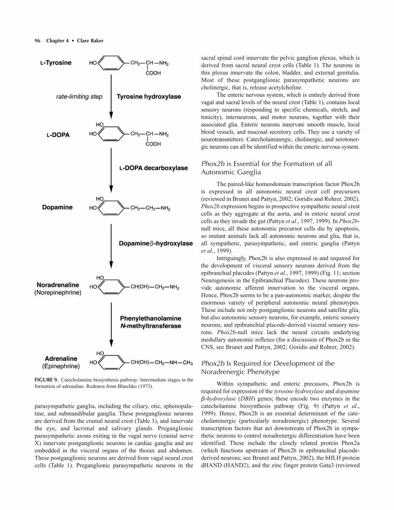
96 Chapter 4 • Clare Baker
parasympathetic ganglia, including the ciliary, otic, sphenopala-tine, and submandibular ganglia. These postganglionic neuronsare derived from the cranial neural crest (Table 1), and innervatethe eye, and lacrimal and salivary glands. Preganglionicparasympathetic axons exiting in the vagal nerve (cranial nerveX) innervate postganglionic neurons in cardiac ganglia and areembedded in the visceral organs of the thorax and abdomen.These postganglionic neurons are derived from vagal neural crestcells (Table 1). Preganglionic parasympathetic neurons in the
sacral spinal cord innervate the pelvic ganglion plexus, which isderived from sacral neural crest cells (Table 1). The neurons inthis plexus innervate the colon, bladder, and external genitalia.Most of these postganglionic parasympathetic neurons arecholinergic, that is, release acetylcholine.
The enteric nervous system, which is entirely derived fromvagal and sacral levels of the neural crest (Table 1), contains localsensory neurons (responding to specific chemicals, stretch, andtonicity), interneurons, and motor neurons, together with theirassociated glia. Enteric neurons innervate smooth muscle, localblood vessels, and mucosal secretory cells. They use a variety ofneurotransmitters: Catecholaminergic, cholinergic, and serotoner-gic neurons can all be identified within the enteric nervous system.
Phox2b is Essential for the Formation of allAutonomic Ganglia
The paired-like homeodomain transcription factor Phox2bis expressed in all autonomic neural crest cell precursors(reviewed in Brunet and Pattyn, 2002; Goridis and Rohrer, 2002).Phox2b expression begins in prospective sympathetic neural crestcells as they aggregate at the aorta, and in enteric neural crestcells as they invade the gut (Pattyn et al., 1997, 1999). In Phox2b-null mice, all these autonomic precursor cells die by apoptosis, so mutant animals lack all autonomic neurons and glia, that is, all sympathetic, parasympathetic, and enteric ganglia (Pattynet al., 1999).
Intriguingly, Phox2b is also expressed in and required forthe development of visceral sensory neurons derived from theepibranchial placodes (Pattyn et al., 1997, 1999) (Fig. 11; sectionNeurogenesis in the Epibranchial Placodes). These neurons pro-vide autonomic afferent innervation to the visceral organs.Hence, Phox2b seems to be a pan-autonomic marker, despite theenormous variety of peripheral autonomic neural phenotypes.These include not only postganglionic neurons and satellite glia,but also autonomic sensory neurons, for example, enteric sensoryneurons, and epibranchial placode-derived visceral sensory neu-rons. Phox2b-null mice lack the neural circuits underlyingmedullary autonomic reflexes (for a discussion of Phox2b in theCNS, see Brunet and Pattyn, 2002; Goridis and Rohrer, 2002).
Phox2b Is Required for Development of theNoradrenergic Phenotype
Within sympathetic and enteric precusors, Phox2b isrequired for expression of the tyrosine hydroxylase and dopamine�-hydroxylase (DBH) genes; these encode two enzymes in thecatecholamine biosynthesis pathway (Fig. 9) (Pattyn et al.,1999). Hence, Phox2b is an essential determinant of the cate-cholaminergic (particularly noradrenergic) phenotype. Severaltranscription factors that act downstream of Phox2b in sympa-thetic neurons to control noradrenergic differentiation have beenidentified. These include the closely related protein Phox2a(which functions upstream of Phox2b in epibranchial placode-derived neurons; see Brunet and Pattyn, 2002), the bHLH proteindHAND (HAND2), and the zinc finger protein Gata3 (reviewed
FIGURE 9. Catecholamine biosynthesis pathway: Intermediate stages in theformation of adrenaline. Redrawn from Blaschko (1973).
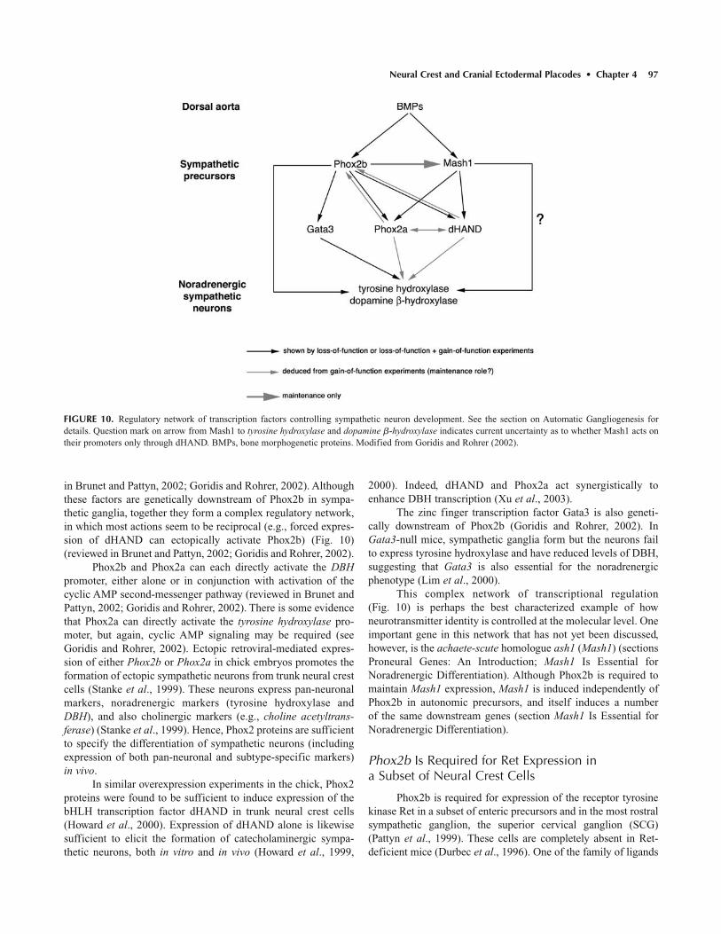
Neural Crest and Cranial Ectodermal Placodes • Chapter 4 97
in Brunet and Pattyn, 2002; Goridis and Rohrer, 2002). Althoughthese factors are genetically downstream of Phox2b in sympa-thetic ganglia, together they form a complex regulatory network,in which most actions seem to be reciprocal (e.g., forced expres-sion of dHAND can ectopically activate Phox2b) (Fig. 10)(reviewed in Brunet and Pattyn, 2002; Goridis and Rohrer, 2002).
Phox2b and Phox2a can each directly activate the DBHpromoter, either alone or in conjunction with activation of thecyclic AMP second-messenger pathway (reviewed in Brunet andPattyn, 2002; Goridis and Rohrer, 2002). There is some evidencethat Phox2a can directly activate the tyrosine hydroxylase pro-moter, but again, cyclic AMP signaling may be required (seeGoridis and Rohrer, 2002). Ectopic retroviral-mediated expres-sion of either Phox2b or Phox2a in chick embryos promotes theformation of ectopic sympathetic neurons from trunk neural crestcells (Stanke et al., 1999). These neurons express pan-neuronalmarkers, noradrenergic markers (tyrosine hydroxylase andDBH), and also cholinergic markers (e.g., choline acetyltrans-ferase) (Stanke et al., 1999). Hence, Phox2 proteins are sufficientto specify the differentiation of sympathetic neurons (includingexpression of both pan-neuronal and subtype-specific markers)in vivo.
In similar overexpression experiments in the chick, Phox2proteins were found to be sufficient to induce expression of thebHLH transcription factor dHAND in trunk neural crest cells(Howard et al., 2000). Expression of dHAND alone is likewisesufficient to elicit the formation of catecholaminergic sympa-thetic neurons, both in vitro and in vivo (Howard et al., 1999,
2000). Indeed, dHAND and Phox2a act synergistically toenhance DBH transcription (Xu et al., 2003).
The zinc finger transcription factor Gata3 is also geneti-cally downstream of Phox2b (Goridis and Rohrer, 2002). InGata3-null mice, sympathetic ganglia form but the neurons failto express tyrosine hydroxylase and have reduced levels of DBH,suggesting that Gata3 is also essential for the noradrenergic phenotype (Lim et al., 2000).
This complex network of transcriptional regulation(Fig. 10) is perhaps the best characterized example of how neurotransmitter identity is controlled at the molecular level. Oneimportant gene in this network that has not yet been discussed,however, is the achaete-scute homologue ash1 (Mash1) (sectionsProneural Genes: An Introduction; Mash1 Is Essential forNoradrenergic Differentiation). Although Phox2b is required tomaintain Mash1 expression, Mash1 is induced independently ofPhox2b in autonomic precursors, and itself induces a number of the same downstream genes (section Mash1 Is Essential forNoradrenergic Differentiation).
Phox2b Is Required for Ret Expression in a Subset of Neural Crest Cells
Phox2b is required for expression of the receptor tyrosinekinase Ret in a subset of enteric precursors and in the most rostralsympathetic ganglion, the superior cervical ganglion (SCG)(Pattyn et al., 1999). These cells are completely absent in Ret-deficient mice (Durbec et al., 1996). One of the family of ligands
FIGURE 10. Regulatory network of transcription factors controlling sympathetic neuron development. See the section on Automatic Gangliogenesis fordetails. Question mark on arrow from Mash1 to tyrosine hydroxylase and dopamine �-hydroxylase indicates current uncertainty as to whether Mash1 acts ontheir promoters only through dHAND. BMPs, bone morphogenetic proteins. Modified from Goridis and Rohrer (2002).
98 Chapter 4 • Clare Baker
that signal through Ret, glial cell line-derived neurotrophic fac-tor (GDNF), is essential for the development of the entire entericnervous system (Moore et al., 1996) (section The Differentiationof Enteric Neurons; reviewed in Young and Newgreen, 2001;Airaksinen and Saarma, 2002).
Mash1 Is Essential for Noradrenergic Differentiation
Mash1 (mouse Ash1), a bHLH transcription factor relatedto the invertebrate proneural Achaete-Scute complex (sectionProneural Genes: An Introduction; Chapter 5), was the first transcription factor found to be necessary for sympathetic devel-opment. Like Phox2b, Mash1 is expressed in all neural crest-derived autonomic precursors (sympathetic, parasympathetic,and enteric). Unlike Phox2b, however, it is not expressed inepibranchial placode-derived visceral sensory neurons. Mash1 isfirst expressed in sympathetic precursors shortly after they settlenear the dorsal aorta. Like Phox2b, Mash1 is essential for DBHexpression in all cell types except epibranchial placode-derivedneurons; that is, Mash1 is a noradrenergic determinant, indepen-dent of Phox2b (Hirsch et al., 1998).
In Mash1-null mice, sympathetic and parasympatheticganglia form (and express Phox2b), but pan-neuronal markers,Phox2a, tyrosine hydroxylase, and DBH are all lacking, and most(but not all) sympathetic and parasympathetic neuroblasts subse-quently degenerate (Guillemot et al., 1993; Hirsch et al., 1998).dHAND expression is also reported to be missing in theseembryos (Anderson and Jan, 1997). If Mash1 is constitutivelyexpressed in cultured neural crest stem cells, it induces bothPhox2a and Ret, together with pan-neuronal markers and morphological neuronal differentiation (Lo et al., 1998). Hence,Phox2a, dHAND, and Ret expression are induced not only byPhox2b, but also by Mash1. Mash1, like Phox2b, therefore, couples expression of pan-neuronal and neuronal subtype-specific markers (Fig. 10) (reviewed in Goridis and Rohrer,2002). However, this linkage can be uncoupled experimentally:Floorplate ablation in the chick abolishes Phox2a and tyrosinehydroxylase expression, but not Cash1 (chick Ash1) or pan-neuronal marker expression, in neural crest cells near the dorsalaorta (Groves et al., 1995). This suggests that a floorplate-derived signal, in addition to Mash1, is required for noradre-nergic identity in prospective sympathetic neurons (sectionFloorplate-Derived Signals). Hence, Mash1 expression is notsufficient, in all contexts, to promote noradrenergic identity.Indeed, Mash1 alone does not promote autonomic neurogenesisin vitro in the absence of BMP2; hence it must interact with otherfactors induced by BMP2, such as Phox2b (Lo et al., 2002) (section BMPs Induce Both Mash1 and Phox2b in SympatheticPrecursors).
Interestingly, given the requirement of Gata3 for nora-drenergic development (section Phox2b Is Required forDevelopment of the Noradrenergic Phenotype), the DrosophilaGata factor Pannier can either activate or repress achaete-scutecomplex genes, in association with various transcriptional cofactors (Ramain et al., 1993; Skaer et al., 2002). This suggests
a mechanism whereby Gata3 might also interact with Mash1, aswell as being downstream of Phox2b, although currently there isno evidence for this (Goridis and Rohrer, 2002).
A subset of enteric neurons, including apparently all serotonergic enteric neurons, is also missing in Mash1-null mice(Blaugrund et al., 1996; Hirsch et al., 1998). Since serotonergicenteric neurons seem to develop from tyrosine hydroxylase-expressing precursors, this loss is perhaps to be expected(Blaugrund et al., 1996).
Mash1 Also Plays Roles in Sensory Neurogenesis
Mash1 is not only required for the development of auto-nomic neurons, and it does not always function by inducingPhox2a. The mesencephalic nucleus of the trigeminal nerve,which was introduced in the section on Neural Crest Derivativesas a (somewhat controversial) neural crest derivative within thebrain, also depends on Mash1, but never expresses Phox2a(Hirsch et al., 1998). Mash1 is also essential for the developmentof olfactory neuron progenitors in the olfactory placode, whichlikewise do not express Phox2a (Guillemot et al., 1993; Cauet al., 1997) (section A bHLH Transcription Factor CascadeControls Olfactory Neurogenesis). Hence, different neuronalsubtype-specific factors must cooperate with Mash1 in theformation of these cell types.
BMPs Induce Both Mash1 and Phox2b inSympathetic Precursors
Neural crest cells that migrate past the notochord and stopin the vicinity of the dorsal aorta (section Migration Arrest atTarget Sites) will form the neurons and satellite cells of the sym-pathetic ganglia. Transplantation, rotation, and ablation experi-ments in the chick suggest that catecholaminergic neuronaldifferentiation only occurs near the aorta/mesonephros and alsorequires the presence of either the ventral neural tube or the noto-chord (Teillet and Le Douarin, 1983; Stern et al., 1991; Groveset al., 1995).
As described above, both Phox2b and Mash1 are firstexpressed shortly after neural crest cells arrive at the dorsal aorta.At this time, the dorsal aorta expresses Bmp2, Bmp4, and Bmp7(Reissmann et al., 1996; Shah et al., 1996). All three factorsinduce increased numbers of catecholaminergic cells in neuralcrest cell cultures, as does forced expression of a constitutivelyactive BMP receptor (reviewed in Goridis and Rohrer, 2002).BMP2 induces Mash1 and Phox2a in cultured neural crest stemcells (Shah et al., 1996; Lo et al., 1998). Overexpression of BMP4near the developing sympathetic ganglia leads to the ectopic formation of catecholaminergic cells in vivo (Reissmann et al.,1996). Conversely, when beads soaked in the BMP inhibitorNoggin are placed near the dorsal aorta in the chick, sympatheticganglia initially form, but sympathetic neurons do not develop(Schneider et al., 1999). In these Noggin-treated embryos, sym-pathetic ganglia lack expression of pan-neuronal markers, and ofPhox2b, Phox2a, DBH, and tyrosine hydroxylase, while Cash1 isstrongly reduced (Schneider et al., 1999). Together, these results

Neural Crest and Cranial Ectodermal Placodes • Chapter 4 99
provide overwhelming evidence that dorsal aorta-derived BMPsinduce expression of both Phox2b and Mash1, thus initiating theregulatory network of transcription factors that leads eventuallyto sympathetic neuron differentiation. However, these cues maybe insufficient for catecholaminergic differentiation in vivo, asdiscussed in the following section.
Floorplate-Derived Signals Are Also Required forCatecholaminergic Differentiation
In addition to signals from the dorsal aorta, the presence offloorplate and/or notochord is also required for catecholaminer-gic differentiation (Teillet and Le Douarin, 1983; Stern et al.,1991; Groves et al., 1995). In particular, although neurons dif-ferentiate in the sympathetic ganglia in the absence of floorplate,they do not express catecholaminergic markers (Groves et al.,1995). This suggests that in addition to BMPs from the dorsalaorta (which induce Phox2b and Mash1), floorplate-derivedsignals are also required to induce or maintain subtype-specificmarkers in the sympathetic ganglia (Groves et al., 1995). Sonichedgehog (see Chapter 3) seems to have little effect on cate-cholaminergic differentiation (Reissmann et al., 1996), and themolecular nature of the floorplate-derived signal(s) remainsunclear. It may be relevant in this context that enhanced cyclicAMP signaling is required for efficient activation of the tyrosinehydroxylase promoter by Phox2a in vitro (reviewed in Goridisand Rohrer, 2002). Also, activation of the mitogen-activated pro-tein (MAP) kinase signaling cascade in avian neural crest cellscauses catecholaminergic differentiation independently of BMP4(Wu and Howard, 2001). Clearly, there is still much to learnabout the control of sympathetic neuron differentiation.
BMPs and Parasympathetic vs SympatheticDifferentiation
The differentiation of parasympathetic vs sympatheticautonomic neurons may be determined by local concentrations ofBMPs at different neural crest target sites, as well as, perhaps,differential sensitivities of responding neural crest cells to BMPs(White et al., 2001). Postmigratory rat neural crest stem cells,isolated from fetal sciatic nerve, are more likely to differentiateas cholinergic parasympathetic neurons than as catecholaminer-gic sympathetic neurons when back-grafted into chick neuralcrest migratory pathways (White et al., 2001). After such grafts,they form cholinergic neurons in both sympathetic ganglia andparasympathetic ganglia, such as the pelvic plexus (White et al.,2001). In culture, they respond to BMP2 by differentiating asboth cholinergic and noradrenergic autonomic neurons. However,they are significantly less sensitive to the neuronal differentia-tion-inducing activity of BMP2 than are migrating neural creststem cells (section Differences in the Sensitivity of DifferentNeural Crest Stem Cells to Gliogenic Cues), and differentiate ascholinergic neurons at lower BMP2 concentrations (White et al.,2001). The molecular basis for this cholinergic bias is unknown.
BMPs are expressed at some sites of parasympathetic gan-gliogenesis. For example, the caudal cloaca, located proximal to
the forming parasympathetic pelvic plexus, expresses BMP2 atan appropriate time to be involved in inducing parasympatheticneuronal differentiation (White et al., 2001).
The Differentiation of Enteric Neurons
BMP2, which is expressed in gut mesenchyme, promotesthe neuronal maturation of postmigratory enteric neural precur-sors isolated from the rat gut (Pisano et al., 2000). However,several other growth factors have also been found to affectenteric neuronal differentiation.
Glial cell line-derived neurotrophic factor (GDNF) is thefounding member of a family of ligands that act via a commonsignal transducer, the receptor tyrosine kinase Ret, complexedwith ligand-specific receptors, the GDNF family receptor-�(GFR�) receptors (reviewed in Airaksinen and Saarma, 2002).GDNF is expressed in gut mesenchymal cells, and the entireenteric nervous system is missing in GDNF-deficient mice(Moore et al., 1996). In Ret-deficient mice, all enteric neuronsand glia are missing from the gut below the level of the esopha-gus and the immediately adjacent stomach (Durbec et al., 1996).GDNF and Neurturin, another GDNF family ligand, promote thein vitro survival, proliferation, and neuronal differentiation ofmigrating and postmigratory Ret� enteric precursors from the ratgut (Taraviras et al., 1999).
The growth factor Endothelin3 (Edn3), conversely, seemsto inhibit the neuronal differentiation of enteric precursors, thusmaintaining a sufficiently large pool of migratory, undifferenti-ated precursors to colonize the entire gut (Hearn et al., 1998;Shin et al., 1999). Endothelin3 prevents the neurogenic activityof GDNF on migrating enteric neural precursors isolated fromthe quail embryo gut (Hearn et al., 1998).
Mutations that affect the Ret or Endothelin signaling path-ways cause Hirschsprung’s disease in humans, in which entericganglia are missing from the terminal colon (reviewed in Gershon,1999; Manie et al., 2001; McCallion and Chakravarti, 2001).
Differentiation of Satellite Cells in Autonomic Ganglia
Strong autonomic neurogenic cues, such as BMP2, areclearly present at sites of autonomic gangliogenesis. How, then, do satellite glia form within autonomic ganglia? Exposure to gliogenesis-promoting factors such as NRG1 type II (sectionDifferentiation of DRG Satellite Cells) is insufficient. Cultured ratneural crest stem cells rapidly commit to an autonomic neuronalfate on exposure to BMP2, but only commit to a glial fate afterprolonged exposure to NRG1 type II (Shah and Anderson, 1997).Furthermore, saturating concentrations of BMP2 are dominantover NRG1 type II (although at low BMP2 concentrations, NRG1type II can attenuate Mash1 induction by BMP2) (Shah andAnderson, 1997). These results may explain why, in vivo, neuronsdifferentiate before glia in autonomic ganglia. What, then, preventsall autonomic progenitors from differentiating into neurons?
Activation of the Notch signaling pathway seems to beessential for adoption of a glial fate in the presence of BMP2

100 Chapter 4 • Clare Baker
(Morrison et al., 2000b). As discussed in the section on NotchActivation Leads to Gliogenesis by Neural Crest Stem Cells,even transient activation of Notch signaling inhibits neuronal dif-ferentiation and instructively promotes glial differentiation, incultures of postmigratory neural crest stem cells isolated fromfetal rat sciatic nerve (Morrison et al., 2000b). This action ofNotch is dominant over that of BMP2, blocking neurogenesis ata point upstream of Mash1 induction (Morrison et al., 2000b). Itis likely that a similar mechanism of Notch activation acts withinautonomic ganglia to promote satellite cell differentiation in thepresence of BMP2. One model suggested by these results is thatdifferentiating autonomic neurons express Notch ligands; thesethen activate Notch signaling in neighboring non-neuronal cells,which are then able to differentiate as glia (Morrison et al.,2000b). Other gliogenesis-promoting factors, such as NRG1 type II, may also act in concert with, or reinforce, the gliogenicaction of Notch in peripheral autonomic ganglia (Hagedornet al., 2000b). It is possible that once Notch is activated, prevent-ing a neuronal fate and promoting a glial fate, NRG1 type II maythen be able to promote a satellite cell fate (Hagedorn et al.,2000b; Leimeroth et al., 2002).
Summary of Autonomic Gangliogenesis
Phox2b is required for the formation of the entire periph-eral autonomic nervous system. It is also necessary and sufficientfor catecholaminergic (particularly noradrenergic) neuronaldifferentiation. Mash1 is necessary, but not sufficient, for norad-renergic differentiation. Phox2b and Mash1 interact in a complexregulatory network of transcription factors to induce noradrener-gic differentiation. They are independently induced in sympa-thetic precursors by BMPs from the dorsal aorta; however,additional floorplate-derived signals are also required for cate-cholaminergic differentiation of sympathetic neurons. BMPs mayalso induce parasympathetic fates: The choice between parasym-pathetic and sympathetic fates may depend on local BMP con-centrations and intrinsic differences in the sensitivity of differentpostmigratory neural crest cell populations to BMPs. BMPs andGDNF promote the differentiation of enteric neurons, while Edn3may prevent enteric neuronal differentiation. Satellite cell differ-entiation requires Notch activation, which is dominant to the neu-rogenesis-promoting activity of BMPs. The gliogenic activity ofNRG1 type II is subordinate to BMPs, but may be able to pro-mote satellite cell differentiation once Notch has been activated.
Community Effects Alter Fate Decisions
A multipotent neural crest cell may adopt one fate inresponse to a given instructive growth factor when it is alone, buta different fate when it is part of a cluster (“community”) ofneural crest cells (reviewed in Sommer, 2001). Individual post-migratory multipotent cells isolated from embryonic rat DRGrespond to BMP2 by forming both autonomic neurons andsmooth muscle cells, while clusters of the same multipotent cells form significantly more autonomic neurons, at the expense of smooth muscle cells (Hagedorn et al., 1999, 2000a).
This “community effect” (Gurdon et al., 1993) may preventneural crest cells in autonomic ganglia from adopting an aberrant(smooth muscle) fate in response to BMP2 in vivo.
Different concentrations of the same factor can also havedifferent effects when local neural crest cell–cell signaling isallowed to occur. Individual postmigratory progenitors from ratDRG respond to TGF� by adopting a predominantly smoothmuscle fate; they never form neurons (Hagedorn et al., 1999,2000a). Although high doses of TGF� cause some cell death, thepredominant fate choice is still smooth muscle (Hagedorn et al.,2000a). Clusters of these progenitors, in contrast, respond to highTGF� doses by dying, and to low TGF� doses by forming autonomic neurons (Hagedorn et al., 1999, 2000a).
Similar community effects may underlie the results discussed in the section on Axial Fate-Restriction, in which individual trunk neural crest cells form cartilage in the headwhen surrounded by host cartilage cells, but coherent masses oftrunk neural crest cells do not (McGonnell and Graham, 2002).Community effects also help to maintain neural crest cellregional identity: Individual neural crest cells will change theirHox gene expression patterns in response to environmental cues,while large groups of neural crest cells do not (e.g., Goldinget al., 2000; Trainor and Krumlauf, 2000; Schilling et al., 2001).
In summary, local neural crest cell–cell interactions mayreinforce fate choice in particular environmental contexts, and pre-vent inappropriate fate choices in response to environmental cues.
NEURAL CREST SUMMARY
Since the last edition of this book, in 1991, there has beenan explosion of information about the genes and signaling path-ways important for neural crest cell development. Molecular cuesinvolved in neural crest cell induction at the neural plate borderhave now been identified. These include BMPs, which are impor-tant for setting up the neural plate border itself and, later, forneural crest cell delamination, and Wnts, which are both neces-sary and sufficient for neural crest precursor cell inductionwithin the neural plate border. Numerous repulsive guidancecues, including ephrins, are now known to play essential roles insculpting the migration pathways of both cranial and trunk neuralcrest cells, and some progress has been made in understandingmigration arrest at target sites. The migrating neural crest cellpopulation is heterogeneous, containing multipotent and fate-restricted cells; however, the latter do not seem to be determined;that is, they retain the potential to adopt other fates when challenged experimentally. There is a greater molecular under-standing of lineage diversification, and it is becoming apparentthat the sensory–autonomic lineage decision is taken before theneuronal–glial fate decision. Various transcription factors areknown to be essential for the formation of particular neural crestlineages, including Phox2b for the autonomic lineage and Sox10for the glial lineage. Several instructive differentiation cues thatact on multipotent neural crest cells, including BMPs and NRGs,have been identified. Finally, an emerging theme is that neuralcrest cell–cell interactions, including community effects, are

Neural Crest and Cranial Ectodermal Placodes • Chapter 4 101
important in determining neural crest cell fate choice. Clearly, agreat deal has been learned about neural crest cell induction,migration, and differentiation. However, many questions stillremain, and there are many fruitful avenues for future researchinto the development of these fascinating cells.
OVERVIEW OF CRANIAL ECTODERMAL PLACODES
Cranial ectodermal placodes (Greek root ����, i.e., flatplate, tablet) are discrete patches of thickened ectoderm thatappear transiently in the head of all vertebrate embryos (reviewedin Webb and Noden, 1993; Baker and Bronner-Fraser, 2001;Begbie and Graham, 2001a). They were discovered 120 years ago(van Wijhe, 1883) and given the name “placode” by von Kupffer(1894). Placodes give rise to the bulk of the peripheral sensorynervous system in the head. The olfactory, otic, and lateral lineplacodes give rise to the paired peripheral sense organs (olfactoryepithelium, inner ears, and lateral line system of anamniotes)together with their afferent innervating neurons. The lens pla-codes give rise to the lenses of the eye. The trigeminal placodesform many of the cutaneous sensory neurons that innervate thehead, including the jaws and teeth. The epibranchial placodesgive rise to visceral sensory neurons that provide afferent inner-vation for tastebuds, and afferent autonomic innervation for the
visceral organs. Finally, the hypophyseal (or adenohypophyseal)placode gives rise to all of the endocrine cells and supportingcells of the adenohypophysis (anterior pituitary gland). Althoughthe molecular mechanisms underlying the induction and devel-opment of the hypophyseal placode are perhaps the best under-stood of all the placodes, its development will not be discussedhere (for detailed reviews of hypophyseal placode induction anddevelopment, see Baker and Bronner-Fraser, 2001; Dasen andRosenfeld, 2001; Scully and Rosenfeld, 2002).
Like the neural crest, therefore, placodes give rise to a verydiverse array of cell types, including sensory receptors, sensoryneurons, supporting cells, secretory cells, glia, neuroendocrine,and endocrine cells (Table 3). Figure 11 shows a fate-map of theplacode-forming ectoderm in the head of the 8-somite stagechick embryo, together with the respective neuronal contribu-tions to cranial sensory ganglia of placodes and the neural crest.Figure 12 shows the location of the different placodes in the headof the 19-somite stage Xenopus embryo. It is evident from theseschematics that a relatively large proportion of dorsal head ectoderm contributes to placodal tissue.
Also like the neural crest, cranial ectodermal placodes areusually considered to be a defining characteristic of the craniates(vertebrates plus hagfish) (Gans and Northcutt, 1983; Northcuttand Gans, 1983; Baker and Bronner-Fraser, 1997). However,molecular analyses suggest that at least some vertebrate pla-codes may have homologues in non-vertebrate chordates (e.g.,Boorman and Shimeld, 2002; Christiaen et al., 2002). Placodes
TABLE 3. Cell Types and Cells Derived from Cranial Ectodermal Placodes
Placode General cell type Cells
Olfactory Sensory ciliary receptor Chemoreceptive olfactory receptor neuronsSensory neurons Olfactory receptor neuronsGlia Olfactory ensheathing gliaNeuroendocrine cells Gonadotropin-releasing hormone (GnRH)-producing neuronsSecretory/support cells Sustentacular cells (secrete mucus; provide support)
Otic Sensory ciliary receptor Mechanosensory hair cellsSensory neurons Otic hair cell-innervating neurons, collected in vestibulo-cochlear ganglion of cranial
nerve VIIISecretory cells Cupula-secreting cells; endolymph-secreting cells; cells secreting biomineralized matrix
of otoliths/otoconiaSupporting cells Hair cell support cells; non-sensory epithelia
Lateral line Sensory ciliary receptor Mechanosensory hair cells in neuromastsElectroreceptive cells in ampullary organs
Sensory neurons Lateral line hair cell-innervating neurons, collected in lateral line gangliaSecretory cells Cupula-secreting cells in neuromastsSupporting cells Hair cell support cells in neuromasts
Lens Specialized epithelium Lens fiber cellsOphthalmic and Sensory neurons Cutaneous sensory neurons (pain, touch, temperature), collected in trigeminal ganglion ofmaxillomandibular cranial nerve Vtrigeminal
Epibranchial Sensory neurons Afferent neurons for taste buds and visceral organs, collected in geniculate, petrosal, and nodoseganglia (distal ganglia of cranial nerves VII, IX, and X, respectively)
Hypophyseal Endocrine cells All endocrine cells of the adenohypophysis (anterior pituitary gland)Supporting cells Support cells of the adenohypophysis

102 Chapter 4 • Clare Baker
have been studied in all craniate classes, including hagfish (e.g., Wicht and Northcutt, 1995) and lamprey (e.g., Bodznick andNorthcutt, 1981; Neidert et al., 2001; McCauley and Bronner-Fraser, 2002). Although most research has been done on the senseorgan placodes, in particular the lens and otic placodes, as well asthe hypophyseal placode, molecular information has also enabledcloser investigation of the development of the trigeminal and epi-branchial placodes. Here, a relatively brief summary is providedof the current state of knowledge of the induction and develop-ment of the different placodes. For a more detailed review of classical and modern research into placode induction, the readeris referred to Baker and Bronner-Fraser (2001).
A PREPLACODAL FIELD AT THE ANTERIORNEURAL PLATE BORDER
All fate-mapping studies to date have shown that placodesarise from ectoderm at the neural plate border in the prospectivehead region (Baker and Bronner-Fraser, 2001). Older fate mapssuggest that placodes originate from ectoderm lying lateral to theneural crest-forming area, except in the most rostral region,where no neural crest cells form and olfactory and hypophysealplacodes directly abut prospective neural plate territories (Bakerand Bronner-Fraser, 2001). However, cell lineage analysis shows that placodal precursors, like neural crest precursors (section Embryonic Origin of the Neural Crest), do not exist
FIGURE 12. Location of placodes in the head of a 19-somite stage Xenopusembryo. epi, epibranchial placode; epi VII, facial/geniculate placode; epi IX,glossopharyngeal/petrosal placode; epi X, vagal/nodose placodes; lat, lateralline placode; latAD, anterodorsal lateral line placode; latAV, anteroventral lat-eral line placode; latM, middle lateral line placode; latP, posterior lateral lineplacode; mmV, maxillomandibular trigeminal placode; olf., olfactory pla-code; opV, ophthalmic trigeminal placode. Redrawn from Schlosser andNorthcutt (2000).
FIGURE 11. Fate-map of placodes (black ovals) and neural crest (dark blue) in the head of an 8-somite stage chick embryo, and their neuronal contributionto the sensory ganglia of the cranial nerves (Roman numerals). All satellite cells in cranial sensory ganglia are derived from the neural crest. fb, forebrain; G., ganglion; gen, geniculate; ln, lens; mb, midbrain; mmV, maxillomandibular trigeminal; nod, nodose; olf., olfactory; opV, ophthalmic trigeminal; pet, petrosal; prox., proximal; sup., superior; vest.-coch., vestibulocochlear. Redrawn from D’Amico-Martel and Noden (1983).

Neural Crest and Cranial Ectodermal Placodes • Chapter 4 103
as a segregated population (Streit, 2002). Although prospectiveplacodal territory extends more laterally than prospective neuralcrest territory, placodal and neural crest precursors are mingledtogether more medially (Streit, 2002).
Molecular evidence supports some early morphologicalobservations in suggesting that there is a preplacodal field, orpanplacodal anlage, around the anterior neural plate. This field ismorphologically visible in the frog, Rana, which has a continu-ous band of thickened ectoderm around the edge of the anteriorneural plate, from which most placodes originate (Knouff, 1935)(Fig. 13A). Molecularly, this field seems to be characterized in multiple species by the expression of various genes in a horse-shoe-shaped band around the anterior neural plate border(Figs 13B, C). These genes, which primarily encode transcriptionfactors, are often subsequently maintained in all or multipleplacodes. They include the homeodomain transcription factorsSix1, Six4, Dlx3, Dlx5, and Dlx7, the HMG-domain transcrip-tion factor Sox3 (which is also expressed in the neural plate), andthe transcription cofactors Eya1 and Eya2 (for original refer-ences, see Baker and Bronner-Fraser, 2001; also David et al.,2001; Ghanbari et al., 2001). See the section on Establishment ofthe Neural Plate Border for a discussion of the role of Dlx genesin positioning the neural plate border. In the chick, the expressiondomains of these genes are not coincident; rather, they areexpressed in a series of overlapping domains that shift both spa-tially and temporally with the position of placodal precursors(Streit, 2002).
It is clear that several of these genes play important rolesin the development of multiple placodes. For example, dlx3,acting in concert with dlx7, is necessary for the formation of both olfactory and otic placodes in the zebrafish (Solomon andFritz, 2002). Ectopic expression of Sox3 in another teleost fish,medaka, causes ectopic lens and otic vesicle formation in ecto-dermal regions relatively close to the endogenous lens and oticplacodes (Köster et al., 2000). Sox3 may, therefore, act as a com-petence factor, enabling ectopic ectoderm to respond to placode-inducing signals (Köster et al., 2000).
However, the precise significance of the preplacodaldomain of gene expression remains unclear: It does not seem tocorrelate either with the site of origin of all placodal precursorcells, or with determination toward a placodal fate. A cell lineageanalysis in the chick showed that some otic placode precursorsarise from ectoderm lying medial to the Six4 expression domain(Streit, 2002). Hence, not all placodal precursors originate fromthe Six4� domain. Furthermore, cells within the Six4� domainform not only placodal derivatives, but also neural crest and epi-dermis (and neural tube until the 2-somite stage, at the level ofthe future otic placode) (Streit, 2002). Hence, cells within thepreplacodal domain are not all determined toward a placodal fate.
Some insight into the function of the preplacodal domainmay come from observations showing that there is a large degreeof ectodermal cell movement in the neural plate border region(Whitlock and Westerfield, 2000; Streit, 2002). These studiescombined cell lineage analysis (using DiI or fluorescent dex-trans) with time-lapse analysis and in situ hybridization.Precursors of a particular placode, such as the olfactory placodein zebrafish (Whitlock and Westerfield, 2000) or the otic placodein chick (Streit, 2002), originate from a fairly large region ofectoderm at the anterior neural plate border, and subsequentlyconverge to form the final placode. This may suggest a modelwhereby cells that move into the preplacodal gene expressiondomain upregulate the genes defining the domain, while cellsthat move out of the domain downregulate these genes. Cells thatexpress the “preplacodal” genes may be rendered competent torespond to specific placode-inducing signals. However, the fateof a given cell within the preplacodal domain will depend on theprecise combination of signals it subsequently receives. Hence,although it is competent to form placode, it may give rise toneural crest or epidermis instead.
The Pax/Six/Eya/Dach Regulatory Network
The overlapping expression of various Six and Eya genesin the preplacodal domain is of particular interest. Six and Eya
FIGURE 13. A preplacodal domain of ectoderm can be recognized around the anterior neural plate border, occasionally by morphology alone, more often byspecific gene expression. (A) Fate map of open neural plate stage Rana embryo (dorsal view), showing the preplacodal domain, recognizable morphologicallyas a continuous strip of thickened ectoderm around the prospective neural crest (NC) domain. cg, prospective cement gland; epid, epidermis; NC, neural crest.Redrawn from Knouff (1935). (B) Eya2 expression (dark staining) around the anterior neural plate border in a stage 6 (neurula-stage) chick embryo (dorsalview). (C) Six4 expression (dark staining) around the anterior neural plate border in a 2-somite stage chick embryo (dorsal view). Both Eya2 and Six4 are sub-sequently maintained in most placodes (section A Preplacodal Field at the Anterior Neural Plate Border for details). Photographs courtesy of Dr. Andrea Streitand Anna Litsiou, King’s College, London, United Kingdom. Chick staging after Hamburger and Hamilton (1951).
104 Chapter 4 • Clare Baker
family members function in a complex cross-regulatory networkwith Pax transcription factors and the transcription cofactorDachshund (Dach, also a multimember family in vertebrates), ina variety of developmental contexts. These include eye develop-ment in Drosophila (reviewed in Wawersik and Maas, 2000) andvertebrate muscle development (Heanue et al., 1999). Dach fam-ily members are expressed in part of the preplacodal domain andin various placodes (Davis et al., 2001; Hammond et al., 2002;Heanue et al., 2002; Loosli et al., 2002). While Pax genes are notexpressed in the preplacodal domain, most individual placodesare characterized by a particular combination of Pax gene expres-sion. For example, Pax6 is expressed in the olfactory and lensplacodes, Pax3 in the ophthalmic trigeminal placode, Pax2/5/8 inthe otic placode, and Pax2 in the epibranchial placodes (reviewedin Baker and Bronner-Fraser, 2001). Mouse knockout studieshave shown that Pax gene expression within the placodes isimportant for their proper development. For example, Pax6is essential for olfactory and lens placode formation, while Pax2is important for aspects of otic placode development (reviewed inBaker and Bronner-Fraser, 2001).
Given the above, it is possible that expression of Six, Eya,and perhaps also Dach genes within the preplacodal domain mayrepresent a molecular framework common to all placodes. Thisnetwork might then be able to interact with different Pax genes,induced in different regions of the preplacodal domain by spe-cific placode-inducing signals, to specify individual placodeidentities. Although this model is attractive, further supportingevidence is required.
Models of Individual Placode Formation in the Preplacodal Domain
The active convergence of cells at the neural plate borderto form specific placodes (Whitlock and Westerfield, 2000;Streit, 2002) suggests two possible models for the formation ofindividual placodes from ectoderm in this region (Streit, 2002).The first model proposes that cells in a large region of ectodermreceive a specific placode-inducing signal: Those cells thatrespond to the signal “sort out” from non-responding cells andactively migrate to the site of formation of the placode. The sec-ond model proposes that ectodermal cells move at random: Thosethat come within range of localized placode-inducing signalsadopt a specific placodal fate. Evidence exists to support theexistence of both localized and widespread placode-inducing signals (e.g., sections Induction of the Otic Placodes; Inductionof the Trigeminal Placodes). However, it is currently unknownwhether active sorting processes occur within the ectoderm tocause the aggregation of specific placode precursors.
Summary
All placodes originate from ectoderm at the anterior neuralplate border. A horseshoe-shaped domain of ectoderm at theanterior neural plate border expresses numerous specific tran-scription factors, such as Six, Eya, and Dlx family members, all
of which have roles in placode development. Cells expressing anappropriate combination of these genes may be competent toadopt a placodal fate in response to placode-inducing signals.Cells that respond to specific placode-inducing signals may sortout and aggregate to form the placode. Alternatively, cells mayrandomly move within range of specific placode-inducing sig-nals, cease migrating, and differentiate accordingly. Further evi-dence is required to distinguish between these models.
In the following sections, the induction and some aspectsof the development of individual placodes are discussed in turn,beginning with the sense organ placodes (olfactory, otic, lateralline, and lens), and ending with the trigeminal and epibranchialplacodes.
SENSE ORGAN PLACODES
Olfactory Placodes
Olfactory Placode Derivatives
The paired olfactory placodes, which invaginate towardthe telencephalon to form olfactory pits, give rise to the entireolfactory (odorant-sensing) and, where present, vomeronasal(pheromone-sensing) epithelia, together with the respiratoryepithelium that lines the nasal passages. The olfactory andvomeronasal epithelia contain ciliated sensory receptor neurons,each of which bind odorants via a single member of an enormousfamily of G-protein-coupled, seven-transmembrane domainreceptor molecules (reviewed in Mombaerts, 2001; Ronnett andMoon, 2002). The epithelia also contain basal cells, which gen-erate olfactory sensory neurons throughout life (for a review onstem cells in the olfactory epithelium, see Calof et al., 1998), andsupporting sustentacular cells, which share some characteristicswith glia (reviewed in Ronnett and Moon, 2002). All of thesecells are derived from the olfactory placode.
The cell bodies of the olfactory receptor neurons remain inthe placode, while their axons extend into the brain to form theolfactory, vomeronasal, and terminal nerves (for reviews ofolfactory axon pathfinding, see Mombaerts, 2001; St. John et al.,2002). These nerves are ensheathed by olfactory placode-derivedglial cells (reviewed in Wewetzer et al., 2002) that leave the pla-code and migrate along the nerves into the brain. In thezebrafish, pioneer neurons, distinct from olfactory receptor neu-rons, differentiate early within the placode and send their axonsto the telencephalon (Whitlock and Westerfield, 1998). Axonsfrom the olfactory receptor neurons follow this initial scaffold-ing, and the pioneer neurons subsequently die by apoptosis(Whitlock and Westerfield, 1998). Olfactory axons are the firstperipheral input to reach the brain during development. Theaxons of pioneer neurons in the rat induce formation of the olfac-tory bulbs (Gong and Shipley, 1995), which fail to form if theolfactory placodes are missing or if olfactory axons fail to reachthe brain (reviewed in Baker and Bronner-Fraser, 2001). Theolfactory epithelium is also required for induction of the carti-laginous nasal capsule, which is derived from the neural crest.

Neural Crest and Cranial Ectodermal Placodes • Chapter 4 105
The olfactory placode also forms neuroendocrine cells thatmigrate along the olfactory nerve into the forebrain and dien-cephalon. These neurons produce gonadotropin-releasing hor-mone (GnRH) and form the terminal nerve-septo-preoptic GnRHsystem (reviewed in Dubois et al., 2002). This system regulatesgonadotropin release from the adenohypophysis (anterior pitu-itary), another placodal derivative (section Overview of CranialEctodermal Placodes). Hence, the olfactory placode is not onlyessential for olfaction, but also for reproduction. This is seenclinically in Kallmann’s syndrome, in which olfactory axons andGnRH neurons fail to migrate into the brain, resulting in anosmiaand sterility (hypogonadism) (reviewed in MacColl et al., 2002).An early-stage fate-map in zebrafish, however, challenges the olfactory placode origin of GnRH neurons, suggesting that terminal nerve GnRH neurons originate from the neural crest,and hypothalamic GnRH neurons from the hypophyseal placode(adenohypophysis) (Whitlock et al., 2003). More early-stagefate-map data are needed from multiple species to resolve thiscontroversy.
Olfactory Placode Formation Involves theConvergence of Cellular Fields
In the 4-somite stage zebrafish embryo, the olfactory pla-codes fate-map to bilateral regions of Dlx3� ectoderm at the lateralborders of the anterior-most neural plate, much longer in rostro-caudal extent than the final placodes, abutting prospective telen-cephalic territory rostrally and prospective neural crest caudally(Whitlock and Westerfield, 2000). Dlx3, acting in concert withDlx7, is essential for the formation of the olfactory placode in thezebrafish, as shown both by mutant and knockdown analysis usingantisense morpholino oligonucleotides (Solomon and Fritz, 2002).Each of these long bilateral Dlx3� cellular fields converges toform an olfactory placode (Whitlock and Westerfield, 2000).
In neurula-stage Xenopus embryos, and 3-somite stagechick and mouse embryos, the olfactory placodes fate-map to theouter edge of the anterior neural ridge (the rostral boundary ofthe neural plate) (Couly and Le Douarin, 1988; Eagleson andHarris, 1990; Osumi-Yamashita et al., 1994). Future olfactoryplacode and olfactory bulb tissues are contiguous within theanterior neural plate. It is currently unknown whether olfactoryplacode formation in these species also involves cellular conver-gence, as in the zebrafish.
Induction of the Olfactory Placodes
Classical grafting and coculture experiments in amphibianembryos (reviewed in Baker and Bronner-Fraser, 2001) sug-gested that anterior mesendoderm is an important source ofolfactory placode-inducing signals. This tissue is also importantfor forebrain induction (reviewed in Foley and Stern, 2001)(Chapter 3). Forebrain tissue is also important for olfactory pla-code induction and/or maintenance (reviewed in Baker andBronner-Fraser, 2001). Nothing is currently known about whichmolecular signals from these tissues, or others, might be involvedin the induction of the olfactory placode.
In the chick, FGF8 from the midfacial ectoderm is neces-sary and sufficient to induce the genes erm and pea3 in the olfac-tory pits (Firnberg and Neubüser, 2002). (The induction andmaintenance of these two Ets-domain transcription factors isgenerally tightly coupled to FGF signaling; Raible and Brand,2001; Roehl and Nüsslein-Volhard, 2001.) FGFs stimulate theproliferation of olfactory receptor neuron progenitors in vitro(reviewed in Calof et al., 1998) (see next section), hence, FGF8may play a similar role in promoting olfactory neurogenesis in vivo. However, this remains to be demonstrated.
Experiments in mice and zebrafish have shown that thetranscription factors Otx2, Pax6, Dlx5, and Dlx3 (acting in con-cert with Dlx7), all of which are expressed in the anterior neuralridge and in the olfactory placodes, are required for olfactoryplacode development (reviewed in Baker and Bronner-Fraser,2001) (also Solomon and Fritz, 2002). However, their preciseroles are currently undefined.
A bHLH Transcription Factor Cascade ControlsOlfactory Neurogenesis
Mice lacking the Achaete-Scute homologue Mash1 (section Mash1 Is Essential for Noradrenergic Differentiation)have a drastically reduced number of olfactory receptor neurons,due to the death of most olfactory neuron progenitors (Guillemotet al., 1993; Cau et al., 1997). Mash1 is required for the expres-sion of the atonal-related bHLH genes Neurogenin1 (Ngn1)(section Neurogenins Are Essential for the Formation of DorsalRoot Ganglia) and NeuroD (Cau et al., 1997), and for activationof the Notch signaling pathway (Cau et al., 2000, 2002). Ngn1 isrequired for neuronal differentiation; it does not affect eitherMash1 expression or the Notch signaling pathway (Cau et al.,2002).
Mash1 is not expressed in the earliest-differentiating neurons in the olfactory placode, whose formation is unaffectedin Mash1-null mice (Cau et al., 1997, 2002). These Mash1-independent progenitors express Ngn2 as well as Ngn1, but theirdifferentiation is blocked in Ngn1-mutant mice (Cau et al., 1997,2002). Given their early differentiation, these could represent the pioneer neurons whose axons set up the initial scaffold for the olfactory nerve (section Olfactory Placode Derivatives).Interestingly, vomeronasal sensory neurons are relatively unaf-fected in mice that are double mutant for both Mash1 and Ngn1(Cau et al., 2002). This suggests both that a different gene controls their development, and that vomeronasal and olfactorysensory neuron progenitors are molecularly distinct (Cau et al.,2002).
As described in the previous section, FGF8, which isexpressed in the epithelium around the placodes, may stimulatethe proliferation of olfactory neuron stem cells and neuronalprogenitors (reviewed in Calof et al., 1998) (also see LaMantiaet al., 2000). Treatment of olfactory placode explants with afunction-blocking FGF8 antibody causes a reduction in the num-bers of neurons relative to controls, though not a complete loss,suggesting that FGF8 is not only sufficient but also necessary forolfactory neurogenesis (LaMantia et al., 2000).

106 Chapter 4 • Clare Baker
BMPs also play an important role in olfactory neurogene-sis: In fact, they can both promote and inhibit neurogenesis incultures of olfactory epithelium, depending on the concentration,the specific ligand, and the cellular context (Shou et al., 1999,2000). Exposure of Mash1� olfactory neuron progenitors to highconcentrations of BMP4 or BMP7 in culture leads to the degra-dation of Mash1 protein via the proteasome pathway, and henceto inhibition of neuronal differentiation (Shou et al., 1999).However, treatment of olfactory epithelium cultures with theBMP antagonist Noggin inhibits neuronal differentiation, show-ing a requirement for BMP signaling in neurogenesis (Shouet al., 2000). This requirement is explained by the observationthat low concentrations of BMP4, but not BMP7, promote thesurvival of newly born olfactory receptor neurons (Shou et al.,2000). Hence, BMP4 inhibits the production of olfactory recep-tor neurons at high concentrations and promotes the survival ofdifferentiated neurons at low concentrations. This may provide afeedback mechanism to maintain an appropriate number of olfac-tory receptor neurons in the epithelium, particularly as BMP4may be produced by the olfactory receptor neurons themselves(Shou et al., 2000).
Retinoic acid is produced by the neural crest-derived frontonasal mesenchyme between the olfactory placode and theventrolateral forebrain (LaMantia et al., 1993, 2000). In vitro,retinoic acid stimulates the maturation of olfactory receptor neu-rons from immortalized clonal cell lines derived from the mouseolfactory placode, suggesting a possible role in this process in vivo (Illing et al., 2002). However, retinoic acid treatment ofolfactory placode explants leads to reduced neuronal differentia-tion (LaMantia et al., 2000). Further evidence is thereforerequired to establish the precise role of retinoic acid.
Lateral Line Placodes
Lateral Line Placode Derivatives
The lateral line system is a mechanosensory and electrore-ceptive sensory system in which individual sense organs arearranged in characteristic lines along the head and trunk of fishand amphibians (Fig. 14C) (Coombs et al., 1989). The entire lat-eral line system seems to have been lost in amniotes, presumablyin association with the transition to a primarily terrestriallifestyle; it is also often lost in amphibians at metamorphosis.Lateral line electroreception was lost in most teleost fish and inanuran amphibians; indeed, different elements of the lateral linesystem have been lost independently in multiple vertebrate lineages (reviewed in Northcutt, 1997; Schlosser, 2002b).
There are two types of lateral line sense organs:Mechanosensory neuromasts (Fig. 14B) that respond to local dis-turbances in the water surrounding the animal, and electrorecep-tive ampullary organs that respond to weak electric fields. Theyare used in various behaviors, including schooling, obstacleavoidance, and prey detection. The sense organs themselves, andthe neurons that provide their afferent innervation, are derivedfrom a series of paired lateral line placodes on the head (reviewedin Winklbauer, 1989; Baker and Bronner-Fraser, 2001; Schlosser,2002a) (Fig. 12). The same lateral line placode can form bothmechanosensory neuromasts and electroreceptive ampullaryorgans (Northcutt et al., 1995). Primitively, there were probablyat least three pre-otic and three post-otic lateral line placodes(Northcutt, 1997). One pole of each lateral line placode gives riseto neuroblasts, which exit the placode and aggregate nearby toform the sensory neurons of a lateral line ganglion (the satellite
FIGURE 14. The mechanosensory lateral line system. (A) Schematic showing an individual unit of a lateral line neuromast: A mechanosensory hair cell bear-ing a true cilium (kinocilium) and stereocilia, innervated by lateral line neurites, with an adjacent support cell. Redrawn from Kardong (1998). (B) Schematicsection through a neuromast organ: The cilia and stereocilia of each hair cell project into a gelatinous cupula, secreted by the supporting cells. Redrawn fromKardong (1998). (C) Lateral line neuromasts on the head (particularly visible in a ring around the eye) and along the trunk of a live stage 45 (4-day) Xenopustadpole, visualized with the vital mitochondrial stain DASPEI. (D) Scanning electron micrograph of lateral line neuromasts along the trunk of a stage 49 (12-day) Xenopus tadpole. (E) High-power view of individual neuromast from (D), showing bundle of long kinocilia with smaller stereocilia at its base.Xenopus staging after Nieuwkoop and Faber (1967).

Neural Crest and Cranial Ectodermal Placodes • Chapter 4 107
cells of the ganglion are derived from the neural crest). Neuritesfrom these neurons, followed and ensheathed by neural crest-derived glial cells (Gilmour et al., 2002), track the remainingnon-neurogenic part of the placode as it elongates to form a lat-eral line primordium and undergoes a remarkable migrationthrough the epidermis, depositing clusters of cells in its wake.These cells give rise to the supporting and mechanosensory haircells of the lateral line sense organ (neuromast). Hence, the pla-code that forms a given line of sense organs also typically formstheir afferent innervating neurons. In the zebrafish, the atonal-related bHLH transcription factor Neurogenin1 (Ngn1; sectionProneural Genes: An Introduction) is required for the formationof lateral line neurons, but its loss has no effect on the migrationof the lateral line primordia, or on neuromast development(Andermann et al., 2002). Hence, as previously demonstrated inamphibian embryos (Tweedle, 1977), lateral line sense organdevelopment is independent of innervation.
A single mechanosensory neuromast is composed ofseveral hair cells, together with supporting cells; each hair cellhas a single true cilium (kinocilium) with a bundle of stereociliaat its base (Figs. 14A, E). Lateral line hair cells are very similarin structure to inner ear hair cells derived from the otic placode(section Otic Placode Derivatives). The kinocilia and sterocilia ofthe hair cells in each lateral line neuromast are embedded in agelatinous sheath, or cupula, which is secreted by the supportingcells of the neuromast (Fig. 14B). Water movements deflect thecupula. If the cupula movement bends the stereocilia toward thekinocilium, mechanosensitive ion channels in the hair cell open,depolarizing the hair cell and stimulating the afferent fibers ofthe lateral line nerve which synapses onto its basal surface. If thestereocilia are bent away from the kinocilium, this closes the fewion channels that are open at rest, causing hyperpolarization ofthe hair cell and neuronal inhibition (reviewed in Winklbauer,1989; Pickles and Corey, 1992).
Electroreceptor cells are structurally similar to neuromasthair cells, although they are more variable across taxa (Bodznick,1989). They are secondary sense cells (i.e., they require affer-ent innervation) with apical microvilli and/or a kinocilium.Interestingly, while teleost electroreceptors, where present, seemto have secondarily evolved from neuromast hair cells, nonteleostelectroreceptors (i.e., those found in lamprey, nonteleost fish,and amphibia) are phylogenetically as old or older than neuro-mast hair cells (Bodznick and Northcutt, 1981; Bodznick, 1989).From the phylogenetic evidence, it is equally likely that neuro-mast hair cells evolved from electroreceptors, that electro-receptors evolved from mechanosensory hair cells, or that bothevolved independently from a common ancestral ciliated celltype (Bodznick, 1989).
Induction of the Lateral Line Placodes
Relatively little is currently known about the sources of lateral line placode-inducing signals, and nothing of their molecular nature (reviewed in Baker and Bronner-Fraser, 2001;Schlosser, 2002a). Lateral line placodes are induced separatelyfrom the otic placodes (section Otic Placodes), despite their
proximity and even (in some species) their apparent derivationfrom a common Pax2� “dorsolateral placode area” (Schlosser andNorthcutt, 2000; Schlosser, 2002a). Lateral line and otic placodescan be induced independently in grafting experiments, and ecto-dermal competence to form lateral line placodes persists muchlonger than that to form otic placodes (reviewed in Baker andBronner-Fraser, 2001; Schlosser, 2002a). Furthermore, mutationsin zebrafish can affect otic placodes but not lateral line placodes,and vice versa (Whitfield et al., 1996), and lateral line placodeshave been lost multiple times in evolution with no effect on theotic placodes (e.g., Schlosser et al., 1999).
In the axolotl, lateral line placodes are determined (i.e., develop autonomously in ectopic locations) by late neuralfold stages (Schlosser and Northcutt, 2001). Hence, lateral line placode-inducing signals must act before this time, although theypersist until relatively late stages, as shown by grafts of non-placodal ectoderm to the placode-forming region of tailbud stageembryos (Schlosser and Northcutt, 2001). Grafting experimentsin amphibians have implicated both mesoderm and neural plateas sources of lateral line placode-inducing signals (reviewed inBaker and Bronner-Fraser, 2001; Schlosser, 2002a). Their mole-cular nature is currently unknown. Migrating lateral line primor-dia in zebrafish express the FGF target genes, erm and pea3(Münchberg et al., 1999; Raible and Brand, 2001; Roehl andNüsslein-Volhard, 2001), the Wnt receptor gene Frizzled7a(Sumanas et al., 2002), and the BMP inhibitor follistatin(Mowbray et al., 2001). Hence, FGF, Wnt, and BMP signalingmay all be involved in aspects of lateral line placode develop-ment; however, any role for these signaling pathways in lateralline placode induction remains to be demonstrated. Early devel-opment and migration of lateral line placodes in fgf8 mutantzebrafish appears normal, although the number of neuromastsformed is strongly reduced, suggesting an involvement of FGF8at later stages (Léger and Brand, 2002).
Individual lateral line placodes differ in the number andtype of sense organs that they form, and also in their geneexpression patterns (e.g., only the middle lateral line placodeexpresses Hoxb3 in the axolotl; Metscher et al., 1997).Additional inducing signals are presumably involved, therefore,in specifying individual lateral line placode identity, but theseare wholly unknown.
Migration of Lateral Line Primordia
Cells of the lateral line primordia in amphibian embryosactively migrate through and displace the inner cells of the bilay-ered epidermis. Cell division also occurs within the primordia as they migrate (Winklbauer, 1989; Schlosser, 2002a). In zebrafish,lateral line primordia migrate just beneath the epidermis; time-lapseanalysis of living embryos shows that each cell migrates indepen-dently, rather than the whole primordium moving as a solid block ofcells, but they generally retain their neighbor relationships (Gompelet al., 2001). The clusters of undifferentiated cells that will form theneuromasts are deposited when a group of cells at the trailing edgeof the primordium progressively slows down relative to the rest ofthe primordium (Gompel et al., 2001).

108 Chapter 4 • Clare Baker
During normal development, lateral line primordiamigrate along invariant pathways. When pre-otic and post-oticlateral line placodes are exchanged, they migrate along the path-way appropriate for their new position (reviewed in Schlosser,2002a), suggesting that they are following extrinsic guidancecues. Several such guidance cues have now been identified inzebrafish. The chemokine SDF1 is expressed in a trail along themigration pathway of the posterior lateral line primordium(David et al., 2002). The migrating cells of the lateral line pri-mordium express the SDF1 receptor, CXCR4, and inactivationof either the receptor or its ligand blocks migration (David et al.,2002). Also, the posterior lateral line primordium migratesalong the trunk at the level of the horizontal myoseptum thatdivides the axial muscles into dorsal and ventral halves.Semaphorin3A1 (Sema3A1) is expressed throughout thesomites except in the horizontal myoseptum, and when themyoseptum is missing, as in certain mutant strains, the lateralline primordium migrates aberrantly (Shoji et al., 1998). Theseresults suggest that in addition to the SDF1-CSCR4 system, theprimordium may be directed along the myoseptum by repulsiveSema3A1 migration cues from the dorsal and ventral somites(Shoji et al., 1998). Finally, the posterior lateral line primordiumalso expresses robo1 (Lee et al., 2001), which encodes a recep-tor for the repulsive migration cue Slit (reviewed in Ghose and Van Vactor, 2002). Hence, Slit-Robo signaling may also be involved in guiding lateral line primordia along specificmigration pathways.
The lateral line axons that track the migrating primordiumare ensheathed in neural crest-derived glial cells, which lagslightly behind the axonal growth cones (Gilmour et al., 2002).These are not required for the growth or pathfinding of the lat-eral line axons, but their genetic ablation leads to defasciculationof the lateral line nerves (Gilmour et al., 2002). Hence, neuralcrest-derived glia are required for the organization of the lateralline nerves (Gilmour et al., 2002).
Cell Fate Determination Within Lateral Line Neuromasts
The bHLH atonal homologue zath1 (section ProneuralGenes: An Introduction) is progressively restricted to prospectivemechanosensory hair cells in lateral line neuromasts in thezebrafish, suggesting that its expression defines the cells with thepotential to form hair cells (Itoh and Chitnis, 2001). The mouseath1 homologue, Math1, is Specifically required for inner ear(otic placode-derived) hair cell formation (Bermingham et al.,1999) (section Hair Cell Specification Requires Notch Inhibitionand Math1), so it is likely that zath1 may similarly be required for lateral line hair cell formation. Determination oflateral line hair cell vs support cell fate probably involves Notch-mediated lateral inhibition (e.g., sections Differentiation of DRGNeurons Depends on Inhibition of Notch Signaling; Hair CellSpecification Requires Notch Inhibition and Math1): Expressionof the Notch ligand DeltaB correlates with zath1 expression inprospective hair cells, while Notch3 expression is excluded fromprospective hair cells (Itoh and Chitnis, 2001).
Otic Placodes
Otic Placode Derivatives
The paired otic placodes, which form in the ectoderm adja-cent to the hindbrain (Figs. 3 and 10), invaginate to form closedotic vesicles (otocysts). In zebrafish, the placodes sink beneath the surface ectoderm and the vesicles form by cavitation(Whitfield et al., 2002). Each simple hollow epithelial ball under-goes profound morphogenetic changes to produce the highly com-plex, three-dimensional structure of the inner ear, or vestibularapparatus (membranous labyrinth) (Fig. 15). A neurogenic region
FIGURE 15. The complex structure of the inner ear. (A) Schematic showinga generalized vertebrate vestibular apparatus, with the three semicircularcanals and major compartments: utriculus, sacculus, and lagena. The cochlearduct is an extension of the lagena that forms only in terrestrial vertebrates;hence it is shown as a dashed line. Specialized auditory hair cells are col-lected in a strip in the cochlear duct called the basilar papilla in birds, and theorgan of Corti in the coiled mammalian cochlea. See section Otic PlacodeDerivatives for details. a, anterior crista; h, horizontal crista; l, lagenar mac-ula; n, macula neglecta; p, posterior crista; s, saccular macula; u, utricularmacula. Redrawn from Kardong (1998). (B) Confocal image of a 5-dayzebrafish larva in dorsal view (rostral to the top), dissected to show the twoears on either side of the notochord (nc). The hair cells are visualized usingfluorescent phalloidin, which binds the actin-rich stereocilia on the surface ofeach hair cell. Photograph courtesy of Dr. Tanya Whitfield, University ofSheffield, United Kingdom. a, anterior crista; h, horizontal (lateral) crista; nc,notochord; p, posterior crista; sm, saccular macula; um, utricular macula.

Neural Crest and Cranial Ectodermal Placodes • Chapter 4 109
in each otic vesicle also gives rise to the sensory neurons that pro-vide afferent innervation for the mechanosensory hair cells of theinner ear; these neurons are collected in the VIIIth cranial ganglion(vestibulocochlear/statoacoustic) (Fig. 11). The otic vesicle itselfinduces the formation of the cartilaginous otic capsule, which sur-rounds and protects the vestibular apparatus, from adjacent headmesenchyme (Frenz et al., 1994).
The vestibular apparatus contains three semicircularcanals, oriented roughly in the three planes of space, and two orthree relatively distinct chambers, the utriculus, sacculus andlagena (the latter being an extension of the sacculus) (Fig. 15A).These compartments all contain both non-sensory epithelium,and sensory vestibular epithelium containing neuromasts, that is,collections of mechanosensory hair cells and supporting cells.These are similar to those found in the lateral line system (sectionLateral Line Placode Derivatives), except that otic neuromastsare usually much larger and contain many more hair cells thanlateral line neuromasts. Each hair cell has a true cilium (kinocil-ium) with a bundle of stereocilia at its base; these are all embed-ded in a gelatinous cupula secreted by the supporting cells of theneuromast (Figs. 14A, B). Deflection of the cupula in a particular direction triggers depolarization of the hair cell, stim-ulating the afferent fibers of the sensory neurons that synapseonto the hair cell base. The high K�/low Na� concentration ofthe endolymph filling the vestibular apparatus is essential forsensory tranduction by inner ear hair cells. Specialized epithelialcells within the vestibular apparatus (stria vascularis in mam-mals; tegmentum vasculosum in birds) secrete the endolymph.
Dilated ampullae at the base of each semicircular canalcontain expanded neuromast organs, the cristae (Figs. 15A, B).When the head is turned, the semicircular canals are accelerated,but fluid inertia causes the endolymph to lag behind, relative tothe semicircular canal itself, deflecting the cupula and stimulatingthe hair cells. The cristae therefore detect angular acceleration(rotation). Their afferent innervation is from otic placode-derivedneurons in the vestibular part of the VIIIth ganglion (Fig. 11).
The utriculus and sacculus also contain large, modifiedneuromast organs, the maculae (Figs. 15A, B). The utricularmacula in the adult zebrafish contains approximately 6,000 haircells (Platt, 1993), which gives some idea of the size of theseneuromasts. Each macula has dense crystals composed of proteinand calcium carbonate, called otoliths or otoconia, embedded inthe cupular surface (Riley et al., 1997). Otoliths intensify the dis-placements of the hair cells in response to linear acceleration.The maculae therefore detect gravity and linear acceleration.Like the cristae, their afferent innervation is provided by otic placode-derived vestibular ganglion neurons.
The maculae in the sacculus and lagena are also involvedin hearing in all vertebrates, including fish, where compressionwaves cause movement of the maculae in relation to the otolithsresting on them. The lagena is lengthened in terrestrial verte-brates to form the cochlear duct, which is coiled in mammals(Fig. 15A). Cochlear auditory hair cells, which are often highlymodified in structure, are collected in a specialized strip calledthe basilar papilla in birds and the organ of Corti in mammals.The afferent innervation for auditory hair cells is provided by otic
placode-derived neurons in the auditory (cochlear) part of theVIIIth ganglion.
In summary, the entire vestibular apparatus, together withits afferent neurons, is derived from a simple epithelial ball, theotic vesicle, which in turn is derived from the otic placode. Theformation of the inner ear is clearly a highly complex process,and only selected aspects will be discussed here. For moredetailed analysis of otic morphogenesis, the reader is referred toseveral recent reviews (Torres and Giraldez, 1998; Rinkwitzet al., 2001; Whitfield et al., 2002).
Otic Placode Formation Involves Cell Movement and Convergence
Cell lineage analysis in the chick has shown that up to the 1-somite stage, otic placode precursors are found in a large regionof ectoderm at the neural plate border, intermingled with precursorsof neural tube, neural crest, epibranchial placodes (sectionEpibranchial Placodes), and epidermis (Streit, 2002). By the 4-somite stage, otic placode precursors extend from the level of theanterior hindbrain to the level of the first somite (Streit, 2002). Bythe 8-somite stage, a few hours before the otic placode is morpho-logically visible, quail-chick chimera analysis shows that prospec-tive otic placode cells are found in a relatively small area adjacentto rhombomeres 5 and 6, just rostral to the first somite (Fig. 11)(D’Amico-Martel and Noden, 1983). A few hours later, at the 10-somite stage, the otic placode becomes morphologically visible.
Time-lapse video analysis of DiI-labeled ectodermal cellsshows that the convergence of otic placode precursors to the finalplacode area results from extensive cell movement within the ecto-derm (Streit, 2002). It is currently unclear whether this reflectsactive migration of otic-specified cells to the location of the future placode, or capture of randomly moving cells by progressively more localized otic placode-inducing signals (sectionModels of Individual Placode Formation in the PreplacodalDomain). Pax2, which is essential for proper otic placode develop-ment (Torres and Giraldez, 1998) is induced at the 4-somite stagein the broad region of ectoderm that contains otic precursors(Groves and Bronner-Fraser, 2000; Streit, 2002). However, not allcells within the Pax2� domain contribute to the otic placode: Evenat the 7–10-somite stage, some cells in this domain contribute toepidermis, or to the epibranchial placodes, which also express Pax2(Fig. 11; section Epibranchial Placodes) (Streit, 2002). Hence, Pax2expression does not correlate with determination toward an otic fate(also see Groves and Bronner-Fraser, 2000). Nonetheless, itremains to be seen whether there is directed migration of otic pre-cursor cells toward the site of the future otic placode.
Interestingly, a fate-map of different sensory areas withinthe otic placode in Xenopus suggests that extensive cell move-ment continues within the otic placode and vesicle until fairlylate stages (Kil and Collazo, 2001).
Induction of the Otic Placodes
More detailed information on the induction of the otic pla-codes can be found in various reviews (Baker and Bronner-Fraser,

110 Chapter 4 • Clare Baker
2001; Noramly and Grainger, 2002; Whitfield et al., 2002; Rileyand Phillips, 2003). Ablation and grafting experiments in a vari-ety of species suggested that both mesendoderm and hindbrain aresources of otic placode-inducing signals. They led to a model inwhich the earliest otic placode-inducing signals are derived frommesendoderm, with later signals from the hindbrain. This modelhas been supported by experiments that have identified some ofthe signals involved in otic placode development.
Fgf3 is dynamically expressed in several vertebrates inrhombomeres 4–6 (r4–6), adjacent to the site of formation of theotic placodes (Fig. 11); however, otic vesicles form normally inFgf3-null mice (Mansour et al., 1993). Experiments in zebrafishand mouse suggest that FGF3 acts redundantly with a secondFGF family member to induce the otic placodes (Phillips et al.,2001; Maroon et al., 2002; Wright and Mansour, 2003). Inzebrafish, simultaneous knockdown of FGF3 and FGF8 functionusing antisense morpholino oligonucleotides results in the loss ofearly otic markers such as pax2.1 and dlx3 (see previous sectionand section A Preplacodal Field at the Anterior Neural PlateBorder) and of the otic vesicles (Maroon et al., 2002; Phillipset al., 2001). Fgf3 and Fgf8 are co-expressed in r4 in thezebrafish, but Fgf8 is not expressed in the hindbrain in chick ormouse. However, at neural plate stages in the chick, Fgf4 isco-expressed with Fgf3 in prospective hindbrain neuroectoderm(Maroon et al., 2002), while Fgf19 is expressed in paraxial meso-derm at same axial level (Ladher et al., 2000) (see below). In themouse, Fgf10 is expressed in the mesenchyme underlying theprospective otic placode, and mice lacking both Fgf3 and Fgf10fail to form otic vesicles and show abnormal otic placode markerexpression patterns (Wright and Mansour, 2003). Hence, FGF3may cooperate with other FGF family members in different ver-tebrate species to induce expression of early otic markers such asPax2, and the otic vesicles themselves.
Nonetheless, abrogation of FGF3 and FGF8 function inzebrafish does not affect expression of Pax8, the earliest knownspecific marker for prospective otic placode ectoderm (Maroonet al., 2002). Pax8 expression, which normally appears at lategastrula/neural plate stages, is delayed in mutant zebrafishembryos that lack cranial mesendoderm (Mendonsa and Riley,1999; Phillips et al., 2001). It is possible, therefore, that early sig-nals from cranial mesendoderm normally induce Pax8, but that inthe mutant embryos, Pax8 expression is rescued by later, as-yetunidentified hindbrain-derived signals.
Foxi1, a member of the forkhead family of winged-helixtranscription factors, is expressed prior to pax8 and is required forpax8 expression in zebrafish (Solomon et al., 2003). Foxi1 mutantzebrafish show a severe reduction or loss both of pax8 expressionand the otic placodes, and foxi1 misexpression induces ectopicpax8 expression (Solomon et al., 2003). However, while neces-sary for pax8 expression, foxi1 is not in fact sufficient, becausepax8 is not expressed in every cell that expresses foxi1 (Solomonet al., 2003). Hence, pax8 expression requires additional regula-tory factors besides Foxi1. Also, foxi1 is expressed in and requiredfor the development of the epibranchial placodes (section ACommon Primordium for Epibranchial and Otic Placodes?), so foxi1 is not specific to the otic placodes (Lee et al., 2003).
In the chick, Wnt8c from the hindbrain and FGF19 fromthe paraxial mesoderm have been suggested to be involved in oticplacode induction (Ladher et al., 2000). However, loss of Wnt8c(via inhibition of retinoic acid signaling) does not affect oticvesicle formation in the chick, and FGF19 acts specificallythrough FGF receptor 4, whose loss does not affect otic placodedevelopment in the mouse (see discussion in Maroon et al.,2002).
In summary, signals from cranial mesendoderm and thehindbrain are involved in induction of the otic placodes. FGF3from the hindbrain, acting redundantly with another FGF familymember, is required for otic vesicle formation: In their absence,Pax8 expression in prospective otic territory is unaffected, butPax2 is not expressed and subsequent otic development is blocked.Wnts may also be involved in otic placode development, but arequirement for Wnt signaling has not yet been demonstrated.
Neurogenesis in the Otic Vesicle RequiresNeurogenin1 and Notch Inhibition
The neuroblasts that will form the neurons of the vestibu-locochlear (VIIIth) ganglion delaminate from the ventromedialregion of the otic vesicle and aggregate nearby to form theganglion (Fig. 11). All satellite glia within the ganglion arederived from the neural crest.
In the mouse, Atonal-related neural bHLH factors such asNeurogenin1 (Ngn1; section Proneural Genes: An Introduction)and NeuroD are expressed in epithelial cells within the otic vesi-cle prior to delamination (Ma et al., 1998). Vestibulocochlearneurons are entirely missing in Ngn1-null mice (Ma et al., 1998).Ngn1 is required prior to the delamination of otic neuroblastsfrom the otic vesicle, since NeuroD and the expression of theNotch ligand Delta-like1 are both missing from the otic epithe-lium in Ngn1-null mice (Ma et al., 1998). Evidence that Notchinhibition (see section Proneural Genes: An Introduction) isinvolved in selection of neuronal cell fate within the otic vesiclecomes from zebrafish carrying the mindbomb mutation: Theseembryos, in which Notch activation is blocked, have double thewildtype number of statoacoustic ganglion neurons (Haddonet al., 1998).
Hair Cell Specification Requires Notch Inhibition and Math1
Cell fate specification in the inner ear is discussed in moredetail in several recent reviews (Fekete and Wu, 2002; Whitfieldet al., 2002; Riley and Philllips, 2003). Cell fate specificationwithin the sensory patches of the inner ear (the areas containingmechanosensory hair cells and supporting cells) depends onNotch signaling (section Proneural Genes: An Introduction).Well before hair cell differentiation occurs, prospective sensorypatches are prefigured by their expression of Notch (initiallyexpressed throughout the otic placode and later restricted to sen-sory epithelium) and its ligands Delta and Serrate. Delta expres-sion eventually becomes restricted to nascent hair cells. Cellswith low levels of Notch activity differentiate as hair cells, while

Neural Crest and Cranial Ectodermal Placodes • Chapter 4 111
Notch activation leads to supporting cell differentiation (cf. glialdifferentiation, e.g., section Differentiation of DRG SatelliteCells Depends on Notch Activation and Instructive GliogenicCues) (reviewed in Eddison et al., 2000; Fekete and Wu, 2002;Whitfield et al., 2002). Numerous lines of evidence support thismodel. For example, in mindbomb mutant zebrafish (whereNotch activation is blocked and cells are “deaf ” to Delta signal-ing), sensory patch cells differentiate as hair cells at the expenseof supporting cells (Haddon et al., 1998). The Notch effectorHes1, a bHLH transcriptional repressor (Davis and Turner,2001), negatively regulates hair cell production: Hes1-null micehave extra inner ear hair cells (Zheng et al., 2000). Finally, theNotch antagonist Numb is expressed at high levels in hair cells inthe chick (Eddison et al., 2000).
The mouse Atonal homologue Math1 (section ProneuralGenes: An Introduction) is both necessary and sufficient for haircell differentiation, as shown by knockout and overexpressionstudies (Bermingham et al., 1999; Zheng and Gao, 2000). Math1expression first begins in nascent hair cells; it is not required toset up the area that will form a sensory patch (Chen et al., 2002).Hence, Math1 may specify hair cell identity in the inner ear.
Lens Placodes
Lens Placode Derivatives
The lens placodes are unusual among the cranial ectoder-mal placodes, as they do not produce either sensory receptor cellsor neurons. Where the evaginating optic vesicles approach theoverlying surface ectoderm, it thickens to form the lens placodes;these invaginate and pinch off to form the eye lenses. The newlyformed lenses have a distinct polarity, maintained throughoutlife, with proliferating cuboidal cells covering the anterior sur-face, and terminally differentiated lens fiber cells making up thebulk of the lens. Successive layers of lens fiber cells differentiallyaccumulate highly stable, soluble proteins called crystallins,giving a smooth decreasing gradient of refractive index from thecenter to the periphery.
The Importance of Pax6 for Lens Placode Development
Pax6 has been implicated in eye and anterior head develop-ment in all major animal groups. The Pax/Six/Eya/Dach regulatorynetwork (section The Pax/Six/Eya/Dach Regulatory Network) wasfirst identified in studies of the Pax6 homologue eyeless inDrosophila eye development (Wawersik and Maas, 2000). Invertebrates, Pax6 is essential for both lens placode and retinaldevelopment (reviewed in Ashery-Padan and Gruss, 2001; Bakerand Bronner-Fraser, 2001). Pax6 is initially expressed in a broadregion of head ectoderm and is eventually restricted to the lensplacode itself. The homeobox transcription factors Meis1 andMeis2 are direct upstream regulators of Pax6 expression in lensectoderm (Zhang et al., 2002). Knockout experiments in mice haveshown that Pax6 is required in head ectoderm both for competence
to respond to a lens-inducing signal from the optic vesicle (seenext section) and also for subsequent steps in lens placode devel-opment (reviewed in Ashery-Padan and Gruss, 2001; Baker andBronner-Fraser, 2001). Pax6 is required for the upregulation of theHMG-domain transcription factor Sox2 (and/or Sox1, Sox3) inprospective lens ectoderm after it is contacted by the optic vesicles:These genes are essential for lens differentiation (Baker andBronner-Fraser, 2001). Sox2 and Pax6 act together in subsequentsteps of lens differentiation, by cooperatively binding crystallingene enhancers and activating their expression (Kamachi et al.,2001). Pax6 can induce ectopic lenses (and eyes) in Xenopus headectoderm (Altmann et al., 1997; Chow et al., 1999), although inchick head ectoderm, ectopic lens induction requires both Pax6and Sox2 (Kamachi et al., 2001). Hence, Pax6 is necessary, thoughnot sufficient, for lens formation.
Induction of the Lens Placodes
More detailed descriptions of lens placode induction canbe found in recent reviews (Ogino and Yasuda, 2000; Ashery-Padan and Gruss, 2001; Baker and Bronner-Fraser, 2001).Classical grafting experiments demonstrated that anteriormesendoderm, anterior neural plate, and the optic vesicles mayall play roles in lens placode induction (reviewed in Baker andBronner-Fraser, 2001). Some of the signals from the optic vesi-cles have now been identified. Knockout experiments in micehave shown that optic vesicle-derived BMP7 is required for Sox2expression and Pax6 maintenance in presumptive lens ectoderm(Wawersik et al., 1999). FGF8 is expressed in the optic vesiclesin the chick and can induce lens placode markers (Vogel-Höpkeret al., 2000), and genetic block of FGF signaling in prospectivelens ectoderm in the mouse leads to defects in lens formation(Faber et al., 2001). Presumptive lens ectoderm in the mouse alsoreceives retinoic acid signals, as demonstrated by the activationof retinoic acid-responsive transgenes (Baker and Bronner-Fraser, 2001). Finally, optic vesicle-derived BMP4 is involved ina somewhat later phase of lens placode formation, downstream ofPax6 (Furuta and Hogan, 1998).
Lens Fiber Differentiation
Factors that induce lens fiber differentiation are found inthe vitreous and aqueous humors of the eye, and several differentfamilies of growth factors have been implicated in lens fiber dif-ferentiation. FGFs can stimulate the differentiation of lens fibercells from lens epithelial cells (Govindarajan and Overbeek,2001), and transgenic expression of dominant negative FGFreceptors in mouse lenses or eyes leads to delayed lens fiberdifferentiation and apoptosis (Robinson et al., 1995;Govindarajan and Overbeek, 2001). Transgenic expression ofdominant negative TGF� receptors in the mouse lens also disrupts lens fiber differentiation, suggesting a role for TGF�family members as well as FGFs (de Iongh et al., 2001). Finally,retroviral-mediated overexpression of the BMP antagonistNoggin in chick eyes delays lens fiber development and results inlens cell death, suggesting that BMPs are also involved in lensfiber differentiation and survival (Belecky-Adams et al., 2002).

112 Chapter 4 • Clare Baker
Transgenic expression of a dominant negative BMP type I recep-tor in the mouse eye also leads to defects in lens fiber formation(Faber et al., 2002). Hence, FGF, TGF�, and BMP family mem-bers may all be involved in triggering lens fiber differentiation.
TRIGEMINAL AND EPIBRANCHIAL PLACODES
The trigeminal and epibranchial placodes (Figs. 10 and 11)do not contribute to the paired sense organs. However, trigeminalplacode-derived neurons are important for touch, pain, andtemperature sensations from the head, including the jaws andteeth, while epibranchial placode-derived neurons provideafferent innervation for taste buds, and autonomic afferentinnervation for the visceral organs. The trigeminal placodes form in the surface ectoderm adjacent to the midbrain and rostralhindbrain, while the epibranchial placodes form above each pharyngeal (branchial) cleft (Figs. 3 and 10).
Trigeminal Placodes
Trigeminal Placode Derivatives
The sensory trigeminal ganglion complex of cranial nerveV is formed in most craniates by the fusion of two separate gan-glia during development: the ophthalmic trigeminal (opV; some-times called profundal) and maxillomandibular trigeminal(mmV; sometimes called gasserian) ganglia. The neurons in thetrigeminal ganglion are of mixed origin, being derived both fromneural crest and from the two separate trigeminal placodes (opVand mmV) (see Figs. 10 and 11). All satellite glial cells in theganglion are derived from the neural crest. In the chick, both theopV lobe and the mmV lobe of the trigeminal ganglion containlarge-diameter placode-derived neurons distally, and small-diam-eter neural crest-derived neurons proximally (Hamburger, 1961;D’Amico-Martel and Noden, 1983) (Fig. 11).
Trigeminal ganglion neurons are primary sensory neurons,like those in the dorsal root ganglia, transmitting cutaneous(touch, pain, and temperature) information from the skin andproprioceptive information from muscles. Neurons in the opVlobe/ganglion innervate the head, including the nose and eye-balls, while neurons in the mmV lobe/ganglion innervate thelower face, jaws, tongue, and teeth. Cutaneous neurons arederived from both the neural crest and the two placodes, whileproprioceptive neurons seem only to be derived from the neuralcrest, at least in the chick (Noden, 1980). (Most of the proprio-ceptive neurons that innervate the jaws are found in the mesen-cephalic nucleus of the trigeminal nerve (mesV), which seems tobe a neural crest-derived sensory ganglion within the brain; seesection Neural Crest Derivatives.)
In fish and amphibians, trigeminal neurons are born veryearly and make up part of the primary nervous system that mediatesswimming reflexes. Judging by their position, lateral to the FoxD3�
neural crest domain in zebrafish (e.g., Kim et al., 2000; Andermannet al., 2002; Itoh et al., 2002), these early-born trigeminal neuronsare placode-derived. Like all other placode-derived neurons in the
zebrafish, they express the atonal-related proneural bHLH genengn1 (section Proneural Genes: An Introduction) (Andermannet al., 2002). Their early differentiation is consistent with the earlybirth of placode-derived trigeminal neurons relative to that of neuralcrest-derived neurons in other vertebrates, such as the chick(D’Amico-Martel and Noden, 1980).
Induction of the Trigeminal Placodes
The trigeminal placodes form in the surface ectodermadjacent to the midbrain and rostral hindbrain (Figs. 3 and 10;D’Amico-Martel and Noden, 1983). For more detailed informa-tion about classical experiments on induction of the trigeminalplacodes, see Baker and Bronner-Fraser (2001). Very little isknown about the formation of the mmV placode. More informa-tion is available on induction of the opV placode in the chick,which begins to express Pax3 from the 4-somite stage (Starket al., 1997). Pax3 expression correlates with the determinationof opV placode-derived cells to adopt a cutaneous sensory neu-ron fate (Baker and Bronner-Fraser, 2000; Baker et al., 2002).The importance of Pax3 is shown by the severe reduction of theopV lobe of the trigeminal ganglion in mice carrying a mutatedPax3 gene (Tremblay et al., 1995). Barrier implantation andcoculture experiments in the chick have shown that Pax3 isinduced in head ectoderm by an unidentified neural tube-derivedsignal (Stark et al., 1997; Baker et al., 1999). The Pax3-inducingsignal is produced along the entire length of the neuraxis; how-ever, restriction of Pax3 expression to the forming opV placodemay result, at least in part, from spatiotemporal changes in ecto-dermal competence to respond to this signal (Baker et al., 1999).
Experiments in the zebrafish have shown that homologuesof the Iroquois family of homeodomain transcription factors,which are required for the expression of proneural achaete-scutegenes in Drosophila (section Proneural Genes: An Introduction),are involved in the formation of the trigeminal placodes (Itohet al., 2002). Zebrafish iro1 and iro7 are expressed at neural platestages in a region of neuroectoderm extending from the midbrainto r4 (Itoh et al., 2002). As somitogenesis begins, the expressionof both genes expands into the ectoderm where the trigeminalplacodes form, as defined by expression of the atonal homologueneurogenin1 (ngn1) (Itoh et al., 2002) (see next section).Functional knockdown of Iro7 (though not Iro1) using antisensemorpholino oligonucleotides leads to loss of ngn1 expression inthe trigeminal placode (Itoh et al., 2002). Ngn1 in the mouse isessential for neurogenesis in the trigeminal placodes (see nextsection). Hence, iro7 is required for trigeminal placode-derivedneurogenesis.
The rostral border of iro1 and iro7 expression in thetrigeminal placode ectoderm is expanded rostrally in zebrafishmutants with increased Wnt signaling (Itoh et al., 2002); this cor-relates with the rostral expansion of ngn1� trigeminal placode-derived neurons seen in such mutants (Kim et al., 2000; Itohet al., 2002). Wnt signaling may, therefore, be involved in trigem-inal placode induction and/or neurogenesis. Several Wnt recep-tors are expressed broadly in rostral head ectoderm at appropriatestages in the chick (Stark et al., 2000).

Neural Crest and Cranial Ectodermal Placodes • Chapter 4 113
In the chick, the FGF receptor FREK is expressed in theopV placode, but only from the 10-somite stage, well after initialinduction of Pax3 (Stark et al., 1997). It continues to beexpressed in delaminating neuroblasts, but is not maintainedafter gangliogenesis (Stark et al., 1997). FGF family membersmay, therefore, play a role in trigeminal placode-derived cellmigration.
Neurogenesis in the Trigeminal Placodes Requires Neurogenin1
In zebrafish, the trigeminal placodes are first detectable byngn1 expression in lateral patches of ectoderm at late gastrulastages; antisense morpholino-mediated functional knockdown ofNgn1 completely abrogates formation of the trigeminal ganglia(Andermann et al., 2002). In the mouse, Ngn1 is expressed insubsets of cells in the trigeminal placodes, in delaminatingtrigeminal neuroblasts, and in condensing trigeminal ganglionneurons in the mouse (Ma et al., 1998). Ngn2 is weaklyexpressed in the trigeminal ganglion well after Ngn1 (Fode et al.,1998; Ma et al., 1998), and Ngn2-null mice have no trigeminalganglion defects (Fode et al., 1998). In contrast, the trigeminalganglia are totally absent in Ngn1-null mice (Ma et al., 1998).Ngn1 is required in the trigeminal placodes for neuroblast delam-ination, and for expression of downstream neural bHLH genes,such as the atonal-related NeuroD family members NeuroD andMath3, the achaete-scute-related gene NSCL1, and Ngn2 (Maet al., 1998) (for family relationships of proneural genes, seeBertrand et al., 2002). Ngn1 is also required for expression of theNotch ligand Delta-like1 in the trigeminal placodes (Ma et al.,1998); Delta–Notch signaling is presumably also involved in theselection of neuronal fate (section Proneural Genes: AnIntroduction). Notch expression within the trigeminal placodes isseen at the same time as Ngn1 expression (Reaume et al., 1992).However, abrogation of Notch signaling (in mice with mutationsin a transcriptional effector of the Notch signaling pathway) hasno effect on the initial expression of Ngn1 in the trigeminal pla-codes (Ma et al., 1998). Hence, the establishment of Ngn1expression in the trigeminal placodes is independent of Notchsignaling.
The total absence of the trigeminal ganglion in Ngn1-nullmice is due not only to trigeminal placode defects: Neural crestcells condense to form the trigeminal ganglionic primordium inNgn1-null mice, but fail to form neurons (Ma et al., 1998). Theother proximal cranial sensory ganglia, whose neurons are allderived from the neural crest (Fig. 11), also fail to form in Ngn1-null mice (Ma et al., 1998).
Interactions Between Neural Crest-Derived and Placode-Derived Trigeminal Cells in Gangliogenesis
Placode-derived neurons differentiate before neural crest-derived neurons in the trigeminal ganglion (D’Amico-Martel andNoden, 1980). However, the first ganglionic condensation ismade up of neural crest cells, which are only later joined by
placode-derived neurons (Covell and Noden, 1989). Neural crestcells are not required for induction at least of the opV placodes(Stark et al., 1997), and their ablation delays, but does not abol-ish, gangliogenesis and pathfinding by placode-derived trigemi-nal neurons (Hamburger, 1961; Moody and Heaton, 1983b). Inthe absence of neural crest cells, the placode-derived ganglia tendto remain as two separate ganglia, suggesting the neural crestcells act as an aggregation center for ganglionic fusion (Yntema,1944; Hamburger, 1961). In contrast, when the trigeminal pla-codes are ablated, neural crest-derived trigeminal neurons do notmake appropriate peripheral projections (Hamburger, 1961;Lwigale, 2001). Furthermore, the central projections of trigemi-nal placode-derived neurons are required for trigeminal motorneuron migration and axonal projection (Moody and Heaton,1983a, b).
Epibranchial Placodes
Epibranchial Placode Derivatives
The epibranchial placodes form above the pharyngeal(branchial) clefts (section Pharyngeal Arches and Neural CrestStreams; Figs. 3 and 10). The first epibranchial placode (facial orgeniculate) forms above the first pharyngeal cleft, and gives riseto sensory neurons in the distal (geniculate) ganglion of cranialnerve VII (facial) (Fig. 11). These neurons primarily provideafferent innervation for the taste buds. The second epibranchialplacode (glossopharyngeal or petrosal) forms above the secondpharyngeal cleft and gives rise to sensory neurons in the distal(petrosal) ganglion of cranial nerve IX (glossopharyngeal)(Fig. 11). These neurons provide afferent innervation for tastebuds, and afferent autonomic innervation for visceral organs suchas the heart. The third epibranchial placode (vagal or nodose)forms above the third pharyngeal cleft, and gives rise to sensoryneurons in the distal (nodose) ganglion of cranial nerve X (vagal)(Fig. 11). These neurons primarily provide afferent autonomicinnervation for the heart and other visceral organs. Additionalvagal epibranchial placodes form above more posterior pharyn-geal clefts and contribute neurons to the nodose ganglion or gan-glia (see Baker and Bronner-Fraser, 2001). Satellite cells in all ofthese ganglia are derived from the neural crest (Narayanan andNarayanan, 1980).
The geniculate placode in nonteleost fish and birds has alsobeen described as giving rise to a pouch-like sense organ associ-ated with the first pharyngeal cleft, lined with mechanosensoryhair cells (Vitali, 1926; Yntema, 1944; D’Amico-Martel andNoden, 1983; Baker and Bronner-Fraser, 2001). This organappears to have been lost in teleosts, amphibians, reptiles, andmammals. In nonteleost fish, this “spiracular organ” is consideredto be a specialized lateral line organ (reviewed in Barry andBennett, 1989). However, if it is indeed derived from the genicu-late placode and not from a lateral line placode, then it wouldappear that epibranchial placodes are able to form not only sen-sory neurons, but also mechanosensory hair cells like those of theinner ear and lateral line.

114 Chapter 4 • Clare Baker
A Common Primordium for Epibranchial and Otic Placodes?
In the 10-somite stage chick embryo, the HMG-domaintranscription factor Sox3, which labels the thickened ectoderm ofthe neural plate and cranial ectodermal placodes, is expressed intwo narrow domains near the otic placode (Ishii et al., 2001).One of these contains the otic placode itself plus prospectivegeniculate placode ectoderm; the other, more ventrocaudaldomain, fate-maps to the petrosal and nodose placodes (Fig. 11)(Ishii et al., 2001). Ectoderm that will eventually form the epi-branchial placodes remains thickened and retains Sox3 expres-sion, while the ectoderm between the placodes thins and losesSox3 expression (Ishii et al., 2001). These results suggest that abroad domain of thickened ectoderm is partitioned into the dif-ferent epibranchial placodes in the chick.
Intriguingly, the broad domain of Sox3� ectoderm thateventually forms the geniculate placode also contains the oticplacode (Ishii et al., 2001). Pax2 is also expressed in a broadregion of ectoderm that includes precursors of both the otic andepibranchial placodes in the chick (Groves and Bronner-Fraser,2000; Streit, 2002) (section Otic Placode Formation). It has beensuggested in Xenopus that the Pax2� “dorsolateral placode area,”which includes otic and lateral line placodes (section LateralLine Placode Derivatives), may also include the epibranchialplacodes (Schlosser, 2002a), although this remains to bedemonstrated. Furthermore, the winged-helix transcrip-tion factor Foxi1, which is required for otic placode formation(Solomon et al., 2003; section Induction of the Otic Placodes) isalso expressed in and required for epibranchial placode develop-ment (Lee et al., 2003) (section Neurogenesis in the EpibranchialPlacodes). The domain of foxi1 expression in the zebrafish hasbeen described as a “lateral cranial placodal domain” thatencompasses otic and epibranchial placodes (Lee et al., 2003).As described in the previous section, the geniculate placode mayform mechanosensory hair cells, like otic and lateral line hair cells, during normal development in chick andnonteleost fish. It is possible, therefore, that the close spatialassociation of epibranchial placodes with the otic placodes,together with their shared expression of Pax2, might reflect pre-viously unrecognized embryonic and, potentially, evolutionaryrelationships. However, additional evidence is required to support this hypothesis.
Induction of the Epibranchial Placodes
In all vertebrate species, epibranchial placode formationoccurs in close spatiotemporal association with (1) contactbetween the outpocketing pharyngeal endoderm and the overly-ing surface ectoderm, and (2) migrating neural crest streams(Fig. 3; Baker and Bronner-Fraser, 2001). Mechanical andgenetic ablation experiments have shown that neural crest cellsare not required for the formation of the epibranchial placodes(Yntema, 1944; Begbie et al., 1999; Gavalas et al., 2001).Instead, signals from the pharyngeal endoderm seem to beimportant, at least for the induction of neurogenesis within the
epibranchial placodes in the chick (Begbie et al., 1999).Pharyngeal endoderm is sufficient to induce epibranchial neu-rons (Phox2a�; see next section) from non-placode-formingchick head ectoderm in vitro (Begbie et al., 1999). BMP7, whichis produced by pharyngeal endoderm, is also sufficient to induceepibranchial neurons from this ectoderm in vitro (Begbie et al.,1999). Furthermore, the BMP7 inhibitor follistatin reduces neu-ronal induction by pharyngeal endoderm in vitro, suggesting thatBMP7 might be the pharyngeal endoderm-derived signal in vivo(Begbie et al., 1999). Nonetheless, pharyngeal endoderm cannotinduce epibranchial neurons from trunk ectoderm (Begbie et al.,1999), which is competent to make nodose placode neuronswhen grafted to the nodose placode region (Vogel and Davies,1993). Hence, additional signals in the pharyngeal region mustenable trunk ectoderm to form epibranchial neurons in responseto signals from pharyngeal endoderm.
Neurogenesis in the Epibranchial PlacodesRequires Neurogenin2, Phox2b, and Phox2a
The bHLH proneural transcription factor Neurogenin2(Ngn2) (section Proneural Genes: An Introduction) is expressedin epibranchial placodes and delaminating cells prior to overtneuronal differentiation in the mouse (Fode et al., 1998). InNgn2-mutant mice, geniculate and petrosal placode-derived cellsfail to delaminate, migrate, or differentiate (Fode et al., 1998). Inthe nodose placode, which develops normally in Ngn2 mutants,Ngn2 may act redundantly with Ngn1 (Fode et al., 1998; Maet al., 1998). In all three epibranchial placodes, Ngn2 is requiredfor Delta-like1 expression, suggesting that Notch–Delta signal-ing is also involved in epibranchial placode-derived neurogenesis(Fode et al., 1998).
In the zebrafish, Ngn1 seems to encompass all functions ofmurine Ngn1 and Ngn2, and ngn1 is expressed in the epibranchialplacodes (Andermann et al., 2002). All peripheral ganglia, includ-ing the epibranchial placode-derived ganglia, are missing afterantisense morpholino-mediated functional knockdown of Ngn1(Andermann et al., 2002). The winged-helix transcription factorFoxi1, which is expressed prior to ngn1 in prospective epibranchialplacode ectoderm, is required for ngn1 expression in theepibranchial placodes (Lee et al., 2003).
As described in the section Phox2b Is Essential for theFormation of All Autonomic Ganglia, the paired-like home-odomain transcription factor Phox2b is required for the develop-ment of all autonomic ganglia, including the epibranchialplacode-derived ganglia (Pattyn et al., 1999). The neurons inthese ganglia provide autonomic afferent innervation to the vis-ceral organs and transiently express the noradrenergic markerstyrosine hydroxylase and dopamine �-hydroxylase (DBH)(Fig. 9) (e.g., Katz and Erb, 1990; Morin et al., 1997). Asdescribed in section Phox2b Is Required for Development of theNoradrenergic Phenotype, Phox2b and the related factor Phox2adirectly activate the DBH promoter (reviewed in Brunet and Pattyn, 2002; Goridis and Rohrer, 2002). Phox2b-mutantmice show severe apoptotic atrophy of all three epibranchial placode-derived ganglia (Pattyn et al., 1999).

Neural Crest and Cranial Ectodermal Placodes • Chapter 4 115
In epibranchial placode-derived ganglia, unlike sympa-thetic ganglia (section Phox2b Is Required for Development ofthe Noradrenergic Phenotype), Phox2a lies genetically upstreamof Phox2b, which is in turn required for DBH expression (Pattynet al., 1999, 2000). Phox2a is not required for delamination oraggregation of epibranchial placode-derived cells, or for theexpression of certain neuronal markers, but is required for DBHand Ret expression (hence probably for neuronal survival inresponse to the Ret ligand GDNF) (Morin et al., 1997). Phox2a-mutant mice show severe atrophy of the petrosal and nodose ganglia, while the geniculate ganglion is relatively unaffected(Morin et al., 1997), perhaps via redundancy with Phox2b. Likesympathetic neurons (section BMPs Induce Both Mash1 andPhox2b in Sympathetic Precursors), a BMP family member, inthis case BMP7, is able to induce Phox2a expression in headectoderm (see previous section) (Begbie et al., 1999).
Interactions Between Neural Crest-Derivedand Epibranchial Placode-Derived Cells inGangliogenesis
Although neural crest cells are not required for epi-branchial placode formation or neurogenesis (Yntema, 1944;Begbie et al., 1999; Gavalas et al., 2001), they seem to play animportant role in guiding the migration and projection patterns ofepibranchial placode-derived neurons (Begbie and Graham,2001b). After neural crest ablation, epibranchial placode-derivedneurons remain subectodermal and make aberrant projections(Begbie and Graham, 2001b).
It is possible that some neural crest cells initially formneurons in the epibranchial ganglia in the chick (Kious et al.,2002), although these presumably die, as they are not seen at laterstages of development (D’Amico-Martel and Noden, 1983).Neural crest cells can also compensate to some extent for loss ofthe epibranchial placodes. Neural crest cells may form neurons inthe geniculate ganglion in Ngn2-mutant mice, which lack epi-branchial placode-derived neurons (Fode et al., 1998). Also,neural crest cells from the same axial level as the nodose placodecan form neurons in the nodose ganglion after the nodose placodeis ablated (Harrison et al., 1995). However, these neurons may notsubstitute functionally for nodose placode-derived neurons, asnodose placode-ablated embryos have abnormal cardiac function(Harrison et al., 1995).
PLACODE SUMMARY
Cranial ectodermal placodes are, at first sight, a disparatecollection of embryonic structures, united by their early-thickenedmorphology and association with the paired sense organs and/orcranial sensory ganglia. Each individual placode gives rise to verydifferent derivatives, from mechanosensory hair cells to lens fibersto visceral sensory neurons. However, some early steps in placodeinduction may be common to all placodes. They share a commonorigin from a preplacodal field of ectoderm around the anteriorborder of the neural plate that can be identified molecularly and, in
some species, morphologically. The Pax/Six/Eya/Dach geneticregulatory network seems to be active in all placodes, with differentcombinations of Pax genes, in particular, expressed in differentplacodes and possibly serving to determine placode identity.Recent evidence suggests that there is a substantial degree of cellmovement within the pre-placodal field. Individual placodes mayform within this field either by differential cellular responses towidespread inducing signals and active convergence to the form-ing placode, or by the “trapping” of randomly moving cells bylocalized placode-inducing signals. Current evidence cannot dis-tinguish between these two hypotheses. Each individual placodeseems to be induced by a different combination of tissues (neuraltube, pharyngeal endoderm, paraxial mesoderm, etc) and mole-cules: where identified, the latter include members of the BMP,FGF, and Wnt families. Neurogenesis within all neurogenic pla-codes involves one or both Ngns, and probably Delta–Notch sig-naling, showing clear parallels with sensory neurogenesis in theneural crest. As is the case for autonomic neural crest-derived neu-rons, Phox2a and Phox2b are required for the transient expressionof the catecholaminergic phenotype within epibranchial placode-derived neurons, which provide afferent autonomic innervation tothe visceral organs.
As should be evident from this section of the chapter, greatstrides have been made in our understanding of placode inductionand development, particularly with the application of moleculartechniques. However, there is much still to learn, from the earlieststages of placode induction at the neural plate border, to the finalpatterning and morphogenesis of their diverse derivatives.
OVERALL SUMMARY
Hopefully, this chapter has succeeded in giving a flavor ofthe complexity that underlies the induction and development ofthe neural crest and cranial ectodermal placodes. The neural crestforms the entire PNS in the trunk, while placodes are essentialfor the formation of the paired peripheral sense organs and mostcranial sensory neurons. Although for the most part they havebeen treated separately, it is important to realize that neural crestand placodes do not develop in isolation from one another. Asdiscussed in the preceding sections, placode-derived neurons incranial sensory ganglia are supported by neural crest-derivedsatellite glia. Neural crest-derived trigeminal neurons need pla-code-derived trigeminal neurons in order to make appropriateperipheral projections. Migrating streams of cranial neural crestcells are required for proper migration of epibranchial placode-derived neurons. Hence, both the formation and interaction ofplacodes and neural crest cells are essential for the developmentof a fully functional peripheral nervous system. The mutualinterdependence of these two cell populations reflects their longevolutionary history together: Both neural crest and placodes arepresent in hagfish, the most primitive extant craniate.
Since the last edition of this book, in 1991, our understand-ing of the induction and development of both neural crest and cranial ectodermal placodes has advanced in leaps and bounds. Itis to be hoped that the next decade will prove similarly fruitful.

116 Chapter 4 • Clare Baker
REFERENCES
Abzhanov, A., Tzahor, E., Lassar, A.B., and Tabin, C.J., 2003, Dissimilar regulation of cell differentiation in mesencephalic (cranial) and sacral(trunk) neural crest cells in vitro, Development 130:4567–4579.
Adams, R.H., Diella, F., Hennig, S., Helmbacher, F., Deutsch, U., and Klein, R., 2001, The cytoplasmic domain of the ligand ephrinB2 isrequired for vascular morphogenesis but not cranial neural crestmigration, Cell 104:57–69.
Adelmann, H.B., 1925, The development of the neural folds and cranial ganglia of the rat, J. Comp. Neurol. 39: 19–171.
Adlkofer, K. and Lai, C., 2000, Role of neuregulins in glial cell development,Glia 29: 104–111.
Agrawal, M. and Brauer, P.R., 1996, Urokinase-type plasminogen activatorregulates cranial neural crest cell migration in vitro, Dev. Dyn.207:281–290.
Airaksinen, M.S. and Saarma, M., 2002, The GDNF family: Signalling, bio-logical functions and therapeutic value, Nat. Rev. Neurosci.3:383–394.
Akitaya, T. and Bronner-Fraser, M., 1992, Expression of cell adhesion mole-cules during initiation and cessation of neural crest cell migration,Dev. Dyn. 194:12–20.
Alfandari, D., Cousin, H., Gaultier, A., Smith, K., White, J.M., Darribère, T.,and DeSimone, D.W., 2001, Xenopus ADAM 13 is a metalloproteaserequired for cranial neural crest-cell migration, Curr. Biol.11:918–930.
Alfandari, D., Wolfsberg, T.G., White, J.M., and DeSimone, D.W., 1997,ADAM 13: A novel ADAM expressed in somitic mesoderm andneural crest cells during Xenopus laevis development, Dev. Biol.182:314–330.
Altmann, C.R., Chow, R.L., Lang, R.A., and Hemmati-Brivanlou, A., 1997,Lens induction by Pax-6 in Xenopus laevis, Dev. Biol. 185:119–123.
Andermann, P., Ungos, J., and Raible, D.W., 2002, Neurogenin1 defineszebrafish cranial sensory ganglia precursors, Dev. Biol. 251:45–58.
Anderson, D.J., 1993, Molecular control of cell fate in the neural crest: Thesympathoadrenal lineage. Annu. Rev. Neurosci. 16:129–158.
Anderson, D.J., 1997, Cellular and molecular biology of neural crest cell lin-eage determination, Trends Genet. 13:276–280.
Anderson, D.J., 1999, Lineages and transcription factors in the specificationof vertebrate primary sensory neurons, Curr. Opin. Neurobiol.9:517–524.
Anderson, D.J., 2000, Genes, lineages and the neural crest: A speculativereview, Phil. Trans. R. Soc. Lond. B, 355:953–964.
Anderson, D.J. and Jan, Y.N., 1997, The determination of the neuronal phe-notype, in Molecular and Cellular Approaches to NeuralDevelopment (W.M. Cowan, T.M. Jessell, and S.L. Zipursky, eds.),Oxford University Press, Oxford, pp. 26–63.
Artinger, K.B. and Bronner-Fraser, M., 1992, Partial restriction in the devel-opmental potential of late emigrating avian neural crest cells, Dev.Biol. 149:149–157.
Artinger, K.B., Chitnis, A.B., Mercola, M., and Driever, W., 1999, Zebrafishnarrowminded suggests a genetic link between formation of neuralcrest and primary sensory neurons, Development 126:3969–3979.
Ashery-Padan, R. and Gruss, P., 2001, Pax6 lights-up the way for eye devel-opment, Curr. Opin. Cell Biol. 13:706–714.
Ayer-Le Lièvre, C.S. and Le Douarin, N.M., 1982, The early development ofcranial sensory ganglia and the potentialities of their component cellsstudied in quail-chick chimeras, Dev. Biol. 94:291–310.
Baker, C.V.H. and Bronner-Fraser, M., 1997, The origins of the neural crest.Part II: an evolutionary perspective, Mech. Dev. 69:13–29.
Baker, C.V.H. and Bronner-Fraser, M., 2000, Establishing neuronal identityin vertebrate neurogenic placodes, Development 127:3045–3056.
Baker, C.V.H. and Bronner-Fraser, M., 2001, Vertebrate cranial placodes I.Embryonic induction, Dev. Biol. 232:1–61.
Baker, C.V.H., Bronner-Fraser, M., Le Douarin, N.M., and Teillet, M.-A.,1997, Early- and late-migrating cranial neural crest cell populationshave equivalent developmental potential in vivo, Development124:3077–3087.
Baker, C.V.H., Stark, M.R., and Bronner-Fraser, M., 2002, Pax3-expressingtrigeminal placode cells can localize to trunk neural crest sites butare committed to a cutaneous sensory neuron fate, Dev. Biol.249:219–236.
Baker, C.V.H., Stark, M.R., Marcelle, C., and Bronner-Fraser, M., 1999,Competence, specification and induction of Pax-3 in the trigeminalplacode, Development 126:147–156.
Baker, R.C. and Graves, G.O., 1939, The behaviour of the neural crest in theforebrain region of Amblystoma, J. Comp. Neurol. 71:389–415.
Bang, A.G., Papalopulu, N., Goulding, M.D., and Kintner, C., 1999,Expression of Pax-3 in the lateral neural plate is dependent on a Wnt-mediated signal from posterior nonaxial mesoderm, Dev. Biol.212:366–380.
Bang, A.G., Papalopulu, N., Kintner, C., and Goulding, M.D., 1997,Expression of Pax-3 is initiated in the early neural plate by posterior-izing signals produced by the organizer and by posterior non-axialmesoderm, Development 124:2075–2085.
Barald, K.F., 1988a, Antigen recognized by monoclonal antibodies to mesen-cephalic neural crest and to ciliary ganglion neurons is involved in thehigh affinity choline uptake mechanism in these cells, J. Neurosci.Res. 21:119–134.
Barald, K.F., 1988b, Monoclonal antibodies made to chick mesencephalicneural crest cells and to ciliary ganglion neurons identify a commonantigen on the neurons and a neural crest subpopulation, J. Neurosci.Res. 21:107–118.
Baroffio, A., Dupin, E., and Le Douarin, N.M., 1988, Clone-forming abilityand differentiation potential of migratory neural crest cells, Proc.Natl. Acad. Sci. USA 85:5325–5329.
Baroffio, A., Dupin, E., and Le Douarin, N.M., 1991, Common precursorsfor neural and mesectodermal derivatives in the cephalic neural crest,Development 112:301–305.
Barry, M.A. and Bennett, M.V.L., 1989, Specialised lateral line receptor systems in elasmobranchs: The spiracular organs and vesicles of Savi,in The Mechanosensory Lateral Line. Neurobiology and Evolution(S. Coombs, P. Görner, and H. Münz, eds.), Springer-Verlag, NewYork, pp. 591–606.
Batlle, E., Sancho, E., Franci, C., Dominguez, D., Monfar, M., Baulida, J.et al., 2000, The transcription factor Snail is a repressor of E-cadheringene expression in epithelial tumour cells. Nat. Cell Biol. 2:84–89.
Begbie, J., Brunet, J.-F., Rubenstein, J.L., and Graham, A., 1999, Induction ofthe epibranchial placodes, Development 126:895–902.
Begbie, J. and Graham, A., 2001a, The ectodermal placodes: A dysfunctionalfamily, Phil. Trans. R. Soc. Lond. B 356:1655–1660.
Begbie, J. and Graham, A., 2001b, Integration between the epibranchialplacodes and the hindbrain, Science 294:595–598.
Belecky-Adams, T.L., Adler, R., and Beebe, D.C., 2002, Bone morphogeneticprotein signaling and the initiation of lens fiber cell differentiation,Development 129:3795–3802.
Bellmeyer, A., Krase, J., Lindgren, J., and LaBonne, C., 2003, The pro-tooncogene c-Myc is an essential regulator of neural crest formationin Xenopus, Dev. Cell 4:827–839.
Bermingham, N.A., Hassan, B.A., Price, S.D., Vollrath, M.A., Ben-Arie, N.,Eatock, R.A. et al., 1999, Math1: An essential gene for the generationof inner ear hair cells, Science 284:1837–1841.
Bertrand, N., Castro, D.S., and Guillemot, F., 2002, Proneural genes and thespecification of neural cell types, Nat. Rev. Neurosci. 3:517–530.
Bixby, S., Kruger, G., Mosher, J., Joseph, N., and Morrison, S., 2002, Cell-intrinsic differences between stem cells from different regions of theperipheral nervous system regulate the generation of neural diversity,Neuron 35:643–656.

Neural Crest and Cranial Ectodermal Placodes • Chapter 4 117
Blaschko, H., 1973, Catecholamine biosynthesis, Br. Med. Bull. 29:105–109.Blaugrund, E., Pham, T.D., Tennyson, V.M., Lo, L., Sommer, L., Anderson, D.J.
et al., 1996, Distinct subpopulations of enteric neuronal pro-genitors defined by time of development, sympathoadrenal lineage markers and Mash-1-dependence, Development 122:309–320.
Bodznick, D., 1989, Comparisons between electrosensory and mechano-sensory lateral line systems, in The Mechanosensory Lateral Line.Neurobiology and Evolution (S. Coombs, P. Görner, and H. Münz,eds.), Springer-Verlag, New York, pp. 655–678.
Bodznick, D. and Northcutt, R.G., 1981, Electroreception in lampreys:Evidence that the earliest vertebrates were electroreceptive, Science212:465–467.
Bonstein, L., Elias, S., and Frank, D., 1998, Paraxial-fated mesoderm isrequired for neural crest induction in Xenopus embryos, Dev. Biol.193:156–168.
Boorman, C.J. and Shimeld, S.M., 2002, Pitx homeobox genes in Ciona andamphioxus show left-right asymmetry is a conserved chordate character and define the ascidian adenohypophysis, Evol. Dev.4:354–365.
Borchers, A., David, R., and Wedlich, D., 2001, Xenopus cadherin-11restrains cranial neural crest migration and influences neural crestspecification, Development 128:3049–3060.
Bordzilovskaya, N.P., Dettlaff, T.A., Duhon, S.T., and Malacinski, G.M.,1989, Developmental-stage series of axolotl embryos, in Develop-mental Biology of the Axolotl (J.B. Armstrong and G.M. Malacinski,eds.), Oxford University Press, Oxford, pp. 176–186.
Brachet, A., 1907, Recherches sur l’ontogénèse de la tête chez lesAmphibiens, Arch. Biol. 23:165–257.
Britsch, S., Goerich, D.E., Riethmacher, D., Peirano, R.I., Rossner, M., Nave,K.A. et al., 2001, The transcription factor Sox10 is a key regulator ofperipheral glial development, Genes Dev. 15:66–78.
Bronner-Fraser, M., 1986, Analysis of the early stages of trunk neural crestmigration in avian embryos using monoclonal antibody HNK-1, Dev.Biol. 115:44–55.
Bronner-Fraser, M. and Fraser, S., 1989, Developmental potential of aviantrunk neural crest cells in situ, Neuron 3:755–766.
Bronner-Fraser, M. and Fraser, S.E., 1988, Cell lineage analysis reveals multipotency of some avian neural crest cells, Nature 335:161–164.
Bronner-Fraser, M. and Stern, C., 1991, Effects of mesodermal tissues onavian neural crest cell migration, Dev. Biol. 143:213–217.
Brunet, J.F. and Pattyn, A., 2002, Phox2 genes—from patterning to connec-tivity, Curr. Opin. Genet. Dev. 12:435–440.
Burstyn-Cohen, T. and Kalcheim, C., 2002, Association between the cellcycle and neural crest delamination through specific regulation ofG1/S transition, Dev. Cell 3:383–395.
Cai, H., Kratzschmar, J., Alfandari, D., Hunnicutt, G., and Blobel, C.P., 1998,Neural crest-specific and general expression of distinct metallo-protease-disintegrins in early Xenopus laevis development, Dev. Biol.204:508–524.
Calof, A.L., Mumm, J.S., Rim, P.C., and Shou, J., 1998, The neuronal stemcell of the olfactory epithelium, J. Neurobiol. 36:190–205.
Cambronero, F. and Puelles, L., 2000, Rostrocaudal nuclear relationships inthe avian medulla oblongata: A fate map with quail chick chimeras, J. Comp. Neurol. 427:522–545.
Cameron-Curry, P., Dulac, C., and Le Douarin, N.M., 1993, Negative regula-tion of Schwann cell myelin protein gene expression by the dorsalroot ganglionic microenvironment, Eur. J. Neurosci. 5:594–604.
Cano, A., Pérez-Moreno, M.A., Rodrigo, I., Locascio, A., Blanco, M.J., delBarrio, M.G. et al., 2000, The transcription factor snail controlsepithelial–mesenchymal transitions by repressing E-cadherin expres-sion, Nat. Cell Biol. 2:76–83.
Carl, T.F., Dufton, C., Hanken, J., and Klymkowsky, M.W., 1999, Inhibitionof neural crest migration in Xenopus using antisense slug RNA, Dev.Biol. 213:101–115.
Carr, V.M. and Simpson, S.B., Jr., 1978, Proliferative and degenerative eventsin the early development of chick dorsal root ganglia. I. Normaldevelopment, J. Comp. Neurol. 182:727–739.
Catala, M., Teillet, M.-A., De Robertis, E.M., and Le Douarin, M.L., 1996, Aspinal cord fate map in the avian embryo: While regressing, Hensen’snode lays down the notochord and floor plate thus joining the spinalcord lateral walls, Development 122:2599–2610.
Catala, M., Ziller, C., Lapointe, F., and Le Douarin, N.M., 2000, The devel-opmental potentials of the caudalmost part of the neural crest arerestricted to melanocytes and glia, Mech. Dev. 95:77–87.
Cau, E., Casarosa, S., and Guillemot, F., 2002, Mash1 and Ngn1 control dis-tinct steps of determination and differentiation in the olfactory sen-sory neuron lineage, Development 129:1871–1880.
Cau, E., Gradwohl, G., Casarosa, S., Kageyama, R., and Guillemot, F., 2000,Hes genes regulate sequential stages of neurogenesis in the olfactoryepithelium, Development 127:2323–2332.
Cau, E., Gradwohl, G., Fode, C., and Guillemot, F., 1997, Mash1 activates acascade of bHLH regulators in olfactory neuron progenitors,Development 124:1611–1621.
Chai, Y., Jiang, X., Ito, Y., Bringas, P., Han, J., Rowitch, D.H. et al., 2000, Fate of the mammalian cranial neural crest during tooth and mandi-bular morphogenesis, Development 127:1671–1679.
Chang, C. and Hemmati-Brivanlou, A., 1998, Neural crest induction byXwnt7B in Xenopus, Dev. Biol. 194:129–134.
Chen, P., Johnson, J.E., Zoghbi, H.Y., and Segil, N., 2002, The role of Math1in inner ear development: Uncoupling the establishment of the sen-sory primordium from hair cell fate determination, Development129:2495–2505.
Cheung, M. and Briscoe, J., 2003, Neural crest development is regulated bythe transcription factor Sox9, Development 130:5681–5693.
Chibon, P., 1964, Analyse par la méthode de marquage nucléaire à la thymi-dine tritiée des dérivés de la crête neurale céphalique chez l’UrodèlePleurodeles waltlii, C. R. Acad. Sci., Ser. III 259:3624–3627.
Chibon, P., 1966, Analyse expérimentale de la régionalisation et des capacitésmorphogénétiques de la crête neurale chez l’Amphibien UrodèlePleurodeles waltlii Michah, Mem. Soc. Fr. Zool. 36:1–107.
Chibon, P., 1967a, Etude expérimentale par ablations, greffes et auto-radiographie, de l’origine des dents chez l’Amphibien UrodèlePleurodeles waltlii Michah, Arch. Oral Biol. 12:745–753.
Chibon, P., 1967b, Marquage nucléaire par la thymidine tritiée des dérivésde la crête neurale chez l’Amphibien Urodèle Pleurodeles waltliiMichah, J. Embryol. Exp. Morphol. 18:343–358.
Chow, R.L., Altmann, C.R., Lang, R.A., and Hemmati-Brivanlou, A., 1999,Pax6 induces ectopic eyes in a vertebrate, Development126:4213–4222.
Christiaen, L., Burighel, P., Smith, W.C., Vernier, P., Bourrat, F., and Joly, J.S.,2002, Pitx genes in Tunicates provide new molecular insight into theevolutionary origin of pituitary, Gene 287:107–113.
Ciment, G. and Weston, J.A., 1982, Early appearance in neural crest andcrest-derived cells of an antigenic determinant present in avian neu-rons, Dev. Biol. 93:355–367.
Cohen, A.M. and Königsberg, I.R., 1975, A clonal approach to the problemof neural crest determination, Dev. Biol. 46:262–280.
Collazo, A., Bronner-Fraser, M., and Fraser, S.E., 1993, Vital dye labelling ofXenopus laevis trunk neural crest reveals multipotency and novelpathways of migration, Development 118:363–376.
Coombs, S., Görner, P., and Münz, H., 1989, The Mechanosensory LateralLine. Neurobiology and Evolution, Springer-Verlag, New York.
Cornell, R.A. and Eisen, J.S., 2000, Delta signaling mediates segregation ofneural crest and spinal sensory neurons from zebrafish lateral neuralplate, Development 127:2873–2882.
Cornell, R.A. and Eisen, J.S., 2002, Delta/Notch signaling promotes forma-tion of zebrafish neural crest by repressing Neurogenin 1 function,Development 129:2639–2648.

118 Chapter 4 • Clare Baker
Couly, G., Creuzet, S., Bennaceur, S., Vincent, C., and Le Douarin, N.M.,2002, Interactions between Hox-negative cephalic neural crest cellsand the foregut endoderm in patterning the facial skeleton in the vertebrate head, Development 129:1061–1073.
Couly, G. and Le Douarin, N.M., 1988, The fate map of the cephalic neuralprimordium at the presomitic to the 3-somite stage in the avianembryo, Development 103:101–113.
Couly, G.F., Coltey, P.M., and Le Douarin, N.M., 1993, The triple origin ofskull in higher vertebrates: A study in quail-chick chimeras,Development 117:409–429.
Couly, G.F. and Le Douarin, N.M., 1985, Mapping of the early neural primordium in quail-chick chimeras. I. Developmental relationshipsbetween placodes, facial ectoderm, and prosencephalon, Dev. Biol.110:422–439.
Covell, D.A., Jr. and Noden, D.M., 1989, Embryonic development of thechick primary trigeminal sensory-motor complex, J. Comp. Neurol.286:488–503.
D’Amico-Martel, A. and Noden, D.M., 1980, An autoradiographic analysis of the development of the chick trigeminal ganglion, J. Embryol. Exp. Morphol. 55:167–182.
D’Amico-Martel, A. and Noden, D.M., 1983, Contributions of placodal andneural crest cells to avian cranial peripheral ganglia, Am. J. Anat.166:445–468.
Dasen, J.S. and Rosenfeld, M.G., 2001, Signaling and transcriptional mecha-nisms in pituitary development, Annu. Rev. Neurosci. 24:327–355.
David, R., Ahrens, K., Wedlich, D., and Schlosser, G., 2001, Xenopus Eya1demarcates all neurogenic placodes as well as migrating hypaxialmuscle precursors, Mech. Dev. 103:189–192.
Davis, R.J., Shen, W., Sandler, Y.I., Heanue, T.A., and Mardon, G., 2001,Characterization of mouse Dach2, a homologue of Drosophiladachshund, Mech. Dev. 102:169–179.
Davis, R.L. and Turner, D.L., 2001, Vertebrate hairy and Enhancer of splitrelated proteins: Transcriptional repressors regulating cellular differ-entiation and embryonic patterning, Oncogene 20:8342–8357.
De Bellard, M., Ching, W., Gossler, A., and Bronner-Fraser, M., 2002,Disruption of segmental neural crest migration and ephrin expressionin Delta-1 null mice, Dev. Biol. 249:121–130.
de Iongh, R.U., Lovicu, F.J., Overbeek, P.A., Schneider, M.D., Joya, J.,Hardeman, E.D. et al., 2001, Requirement for TGF� receptor signal-ing during terminal lens fiber differentiation, Development128:3995–4010.
Deardorff, M.A., Tan, C., Saint-Jeannet, J.P., and Klein, P.S., 2001, A role forfrizzled 3 in neural crest development, Development 128:3655–3663.
Debby-Brafman, A., Burstyn-Cohen, T., Klar, A., and Kalcheim, C., 1999, F-Spondin, expressed in somite regions avoided by neural crest cells,mediates inhibition of distinct somite domains to neural crest migra-tion, Neuron 22:475–488.
del Barrio, M.G. and Nieto, M.A., 2002, Overexpression of Snail familymembers highlights their ability to promote chick neural crest forma-tion, Development 129:1583–1593.
Derby, M.A. and Newgreen, D.F., 1982, Differentiation of avian neural crestcells in vitro: Absence of a developmental bias toward melanogenesis,Cell Tissue Res. 225:365–378.
Detwiler, S.R., 1937, Observations upon the migration of neural crest cells,and upon the development of the spinal ganglia and vertebral archesin Amblystoma, Am. J. Anat. 61:63–94.
Dickinson, M.E., Selleck, M.A., McMahon, A.P., and Bronner-Fraser, M.,1995, Dorsalization of the neural tube by the non-neural ectoderm,Development 121:2099–2106.
Dorsky, R.I., Moon, R.T., and Raible, D.W., 1998, Control of neural crest cellfate by the Wnt signalling pathway, Nature 396:370–373.
Dorsky, R.I., Moon, R.T., and Raible, D.W., 2000a, Environmental signalsand cell fate specification in premigratory neural crest, Bioessays22:708–716.
Dorsky, R.I., Raible, D.W., and Moon, R.T., 2000b, Direct regulation ofnacre, a zebrafish MITF homolog required for pigment cell forma-tion, by the Wnt pathway, Genes Dev. 14:158–162.
Dottori, M., Gross, M.K., Labosky, P., and Goulding, M., 2001, The winged-helix transcription factor Foxd3 suppresses interneuron differentia-tion and promotes neural crest cell fate, Development128:4127–4138.
Du Shane, G., 1938, Neural fold derivatives in the Amphibia: Pigment cells,spinal ganglia and Rohon-Beard cells, J. Exp. Zool. 78:485–503.
Dubois, E.A., Zandbergen, M.A., Peute, J., and Goos, H.J., 2002,Evolutionary development of three gonadotropin-releasing hormone(GnRH) systems in vertebrates, Brain Res. Bull. 57:413–418.
Dulac, C. and Le Douarin, N.M., 1991, Phenotypic plasticity of Schwanncells and enteric glial cells in response to the microenvironment,Proc. Natl. Acad. Sci. USA 88:6358–6362.
Dunn, K.J., Williams, B.O., Li, Y., and Pavan, W.J., 2000, Neural crest-directed gene transfer demonstrates Wnt1 role in melanocyte expan-sion and differentiation during mouse development, Proc. Natl. Acad.Sci. USA 97:10,050–10,055.
Dupin, E., Baroffio, A., Dulac, C., Cameron-Curry, P., and Le Douarin, N.M.,1990, Schwann-cell differentiation in clonal cultures of the neuralcrest, as evidenced by the anti-Schwann cell myelin protein mono-clonal antibody, Proc. Natl. Acad. Sci. USA 87:1119–1123.
Durbec, P.L., Larsson-Blomberg, L.B., Schuchardt, A., Costantini, F., andPachnis, V., 1996, Common origin and developmental dependence onc-ret of subsets of enteric and sympathetic neuroblasts, Development122:349–358.
DuShane, G.P., 1935, An experimental study of the origin of pigment cells inamphibia, J. Exp. Zool. 72:1–31.
Dutton, K.A., Pauliny, A., Lopes, S.S., Elworthy, S., Carney, T.J., Rauch, J.et al., 2001, Zebrafish colourless encodes sox10 and specifies non-ectomesenchymal neural crest fates, Development 128:4113–4125.
Eagleson, G.W. and Harris, W.A., 1990, Mapping of the presumptive brainregions in the neural plate of Xenopus laevis, J. Neurobiol. 21:427–40.
Eddison, M., Le Roux, I., and Lewis, J., 2000, Notch signaling in the devel-opment of the inner ear: Lessons from Drosophila, Proc. Natl. Acad.Sci. USA 97:11,692–11,699.
Eickholt, B.J., Mackenzie, S.L., Graham, A., Walsh, F.S., and Doherty, P.,1999, Evidence for collapsin-1 functioning in the control of neuralcrest migration in both trunk and hindbrain regions, Development126:2181–2189.
Epperlein, H.-H., Meulemans, D., Bronner-Fraser, M., Steinbeisser, H., andSelleck, M.A.J., 2000, Analysis of cranial neural crest migratory path-ways in axolotl using cell markers and transplantation, Development127:2751–2761.
Erickson, C.A., 1987, Behavior of neural crest cells on embryonic basal lam-inae, Dev. Biol. 120:38–49.
Erickson, C.A., Duong, T.D., and Tosney, K.W., 1992, Descriptive and exper-imental analysis of the dispersion of neural crest cells along the dor-solateral path and their entry into ectoderm in the chick embryo, Dev.Biol. 151:251–272.
Erickson, C.A. and Goins, T.L., 1995, Avian neural crest cells can migratein the dorsolateral path only if they are specified as melanocytes,Development 121:915–924.
Erickson, C.A., Loring, J.F., and Lester, S.M., 1989, Migratory pathways ofHNK-1-immunoreactive neural crest cells in the rat embryo, Dev.Biol. 134:112–118.
Etchevers, H.C., Vincent, C., Le Douarin, N.M., and Couly, G.F., 2001, Thecephalic neural crest provides pericytes and smooth muscle cellsto all blood vessels of the face and forebrain, Development128:1059–1068.
Faber, S.C., Dimanlig, P., Makarenkova, H.P., Shirke, S., Ko, K., and Lang, R.A., 2001, Fgf receptor signaling plays a role in lens induction, Development 128:4425–4438.

Neural Crest and Cranial Ectodermal Placodes • Chapter 4 119
Faber, S.C., Robinson, M.L., Makarenkova, H.P., and Lang, R.A., 2002, Bmpsignaling is required for development of primary lens fiber cells,Development 129:3727–3737.
Farlie, P.G., Kerr, R., Thomas, P., Symes, T., Minichiello, J., Hearn, C.J. et al.,1999, A paraxial exclusion zone creates patterned cranial neural crestcell outgrowth adjacent to rhombomeres 3 and 5, Dev. Biol.213:70–84.
Fedtsova, N., Perris, R., and Turner, E.E., 2003, Sonic hedgehog regulates theposition of the trigeminal ganglia, Dev. Biol. 261:456–469.
Fekete, D.M. and Wu, D.K., 2002, Revisiting cell fate specification in theinner ear, Curr. Opin. Neurobiol. 12:35–42.
Firnberg, N. and Neubüser, A., 2002, FGF signaling regulates expression ofTbx2, Erm, Pea3, and Pax3 in the early nasal region, Dev. Biol.247:237–250.
Fode, C., Gradwohl, G., Morin, X., Dierich, A., LeMeur, M., Goridis, C.et al., 1998, The bHLH protein NEUROGENIN 2 is a determinationfactor for epibranchial placode-derived sensory neurons, Neuron20:483–494.
Foley, A.C. and Stern, C.D., 2001, Evolution of vertebrate forebrain develop-ment: How many different mechanisms? J. Anat. 199:35–52.
Fontaine-Pérus, J., Halgand, P., Chéraud, Y., Rouaud, T., Velasco, M.E.,Cifuentes Diaz, C. et al., 1997, Mouse–chick chimera: A developmen-tal model of murine neurogenic cells, Development 124:3025–3036.
Fontaine-Pérus, J.C., Chanconie, M., and Le Douarin, N.M., 1982,Differentiation of peptidergic neurones in quail–chick chimaericembryos, Cell Differ. 11:183–193.
Frame, M.C. and Brunton, V.G., 2002, Advances in Rho-dependent actinregulation and oncogenic transformation, Curr. Opin. Genet. Dev.12:36–43.
Frank, E. and Sanes, J.R., 1991, Lineage of neurons and glia in chick dorsalroot ganglia: Analysis in vivo with a recombinant retrovirus,Development 111:895–908.
Fraser, S.E. and Bronner-Fraser, M., 1991, Migrating neural crest cells in thetrunk of the avian embryo are multipotent, Development112:913–920.
Frenz, D.A., Liu, W., Williams, J.D., Hatcher, V., Galinovic-Schwartz, V.,Flanders, K.C. et al., 1994, Induction of chondrogenesis:Requirement for synergistic interaction of basic fibroblast growthfactor and transforming growth factor-beta, Development120:415–424.
Furuta, Y. and Hogan, B.L.M., 1998, BMP4 is essential for lens induction inthe mouse embryo, Genes Dev. 12:3764–3775.
Gaiano, N. and Fishell, G., 2002, The role of Notch in promoting glial andneural stem cell fates, Annu. Rev. Neurosci. 25:471–490.
Gammill, L.S. and Bronner-Fraser, M., 2002, Genomic analysis of neuralcrest induction, Development 129:5731–5741.
Gammill, L.S. and Bronner-Fraser, M., 2003, Neural crest specification:Migrating into genomics, Nat. Rev. Neurosci. 4:795–805.
Gans, C. and Northcutt, R.G., 1983, Neural crest and the origin of verte-brates: A new head, Science 220:268–274.
García-Castro, M.I., Marcelle, C., and Bronner-Fraser, M., 2002, EctodermalWnt function as a neural crest inducer, Science 297:848–851.
Garratt, A.N., Britsch, S., and Birchmeier, C., 2000, Neuregulin, a factor withmany functions in the life of a schwann cell, Bioessays 22:987–996.
Gavalas, A., Trainor, P., Ariza-McNaughton, L., and Krumlauf, R., 2001,Synergy between Hoxa1 and Hoxb1: The relationship between archpatterning and the generation of cranial neural crest, Development128:3017–3027.
Gershon, M.D., 1999, Lessons from genetically engineered animal models.II. Disorders of enteric neuronal development: Insights from trans-genic mice, Am. J. Physiol. 277:G262–G267.
Ghanbari, H., Seo, H., Fjose, A., and Brändli, A.W., 2001, Molecular cloningand embryonic expression of Xenopus Six homeobox genes, Mech.Dev. 101:271–277.
Ghose, A. and Van Vactor, D., 2002, GAPs in Slit-Robo signaling, Bioessays24:401–404.
Gilmour, D.T., Maischein, H.M., and Nusslein-Volhard, C., 2002, Migrationand function of a glial subtype in the vertebrate peripheral nervoussystem, Neuron 34:577–588.
Girdlestone, J. and Weston, J.A., 1985, Identification of early neuronalsubpopulations in avian neural crest cell cultures, Dev. Biol.109:274–287.
Gitelman, I., 1997, Twist protein in mouse embryogenesis, Dev. Biol.189:205–214.
Glimelius, B. and Weston, J.A., 1981, Analysis of developmentally homoge-neous neural crest cell populations in vitro. III. Role of culture envi-ronment in cluster formation and differentiation, Cell Differ.10:57–67.
Golding, J.P., Tidcombe, H., Tsoni, S., and Gassmann, M., 1999, Chondroitinsulphate-binding molecules may pattern central projections of sen-sory axons within the cranial mesenchyme of the developing mouse,Dev. Biol. 216:85–97.
Golding, J.P., Trainor, P., Krumlauf, R., and Gassmann, M., 2000, Defects inpathfinding by cranial neural crest cells in mice lacking the neureg-ulin receptor ErbB4, Nat. Cell Biol. 2:103–109.
Goldstein, R.S. and Kalcheim, C., 1991, Normal segmentation and size of theprimary sympathetic ganglia depend upon the alternation of rostro-caudal properties of the somites, Development 112:327–334.
Gompel, N., Cubedo, N., Thisse, C., Thisse, B., Dambly-Chaudière, C., andGhysen, A., 2001, Pattern formation in the lateral line of zebrafish,Mech. Dev. 105:69–77.
Gong, Q. and Shipley, M.T., 1995, Evidence that pioneer olfactory axons reg-ulate telencephalon cell cycle kinetics to induce the formation of theolfactory bulb, Neuron 14:91–101.
Goridis, C. and Rohrer, H., 2002, Specification of catecholaminergic andserotonergic neurons, Nat. Rev. Neurosci. 3:531–541.
Govindarajan, V. and Overbeek, P.A., 2001, Secreted FGFR3, but not FGFR1,inhibits lens fiber differentiation, Development 128:1617–1627.
Graham, A., Heyman, I., and Lumsden, A., 1993, Even-numbered rhom-bomeres control the apoptotic elimination of neural crest cells fromodd-numbered rhombomeres in the chick hindbrain, Development119:233–245.
Graveson, A.C. and Armstrong, J.B., 1987, Differentiation of cartilage fromcranial neural crest in the axolotl (Ambystoma mexicanum),Differentiation 35:16–20.
Graveson, A.C., Hall, B.K., and Armstrong, J.B., 1995, The relation-ship between migration and chondrogenic potential of trunk neuralcrest cells in Ambystoma mexicanum, Roux’s Arch. Dev. Biol.204:477–483.
Graveson, A.C., Smith, M.M., and Hall, B.K., 1997, Neural crest potential fortooth development in a urodele amphibian: Developmental and evo-lutionary significance, Dev. Biol. 188:34–42.
Greenwood, A.L., Turner, E.E., and Anderson, D.J., 1999, Identification ofdividing, determined sensory neuron precursors in the mammalianneural crest, Development 126:3545–3559.
Groves, A.K. and Bronner-Fraser, M., 2000, Competence, specification andcommitment in otic placode induction, Development 127:3489–3499.
Groves, A.K., George, K.M., Tissier-Seta, J.-P., Engel, J.D., Brunet, J.-F., andAnderson, D.J., 1995, Differential regulation of transcription factorgene expression and phenotypic markers in developing sympatheticneurons, Development 121:887–901.
Guillemot, F., Lo, L.C., Johnson, J.E., Auerbach, A., Anderson, D.J., andJoyner, A.L., 1993, Mammalian achaete-scute homolog 1 is requiredfor the early development of olfactory and autonomic neurons, Cell75:463–476.
Guillory, G. and Bronner-Fraser, M., 1986, An in vitro assay for neural crestcell migration through the somites, J. Embryol. Exp. Morphol.98:85–97.

120 Chapter 4 • Clare Baker
Gurdon, J.B., Lemaire, P., and Kato, K., 1993, Community effects and relatedphenomena in development, Cell 75:831–834.
Haddon, C., Jiang, Y.J., Smithers, L., and Lewis, J., 1998, Delta–Notchsignalling and the patterning of sensory cell differentiation in thezebrafish ear: Evidence from the mind bomb mutant, Development125:4637–4644.
Hagedorn, L., Floris, J., Suter, U., and Sommer, L., 2000a, Autonomic neu-rogenesis and apoptosis are alternative fates of progenitor cell com-munities induced by TGF�, Dev. Biol. 228:57–72.
Hagedorn, L., Paratore, C., Brugnoli, G., Baert, J.L., Mercader, N., Suter, U.et al., 2000b, The Ets domain transcription factor Erm distinguishesrat satellite glia from Schwann cells and is regulated in satellite cellsby neuregulin signaling, Dev. Biol. 219:44–58.
Hagedorn, L., Suter, U., and Sommer, L., 1999, P0 and PMP22 mark a mul-tipotent neural crest-derived cell type that displays community effectsin response to TGF-� family factors, Development 126:3781–3794.
Hall, B.K., 1999, The Neural Crest in Development and Evolution, Springer-Verlag, New York.
Hamburger, V., 1961, Experimental analysis of the dual origin of the trigem-inal ganglion in the chick embryo, J. Exp. Zool. 148:91–117.
Hamburger, V. and Hamilton, H.L., 1951, A series of normal stages in thedevelopment of the chick embryo, J. Morphol. 88:49–92.
Hammond, K.L., Hill, R.E., Whitfield, T.T., and Currie, P.D., 2002, Isolationof three zebrafish dachshund homologues and their expression insensory organs, the central nervous system and pectoral fin buds,Mech. Dev. 112:183–189.
Harrison, R.G., 1938, Die Neuralleiste Erganzheft, Anat. Anz. 85:3–30.Harrison, T.A., Stadt, H.A., Kumiski, D., and Kirby, M.L., 1995,
Compensatory responses and development of the nodose ganglionfollowing ablation of placodal precursors in the embryonic chick(Gallus domesticus), Cell Tissue Res. 281:379–385.
Heanue, T.A., Davis, R.J., Rowitch, D.H., Kispert, A., McMahon, A.P.,Mardon, G. et al., 2002, Dach1, a vertebrate homologue ofDrosophila dachshund, is expressed in the developing eye and ear ofboth chick and mouse and is regulated independently of Pax and Eyagenes, Mech. Dev. 111:75–87.
Heanue, T.A., Reshef, R., Davis, R.J., Mardon, G., Oliver, G., Tomarev, S.et al., 1999, Synergistic regulation of vertebrate muscle developmentby Dach2, Eya2, and Six1, homologs of genes required forDrosophila eye formation, Genes Dev. 13:3231–3243.
Hearn, C.J., Murphy, M., and Newgreen, D., 1998, GDNF and ET-3 differen-tially modulate the numbers of avian enteric neural crest cells andenteric neurons in vitro, Dev. Biol. 197:93–105.
Helbling, P.M., Tran, C.T., and Brändli, A.W., 1998, Requirement for EphAreceptor signaling in the segregation of Xenopus third and fourth archneural crest cells, Mech. Dev. 78:63–79.
Henion, P.D., Blyss, G.K., Luo, R., An, M., Maynard, T.M., Cole, G.J. et al.,2000, Avian transitin expression mirrors glial cell fate restrictionsduring neural crest development, Dev. Dyn. 218:150–159.
Henion, P.D. and Weston, J.A., 1997, Timing and pattern of cell fate restric-tions in the neural crest lineage, Development 124:4351–4359.
Hirsch, M.R., Tiveron, M.C., Guillemot, F., Brunet, J.F., and Goridis, C.,1998, Control of noradrenergic differentiation and Phox2a expres-sion by MASH1 in the central and peripheral nervous system,Development 125:599–608.
Hirsinger, E., Duprez, D., Jouve, C., Malapert, P., Cooke, J., and Pourquié, O.,1997, Noggin acts downstream of Wnt and Sonic Hedgehog toantagonize BMP4 in avian somite patterning, Development124:4605–4614.
His, W., 1868, Untersuchungen über die erste Anlage des Wirbeltierleibes.Die erste Entwicklung des Hühnchens im Ei, F.C.W. Vogel, Leipzig.
Honoré, S.M., Aybar, M.J., and Mayor, R., 2003, Sox10 is required for theearly development of the prospective neural crest in Xenopusembryos, Dev. Biol. 260:79–96.
Howard, M., Foster, D.N., and Cserjesi, P., 1999, Expression of HAND geneproducts may be sufficient for the differentiation of avian neuralcrest-derived cells into catecholaminergic neurons in culture, Dev.Biol. 215:62–77.
Howard, M.J., Stanke, M., Schneider, C., Wu, X., and Rohrer, H., 2000, Thetranscription factor dHAND is a downstream effector of BMPs insympathetic neuron specification, Development 127:4073–4081.
Hunter, E., Begbie, J., Mason, I., and Graham, A., 2001, Early developmentof the mesencephalic trigeminal nucleus, Dev. Dyn. 222:484–493.
Illing, N., Boolay, S., Siwoski, J.S., Casper, D., Lucero, M.T., and Roskams, A.J., 2002, Conditionally immortalized clonal cell linesfrom the mouse olfactory placode differentiate into olfactory receptorneurons, Mol. Cell. Neurosci. 20:225–243.
Ishii, Y., Abu-Elmagd, M., and Scotting, P.J., 2001, Sox3 expression defines a common primordium for the epibranchial placodes in chick, Dev.Biol. 236:344–353.
Itoh, M. and Chitnis, A.B., 2001, Expression of proneural and neurogenicgenes in the zebrafish lateral line primordium correlates with selec-tion of hair cell fate in neuromasts, Mech. Dev. 102:263–266.
Itoh, M., Kudoh, T., Dedekian, M., Kim, C.H., and Chitnis, A.B., 2002, A rolefor iro1 and iro7 in the establishment of an anteroposterior compart-ment of the ectoderm adjacent to the midbrain–hindbrain boundary,Development 129:2317–2327.
Iversen, S., Iversen, L., and Saper, C.B., 2000, The autonomic nervous system and the hypothalamus in Principles of Neural Science(E.R. Kandel, J.H. Schwartz, and T.M. Jessell, eds.), McGraw-Hill,New York, pp. 960–981.
Jacobson, C.-O., 1959, The localization of the presumptive cerebral regionsin the neural plate of the axolotl larva, J. Embryol. Exp. Morphol.7:1–21.
Jessen, K.R. and Mirsky, R., 2002, Signals that determine Schwann cell iden-tity, J. Anat. 200:367–376.
Jesuthasan, S., 1996, Contact inhibition/collapse and pathfinding of neuralcrest cells in the zebrafish trunk, Development 122:381–389.
Jin, E.J., Erickson, C.A., Takada, S., and Burrus, L.W., 2001, Wnt and BMPsignaling govern lineage segregation of melanocytes in the avianembryo, Dev. Biol. 233:22–37.
Johnston, M.C., 1966, A radioautographic study of the migration and fate ofcranial neural crest cells in the chick embryo, Anat. Rec. 156:143–155.
Kalcheim, C., 1996, The role of neurotrophins in development of neural-crestcells that become sensory ganglia, Phil. Trans. R. Soc. Lond. B351:375–381.
Kalcheim, C., 2000, Mechanisms of early neural crest development: Fromcell specification to migration, Int. Rev. Cytol. 200:143–196.
Kalcheim, C. and Teillet, M.A., 1989, Consequences of somite manipulationon the pattern of dorsal root ganglion development, Development106:85–93.
Kamachi, Y., Uchikawa, M., Tanouchi, A., Sekido, R., and Kondoh, H., 2001,Pax6 and SOX2 form a co-DNA-binding partner complex that regu-lates initiation of lens development, Genes Dev. 15:1272–1286.
Kanzler, B., Foreman, R.K., Labosky, P.A., and Mallo, M., 2000, BMP sig-naling is essential for development of skeletogenic and neurogeniccranial neural crest, Development 127:1095–1104.
Kardong, K.V., 1998, Vertebrates: Comparative Anatomy, Function,Evolution, WCB/McGraw-Hill, Boston.
Katz, D.M. and Erb, M.J., 1990, Developmental regulation of tyrosinehydroxylase expression in primary sensory neurons of the rat, Dev.Biol. 137:233–242.
Kawasaki, T., Bekku, Y., Suto, F., Kitsukawa, T., Taniguchi, M., Nagatsu, I.et al., 2002, Requirement of neuropilin 1-mediated Sema3A signalsin patterning of the sympathetic nervous system, Development129:671–680.
Keller, R.E. and Spieth, J., 1984, Neural crest cell behavior in white and darklarvae of Ambystoma mexicanum: Time-lapse cinemicrographic

Neural Crest and Cranial Ectodermal Placodes • Chapter 4 121
analysis of pigment cell movement in vivo and in culture, J. Exp. Zool.229:109–126.
Keynes, R.J. and Stern, C.D., 1984, Segmentation in the vertebrate nervoussystem, Nature 310:786–789.
Kil, S.-H. and Collazo, A., 2001, Origins of inner ear sensory organs revealedby fate map and time-lapse analyses, Dev. Biol. 233:365–379.
Kim, C.H., Oda, T., Itoh, M., Jiang, D., Artinger, K.B., Chandrasekharappa, S.C.et al., 2000, Repressor activity of Headless/Tcf3 is essential for vertebrate head formation, Nature 407:913–916.
Kim, J., Lo, L., Dormand, E., and Anderson, D.J., 2003, SOX10 maintainsmultipotency and inhibits neuronal differentiation of neural crest stemcells, Neuron 38:17–31.
Kious, B.M., Baker, C.V., Bronner-Fraser, M., and Knecht, A.K., 2002,Identification and characterization of a calcium channel gamma sub-unit expressed in differentiating neurons and myoblasts, Dev. Biol.243:249–259.
Kirby, M.L., 1989, Plasticity and predetermination of mesencephalic andtrunk neural crest transplanted into the region of the cardiac neuralcrest, Dev. Biol. 134:402–412.
Kirby, M.L., Gale, T.F., and Stewart, D.E., 1983, Neural crest cells contributeto normal aorticopulmonary septation, Science 220:1059–1061.
Kitamura, K., Takiguchi-Hayashi, K., Sezaki, M., Yamamoto, H., andTakeuchi, T., 1992, Avian neural crest cells express a melanogenictrait during early migration from the neural tube: Observations with the new monoclonal antibody, “MEBL-1”, Development114:367–378.
Knecht, A.K. and Bronner-Fraser, M., 2002, Induction of the neural crest: Amultigene process, Nat. Rev. Genet. 3:453–461.
Knecht, A.K. and Harland, R.M., 1997, Mechanisms of dorsal-ventralpatterning in noggin-induced neural tissue, Development 124:2477–2488.
Knouff, R.A., 1935, The developmental pattern of ectodermal placodes inRana pipiens, J. Comp. Neurol. 62:17–71.
Köntges, G. and Lumsden, A., 1996, Rhombencephalic neural crest segmen-tation is preserved throughout craniofacial ontogeny, Development122:3229–3242.
Korade, Z. and Frank, E., 1996, Restriction in cell fates of developing spinalcord cells transplanted to neural crest pathways, J. Neurosci.16:7638–7648.
Kos, R., Reedy, M.V., Johnson, R.L., and Erickson, C.A., 2001, The winged-helix transcription factor FoxD3 is important for establishing theneural crest lineage and repressing melanogenesis in avian embryos,Development 128:1467–1479.
Köster, R.W., Kühnlein, R.P., and Wittbrodt, J., 2000, Ectopic Sox3 activityelicits sensory placode formation, Mech. Dev. 95:175–187.
Krotoski, D.M., Fraser, S.E., and Bronner-Fraser, M., 1988, Mapping ofneural crest pathways in Xenopus laevis using inter- and intra-specificcell markers, Dev. Biol. 127:119–132.
Kruger, G., Mosher, J., Bixby, S., Joseph, N., Iwashita, T., and Morrison, S.,2002, Neural crest stem cells persist in the adult gut but undergochanges in self-renewal, neuronal subtype potential, and factorresponsiveness, Neuron 35:657–669.
Krull, C.E., 2001, Segmental organization of neural crest migration, Mech.Dev. 105:37–45.
Krull, C.E., Collazo, A., Fraser, S.E., and Bronner-Fraser, M., 1995,Segmental migration of trunk neural crest: Time-lapse analysisreveals a role for PNA-binding molecules, Development121:3733–3743.
Krull, C.E., Lansford, R., Gale, N.W., Collazo, A., Marcelle, C., Yancopoulos, G.D. et al., 1997, Interactions of Eph-related receptorsand ligands confer rostrocaudal pattern to trunk neural crest migra-tion, Curr. Biol. 7:571–580.
Kubota, Y. and Ito, K., 2000, Chemotactic migration of mesencephalic neuralcrest cells in the mouse, Dev. Dyn. 217:170–179.
Kubu, C.J., Orimoto, K., Morrison, S.J., Weinmaster, G., Anderson, D.J., andVerdi, J.M., 2002, Developmental changes in Notch1 and Numbexpression mediated by local cell–cell interactions underlie progres-sively increasing Delta sensitivity in neural crest stem cells, Dev. Biol.244:199–214.
Kulesa, P.M. and Fraser, S.E., 1998, Neural crest cell dynamics revealed bytime-lapse video microscopy of whole embryo chick explant cultures,Dev. Biol. 204:327–344.
Kulesa, P.M. and Fraser, S.E., 2000, In ovo time-lapse analysis of chick hind-brain neural crest cell migration shows cell interactions during migra-tion to the branchial arches, Development 127:1161–1172.
Kullander, K. and Klein, R., 2002, Mechanisms and functions of Eph andephrin signaling, Nat. Rev. Mol. Cell Biol. 3:475–486.
Kuratani, S.C. and Kirby, M.L., 1991, Initial migration and distribution of thecardiac neural crest in the avian embryo: An introduction to the con-cept of the circumpharyngeal crest, Am. J. Anat. 191:215–227.
LaBonne, C. and Bronner-Fraser, M., 1998, Neural crest induction inXenopus: Evidence for a two-signal model, Development125:2403–2414.
LaBonne, C. and Bronner-Fraser, M., 1999, Molecular mechanisms of neuralcrest formation, Annu. Rev. Cell Dev. Biol. 15:81–112.
LaBonne, C. and Bronner-Fraser, M., 2000, Snail-related transcriptionalrepressors are required in Xenopus for both the induction of the neuralcrest and its subsequent migration, Dev. Biol. 221:195–205.
Ladher, R.K. Anakwe, K.U., Gurney, A.L., Schoenwolf, G.C., and Francis-West, P.H., 2000, Identification of synergistic signals initiating innerear development, Science 290:1965–1968.
LaMantia, A., Bhasin, N., Rhodes, K., and Heemskerk, J., 2000,Mesenchymal/epithelial induction mediates olfactory pathway forma-tion, Neuron 28:411–425.
LaMantia, A.S., Colbert, M.C., and Linney, E., 1993, Retinoic acid inductionand regional differentiation prefigure olfactory pathway formation inthe mammalian forebrain, Neuron 10:1035–1048.
Le Douarin, N.M., 1969, Particularités du noyau interphasique chez la Caillejaponaise (Coturnix coturnix japonica). Utilisation de ces partic-ularités comme ‘marquage biologique’ dans les recherches sur lesinteractions tissulaires et les migrations cellulaires au cours del’ontogenèse, Bull. Biol. Fr. Belg. 103:435–452.
Le Douarin, N.M., 1973, A biological cell labelling technique and its use inexperimental embryology, Dev. Biol. 30:217–222.
Le Douarin, N.M., 1986, Cell line segregation during peripheral nervous sys-tem ontogeny, Science 231:1515–1522.
Le Douarin, N.M. and Kalcheim, C., 1999, The Neural Crest, CambridgeUniversity Press, Cambridge.
Le Douarin, N.M., Renaud, D., Teillet, M.-A., and Le Douarin, G.H., 1975,Cholinergic differentiation of presumptive adrenergic neuroblasts ininterspecific chimeras after heterotopic transplantations, Proc. Natl.Acad. Sci. USA 72:728–732.
Le Douarin, N.M. and Teillet, M.-A., 1973, The migration of neural crestcells to the wall of the digestive tract in avian embryo, J. Embryol.Exp. Morphol. 30:31–48.
Le Douarin, N.M. and Teillet, M.-A., 1974, Experimental analysis of themigration and differentiation of neuroblasts of the autonomic ner-vous system and of neurectodermal mesenchymal derivatives, using a biological cell marking technique, Dev. Biol. 41:162–184.
Le Lièvre, C.S. and Le Douarin, N.M., 1975, Mesenchymal derivatives of the neural crest: Analysis of chimaeric quail and chick embryos, J. Embryol. Exp. Morphol. 34:125–154.
Lee, J.S., Ray, R., and Chien, C.B., 2001, Cloning and expression of threezebrafish roundabout homologs suggest roles in axon guidance andcell migration, Dev. Dyn. 221:216–230.
Lee, S.A., Shen, E.L., Fiser, A., Sali, A., and Guo, S., 2003, The zebrafishforkhead transcription factor Foxi1 specifies epibranchial placode-derived sensory neurons, Development 130:2669–2679.

122 Chapter 4 • Clare Baker
Léger, S. and Brand, M., 2002, Fgf8 and Fgf3 are required for zebrafish earplacode induction, maintenance and inner ear patterning, Mech. Dev.119:91–108.
Leimeroth, R., Lobsiger, C., Lussi, A., Taylor, V., Suter, U., and Sommer, L.,2002, Membrane-bound neuregulin 1 type III actively promotesSchwann cell differentiation of multipotent progenitor cells, Dev.Biol. 246:245–258.
Lele, Z., Folchert, A., Concha, M., Rauch, G.J., Geisler, R., Rosa, F. et al.,2002, Parachute/n-cadherin is required for morphogenesis and main-tained integrity of the zebrafish neural tube, Development129:3281–3294.
Liem, K.F., Jr., Jessell, T.M., and Briscoe, J., 2000, Regulation of the neuralpatterning activity of sonic hedgehog by secreted BMP inhibitorsexpressed by notochord and somites, Development 127:4855–4866.
Liem, K.F., Jr., Tremml, G., Roelink, H., and Jessell, T.M., 1995, Dorsal dif-ferentiation of neural plate cells induced by BMP-mediated signalsfrom epidermal ectoderm, Cell 82:969–979.
Lim, K.C., Lakshmanan, G., Crawford, S.E., Gu, Y., Grosveld, F., and Engel, J.D., 2000, Gata3 loss leads to embryonic lethality due to nor-adrenaline deficiency of the sympathetic nervous system, Nat. Genet.25:209–212.
Lim, T.M., Lunn, E.R., Keynes, R.J., and Stern, C.D., 1987, The differingeffects of occipital and trunk somites on neural development in thechick embryo, Development 100:525–533.
Linker, C., Bronner-Fraser, M., and Mayor, R., 2000, Relationship betweengene expression domains of Xsnail, Xslug, and Xtwist and cell move-ment in the prospective neural crest of Xenopus, Dev. Biol.224:215–225.
Liu, J.P. and Jessell, T.M., 1998, A role for rhoB in the delamination of neuralcrest cells from the dorsal neural tube, Development 125:5055–5067.
Lo, L., Dormand, E., Greenwood, A., and Anderson, D.J., 2002, Comparisonof the generic neuronal differentiation and neuron subtype specifica-tion functions of mammalian achaete-scute and atonal homologs incultured neural progenitor cells, Development 129:1553–1567.
Lo, L., Tiveron, M.C., and Anderson, D.J., 1998, MASH1 activates expres-sion of the paired homeodomain transcription factor Phox2a, andcouples pan-neuronal and subtype-specific components of autonomicneuronal identity, Development 125:609–620.
Löfberg, J., Ahlfors, K., and Fällström, C., 1980, Neural crest cell migrationin relation to extracellular matrix organization in the embryonicaxolotl trunk, Dev. Biol. 75:148–167.
Loosli, F., Mardon, G., and Wittbrodt, J., 2002, Cloning and expression ofmedaka Dachshund, Mech. Dev. 112:203–206.
Lumsden, A., 1990, The cellular basis of segmentation in the developinghindbrain, Trends Neurosci. 13:329–335.
Lumsden, A.G., 1988, Spatial organization of the epithelium and the role ofneural crest cells in the initiation of the mammalian tooth germ,Development 103:155–169.
Luo, T., Lee, Y.-H., Saint-Jeannet, J.-P., and Sargent, T.D., 2003, Induction of neural crest in Xenopus by transcription factor AP2�, Proc. Natl.Acad. Sci. USA 100:532–537.
Lwigale, P.Y., 2001, Embryonic origin of avian corneal sensory nerves. Dev.Biol. 239:323–337.
Ma, Q., Chen, Z., del Barco Barrantes, I., de la Pompa, J.L., and Anderson, D.J., 1998, Neurogenin1 is essential for the determinationof neuronal precursors for proximal cranial sensory ganglia, Neuron20:469–482.
Ma, Q., Fode, C., Guillemot, F., and Anderson, D.J., 1999, NEUROGENIN1and NEUROGENIN2 control two distinct waves of neurogenesis indeveloping dorsal root ganglia, Genes Dev. 13:1717–1728.
MacColl, G., Bouloux, P., and Quinton, R., 2002, Kallmann syndrome:Adhesion, afferents, and anosmia, Neuron 34:675–678.
Maisey, J.G., 1986, Heads and tails: A chordate phylogeny, Cladistics2:201–256.
Mancilla, A. and Mayor, R., 1996, Neural crest formation in Xenopus lae-vis:Mechanisms of Xslug induction, Dev. Biol. 177:580–589.
Manie, S., Santoro, M., Fusco, A., and Billaud, M., 2001, The RET receptor:Function in development and dysfunction in congenital malforma-tion, Trends Genet. 17:580–589.
Mansour, S.L., Goddard, J.M., and Capecchi, M.R., 1993, Mice homozygousfor a targeted disruption of the proto-oncogene int-2 have develop-mental defects in the tail and inner ear, Development 117:13–28.
Mansouri, A., Pla, P., Larue, L., and Gruss, P., 2001, Pax3 acts cellautonomously in the neural tube and somites by controlling cell sur-face properties, Development 128:1995–2005.
Marcelle, C., Stark, M.R., and Bronner-Fraser, M., 1997, Coordinate actionsof BMPs, Wnts, Shh and noggin mediate patterning of the dorsalsomite, Development 124:3955–3963.
Marchant, L., Linker, C., Ruiz, P., Guerrero, N., and Mayor, R., 1998, Theinductive properties of mesoderm suggest that the neural crest cellsare specified by a BMP gradient, Dev. Biol. 198:319–329.
Maroon, H., Walshe, J., Mahmood, R., Kiefer, P., Dickson, C., and Mason, I.,2002, Fgf3 and Fgf8 are required together for formation of the oticplacode and vesicle, Development 129:2099–2108.
Martí, E., 2000, Expression of chick BMP-1/Tolloid during patterning of theneural tube and somites, Mech. Dev. 91:415–419.
Mayor, R. and Aybar, M.J., 2001, Induction and development of neural crestin Xenopus laevis, Cell Tissue Res. 305:203–209.
Mayor, R., Guerrero, N., Young, R.M., Gomez-Skarmeta, J.L., and Cuellar, C., 2000, A novel function for the Xslug gene: Control of dorsal mesendoderm development by repressing BMP-4, Mech. Dev.97:47–56.
Mayor, R., Morgan, R., and Sargent, M.G., 1995, Induction of the prospec-tive neural crest of Xenopus, Development 121:767–777.
McCallion, A.S. and Chakravarti, A., 2001, EDNRB/EDN3 andHirschsprung disease type II, Pigment Cell Res. 14:161–169.
McCauley, D.W. and Bronner-Fraser, M., 2002, Conservation of Pax geneexpression in ectodermal placodes of the lamprey, Gene287:129–139.
McGonnell, I.M. and Graham, A., 2002, Trunk neural crest has skeletogenicpotential, Curr. Biol. 12:767–771.
McLarren, K.W., Litsiou, A., and Streit, A., 2003, DLX5 positions the neuralcrest and preplacode region at the border of the neural plate, Dev.Biol. 259:34–47.
Mendonsa, E.S. and Riley, B.B., 1999, Genetic analysis of tissue interactionsrequired for otic placode induction in the zebrafish, Dev. Biol.206:100–112.
Metscher, B.D., Northcutt, R.G., Gardiner, D.M., and Bryant, S.V., 1997,Homeobox genes in axolotl lateral line placodes and neuromasts, Dev.Genes Evol. 207:287–295.
Meyer, D., Yamaai, T., Garratt, A., Riethmacher-Sonnenberg, E., Kane, D.,Theill, L.E. et al., 1997, Isoform-specific expression and function ofneuregulin, Development 124:3575–3586.
Mikkola, I., Heavey, B., Horcher, M., and Busslinger, M., 2002, Reversionof B cell commitment upon loss of Pax5 expression, Science297:110–113.
Mitsiadis, T.A., Cheraud, Y., Sharpe, P., and Fontaine-Perus, J., 2003,Development of teeth in chick embryos after mouse neural crest trans-plantations, Proc. Natl. Acad. Sci. USA 100:6541–6545.
Mombaerts, P., 2001, How smell develops, Nat. Neurosci. 4 Suppl.1:1192–1198.
Monsoro-Burq, A.H., Fletcher, R.B., and Harland, R.M., 2003, Neural crestinduction by paraxial mesoderm in Xenopus embryos requires FGFsignals, Development 130:3111–3124.
Moody, S.A. and Heaton, M.B., 1983a, Developmental relationships betweentrigeminal ganglia and trigeminal motoneurons in chick embryos. I.Ganglion development is necessary for motoneuron migration, J. Comp. Neurol. 213:327–343.

Neural Crest and Cranial Ectodermal Placodes • Chapter 4 123
Moody, S.A. and Heaton, M.B., 1983b, Developmental relationships betweentrigeminal ganglia and trigeminal motoneurons in chick embryos. II.Ganglion axon ingrowth guides motoneuron migration, J. Comp.Neurol. 213:344–349.
Moore, M.W., Klein, R.D., Farinas, I., Sauer, H., et al., 1996, Renal and neu-ronal abnormalities in mice lacking GDNF, Nature 382:76–79.
Mori-Akayama, Y., Akiyama, H., Rowitch, D., and de Crombrugghe, B.,2003, Sox9 is required for determination of the chondrogenic cell lineage in the cranial neural crest, Proc. Natl. Acad. Sci. USA100:9360–9365.
Morin, X., Cremer, H., Hirsch, M.R., Kapur, R.P., Goridis, C., and Brunet, J.-F., 1997, Defects in sensory and autonomic ganglia andabsence of locus coeruleus in mice deficient for the homeobox genePhox2a, Neuron 18:411–423.
Morin-Kensicki, E.M. and Eisen, J.S., 1997, Sclerotome development andperipheral nervous system segmentation in embryonic zebrafish,Development 124:159–167.
Morrison, S.J., 2001, Neuronal potential and lineage determination by neuralstem cells, Curr. Opin. Cell Biol. 13:666–672.
Morrison, S.J., Csete, M., Groves, A.K., Melega, W., Wold, B., and Anderson, D.J., 2000a, Culture in reduced levels of oxygen promotesclonogenic sympathoadrenal differentiation by isolated neural creststem cells, J. Neurosci. 20:7370–7376.
Morrison, S.J., Perez, S.E., Qiao, Z., Verdi, J.M., Hicks, C., Weinmaster, G.et al., 2000b, Transient Notch activation initiates an irreversibleswitch from neurogenesis to gliogenesis by neural crest stem cells,Cell 101:499–510.
Morrison, S.J., White, P.M., Zock, C., and Anderson, D.J., 1999, Prospectiveidentification, isolation by flow cytometry, and in vivo self-renewal ofmultipotent mammalian neural crest stem cells, Cell 96:737–749.
Moury, J.D. and Jacobson, A.G., 1989, Neural fold formation at newly cre-ated boundaries between neural plate and epidermis in the axolotl,Dev. Biol. 133:44–57.
Moury, J.D. and Jacobson, A.G., 1990, The origins of neural crest cells in theaxolotl, Dev. Biol. 141:243–253.
Mowbray, C., Hammerschmidt, M., and Whitfield, T.T., 2001, Expression ofBMP signalling pathway members in the developing zebrafish innerear and lateral line, Mech. Dev. 108:179–184.
Muhr, J., Graziano, E., Wilson, S., Jessell, T.M., and Edlund, T., 1999,Convergent inductive signals specify midbrain, hindbrain, and spinalcord identity in gastrula stage chick embryos, Neuron 23:689–702.
Muhr, J., Jessell, T.M., and Edlund, T., 1997, Assignment of early caudal iden-tity to neural plate cells by a signal from caudal paraxial mesoderm,Neuron 19:487–502.
Mujtaba, T., Mayer-Proschel, M., and Rao, M.S., 1998, A common neuralprogenitor for the CNS and PNS, Dev. Biol. 200:1–15.
Münchberg, S.R., Ober, E.A., and Steinbeisser, H., 1999, Expression of theEts transcription factors erm and pea3 in early zebrafish develop-ment, Mech. Dev. 88:233–236.
Muñoz-Sanjuán, I. and Hemmati-Brivanlou, A., 2002, Neural induction, thedefault model and embryonic stem cells, Nat. Rev. Neurosci.3:271–280.
Murphy, P., Topilko, P., Schneider-Maunoury, S., Seitanidou, T., Baron-VanEvercooren, A., and Charnay, P., 1996, The regulation of Krox-20expression reveals important steps in the control of peripheral glialcell development, Development 122:2847–2857.
Nakagawa, S. and Takeichi, M., 1995, Neural crest cell-cell adhesion con-trolled by sequential and subpopulation-specific expression of novelcadherins, Development 121:1321–1332.
Nakagawa, S. and Takeichi, M., 1998, Neural crest emigration from theneural tube depends on regulated cadherin expression, Development125:2963–2971.
Nakamura, H. and Ayer-Le Lièvre, C.S., 1982, Mesectodermal capabilities ofthe trunk neural crest of birds, J. Embryol. Exp. Morphol. 70:1–18.
Nakata, K., Koyabu, Y., Aruga, J., and Mikoshiba, K., 2000, A novel memberof the Xenopus Zic family, Zic5, mediates neural crest development,Mech. Dev. 99:83–91.
Narayanan, C.H. and Narayanan, Y., 1978, Determination of the embryonicorigin of the mesencephalic nucleus of the trigeminal nerve in birds,J. Embryol. Exp. Morphol. 43:85–105.
Narayanan, C.H. and Narayanan, Y., 1980, Neural crest and placodal contri-butions in the development of the glossopharyngeal-vagal complex inthe chick, Anat. Rec. 196:71–82.
Nataf, V., Lecoin, L., Eichmann, A., and Le Douarin, N.M., 1996, Endothelin-B receptor is expressed by neural crest cells in the avianembryo, Proc. Natl. Acad. Sci. USA 93:9645–9650.
Neidert, A.H., Virupannavar, V., Hooker, G.W., and Langeland, J.A., 2001,Lamprey Dlx genes and early vertebrate evolution, Proc. Natl. Acad.Sci. USA 98:1665–1670.
Newgreen, D.F. and Gooday, D., 1985, Control of the onset of migration ofneural crest cells in avian embryos. Role of Ca��-dependent celladhesions, Cell Tissue Res. 239:329–336.
Newgreen, D.F. and Minichiello, J., 1995, Control of epitheliomesenchymaltransformation. I. Events in the onset of neural crest cell migration areseparable and inducible by protein kinase inhibitors, Dev. Biol.170:91–101.
Newgreen, D.F. and Minichiello, J., 1996, Control of epitheliomesenchymaltransformation. II. Cross-modulation of cell adhesion andcytoskeletal systems in embryonic neural cells, Dev. Biol.176:300–312.
Nguyen, V.H., Schmid, B., Trout, J., Connors, S.A., Ekker, M., and Mullins, M.C., 1998, Ventral and lateral regions of the zebrafish gastrula, including the neural crest progenitors, are established by a bmp2b/swirl pathway of genes, Dev. Biol. 199:93–110.
Nichols, D.H., 1981, Neural crest formation in the head of the mouse embryoas observed using a new histological technique, J. Embryol. Exp.Morphol. 64:105–120.
Nieto, M.A., 2002, The Snail superfamily of zinc-finger transcription factors,Nat. Rev. Mol. Cell Biol. 3:155–166.
Nieto, M.A., Sargent, M.G., Wilkinson, D.G., and Cooke, J., 1994, Control ofcell behavior during vertebrate development by Slug, a zinc fingergene, Science 264:835–839.
Nieuwkoop, P.D. and Faber, J., 1967, Normal Table of Xenopus laevis(Daudin), North-Holland, Amsterdam.
Noden, D.M., 1975, An analysis of migratory behavior of avian cephalicneural crest cells, Dev. Biol. 42:106–130.
Noden, D.M., 1978a, The control of avian cephalic neural crest cytodifferen-tiation. I. Skeletal and connective tissues, Dev. Biol. 67:296–312.
Noden, D.M., 1978b, The control of avian cephalic neural crest cytodifferen-tiation. II. Neural tissues, Dev. Biol. 67:313–329.
Noden, D.M., 1980, Somatotopic and functional organization of the aviantrigeminal ganglion: An HRP analysis in the hatchling chick, J. Comp.Neurol. 190:405–428.
Noden, D.M., 1983, The role of the neural crest in patterning of avian cranialskeletal, connective, and muscle tissues, Dev. Biol. 96:144–165.
Noden, D.M., 1991, Vertebrate craniofacial development: the relationbetween ontogenetic process and morphological outcome, BrainBehav. Evol. 38:190–225.
Noramly, S. and Grainger, R.M., 2002, Determination of the embryonic innerear, J. Neurobiol. 53:100–128.
Nordström, U., Jessell, T.M., and Edlund, T., 2002, Progressive induction ofcaudal neural character by graded Wnt signaling, Nat. Neurosci.5:525–532.
Northcutt, R.G., 1997, Evolution of gnathostome lateral line ontogenies,Brain Behav. Evol. 50:25–37.
Northcutt, R.G., Brändle, K., and Fritzsch, B., 1995, Electroreceptors andmechanosensory lateral line organs arise from single placodes inaxolotls, Dev. Biol. 168:358–373.

124 Chapter 4 • Clare Baker
Northcutt, R.G. and Gans, C., 1983, The genesis of neural crest and epider-mal placodes: A reinterpretation of vertebrate origins, Quart. Rev.Biol. 58:1–28.
Oakley, R.A., Lasky, C.J., Erickson, C.A., and Tosney, K.W., 1994,Glycoconjugates mark a transient barrier to neural crest migration inthe chicken embryo, Development 120:103–114.
Ogino, H. and Yasuda, K., 2000, Sequential activation of transcription factorsin lens induction, Dev. Growth Differ. 42:437–448.
Olsson, L., Falck, P., Lopez, K., Cobb, J., and Hanken, J., 2001, Cranial neuralcrest cells contribute to connective tissue in cranial muscles in theanuran amphibian, Bombina orientalis, Dev. Biol. 237:354–367.
Olsson, L. and Hanken, J., 1996, Cranial neural-crest migration and chon-drogenic fate in the Oriental fire-bellied toad Bombina orientalis:Defining the ancestral pattern of head development in anuranamphibians, J. Morph. 229:105–120.
Osumi-Yamashita, N., Ninomiya, Y., Doi, H., and Eto, K., 1994, The con-tribution of both forebrain and midbrain crest cells to the mes-enchyme in the frontonasal mass of mouse embryos, Dev. Biol.164:409–419.
Papan, C. and Campos-Ortega, J.A., 1994, On the formation of the neuralkeel and neural tube in the zebrafish Danio (Brachydanio) rerio,Roux’s Arch. Dev. Biol. 203:178–186.
Paratore, C., Goerich, D.E., Suter, U., Wegner, M., and Sommer, L., 2001,Survival and glial fate acquisition of neural crest cells are regulatedby an interplay between the transcription factor Sox10 and extrinsiccombinatorial signaling, Development 128:3949–3961.
Pattyn, A., Goridis, C., and Brunet, J.-F., 2000, Specification of the centralnoradrenergic phenotype by the homeobox gene Phox2b, Mol. Cell.Neurosci. 15:235–243.
Pattyn, A., Morin, X., Cremer, H., Goridis, C., and Brunet, J.-F., 1997,Expression and interactions of the two closely related homeoboxgenes Phox2a and Phox2b during neurogenesis, Development124:4065–4075.
Pattyn, A., Morin, X., Cremer, H., Goridis, C., and Brunet, J.F., 1999, Thehomeobox gene Phox2b is essential for the development of autonomicneural crest derivatives, Nature 399:366–370.
Perez, S.E., Rebelo, S., and Anderson, D.J., 1999, Early specification of sen-sory neuron fate revealed by expression and function of neurogeninsin the chick embryo, Development 126:1715–1728.
Perissinotto, D., Iacopetti, P., Bellina, I., Doliana, R., Colombatti, A., Pettway, Z. et al., 2000, Avian neural crest cell migration is diverselyregulated by the two major hyaluronan-binding proteoglycans PG-M/versican and aggrecan, Development 127:2823–2842.
Perris, R., 1997, The extracellular matrix in neural crest-cell migration,Trends Neurosci. 20:23–31.
Perris, R. and Perissinotto, D., 2000, Role of the extracellular matrix duringneural crest cell migration, Mech. Dev. 95:3–21.
Perris, R., von Boxberg, Y., and Lofberg, J., 1988, Local embryonic matricesdetermine region-specific phenotypes in neural crest cells, Science241:86–89.
Pettway, Z., Domowicz, M., Schwartz, N.B., and Bronner-Fraser, M., 1996,Age-dependent inhibition of neural crest migration by the notochordcorrelates with alterations in the S103L chondroitin sulfate proteo-glycan, Exp. Cell Res. 225:195–206.
Phillips, B.T., Bolding, K., and Riley, B.B., 2001, Zebrafish fgf3 and fgf8encode redundant functions required for otic placode induction, Dev.Biol. 235:351–365.
Pickles, J.O. and Corey, D.P., 1992, Mechanoelectrical transduction by haircells, Trends Neurosci. 15:254–259.
Pisano, J.M., Colon-Hastings, F., and Birren, S.J., 2000, Postmigratoryenteric and sympathetic neural precursors share common, develop-mentally regulated, responses to BMP2, Dev. Biol. 227:1–11.
Platt, C., 1993, Zebrafish inner ear sensory surfaces are similar to those ingoldfish, Hear. Res. 65:133–140.
Pohl, B.S. and Knöchel, W., 2001, Overexpression of the transcriptionalrepressor FoxD3 prevents neural crest formation in Xenopus embryos,Mech. Dev. 103:93–106.
Pourquié, O., 2001, Vertebrate somitogenesis, Annu. Rev. Cell Dev. Biol.17:311–350.
Raible, D.W. and Eisen, J.S., 1994, Restriction of neural crest cell fate in thetrunk of the embryonic zebrafish, Development 120:495–503.
Raible, D.W. and Eisen, J.S., 1996, Regulative interactions in zebrafish neuralcrest, Development 122:501–507.
Raible, D.W., Wood, A., Hodsdon, W., Henion, P.D., Weston, J.A., and Eisen, J.S., 1992, Segregation and early dispersal of neural crest cellsin the embryonic zebrafish, Dev. Dyn. 195:29–42.
Raible, F. and Brand, M., 2001, Tight transcriptional control of the ETSdomain factors Erm and Pea3 by Fgf signaling during early zebrafishdevelopment, Mech. Dev. 107:105–117.
Ramain, P., Heitzler, P., Haenlin, M., and Simpson, P., 1993, pannier, a negative regulator of achaete and scute in Drosophila, encodes a zincfinger protein with homology to the vertebrate transcription factorGATA-1, Development 119:1277–1291.
Rathjen, J., Haines, B.P., Hudson, K.M., Nesci, A., Dunn, S., and Rathjen, P.D., 2002, Directed differentiation of pluripotent cells toneural lineages: Homogeneous formation and differentiation of aneurectoderm population, Development 129:2649–2661.
Raven, C.P., 1931, Zur Entwicklung der Ganglienleiste. I: Die Kinematik derGanglienleisten Entwicklung bei den Urodelen, Wilhelm Roux Arch.EntwMech. Org. 125:210–293.
Raven, C.P., 1936, Zur Entwicklung der Ganglienleiste. V: Uber dieDifferenzierung des Rumpfganglienleistenmaterials, Wilhelm RouxArch. EntwMech. Org. 134:122–145.
Raven, C.P. and Kloos, J., 1945, Induction by medial and lateral pieces of thearchenteron roof with special reference to the determination of theneural crest, Acta. Néerl. Morph. 5:348–362.
Rawls, J.F., Mellgren, E.M., and Johnson, S.L., 2001, How the zebrafish getsits stripes, Dev. Biol. 240:301–314.
Reaume, A.G., Conlon, R.A., Zirngibl, R., Yamaguchi, T.P., and Rossant, J.,1992, Expression analysis of a Notch homologue in the mouseembryo, Dev. Biol. 154:377–387.
Reedy, M.V., Faraco, C.D., and Erickson, C.A., 1998, Specification andmigration of melanoblasts at the vagal level and in hyperpigmentedSilkie chickens, Dev. Dyn. 213:476–485.
Reissmann, E., Ernsberger, U., Francis-West, P.H., Rueger, D., Brickell, P.M.,and Rohrer, H., 1996, Involvement of bone morphogenetic protein-4and bone morphogenetic protein-7 in the differentiation of the adren-ergic phenotype in developing sympathetic neurons, Development122:2079–2088.
Richardson, M.K. and Sieber-Blum, M., 1993, Pluripotent neural crest cells in the developing skin of the quail embryo, Dev. Biol.157:348–358.
Richman, J.M. and Lee, S.-H., 2003, About face: Signals and genes control-ling jaw patterning and identity in vertebrates, Bioessays 25:554–568.
Rickmann, M., Fawcett, J.W., and Keynes, R.J., 1985, The migration of neuralcrest cells and the growth of motor axons through the rostral half ofthe chick somite, J. Embryol. Exp. Morphol. 90:437–455.
Riley, B.B. and Phillips, B.T., 2003, Ringing in the new ear: Resolution of cellinteractions in otic development, Dev. Biol. 261:289–312.
Riley, B.B., Zhu, C., Janetopoulos, C., and Aufderheide, K.J., 1997, A critical period of ear development controlled by distinct populationsof ciliated cells in the zebrafish, Dev. Biol. 191:191–201.
Rinkwitz, S., Bober, E., and Baker, R., 2001, Development of the vertebrateinner ear, Ann. N. Y. Acad. Sci. 942:1–14.
Robinson, M.L., MacMillan-Crow, L.A., Thompson, J.A., and Overbeek,P.A., 1995, Expression of a truncated FGF receptor results indefective lens development in transgenic mice, Development121:3959–3967.

Neural Crest and Cranial Ectodermal Placodes • Chapter 4 125
Robinson, V., Smith, A., Flenniken, A.M., and Wilkinson, D.G., 1997, Rolesof Eph receptors and ephrins in neural crest pathfinding, Cell TissueRes. 290:265–274.
Roehl, H. and Nüsslein-Volhard, C., 2001, Zebrafish pea3 and erm are gen-eral targets of FGF8 signaling, Curr. Biol. 11:503–507.
Rollhäuser-ter Horst, J., 1979, Artificial neural crest formation in amphibia,Anat. Embryol. 157:113–120.
Rollhäuser-ter Horst, J., 1980, Neural crest replaced by gastrula ectoderm inamphibia. Effect on neurulation, CNS, gills and limbs, Anat.Embryol. 160:203–211.
Ronnett, G.V. and Moon, C., 2002, G proteins and olfactory signal transduc-tion, Annu. Rev. Physiol. 64:189–222.
Rosenquist, G.C., 1981, Epiblast origin and early migration of neural crestcells in the chick embryo, Dev. Biol. 87:201–211.
Rothman, T.P., Sherman, D., Cochard, P., and Gershon, M.D., 1986,Development of the monoaminergic innervation of the avian gut:Transient and permanent expression of phenotypic markers, Dev.Biol. 116:357–380.
Sadaghiani, B. and Thiébaud, C.H., 1987, Neural crest development in theXenopus laevis embryo, studied by interspecific transplantation andscanning electron microscopy, Dev. Biol. 124:91–110.
Santagati, F. and Rijli, F.M., 2003, Cranial neural crest and the building of thevertebrate head, Nat. Rev. Neurosci. 4:806–818.
Saint-Jeannet, J.P., He, X., Varmus, H.E., and Dawid, I.B., 1997, Regulationof dorsal fate in the neuraxis by Wnt-1 and Wnt-3a, Proc. Natl. Acad.Sci. USA 94:13,713–13,718.
Santiago, A. and Erickson, C.A., 2002, Ephrin-B ligands play a dual rolein the control of neural crest cell migration, Development129:3621–3632.
Sarkar, S., Petiot, A., Copp, A., Ferretti, P., and Thorogood, P., 2001, FGF2promotes skeletogenic differentiation of cranial neural crest cells,Development 128:2143–2152.
Sasai, N., Mizuseki, K., and Sasai, Y., 2001, Requirement of FoxD3-class sig-naling for neural crest determination in Xenopus, Development128:2525–2536.
Savagner, P., Yamada, K.M., and Thiery, J.P., 1997, The zinc-finger proteinslug causes desmosome dissociation, an initial and necessary step forgrowth factor-induced epithelial-mesenchymal transition, J. Cell Biol.137:1403–1419.
Schebesta, M., Heavey, B., and Busslinger, M., 2002, Transcriptional controlof B-cell development, Curr. Opin. Immunol. 14:216–223.
Schilling, T.F. and Kimmel, C.B., 1994, Segment and cell type lineage restric-tions during pharyngeal arch development in the zebrafish embryo,Development 120:483–494.
Schilling, T.F., Prince, V., and Ingham, P.W., 2001, Plasticity in zebrafish hoxexpression in the hindbrain and cranial neural crest, Dev. Biol.231:201–216.
Schlosser, G., 2002a, Development and evolution of lateral line placodes inamphibians. I. Development, Zoology 105:119–146.
Schlosser, G., 2002b, Development and evolution of lateral line placodes in amphibians. II. Evolutionary diversification, Zoology105:177–193.
Schlosser, G., Kintner, C., and Northcutt, R.G., 1999, Loss of ectodermalcompetence for lateral line placode formation in the direct developingfrog Eleutherodactylus coqui, Dev. Biol. 213:354–369.
Schlosser, G. and Northcutt, R.G., 2000, Development of neurogenic pla-codes in Xenopus laevis, J. Comp. Neurol. 418:121–146.
Schlosser, G. and Northcutt, R.G., 2001, Lateral line placodes are inducedduring neurulation in the axolotl, Dev. Biol. 234:55–71.
Schmitz, B., Papan, C., and Campos-Ortega, J.A., 1993, Neurulation in theanterior trunk region of the zebrafish Brachydanio rerio, Roux’s Arch.Dev. Biol. 203:250–259.
Schneider, R.A. and Helms, J.A., 2003, The cellular and molecular origins ofbeak morphology, Science 299:565–568.
Schneider, C., Wicht, H., Enderich, J., Wegner, M., and Rohrer, H., 1999,Bone morphogenetic proteins are required in vivo for the generationof sympathetic neurons, Neuron 24:861–870.
Schroeder, T.E., 1970, Neurulation in Xenopus laevis. An analysis and modelbased upon light and electron microscopy, J. Embryol. Exp. Morphol.23:427–462.
Schweizer, G., Ayer-Le Lièvre, C., and Le Douarin, N.M., 1983, Restrictionsof developmental capacities in the dorsal root ganglia during thecourse of development, Cell Differ. 13:191–200.
Scully, K.M. and Rosenfeld, M.G., 2002, Pituitary development: Regulatorycodes in mammalian organogenesis, Science 295:2231–2235.
Sechrist, J., Serbedzija, G.N., Scherson, T., Fraser, S.E., and Bronner-Fraser, M., 1993, Segmental migration of the hindbrain neural crestdoes not arise from its segmental generation, Development118:691–703.
Sela-Donenfeld, D. and Kalcheim, C., 1999, Regulation of the onset of neuralcrest migration by coordinated activity of BMP4 and Noggin in thedorsal neural tube, Development 126:4749–4762.
Sela-Donenfeld, D. and Kalcheim, C., 2000, Inhibition of noggin expression inthe dorsal neural tube by somitogenesis: A mechanism for coordinatingthe timing of neural crest emigration, Development 127:4845–4854.
Selleck, M.A. and Bronner-Fraser, M., 1995, Origins of the avian neuralcrest: the role of neural plate-epidermal interactions, Development121:525–538.
Serbedzija, G.N., Bronner-Fraser, M., and Fraser, S.E., 1989, A vital dyeanalysis of the timing and pathways of avian trunk neural crest cellmigration, Development 106:809–816.
Serbedzija, G.N., Bronner-Fraser, M., and Fraser, S.E., 1994, Developmentalpotential of trunk neural crest cells in the mouse, Development120:1709–1718.
Serbedzija, G.N., Fraser, S.E., and Bronner-Fraser, M., 1990, Pathways oftrunk neural crest cell migration in the mouse embryo as revealed byvital dye labelling, Development 108:605–612.
Shah, N.M. and Anderson, D.J., 1997, Integration of multiple instructive cues by neural crest stem cells reveals cell-intrinsic biases in relative growth factor responsiveness, Proc. Natl. Acad. Sci. USA94:11,369–11,374.
Shah, N.M., Groves, A.K., and Anderson, D.J., 1996, Alternative neural crestcell fates are instructively promoted by TGF� superfamily members,Cell 85:331–343.
Shah, N.M., Marchionni, M.A., Isaacs, I., Stroobant, P., and Anderson, D.J.,1994, Glial growth factor restricts mammalian neural crest stem cellsto a glial fate, Cell 77:349–360.
Sharma, K., Korade, Z., and Frank, E., 1995, Late-migrating neuroepithelialcells from the spinal cord differentiate into sensory ganglion cells andmelanocytes, Neuron 14:143–152.
Shin, M.K., Levorse, J.M., Ingram, R.S., and Tilghman, S.M., 1999, The tem-poral requirement for endothelin receptor-B signalling during neuralcrest development, Nature 402:496–501.
Shoji, W., Yee, C.S., and Kuwada, J.Y., 1998, Zebrafish semaphorin Z1a col-lapses specific growth cones and alters their pathway in vivo,Development 125:1275–1283.
Shou, J., Murray, R.C., Rim, P.C., and Calof, A.L., 2000, Opposing effects of bone morphogenetic proteins on neuron production and survival in the olfactory receptor neuron lineage, Development127:5403–5413.
Shou, J., Rim, P.C., and Calof, A.L., 1999, BMPs inhibit neurogenesis by amechanism involving degradation of a transcription factor, Nat.Neurosci. 2:339–345.
Sieber-Blum, M., 1989, SSEA-1 is a specific marker for the spinal sensoryneuron lineage in the quail embryo and in neural crest cell cultures,Dev. Biol. 134:362–375.
Sieber-Blum, M., 2000, Factors controlling lineage specification in the neuralcrest, Int. Rev. Cytol. 197:1–33.
126 Chapter 4 • Clare Baker
Sieber-Blum, M. and Cohen, A.M., 1980, Clonal analysis of quail neuralcrest cells: they are pluripotent and differentiate in vitro in the absenceof noncrest cells, Dev. Biol. 80:96–106.
Skaer, N., Pistillo, D., Gibert, J.M., Lio, P., Wulbeck, C., and Simpson, P., 2002,Gene duplication at the achaete-scute complex and morphologicalcomplexity of the peripheral nervous system in Diptera, Trends Genet.18:399–405.
Smith, A., Robinson, V., Patel, K., and Wilkinson, D.G., 1997, The EphA4and EphB1 receptor tyrosine kinases and ephrin-B2 ligand regulatetargeted migration of branchial neural crest cells, Curr. Biol.7:561–570.
Smith, M., Hickman, A., Amanze, D., Lumsden, A., and Thorogood, P., 1994,Trunk neural crest origin of caudal fin mesenchyme in the zebrafishBrachydanio rerio, Proc. R. Soc. Lond. B 256:137–145.
Solomon, K.S. and Fritz, A., 2002, Concerted action of two dlx paralogs insensory placode formation, Development 129:3127–3136.
Solomon, K.S., Kudoh, T., Dawid, I.B., and Fritz, A., 2003, Zebrafish foxi1mediates otic placode formation and jaw development, Development130:929–940.
Sommer, L., 2001, Context-dependent regulation of fate decisions in multi-potent progenitor cells of the peripheral nervous system, Cell TissueRes. 305:211–216.
Spokony, R.F., Aoki, Y., Saint-Germain, N., Magner-Fink, E., and Saint-Jeannet, J.-P., 2002, The transcription factor Sox9 is required for cra-nial neural crest development in Xenopus, Development 129:421–432.
St. John, J.A., Clarris, H.J., and Key, B., 2002, Multiple axon guidance cuesestablish the olfactory topographic map: How do these cues interact?Int. J. Dev. Biol. 46:639–647.
Stanke, M., Junghans, D., Geissen, M., Goridis, C., Ernsberger, U., andRohrer, H., 1999, The Phox2 homeodomain proteins are sufficient topromote the development of sympathetic neurons, Development126:4087–4094.
Stark, M.R., Biggs, J.J., Schoenwolf, G.C., and Rao, M.S., 2000,Characterization of avian frizzled genes in cranial placode develop-ment, Mech. Dev. 93:195–200.
Stark, M.R., Sechrist, J., Bronner-Fraser, M., and Marcelle, C., 1997, Neuraltube-ectoderm interactions are required for trigeminal placode forma-tion, Development 124:4287–4295.
Stemple, D.L. and Anderson, D.J., 1992, Isolation of a stem cell for neuronsand glia from the mammalian neural crest, Cell 71:973–985.
Stern, C.D., Artinger, K.B., and Bronner-Fraser, M., 1991, Tissue interactionsaffecting the migration and differentiation of neural crest cells in thechick embryo, Development 113:207–216.
Stockdale, F.E., Nikovits, W., Jr., and Christ, B., 2000, Molecular and cellu-lar biology of avian somite development, Dev. Dyn. 219:304–321.
Streit, A., 2002, Extensive cell movements accompany formation of the oticplacode, Dev. Biol. 249:237–254.
Streit, A. and Stern, C.D., 1999, Establishment and maintenance of the bor-der of the neural plate in the chick: involvement of FGF and BMPactivity, Mech. Dev. 82:51–66.
Sumanas, S., Kim, H.J., Hermanson, S.B., and Ekker, S.C., 2002, Lateral line,nervous system, and maternal expression of Frizzled 7a duringzebrafish embryogenesis, Mech. Dev. 115:107–111.
Tan, C., Deardorff, M.A., Saint-Jeannet, J.P., Yang, J., Arzoumanian, A., andKlein, P.S., 2001, Kermit, a frizzled interacting protein, regulates friz-zled 3 signaling in neural crest development, Development128:3665–3674.
Taraviras, S., Marcos-Gutierrez, C.V., Durbec, P., Jani, H., Grigoriou, M.,Sukumaran, M. et al., 1999, Signalling by the RET receptor tyrosinekinase and its role in the development of the mammalian enteric ner-vous system, Development 126:2785–2797.
Teillet, M.-A., 1978, Evolution of the lumbo-sacral neural crest in the avianembryo: Origin and differentiation of the ganglionated nerve of
Remak studied in interspecific quail-chick chimaerae, Roux’s Arch.Dev. Biol. 184:251–268.
Teillet, M.-A., Kalcheim, C., and Le Douarin, N.M., 1987, Formation of thedorsal root ganglia in the avian embryo: Segmental origin and migra-tory behavior of neural crest progenitor cells, Dev. Biol.120:329–347.
Teillet, M.A. and Le Douarin, N.M., 1983, Consequences of neural tube andnotochord excision on the development of the peripheral nervous sys-tem in the chick embryo, Dev. Biol. 98:192–211.
Testaz, S., Jarov, A., Williams, K.P., Ling, L.E., Koteliansky, V.E., Fournier-Thibault, C. et al., 2001, Sonic hedgehog restricts adhesion andmigration of neural crest cells independently of the Patched-Smoothened-Gli signaling pathway, Proc. Natl. Acad. Sci. USA 98:12,521–12,526.
Thisse, C., Thisse, B., and Postlethwait, J.H., 1995, Expression of snail2, asecond member of the zebrafish snail family, in cephalic mesendo-derm and presumptive neural crest of wild-type and spadetail mutantembryos, Dev. Biol. 172:86–99.
Torres, M. and Giraldez, F., 1998, The development of the vertebrate innerear, Mech. Dev. 71:5–21.
Tosney, K.W., 1978, The early migration of neural crest cells in the trunkregion of the avian embryo: An electron microscopic study, Dev. Biol.62:317–333.
Tosney, K.W., 1982, The segregation and early migration of cranial neuralcrest cells in the avian embryo, Dev. Biol. 89:13–24.
Trainor, P. and Krumlauf, R., 2000, Plasticity in mouse neural crest cellsreveals a new patterning role for cranial mesoderm, Nat. Cell Biol.2:96–102.
Trainor, P.A., Ariza-McNaughton, L., and Krumlauf, R., 2002a, Role of theisthmus and FGFs in resolving the paradox of neural crest plasticityand prepatterning, Science 295:1288–1291.
Trainor, P.A., Sobieszczuk, D., Wilkinson, D., and Krumlauf, R., 2002b,Signalling between the hindbrain and paraxial tissues dictates neuralcrest migration pathways, Development 129:433–442.
Tremblay, P., Kessel, M., and Gruss, P., 1995, A transgenic neuroanatomicalmarker identifies cranial neural crest deficiencies associated with thePax3 mutant Splotch, Dev. Biol. 171:317–329.
Tucker, G.C., Ciment, G., and Thiery, J.P., 1986, Pathways of avian neuralcrest cell migration in the developing gut, Dev. Biol. 116:439–450.
Tweedle, C.D., 1977, Ultrastructure of lateral line organs in aneurogenicamphibian larvae (Ambystoma), Cell Tissue Res. 185:191–197.
Valinsky, J.E. and Le Douarin, N.M., 1985, Production of plasminogenactivator by migrating cephalic neural crest cells, EMBO J.4:1403–1406.
Vallin, J., Thuret, R., Giacomello, E., Faraldo, M.M., Thiery, J.-P., andBroders, F., 2001, Cloning and characterization of three Xenopus Slugpromoters reveal direct regulation by Lef/�-catenin signaling, J. Biol.Chem. 276:30,350–30,358.
van Wijhe, J.W., 1883, Uber die Mesodermsegmente und die Entwicklung der Nerven des Selachierkopfes, Verhandelingen der KoninklijkeAkademie van Wetenschappen (Amsterdam) 22(E):1–50.
Veitch, E., Begbie, J., Schilling, T.F., Smith, M.M., and Graham, A., 1999,Pharyngeal arch patterning in the absence of neural crest, Curr. Biol.9:1481–1484.
Villanueva, S., Glavic, A., Ruiz, P., and Mayor, R., 2002, Posteriorization byFGF, Wnt, and retinoic acid is required for neural crest induction, Dev.Biol. 241:289–301.
Vitali, G., 1926, La façon de se comporter du placode de la première fentebranchiale (placode épibranchial) dans la série des vertébrés, Arch.Ital. Biol. 76:94–106.
Vogel, K.S. and Davies, A.M., 1993, Heterotopic transplantation of presump-tive placodal ectoderm changes the fate of sensory neuron precursors,Development 119:263–276.

Neural Crest and Cranial Ectodermal Placodes • Chapter 4 127
Vogel-Höpker, A., Momose, T., Rohrer, H., Yasuda, K., Ishihara, L., andRapaport, D.H., 2000, Multiple functions of fibroblast growth factor-8 (FGF-8) in chick eye development, Mech. Dev. 94:25–36.
Vogt, W., 1929, Gestaltungsanalyse am Amphibienkeim mit örtlicherVitalfärbung Vorwort über Wege une Ziele. II: Gastrulation undMesodermbilding bei Urodelen und Anuren, Wilhelm Roux Arch.EntwMech. Org. 120:384–706.
von Kupffer, C., 1894, Ueber Monorhinie und Amphirhinie, Sitzungsberichteder mathematisch-physikalischen Classe der k. BayerischenAkademie der Wissenschaften zu München 24:51–60.
Wagner, G., 1949, Die Bedeutung der Neuralleiste für die Kopfgestaltung derAmphibienlarven. Untersuchungen an Chimaeren von Triton, Rev.Suisse Zool. 56:519–620.
Wakamatsu, Y., Maynard, T.M., and Weston, J.A., 2000, Fate determination ofneural crest cells by NOTCH-mediated lateral inhibition and asym-metrical cell division during gangliogenesis, Development127:2811–2821.
Wang, H.U. and Anderson, D.J., 1997, Eph family transmembrane ligands canmediate repulsive guidance of trunk neural crest migration and motoraxon outgrowth, Neuron 18:383–396.
Wawersik, S. and Maas, R.L., 2000, Vertebrate eye development as modeledin Drosophila, Hum. Mol. Genet. 9:917–925.
Wawersik, S., Purcell, P., Rauchman, M., Dudley, A.T., Robertson, E.J., andMaas, R., 1999, BMP7 acts in murine lens placode development, Dev.Biol. 207:176–188.
Webb, J.F. and Noden, D.M., 1993, Ectodermal placodes: Contributions to thedevelopment of the vertebrate head, Amer. Zool. 33:434–447.
Weston, J.A., 1963, A radioautographic analysis of the migration and local-ization of trunk neural crest cells in the chick, Dev. Biol. 6:279–310.
Wewetzer, K., Verdú, E., Angelov, D.N., and Navarro, X., 2002, Olfactoryensheathing glia and Schwann cells: Two of a kind? Cell Tissue Res.309:337–345.
White, P.M. and Anderson, D.J., 1999, In vivo transplantation of mammalianneural crest cells into chick hosts reveals a new autonomic sublineagerestriction, Development 126:4351–4363.
White, P.M., Morrison, S.J., Orimoto, K., Kubu, C.J., Verdi, J.M., andAnderson, D.J., 2001, Neural crest stem cells undergo cell-intrinsicdevelopmental changes in sensitivity to instructive differentiation signals, Neuron 29:57–71.
Whitfield, T.T., Granato, M., van Eeden, F.J., Schach, U., Brand, M., Furutani-Seiki, M. et al., 1996, Mutations affecting development of thezebrafish inner ear and lateral line, Development 123:241–254.
Whitfield, T.T., Riley, B.B., Chiang, M.Y., and Phillips, B., 2002,Development of the zebrafish inner ear, Dev. Dyn. 223:427–458.
Whitlock, K.E. and Westerfield, M., 1998, A transient population of neuronspioneers the olfactory pathway in the zebrafish, J. Neurosci.18:8919–8927.
Whitlock, K.E. and Westerfield, M., 2000, The olfactory placodes of thezebrafish form by convergence of cellular fields at the edge of theneural plate, Development 127:3645–3653.
Whitlock, K.E., Wolf, C.D., and Boyce, M.L., 2003, Gonadotropin-releasinghormone (GnRH) cells arise from cranial neural crest and adenohy-pophyseal regions of the neural plate in the zebrafish, Danio rerio,Dev. Biol. 257:140–152.
Wicht, H. and Northcutt, R.G., 1995, Ontogeny of the head of the Pacifichagfish (Eptatretus stouti, Myxinoidea): Development of the lateralline system, Phil. Trans. R. Soc. Lond. B 349:119–134.
Wilson, P.A. and Hemmati-Brivanlou, A., 1995, Induction of epidermis andinhibition of neural fate by Bmp-4, Nature 376:331–333.
Wilson, P.A., Lagna, G., Suzuki, A., and Hemmati-Brivanlou, A., 1997,Concentration-dependent patterning of the Xenopus ectoderm byBMP4 and its signal transducer Smad1, Development124:3177–3184.
Winklbauer, R., 1989, Development of the lateral line system in Xenopus,Prog. Neurobiol. 32:181–206.
Woda, J.M., Pastagia, J., Mercola, M., and Artinger, K.B., 2003, Dlx proteinsposition the neural plate border and determine adjacent cell fates,Development 130:331–342.
Wright, T.J. and Mansour, S.L., 2003, Fgf3 and Fgf10 are required for mouseotic placode induction, Development 130:3379–3390.
Wu, X. and Howard, M.J., 2001, Two signal transduction pathways involvedin the catecholaminergic differentiation of avian neural crest-derivedcells in vitro, Mol. Cell. Neurosci. 18:394–406.
Wu, J., Saint-Jeannet, J.-P., and Klein, P.S., 2003, Wnt-frizzled signaling inneural crest formation, Trends Neurosci. 26:40–45.
Xu, H., Firulli, A.B., Zhang, X., and Howard, M.J., 2003, HAND2 synergis-tically enhances transcription of dopamine-�-hydroxylase in the presence of Phox2a, Dev. Biol. 262:183–193.
Yan, Y.L., Miller, C.T., Nissen, R.M., Singer, A., Liu, D., Kirn, A. et al., 2002,A zebrafish sox9 gene required for cartilage morphogenesis,Development 129:5065–5079.
Yip, J.W., 1986, Migratory patterns of sympathetic ganglioblasts and otherneural crest derivatives in chick embryos, J. Neurosci. 6:3465–3473.
Yntema, C.L., 1944, Experiments on the origin of the sensory ganglia of thefacial nerve in the chick, J. Comp. Neurol. 81:147–167.
Young, H.M., Hearn, C.J., Farlie, P.G., Canty, A.J., Thomas, P.Q., andNewgreen, D.F., 2001, GDNF is a chemoattractant for enteric neuralcells, Dev. Biol. 229:503–516.
Young, H.M. and Newgreen, D., 2001, Enteric neural crest-derived cells: Origin,identification, migration, and differentiation, Anat. Rec. 262:1–15.
Yu, T.W. and Bargmann, C.I., 2001, Dynamic regulation of axon guidance,Nat. Neurosci. 4 Suppl. 1:1169–1176.
Zhang, X., Friedman, A., Heaney, S., Purcell, P., and Maas, R.L., 2002, Meishomeoproteins directly regulate Pax6 during vertebrate lens morpho-genesis, Genes Dev. 16:2097–2107.
Zheng, J.L. and Gao, W.Q., 2000, Overexpression of Math1 induces robustproduction of extra hair cells in postnatal rat inner ears, Nat. Neurosci.3:580–586.
Zheng, J.L., Shou, J., Guillemot, F., Kageyama, R., and Gao, W.Q., 2000,Hes1 is a negative regulator of inner ear hair cell differentiation,Development 127:4551–4560.
Zilian, O., Saner, C., Hagedorn, L., Lee, H.Y., Sauberli, E., Suter, U. et al.,2001, Multiple roles of mouse Numb in tuning developmental cellfates, Curr. Biol. 11:494–501.
Zirlinger, M., Lo, L., McMahon, J., McMahon, A.P., and Anderson, D.J.,2002, Transient expression of the bHLH factor neurogenin-2 marks asubpopulation of neural crest cells biased for a sensory but not a neu-ronal fate, Proc. Natl. Acad. Sci. USA 99:8084–8089.