Embed Size (px)
Citation preview
Linking Cancer Metabolism to DNA Repair and Accelerated Senescence
Elena V. Efimova*1,4, Satoe Takahashi*1,4, Noumaan A. Shamsi2, Ding Wu1,4, Edwardine
Labay3,4, Olesya A. Ulanovskaya2, Ralph R. Weichselbaum3,4, Sergey A. Kozmin2, and Stephen
J. Kron1,4
* Equal contribution
1Department of Molecular Genetics and Cell Biology, The University of Chicago, Chicago IL
60637
2Department of Chemistry, The University of Chicago, Chicago IL 60637
3Department of Radiation and Cellular Oncology, The University of Chicago, Chicago IL 60637
4Ludwig Center for Metastasis Research, The University of Chicago, Chicago, IL 60637
Running title: Cancer metabolism in DNA repair
Keywords: Warburg effect, glutaminolysis, oncometabolite, DNA repair
Financial support: This work was generously supported by NIH R01s CA164492 and
CA176843, metabolomics supplement CA164492-S1, and a grant from Grant Achatz and the
Alinea team to S.J.K., by a Susan G. Komen Postdoctoral Fellowship KG101224 to S.T., by P50
GM086145 to S.A.K., and by funds from the Ludwig Center for Metastasis Research to R.R.W.
on September 7, 2018. © 2015 American Association for Cancer Research. mcr.aacrjournals.org Downloaded from
Author manuscripts have been peer reviewed and accepted for publication but have not yet been edited. Author Manuscript Published OnlineFirst on November 4, 2015; DOI: 10.1158/1541-7786.MCR-15-0263
2
Corresponding author: Stephen J. Kron, 929 East 57th Street, GCIS W522A, Chicago IL
60637. Phone: (773)834-0250; Fax: (773) 834-1815; E-mail: [email protected]
Potential conflict of interest: The authors report no conflicts.
on September 7, 2018. © 2015 American Association for Cancer Research. mcr.aacrjournals.org Downloaded from
Author manuscripts have been peer reviewed and accepted for publication but have not yet been edited. Author Manuscript Published OnlineFirst on November 4, 2015; DOI: 10.1158/1541-7786.MCR-15-0263
3
ABSTRACT
Conventional wisdom ascribes metabolic reprogramming in cancer to meeting increased
demands for intermediates to support rapid proliferation. Prior models have proposed benefits
toward cell survival, immortality and stress resistance while the recent discovery of
oncometabolites has shifted attention to chromatin targets affecting gene expression. To explore
further effects of cancer metabolism and epigenetic deregulation, DNA repair kinetics were
examined in cells treated with metabolic intermediates, oncometabolites and/or metabolic
inhibitors by tracking resolution of double strand breaks (DSBs) in irradiated MCF7 breast
cancer cells. Disrupting cancer metabolism revealed roles for both glycolysis and glutaminolysis
in promoting DSB repair and preventing accelerated senescence after irradiation. Targeting
pathways common to glycolysis and glutaminolysis uncovered opposing effects of the
hexosamine biosynthetic pathway (HBP) and tricarboxylic acid (TCA) cycle. Treating cells with
the HBP metabolite N-acetylglucosamine (GlcNAc) or augmenting protein O-GlcNAcylation
with small molecules or RNAi targeting O-GlcNAcase enhanced DSB repair, while targeting O-
GlcNAc transferase reversed GlcNAc's effects. Opposing the HBP, TCA metabolites including
α-ketoglutarate blocked DSB resolution. Strikingly, DNA repair could be restored by the
oncometabolite 2-hydroxyglutarate (2-HG). Targeting downstream effectors of histone
methylation and demethylation implicated the PRC1/2 polycomb complexes as the ultimate
targets for metabolic regulation, reflecting known roles forPolycomb group proteins in non-
homologous end-joining (NHEJ) DSB repair. Our findings that epigenetic effects of cancer
metabolic reprogramming may promote DNA repair provide a molecular mechanism by which
deregulation of metabolism may not only support cell growth but also maintain cell immortality,
drive therapeutic resistance and promote genomic instability.
on September 7, 2018. © 2015 American Association for Cancer Research. mcr.aacrjournals.org Downloaded from
Author manuscripts have been peer reviewed and accepted for publication but have not yet been edited. Author Manuscript Published OnlineFirst on November 4, 2015; DOI: 10.1158/1541-7786.MCR-15-0263
4
Implications:
By defining a pathway from deregulated metabolism to enhanced DNA damage response in
cancer, these data provide a rationale for targeting downstream epigenetic effects of metabolic
reprogramming to block cancer cell immortality and overcome resistance to genotoxic stress.
on September 7, 2018. © 2015 American Association for Cancer Research. mcr.aacrjournals.org Downloaded from
Author manuscripts have been peer reviewed and accepted for publication but have not yet been edited. Author Manuscript Published OnlineFirst on November 4, 2015; DOI: 10.1158/1541-7786.MCR-15-0263
5
INTRODUCTION
Otto Warburg was first to describe a diminished Pasteur effect in tumors, which actively take
up and ferment glucose even in the presence of oxygen (1). Prior models ascribed deregulated
glucose fermentation in cancer cells primarily to compensation for mitochondrial defects or
adaptation to tumor hypoxia (2), but the focus has shifted to roles for metabolic products beyond
ATP. Indeed, aerobic glycolysis promotes accumulation of intermediates that can serve as
precursors for the proteins, lipids, and nucleic acids needed to support rapid cancer cell growth
(3, 4). In turn, glutamine "addiction" in cancer, first observed by Eagle as an elevated
requirement for cells in culture (5), has similarly been ascribed to answering increased demand
for building blocks for cell proliferation (6-8). Beyond biosynthesis, recent attention has focused
on potential regulatory functions for metabolic intermediates produced by glycolysis and/or
glutaminolysis, via their roles as co-factors and inhibitors of chromatin-modifying enzymes (9-
11, 12 ). Relevant chromatin-modifying enzyme/coenzyme pairs include histone
acetyltransferase (HAT) and acetyl-CoA, poly(ADP-ribose) polymerase (PARP) and NAD+,
histone lysine methyltransferases (HMT) and S-adenosyl methionine, Jumonji-domain
containing histone lysine demethylases (JmjC HDM) and α-ketoglutarate (α-KG), O-linked N-
acetylglucosamine (O-GlcNAc) transferase (OGT) and GlcNAc and, of course, Ser/Thr and Tyr
protein kinases and ATP. These considerations have raise the hypothesis that via its epigenetic
effects, cancer metabolic reprogramming may influence gene expression to drive oncogenesis
and maintain cancer cell identity. For example, glycolytic metabolism in cancer cells impacts
global chromatin structure by modulating histone acetylation (13), potentially altering
transcription but also impinging on DNA repair. Indeed, along with their well-studied roles in
epigenetic regulation of transcription, HATs, PARPs, and HMTs are also key regulators of DNA
on September 7, 2018. © 2015 American Association for Cancer Research. mcr.aacrjournals.org Downloaded from
Author manuscripts have been peer reviewed and accepted for publication but have not yet been edited. Author Manuscript Published OnlineFirst on November 4, 2015; DOI: 10.1158/1541-7786.MCR-15-0263
6
damage response (DDR) (14), suggesting a mechanism by which cancer metabolism might
directly influence genomic instability and resistance to genotoxic stress.
In particular, specific patterns of histone modification are associated with ionizing radiation
induced foci (IRIF), the multi-kilobase chromatin domains that form rapidly at sites of
chromosomal double strand breaks (DSBs) and mark eroded telomeres (15-18). While DSBs are
difficult to visualize in intact nuclei, IRIF are easily detected and can serve as a proxy for DNA
damage (19). To probe the interaction of cancer metabolism and DNA repair, we used small
molecule inhibitors, cell-permeable metabolic intermediates and RNA interference to perturb
metabolic pathways in MCF7 breast adenocarcinoma cells. We observed that inhibition of
glycolysis before irradiation allowed IRIF to form but blocked their timely resolution. Detecting
residual DNA breaks by comet assays confirmed a defect in DSB repair. In the face of persistent
damage, rather than undergoing apoptosis, many cells entered accelerated senescence. Additional
chemical probes pointed to two pathways downstream of glycolysis, the hexosamine biosynthetic
pathway (HBP) and tricarboxylic acid (TCA) cycle, mediating opposing effects on IRIF
persistence, DSB repair and cell senescence. Finally, we were able to implicate Polycomb
Repressive Complex (PRC) 1 and 2 as the ultimate targets of cancer metabolic reprogramming in
DSB repair. Taken together, these findings reveal critical connections between cancer cell
metabolism, DSB repair and senescence with implications for genomic instability,
carcinogenesis and therapeutic resistance.
on September 7, 2018. © 2015 American Association for Cancer Research. mcr.aacrjournals.org Downloaded from
Author manuscripts have been peer reviewed and accepted for publication but have not yet been edited. Author Manuscript Published OnlineFirst on November 4, 2015; DOI: 10.1158/1541-7786.MCR-15-0263
7
METHODS
Cell Lines and Tissue Culture
The MCF7 Tet-On Advanced cell line was obtained from Clontech. The generation and
characterization of the MCF7GFP-IBD cell line has been described (24) and was used with further
authentication by IDEXX BioResearch within the last 6 months. Panc 02GFP-IBD, U-87 MGGFP-
IBD, and hTERT-HME1GFP-IBD cell lines were developed similarly from parent cell lines
purchased from American Type Culture Collection (ATCC). Briefly, GFP-IBD cloned into the
pLVX-Tight-Puro vector was transfected along with pLVX-Tet-On Advanced vector (Clontech)
into each cell line. Following G418 and puromycin selection, cells were induced with 1 µg/mL
doxycycline and sorted to establish the IRIF reporter cell lines. The Panc 02GFP-IBD and U-87
MGGFP-IBD cell lines were maintained in RPMI (Invitrogen), supplemented with 10% Tet system
approved FBS (Clontech). The hTERT-HME1GFP-IBD cell line was maintained in MEBM media
supplemented with MEGM SingleQuot (Lonza). For studies requiring glucose and glutamine
limitation, media was prepared using DMEM base, D-glucose (Sigma), and L-glutamine
solutions (Gemini Bioproducts) at appropriate concentrations with 10% FBS.
IRIF Imaging
MCF7GFP-IBD cells were seeded at 2.5 x 10⁴ per well in 24-well plates on cover glass (Fisher
Scientific) in 4.5 g/L glucose media. GFP-IBD expression was induced with 1 µg/mL
doxycycline (Sigma) for 48 hours. For glucose- and glutamine-limiting conditions, media was
exchanged one day after plating. Cells were incubated with small molecule inhibitors or cell-
permeable metabolites (Supplementary Table 1) for 1 hour prior to 6 Gy irradiation by a
GammaCell (MDS Nordion) 60Co source unless otherwise noted and IRIF persistence was
evaluated at 24 hours. Control, nonirradiated cells treated with each inhibitor or metabolite were
on September 7, 2018. © 2015 American Association for Cancer Research. mcr.aacrjournals.org Downloaded from
Author manuscripts have been peer reviewed and accepted for publication but have not yet been edited. Author Manuscript Published OnlineFirst on November 4, 2015; DOI: 10.1158/1541-7786.MCR-15-0263
8
examined at 24 hours to confirm lack of toxicity and no increase in IRIF formation. In turn, cells
treated with inhibitors or metabolites were examined at 2 hours after irradiation to detect any
suppression of IRIF formation.
For imaging, cells were fixed with 2% paraformaldehyde in PBS for 5 minutes, followed by
two washes with PBS. Slides were mounted with ProLong Gold (Invitrogen) after staining with 5
µg/mL Hoechst 33342 (Sigma) or mounted with SlowFade Gold anti-fade reagent with DAPI
(Invitrogen). Images were captured on a Zeiss Axiovert 40 CFL microscope with a 40X Plan-
Neofluar objective and Axiocam digital camera controlled by AxioVision 4.8 software and
pseudo-colored in Adobe Photoshop or ImageJ (http://imagej.nih.gov/ij/). Numbers of foci per
nucleus were determined using ImageJ, and means ± SEM were plotted. Statistical significance
of IRIF phenotypes was determined by two-tailed, unpaired t-test with Welch’s correction using
GraphPad Prism 6 software. P ≤ 0.05 are considered to be significant, with *** denoting P ≤
0.001; **, P ≤ 0.01; *, P ≤ 0.05. P > 0.05 is not significant (n.s.).
RNAi Gene Silencing Experiments
Sets of 3 validated gene-specific Trilencer-27 siRNA duplexes targeting expression of OGT
and OGA (MGEA5) and the Trilencer-27 Universal scrambled negative control siRNA duplex
were obtained from OriGene Technologies.
The siRNA sequences used in this study were:
OGT(a) - ACUACUCAGAUCAACAAUAAGGCTG;
OGT(b) - CCUACUCUAAUAUGGGAAACACUCT;
OGT(c) - GGCACAUCGAGAAUAUCAGGCAGGA;
MGEA5(a) - CCUCUAGAAUGGUAACAAAUCAGCC;
on September 7, 2018. © 2015 American Association for Cancer Research. mcr.aacrjournals.org Downloaded from
Author manuscripts have been peer reviewed and accepted for publication but have not yet been edited. Author Manuscript Published OnlineFirst on November 4, 2015; DOI: 10.1158/1541-7786.MCR-15-0263
9
MGEA5(b) - GCACGAGAAUAUGAGAUAGAGUUCA;
MGEA5(c) - CGAGCAAAUAGUAGUGUUGUCAGTG.
For siRNA analysis, MCF7GFP-IBD cells were seeded in 6-well plates to achieve 60-80%
confluence after 24 hours. Transfections of the individual duplex siRNAs, mixtures of 3 gene-
specific duplex siRNAs or the scrambled control were performed using FuGENE HD (Promega)
according to the manufacturer’s instruction. After 24 hours, transfected cells were seeded in 24-
well plates with coverglasses in high or low glucose media with 1 µg /mL doxycycline with or
without PUGNAc. After overnight incubation, cells were irradiated with 6 Gy. After 24 hours,
cells were fixed and GFP-IBD foci were imaged. OGT protein expression and O-GlcNAc protein
modification were analyzed by Western blot 48 hours after siRNA transfection. O-GlcNAc
transferase antibody from Thermo Fisher Scientific was used for Western blot analysis of OGT
protein expression and anti-O-linked N-acetylglucosamine antibody from Abcam for detection of
O-GlcNAc protein modification.
SA-β-Gal Senescence Assay
MCF7GFP-IBD cells were seeded at 3 x 10⁴ per 35 mm Fluorodish (World Precision
Instruments) in 4.5 g/L glucose media. After 1 day, cells were treated with small molecule agents
for 1 hour prior to irradiation at 6 Gy. To monitor effects of glucose or glutamine limitation on
senescence induction, growth media was changed to the appropriate media 1 day after seeding
and cells cultured overnight before further treatment. The SA-β-Gal assay was performed as
described (66), fixing cells 5 days after irradiation. Images were captured on a Zeiss Axiovert
200M microscope with 20X Plan-NeoFluar objective and Axiocam digital camera controlled by
OpenLab software. Images were corrected for white balance using an ImageJ macro
on September 7, 2018. © 2015 American Association for Cancer Research. mcr.aacrjournals.org Downloaded from
Author manuscripts have been peer reviewed and accepted for publication but have not yet been edited. Author Manuscript Published OnlineFirst on November 4, 2015; DOI: 10.1158/1541-7786.MCR-15-0263
10
(http://digital.bsd.uchicago.edu/%5Cimagej_macros.htmL).
To estimate the level of SA-β-Gal staining for each condition, SA-β-Gal-positive and SA-β-
Gal-negative cells were counted in multiple fields, percent SA-β-Gal-positive determined, and
these values were averaged. Percent positive staining is indicated in each SA-β-Gal image as
mean ± SEM.
Western Blotting
MCF7GFP-IBD cells were seeded at 1 x 10⁶ on 10 cm dishes. 2-DG, 2-FDG, and mannose were
added to the media 1 hour before irradiation. Cells were harvested the next day, and lysed in
lysis buffer (50 mmol/L Tris-HCl pH 7.5, 150 mmol/L NaCl, 1% NP-40, 1 mmol/L EGTA,
0.05% SDS) with HALT protease and phosphatase inhibitor cocktail (Thermo Scientific). 10 µg
of total cell lysates were loaded per lane on NuPage Bis-Tris precast gels (Invitrogen),
transferred to a 0.45 µmeter nitrocellulose membrane (Bio-Rad), and probed with an anti-BiP
antibody (C50B12, Cell Signaling). For the detection of poly(ADP-ribose) (PAR) chains, cells
were treated with 50 mmol/L 2-DG and/or 10 µmol/L PARP inhibitor veliparib (ChemieTek) 1
hour before the induction of DNA damage using 1 mmol/L H2O2 for 10 minutes. 20 µg of total
cell lysate was loaded per lane, and analyzed by Western blotting using an anti-PAR antibody
(10H, GeneTex).
Detection of DNA Damage
Neutral comet assays were performed according to the manufacturer’s protocol
(CometAssay, Trevigen). Briefly, MCF7GFP-IBD cells were seeded at 2 x 10⁵ per well on 6-well
plates, treated as indicated for IRIF imaging and harvested at 24 hours by trypsinization, washed
on September 7, 2018. © 2015 American Association for Cancer Research. mcr.aacrjournals.org Downloaded from
Author manuscripts have been peer reviewed and accepted for publication but have not yet been edited. Author Manuscript Published OnlineFirst on November 4, 2015; DOI: 10.1158/1541-7786.MCR-15-0263
11
once with PBS, and re-suspended at 1 x 10⁵ cells/mL. Cells were then mixed with Comet LM
agarose at 1:10, and 5000 cells were spotted per area on CometSlides. After incubation in Lysis
Solution for 1 hour at 4º C, electrophoresis was performed as recommended. Slides were fixed
with 70% methanol for 30 minutes at room temperature, dried, and stained with SYBR green for
imaging using a Zeiss Axiovert 40 CFL with 10X Plan-NeoFluar objective, and an Axiocam
digital camera controlled by AxioVision 4.8 software. Images were analyzed using an Image J
comet assay macro (http://www.med.unc.edu/microscopy/resources/imagej-plugins-and-
macros/comet-assay), and pseudo-colored in Adobe Photoshop.
Synthesis of Cell Permeable (R)-2-HG and (S)-2-HG
Ester-protected analogs of (R)-2-HG and (S)-2-HG were synthesized as previously described
(42). (R) or (S)-5-oxotetrahydrofuran-2-carboxylic acid (650 mg, 5.0 mmol/L) was dissolved in
acetonitrile (15 mL), followed by addition of i-Pr2NEt (1.05 mL, 6.0 mmol/L, 1.2 equiv) and 3-
(trifluoromethyl)benzyl bromide (0.92 mL, 6.0 mmol/L, 1.2 equiv). The mixture was refluxed for
15 minutes, allowed to cool to RT and then stirred overnight. The solvent was removed under
vacuum and the resulting white residue was re-dissolved in ethyl acetate (50 mL). The organic
layer was washed with 10% HCl (50 mL), 10% sat NaHCO3 (50 mL) and brine (50 mL), and
dried with Na2SO4. Rotary evaporation yielded a yellow oil which was purified using flash
chromatography, eluting with 1:1 hexanes:ethyl acetate to give the pure compound as an oil [(R)-
2-HG: 1.26g, 89%; or (S)-2-HG: 1.08g, 75%] which solidified to a white solid after co-
evaporation with ether.
1H NMR (500 MHz, CDCl3) δ 7.65 – 7.56 (m, 2H), 7.56 – 7.51 (m, 1H), 7.48 (t, J = 7.9Hz,
1H), 5.24 (s, 2H), 5.00 – 4.94 (m, 1H), 2.61 – 2.47 (m, 3H), 2.33 – 2.23 (m, 1H).13C NMR
on September 7, 2018. © 2015 American Association for Cancer Research. mcr.aacrjournals.org Downloaded from
Author manuscripts have been peer reviewed and accepted for publication but have not yet been edited. Author Manuscript Published OnlineFirst on November 4, 2015; DOI: 10.1158/1541-7786.MCR-15-0263
12
(126MHz, CDCl3) δ 175.83, 169.59, 135.77, 131.67, 129.35, 125.59, 125.56, 125.53, 125.50,
125.06, 125.03, 125.00, 124.97, 75.56, 66.58, 26.65, 25.75.
on September 7, 2018. © 2015 American Association for Cancer Research. mcr.aacrjournals.org Downloaded from
Author manuscripts have been peer reviewed and accepted for publication but have not yet been edited. Author Manuscript Published OnlineFirst on November 4, 2015; DOI: 10.1158/1541-7786.MCR-15-0263
13
RESULTS
Targeting glycolysis blocks IRIF resolution, slows DSB repair and accelerates senescence
The constitutively nuclear-localized DNA damage and repair signaling adapter protein
53BP1 is recruited to IRIF within minutes after DNA damage and then disperses as DSBs are
repaired (20). We have exploited GFP fused to the 53BP1 minimal IRIF binding domain (IBD),
which consists of a nuclear localization domain, dimerization domain, paired Tudor domains and
an ubiquitination-dependent recruitment (UDR) motif (21-23), as a live-cell reporter for DNA
double strand break formation and repair in MCF7 human breast cancer cells (MCF7GFP-IBD) (24).
Upon irradiation of MCF7GFP-IBD cells with 6 Gy, GFP-IBD relocalizes to form several dozen
IRIF. In response to the DNA damage signal, cells arrest proliferation, primarily in G1. A phase
of rapid repair ensues during the first 2 hours and most remaining IRIF resolve over 48 hours.
While most surviving cells return to proliferation, a fraction fail to complete repair and remain
arrested. Over several days, these cells eventually adopt features characteristic of senescent cells
including a flat morphology, altered ploidy and expression of senescence-associated β-
galactosidase (SA-β-Gal). When MCF7GFP-IBD cells are treated with higher radiation doses and/or
radiation sensitizers such as the PARP inhibitor veliparib, IRIF persistence and the proportion of
senescent cells each increase (24, 25). Consistent with the known role of PARP in non-
homologous endjoining (NHEJ) DSB repair (26), treating MCF7GFP-IBD cells with 10 µmol/L
veliparib for 1 hour prior to 6 Gy irradiation significantly increased both IRIF persistence (P ≤
0.001) (Supplementary Fig. S1A-B) and DNA fragmentation measured by neutral comet assays
(P ≤ 0.001) at 24 hours (Supplementary Fig. S1C-D).
In searching for other agents that might similarly impair the DNA damage response, we
observed that treating MCF7GFP-IBD cells with the novel glucose transporter (GLUT1) inhibitors
on September 7, 2018. © 2015 American Association for Cancer Research. mcr.aacrjournals.org Downloaded from
Author manuscripts have been peer reviewed and accepted for publication but have not yet been edited. Author Manuscript Published OnlineFirst on November 4, 2015; DOI: 10.1158/1541-7786.MCR-15-0263
14
Compounds 11 and 12 (27) (see Supplementary Table 1 for structures and references for all
small-molecule probes) prior to irradiation induced both significant IRIF persistence (P ≤ 0.001)
and increased senescence at 5 days (Fig. 1A and Supplementary Fig. S2A). Suggesting a role for
glucose uptake in DNA repair, limiting glucose transport with the conventional inhibitors 2-
deoxy-D-glucose (2-DG) and phloretin as well as simply lowering media glucose from 4.5 g/L to
1 g/L or removing glucose altogether each recapitulated this effect (Fig. 1A-C and
Supplementary Fig. S2A-B). In turn, conventional small-molecule probes of glycolysis, 3-
bromopyruvate (3-BP) to inhibit hexokinase, 3-(3-pyridinyl)-1-(4-pyridinyl)-2-propen-1-one (3-
PO) to target phosphofructokinase-2 (PFK-2), alizarin red S to block phosphoglycerate mutase 1
(PGAM1) or oxamate to inhibit lactate dehydrogenase (LDH) (Fig. 1D), each promoted IRIF
persistence and senescence (P ≤ 0.001, Fig. 1E and Supplementary Fig. S2C), establishing a
requirement for glucose metabolism in IRIF resolution. Consistent with effects observed with
MCF7GFP-IBD cells, the glucose uptake inhibitor phloretin and hexokinase inhibitor 3-BP
similarly promoted IRIF persistence in other cell lines. When either the mouse pancreatic cell
line Panc 02GFP-IBD or human glioma cell line U-87 MGGFP-IBD were irradiated with 6 Gy in 4.5
g/L glucose media, both resolved IRIF by 24 hours. While treatment with phloretin or 3-BP prior
to irradiation did not induce IRIF on its own, blocking glycolysis delayed IRIF resolution at 24
hours after irradiation in both cell lines (Supplementary Fig. S2E and S2F).
In some cells, glycolysis is critical for maintaining ATP levels, raising the concern that
glycolysis inhibitors might delay DNA repair via ATP depletion. However, that MCF7 cells
display active respiration and avid utilization of alternate fuels (28) suggests they can
compensate for decreased glycolytic flux. Alternatively, glucose fermentation might augment
NAD+ pools, supporting PARP activity to promote NHEJ repair. However, neither media
on September 7, 2018. © 2015 American Association for Cancer Research. mcr.aacrjournals.org Downloaded from
Author manuscripts have been peer reviewed and accepted for publication but have not yet been edited. Author Manuscript Published OnlineFirst on November 4, 2015; DOI: 10.1158/1541-7786.MCR-15-0263
15
glucose limitation nor treatment with 2-DG blocked PARylation upon H2O2 treatment
(Supplementary Fig. S3A-D). To explain his observation that elevating glycolysis blocked
senescence (29), Kondoh invoked the Crabtree effect, where increased fermentation suppresses
respiration. He proposed that the resulting decrease in mitochondrial reactive oxygen species
(ROS) would protect cells from DNA damage. To test this hypothesis, cells were irradiated in 1
g/L glucose in the presence of antioxidants N-acetyl-L-cysteine (NAC), butylated
hydroxyanisole (BHA), or EUK134. The antioxidants failed to restore IRIF kinetics
(Supplementary Fig. S4A-B), suggesting a direct role for glucose metabolism in DNA repair.
Glucose limitation results in IRIF persistence independent of UPR activation
As a potential confounding factor linking glucose limitation to IRIF persistence, we
considered the potential impact of inhibition of secretory pathway N-glycosylation and resulting
activation of the unfolded protein response (UPR) (30). Much like 1 g/L glucose media, both the
N-glycosylation inhibitor tunicamycin (TM) and SERCA inhibitor thapsigargin (THA) induced
IRIF persistence (Fig. 2A-B). Arguing against a primary role for UPR in IRIF kinetics, 1 g/L
glucose media failed to induce upregulation of the UPR marker GRP78/BiP (31) (Fig. 2C). To
dissect this further, we examined suppression of UPR by mannose in cells treated with 2-DG
versus the negative control 2-FDG (32) (Fig. 2D). Even though it blocked GRP78/BiP induction
in 2-DG-treated cells (Fig. 2E), mannose failed to restore IRIF resolution (Fig. 2F-G), unlinking
the UPR and DSB repair pathways.
Targeting the hexosamine biosynthetic pathway (HBP) with chemical probes or RNA
interference similarly modulates IRIF kinetics
on September 7, 2018. © 2015 American Association for Cancer Research. mcr.aacrjournals.org Downloaded from
Author manuscripts have been peer reviewed and accepted for publication but have not yet been edited. Author Manuscript Published OnlineFirst on November 4, 2015; DOI: 10.1158/1541-7786.MCR-15-0263
16
In glucose-depleted cells, the core glycosylation subunit N-acetylglucosamine (GlcNAc) can
enter the hexosamine biosynthetic pathway (HBP) to restore levels of UDP-GlcNAc (Fig. 3A)
but fails to repopulate glycolysis, the pentose phosphate pathway, or TCA cycle (33). Beyond
secretory pathway glycosylation, UDP-GlcNAc serves as a co-factor for nucleocytoplasmic O-
GlcNAc transferase (OGT) in O-linked protein GlcNAcylation (34, 35) (Fig. 3A). Suggesting a
role for the HBP pathway in DNA repair, the addition of GlcNAc to 1 g/L glucose media
restored IRIF resolution and shortened comet tails in irradiated MCF7GFP-IBD cells (Fig. 3B-C).
Confirming a role for the HBP and OGT in DNA repair, inhibiting the HBP rate-limiting enzyme
glutamine fructose-6-phosphate amidotransferase (GFAT) with azaserine or directly blocking
OGT with either alloxan, BADGP or ST060226 each significantly induced IRIF persistence in
4.5 g/L glucose (P ≤ 0.001, Fig. 3D-E). In turn, blocking the deglycosylating enzyme O-
GlcNAcase (OGA) with PUGNAc restored IRIF resolution in 1 g/L glucose (P ≤ 0.01, Fig. 3D-
E, Supplementary Figure S5B). Consistent with the IRIF kinetics, neutral comet assays
demonstrated that GlcNAc and PUGNAc restored DSB repair in 1 g/L glucose (P ≤ 0.001) while
alloxan had an opposing effect (P ≤ 0.01, Fig. 3F-G).
To confirm links between the HBP pathway and DNA repair in a non-transformed cell line,
we used hTERT-HME1GFP-IBD, a human telomerase reverse transcriptase immortalized mammary
epithelial cell line expressing the IRIF reporter. Like MCF7GFP-IBD, hTERT-HME1GFP-IBD
displayed greater IRIF persistence at 24 hours after irradiation when treated with glucose uptake
inhibitor Compound 11 or OGT inhibitor ST060266 and faster IRIF resolution when treated with
GlcNac or the OGA inhibitor PUGNAc (Supplementary Figure S5C).
As a complementary strategy to analysis with chemical probes, we examined targeting O-
GlcNAcylation via RNA interference to knock down expression of OGT or OGA in MCF7GFP-
on September 7, 2018. © 2015 American Association for Cancer Research. mcr.aacrjournals.org Downloaded from
Author manuscripts have been peer reviewed and accepted for publication but have not yet been edited. Author Manuscript Published OnlineFirst on November 4, 2015; DOI: 10.1158/1541-7786.MCR-15-0263
17
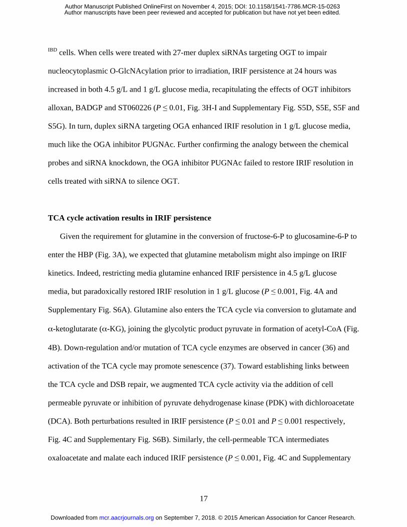
IBD cells. When cells were treated with 27-mer duplex siRNAs targeting OGT to impair
nucleocytoplasmic O-GlcNAcylation prior to irradiation, IRIF persistence at 24 hours was
increased in both 4.5 g/L and 1 g/L glucose media, recapitulating the effects of OGT inhibitors
alloxan, BADGP and ST060226 (P ≤ 0.01, Fig. 3H-I and Supplementary Fig. S5D, S5E, S5F and
S5G). In turn, duplex siRNA targeting OGA enhanced IRIF resolution in 1 g/L glucose media,
much like the OGA inhibitor PUGNAc. Further confirming the analogy between the chemical
probes and siRNA knockdown, the OGA inhibitor PUGNAc failed to restore IRIF resolution in
cells treated with siRNA to silence OGT.
TCA cycle activation results in IRIF persistence
Given the requirement for glutamine in the conversion of fructose-6-P to glucosamine-6-P to
enter the HBP (Fig. 3A), we expected that glutamine metabolism might also impinge on IRIF
kinetics. Indeed, restricting media glutamine enhanced IRIF persistence in 4.5 g/L glucose
media, but paradoxically restored IRIF resolution in 1 g/L glucose (P ≤ 0.001, Fig. 4A and
Supplementary Fig. S6A). Glutamine also enters the TCA cycle via conversion to glutamate and
α-ketoglutarate (α-KG), joining the glycolytic product pyruvate in formation of acetyl-CoA (Fig.
4B). Down-regulation and/or mutation of TCA cycle enzymes are observed in cancer (36) and
activation of the TCA cycle may promote senescence (37). Toward establishing links between
the TCA cycle and DSB repair, we augmented TCA cycle activity via the addition of cell
permeable pyruvate or inhibition of pyruvate dehydrogenase kinase (PDK) with dichloroacetate
(DCA). Both perturbations resulted in IRIF persistence (P ≤ 0.01 and P ≤ 0.001 respectively,
Fig. 4C and Supplementary Fig. S6B). Similarly, the cell-permeable TCA intermediates
oxaloacetate and malate each induced IRIF persistence (P ≤ 0.001, Fig. 4C and Supplementary
on September 7, 2018. © 2015 American Association for Cancer Research. mcr.aacrjournals.org Downloaded from
Author manuscripts have been peer reviewed and accepted for publication but have not yet been edited. Author Manuscript Published OnlineFirst on November 4, 2015; DOI: 10.1158/1541-7786.MCR-15-0263
18
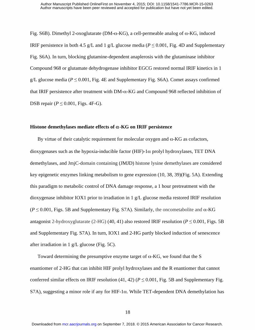
Fig. S6B). Dimethyl 2-oxoglutarate (DM-α-KG), a cell-permeable analog of α-KG, induced
IRIF persistence in both 4.5 g/L and 1 g/L glucose media (P ≤ 0.001, Fig. 4D and Supplementary
Fig. S6A). In turn, blocking glutamine-dependent anaplerosis with the glutaminase inhibitor
Compound 968 or glutamate dehydrogenase inhibitor EGCG restored normal IRIF kinetics in 1
g/L glucose media (P ≤ 0.001, Fig. 4E and Supplementary Fig. S6A). Comet assays confirmed
that IRIF persistence after treatment with DM-α-KG and Compound 968 reflected inhibition of
DSB repair (P ≤ 0.001, Figs. 4F-G).
Histone demethylases mediate effects of α-KG on IRIF persistence
By virtue of their catalytic requirement for molecular oxygen and α-KG as cofactors,
dioxygenases such as the hypoxia-inducible factor (HIF)-1α prolyl hydroxylases, TET DNA
demethylases, and JmjC-domain containing (JMJD) histone lysine demethylases are considered
key epigenetic enzymes linking metabolism to gene expression (10, 38, 39)(Fig. 5A). Extending
this paradigm to metabolic control of DNA damage response, a 1 hour pretreatment with the
dioxygenase inhibitor IOX1 prior to irradiation in 1 g/L glucose media restored IRIF resolution
(P ≤ 0.001, Figs. 5B and Supplementary Fig. S7A). Similarly, the oncometabolite and α-KG
antagonist 2-hydroxyglutarate (2-HG) (40, 41) also restored IRIF resolution (P ≤ 0.001, Figs. 5B
and Supplementary Fig. S7A). In turn, IOX1 and 2-HG partly blocked induction of senescence
after irradiation in 1 g/L glucose (Fig. 5C).
Toward determining the presumptive enzyme target of α-KG, we found that the S
enantiomer of 2-HG that can inhibit HIF prolyl hydroxylases and the R enantiomer that cannot
conferred similar effects on IRIF resolution (41, 42) (P ≤ 0.001, Fig. 5B and Supplementary Fig.
S7A), suggesting a minor role if any for HIF-1α. While TET-dependent DNA demethylation has
on September 7, 2018. © 2015 American Association for Cancer Research. mcr.aacrjournals.org Downloaded from
Author manuscripts have been peer reviewed and accepted for publication but have not yet been edited. Author Manuscript Published OnlineFirst on November 4, 2015; DOI: 10.1158/1541-7786.MCR-15-0263
19
been linked to excision repair, a role for TET proteins in DSB repair remains to be established.
By contrast, activation of JMJD proteins to demethylate histones H3 at K4, K9, K36, K79 and/or
H4 at K20 could antagonize well-known protein-protein interactions critical to IRIF function and
DSB repair (17). Indeed, treating cells with either PBIT to inhibit H3 K4 demethylase Jarid1A/B
or with GSKJ4 to block H3 K27 demethylase JMJD3 each restored IRIF resolution after
irradiation in 1 g/L glucose (P ≤ 0.01 and P ≤ 0.001 respectively, Fig. 5B and Supplementary Fig.
S7A), decreased senescence (Fig. 5C), and accelerated DNA repair as measured by comet assay
(P ≤ 0.001, Supplementary Fig. S7B). In turn, R-2-HG, IOX1 and GSKJ4 each blocked the
effect of DM-α-KG, restoring IRIF resolution and DNA repair (P ≤ 0.001, Fig. 5D-E and
Supplementary Fig. S7B).
Toward determining the order of function between O-GlcNAcylation and histone
demethylase activity in IRIF resolution, we examined relevant combinations of cell-permeable
metabolites and specific inhibitors. Both GlcNAc and PUGNAc restored IRIF resolution and
DNA repair in cells treated with DM-α-KG, suggesting that O-GlcNAcylation might promote
methylation or protect against demethylation (P ≤ 0.001, Fig. 5D-E and Supplementary Fig.
S7C-D). In a reciprocal experiment, the demethylase inhibitors IOX1 and R-2-HG overcame the
effects on IRIF resolution of OGT inhibitors alloxan and ST060266 (P ≤ 0.001, Fig. 5F and
Supplementary Fig. S7E). A conservative interpretation places α-KG-dependent lysine
demethylation downstream of O-GlcNAcylation in DNA repair and IRIF resolution.
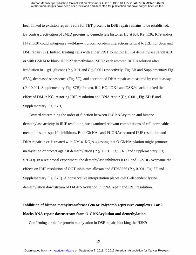
Inhibition of histone methyltransferase G9a or Polycomb repressive complexes 1 or 2
blocks DNA repair downstream from O-GlcNAcylation and demethylation
Confirming a role for protein methylation in DSB repair, blocking the H3K9
on September 7, 2018. © 2015 American Association for Cancer Research. mcr.aacrjournals.org Downloaded from
Author manuscripts have been peer reviewed and accepted for publication but have not yet been edited. Author Manuscript Published OnlineFirst on November 4, 2015; DOI: 10.1158/1541-7786.MCR-15-0263
20
methyltransferase G9a with BRD4770 or the H3K27 methyltransferase EZH2 with GSK126
each promoted IRIF persistence and delayed repair in both 4.5 and 1 g/L glucose (P ≤ 0.001, Fig.
6A-D). We placed O-GlcNAcylation upstream of histone methylation, insofar as neither GlcNAc
nor PUGNAc could overcome inhibition of IRIF resolution after treatment with
methyltransferase G9a inhibitor BRD4770 (Supplementary Fig. S7F-G).
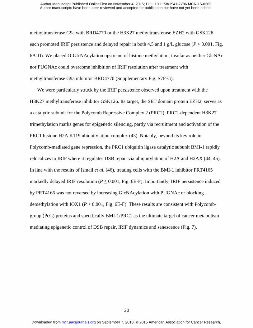
We were particularly struck by the IRIF persistence observed upon treatment with the
H3K27 methyltransferase inhibitor GSK126. Its target, the SET domain protein EZH2, serves as
a catalytic subunit for the Polycomb Repressive Complex 2 (PRC2). PRC2-dependent H3K27
trimethylation marks genes for epigenetic silencing, partly via recruitment and activation of the
PRC1 histone H2A K119 ubiquitylation complex (43). Notably, beyond its key role in
Polycomb-mediated gene repression, the PRC1 ubiquitin ligase catalytic subunit BMI-1 rapidly
relocalizes to IRIF where it regulates DSB repair via ubiquitylation of H2A and H2AX (44, 45).
In line with the results of Ismail et al. (46), treating cells with the BMI-1 inhibitor PRT4165
markedly delayed IRIF resolution (P ≤ 0.001, Fig. 6E-F). Importantly, IRIF persistence induced
by PRT4165 was not reversed by increasing GlcNAcylation with PUGNAc or blocking
demethylation with IOX1 (P ≤ 0.001, Fig. 6E-F). These results are consistent with Polycomb-
group (PcG) proteins and specifically BMI-1/PRC1 as the ultimate target of cancer metabolism
mediating epigenetic control of DSB repair, IRIF dynamics and senescence (Fig. 7).
on September 7, 2018. © 2015 American Association for Cancer Research. mcr.aacrjournals.org Downloaded from
Author manuscripts have been peer reviewed and accepted for publication but have not yet been edited. Author Manuscript Published OnlineFirst on November 4, 2015; DOI: 10.1158/1541-7786.MCR-15-0263
21
DISCUSSION
Among the many hallmarks of cancer (47), altered metabolism has returned to the forefront
as a potential therapeutic target. From Warburg's initial description of aerobic glycolysis to the
present, the defining feature of cancer metabolic reprogramming has remained the deregulated
uptake and utilization of glucose beyond cellular needs for ATP production. This apparent
inefficiency is commonly ascribed to the elevated demands for biosynthetic intermediates
required for rapid cell proliferation (3, 6, 7, 48) and/or as a means of resistance to cell death (49).
Supporting the former model, a moderate correlation has been observed across multiple studies
between 18F-fluorodeoxyglucose (FDG) uptake as measured by PET in human tumors and
fraction of Ki-67 positive nuclei or other measures of cell proliferation in biopsies. However,
recent discoveries that the oncogenicity of mutations of TCA cycle enzymes IDH1 and IDH2 is
mediated by excess production of the oncometabolite 2-HG, leading to inhibition of histone
and/or DNA demethylases (10, 50, 51), have lent support to an alternative model that implicates
metabolic reprogramming in deregulation of gene expression. Our work extends this model
beyond epigenetic regulation of transcription to effects of chromatin modification on DNA repair.
We found that disrupting glycolysis and/or glutaminolysis impaired the response to ionizing
radiation, thereby linking cancer metabolism to enhanced DNA double strand break repair. The
increased tolerance to genotoxic stress induced by metabolic reprogramming may promote
genomic instability and cell immortality in cancer cells by evading DNA-damage induced
senescence, independent of cell proliferation per se.
We note that our primary strategy was to exploit small molecule probes rather than genetic
tools to perturb cell metabolism. Beyond offering finer control of dose and timing compared to
RNAi (52), chemical probes are particularly suited to the analysis of metabolic networks, where
on September 7, 2018. © 2015 American Association for Cancer Research. mcr.aacrjournals.org Downloaded from
Author manuscripts have been peer reviewed and accepted for publication but have not yet been edited. Author Manuscript Published OnlineFirst on November 4, 2015; DOI: 10.1158/1541-7786.MCR-15-0263
22
intermediates and metabolites are often considered nodes connected by metabolic enzymes as
edges. While RNAi, CRISPR or other nucleic acid-targeted perturbations are powerful tools for
analysis of specific gene products and linear pathways, they are poorly matched to analysis of
cell metabolism, given the complex web of reactions that affect many intermediates. By contrast,
a single chemical probe may target multiple isoforms or whole classes of binding sites. In turn,
probes are readily used in combination and temporal order, allowing straightforward
interrogation of order-of-function, functional redundancy and other pathway relationships (53,
54). Of translational significance, many of the small molecules used here have been evaluated for
therapeutic use (55).
Among the most dramatic results was the block to DNA repair induced by TCA cycle
intermediates including the cell permeable analog of α-KG. In turn, the TCA cycle enzyme-
derived oncometabolite 2-HG blocked all these effects. Providing a strong argument that α-KG
and its competitive inhibitor 2-HG mediate their effects via epigenetic targets, 1) α-KG serves as
a co-factor for multiple dioxygenases including the Jumonji domain-containing (JMJD) histone
lysine demethylases while 2-HG is a dioxygenase and JMJD inhibitor, 2) α-KG effects were
recapitulated by G9a histone lysine methyltransferase inhibitor BRD4770 and EZH2
methyltransferase inhibitor GSK126, and 3) α-KG effects were blocked by Jarid1A/B histone
lysine demethylase inhibitor PBIT and JMJD3 demethylase inhibitor GSKJ4. These results
appear to provide a link between mitochondrial metabolism and genomic instability that may be
independent of oxidative phosphorylation and reactive oxygen species.
Among our results with the greatest potential for translation, we implicated the hexosamine
biosynthetic pathway (HBP) in regulation of DNA repair and senescence. We found that
activating OGT by treating cells with GlcNAc, a readily available neutraceutical used to relieve
on September 7, 2018. © 2015 American Association for Cancer Research. mcr.aacrjournals.org Downloaded from
Author manuscripts have been peer reviewed and accepted for publication but have not yet been edited. Author Manuscript Published OnlineFirst on November 4, 2015; DOI: 10.1158/1541-7786.MCR-15-0263
23
inflammation, or targeting OGA with its inhibitor PUGNAc or by knockdown with siRNA each
promote DSB repair. Blocking the pathway with small-molecule OGT inhibitors or by OGT
knockdown had the opposite effect. Of potentially relevant OGT targets, surveys of the O-
GlcNAc-ome have revealed modification of histones, DNA damage repair proteins, histone
methyltransferases and PcG proteins (56-58). Specifically, OGT mediates O-GlcNAcylation of
EZH2 on Ser75 in MCF7 cells, enhancing protein stability and maintaining H3 K27
trimethylation (59). Depletion of EZH2 is sufficient to delay DSB repair (60). However, given
the multiple targets of O-GlcNAcylation in epigenetic regulation, its precise roles in DSB repair
and IRIF kinetics remain to be elucidated.
Multiple lines of evidence point to cancer metabolism accelerating DSB repair via activation
of non-homologous end-joining (NHEJ). First, our use of GFP-IBD as a reporter may have
skewed our study toward tracking end-joining, given the known role of 53BP1 in promoting
NHEJ over homologous recombination (HR) (20, 61). In turn, NHEJ inhibitors promote IRIF
persistence and senescence after irradiation (62), while candidate HR inhibitors fail to promote
either response (our unpublished results). Providing a potential mechanistic link, several PcG
proteins have been linked to NHEJ by detection of physical interactions and/or functional studies.
One implication is that cancer metabolic reprogramming may speed resolution of DSBs by rapid
NHEJ repair before they can initiate slow, but accurate, HR repair. Cancer cells may gain an
advantage not only from more efficient DSB repair that supports proliferation in the face of
genotoxic stress, but also from the increased genomic instability due to error-prone repair.
The Warburg effect has been widely recognized as a driver of cancer cell growth,
tumorigenicity and therapeutic resistance. Our chemical genetics approach revealed that cancer
metabolism also promotes DNA repair and thereby blocks accelerated senescence, inactivating a
on September 7, 2018. © 2015 American Association for Cancer Research. mcr.aacrjournals.org Downloaded from
Author manuscripts have been peer reviewed and accepted for publication but have not yet been edited. Author Manuscript Published OnlineFirst on November 4, 2015; DOI: 10.1158/1541-7786.MCR-15-0263
24
recognized tumor suppressor mechanism that serves as a critical barrier to carcinogenesis (63-
65). These studies identified roles for both glycolysis and glutaminolysis via their dual
contributions to the TCA cycle and HBP as key determinants of cellular responses to DNA
damage. Our data also suggest a reexamination of the mechanisms of carcinogenesis by
oncometabolites to include mechanisms beyond deregulation of gene expression. Indeed, 2-HG
alone appears sufficient to recapitulate the benefits of metabolic reprogramming in promoting
DNA repair and blocking cell senescence. Similarly, that treating cells with GlcNAc mirrors the
effects of the oncometabolite 2-HG supports a reevaluation of O-GlcNAcylation as a mediator
and target in cancer and aging.
Taken together, our results suggest that along with an established role in supporting cell
proliferation, increased glycolysis and glutaminolysis may also support cancer cell survival in
the face of genotoxic stress. By promoting DNA repair by error-prone non-homologous end-
joining, metabolic reprogramming may serve a previously unrecognized role in tumor
progression to increase genomic instability and maintain cellular immortality.
on September 7, 2018. © 2015 American Association for Cancer Research. mcr.aacrjournals.org Downloaded from
Author manuscripts have been peer reviewed and accepted for publication but have not yet been edited. Author Manuscript Published OnlineFirst on November 4, 2015; DOI: 10.1158/1541-7786.MCR-15-0263
25
Author’s Contributions
Conception and design: E.V. Efimova and S.J. Kron
Development and methodology: E.V. Efimova, S. Takahashi, N.A. Shamsi, D. Wu, E. Labay,
O.A. Ulanovskaya
Acquisition of data: E.V. Efimova, S.Takahashi, N.A. Shamsi, D. Wu, E. Labay, O.A.
Ulanovskaya
Analysis and interpretation of data: E.V. Efimova, S. Takahashi, N.A. Shamsi, D. Wu, E.
Labay, O.A. Ulanovskaya, R.R. Weichselbaum, S.A. Kozmin, and S.J. Kron
Writing, review, and/or revision of the manuscript: E.V. Efimova, S.Takahashi, N. A.
Shamsi, D. Wu, E. Labay, O.A. Ulanovskaya, R.R. Weichselbaum, S.A. Kozmin, and S.J. Kron
Study supervision: R.R. Weichselbaum, S.A. Kozmin, and S.J. Kron
Acknowledgements
We thank S. Bond, C. Labno, and V. Bindokas in The University of Chicago Integrated
Microscopy Core Facility for technical assistance, V.Boilot and A. Ramamurthy for technical
assistance and proofreading, and W. Lu and J. Rabinowitz at Princeton University for helpful
discussions.
on September 7, 2018. © 2015 American Association for Cancer Research. mcr.aacrjournals.org Downloaded from
Author manuscripts have been peer reviewed and accepted for publication but have not yet been edited. Author Manuscript Published OnlineFirst on November 4, 2015; DOI: 10.1158/1541-7786.MCR-15-0263

26
REFERENCES
1. Warburg O. On the origin of cancer cells. Science. 1956;123:309-14.
2. Frezza C, Gottlieb E. Mitochondria in cancer: not just innocent bystanders. Semin Cancer
Biol. 2009;19:4-11.
3. Vander Heiden MG, Cantley LC, Thompson CB. Understanding the Warburg effect: the
metabolic requirements of cell proliferation. Science. 2009;324:1029-33.
4. Cantor JR, Sabatini DM. Cancer cell metabolism: one hallmark, many faces. Cancer
Discovery. 2012;2:881-98.
5. Eagle H. Nutrition needs of mammalian cells in tissue culture. Science. 1955;122:501-14.
6. Wise DR, Thompson CB. Glutamine addiction: a new therapeutic target in cancer. Trends in
Biochemical Sciences. 2010;35:427-33.
7. DeBerardinis RJ, Cheng T. Q's next: the diverse functions of glutamine in metabolism, cell
biology and cancer. Oncogene. 2010;29:313-24.
8. Dang CV. Rethinking the Warburg effect with Myc micromanaging glutamine metabolism.
Cancer Res. 2010;70:859-62.
9. Lu C, Thompson CB. Metabolic regulation of epigenetics. Cell Metab. 2012;16:9-17.
10. Kaelin WG, Jr., McKnight SL. Influence of metabolism on epigenetics and disease. Cell.
2013;153:56-69.
11. Gut P, Verdin E. The nexus of chromatin regulation and intermediary metabolism. Nature.
2013;502:489-98.
12. Badeaux A, Shi Y. Emerging roles for chromatin as a signal integration and storage platform.
Nat Rev Mol Cell Biol. 2013;14:211-24.
on September 7, 2018. © 2015 American Association for Cancer Research. mcr.aacrjournals.org Downloaded from
Author manuscripts have been peer reviewed and accepted for publication but have not yet been edited. Author Manuscript Published OnlineFirst on November 4, 2015; DOI: 10.1158/1541-7786.MCR-15-0263
27
13. Liu XS, Little JB, Yuan ZM. Glycolytic metabolism influences global chromatin structure.
Oncotarget. 2015;6:4214-25.
14. Liu J, Kim J, Oberdoerffer P. Metabolic modulation of chromatin: implications for DNA
repair and genomic integrity. Front Genet. 2013;4:182.
15. Bonner WM, Redon CE, Dickey JS, Nakamura AJ, Sedelnikova O, Solier S, et al.
GammaH2AX and cancer. Nat Rev Cancer. 2008;8:957-67.
16. Rossetto D, Truman AW, Kron SJ, Côté J. Epigenetic modifications in double-strand break
DNA damage signaling and repair. Clin Cancer Res. 2010;16:4543-52.
17. Hunt CR, Ramnarain D, Horikoshi N, Iyengar P, Pandita RK, Shay JW, et al. Histone
modifications and DNA double-strand break repair after exposure to ionizing radiations.
Radiat Res. 2013;179:383-92.
18. Smeenk G, van Attikum H. The chromatin response to DNA breaks: leaving a mark on
genome integrity. Annu Rev Biochem. 2013;82:55-80.
19. Goodarzi A, Jeggo P. Irradiation induced foci (IRIF) as a biomarker for radiosensitivity.
Mutat Res. 2012;736:39-47.
20. Panier S, Boulton SJ. Double-strand break repair: 53BP1 comes into focus. Nat Rev Mol Cell
Biol. 2014;15:7-18.
21. Huyen Y, Zgheib O, Ditullio RA, Gorgoulis VG, Zacharatos P, Petty TJ, et al. Methylated
lysine 79 of histone H3 targets 53BP1 to DNA double-strand breaks. Nature. 2004;432:406-
11.
22. Botuyan MV, Lee J, Ward IM, Kim JE, Thompson JR, Chen J, et al. Structural basis for the
methylation state-specific recognition of histone H4-K20 by 53BP1 and Crb2 in DNA repair.
Cell. 2006;127:1361-73.
on September 7, 2018. © 2015 American Association for Cancer Research. mcr.aacrjournals.org Downloaded from
Author manuscripts have been peer reviewed and accepted for publication but have not yet been edited. Author Manuscript Published OnlineFirst on November 4, 2015; DOI: 10.1158/1541-7786.MCR-15-0263
28
23. Fradet-Turcotte A, Canny MD, Escribano-Díaz C, Orthwein A, Leung CCY, Huang H, et al.
53BP1 is a reader of the DNA-damage-induced H2A Lys 15 ubiquitin mark. Nature.
2013;499:50-4.
24. Efimova EV, Mauceri HJ, Golden DW, Labay E, Bindokas VP, Darga TE, et al. Poly(ADP-
Ribose) Polymerase Inhibitor Induces Accelerated Senescence in Irradiated Breast Cancer
Cells and Tumors. Cancer Res. 2010;70:6277-82.
25. Labay E, Efimova E, Quarshie B, Golden D, Weichselbaum R, Kron S. Ionizing radiation-
induced foci persistence screen to discover enhancers of accelerated senescence. Int J High
Throughput Screen. 2011;2:1-13.
26. Ciccia A, Elledge SJ. The DNA damage response: making it safe to play with knives. Mol
Cell. 2010;40:179-204.
27. Ulanovskaya O, Cui J, Kron S, Kozmin S. A Pairwise Chemical Genetic Screen Identifies
New Inhibitors of Glucose Transport. Chemistry & Biology. 2011;18:222-30.
28. Guppy M, Leedman P, Zu X, Russell V. Contribution by different fuels and metabolic
pathways to the total ATP turnover of proliferating MCF-7 breast cancer cells. Biochem J.
2002;364:309-15.
29. Kondoh H, Lleonart ME, Gil J, Wang J, Degan P, Peters G, et al. Glycolytic enzymes can
modulate cellular life span. Cancer Res. 2005;65:177-85.
30. Wang M, Kaufman RJ. The impact of the endoplasmic reticulum protein-folding
environment on cancer development. Nat Rev Cancer. 2014;14:581-97.
31. Lee AS. The ER chaperone and signaling regulator GRP78/BiP as a monitor of endoplasmic
reticulum stress. Methods. 2005;35:373-81.
on September 7, 2018. © 2015 American Association for Cancer Research. mcr.aacrjournals.org Downloaded from
Author manuscripts have been peer reviewed and accepted for publication but have not yet been edited. Author Manuscript Published OnlineFirst on November 4, 2015; DOI: 10.1158/1541-7786.MCR-15-0263
29
32. Kurtoglu M, Gao N, Shang J, Maher J, Lehrman MA, Wangpaichitr M, et al. Under
normoxia, 2-deoxy-D-glucose elicits cell death in select tumor types not by inhibition of
glycolysis but by interfering with N-linked glycosylation. Molecular Cancer Therapeutics.
2007;6:3049-58.
33. Wellen KE, Thompson CB. Cellular metabolic stress: considering how cells respond to
nutrient excess. Molecular Cell. 2010;40:323-32.
34. Hart G, Slawson C, Ramirez-Correa G, Lagerlof O. Cross talk between O-GlcNAcylation
and phosphorylation: roles in signaling, transcription, and chronic disease. Annu Rev
Biochem. 2011;80:825-58.
35. Hanover JA, Krause MW, Love DC. Bittersweet memories: linking metabolism to
epigenetics through O-GlcNAcylation. Nat Rev Mol Cell Biol. 2012;13:312-21.
36. Wallace DC. Mitochondria and cancer. Nat Rev Cancer. 2012;12:685-98.
37. Kaplon J, Zheng L, Meissl K, Chaneton B, Selivanov VA, Mackay G, et al. A key role for
mitochondrial gatekeeper pyruvate dehydrogenase in oncogene-induced senescence. Nature.
2013;498:109-12.
38. Branco MR, Ficz G, Reik W. Uncovering the role of 5-hydroxymethylcytosine in the
epigenome. Nat Rev Genet. 2011;13:7-13.
39. Ward PS, Thompson CB. Metabolic reprogramming: a cancer hallmark even warburg did not
anticipate. Cancer Cell. 2012;21:297-308.
40. Xu W, Yang H, Liu Y, Yang Y, Wang P, Kim S, et al. Oncometabolite 2-hydroxyglutarate is
a competitive inhibitor of α-ketoglutarate-dependent dioxygenases. Cancer Cell. 2011;19:17-
30.
on September 7, 2018. © 2015 American Association for Cancer Research. mcr.aacrjournals.org Downloaded from
Author manuscripts have been peer reviewed and accepted for publication but have not yet been edited. Author Manuscript Published OnlineFirst on November 4, 2015; DOI: 10.1158/1541-7786.MCR-15-0263
30
41. Chowdhury R, Yeoh KK, Tian Y, Hillringhaus L, Bagg EA, Rose N, et al. The
oncometabolite 2-hydroxyglutarate inhibits histone lysine demethylases. EMBO Rep.
2011;12:463-9.
42. Losman JA, Looper RE, Koivunen P, Lee S, Schneider RK, McMahon C, et al. (R)-2-
hydroxyglutarate is sufficient to promote leukemogenesis and its effects are reversible.
Science. 2013;339:1621-5.
43. Margueron R, Reinberg D. The Polycomb complex PRC2 and its mark in life. Nature.
2011;469:343-9.
44. Liu J, Cao L, Chen J, Song S, Lee IH, Quijano C, et al. Bmi1 regulates mitochondrial
function and the DNA damage response pathway. Nature. 2009;459:387-92.
45. Gieni RS, Ismail IH, Campbell S, Hendzel MJ. Polycomb group proteins in the DNA damage
response: a link between radiation resistance and "stemness". Cell Cycle. 2011;10:883-94.
46. Ismail IH, McDonald D, Strickfaden H, Xu Z, Hendzel MJ. A small molecule inhibitor of
polycomb repressive complex 1 inhibits ubiquitin signaling at DNA double-strand breaks. J
Biol Chem. 2013;288:26944-54.
47. Hanahan D, Weinberg RA. Hallmarks of cancer: the next generation. Cell. 2011;144:646-74.
48. Koppenol WH, Bounds PL, Dang CV. Otto Warburg's contributions to current concepts of
cancer metabolism. Nat Rev Cancer. 2011;11:325-37.
49. Buchakjian MR, Kornbluth S. The engine driving the ship: metabolic steering of cell
proliferation and death. Nat Rev Mol Cell Biol. 2010;11:715-27.
50. Yuan H, Xiong Y, Guan K. Nutrient sensing, metabolism, and cell growth control. Molecular
Cell. 2013;49:379-87.
on September 7, 2018. © 2015 American Association for Cancer Research. mcr.aacrjournals.org Downloaded from
Author manuscripts have been peer reviewed and accepted for publication but have not yet been edited. Author Manuscript Published OnlineFirst on November 4, 2015; DOI: 10.1158/1541-7786.MCR-15-0263
31
51. Delatte B, Deplus R, Fuks F. Playing TETris with DNA modifications. EMBO J.
2014;33:1198-211.
52. Frye SV. The art of the chemical probe. Nat Chem Biol. 2010;6:159-61.
53. Lehar J, Stockwell BR, Giaever G, Nislow C. Combination chemical genetics. Nat Chem
Biol. 2008;4:674-81.
54. Castoreno AB, Eggert US. Small molecule probes of cellular pathways and networks. ACS
Chem Biol. 2011;6:86-94.
55. Galluzzi L, Kepp O, Vander Heiden MG, Kroemer G. Metabolic targets for cancer therapy.
Nat Rev Drug Discov. 2013;12:829-46.
56. Nandi A, Sprung R, Barma DK, Zhao Y, Kim SC, Falck JR, et al. Global identification of O-
GlcNAc-modified proteins. Anal Chem. 2006;78:452-8.
57. Love DC, Krause MW, Hanover JA. O-GlcNAc cycling: emerging roles in development and
epigenetics. Semin Cell Dev Biol. 2010;21:646-54.
58. Zachara N, Molina H, Wong K, Pandey A, Hart G. The dynamic stress-induced "O-GlcNAc-
ome" highlights functions for O-GlcNAc in regulating DNA damage/repair and other cellular
pathways. Amino Acids. 2011;40:793-808.
59. Chu CS, Lo PW, Yeh YH, Hsu PH, Peng SH, Teng YC, et al. O-GlcNAcylation regulates
EZH2 protein stability and function. Proc Natl Acad Sci U S A. 2014;111:1355-60.
60. Campbell S, Ismail IH, Young LC, Poirier GG, Hendzel MJ. Polycomb repressive complex 2
contributes to DNA double-strand break repair. Cell Cycle. 2013;12:2675-83.
61. Zimmermann M, de Lange T. 53BP1: pro choice in DNA repair. Trends Cell Biol.
2014;24:108-17.
on September 7, 2018. © 2015 American Association for Cancer Research. mcr.aacrjournals.org Downloaded from
Author manuscripts have been peer reviewed and accepted for publication but have not yet been edited. Author Manuscript Published OnlineFirst on November 4, 2015; DOI: 10.1158/1541-7786.MCR-15-0263
32
62. Azad A, Bukczynska P, Jackson S, Haupt Y, Cullinane C, McArthur GA, et al. Co-targeting
deoxyribonucleic acid-dependent protein kinase and poly(adenosine diphosphate-ribose)
polymerase-1 promotes accelerated senescence of irradiated cancer cells. Int J Radiat Oncol
Biol Phys. 2014;88:385-94.
63. Lowe SW, Cepero E, Evan G. Intrinsic tumour suppression. Nature. 2004;432:307-15.
64. Campisi J, D'adda Di Fagagna F. Cellular senescence: when bad things happen to good cells.
Nat Rev Mol Cell Biol. 2007;8:729-40.
65. D'adda Di Fagagna F. Living on a break: cellular senescence as a DNA-damage response.
Nat Rev Cancer. 2008;8:512-22.
66. Dimri GP, Lee X, Basile G, Acosta M, Scott G, Roskelley C, et al. A biomarker that
identifies senescent human cells in culture and in aging skin in vivo. Proc Natl Acad Sci
USA. 1995;92:9363-7.
on September 7, 2018. © 2015 American Association for Cancer Research. mcr.aacrjournals.org Downloaded from
Author manuscripts have been peer reviewed and accepted for publication but have not yet been edited. Author Manuscript Published OnlineFirst on November 4, 2015; DOI: 10.1158/1541-7786.MCR-15-0263
33
Figure Legends
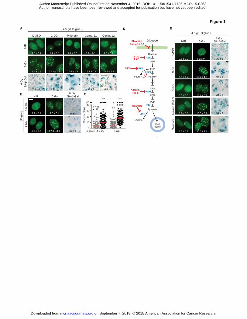
Figure 1. Targeting glycolysis blocks IRIF resolution and accelerates senescence. A, Glucose
uptake inhibitors do not induce DNA damage in nonirradiated cells (NIR) but promote IRIF
persistence and accelerated senescence after irradiation with 6 Gy (IR) in 4.5 g/L glucose.
Representative cell images, with GFP-IBD fluorescence examined 24 hours after irradiation
(IRIF, green; foci numbers per nucleus are shown (mean ± SEM) and senescence-associated β-
galactosidase activity detected 5 days after irradiation by X-Gal (SA-β-Gal, blue). Degree of
senescence phenotype was assessed by % SA-β-Gal (+) cells and indicated as mean ± SEM. B,
Lowering media glucose to 1 g/L promotes IRIF persistence and senescence after irradiation.
Representative images, with IRIF per nucleus (mean ± SEM) indicated. C, Plots of IRIF per
nucleus in individual cells after 6 Gy in 4.5 g/L and 1 g /L glucose (NIR,�; IR,�), red bar
indicates mean ± SEM. ***, P ≤ 0.001, unpaired t-test. D, Glycolysis pathway and chemical
probes. E, Small molecule inhibitors of glycolysis induce IRIF persistence and accelerated
senescence in 4.5 g/L glucose. See Supplementary Table 1 for concentrations of all probes used.
Shown are representative images of cell nuclei with IRIF, with means ± SEM indicated, and SA-
β-Gal assay cell images. Scale bars, 10 µm (IRIF); 50 µm (SA-β-Gal).
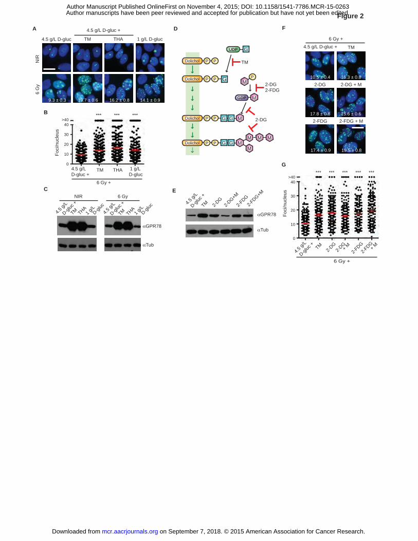
Figure 2. Inhibition of O-GlcNAcylation results in IRIF persistence independent of UPR
activation. A, Induction of unfolded protein response (UPR) causes IRIF persistence. MCF7GFP-
IBD cells were grown in 1 g/L or 4.5 g/L glucose media, treated with TM or THA 1 hour before 6
Gy irradiation, and observed after 24 hours. IRIF (green) per nucleus (blue) are shown for
irradiated cells (mean ± SEM). Scale bar, 10 µm. B, Plots of IRIF per nucleus from A. Red bar,
on September 7, 2018. © 2015 American Association for Cancer Research. mcr.aacrjournals.org Downloaded from
Author manuscripts have been peer reviewed and accepted for publication but have not yet been edited. Author Manuscript Published OnlineFirst on November 4, 2015; DOI: 10.1158/1541-7786.MCR-15-0263
34
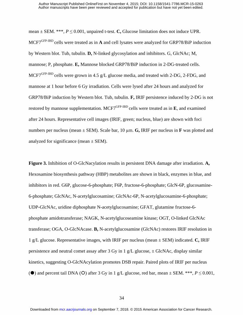
mean ± SEM. ***, P ≤ 0.001, unpaired t-test. C, Glucose limitation does not induce UPR.
MCF7GFP-IBD cells were treated as in A and cell lysates were analyzed for GRP78/BiP induction
by Western blot. Tub, tubulin. D, N-linked glycosylation and inhibitors. G, GlcNAc; M,
mannose; P, phosphate. E, Mannose blocked GRP78/BiP induction in 2-DG-treated cells.
MCF7GFP-IBD cells were grown in 4.5 g/L glucose media, and treated with 2-DG, 2-FDG, and
mannose at 1 hour before 6 Gy irradiation. Cells were lysed after 24 hours and analyzed for
GRP78/BiP induction by Western blot. Tub, tubulin. F, IRIF persistence induced by 2-DG is not
restored by mannose supplementation. MCF7GFP-IBD cells were treated as in E, and examined
after 24 hours. Representative cell images (IRIF, green; nucleus, blue) are shown with foci
numbers per nucleus (mean ± SEM). Scale bar, 10 µm. G, IRIF per nucleus in F was plotted and
analyzed for significance (mean ± SEM).
Figure 3. Inhibition of O-GlcNacylation results in persistent DNA damage after irradiation. A,
Hexosamine biosynthesis pathway (HBP) metabolites are shown in black, enzymes in blue, and
inhibitors in red. G6P, glucose-6-phosphate; F6P, fructose-6-phosphate; GlcN-6P, glucosamine-
6-phosphate; GlcNAc, N-acetylglucosamine; GlcNAc-6P, N-acetylglucosamine-6-phosphate;
UDP-GlcNAc, uridine diphosphate N-acetylglucosamine; GFAT, glutamine fructose-6-
phosphate amidotransferase; NAGK, N-acetylglucoseamine kinase; OGT, O-linked GlcNAc
transferase; OGA, O-GlcNAcase. B, N-acetylglucosamine (GlcNAc) restores IRIF resolution in
1 g/L glucose. Representative images, with IRIF per nucleus (mean ± SEM) indicated. C, IRIF
persistence and neutral comet assay after 3 Gy in 1 g/L glucose, ± GlcNAc, display similar
kinetics, suggesting O-GlcNAcylation promotes DSB repair. Paired plots of IRIF per nucleus
(�) and percent tail DNA (�) after 3 Gy in 1 g/L glucose, red bar, mean ± SEM. ***, P ≤ 0.001,
on September 7, 2018. © 2015 American Association for Cancer Research. mcr.aacrjournals.org Downloaded from
Author manuscripts have been peer reviewed and accepted for publication but have not yet been edited. Author Manuscript Published OnlineFirst on November 4, 2015; DOI: 10.1158/1541-7786.MCR-15-0263
35
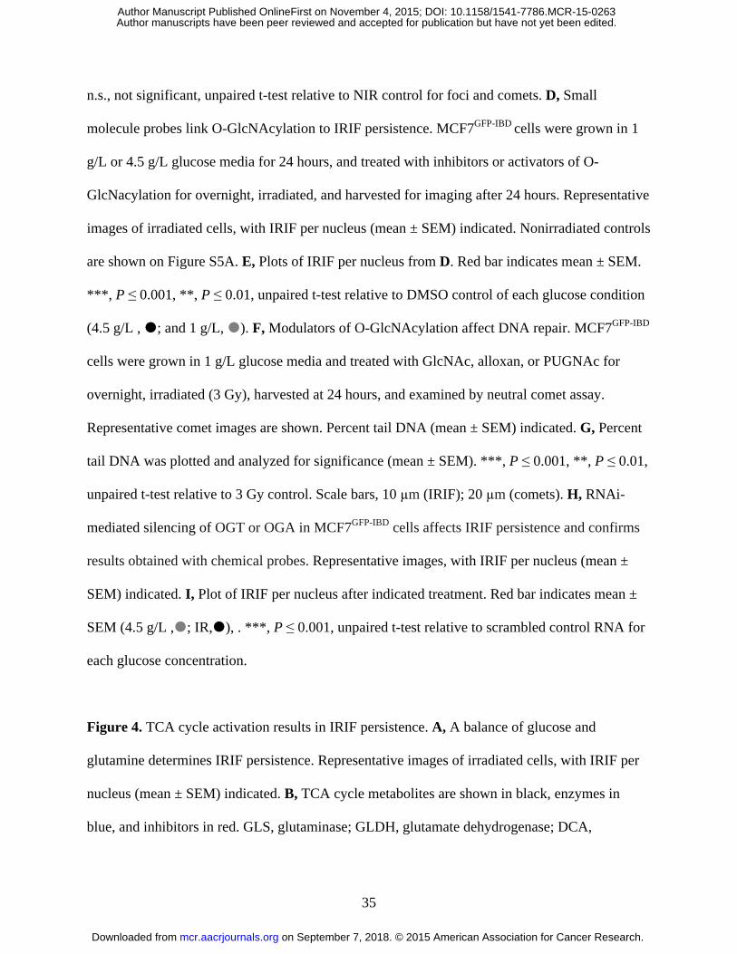
n.s., not significant, unpaired t-test relative to NIR control for foci and comets. D, Small
molecule probes link O-GlcNAcylation to IRIF persistence. MCF7GFP-IBD cells were grown in 1
g/L or 4.5 g/L glucose media for 24 hours, and treated with inhibitors or activators of O-
GlcNacylation for overnight, irradiated, and harvested for imaging after 24 hours. Representative
images of irradiated cells, with IRIF per nucleus (mean ± SEM) indicated. Nonirradiated controls
are shown on Figure S5A. E, Plots of IRIF per nucleus from D. Red bar indicates mean ± SEM.
***, P ≤ 0.001, **, P ≤ 0.01, unpaired t-test relative to DMSO control of each glucose condition
(4.5 g/L , �; and 1 g/L, �). F, Modulators of O-GlcNAcylation affect DNA repair. MCF7GFP-IBD
cells were grown in 1 g/L glucose media and treated with GlcNAc, alloxan, or PUGNAc for
overnight, irradiated (3 Gy), harvested at 24 hours, and examined by neutral comet assay.
Representative comet images are shown. Percent tail DNA (mean ± SEM) indicated. G, Percent
tail DNA was plotted and analyzed for significance (mean ± SEM). ***, P ≤ 0.001, **, P ≤ 0.01,
unpaired t-test relative to 3 Gy control. Scale bars, 10 µm (IRIF); 20 µm (comets). H, RNAi-
mediated silencing of OGT or OGA in MCF7GFP-IBD cells affects IRIF persistence and confirms
results obtained with chemical probes. Representative images, with IRIF per nucleus (mean ±
SEM) indicated. I, Plot of IRIF per nucleus after indicated treatment. Red bar indicates mean ±
SEM (4.5 g/L ,�; IR,�), . ***, P ≤ 0.001, unpaired t-test relative to scrambled control RNA for
each glucose concentration.
Figure 4. TCA cycle activation results in IRIF persistence. A, A balance of glucose and
glutamine determines IRIF persistence. Representative images of irradiated cells, with IRIF per
nucleus (mean ± SEM) indicated. B, TCA cycle metabolites are shown in black, enzymes in
blue, and inhibitors in red. GLS, glutaminase; GLDH, glutamate dehydrogenase; DCA,
on September 7, 2018. © 2015 American Association for Cancer Research. mcr.aacrjournals.org Downloaded from
Author manuscripts have been peer reviewed and accepted for publication but have not yet been edited. Author Manuscript Published OnlineFirst on November 4, 2015; DOI: 10.1158/1541-7786.MCR-15-0263
36
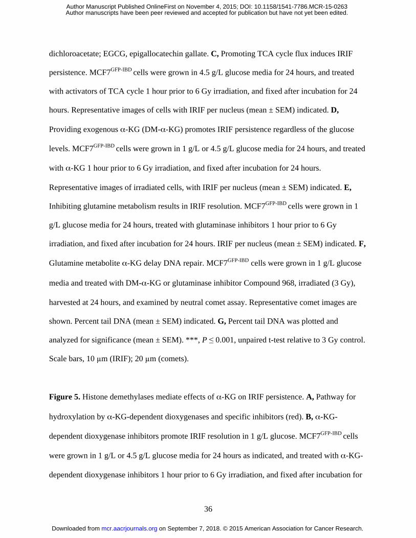
dichloroacetate; EGCG, epigallocatechin gallate. C, Promoting TCA cycle flux induces IRIF
persistence. MCF7GFP-IBD cells were grown in 4.5 g/L glucose media for 24 hours, and treated
with activators of TCA cycle 1 hour prior to 6 Gy irradiation, and fixed after incubation for 24
hours. Representative images of cells with IRIF per nucleus (mean ± SEM) indicated. D,
Providing exogenous α-KG (DM-α-KG) promotes IRIF persistence regardless of the glucose
levels. MCF7GFP-IBD cells were grown in 1 g/L or 4.5 g/L glucose media for 24 hours, and treated
with α-KG 1 hour prior to 6 Gy irradiation, and fixed after incubation for 24 hours.
Representative images of irradiated cells, with IRIF per nucleus (mean ± SEM) indicated. E,
Inhibiting glutamine metabolism results in IRIF resolution. MCF7GFP-IBD cells were grown in 1
g/L glucose media for 24 hours, treated with glutaminase inhibitors 1 hour prior to 6 Gy
irradiation, and fixed after incubation for 24 hours. IRIF per nucleus (mean ± SEM) indicated. F,
Glutamine metabolite α-KG delay DNA repair. MCF7GFP-IBD cells were grown in 1 g/L glucose
media and treated with DM-α-KG or glutaminase inhibitor Compound 968, irradiated (3 Gy),
harvested at 24 hours, and examined by neutral comet assay. Representative comet images are
shown. Percent tail DNA (mean ± SEM) indicated. G, Percent tail DNA was plotted and
analyzed for significance (mean ± SEM). ***, P ≤ 0.001, unpaired t-test relative to 3 Gy control.
Scale bars, 10 µm (IRIF); 20 µm (comets).
Figure 5. Histone demethylases mediate effects of α-KG on IRIF persistence. A, Pathway for
hydroxylation by α-KG-dependent dioxygenases and specific inhibitors (red). B, α-KG-
dependent dioxygenase inhibitors promote IRIF resolution in 1 g/L glucose. MCF7GFP-IBD cells
were grown in 1 g/L or 4.5 g/L glucose media for 24 hours as indicated, and treated with α-KG-
dependent dioxygenase inhibitors 1 hour prior to 6 Gy irradiation, and fixed after incubation for
on September 7, 2018. © 2015 American Association for Cancer Research. mcr.aacrjournals.org Downloaded from
Author manuscripts have been peer reviewed and accepted for publication but have not yet been edited. Author Manuscript Published OnlineFirst on November 4, 2015; DOI: 10.1158/1541-7786.MCR-15-0263
37
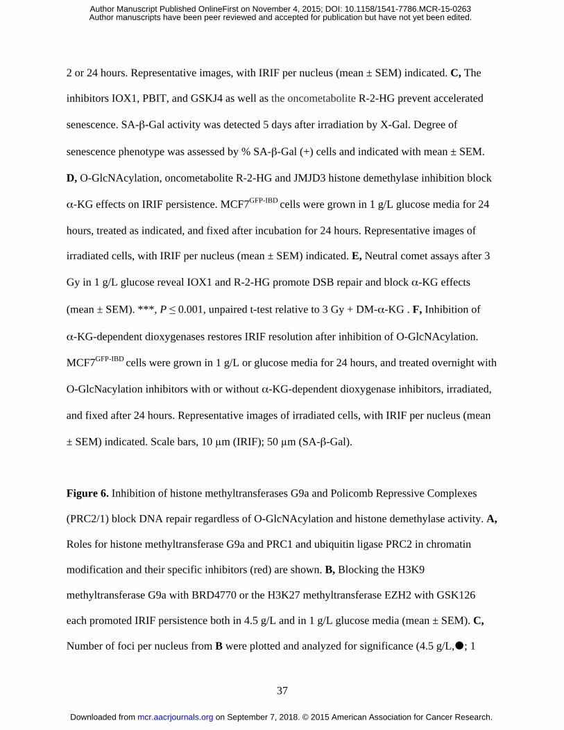
2 or 24 hours. Representative images, with IRIF per nucleus (mean ± SEM) indicated. C, The
inhibitors IOX1, PBIT, and GSKJ4 as well as the oncometabolite R-2-HG prevent accelerated
senescence. SA-β-Gal activity was detected 5 days after irradiation by X-Gal. Degree of
senescence phenotype was assessed by % SA-β-Gal (+) cells and indicated with mean ± SEM.
D, O-GlcNAcylation, oncometabolite R-2-HG and JMJD3 histone demethylase inhibition block
α-KG effects on IRIF persistence. MCF7GFP-IBD cells were grown in 1 g/L glucose media for 24
hours, treated as indicated, and fixed after incubation for 24 hours. Representative images of
irradiated cells, with IRIF per nucleus (mean ± SEM) indicated. E, Neutral comet assays after 3
Gy in 1 g/L glucose reveal IOX1 and R-2-HG promote DSB repair and block α-KG effects
(mean ± SEM). ***, P ≤ 0.001, unpaired t-test relative to 3 Gy + DM-α-KG . F, Inhibition of
α-KG-dependent dioxygenases restores IRIF resolution after inhibition of O-GlcNAcylation.
MCF7GFP-IBD cells were grown in 1 g/L or glucose media for 24 hours, and treated overnight with
O-GlcNacylation inhibitors with or without α-KG-dependent dioxygenase inhibitors, irradiated,
and fixed after 24 hours. Representative images of irradiated cells, with IRIF per nucleus (mean
± SEM) indicated. Scale bars, 10 µm (IRIF); 50 µm (SA-β-Gal).
Figure 6. Inhibition of histone methyltransferases G9a and Policomb Repressive Complexes
(PRC2/1) block DNA repair regardless of O-GlcNAcylation and histone demethylase activity. A,
Roles for histone methyltransferase G9a and PRC1 and ubiquitin ligase PRC2 in chromatin
modification and their specific inhibitors (red) are shown. B, Blocking the H3K9
methyltransferase G9a with BRD4770 or the H3K27 methyltransferase EZH2 with GSK126
each promoted IRIF persistence both in 4.5 g/L and in 1 g/L glucose media (mean ± SEM). C,
Number of foci per nucleus from B were plotted and analyzed for significance (4.5 g/L,�; 1
on September 7, 2018. © 2015 American Association for Cancer Research. mcr.aacrjournals.org Downloaded from
Author manuscripts have been peer reviewed and accepted for publication but have not yet been edited. Author Manuscript Published OnlineFirst on November 4, 2015; DOI: 10.1158/1541-7786.MCR-15-0263
38
g/L,�; mean ± SEM). ***, P ≤ 0.001, unpaired t-test relative to IR control (6 Gy) of each
glucose condition. D, Inhibitors of histone methyltransferases G9a and EZH2 block DSB repair.
Neutral comet assays after 3 Gy in 1 g/L glucose. Percent tail DNA was plotted and analyzed for
significance (mean ± SEM). ***, P ≤ 0.001, unpaired t-test relative to 3 Gy control. E, Small-
molecule inhibition of PRC1 (BMI1/RNF2) E3 ubiquitin ligase induces IRIF persistence,
independent of α-KG-dependent dioxygenase activity or O-GlcNAcylation (mean ± SEM). F,
Number of IRIF per nucleus from (E) was plotted and analyzed for significance. ***, P ≤ 0.001,
n.s., not significant, unpaired t-test relative to IR control. Scale bars, 10 µm.
Figure 7. Model linking metabolic reprogramming to cancer cell immortality via modulation of
DNA repair. These studies with chemical probes and RNA interference have implicated
increased glucose and glutamine metabolism in mediated increased DNA double strand break
repair, thereby supporting continued proliferation and resisting accelerated senescence. Prior
work has established a key role for chromatin in regulation of DNA damage repair, mediated by
phosphorylation of histone H2AX at sites of damage, leading to recruitment of 53BP1 and other
repair and signaling factors, inducing a checkpoint signal that promotes accelerated senescence.
Our data implicate cancer metabolic reprogramming in accelerating DNA repair, leading to
H2AX dephosphorylation, release of 53BP1 and rapid return to proliferation. We found a role for
the hexosamine biosynthetic pathway (HBP) metabolite N-acetyl-glucosamine and O-
GlcNAcylation (O-GlcNAc) in promoting histone methylation by EZH2 and/or G9A upstream of
the E3 ubiquitin ligase activity of the PRC1 polycomb group complex, which has a well-
established role in non-homologous end-joining repair. This activity is normally opposed by the
TCA cycle product α-ketoglutarate (α-KG), which can promote histone demethylation by JMJD
on September 7, 2018. © 2015 American Association for Cancer Research. mcr.aacrjournals.org Downloaded from
Author manuscripts have been peer reviewed and accepted for publication but have not yet been edited. Author Manuscript Published OnlineFirst on November 4, 2015; DOI: 10.1158/1541-7786.MCR-15-0263
39
demethylases. Downregulation of the TCA cycle or overproduction of the oncometabolite 2-
hydroxyglutarate (2-HG) blocks demethylation to accelerate DNA repair and resist senescence.
on September 7, 2018. © 2015 American Association for Cancer Research. mcr.aacrjournals.org Downloaded from
Author manuscripts have been peer reviewed and accepted for publication but have not yet been edited. Author Manuscript Published OnlineFirst on November 4, 2015; DOI: 10.1158/1541-7786.MCR-15-0263
A E
B
Glucose
Glucose
G6P
F6P
Pyruvate
LactateTCA cycle
LDH
PFK1
PEPOxamate PK
HXK2-DG3-BP
F1,6BP
2PG
3PGPGM
3-PO
AlizarinRed S
PFK2
F2,6BP +
PhloretinComp.11, 12
3-B
PA
lizar
in R
ed S
Oxa
mat
e3-
PO
DM
SO
NIR 6 Gy SA-β-Gal
2.5 ± 0.2
3.5 ± 0.4
3.5 ± 0.3
3.2 ± 0.3
1.6 ± 0.2
8.2 ± 0.7
19.2 ± 1.5
15.4 ± 0.7
15.5 ± 0.7
30.5 ± 1.9
D
Foci
/nuc
leus
4.5 g/L 1 g/L[D-gluc]:0
20
40
30
>40
10
*** ***
NIRIR
NIR
6 G
y
Comp. 11 Comp. 12DMSO Phloretin2-DG
SA
-β-G
al
2.3 ± 0.2
8.4 ± 0.5
1.4 ± 0.3 2.2 ± 0.3 1.6 ± 0.1 1.6 ± 0.2
23.3 ± 2.3 17.4 ± 1.3 15.1 ± 0.7 24.6 ± 2.3
4.5
g/L
1 g/
L
NIR 6 Gy SA-β-Gal
3.9 ± 0.4
5.5 ± 0.7
9.5 ± 0.4
17.4 ± 0.9
[D-g
luc]
C23 ± 2 85 ± 2 97 ± 1 53 ± 3 83 ± 4
84 ± 2
21 ± 1
49 ± 2
100
20 ± 3
97 ± 1
49 ± 2
52 ± 2
.
Figure 16
Gy
6 Gy
6 Gy
4.5 g/L D-gluc +4.5 g/L D-gluc +
on September 7, 2018. © 2015 American Association for Cancer Research. mcr.aacrjournals.org Downloaded from
Author manuscripts have been peer reviewed and accepted for publication but have not yet been edited. Author Manuscript Published OnlineFirst on November 4, 2015; DOI: 10.1158/1541-7786.MCR-15-0263
A
B
C
G
TM
GDP
UDP
2-DG2-FDG
P PDolichol
P PDolichol
2-DGP PDolichol
P PDolichol
MP
G
G G
G G
M
M
MM
M M M
TM THA 1 g/L D-glucN
IR6
Gy
9.3 ± 0.3 13.7 ± 0.6 16.2 ± 0.8 14.1 ± 0.9
*********
0
20
30
>40
Foci
/nuc
leus
40
TM THA 1 g/L D-gluc
10
D
E
F
G
2-FDG
2-DG 2-DG + M
2-FDG + M
TM
10.5 ± 0.4 16.3 ± 0.8
17.8 ± 0.8 15.6 ± 0.6
17.4 ± 0.9 19.5 ± 0.8
Figure 2
6 Gy +4.5 g/L D-gluc4.5 g/L D-gluc +
4.5 g/L D-gluc +
4.5 g/L D-gluc +
G
*********
0
20
30
>40
Foci
/nuc
leus
40
TM2-D
G
10
2-DG+ M
2-FDG
2-FDG
+ M
*** ***
4.5 g/
L
D-gl
uc +
2-DG
TM 2-FDG+M
2-DG+M
2-FDG
αGPR78
αTub
4.5 g/
L
D-gl
uc +
TM THATM THA
1 g/L
αGPR78
αTub
D-gluc
1 g/LD-gl
uc
NIR 6 Gy
4.5 g/
L
D-gl
uc +
4.5 g/
L
D-gl
uc +
6 Gy +
6 Gy +
on September 7, 2018. © 2015 American Association for Cancer Research. mcr.aacrjournals.org Downloaded from
Author manuscripts have been peer reviewed and accepted for publication but have not yet been edited. Author Manuscript Published OnlineFirst on November 4, 2015; DOI: 10.1158/1541-7786.MCR-15-0263
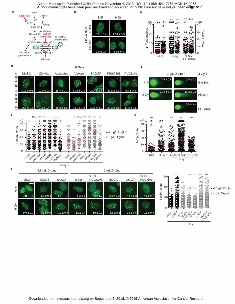
A C
D
B
E
1 g/
L D
-glu
cG
lcN
Ac
NIR 6 Gy
17.4 ± 0.9
3.8 ± 0.4
5.5 ± 0.7
9.1 ± 0.4
DM
SO
DMSO ST060266GlcNAc Azaserine PUGNAcBADGPAlloxan
4.5
g/L
D-g
luc
1 g/
L D
-glu
c
10.3 ± 0.5
15.7 ± 1.2
10.9 ± 0.7
7.3 ± 0.4
15.4 ± 0.7
16.8 ± 1.8
31.3 ± 2.4
25.0 ± 1.8
22.4 ± 1.1
32.8 ± 2.0
33.8 ± 2.2
27.3 ± 1.5
8.0 ± 0.4
11.8 ± 0.9
3 GyNIR 3 Gy + GlcNAc
%Tail D
NA
Foci
/nuc
leus
0
>30
20
30
10
0
20
40
30
>40
10
****** n.s.*** n.s.
G6P
F6PGFAT
GlcN-6P
GlcNAc-6P
Azaserine GlcNAc
UDP-GlcNAc
N-Linkedglycosylation
O-GlcNAcmodification
OGT
OGA
UDP
GlcNAc H2O
ST060266
PUGNAc
BADGPAlloxan
NAGK
0
10
20
30
>4040
Foci
/nuc
leus
DMSO
GlcNAc
Azase
rine
Alloxa
n
PUGNAc
BADGP
ST0602
66
DMSO
GlcNAc
Azase
rine
Alloxa
n
PUGNAc
BADGP
ST0602
66
*** ********n.s. ***** ***n.s.*** *** ***
F
NIR 3 Gy GlcNAc AlloxanPUGNAc3 Gy +
*** ********
0
10
20
30
>40
%Ta
il D
NA
40
G
3.9 ± 0.9
11.5 ± 1.3
NIR
3 Gy
6.0 ± 0.6
16.2 ± 0.9
4.5 ± 0.3
GlcNAc
Alloxan
PUGNAc
3 Gy +1 g/L D-gluc
siScr
siOGT
siScr
siOGT
siOGA
siScr+
PUGNAc
siOGT+
PUGNAc
Foci
/nuc
leus
0
40
60
20
*** *** *** ******
H I
siScr siOGT siScrsiScr+PUGNAc siOGT
siOGT+PUGNAcsiOGAsiOGA
9.6 ± 0.3 20 ± 0.9 10 ± 0.4
6.3 ± 1
12 ± 0.4
4.2 ± 1.2
10 ± 0.3 8.8 ± 0.3
4.5 g/L D-gluc
3.8 ± 0.4
16 ± 0.5 16 ± 0.5
1 g/L D-gluc
5.7 ± 0.8 4.5 ± 1.2 7.3 ± 0.8 7.1 ± 0.73.9 ± 0.3
NIR
6 G
y
Figure 3
.
6 Gy +
6 Gy +
4.5 g/L D-gluc1 g/L D-gluc
4.5 g/L D-gluc1 g/L D-gluc
6 Gy
on September 7, 2018. © 2015 American Association for Cancer Research. mcr.aacrjournals.org Downloaded from
Author manuscripts have been peer reviewed and accepted for publication but have not yet been edited. Author Manuscript Published OnlineFirst on November 4, 2015; DOI: 10.1158/1541-7786.MCR-15-0263
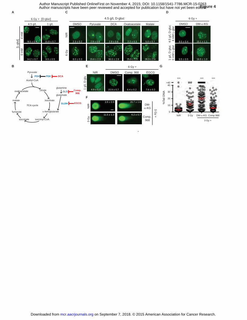
C
B E
A D
F
DMSO DCA OxaloacetatePyruvate Malate
6 G
yN
IR
2.3 ± 0.2 2.9 ± 0.6 2.9 ± 0.6 2.2 ± 0.3 5.0 ± 0.7
8.0 ± 0.3 15.6 ± 2.3 36.0 ± 2.9 25.9 ± 1.5 26.3 ± 2.6
4.5 g/L 1 g/L
[D-gluc]4
mM
0 m
M[L
-glu
t] 9.5 ± 0.4
14.2 ± 0.7
15.9 ± 0.7
9.5 ± 0.5
PDH PDK DCA
TCA cycle
Pyruvate
Acetyl-CoA
citrate
isocitrate
α-ketoglutarate
succinyl-CoAsuccinate
fumarate
malate
oxaloacetateglutamine
glutamateGLS Comp.
968
GLDH EGCG
EGCGComp. 968DMSO
15.9 ± 0.7 6.4 ± 0.2 7.6 ± 0.21 g/
L D
-glu
6 Gy +
DM-α-KG
3.9 ± 0.9
11.5 ± 1.3Comp.
968
20.7 ± 1.6
6.2 ± 0.7
NIR
3 G
y3 G
y +
0
10
20
30
>40
NIR 3 Gy DM-α-KG Comp.968
3 Gy +
%Ta
il D
NA
40
*** ******
G
Figure 4
.
1 g/
L D
-glu
c0
mM
L-g
lut
DMSO DM-α-KG
4.5
g/L
D-g
luc
4 m
M L
-glu
t
9.5 ± 0.5 16.5 ± 1.0
15.1 ± 1.19.5 ± 0.4
6 Gy + 6 Gy +4.5 g/L D-gluc
NIR
4.9 ± 0.7
on September 7, 2018. © 2015 American Association for Cancer Research. mcr.aacrjournals.org Downloaded from
Author manuscripts have been peer reviewed and accepted for publication but have not yet been edited. Author Manuscript Published OnlineFirst on November 4, 2015; DOI: 10.1158/1541-7786.MCR-15-0263
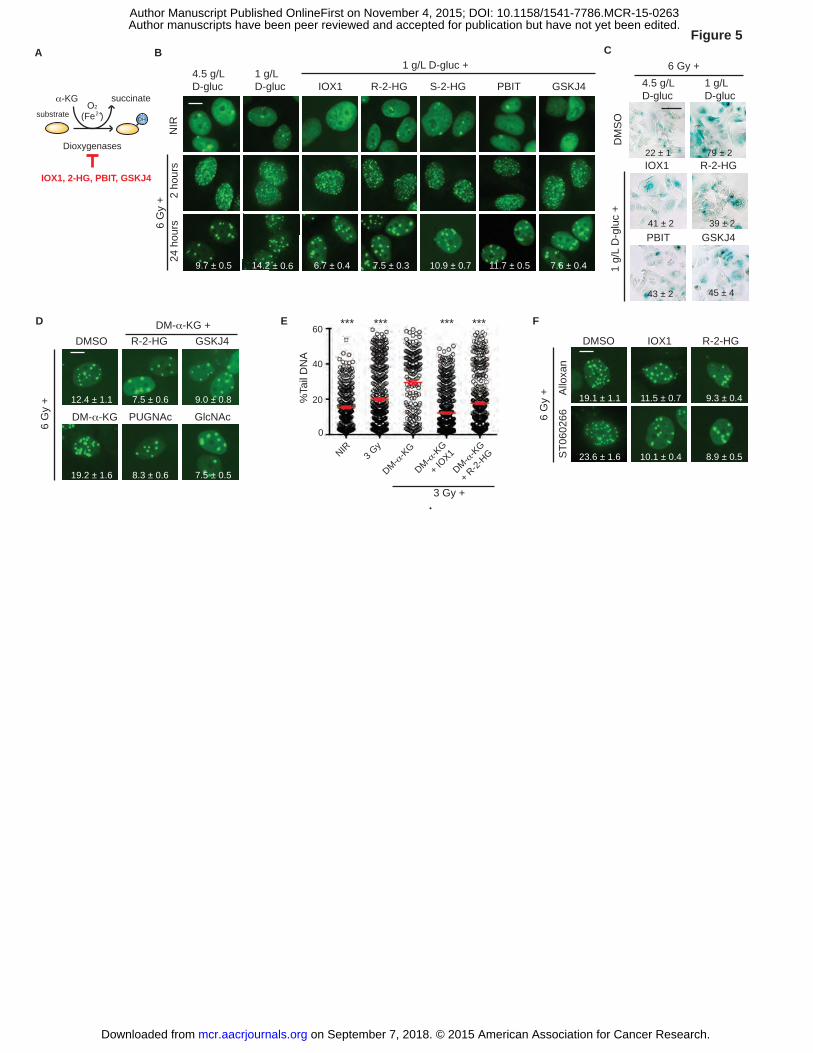
C
D E
A
α-KG succinateO2
(Fe )2+OH
Dioxygenases
substrate
IOX1, 2-HG, PBIT, GSKJ4
R-2-HGIOX1DMSO
Allo
xan
ST0
6026
6
19.1 ± 1.1 11.5 ± 0.7 9.3 ± 0.4
8.9 ± 0.510.1 ± 0.423.6 ± 1.6
B
F
PUGNAc
DMSO
GlcNAc
R-2-HG GSKJ4
DM-α-KG
12.4 ± 1.1
19.2 ± 1.6 8.3 ± 0.6 7.5 ± 0.5
7.5 ± 0.6 9.0 ± 0.8
DM-α-KG +
%Ta
il D
NA
0
20
40
60 *** *** ******
NIR3 G
y
3 Gy +
DM-α-K
G
DM-α-K
G
+ IOX1
DM-α-K
G
+ R-2-
HG
Figure 5
.
6 G
y +
6 G
y +
R-2-HG22 ± 1
1 g/
L D
-glu
c +
41 ± 2
IOX179 ± 2
39 ± 2
4.5 g/LD-gluc
1 g/LD-gluc
6 Gy +
DM
SO
4.5 g/L D-gluc
1 g/L D-gluc GSKJ4
7.6 ± 0.4
PBITR-2-HG
7.5 ± 0.3
IOX1
14.2 ± 0.6 6.7 ± 0.4
S-2-HG
10.9 ± 0.7 11.7 ± 0.59.7 ± 0.5
1 g/L D-gluc +
6 G
y +
NIR
24 h
ours
2 ho
urs
PBIT GSKJ4
43 ± 2 45 ± 4
on September 7, 2018. © 2015 American Association for Cancer Research. mcr.aacrjournals.org Downloaded from
Author manuscripts have been peer reviewed and accepted for publication but have not yet been edited. Author Manuscript Published OnlineFirst on November 4, 2015; DOI: 10.1158/1541-7786.MCR-15-0263
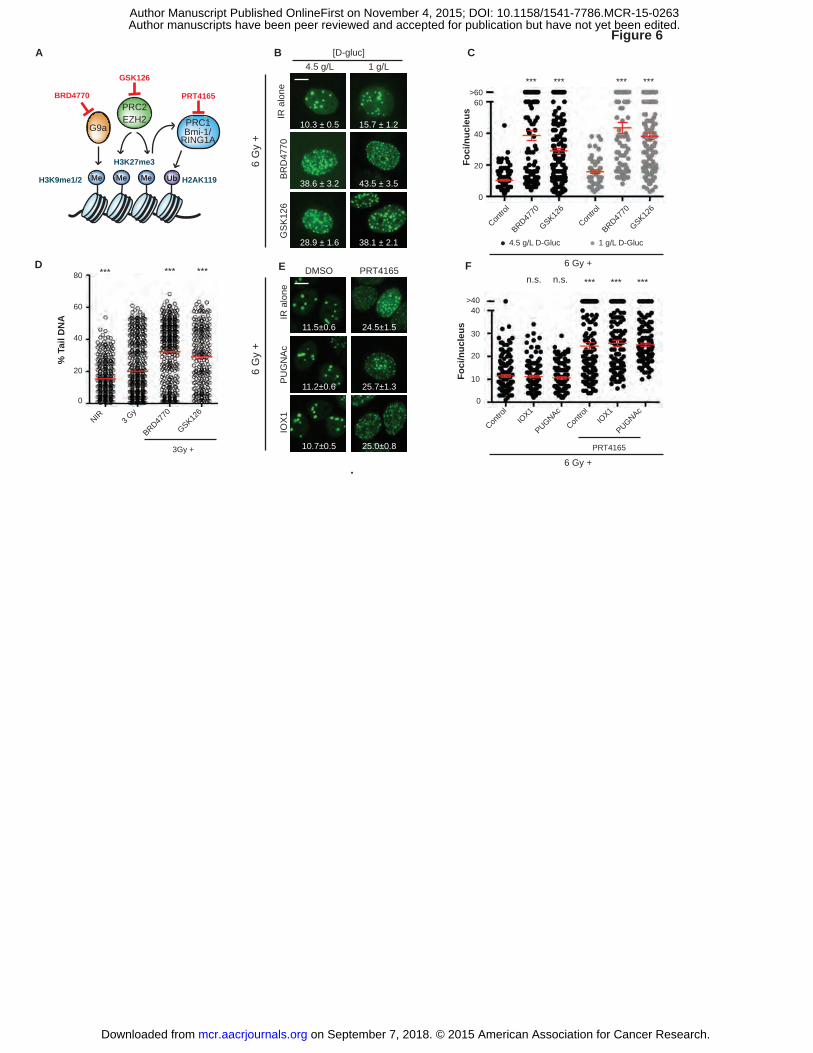
CB
DP
UG
NA
cDMSO PRT4165
IOX
1
A
BR
D47
70G
SK
126
[D-gluc]1 g/L
15.7 ± 1.2
43.5 ± 3.5
38.1 ± 2.1
4.5 g/L
10.3 ± 0.5
38.6 ± 3.2
28.9 ± 1.6
8.9±0.510.1±0.423.6±1.6
11.5±0.6
11.2±0.6
10.7±0.5
24.5±1.5
25.7±1.3
25.0±0.8
PRT4165
Me UbMeMe H2AK119
H3K27me3
H3K9me1/2
G9a
PRC2EZH2
Bmi-1/RING1A
PRC1
BRD4770
GSK126
1 g/L D-Gluc4.5 g/L D-Gluc
Contro
l
BRD4770
GSK126
Contro
l
BRD4770
GSK126
0
20
40
60>60
*** *** *** ***
Foci
/nuc
leus
% T
ail D
NA
EIR
alo
ne
Foci
/nuc
leus
***
20
40>40
30
10
PRT4165
Contro
l0
IOX1
PUGNAcIO
X1
PUGNAc
Contro
l
******n.s. n.s.F
0
20
40
60
80
NIR
BRD4770
GSK126
3 Gy
3Gy +
*** ******
Figure 6
.
6 G
y +
6 G
y +
6 Gy +
6 Gy +
IR a
lone
on September 7, 2018. © 2015 American Association for Cancer Research. mcr.aacrjournals.org Downloaded from
Author manuscripts have been peer reviewed and accepted for publication but have not yet been edited. Author Manuscript Published OnlineFirst on November 4, 2015; DOI: 10.1158/1541-7786.MCR-15-0263
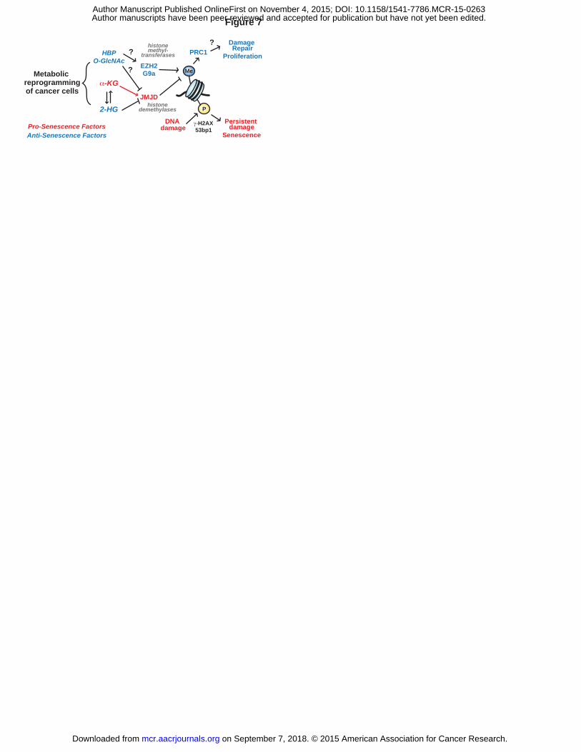
DNA damage
Persistent damage
Senescence
EZH2
JMJD
histonemethyl-
transferases
γ-H2AX 53bp1
HBPO-GlcNAc
α-KG
histonedemethylases2-HG
G9a Me
P
Metabolic reprogrammingof cancer cells
?
PRC1?
Pro-Senescence FactorsAnti-Senescence Factors
Damage Repair
Proliferation
?
Figure 7
on September 7, 2018. © 2015 American Association for Cancer Research. mcr.aacrjournals.org Downloaded from
Author manuscripts have been peer reviewed and accepted for publication but have not yet been edited. Author Manuscript Published OnlineFirst on November 4, 2015; DOI: 10.1158/1541-7786.MCR-15-0263
Published OnlineFirst November 4, 2015.Mol Cancer Res Elena V. Efimova, Satoe Takahashi, Noumaan A. Shamsi, et al. SenescenceLinking Cancer Metabolism to DNA Repair and Accelerated
Updated version
10.1158/1541-7786.MCR-15-0263doi:
Access the most recent version of this article at:
Material
Supplementary
http://mcr.aacrjournals.org/content/suppl/2015/11/06/1541-7786.MCR-15-0263.DC2
Access the most recent supplemental material at:
Manuscript
Authoredited. Author manuscripts have been peer reviewed and accepted for publication but have not yet been
E-mail alerts related to this article or journal.Sign up to receive free email-alerts
Subscriptions
Reprints and
To order reprints of this article or to subscribe to the journal, contact the AACR Publications
Permissions
Rightslink site. Click on "Request Permissions" which will take you to the Copyright Clearance Center's (CCC)
.http://mcr.aacrjournals.org/content/early/2015/11/04/1541-7786.MCR-15-0263To request permission to re-use all or part of this article, use this link
on September 7, 2018. © 2015 American Association for Cancer Research. mcr.aacrjournals.org Downloaded from
Author manuscripts have been peer reviewed and accepted for publication but have not yet been edited. Author Manuscript Published OnlineFirst on November 4, 2015; DOI: 10.1158/1541-7786.MCR-15-0263
![Prostaglandin H Synthase-catalyzed Metabolism and DNA ...[CANCER RESEARCH 47, 4007-4014, August 1, 1987] Prostaglandin H Synthase-catalyzed Metabolism and DNA Binding of 2-Naphthylamine](https://img.pdfslide.net/doc/110x75/6125125eba335f0b336d21dc/prostaglandin-h-synthase-catalyzed-metabolism-and-dna-cancer-research-47-4007-4014.jpg)
![Cross-linking of DNA by Alkylating Agents and …cancerres.aacrjournals.org/content/canres/31/11/1573...(CANCER RESEARCH 31, 1573-1579, November 1971] Cross-linking of DNA by Alkylating](https://img.pdfslide.net/doc/110x75/5e8c9e9d237ced4c0908422b/cross-linking-of-dna-by-alkylating-agents-and-cancer-research-31-1573-1579.jpg)