Embed Size (px)
Citation preview
US HUPO 2009
February 22, 2009
Liquid Chromatography
Techniques for Sample
Preparation
US HUPO 2009 Sample Prep Workshop
February 22, 2009
US HUPO 2009 Sample Prep Workshop
February 22, 2009
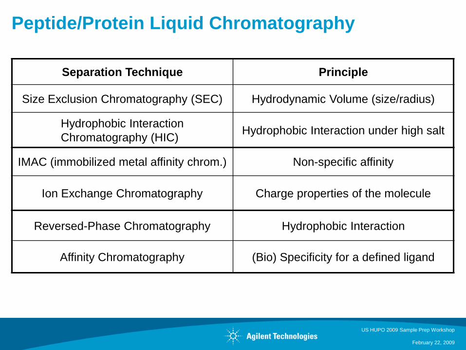
Peptide/Protein Liquid Chromatography
Separation Technique Principle
Size Exclusion Chromatography (SEC) Hydrodynamic Volume (size/radius)
Hydrophobic Interaction
Chromatography (HIC)Hydrophobic Interaction under high salt
IMAC (immobilized metal affinity chrom.) Non-specific affinity
Ion Exchange Chromatography Charge properties of the molecule
Reversed-Phase Chromatography Hydrophobic Interaction
Affinity Chromatography (Bio) Specificity for a defined ligand
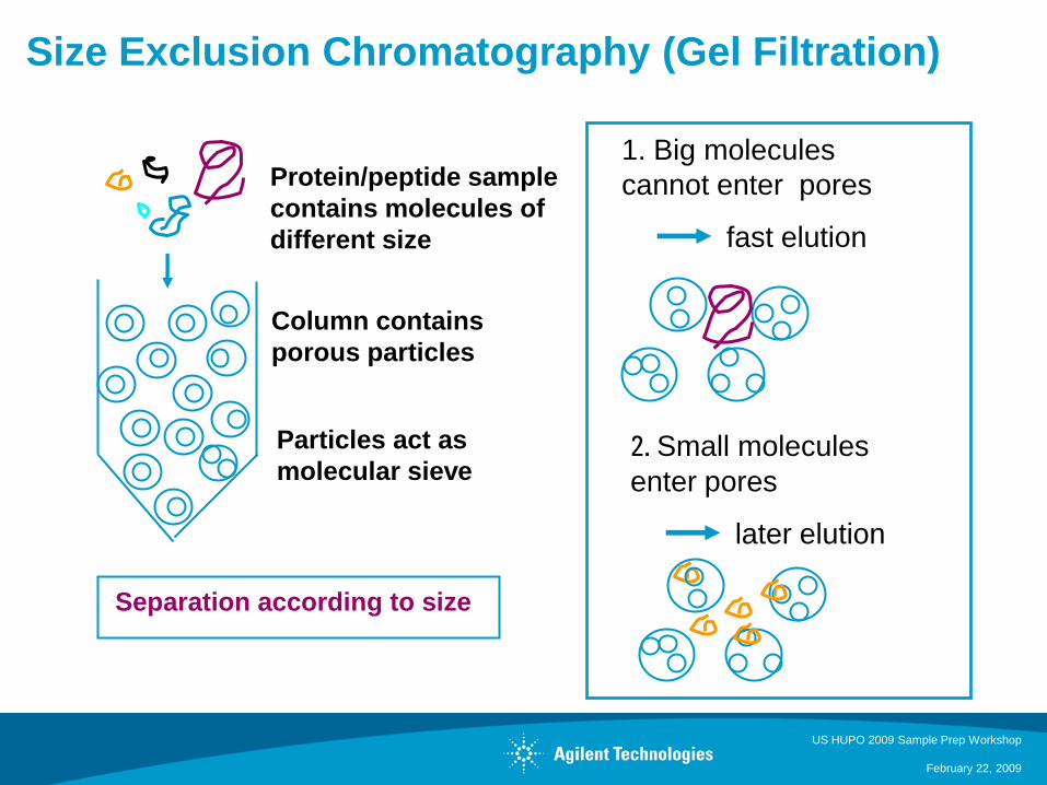
Size Exclusion Chromatography (Gel Filtration)
US HUPO 2009 Sample Prep Workshop
February 22, 2009
1. Big molecules
cannot enter pores
fast elution
Separation according to size
Protein/peptide sample
contains molecules of
different size
Column contains
porous particles
Particles act as
molecular sieve2. Small molecules
enter pores
later elution
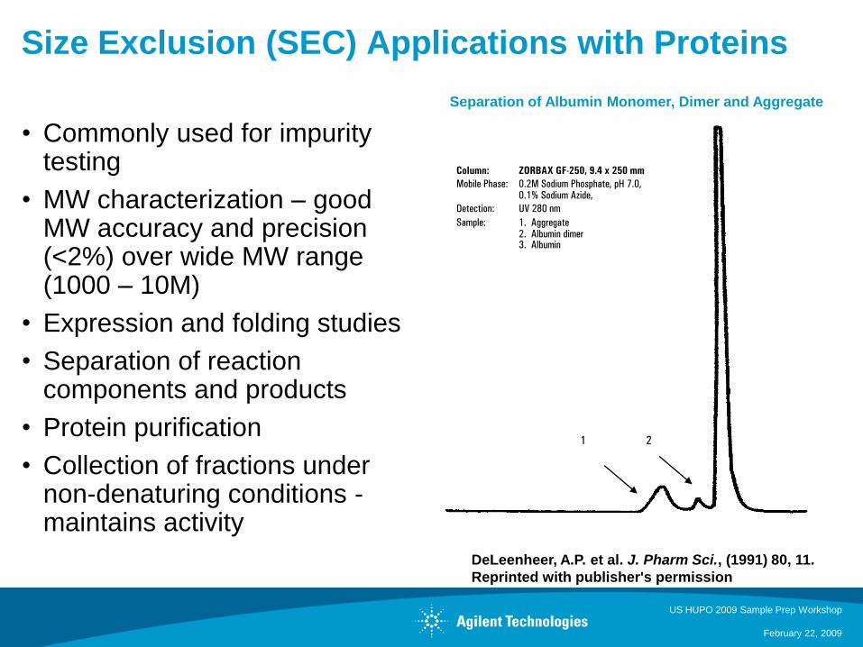
Size Exclusion (SEC) Applications with Proteins
• Commonly used for impurity testing
• MW characterization – good MW accuracy and precision (<2%) over wide MW range (1000 – 10M)
• Expression and folding studies
• Separation of reaction components and products
• Protein purification
• Collection of fractions under non-denaturing conditions -maintains activity
US HUPO 2009 Sample Prep Workshop
February 22, 2009
1 2
Column: ZORBAX GF-250, 9.4 x 250 mm
Mobile Phase: 0.2M Sodium Phosphate, pH 7.0,0.1% Sodium Azide,
Detection: UV 280 nm
Sample: 1. Aggregate2. Albumin dimer3. Albumin
DeLeenheer, A.P. et al. J. Pharm Sci., (1991) 80, 11.
Reprinted with publisher's permission
Separation of Albumin Monomer, Dimer and Aggregate
Hydrophobic Interaction Chromatography
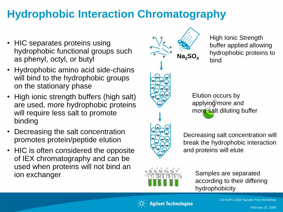
• HIC separates proteins using hydrophobic functional groups such as phenyl, octyl, or butyl
• Hydrophobic amino acid side-chains will bind to the hydrophobic groups on the stationary phase
• High ionic strength buffers (high salt) are used, more hydrophobic proteins will require less salt to promote binding
• Decreasing the salt concentration promotes protein/peptide elution
• HIC is often considered the opposite of IEX chromatography and can be used when proteins will not bind an ion exchanger
US HUPO 2009 Sample Prep Workshop
February 22, 2009
-- -- -- -
-----
- -- -
++
+
+++
+
+
-- -- -- -
-----
- -- -
High Ionic Strength
buffer applied allowing
hydrophobic proteins to
bind
Elution occurs by
applying more and
more salt diluting buffer
Decreasing salt concentration will
break the hydrophobic interaction
and proteins will elute
Samples are separated
according to their differing
hydrophobicity
Na2SO4
US HUPO 2009 Sample Prep Workshop
February 22, 2009
IMAC - Immobilized Metal Affinity Chromatography
Principle
• IMAC is based on covalent binding of amino acids, particularly histidine, to metals
• Proteins with an affinity for metal ions to be retained in a column containing immobilized metal ions, such as cobalt, nickel, copper, zinc, or iron ions
• Naturally occurring proteins often do not have affinity for metal ions and recombinant DNA can be used to introduce metal affinity into a targeted protein
• Common elution methods include changing the pH and/or adding a competition molecule, such as imidazole.
Key Application
• Isolation of phosphorylated proteins or peptides from complex mixtures
• The stationary phase containing an immobilized transition metal is “charged” (Ga(III) or Fe(III)) causing the metals to form tight complexes
• Phosphorylated protein digests are loaded onto the column, the column is washed and then elution occurs either with pH change or imidazole buffer
• Detection of low-abundant phosphopeptides is difficult due to non-phosphorylated high-abundant proteins containing histidine
• Titanium oxide (TiO2) is another metal-based chromatography method for phosphopeptide analysis
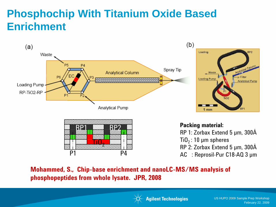
Phosphochip With Titanium Oxide Based
Enrichment
US HUPO 2009 Sample Prep Workshop
February 22, 2009
Mohammed, S., Chip-base enrichment and nanoLC-MS/MS analysis of
phosphopeptides from whole lysate. JPR, 2008
Packing material:
RP 1: Zorbax Extend 5 µm, 300Å
TiO2 : 10 µm spheres
RP 2: Zorbax Extend 5 µm, 300Å
AC : Reprosil-Pur C18-AQ 3 µmP4P1
TiO2
RP1 RP2
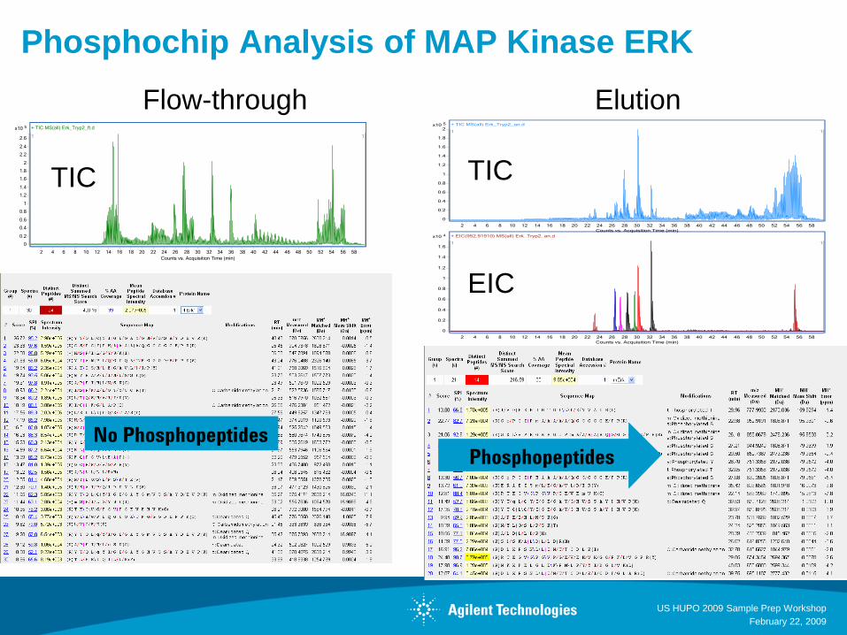
Phosphochip Analysis of MAP Kinase ERK
US HUPO 2009 Sample Prep Workshop
February 22, 2009
Flow-through Elution
TIC TIC
EIC
No PhosphopeptidesPhosphopeptides
US HUPO 2009 Sample Prep Workshop
February 22, 2009
Cation Exchange Chromatography
-- -- -- -
-----
- -- -
Na+
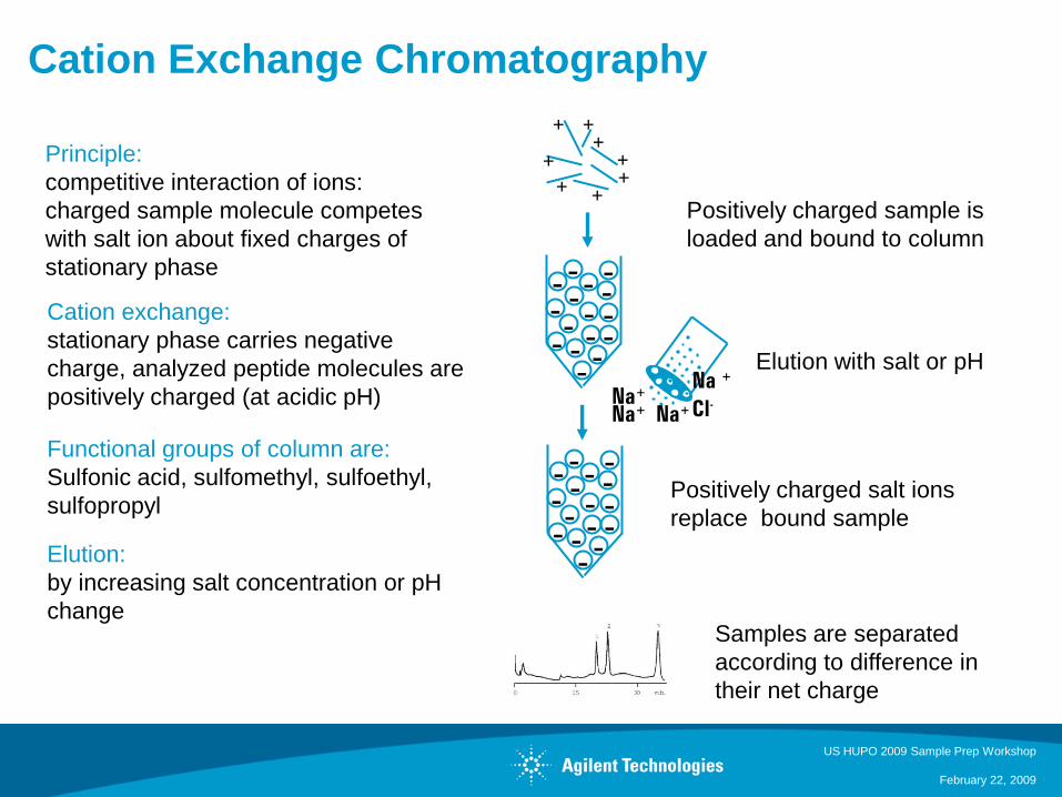
Principle:
competitive interaction of ions:
charged sample molecule competes
with salt ion about fixed charges of
stationary phase
Cation exchange:
stationary phase carries negative
charge, analyzed peptide molecules are
positively charged (at acidic pH)
Functional groups of column are:
Sulfonic acid, sulfomethyl, sulfoethyl,
sulfopropyl
Elution:
by increasing salt concentration or pH
change
++
+
+++
+
+
-- -- -- -
-----
- -- -
Positively charged sample is
loaded and bound to column
Elution with salt or pH
Positively charged salt ions
replace bound sample
Samples are separated
according to difference in
their net charge
Na+Na+
Na +
Cl-
US HUPO 2009 Sample Prep Workshop
February 22, 2009000855P1.PPT
Ion Exchange Chromatography of Proteins
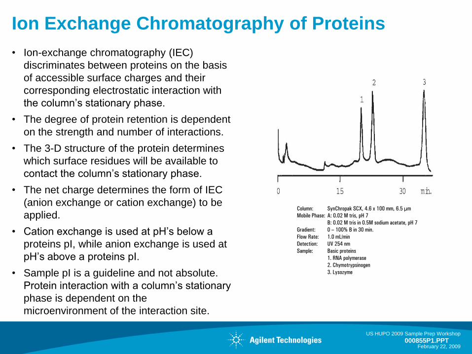
• Ion-exchange chromatography (IEC)
discriminates between proteins on the basis
of accessible surface charges and their
corresponding electrostatic interaction with
the column’s stationary phase.
• The degree of protein retention is dependent
on the strength and number of interactions.
• The 3-D structure of the protein determines
which surface residues will be available to
contact the column’s stationary phase.
• The net charge determines the form of IEC
(anion exchange or cation exchange) to be
applied.
• Cation exchange is used at pH’s below a
proteins pI, while anion exchange is used at
pH’s above a proteins pI.
• Sample pI is a guideline and not absolute.
Protein interaction with a column’s stationary
phase is dependent on the
microenvironment of the interaction site.
Column: SynChropak SCX, 4.6 x 100 mm, 6.5 m
Mobile Phase: A: 0.02 M tris, pH 7
B: 0.02 M tris in 0.5M sodium acetate, pH 7
Gradient: 0 – 100% B in 30 min.
Flow Rate: 1.0 mL/min
Detection: UV 254 nm
Sample: Basic proteins
1. RNA polymerase
2. Chymotrypsinogen
3. Lysozyme
US HUPO 2009 Sample Prep Workshop
February 22, 2009
Reversed Phase Chromatography
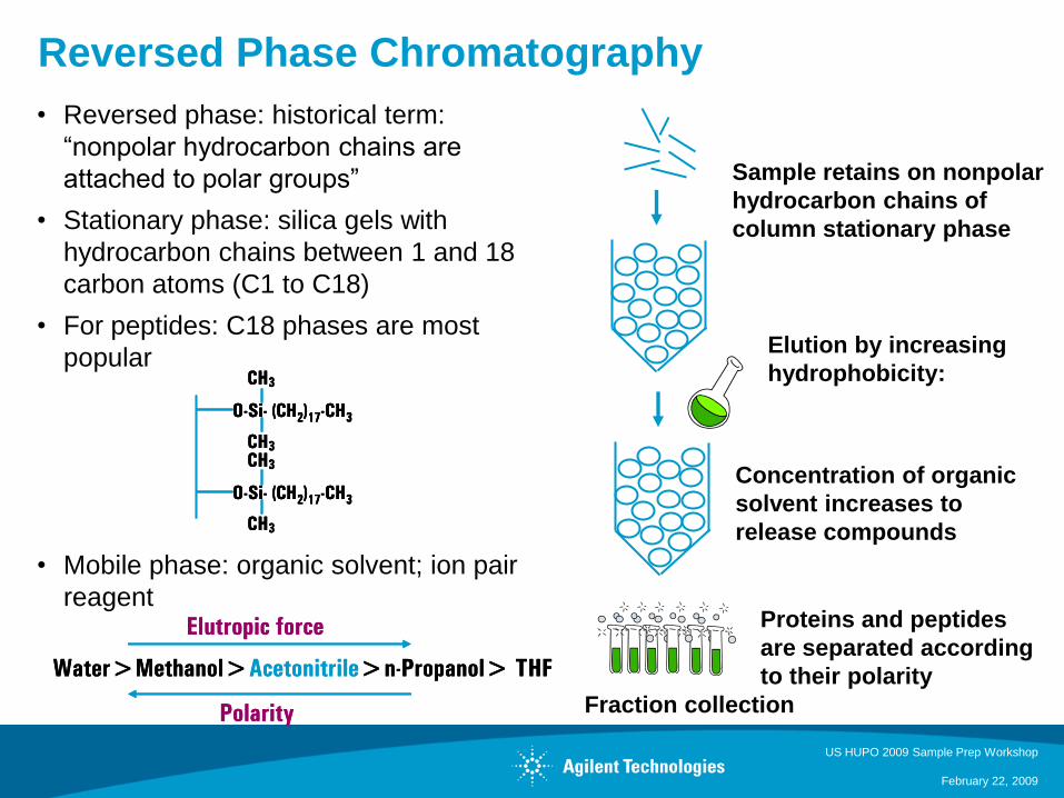
• Reversed phase: historical term:
“nonpolar hydrocarbon chains are
attached to polar groups”
• Stationary phase: silica gels with
hydrocarbon chains between 1 and 18
carbon atoms (C1 to C18)
• For peptides: C18 phases are most
popular
• Mobile phase: organic solvent; ion pair
reagent
Fraction collection
Sample retains on nonpolar
hydrocarbon chains of
column stationary phase
Elution by increasing
hydrophobicity: CH3
O-Si- (CH2)17-CH3
CH3
CH3
O-Si- (CH2)17-CH3
CH3
CH3
O-Si- (CH2)17-CH3
CH3
CH3
O-Si- (CH2)17-CH3
CH3
CH3
O-Si- (CH2)17-CH3
CH3
CH3
O-Si- (CH2)17-CH3
CH3
Water>Methanol>Acetonitrile>n-Propanol> THF
Elutropic force
Polarity
Water>Methanol>Acetonitrile>n-Propanol> THF
Elutropic force
Polarity
Concentration of organic
solvent increases to
release compounds
Proteins and peptides
are separated according
to their polarity
US HUPO 2009 Sample Prep Workshop
February 22, 2009
Reversed Phase Columns for Separations
of Proteins and Peptides
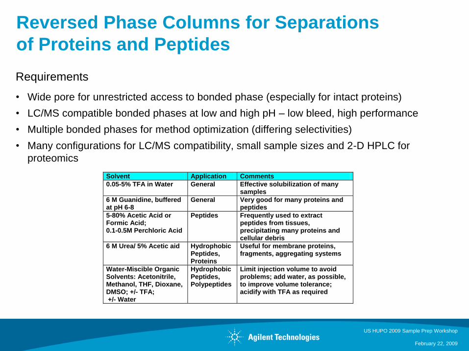
Requirements
• Wide pore for unrestricted access to bonded phase (especially for intact proteins)
• LC/MS compatible bonded phases at low and high pH – low bleed, high performance
• Multiple bonded phases for method optimization (differing selectivities)
• Many configurations for LC/MS compatibility, small sample sizes and 2-D HPLC for
proteomics
Solvent Application Comments
0.05-5% TFA in Water General Effective solubilization of many samples
6 M Guanidine, buffered at pH 6-8
General Very good for many proteins and peptides
5-80% Acetic Acid or Formic Acid; 0.1-0.5M Perchloric Acid
Peptides
Frequently used to extract peptides from tissues, precipitating many proteins and cellular debris
6 M Urea/ 5% Acetic aid Hydrophobic Peptides, Proteins
Useful for membrane proteins, fragments, aggregating systems
Water-Miscible Organic Solvents: Acetonitrile, Methanol, THF, Dioxane, DMSO; +/- TFA; +/- Water
Hydrophobic Peptides, Polypeptides
Limit injection volume to avoid problems; add water, as possible, to improve volume tolerance; acidify with TFA as required
US HUPO 2009 Sample Prep Workshop
February 22, 2009
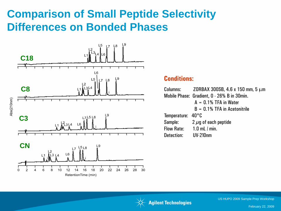
Conditions:
Columns: ZORBAX 300SB, 4.6 x 150 mm, 5 m
Mobile Phase: Gradient, 0 - 26% B in 30min.
A = 0.1% TFA in Water
B = 0.1% TFA in Acetonitrile
Temperature: 40°C
Sample: 2 µg of each peptide
Flow Rate: 1.0 mL / min.
Detection: UV-210nm
C18
C8
C3
CN
Comparison of Small Peptide Selectivity
Differences on Bonded Phases

US HUPO 2009 Sample Prep Workshop
February 22, 2009
Evaluate High pH for Improved Selectivity and
Resolution
• Changing pH of the mobile phase from low pH to high pH can
change selectivity and retention
• Special columns are required to work at high pH – many
manufacturer’s have columns specific for high pH
• Ideal for analysis of proteins and peptides at mid and high pH
• Ammonium hydroxide is an excellent mobile phase additive for LC
and LC/MS (can use instead of TFA)
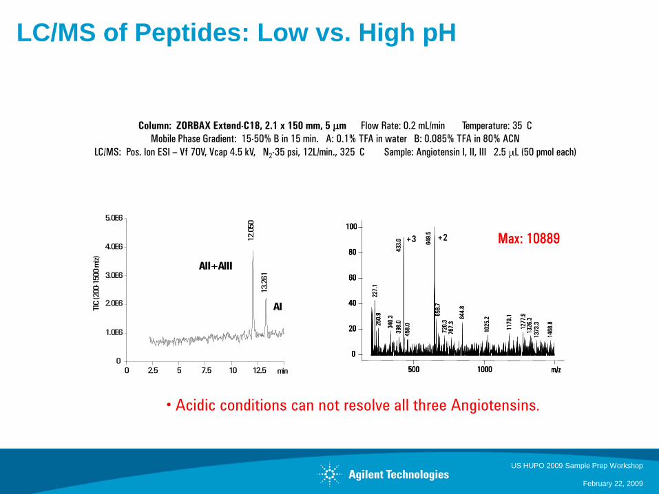
LC/MS of Peptides: Low vs. High pH
US HUPO 2009 Sample Prep Workshop
February 22, 2009
Column: ZORBAX Extend-C18, 2.1 x 150 mm, 5 m Flow Rate: 0.2 mL/min Temperature: 35 C
Mobile Phase Gradient: 15-50% B in 15 min. A: 0.1% TFA in water B: 0.085% TFA in 80% ACN
LC/MS: Pos. Ion ESI – Vf 70V, Vcap 4.5 kV, N2-35 psi, 12L/min., 325 C Sample: Angiotensin I, II, III 2.5 L (50 pmol each)
• Acidic conditions can not resolve all three Angiotensins.
min0 2.5 5 7.5 10 12.5
0
1.0E6
2.0E6
3.0E6
4.0E6
5.0E6
12.0
50
13.2
61
AII+AIII
AITIC
(200-1
500 m
/z)
m/z500 1000
0
20
40
60
80
100
Max: 1088964
9.5
43
3.0
22
7.1
65
9.7
84
4.8
25
0.9
34
0.3
12
77
.9
11
79
.1
10
25
.2
72
0.3
13
26
.3
39
8.0
76
7.3
45
8.0
14
68
.8
13
73
.3
+3 +2
m/z500 1000
0
20
40
60
80
100
m/z500 1000
0
20
40
60
80
100
Max: 1088964
9.5
43
3.0
22
7.1
65
9.7
84
4.8
25
0.9
34
0.3
12
77
.9
11
79
.1
10
25
.2
72
0.3
13
26
.3
39
8.0
76
7.3
45
8.0
14
68
.8
13
73
.3
+3 +2
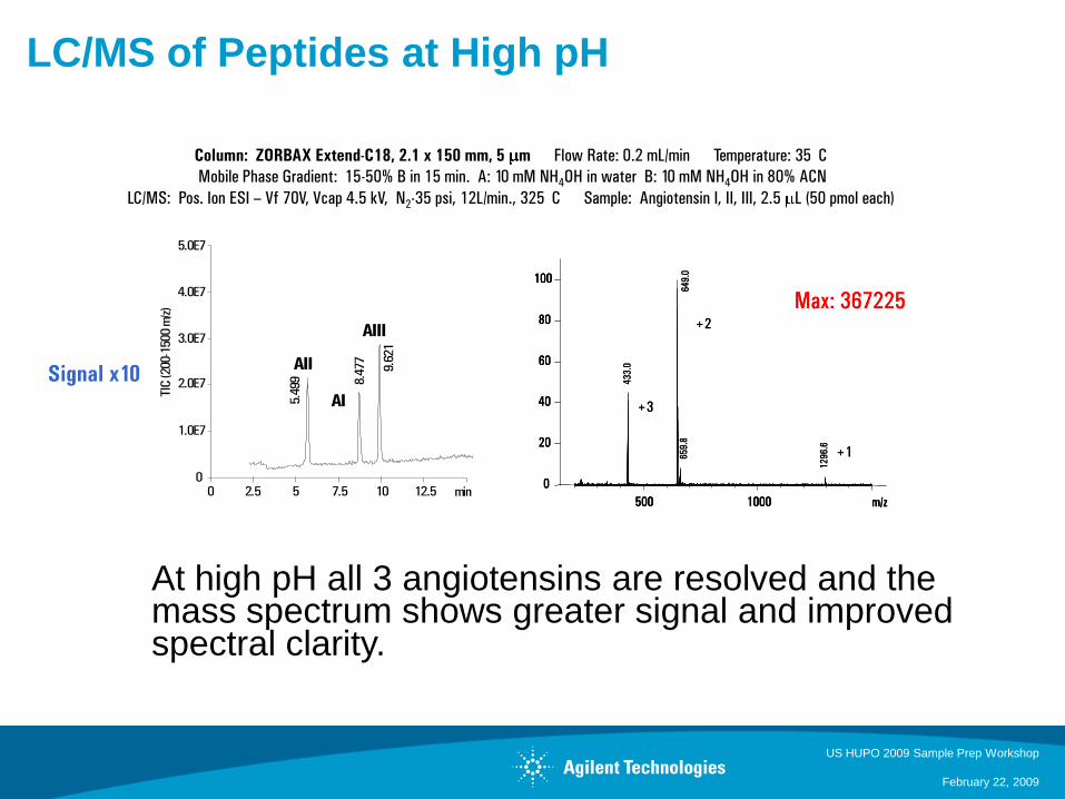
LC/MS of Peptides at High pH
US HUPO 2009 Sample Prep Workshop
February 22, 2009
Column: ZORBAX Extend-C18, 2.1 x 150 mm, 5 m Flow Rate: 0.2 mL/min Temperature: 35 C
Mobile Phase Gradient: 15-50% B in 15 min. A: 10 mM NH4OH in water B: 10 mM NH4OH in 80% ACN
LC/MS: Pos. Ion ESI – Vf 70V, Vcap 4.5 kV, N2-35 psi, 12L/min., 325 C Sample: Angiotensin I, II, III, 2.5 L (50 pmol each)
Signal x10
m/z500 1000
0
20
40
60
80
100
Max: 367225
64
9.0
43
3.0
65
9.8
12
96
.6
+1
+2
+3
m/z500 1000
0
20
40
60
80
100
m/z500 1000
0
20
40
60
80
100
Max: 367225
64
9.0
43
3.0
65
9.8
12
96
.6
+1
+2
+3
min0 2.5 5 7.5 10 12.50
1.0E7
2.0E7
3.0E7
4.0E7
5.0E7
5.4
99 8.4
77AII
AI
TIC
(200-1
500 m
/z)
AIII
9.6
21
At high pH all 3 angiotensins are resolved and the mass spectrum shows greater signal and improved spectral clarity.
US HUPO 2009 Sample Prep Workshop
February 22, 2009
Disadvantages of Traditional Reverse Phase
Techniques for Protein Fractionation
• Recovery – typical reverse phase can range from 30-80% recovery
• Reproducibility – due to poor recovery, the reproducibility often suffers from
carryover
• Capacity – limited in ability to load AND resolve proteins
• Is it possible with reverse phase to:
– Extremely high recoveries
– High column loads
– Improved protein resolution
• What does it take:
– Column Packing Materials - strong effect on separation characteristics-
resolution, selectivity, reproducibility, load and recovery
– Gradient – improves recovery and enhances higher abundant protein resolution
– Temperature- Improves protein separations and aids in recovery
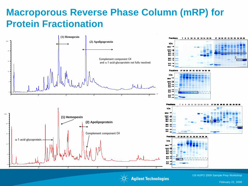
Macroporous Reverse Phase Column (mRP) for
Protein Fractionation
US HUPO 2009 Sample Prep Workshop
February 22, 2009
200.0
116.3
97.4
66.3
55.4
36.5
31.0
21.5
14.4
Fractions 7 8 9 10 11 12 13 14 15 16 17 18 19 20 21 22 23 24 25 26
200.0
116.3
97.4
66.3
55.4
36.5
31.0
21.5
14.4
Fractions 27 28 29 30 31 32 33 34 35 36
kDa
kDa
200.0
116.3
97.4
66.3
55.4
36.5
31.0
21.5
14.4
Fractions 7 8 9 10 11 12 13 14 15 16 17 18 19 20 21 22 23 24 25 26
200.0
116.3
97.4
66.3
55.4
36.5
31.0
21.5
14.4
Fractions 27 28 29 30 31 32 33 34 35 36
kDa
kDa
1
2
2
200.0
116.3
97.4
66.3
55.4
36.5
31.0
21.5
14.4
Fractions 7 8 9 10 11 12 13 14 15 16 17 18 19 20 21 22 23 24 25 26
200.0
116.3
97.4
66.3
55.4
36.5
31.0
21.5
14.4
Fractions 27 28 29 30 31 32 33 34 35 36
kDa
kDa
200.0
116.3
97.4
66.3
55.4
36.5
31.0
21.5
14.4
Fractions 7 8 9 10 11 12 13 14 15 16 17 18 19 20 21 22 23 24 25 26
200.0
116.3
97.4
66.3
55.4
36.5
31.0
21.5
14.4
Fractions 27 28 29 30 31 32 33 34 35 36
kDa
kDa
1
2
2
Fractions 27 28 29 30 31 32 33 34 35 36
200.0
116.3
97.4
66.3
55.4
36.5
31.0
21.5
14.4
Fractions 7 8 9 10 11 12 13 14 15 16 17 18 19 20 21 22 23 24 25 26
200.0
116.3
97.4
66.3
55.4
36.5
31.0
21.5
14.4
kDa
kDa
Fractions 27 28 29 30 31 32 33 34 35 36
200.0
116.3
97.4
66.3
55.4
36.5
31.0
21.5
14.4
Fractions 7 8 9 10 11 12 13 14 15 16 17 18 19 20 21 22 23 24 25 26
200.0
116.3
97.4
66.3
55.4
36.5
31.0
21.5
14.4
Fractions 7 8 9 10 11 12 13 14 15 16 17 18 19 20 21 22 23 24 25 26Fractions 7 8 9 10 11 12 13 14 15 16 17 18 19 20 21 22 23 24 25 26
200.0
116.3
97.4
66.3
55.4
36.5
31.0
21.5
14.4
kDa
kDa
1
2
2
min
min
0 10 20 30 40 50
mAU
0
5
10
15
20
0 10 20 30 40 50
mAU
0
5
10
15
20
Complement component C4
Complement component C4
and -1-acid-glycoprotein not fully resolved.
-1-acid-glycoprotein.
(1) Hemopexin
(2) Apolipoprotein
(1) Hemopexin
(2) Apolipoprotein
min
min
0 10 20 30 40 50
mAU
0
5
10
15
20
0 10 20 30 40 50
mAU
0
5
10
15
20
Complement component C4
Complement component C4
and -1-acid-glycoprotein not fully resolved.
-1-acid-glycoprotein.
(1) Hemopexin
(2) Apolipoprotein
(1) Hemopexin
(2) Apolipoprotein
US HUPO 2009 Sample Prep Workshop
February 22, 2009
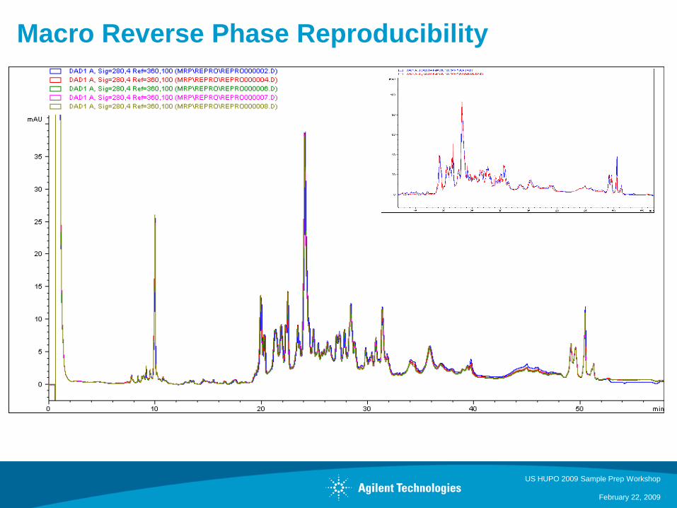
Macro Reverse Phase Reproducibility
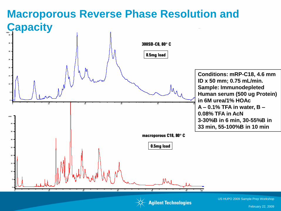
Macroporous Reverse Phase Resolution and
Capacity
US HUPO 2009 Sample Prep Workshop
February 22, 2009
min0 10 20 30 40 50
mAU
0
5
10
15
20
25
30
35
40
min0 10 20 30 40 50
mAU
0
5
10
15
20
25
30
35
40
macroporous C18, 80° C
300SB-C8, 80° C
0.5mg load
0.5mg load
min0 10 20 30 40 50
mAU
0
5
10
15
20
25
30
35
40
min0 10 20 30 40 50
mAU
0
5
10
15
20
25
30
35
40
macroporous C18, 80° C
300SB-C8, 80° C
0.5mg load
0.5mg load
min0 10 20 30 40 50
mAU
0
5
10
15
20
25
30
35
40
min0 10 20 30 40 50
mAU
0
5
10
15
20
25
30
35
40
macroporous C18, 80° C
300SB-C8, 80° C
0.5mg load
0.5mg load
min0 10 20 30 40 50
mAU
0
5
10
15
20
25
30
35
40
min0 10 20 30 40 50
mAU
0
5
10
15
20
25
30
35
40
macroporous C18, 80° C
300SB-C8, 80° C
0.5mg load
0.5mg load
Conditions: mRP-C18, 4.6 mm
ID x 50 mm; 0.75 mL/min.
Sample: Immunodepleted
Human serum (500 ug Protein)
in 6M urea/1% HOAc
A – 0.1% TFA in water, B –
0.08% TFA in AcN
3-30%B in 6 min, 30-55%B in
33 min, 55-100%B in 10 min
US HUPO 2009 Sample Prep Workshop
February 22, 2009
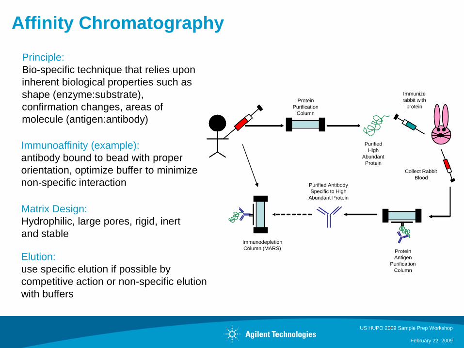
Affinity Chromatography
Principle:
Bio-specific technique that relies upon
inherent biological properties such as
shape (enzyme:substrate),
confirmation changes, areas of
molecule (antigen:antibody)
Immunoaffinity (example):
antibody bound to bead with proper
orientation, optimize buffer to minimize
non-specific interaction
Matrix Design:
Hydrophilic, large pores, rigid, inert
and stable
Elution:
use specific elution if possible by
competitive action or non-specific elution
with buffers
Protein
Purification
Column
Purified
High
Abundant
Protein
Immunize
rabbit with
protein
Protein
Antigen
Purification
Column
Purified Antibody
Specific to High
Abundant Protein
Immunodepletion
Column (MARS)
Collect Rabbit
Blood
US HUPO 2009 Sample Prep Workshop
February 22, 2009
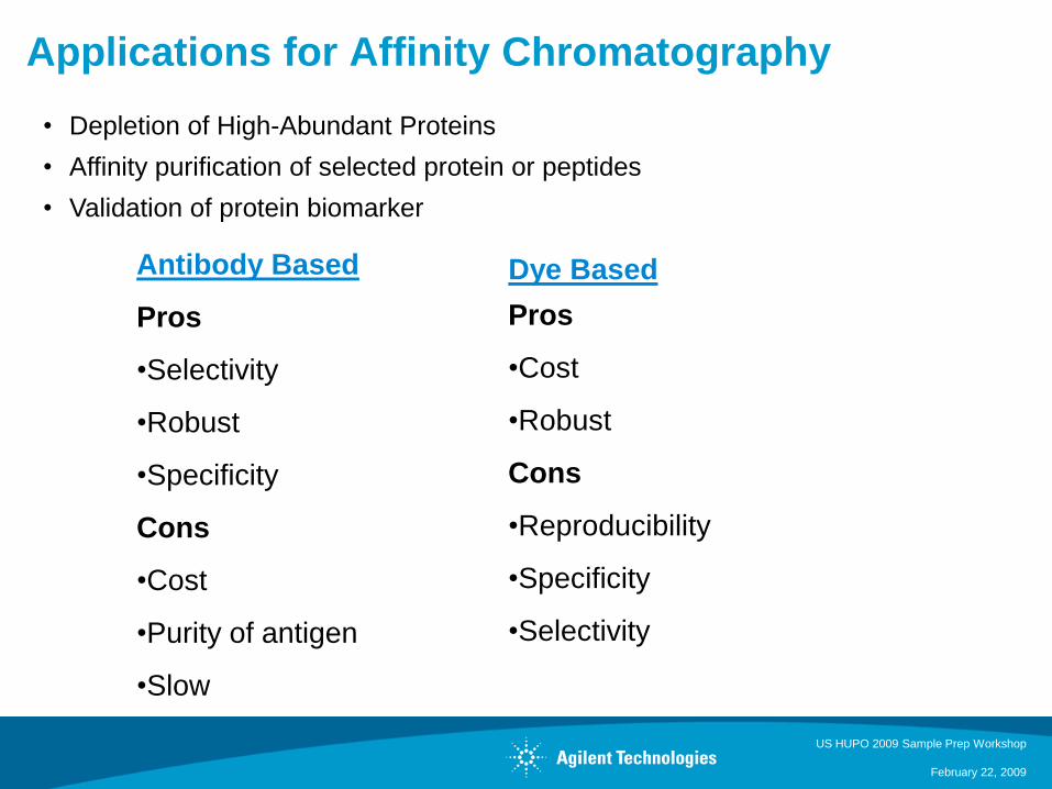
Applications for Affinity Chromatography
Antibody Based
Pros
•Selectivity
•Robust
•Specificity
Cons
•Cost
•Purity of antigen
•Slow
Dye Based
Pros
•Cost
•Robust
Cons
•Reproducibility
•Specificity
•Selectivity
• Depletion of High-Abundant Proteins
• Affinity purification of selected protein or peptides
• Validation of protein biomarker
US HUPO 2009 Sample Prep Workshop
February 22, 2009
HPLC and LC/MS of Proteins for Proteomics
Proteins (mixtures of ~ 104 to 105 different proteins) are present at low – high
levels and one protein results in up to 70 tryptic peptide fragments
• Requires Multi-D techniques to obtain information on all proteins present – use 1 or
more different chromatographic techniques to reduce complexity before LC/MS
analysis
• High Sensitivity LC/MS
MS for identification
• Typically 10 – 30 % sequence coverage is enough for a significant hit in protein
data base search
• If protein mixture is too complex, MS/MS information is lost through co-elution of
too many tryptic peptides -> less sequence coverage or even no protein
identification
US HUPO 2009 Sample Prep Workshop
February 22, 2009
HPLC and LC/MS for Protein Fractionation
Advantages
• Recovery (always improving)
• Resolution of proteins
• Reproducibility
• Visible – one can see the “proteome”
• Automation
Limitations
• Limits visibility of PTM proteome -
(IMAC & TiO2)
• Low abundant proteins are masked by
high abundant proteins
US HUPO 2009 Sample Prep Workshop
February 22, 2009
2-D-HPLC for Proteomics
Advantages
• Most sensitive for low abundance
proteins
• Easier automation
• proteins stay in liquid
• fraction collection
• sample preparation
• Flexibility
• separation technique
• Chemistry
• Time
• Application for most types of proteins
• Concentrates sample
• Direct coupling to MS
Limitations
• Less resolution
• Less comparative data
• Digestion prior to separation
Applications
• Targeted (functional) proteomics
• Identify as many proteins as possible
• Protein expression profiling
• Mapping of protein modifications
• Protein-network mapping
US HUPO 2009 Sample Prep Workshop
February 22, 2009
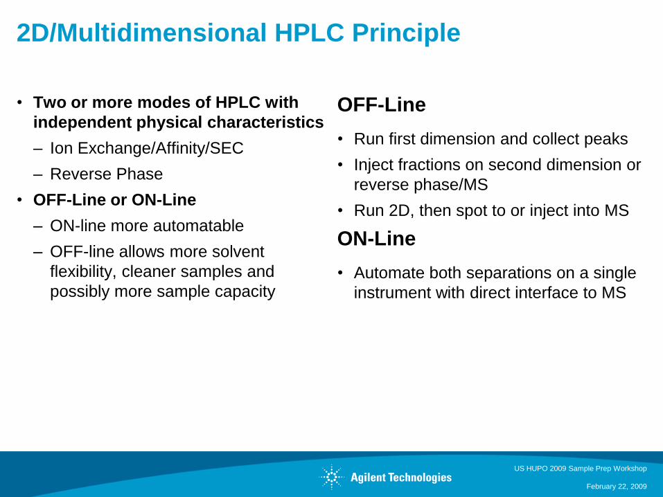
2D/Multidimensional HPLC Principle
• Two or more modes of HPLC with
independent physical characteristics
– Ion Exchange/Affinity/SEC
– Reverse Phase
• OFF-Line or ON-Line
– ON-line more automatable
– OFF-line allows more solvent
flexibility, cleaner samples and
possibly more sample capacity
OFF-Line
• Run first dimension and collect peaks
• Inject fractions on second dimension or
reverse phase/MS
• Run 2D, then spot to or inject into MS
ON-Line
• Automate both separations on a single
instrument with direct interface to MS
US HUPO 2009 Sample Prep Workshop
February 22, 2009
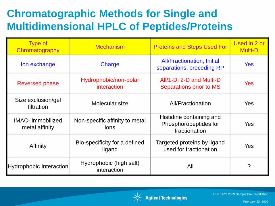
Chromatographic Methods for Single and
Multidimensional HPLC of Peptides/Proteins
Type of
ChromatographyMechanism Proteins and Steps Used For
Used in 2 or
Multi-D
Ion exchange ChargeAll/Fractionation, Initial
separations, preceding RPYes
Reversed phaseHydrophobic/non-polar
interaction
All/1-D, 2-D and Multi-D
Separations prior to MSYes
Size exclusion/gel
filtrationMolecular size All/Fractionation Yes
IMAC- immobilized
metal affinity
Non-specific affinity to metal
ions
Histidine containing and
Phosphoropeptides for
fractionation
Yes
Affinity Bio-specificity for a defined
ligand
Targeted proteins by ligand
used for fractionationYes
Hydrophobic InteractionHydrophobic (high salt)
interactionAll ?
US HUPO 2009 Sample Prep Workshop
February 22, 2009
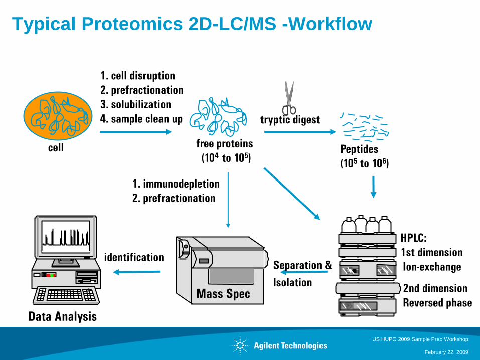
Typical Proteomics 2D-LC/MS -Workflow
HPLC:
1st dimension
Ion-exchange
2nd dimension
Reversed phase
1. cell disruption
2. prefractionation
3. solubilization
4. sample clean up
cell free proteins
(104 to 105)
tryptic digest
Peptides
(105 to 106)
Mass Spec
Data Analysis
Separation &
Isolation
identification
1. immunodepletion
2. prefractionation
US HUPO 2009 Sample Prep Workshop
February 22, 2009
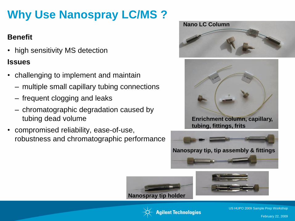
Why Use Nanospray LC/MS ?
Benefit
• high sensitivity MS detection
Issues
• challenging to implement and maintain
– multiple small capillary tubing connections
– frequent clogging and leaks
– chromatographic degradation caused by
tubing dead volume
• compromised reliability, ease-of-use,
robustness and chromatographic performance
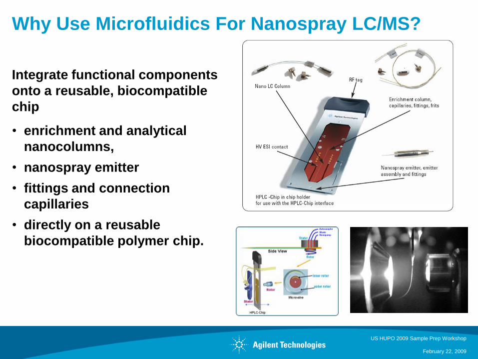
Nano LC Column
Enrichment column, capillary,
tubing, fittings, frits
Nanospray tip, tip assembly & fittings
Nanospray tip holder
US HUPO 2009 Sample Prep Workshop
February 22, 2009
Why Use Microfluidics For Nanospray LC/MS?
Integrate functional components
onto a reusable, biocompatible
chip
• enrichment and analytical
nanocolumns,
• nanospray emitter
• fittings and connection
capillaries
• directly on a reusable
biocompatible polymer chip.
US HUPO 2009 Sample Prep Workshop
February 22, 2009
Separation Options for Protein Identification
• 1D - nano or capillary HPLC for simple samples, e.g. single bands
• Online sample enrichment + 1D nano or capillary HPLC for medium complex
diluted samples
• 2D - capillary + nano HPLC for complex samples
e.g. 1. Cation Exchange, 2. Reversed Phase Separation
• Multidimensional - Fractionation columns + 2D HPLC for complex samples, e.g. 1.
IMAC, SEC, etc. 2. SCX, 3. Reversed Phase Separation
• Identify with MS
US HUPO 2009 Sample Prep Workshop
February 22, 2009
Parting Thoughts . . .
• Ion exchange chromatography, size exclusion chromatography and reversed phase
HPLC columns are the most popular choices for the analysis of proteins and
peptides.
• A variety of reversed phase columns make optimization of protein separations
possible. And a variety of column configurations – length and id make it easy to find
the right column for any size sample.
• Movement towards faster analytical separations, smaller sample sizes, and more
sensitive detection has increased sample throughput and proteomics applications
• Proteomics applications use the same columns and chromatographic techniques,
but with so many proteins present in each sample, pre-fractionation, orthogonal
techniques and nano scale analysis are necessary.
US HUPO 2009 Sample Prep Workshop
February 22, 2009
Conclusions
• Reducing sample complexity through intelligent sample preparation leads to more
positive identification of low abundant proteins
• Separation of proteins prior to sample digestion results in more proteins identified
• Improving the chromatographic separation of peptides results in more proteins
identified
• Multidimensional approaches generally require more analysis time which must be
taken into consideration


![[Kiemnghiemthuoc.com] Sample Preparation in Chromatography](https://img.pdfslide.net/doc/110x75/577ce5251a28abf1038fed3b/kiemnghiemthuoccom-sample-preparation-in-chromatography.jpg)