Embed Size (px)
Citation preview

LIVER CYSTS
By
Maher Osman, MD, Ph.DHPB Surgery, NLINovember 2006

INTRODUCTIONINTRODUCTION
• Non-parasitic cystic lesions are encountered commonly in clinical practice & have two common criteria:– Congenital malformation (inherited or not) of the
intrahepatic bile ducts,– The basic lesion consists of macroscopic or
microscopic cysts.
• Liver cysts may be:– Asymptomatic (in the vast majority of patients)– Symptomatic (in a minority of cases).

Non-Parasitic Liver Cysts Non-Parasitic Liver Cysts
CLASSIFICATION
• Congenital (most common):• Simple• Polycystic liver disease (PLD).
• Inflammatory cysts (liver abscess).• Cystic neoplastic lesions (rare):
• Cystadenoma,• Cystadenocarcinoma.
• Traumatic cysts:• Subcapsular hematoma,• Transected biliary duct (encysted biloma).

CONGENITAL LIVER CYSTSCONGENITAL LIVER CYSTS
Pathology & Pathogenesis
• Macroscopically:
– Spherical or ovoid shape,– Ranges from few mm to 20 cm,– No communication with the intrahepatic bile
ducts,– Solitary or mutiple,– No septations (unilocular),– Clear serous fluid.

CONGENITAL LIVER CYSTSCONGENITAL LIVER CYSTS
Pathology & Pathogenesis
• Microscopically:
– Bordered by a single layer of cuboid or columnar epithelium,
– These cells retain differentiated secretory function.
– The cyst fluid contains water & mineral electrolytes without bile acids or bilirubin (not toxic)
– Regarded as a congenital malformation; an aberrant bile duct have lost communication with the biliary tree an would dilate progressively.

CONGENITAL LIVER CYSTSCONGENITAL LIVER CYSTSPrevalence
• Prevalence:– Considered rare.– The prevalence was 3-5% (In US studies).– The prevalence was 18% (In spiral CT study),
• Incidence:– Age and gender related.– Uncommon before age 40.– Larger in adults > 50 than in young patients. – The female-to-male ratio is 1.5:1 in asymptomatic cysts– The female-to-male ratio is 9:1 in symptomatic cysts.

CONGENITAL LIVER CYSTSCONGENITAL LIVER CYSTS
Clinical Presentation
1) Simple cysts• Usually solitary (can be multiple).• Distributed randomly in the liver.• Vary widely in size (few mm to > 20 cm) • Usually asymptomatic.• Approximately 10% are symptomatic.

CONGENITAL LIVER CYSTSCONGENITAL LIVER CYSTS
Clinical Presentation
1) Simple cysts:• Symptoms develop slowly due to cyst expansion & adjacent organ
compression.
• Complications:• Hemorrhage (sudden acute abdominal pain).• Rupture (sudden pain and hypotension).• Infection (signs and symptoms of sepsis)• Biliary obstruction.

CONGENITAL LIVER CYSTSCONGENITAL LIVER CYSTS
Clinical Presentation
2) Polycystic liver disease:• Have innumerable cysts.
• Distributed throughout the liver with only an occasional segment which is relatively spared of involvement.
• Patients usually present in their 4th-5th decade of life.

CONGENITAL LIVER CYSTSCONGENITAL LIVER CYSTS
Clinical Presentation2) Polycystic liver disease:• Actually presents as a spectrum of disease
with wide variations in the number & site of cysts.
• Gradual onset of:• Severe abdominal distension.• Fatigue.• Satiety.
• Progressive hepatomegaly from cyst enlargement in < 10% of patients.

CONGENITAL LIVER CYSTSCONGENITAL LIVER CYSTS
Clinical Presentation2) Polycystic liver disease:• Complications of PLD:
• Infection• Bile duct obstruction• Budd-chiari syndrome• Perforation• Torsion• Hemorrhage• Hepatic insufficiency is rare

CONGENITAL LIVER CYSTSCONGENITAL LIVER CYSTS
Clinical Presentation2) Polycystic liver disease:• In ½-2/3 of patients will have autosomal dominant
polycystic kidney disease (ADPKD).
• End-stage kidney disease can occur and can be treated effectively.
• Cysts can also occur in the pancreas and spleen.
• Intracebral anurysms are seen in 10% of patients.

CONGENITAL LIVER CYSTSCONGENITAL LIVER CYSTS
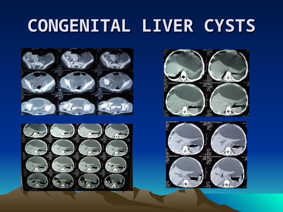
Imaging
• The prevalence of hepatic cysts has approached 10% of the general population.
• The major goal for Imaging of liver cysts is:
• High level of diagnostic accuracy.• Essential for appropriate management.

CONGENITAL LIVER CYSTSCONGENITAL LIVER CYSTS
Imaging
• Imaging characteristics depend on cyst structure:
• Circular to ovoid.
• Lack septations
• Uniformly thin walls.
• No intracystic masses or papillae.
CONGENITAL LIVER CYSTSCONGENITAL LIVER CYSTS

CONGENITAL LIVER CYSTSCONGENITAL LIVER CYSTS

CONGENITAL LIVER CYSTSCONGENITAL LIVER CYSTS

CONGENITAL LIVER CYSTSCONGENITAL LIVER CYSTS
Ultrasound (US)• The most cost-effective imaging
modality.• Can clearly demonstrate the structural
characteristics of a simple cyst.• It can show:
• Minimal ecchogenicity within the cyst.• Enhanced transmission of the acoustic beam
through the cyst walls.

CONGENITAL LIVER CYSTSCONGENITAL LIVER CYSTS
Computerized tomography (CT)
• Similar characteristics to those in US except for the absence of sonographic enhancement.
• Cystic cavities are uniformly hypodense.
• Septations which are inconspicuous on US may be seen in CT.
• Preferable in PLD, to fully detail the extent of intra-abdominal cystic disease.

CONGENITAL LIVER CYSTSCONGENITAL LIVER CYSTS
Magnetic Resonance (MRI) • Demonstrates simple cysts as smooth-walled lesions
with a uniform signal intensity that is generally isotense to CSF.
• Permits definition of hemorrhage or infection within the cyst by differentiating densities between fluid content.
• Defines the intrahepatic vascular relationships in patients with PLD when hepatic venous outflow occlusion or IVC compression is suspected clinically.

CONGENITAL LIVER CYSTSCONGENITAL LIVER CYSTS
• The diagnostic accuracy of US, CT, MRI are equivalent with accuracy around 98% for simple cysts 1 cm or greater.
• Features which should alert the clinician to alternative diagnosis or further studies:
• Mural nodularity,• Irregular wall thickening,• Multiple septations,• Fluid level within a cystic mass.
CONGENITAL LIVER CYSTSCONGENITAL LIVER CYSTS
Management & Results
• Appropriate treatment depends on:• The cause of symptoms,• The extent of cystic disease.
• Several treatment options are available:• Percutaneous aspiration alone.• Percutaneous aspiration and sclerosis.• Cyst fenestration• Combined cyst fenestration and liver resection.• Liver transplantation.

CONGENITAL LIVER CYSTSCONGENITAL LIVER CYSTS
(1 ) Percutaneous Aspiration Alone• Has limited efficacy.• Has two major roles:
– Diagnostic role to determine whether symptoms are related to the cyst or not.
– Temporary therapeptic measure prior to definitive treatment
• Associated with a nearly 100% recurrence rate
CONGENITAL LIVER CYSTSCONGENITAL LIVER CYSTS
(2) Percutaneous Aspiration & Sclerosis
• To ablate the secretory epithelial lining of liver cysts and to reduce recurrence.
• A useful, well-established procedure with increasing evidence to support its long-term efficacy.
CONGENITAL LIVER CYSTSCONGENITAL LIVER CYSTS
• CT or US-guided aspiration of the cyst fluid.• Instillation of radiologic contrast (to
document absence of bile duct communication).
• Absolute alcohol 5-25 ccs is instilled.• Repositioning of the patient several times.• Following adequate alcohol exposure (30
mins), the alcohol is aspirated.

CONGENITAL LIVER CYSTSCONGENITAL LIVER CYSTS
(3) CYST FENESTRATION
• Consists of wide unroofing of the superficial surface of the cyst and excising a portion of the common wall between non-superficial cysts.
• Indicated in:– Recurrent symptomatic cysts after ablation.– Anatomic factors preclude safe percutaneous access.– Radiologic expertise is unavailable.
• Can be performed either laparoscopically or via open laparotomy.

CONGENITAL LIVER CYSTSCONGENITAL LIVER CYSTS
• Laparoscopic procedure is indicated for recurrent symptomatic cysts which are located in the anterior liver segments from III to VI.
• The open laparotomy can be done in patients with recurrent symptomatic cysts, who are not candidates for laparoscopic approach

CONGENITAL LIVER CYSTSCONGENITAL LIVER CYSTS
• Wide excision of the superficial cyst wall is an important technical
factor reducing recurrence.
• Deroofing permits continual peritoneal absorption of further cyst secretions.
• Ablation of the cyst epithelium with coagulation (argon beam) or topical sclerosants may reduce cyst secretion and recurrence.
• Temporary closed suction drainage is optional postoperatively.
• Open laparotomy has a current advantage bec it permits excision or ablation of the non-superficial epithelial lining.

CONGENITAL LIVER CYSTSCONGENITAL LIVER CYSTS
(4) Combined cyst fenestration & excision
• Indicated in:– patients with multiple cysts of the liver if aspiration &
alcohol sclerosis has failed– if cysts communicate with the biliary tree.
• Enucleation can be done for most cysts regardless the size.
• Formal anatomic hepatectomy is rarely required.

CONGENITAL LIVER CYSTSCONGENITAL LIVER CYSTS
• In PLD, this procedure has been advocated increasingly in the last few years as a solution to:
– Reduce symptoms associated with hepatomegaly.
– Preserve liver function– Avoid liver transplantation.

CONGENITAL LIVER CYSTSCONGENITAL LIVER CYSTS
(5) Liver Transplantation • Indicated for patients with:
– Severe PLD and no segmental sparing– Failed resection and fenestration– Liver failure.
• The timing of LTx depends not only on anatomic distribution of liver cysts but also on the performance status and on renal function which also may warrant kidney transplantation.

NEOPLASTIC LIVER CYSTSNEOPLASTIC LIVER CYSTS
• Rare neoplasms that constitute less than 5% of hepatic cysts.
• 80% of patients are women & 80% present with abdominal pain or a mass.

NEOPLASTIC LIVER CYSTSNEOPLASTIC LIVER CYSTS
• In US and CT the following features should suggest a cystic neoplasm:
– Multiloculated cysts from septa of variable thickness.
– Thick wall– Solid intracystic components.– Hypervascularity of the cyst wall is suggestive of
a cystadenocarcinoma.

NEOPLASTIC LIVER CYSTSNEOPLASTIC LIVER CYSTS
CYSTADENOMA
• Contains moderate to dense cellular supporting stroma (like ovarian stroma).
• Lined by cuboidal or columnar mucus-secreting epithelium.
• Should be resected completely bec. of:– The risk of unpredictable malignant progression– The more practical risk of recurrence after partial exicison.
• Can be resected by enucleation.
NEOPLASTIC LIVER CYSTSNEOPLASTIC LIVER CYSTS
CYSTADENOCARCINOMA
• Shows similar stromal composition with a papillary adenocarcinoma lining the cysts with invasion of the cyst wall.
• May only focally involve the cyst (numerous biopsies are required to exclude malignancy).
• Considred as a malignant degeneration of a benign cystadenoma.

NEOPLASTIC LIVER CYSTSNEOPLASTIC LIVER CYSTS
CYSTADENOCARCINOMA
• The pathogenesis of a cystadenocarcinoma from a cystadenoma has been documented.
• Clinical evidence supporting this theory is 17 year disparity in the mean age of patients.
• There is no sex predilection.
• Should be resected widely as any hepatic neoplasm.
• The 5-year survival rate is 25%
TRAUMATIC LIVER CYSTSTRAUMATIC LIVER CYSTS
• Rare cysts and constitute less than 0.5% of liver cysts.
• Occur following significant trauma to the liver with disruption of intrahepatic bile ducts or formation of a subcapsular hematoma.
• Do not have epithelial lining
• Actually represent hepatic pseudocysts.

TRAUMATIC LIVER CYSTSTRAUMATIC LIVER CYSTS
• Present as a cystic mass filled with old blood or bile.
• Clinical presentation, indications and options of treatment are similar to those of simple cysts.
• Occasionally, these cysts spontaneously resolve, therefore, a period of observation (6-12 ms) for potential resolution is preferred.

TRAUMATIC LIVER CYSTSTRAUMATIC LIVER CYSTS
• Surgery is indicated is:– Symptoms persist.– The cyst grows.
• Operation is preferred to address the bile duct disruption.
• Because the cyst lining is non-secretory partial cyst excision is adequate.
• Wide excision of the superficial cyst wall is necessary to identify & ligate the disrupted bile ducts.