Embed Size (px)
Citation preview
Localized Ocular Amyloidosis: Case Series
By: Lindsay Hammons, Dr. Pinky Jha & Dr. Anita D’Souza
Medical College of Wisconsin, Milwaukee, WI, USA

Financial Disclosures� No financial disclosures

Localized Ocular Amyloidosis� Background
� Four patient cases: A, B, C, D
� Workup of systemic amyloidosis (AL)
� Discussion
� Follow-ups
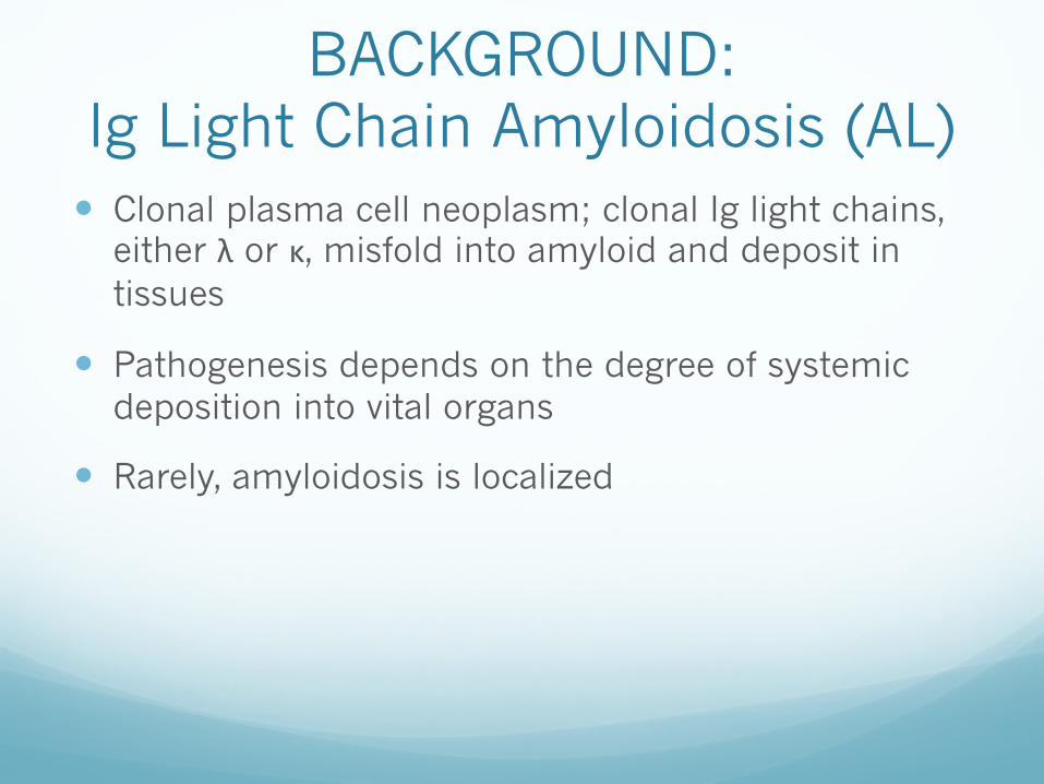
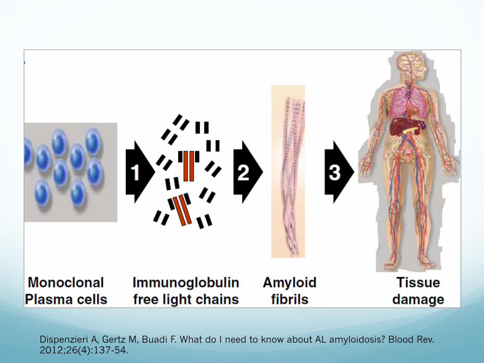
BACKGROUND: Ig Light Chain Amyloidosis (AL)� Clonal plasma cell neoplasm; clonal Ig light chains,
either λ or κ, misfold into amyloid and deposit in tissues
� Pathogenesis depends on the degree of systemic deposition into vital organs
� Rarely, amyloidosis is localized
Dispenzieri A, Gertz M, Buadi F. What do I need to know about AL amyloidosis? Blood Rev. 2012;26(4):137-54.

Case: Patient A
Levin, P. (2009, ). Ptosis surgery. Retrieved from http://www.levinmd.com/galleryptosis-cosmeticsurgeonbay area.htm

Case: Patient A� Clinical: 31yo F with left ptosis, watery eyes, a left
inner eyelid lesion and a foreign body sensation (FBS) in her left eye
� Physical exam: left eye ptosis
� Bx: Congo red + staining on left upper lid with positive AL (lambda) type amyloid deposition

Case: Patient B
Bernadino, C. R. (2003, ). A 56yearold woman with a progressively bulging eye. Retrieved from http://www.medscape.com/viewarticle/708216
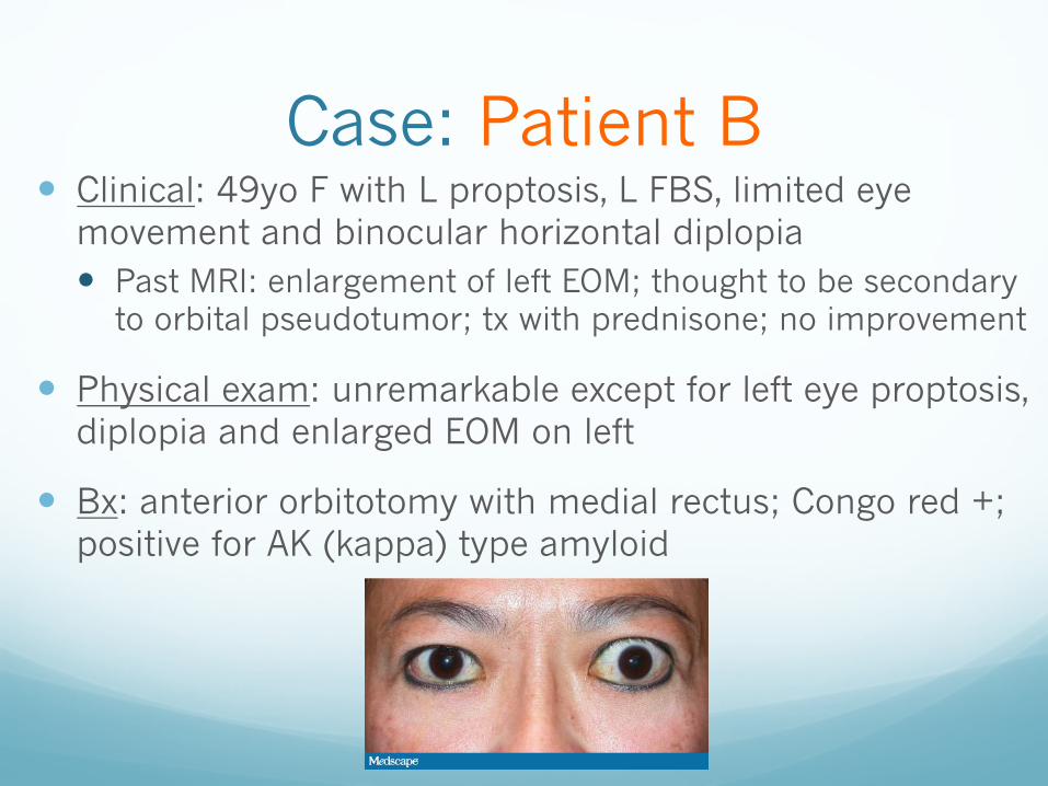
Case: Patient B� Clinical: 49yo F with L proptosis, L FBS, limited eye
movement and binocular horizontal diplopia� Past MRI: enlargement of left EOM; thought to be secondary
to orbital pseudotumor; tx with prednisone; no improvement
� Physical exam: unremarkable except for left eye proptosis, diplopia and enlarged EOM on left
� Bx: anterior orbitotomy with medial rectus; Congo red +; positive for AK (kappa) type amyloid

Case: Patient C
Agerelated vision changes and eyesight problems in the elderly. (2011, ). Retrieved from http://www.seniorhealth365.com/wpcontent/uploads/2011/11/EyesightProblemsinElderly.jpg

Case: Patient C� Clinical: 69yo M with L ptosis for the past month with
a change in vision
� Physical exam: unremarkable except for left upper lid ptosis with significant visual field changes
� Bx: L orbicularis muscle and full-thickness wedge left upper lid excision; immunohistochemistry suggestive of AA, weak staining for λ and κ was also present
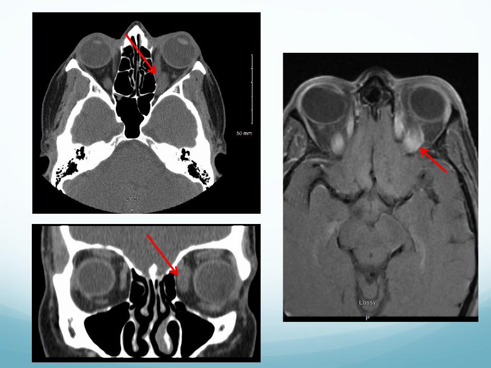

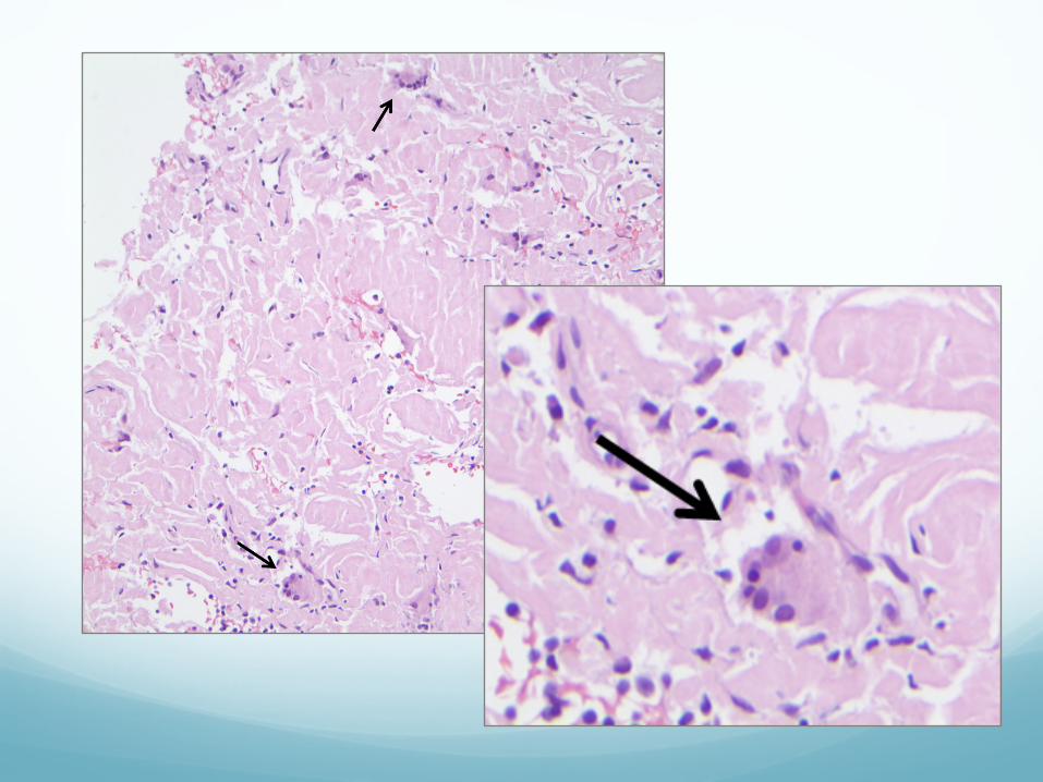
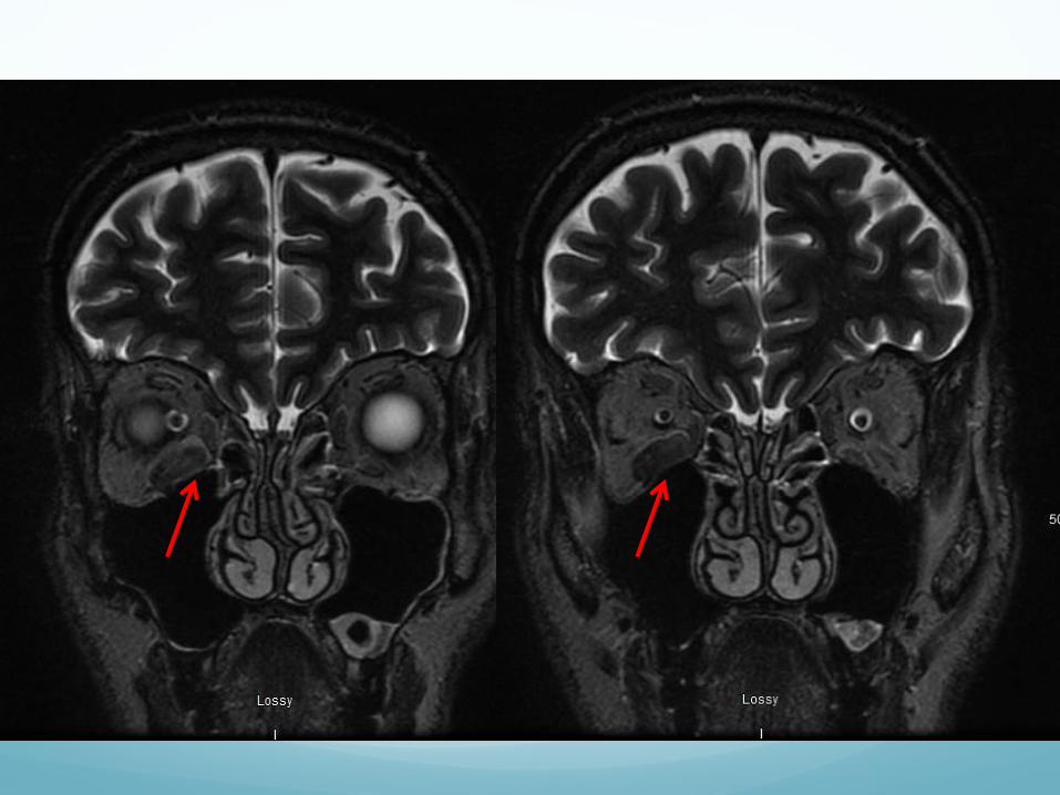
Case: Patient D
Knoop, K. J., Stack, L. B., & Thurman, R. J. (). The atlas of emergency medicine, 3rd edition. Retrieved from http://accessemergencymedicine.mhmedical.com/data/Books/knoo3/knoo3_c002f047c.jpg
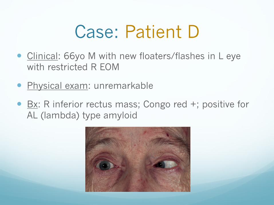
Case: Patient D� Clinical: 66yo M with new floaters/flashes in L eye
with restricted R EOM
� Physical exam: unremarkable
� Bx: R inferior rectus mass; Congo red +; positive for AL (lambda) type amyloid
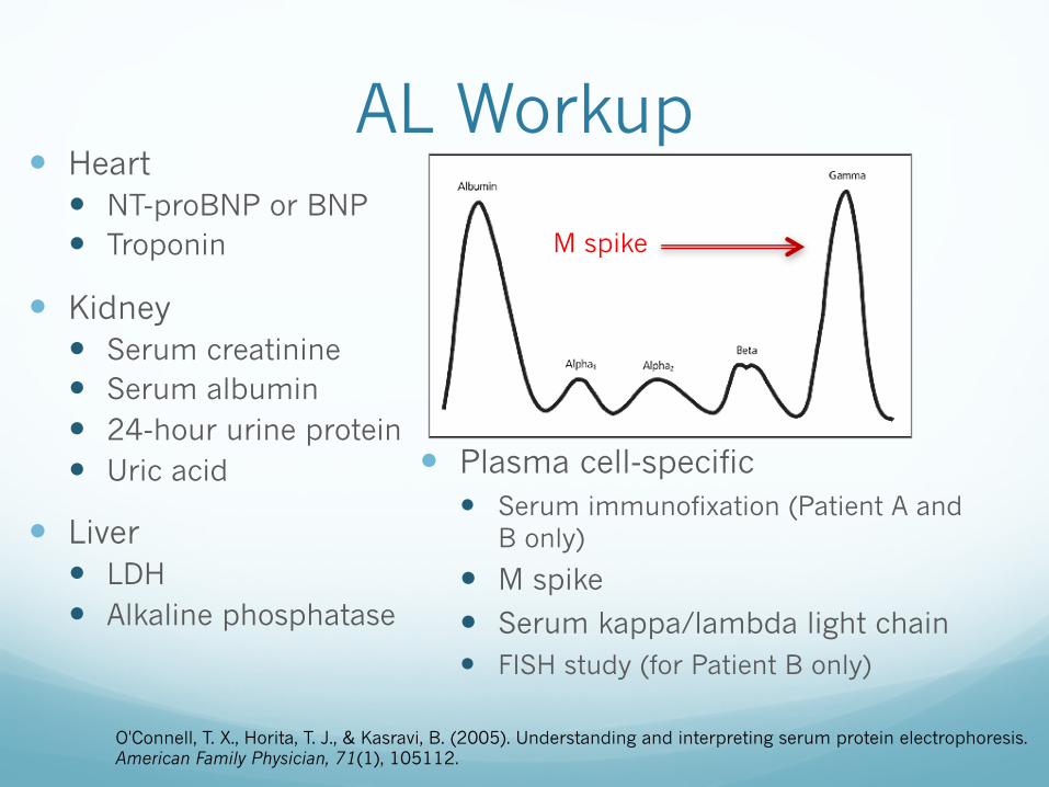
AL Workup� Heart
� NT-proBNP or BNP� Troponin
� Kidney� Serum creatinine� Serum albumin� 24-hour urine protein� Uric acid
� Liver� LDH� Alkaline phosphatase
O'Connell, T. X., Horita, T. J., & Kasravi, B. (2005). Understanding and interpreting serum protein electrophoresis. American Family Physician, 71(1), 105112.
� Plasma cell-specific � Serum immunofixation (Patient A and
B only)
� M spike
� Serum kappa/lambda light chain� FISH study (for Patient B only)
M spike
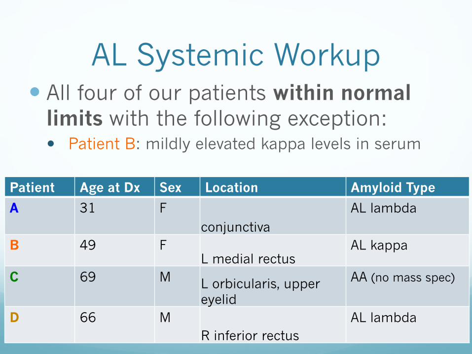
AL Systemic Workup� All four of our patients within normal
limits with the following exception:� Patient B: mildly elevated kappa levels in serum
Patient Age at Dx Sex Location Amyloid Type
A 31 Fconjunctiva
AL lambda
B 49 FL medial rectus
AL kappa
C 69 M L orbicularis, upper eyelid
AA (no mass spec)
D 66 MR inferior rectus
AL lambda

Discussion
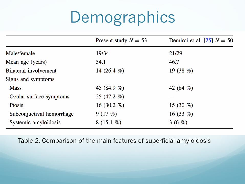
Table 2. Comparison of the main features of superficial amyloidosis
Demographics

Clinical features� Conjunctival
� Eyelid edema� Papules� Yellow subconjunctival plaques� Eyelid ptosis
� Superficial amyloidosis� Alteration in lacrimal film� Superior palpebral conjunctiva/levator muscle involvement� Ptosis
� Deep amyloidosis� EOM, orbital fat, lacrimal gland� Limited movement� Proptosis� Ocuclar displacement� Ptosis

Limitations of the Study� Not focused solely on AL (also AA)
� Some pts have underlying bone marrow malignancies that can complicate diagnosis� Multiple Myeloma� MALT lymphoma
� Solitary osseous plasmacytoma� Waldenström macroglobulinemia
� One patient with systemic amyloidosis
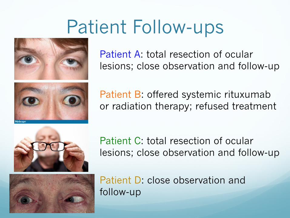
Patient Follow-upsPatient A: total resection of ocular lesions; close observation and follow-up
Patient B: offered systemic rituxumabor radiation therapy; refused treatment
Patient C: total resection of ocular lesions; close observation and follow-up
Patient D: close observation and follow-up
Take Home Points� Patient with unilateral ptosis, consider systemic
amyloidosis on differential diagnosis
� Patients with localized ocular amyloidosis in the conjunctiva and/or extraocular muscles have very low risk of developing systemic involvement
� A watchful waiting approach appears acceptable in the absence of symptoms

References� Agerelated vision changes and eyesight problems in the elderly. (2011, ). Retrieved
from http://www.seniorhealth365.com/wp-content/uploads/2011/11/EyesightProblemsinElderly.jpg
� Bernadino, C. R. (2003, ). A 56yearold woman with a progressively bulging eye. Retrieved from http://www.medscape.com/viewarticle/708216
� Dispenzieri A, Gertz M, Buadi F. What do I need to know about AL amyloidosis? Blood Rev. 2012;26(4):137-54.
� Knoop, K. J., Stack, L. B., & Thurman, R. J. (). The atlas of emergency medicine, 3rd edition. Retrieved from http://accessemergencymedicine.mhmedical.com/data/Books/knoo3/knoo3_c002f047c.jpg
� Levin, P. (2009, ). Ptosis surgery. Retrieved from http://www.levinmd.com/gallery-ptosiscosmeticsurgeonbay area.htm
� MoraHorna, E.R., RojasPadilla, R., López, V.G., Guzmán, M.J., Ceriotto, A., and Salcedo, G. Ocular adnexal and orbital amyloidosis: A case series and literature review. International Opthomology, 2015; 36:281–298.
� O'Connell, T. X., Horita, T. J., & Kasravi, B. (2005). Understanding and interpreting serum protein electrophoresis. American Family Physician, 71(1), 105112.

QUESTIONS?