Embed Size (px)
Citation preview

Published Ahead of Print 2 April 2014. 10.1128/JCM.00525-14.
2014, 52(6):2071. DOI:J. Clin. Microbiol. Herrick and Thomas B. NutmanPapa M. Drame, Doran L. Fink, Joseph Kamgno, Jesica A. Loa loa InfectionRapid and Semiquantitative Detection of Loop-Mediated Isothermal Amplification for
http://jcm.asm.org/content/52/6/2071Updated information and services can be found at:
These include:
REFERENCEShttp://jcm.asm.org/content/52/6/2071#ref-list-1at:
This article cites 44 articles, 14 of which can be accessed free
CONTENT ALERTS more»articles cite this article),
Receive: RSS Feeds, eTOCs, free email alerts (when new
http://journals.asm.org/site/misc/reprints.xhtmlInformation about commercial reprint orders: http://journals.asm.org/site/subscriptions/To subscribe to to another ASM Journal go to:
on June 5, 2014 by UN
IVE
RS
ITE
IT M
AA
ST
RIC
HT
http://jcm.asm
.org/D
ownloaded from
on June 5, 2014 by U
NIV
ER
SIT
EIT
MA
AS
TR
ICH
Thttp://jcm
.asm.org/
Dow
nloaded from

Loop-Mediated Isothermal Amplification for Rapid andSemiquantitative Detection of Loa loa Infection
Papa M. Drame,a Doran L. Fink,a* Joseph Kamgno,b,c Jesica A. Herrick,a* Thomas B. Nutmana
Laboratory of Parasitic Diseases, National Institute of Allergy and Infectious Diseases, National Institutes of Health, Bethesda, Maryland, USAa; Centre for Research onFilariasis and other Tropical Diseases, Yaounde, Cameroonb; Faculty of Medicine and Biomedical Sciences, University of Yaounde I, Yaounde, Cameroonc
Rapid and accurate tests are currently needed to identify individuals with high levels of Loa loa microfilaria (mf), so that theseindividuals may be excluded from mass ivermectin administration campaigns against onchocerciasis and lymphatic filariasisbeing conducted in areas where Onchocerca volvulus, Wuchereria bancrofti, and L. loa are coendemic. To address this need, col-orimetric loop-mediated isothermal amplification (LAMP) assays targeting the L. loa-specific gene sequences LLMF72 andLLMF342 were developed for the detection and quantification of L. loa microfilaremia. Both LAMP assays were highly specific(100%) for L. loa infection compared to the absence of infection or infection with related filarial pathogens. The LLMF72-basedLAMP assay showed greater analytic sensitivity (limit of detection, 0.1 pg/ml of genomic DNA [gDNA] and/or 5 mf/ml) than theLLMF342-based LAMP assay (10 pg/ml of gDNA and/or 50 mf/ml), and its analytic sensitivity was similar to that of LLMF72-based quantitative PCR (qPCR). A high level of correlation was observed between microfilaria counts as determined by LLMF72-based qPCR and time to positivity by the LAMP assay, and performance measures of sensitivity, specificity, and positive and neg-ative predictive values were similar for both assays when applied to field-collected clinical samples. By simply varying the runtime, the LAMP assay was able to accurately distinguish individuals at risk for serious adverse events (SAEs) after exposure toivermectin, using thresholds of >5,000 mf/ml and >30,000 mf/ml as indicators of increasing levels of risk. In summary, LLMF72LAMP represents a new molecular diagnostic tool that is readily applicable as a point-of-care method for L. loa microfilarial de-tection and quantification in resource-limited countries where L. loa infection is endemic.
Loa loa, the causative agent of loiasis, is a parasitic nematode trans-mitted to humans by the tabanid Chrysops fly (1), with transmis-
sion confined to the rainforest and some savannah areas of West andCentral Africa (2). Although the overwhelming majority of L. loa-infected individuals are clinically asymptomatic, Calabar swelling(transient, localized angioedema), and the subconjunctival migrationof an adult worm (“eyeworm”) are the most common clinical man-ifestations (3–5). Rarely, nephropathy, cardiomyopathy, retinopathy,neuropsychiatric complications, and encephalopathy (6–12) can oc-cur as a consequence of chronic infection. Among the 8 filarial infec-tions of humans, L. loa infection has largely been neglected as a publichealth concern in Africa, though it has gained prominence of latebecause of the neurologic serious adverse events (SAEs) that haveoccurred, occasionally leading to death, in highly microfilaremic in-dividuals following exposure to ivermectin (IVM) given during massdrug administration (MDA) for control of Onchocerca volvulusand/or Wuchereria bancrofti in regions where these filarial speciesand L. loa are coendemic. (13, 14).
Although the pathogenesis of the neurologic SAEs is still notwell understood (8, 15), data clearly demonstrate a relationshipbetween the pretreatment L. loa microfilaria (mf) density (14, 16)and the risk of SAEs, with levels of 5,000 mf/ml and 30,000 mf/mlfelt to be the most significant thresholds (13), though other factorsmay also be involved (17–19). Consequently, some IVM-basedMDA programs for onchocerciasis (Ov) have been delayed, andlymphatic filariasis (LF) control programs (using IVM-albenda-zole) have been put on hold in large parts of Central Africa whereL. loa infection is also endemic (20).
In areas where L. loa infection is endemic, the routine methodfor diagnosis and quantification of L. loa is based on calibratedmicroscopic examination of midday blood samples for microfi-lariae, a process that is neither point of care nor high throughput.
Indeed, it is felt to be impractical for use as a widespread screeningtool (21). Alternative serological (22, 23) and molecular (24–26)tests have been developed, but to date, only real-time PCR meth-odology has been able to combine a high degree of sensitivity andspecificity with the ability to accurately quantify L. loa mf levels(26, 27). These methods, however, require a centralized, well-equipped laboratory and relatively expensive reagents.
Loop-mediated isothermal amplification (LAMP) has emerged asa potential alternative to DNA amplification techniques requiringthermocycling and automated fluorescence description (28–30).LAMP relies on an autocycling strand displacement DNA synthesisperformed at a single temperature (30, 31). The by-product of theLAMP reaction is magnesium pyrophosphate that accumulates as thereaction progresses and that can be monitored using turbidity mea-surements or visually using a variety of intercalating dyes (e.g., SYBRgreen I, calcein, hydroxy naphthol blue [HNB]). In particular, addingHNB dye to the reaction tube before amplification has improved thecapacity of the LAMP method to be used as a point-of-care diagnostic
Received 24 February 2014 Returned for modification 14 March 2014Accepted 27 March 2014
Published ahead of print 2 April 2014
Editor: M. J. Loeffelholz
Address correspondence to Thomas B. Nutman, [email protected].
* Present address: Doran L. Fink, Center for Biologics Evaluation and Research,Food and Drug Administration, Silver Spring, Maryland, USA; Jesica A. Herrick,Section of Infectious Diseases, Immunology, and International Medicine,Department of Medicine, University of Illinois at Chicago, Chicago, Illinois, USA.
Copyright © 2014, American Society for Microbiology. All Rights Reserved.
doi:10.1128/JCM.00525-14
June 2014 Volume 52 Number 6 Journal of Clinical Microbiology p. 2071–2077 jcm.asm.org 2071
on June 5, 2014 by UN
IVE
RS
ITE
IT M
AA
ST
RIC
HT
http://jcm.asm
.org/D
ownloaded from
test, since it has made the assay easier to use in resource-limited set-tings (32, 33). Recently, LAMP technology has been applied with highaccuracy to detect infection with a wide array of pathogens, includingbacteria (34, 35), viruses (36), and parasites (28, 37, 38).
In the present study, we have used two L. loa-specific single-copy target genes, LLMF72 and LLMF342, to design LAMPprimers for detection of L. loa mf in human blood and havedeveloped methods that have a sensitivity and specificity sim-ilar to those of the previously described quantitative PCR(qPCR) assay targeting the LLMF72 gene (26). Furthermore,we have identified LAMP conditions that allow for colorimetricdetermination of microfilarial levels above or below specifiedthresholds such that our LAMP method now has the potentialto be a practical point-of-care tool for identifying people at riskfor SAEs when given IVM.
MATERIALS AND METHODSSamples. The source of genomic DNA from L. loa, W. bancrofti, Brugiamalayi, and Mansonella perstans microfilariae (mf) has been describedpreviously (26). For some experiments, 100,000 purified L. loa mf wereplaced in a fixed volume (1 ml) of distilled water and serially diluted tocreate standards for quantification. DNA was then extracted and used forassessment of the sensitivity of the assays. Finally, to evaluate the perfor-mance of the L. loa loop-mediated isothermal amplification (LAMP) assaycompared to the standard quantitative PCR (qPCR) method, 93 middaydried blood spots on filter paper (Whatman no. 1 filter paper) previouslycollected in Cameroon (26) were assessed. These blood spots were ob-tained by pricking the fingers of volunteers living in a region where L. loainfection is endemic. Briefly, capillary blood was spotted onto Whatmanfilter paper, dried, and stored at 4°C until their use for DNA extraction.
DNA extraction for spiked mf and field-collected blood spot sam-ples. Blood spots were punched using a disposable 6-mm biopsy punch(Acuderm, Inc., Ft. Lauderdale, FL, USA). Two punched blood spots (10�l dried blood each) were immersed in 400 �l of water in a Precellys lysingtube (Peqlab, Wilmington, DE, USA) containing glass and ceramic beads.The tube content was homogenized at 6,500 relative centrifugal force (rcf)for 5 min using a Precellys24 bead beating machine (Peqlab, Wilmington,DE, USA). The homogenized samples were then heated for 30 min at 99°Cwhile shaking in a thermomixer (Eppendorf, North America, USA). Fi-nally, the samples were centrifuged at 15,700 rcf for 10 min, and thesupernatant was collected for use. DNA was also isolated from waterspiked with 100,000 purified L. loa mf/ml using the method describedabove.
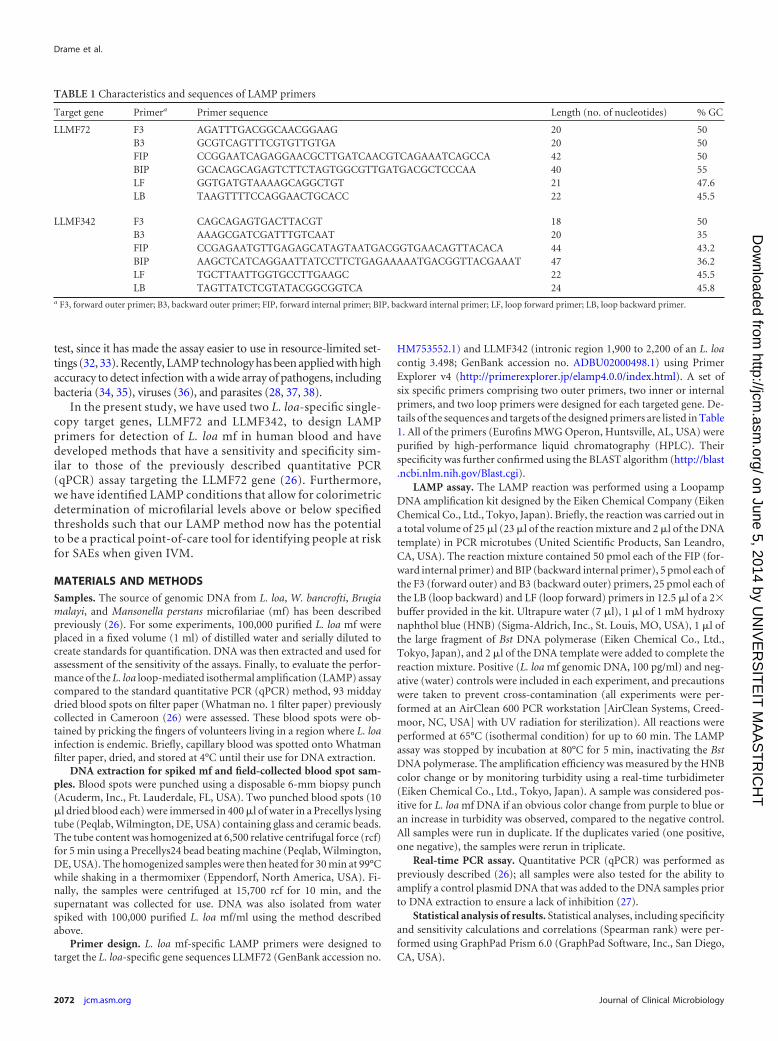
Primer design. L. loa mf-specific LAMP primers were designed totarget the L. loa-specific gene sequences LLMF72 (GenBank accession no.
HM753552.1) and LLMF342 (intronic region 1,900 to 2,200 of an L. loacontig 3.498; GenBank accession no. ADBU02000498.1) using PrimerExplorer v4 (http://primerexplorer.jp/elamp4.0.0/index.html). A set ofsix specific primers comprising two outer primers, two inner or internalprimers, and two loop primers were designed for each targeted gene. De-tails of the sequences and targets of the designed primers are listed in Table1. All of the primers (Eurofins MWG Operon, Huntsville, AL, USA) werepurified by high-performance liquid chromatography (HPLC). Theirspecificity was further confirmed using the BLAST algorithm (http://blast.ncbi.nlm.nih.gov/Blast.cgi).
LAMP assay. The LAMP reaction was performed using a LoopampDNA amplification kit designed by the Eiken Chemical Company (EikenChemical Co., Ltd., Tokyo, Japan). Briefly, the reaction was carried out ina total volume of 25 �l (23 �l of the reaction mixture and 2 �l of the DNAtemplate) in PCR microtubes (United Scientific Products, San Leandro,CA, USA). The reaction mixture contained 50 pmol each of the FIP (for-ward internal primer) and BIP (backward internal primer), 5 pmol each ofthe F3 (forward outer) and B3 (backward outer) primers, 25 pmol each ofthe LB (loop backward) and LF (loop forward) primers in 12.5 �l of a 2�buffer provided in the kit. Ultrapure water (7 �l), 1 �l of 1 mM hydroxynaphthol blue (HNB) (Sigma-Aldrich, Inc., St. Louis, MO, USA), 1 �l ofthe large fragment of Bst DNA polymerase (Eiken Chemical Co., Ltd.,Tokyo, Japan), and 2 �l of the DNA template were added to complete thereaction mixture. Positive (L. loa mf genomic DNA, 100 pg/ml) and neg-ative (water) controls were included in each experiment, and precautionswere taken to prevent cross-contamination (all experiments were per-formed at an AirClean 600 PCR workstation [AirClean Systems, Creed-moor, NC, USA] with UV radiation for sterilization). All reactions wereperformed at 65°C (isothermal condition) for up to 60 min. The LAMPassay was stopped by incubation at 80°C for 5 min, inactivating the BstDNA polymerase. The amplification efficiency was measured by the HNBcolor change or by monitoring turbidity using a real-time turbidimeter(Eiken Chemical Co., Ltd., Tokyo, Japan). A sample was considered pos-itive for L. loa mf DNA if an obvious color change from purple to blue oran increase in turbidity was observed, compared to the negative control.All samples were run in duplicate. If the duplicates varied (one positive,one negative), the samples were rerun in triplicate.
Real-time PCR assay. Quantitative PCR (qPCR) was performed aspreviously described (26); all samples were also tested for the ability toamplify a control plasmid DNA that was added to the DNA samples priorto DNA extraction to ensure a lack of inhibition (27).
Statistical analysis of results. Statistical analyses, including specificityand sensitivity calculations and correlations (Spearman rank) were per-formed using GraphPad Prism 6.0 (GraphPad Software, Inc., San Diego,CA, USA).
TABLE 1 Characteristics and sequences of LAMP primers
Target gene Primera Primer sequence Length (no. of nucleotides) % GC
LLMF72 F3 AGATTTGACGGCAACGGAAG 20 50B3 GCGTCAGTTTCGTGTTGTGA 20 50FIP CCGGAATCAGAGGAACGCTTGATCAACGTCAGAAATCAGCCA 42 50BIP GCACAGCAGAGTCTTCTAGTGGCGTTGATGACGCTCCCAA 40 55LF GGTGATGTAAAAGCAGGCTGT 21 47.6LB TAAGTTTTCCAGGAACTGCACC 22 45.5
LLMF342 F3 CAGCAGAGTGACTTACGT 18 50B3 AAAGCGATCGATTTGTCAAT 20 35FIP CCGAGAATGTTGAGAGCATAGTAATGACGGTGAACAGTTACACA 44 43.2BIP AAGCTCATCAGGAATTATCCTTCTGAGAAAAATGACGGTTACGAAAT 47 36.2LF TGCTTAATTGGTGCCTTGAAGC 22 45.5LB TAGTTATCTCGTATACGGCGGTCA 24 45.8
a F3, forward outer primer; B3, backward outer primer; FIP, forward internal primer; BIP, backward internal primer; LF, loop forward primer; LB, loop backward primer.
Drame et al.
2072 jcm.asm.org Journal of Clinical Microbiology
on June 5, 2014 by UN
IVE
RS
ITE
IT M
AA
ST
RIC
HT
http://jcm.asm
.org/D
ownloaded from

RESULTSAnalytical specificity and sensitivity of Loa loa LAMP assay. Thespecies specificity of the LAMP assays was assessed using a real-time turbidimeter (Fig. 1A) with the LLMF72 set of primers. Ascan be seen, these primers fail to amplify genomic DNA from B.malayi, W. bancrofti, O. volvulus, and M. perstans, whereas theyamplified genomic DNA (gDNA) from L. loa easily and did so in adose-dependent manner. The primers derived from LLMF72 andLLMF342 also failed to amplify genomic DNA from any of thefive Plasmodium species capable of infecting humans (data notshown).
To allow for a simplified detection method that could be visual-ized by the naked eye, colorimetric detection of the LAMP assaysusing a HNB dye was performed next (Fig. 1B). For both LAMPassays targeting the L. loa-specific gene sequences LLMF72 andLLMF342 (not shown), specificity for L. loa could be seen in that only
the L. loa DNA samples were blue (positive), whereas samples witheach of the other related filarial species were purple (negative).
To assess the analytic sensitivity of the LAMP assays, the limitof DNA detection was determined by testing serial dilutions ofgenomic DNA obtained from different numbers of L. loa micro-filariae (mf) (Fig. 2). With the LLMF72 assay, the smallest amountof detectable gDNA was 0.1 pg/ml (Fig. 2A), whereas for theLLMF342 assay, the limit of detection was 10 pg/ml (Fig. 2A).When both sets of primers were used together (Fig. 2A) in anattempt to improve the sensitivity even further, we found little tono improvement over the analytic sensitivity of LLMF72 alone.Similarly, when these assays were performed on DNA extractedfrom different concentrations of mf, we found that the limits ofdetection were 5 mf/ml for LLMF72 primer, 50 mf/ml forLLMF342 primer, and 5 mf/ml for the two sets of primers together(Fig. 2B).
FIG 1 Specificity of LAMP assays for detection of Loa loa DNA. (A) Real-time turbidometry of the LAMP assay targeting the L. loa-specific gene sequenceLLMF72 using genomic DNA from B. malayi (Bm), W. bancrofti (Wb), O. volvulus (Ov), or M. perstans (Mp) (all in black), or L. loa (Ll) (in blue) or DNAextracted from normal whole blood spiked with 10,000 (purple), 1,000 (green), or 100 (red) intact L. loa microfilariae (mf). (B) Colorimetric results of LAMPassay using LLMF72 primers on genomic DNA from L. loa, W. bancrofti, and M. perstans, or water as a negative control.
FIG 2 Analytic performance of colorimetric L. loa LAMP assays. (A and B) Serial dilutions of genomic DNA (A) or DNA extracted from serial dilutions of waterspiked with intact L. loa microfilariae (B) were used as the template in LAMP assays with LLMF72 or LLMF342 primer sets or with multiplexed primer sets(LLMF72 and LLMF342 together). HNB was added to the reaction mixture.
Rapid and Semiquantitative Detection of Loa loa
June 2014 Volume 52 Number 6 jcm.asm.org 2073
on June 5, 2014 by UN
IVE
RS
ITE
IT M
AA
ST
RIC
HT
http://jcm.asm
.org/D
ownloaded from

Time-dependent threshold assessments for performingsemiquantitative LAMP assays. Because the goal of this projectwas to provide a potentially point-of-care method of amplifica-tion, detection, and quantitation, we next assessed whether ourLAMP assay could be used in a semiquantitative manner by ex-ploring how varying the reaction time affected the results of thecolorimetric LAMP assays when performed on DNA obtainedfrom different concentrations of L. loa mf. For each of theLLMF72, LLMF342, and LLMF72-LLMF342 LAMP assays, thecolor change in the reaction tubes from purple (negative) to blue(positive) was monitored every 5 min. The time to positivity wasthen plotted as a function of mf concentration (Fig. 3). For DNAsamples prepared from L. loa mf concentrations of 30,000 mf/mland above, the earliest color change was observed 15 min afterinitiation for LLMF72-based assays, 25 min for LLMF342-based assays, and 20 min for combined LLMF72-LLMF342-
based assays. At the limits of detection for L. loa mf concentra-tion (5 mf/ml in LLMF72 and LLMF72-LLMF342 assays and 10mf/ml in LLMF342 LAMP), a color change occurred after 30min for the LLMF72 assay and after 40 min for LLMF342 andthe combined LLMF72-LLMF342 assays (Fig. 3). In addition,over the range of mf concentration dilutions, there was a highdegree of correlation observed between the time to LAMP re-action positivity (minutes) and the cycle number (thresholdcycle [CT] value) obtained by qPCR for both the LLMF72 assay(Fig. 4A; r � 0.96 and P � 0.0001) and LLMF342 assay (Fig. 4B;r � 0. 97 and P � 0.0001).
On the basis of our observations with varying the LAMP assayrun time, we identified run times that would allow for naked-eyedetermination of whether the L. loa mf concentration in a samplewas above or below specified threshold levels (such as those thatmight be used to estimate the risk of neurologic SAEs after expo-sure to ivermectin). The run times identified for L. loa mf byexamination of the LLMF72 assay were 15 min for a threshold of�30,000 mf/ml, 20 min for a threshold of �5,000 mf/ml, and 25min for a threshold of 100 mf/ml. This difference in time to pos-itivity based on the amount of DNA was also observed in theLLMF342 and LLMF72-LLMF342 assays, though the run time forwhich a positive test corresponded to the specified threshold con-centrations of 30,000, 5,000, and 100 mf/ml differed slightly in thevarious assays.
Loa loa LLMF72 LAMP performance compared to qPCR. Tofurther assess the performance of the LAMP assay, a formal com-parison was made between LLMF72-based qPCR and colorimet-ric LAMP using field-collected dried blood spots obtained from 93individuals living in regions of Cameroon where L. loa infection isendemic. Of the 93 samples tested, 50 were positive and 43 werenegative by qPCR. All qPCR-positive samples were also positiveby LAMP, and there were 3 additional qPCR-negative samplesthat were positive or indeterminate (not purple but not com-pletely blue) in the LAMP assay (presumably false-positive re-sults). Thus, considering qPCR to be the “gold standard,” theLAMP assay has a sensitivity of 100.0% (95% confidence interval[95% CI], 92.9% to 100.0%), a specificity of 93.0% (95% CI,
FIG 3 Semiquantitative application of colorimetric L. loa LAMP assays basedon time to positivity. The time to color change (in minutes) from violet to blueusing primers specific for LLMF72, LLMF342, or both together was deter-mined for samples prepared with different concentrations of L. loa mf.
FIG 4 Correlation between colorimetric L. loa LAMP time to positivity and quantitative PCR (qPCR) cycle number. (A and B) The time to LLMF72 (A) orLLMF342 (B) LAMP assay positivity for samples prepared from different L. loa mf DNA concentrations is correlated with qPCR cycle number when run on thesame samples. The Spearman r and P value for each correlation are indicated.
Drame et al.
2074 jcm.asm.org Journal of Clinical Microbiology
on June 5, 2014 by UN
IVE
RS
ITE
IT M
AA
ST
RIC
HT
http://jcm.asm
.org/D
ownloaded from

80.9% to 98.5%), a positive predictive value of 94.3% (95% CI,94.3% to 98.8%), and a negative predictive value of 100.0% (95%CI, 91.2% to 100.0%).
DISCUSSION
Because coincident L. loa infection has had a severely negativeimpact on control (MDA) programs for onchocerciasis and lym-phatic filariasis in West and Central Africa, there is a consensusthat point-of-care diagnostics are needed for L. loa infection inregions of the world where Onchocerca volvulus, Wuchereria ban-crofti, and L. loa infections are coendemic to achieve global elim-ination goals for O. volvulus and W. bancrofti. Current qPCR-based methods are highly sensitive, specific, and quantitative butrequire sophisticated equipment and are costly. Thus, we devel-oped a LAMP assay and demonstrated its potential for use as apoint-of-care diagnosis tool in field settings.
Both LLMF72- and LLMF342-based LAMP assays were highlyspecific for L. loa with respect to other related filarial parasites (W.bancrofti, M. perstans, B. malayi, and O. volvulus). This result wasexpected based on de novo bioinformatics assessments previouslyperformed in a slightly different context (26). Moreover, its ana-lytic sensitivity was as high as that of qPCR (Fig. 4). The LAMPassay was also highly comparable to the “gold standard” qPCR interms of clinical sensitivity and specificity. In addition, its predic-tive positive (94.3%) and negative (100.0%) values support its usefor clinical diagnostic purposes.
Although LAMP assays (like qPCR) can be quantitated in realtime using a sophisticated turbidimeter (38), having the ability toperform the assays at the point of care or with minimal instrumen-tation led us to monitor our assays using a colorimetric readoutthrough the use of HNB dye, previously shown to be useful inendpoint LAMP assays (32). Thus, the HNB-based LAMP assayprovides a potential point-of-care method of rapid amplificationand easy detection of L. loa DNA that, when standardized, canaccurately distinguish the levels of mf that are correlated withincreased risk for SAEs (�30,000 mf/ml; LAMP assay positive at15 min) from the levels for individuals who might not be at risk(�5,000 mf/ml; LAMP assay positive at 20 min). By following thetime to positivity of our LAMP assays, we were able to make theseassays semiquantitative and useful for specified threshold detec-tion of mf burden. This has important practical implications forMDA efforts in regions where onchocerciasis, lymphatic filariasis,and L. loa infection are endemic and where identifying individualsat increased risk of neurologic SAEs after ivermectin treatment(14, 39) is of paramount usefulness.
LAMP has other significant advantages over current methodsfor the molecular diagnosis of infection. First, the Bst polymerasecatalyzing the LAMP reaction is less likely to be inhibited than isTaq polymerase (40, 41). Second, the colorimetric readout thatoccurs in as little as 15 min means that the LAMP assay results canbe obtained without the use of sophisticated instrumentation. Fi-nally, the ability to perform high-throughput analysis of samplesin 96-well microplates (32, 42) and to run the assay in a heatedwater bath or other constant temperature heating instrument pro-vide advantages that should allow LAMP to move out of the lab-oratory and to be performed in field conditions (43).
Precedence for point-of-care LAMP-based diagnostic assays inlow-resource settings has been reported in two recent studies in-volving detection of malaria parasites in clinical samples in ruralUganda (37, 44). Compared to a single well-nested PCR, the
LAMP-based malaria diagnostic assay importantly lowers the de-tection threshold and thereby opens new avenues for diagnosis,surveillance, and screening in elimination strategies (37). Addi-tionally, LAMP assays offer advantages of speed and accuracycompared to currently available methods when used as primarydiagnostic tools for evaluation of febrile travelers returning toresource-rich countries (44).
In the present study, compared to the reference qPCR assay,the LLMF72 LAMP assay has shown extremely good performancein samples run concurrently in the 2 types of assays (LAMP assaystargeting the L. loa-specific gene sequences LLMF72 andLLMF342) obtained from an area of Cameroon where L. loa in-fection is endemic. One potential limitation of our L. loa LAMPassays is that sensitivity of any molecular assay could be compro-mised by variability in the target gene sequence across regions orover time as the result of selective pressures. The single-copy genesused in the present study, however, do not appear to be underselective pressure (based on small surveys of L. loa from multiplecountries [data not shown]), and thus, these sequences appear tobe invariant.
One additional limitation to LAMP assays as a point-of-caretest is their cost, which at $5.31/test for the Loopamp DNA am-plification kit used in our study is still too high for routine use inresource-limited countries. The use of ready-made reaction mix-tures prepared with individual reagents ($0.28/test) may be amore accessible solution (45). Therefore, future product improve-ments should include scaling up throughput and lowering costs toaddress screening requirements in areas where L. loa infection isendemic.
In conclusion, we have developed LAMP assays capable of es-timating L. loa mf burden. The assays are highly sensitive andspecies specific and can be used with a wide variety of DNA tem-plates (gDNA, extracted DNA, boiled fresh whole blood, or bloodspot samples). When applied to clinical samples, the LAMP assaysperformed similarly to reference qPCR with a greatly reduced costand time. Therefore, LAMP assays represent a powerful alterna-tive to PCR for diagnosis of L. loa infection in resource-limitedcountries, though it may require additional adjustments for field-ready point-of-care use. Nevertheless, they will ultimately benefitglobal health programs aimed at elimination of filarial infections.
ACKNOWLEDGMENTS
We thank Joseph Kubofcik for help with the qPCR methods and SasiBennuru for suggestions throughout.
P.M.D., D.L.F., J.K., and T.B.N. conceived and designed the experi-ments. P.M.D., J.A.H., and D.L.F. performed the experiments. P.M.D.and T.B.N. analyzed the data. P.M.D., D.L.F., and J.K. contributed re-agents/materials/analysis tools. P.M.D. wrote the paper. D.L.F., J.K., andT.B.N. reviewed the paper.
The Division of Intramural Research (DIR) of the NIAID provided allof the funds for this study.
We declare that we have no conflicts of interest.
REFERENCES1. Ratmanov P, Mediannikov O, Raoult D. 2013. Vectorborne diseases in
West Africa: geographic distribution and geospatial characteristics. Trans.R. Soc. Trop. Med. Hyg. 107:273–284. http://dx.doi.org/10.1093/trstmh/trt020.
2. Kelly-Hope LA, Bockarie MJ, Molyneux DH. 2012. Loa loa ecology incentral Africa: role of the Congo River system. PLoS Negl. Trop. Dis.6:e1605. http://dx.doi.org/10.1371/journal.pntd.0001605.
3. Akue JP, Nkoghe D, Padilla C, Moussavou G, Moukana H, Mbou RA,
Rapid and Semiquantitative Detection of Loa loa
June 2014 Volume 52 Number 6 jcm.asm.org 2075
on June 5, 2014 by UN
IVE
RS
ITE
IT M
AA
ST
RIC
HT
http://jcm.asm
.org/D
ownloaded from

Ollomo B, Leroy EM. 2011. Epidemiology of concomitant infection dueto Loa loa and Mansonella perstans in Gabon. PLoS Negl. Trop. Dis.5:e1329. http://dx.doi.org/10.1371/journal.pntd.0001329.
4. Takougang I, Meli J, Lamlenn S, Tatah PN, Ntep M. 2007. Loiasis-aneglected and under-estimated affliction: endemicity, morbidity and per-ceptions in eastern Cameroon. Ann. Trop. Med. Parasitol. 101:151–160.http://dx.doi.org/10.1179/136485907X154511.
5. Eballe AO, Epee E, Koki G, Owono D, Mvogo CE, Bella AL. 2008.Intraocular live male filarial Loa loa worm. Clin. Ophthalmol. 2:965–967.
6. Cruel T, Arborio M, Schill H, Neveux Y, Nedelec G, Chevalier B,Teyssou R, Buisson Y. 1997. Nephropathy and filariasis from Loa loa.Apropos of 1 case of adverse reaction to a dose of ivermectin. Bull. Soc.Pathol. Exot. 90:179 –181. (In French.)
7. Gobbi F, Boussinesq M, Mascarello M, Angheben A, Gobbo M, Ros-sanese A, Corachan M, Bisoffi Z. 2011. Case report: loiasis with periph-eral nerve involvement and spleen lesions. Am. J. Trop. Med. Hyg. 84:733–737. http://dx.doi.org/10.4269/ajtmh.2011.10-0458.
8. Holmes D. 2013. Loa loa: neglected neurology and nematodes. LancetNeurol. 12:631– 632. http://dx.doi.org/10.1016/S1474-4422(13)70139-X.
9. Toussaint D, Danis P. 1965. Retinopathy in generalized Loa loa filariasis.A clinicopathological study. Arch. Ophthalmol. 74:470 – 476. http://dx.doi.org/10.1001/archopht.1965.00970040472007.
10. Andy JJ, Bishara FF, Soyinka OO, Odesanmi WO. 1981. Loasis as apossible trigger of African endomyocardial fibrosis: a case report fromNigeria. Acta Trop. 38:179 –186.
11. Lukiana T, Mandina M, Situakibanza NH, Mbula MM, Lepira BF, OdioWT, Kamgno J, Boussinesq M. 2006. A possible case of spontaneous Loaloa encephalopathy associated with a glomerulopathy. Filaria J. 5:6. http://dx.doi.org/10.1186/1475-2883-5-6.
12. Mackenzie C, Geary T, Prichard R, Boussinesq M. 2007. Where nextwith Loa loa encephalopathy? Data are badly needed. Trends Parasitol.23:237–238. http://dx.doi.org/10.1016/j.pt.2007.04.007.
13. Chippaux JP, Boussinesq M, Gardon J, Gardon-Wendel N, Ernould JC.1996. Severe adverse reaction risks during mass treatment with ivermectinin loiasis-endemic areas. Parasitol. Today 12:448 – 450. http://dx.doi.org/10.1016/0169-4758(96)40006-0.
14. Gardon J, Gardon-Wendel N, Demanga N, Kamgno J, Chippaux JP,Boussinesq M. 1997. Serious reactions after mass treatment of onchocer-ciasis with ivermectin in an area endemic for Loa loa infection. Lancet350:18 –22. http://dx.doi.org/10.1016/S0140-6736(96)11094-1.
15. Boussinesq M, Kamgno J, Pion SD, Gardon J. 2006. What are themechanisms associated with post-ivermectin serious adverse events?Trends Parasitol. 22:244 –246. http://dx.doi.org/10.1016/j.pt.2006.04.006.
16. Boussinesq M, Gardon J, Gardon-Wendel N, Chippaux JP. 2003.Clinical picture, epidemiology and outcome of Loa-associated seriousadverse events related to mass ivermectin treatment of onchocerciasisin Cameroon. Filaria J. 2(Suppl 1):S4. http://dx.doi.org/10.1186/1475-2883-2-S1-S4.
17. Kamgno J, Boussinesq M, Labrousse F, Nkegoum B, Thylefors BI,Mackenzie CD. 2008. Encephalopathy after ivermectin treatment in apatient infected with Loa loa and Plasmodium spp. Am. J. Trop. Med. Hyg.78:546 –551.
18. Kamgno J, Pion SD, Mackenzie CD, Thylefors B, Boussinesq M. 2009.Loa loa microfilarial periodicity in ivermectin-treated patients: compari-son between those developing and those free of serious adverse events.Am. J. Trop. Med. Hyg. 81:1056 –1061. http://dx.doi.org/10.4269/ajtmh.2009.09-0356.
19. Pion SD, Filipe JA, Kamgno J, Gardon J, Basanez MG, Boussinesq M.2006. Microfilarial distribution of Loa loa in the human host: populationdynamics and epidemiological implications. Parasitology 133:101–109.http://dx.doi.org/10.1017/S0031182006000035.
20. World Health Organization. 2010. Progress report 2000 –2009 and stra-tegic plan 2010 –2020 of the global programme to eliminate lymphaticfilariasis: halfway towards eliminating lymphatic filariasis. WHO/HTM/NTD/PCT/2010.6. World Health Organization, Geneva, Switzerland.
21. Molyneux DH. 2009. Filaria control and elimination: diagnostic, moni-toring and surveillance needs. Trans. R. Soc. Trop. Med. Hyg. 103:338 –341. http://dx.doi.org/10.1016/j.trstmh.2008.12.016.
22. Burbelo PD, Ramanathan R, Klion AD, Iadarola MJ, Nutman TB. 2008.Rapid, novel, specific, high-throughput assay for diagnosis of Loa loa in-fection. J. Clin. Microbiol. 46:2298 –2304. http://dx.doi.org/10.1128/JCM.00490-08.
23. Klion AD, Vijaykumar A, Oei T, Martin B, Nutman TB. 2003. Serum
immunoglobulin G4 antibodies to the recombinant antigen, Ll-SXP-1, arehighly specific for Loa loa infection. J. Infect. Dis. 187:128 –133. http://dx.doi.org/10.1086/345873.
24. Jimenez M, Gonzalez LM, Carranza C, Bailo B, Perez-Ayala A, Muro A,Perez-Arellano JL, Garate T. 2011. Detection and discrimination of Loaloa, Mansonella perstans and Wuchereria bancrofti by PCR-RFLP and nest-ed-PCR of ribosomal DNA ITS1 region. Exp. Parasitol. 127:282–286. http://dx.doi.org/10.1016/j.exppara.2010.06.019.
25. Toure FS, Kassambara L, Williams T, Millet P, Bain O, Georges AJ,Egwang TG. 1998. Human occult loiasis: improvement in diagnostic sen-sitivity by the use of a nested polymerase chain reaction. Am. J. Trop. Med.Hyg. 59:144 –149.
26. Fink DL, Kamgno J, Nutman TB. 2011. Rapid molecular assays forspecific detection and quantitation of Loa loa microfilaremia. PLoS Negl.Trop. Dis. 5:e1299. http://dx.doi.org/10.1371/journal.pntd.0001299.
27. Fink DL, Fahle GA, Fischer S, Fedorko DF, Nutman TB. 2011. Towardmolecular parasitologic diagnosis: enhanced diagnostic sensitivity for fi-larial infections in mobile populations. J. Clin. Microbiol. 49:42– 47. http://dx.doi.org/10.1128/JCM.01697-10.
28. Cao L, Cheng R, Yao L, Yuan S, Yao X. 2014. Establishment andapplication of a loop-mediated isothermal amplification method for sim-ple, specific, sensitive, and rapid detection of Toxoplasma gondii. J. Vet.Med. Sci. 76:9 –14. http://dx.doi.org/10.1292/jvms.13-0275.
29. Wang Y, Wang G, Zhang D, Yin H, Wang M. 2013. Detection of acutetoxoplasmosis in pigs using loop-mediated isothermal amplification andquantitative PCR. Kor. J. Parasitol. 51:573–577. http://dx.doi.org/10.3347/kjp.2013.51.5.573.
30. Notomi T, Okayama H, Masubuchi H, Yonekawa T, Watanabe K, Amino N,Hase T. 2000. Loop-mediated isothermal amplification of DNA. Nucleic AcidsRes. 28:E63. http://dx.doi.org/10.1093/nar/28.12.e63.
31. Nagamine K, Hase T, Notomi T. 2002. Accelerated reaction by loop-mediated isothermal amplification using loop primers. Mol. Cell. Probes16:223–229. http://dx.doi.org/10.1006/mcpr.2002.0415.
32. Goto M, Honda E, Ogura A, Nomoto A, Hanaki K. 2009. Colorimetricdetection of loop-mediated isothermal amplification reaction by usinghydroxy naphthol blue. Biotechniques 46:167–172. http://dx.doi.org/10.2144/000113072.
33. Nie K, Zhao X, Ding X, Li XD, Zou SM, Guo JF, Wang DY, Gao RB,Li XY, Huang WJ, Shu YL, Ma XJ. 2013. Visual detection of humaninfection with influenza A (H7N9) virus by subtype-specific reverse tran-scription loop-mediated isothermal amplification with hydroxynaphtholblue dye. Clin. Microbiol. Infect. 19:E372–E375. http://dx.doi.org/10.1111/1469-0691.12263.
34. Aizawa Y, Oishi T, Tsukano S, Taguchi T, Saitoh A. 2014. Clinical utilityof loop-mediated isothermal amplification for rapid diagnosis of Myco-plasma pneumoniae in children. J. Med. Microbiol. 63:248 –251. http://dx.doi.org/10.1099/jmm.0.068288-0.
35. Soli KW, Kas M, Maure T, Umezaki M, Morita A, Siba PM, GreenhillAR, Horwood PF. 2013. Evaluation of colorimetric detection methods forShigella, Salmonella, and Vibrio cholerae by loop-mediated isothermal am-plification. Diagn. Microbiol. Infect. Dis. 77:321–323. http://dx.doi.org/10.1016/j.diagmicrobio.2013.09.009.
36. Wang G, Shang Y, Wang Y, Tian H, Liu X. 2013. Comparison of a loop-mediated isothermal amplification for orf virus with quantitative real-time PCR.Virol. J. 10:138. http://dx.doi.org/10.1186/1743-422X-10-138.
37. Hopkins H, Gonzalez IJ, Polley SD, Angutoko P, Ategeka J, Asiimwe C,Agaba B, Kyabayinze DJ, Sutherland CJ, Perkins MD, Bell D. 2013.Highly sensitive detection of malaria parasitemia in a malaria-endemicsetting: performance of a new loop-mediated isothermal amplification kitin a remote clinic in Uganda. J. Infect. Dis. 208:645– 652. http://dx.doi.org/10.1093/infdis/jit184.
38. Poole CB, Tanner NA, Zhang Y, Evans TC, Jr, Carlow CK. 2012. Diagnosis ofbrugian filariasis by loop-mediated isothermal amplification. PLoS Negl. Trop.Dis. 6:e1948. http://dx.doi.org/10.1371/journal.pntd.0001948.
39. Boussinesq M, Gardon J. 1998. Challenges for the future: loiasis. Ann.Trop. Med. Parasitol. 92(Suppl 1):S147–S151.
40. Kaneko H, Kawana T, Fukushima E, Suzutani T. 2007. Tolerance ofloop-mediated isothermal amplification to a culture medium and biolog-ical substances. J. Biochem. Biophys. Methods 70:499 –501. http://dx.doi.org/10.1016/j.jbbm.2006.08.008.
41. Nkouawa A, Sako Y, Li T, Chen X, Wandra T, Swastika IK, Nakao M,Yanagida T, Nakaya K, Qiu D, Ito A. 2010. Evaluation of a loop-mediated isothermal amplification method using fecal specimens for dif-
Drame et al.
2076 jcm.asm.org Journal of Clinical Microbiology
on June 5, 2014 by UN
IVE
RS
ITE
IT M
AA
ST
RIC
HT
http://jcm.asm
.org/D
ownloaded from
ferential detection of Taenia species from humans. J. Clin. Microbiol.48:3350 –3352. http://dx.doi.org/10.1128/JCM.00697-10.
42. Rijken MJ, Papageorghiou AT, Thiptharakun S, Kiricharoen S, DwellSL, Wiladphaingern J, Pimanpanarak M, Kennedy SH, Nosten F,McGready R. 2012. Ultrasound evidence of early fetal growth restrictionafter maternal malaria infection. PLoS One 7:e31411. http://dx.doi.org/10.1371/journal.pone.0031411.
43. Han ET. 2013. Loop-mediated isothermal amplification test for the mo-lecular diagnosis of malaria. Expert Rev. Mol. Diagn. 13:205–218. http://dx.doi.org/10.1586/erm.12.144.
44. Polley SD, Gonzalez IJ, Mohamed D, Daly R, Bowers K, Watson J, Mewse E,Armstrong M, Gray C, Perkins MD, Bell D, Kanda H, Tomita N, Kubota Y,Mori Y, Chiodini PL, Sutherland CJ. 2013. Clinical evaluation of a loop-mediated amplification kit for diagnosis of imported malaria. J. Infect. Dis. 208:637–644. http://dx.doi.org/10.1093/infdis/jit183.
45. Poon LL, Wong BW, Ma EH, Chan KH, Chow LM, AbeyewickremeW, Tangpukdee N, Yuen KY, Guan Y, Looareesuwan S, Peiris JS.2006. Sensitive and inexpensive molecular test for falciparum malaria:detecting Plasmodium falciparum DNA directly from heat-treated bloodby loop-mediated isothermal amplification. Clin. Chem. 52:303–306.
Rapid and Semiquantitative Detection of Loa loa
June 2014 Volume 52 Number 6 jcm.asm.org 2077
on June 5, 2014 by UN
IVE
RS
ITE
IT M
AA
ST
RIC
HT
http://jcm.asm
.org/D
ownloaded from