Embed Size (px)
Citation preview
RESEARCH Open Access
Loosenin, a novel protein with cellulose-disrupting activity from Bjerkandera adustaRosa E Quiroz-Castañeda1,2, Claudia Martínez-Anaya2, Laura I Cuervo-Soto1, Lorenzo Segovia2,Jorge L Folch-Mallol1*
Abstract
Background: Expansins and expansin-like proteins loosen cellulose microfibrils, possibly through the rupture ofintramolecular hydrogen bonds. Together with the use of lignocellulolytic enzymes, these proteins are potentialmolecular tools to treat plant biomass to improve saccharification yields.
Results: Here we describe a new type of expansin-related fungal protein that we have called loosenin. Itscorresponding gene, loos1, from the basidiomycete Bjerkandera adusta, was cloned and heterologously expressedin Saccharomyces cerevisiae. LOOS1 is distantly related to plant expansins through the shared presence of a DPBBdomain, however domain II found in plant expansins is absent. LOOS1 binds tightly to cellulose and chitin, and wedemonstrate that cotton fibers become susceptible to the action of a commercial cellulase following treatmentwith LOOS1. Natural fibers of Agave tequilana also become susceptible to hydrolysis by cellulases after loosenintreatment.
Conclusions: LOOS1 is a new type of protein with disrupting activity on cellulose. LOOS1 binds polysaccharides,and given its enhancing properties on the action of hydrolytic enzymes, LOOS1 represents a potential additive inthe production of fermentable sugars from lignocellulose.
BackgroundThe central technological impediment to the full indus-trial utilization of lignocellulose is the general absenceof low-cost technology for overcoming its recalcitrance[1]. Filamentous fungi, especially white-rot type basidio-mycetes, efficiently degrade plant cell wall biopolymersdue to the production of a battery of extracellularenzymes such as cellulases, hemicellulases and ligninases[2]. Basidiomycete fungi represent a source of enzymeswith potential applications due to their elevated lignino-lytic activity. Bjerkandera adusta is a basidiomycete fun-gus well known for its high ligninase activity [3] andrecently, its cellulolytic capabilities have been character-ized [4,5].Plant cell walls are physiologically remodeled by a
group of proteins with the ability to relax their compo-nents and promote cell enlargement [6]. These proteins,
called expansins, are also involved in organogenesis [7],abscission [8], initiation of leaves [9], fruit ripening[10,11], pollen tube penetration of the stigma, and otherdevelopmental processes in which cell wall modificationoccurs [12-14]. It has been proposed that expansins dis-rupt non-covalent bonds between cellulose microfibrilsand matrix polymers by a non-enzymatic mechanismleading to wall loosening and extension [15]. The classi-fication of expansins is based on their phylogenetic rela-tionships. In plants, four families form the expansinsuperfamily comprising: a-expansin (EXPA), b-expansin(EXPB), expansin-like A (EXLA) and expansin-like B(EXLB). In contrast to EXPA and EXPB, for whichexperimental data showed cell wall loosening [6,16,17],the function of the EXLAs and EXLBs has been deducedsolely from their gene sequences, and to date, only oneexample of biological activity has been established for amember of the EXLB family, involved in root coloniza-tion by a mycorrhizal fungus in tomato plants [18].Expansin proteins contain between 250-275 aminoacids, divided among two domains and a signal peptide.The N-terminal domain (or domain I) acquires a DPBB
* Correspondence: [email protected] de Biología Molecular de Hongos, Centro de Investigación enBiotecnología, Universidad Autónoma del Estado de Morelos. AvenidaUniversidad 1001 Col. Chamilpa, Cuernavaca 62209, Morelos, MéxicoFull list of author information is available at the end of the article
Quiroz-Castañeda et al. Microbial Cell Factories 2011, 10:8http://www.microbialcellfactories.com/content/10/1/8
© 2011 Quiroz-Castañeda et al; licensee BioMed Central Ltd. This is an Open Access article distributed under the terms of the CreativeCommons Attribution License (http://creativecommons.org/licenses/by/2.0), which permits unrestricted use, distribution, andreproduction in any medium, provided the original work is properly cited.

(double psi beta barrel) fold, with structural andsequence similarity to glycoside hydrolase family 45(GH45) proteins, a b-1,4-endoglucanase family [19,20].Domain II, at the C-terminus, presents homology toproteins of the group 2 pollen allergens, and has beenhypothesized to function as a polysaccharide-bindingdomain, although this is yet to be proven experimentally[19,20]. Another group called expansin-like family X(EXLX) has been identified. This group comprises ofproteins with distant homology to EXPAs and EXPBs,and are present in non-plant organisms [21]. Proteinsequences with homology to expansins have been foundin slime molds [22], bacteria [23,24], and ascomycetefungi [25,26]. Here, we report the identification andcharacterization of loosenin, a novel expansin-type pro-tein, (LOOS1) from Bjerkandera adusta. Part of theloosenin sequence is similar to the DPBB domain pre-sent in plant expansins, and fungal b-1,4-endoglucanasefamily 45. The heterologously expressed LOOS1 pre-parations bind polysaccharides, permit sugar releasefrom cellulose after treatment with a commercial cellu-lase and show loosening activity on cotton fibers.Finally, the recalcitrant natural lignocellulosic substrateAgave tequilana bagasse was 7.5 times more susceptibleto the action of a cocktail of cellulases and xylanasesafter it had been previously treated with LOOS1.
ResultsCloning of loos1 geneWe were interested in finding novel cellulolytic andcellulose-disrupting activities of fungal origin. Uponanalysis of 768 sequenced clones from a subtractedcDNA library from B. adusta (Cuervo et al; manuscriptin preparation), one sequence, that we have termedloos1, was selected because it presented high identityto proteins from fungal species Laccaria bicolor[EMBL:B0CQ69] 64%, Schizophyllum commune [EMBL:D8QC43] 53%, and Flammulina velutipes [EMBL:ACZ59470.1] 54%, annotated as expansin family pro-teins (Additional File 1, Table S1 and Figure S1). Weaimed to determine if loos1 was also expressed underlignocellulose growing conditions. cDNA was amplifiedby RT-PCR from total RNA, obtained from B. adustagrown on wheat straw medium. The PCR product wascloned and its sequence confirmed. The analysis of the390 bp clone suggested that it encodes a novel type ofprotein with distant homology (approximately 20%) tothe family of plant expansins, that we named loosenin[GenBank:GU322016].loos1 genomic DNA sequence includes three introns I, II
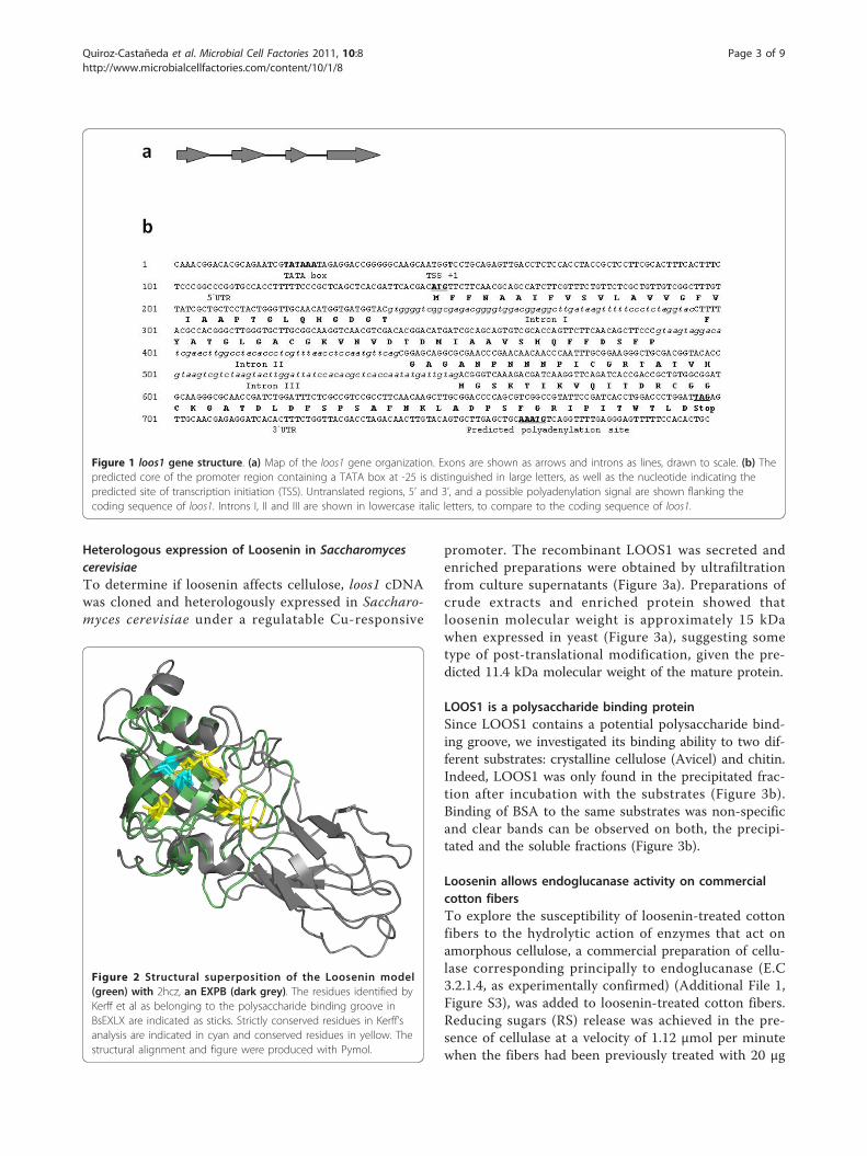
and III, two of which (introns II and III) exhibit the cano-nical 5’-GT....AG-3’ donor-acceptor pairs. Intron lengthsare 55, 53 and 52 nt, respectively, in agreement with theaverage intron size of filamentous fungi (50-70 bp), and
account for 160 extra nucleotides relative to the cDNA(Figure 1a). The 5’ and 3’ UTRs are predicted to consist of98 and 100 nucleotides respectively (Figure 1b).
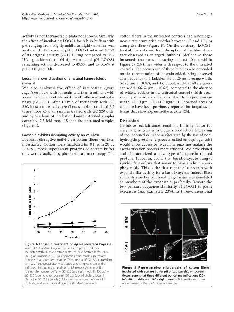
Homology Modeling of LooseninThe loosenin amino acid sequence was used for foldrecognition using the PHYRE web server version 0.1.The top three results were EXLX1 from Bacillus subtilis[PDB:2BH0], the homologue pollen allergen PHL P1N-terminal domain from Phleum pratense [PDB:1N10]and an EXPB and group-1 allergen from maize[PDB:2HCZ]. All had an estimated precision of 100%indicating a successful fold assignment to the DPBB foldfamily. Primary sequences identities were 19, 19 and 20%respectively confirming that loosenin is indeed a remotehomologue of the expansins. We used the alignmentsprovided by PHYRE to construct five models of the com-plete loosenin amino acid sequence with 2BH0, 1N10and 2HCZ as templates using maximum MD-refinement.All five models were essentially identical with an averageRMSD of 0.4 Å. Structures 2BH0 and 1N10 had anRMSD (of the equivalent superimposed alpha-carbons) ofaround 1.4 Å to the models when 2HCZ had an RMSD of0.5 Å, although amino acid identity to loosenin was verymarginally greater (Figure 2). This measure would reflectthe similarity of the proteins cores. Unlike typical expan-sins, loosenin is composed of a single domain, albeit onehighly similar to domain I of expansins, as evidenced byfold recognition. Kerff et al [27] performed a sequenceanalysis of EXLX1 (structure 2BH0) together with otherpolysaccharide recognizing proteins through which theyidentified several conserved residues. Similarly, we super-imposed the loosenin model with the above-mentionedstructures and identified the equivalent residues.In loosenin, T31 and D105 (highlighted in cyan in Addi-tional File 1, Figure S2) correspond to the two strictlyconserved residues, equivalent to T12 and D82 inEXLX1, and known to form a conserved hydrogen bondbetween the OH group of Thr and the carboxylic groupof Asp. Other residues that show perfect sequence con-servation between loosenin and the other three struc-tures are G38, A39, G75, T92, and D93. Four morepositions identified as important by Kerff et al corre-spond to loosenin Y33 (also conserved in 2HCZ and1N10, however it is substituted by a T in 2BH0); A52(conserved in 2BH0, but found as C in the other twostructures); D103 (is substituted by A in 2BH0, or H in1N10 and 2HCZ); and finally F109 (found conserved inthe rest of the structures as an L). Except for G75, allthese residues were identified by Kerff et al. as part ofthe groove which is thought to serve as the polysacchar-ide binding site by means of hydrogen bonding. Themodel thus suggests that loosenin is also a polysacchar-ide binding protein.
Quiroz-Castañeda et al. Microbial Cell Factories 2011, 10:8http://www.microbialcellfactories.com/content/10/1/8
Page 2 of 9
Heterologous expression of Loosenin in SaccharomycescerevisiaeTo determine if loosenin affects cellulose, loos1 cDNAwas cloned and heterologously expressed in Saccharo-myces cerevisiae under a regulatable Cu-responsive
promoter. The recombinant LOOS1 was secreted andenriched preparations were obtained by ultrafiltrationfrom culture supernatants (Figure 3a). Preparations ofcrude extracts and enriched protein showed thatloosenin molecular weight is approximately 15 kDawhen expressed in yeast (Figure 3a), suggesting sometype of post-translational modification, given the pre-dicted 11.4 kDa molecular weight of the mature protein.
LOOS1 is a polysaccharide binding proteinSince LOOS1 contains a potential polysaccharide bind-ing groove, we investigated its binding ability to two dif-ferent substrates: crystalline cellulose (Avicel) and chitin.Indeed, LOOS1 was only found in the precipitated frac-tion after incubation with the substrates (Figure 3b).Binding of BSA to the same substrates was non-specificand clear bands can be observed on both, the precipi-tated and the soluble fractions (Figure 3b).
Loosenin allows endoglucanase activity on commercialcotton fibersTo explore the susceptibility of loosenin-treated cottonfibers to the hydrolytic action of enzymes that act onamorphous cellulose, a commercial preparation of cellu-lase corresponding principally to endoglucanase (E.C3.2.1.4, as experimentally confirmed) (Additional File 1,Figure S3), was added to loosenin-treated cotton fibers.Reducing sugars (RS) release was achieved in the pre-sence of cellulase at a velocity of 1.12 μmol per minutewhen the fibers had been previously treated with 20 μg
Figure 1 loos1 gene structure. (a) Map of the loos1 gene organization. Exons are shown as arrows and introns as lines, drawn to scale. (b) Thepredicted core of the promoter region containing a TATA box at -25 is distinguished in large letters, as well as the nucleotide indicating thepredicted site of transcription initiation (TSS). Untranslated regions, 5’ and 3’, and a possible polyadenylation signal are shown flanking thecoding sequence of loos1. Introns I, II and III are shown in lowercase italic letters, to compare to the coding sequence of loos1.
Figure 2 Structural superposition of the Loosenin model(green) with 2hcz, an EXPB (dark grey). The residues identified byKerff et al as belonging to the polysaccharide binding groove inBsEXLX are indicated as sticks. Strictly conserved residues in Kerff’sanalysis are indicated in cyan and conserved residues in yellow. Thestructural alignment and figure were produced with Pymol.
Quiroz-Castañeda et al. Microbial Cell Factories 2011, 10:8http://www.microbialcellfactories.com/content/10/1/8
Page 3 of 9

of loosenin (Figure 3c), for a specific activity of 56 IU/mg/protein. The velocity of RS release was directly pro-portional to the amount of loosenin added to the reac-tion mixture (2.19 μmol/min when 40 μg of looseninwere added [Figure 3c]), and specific activity of 54.75IU/mg protein). This effect is specific to the presence ofLOOS1, given that addition, at the same concentrations,of proteins from mock supernatants of S. cerevisiae cul-tures or an irrelevant protein (BSA) produced no activityafter treatment with cellulase (Figure 3c; and AdditionalFile 1, Figure S4). Additionally, although mercerizationallowed higher levels of RS release from loosenin treatedfibers in comparison to non-mercerized fibers (compareFigure 3C with Additional File 1, Figure S5a) no RSrelease was detected in the untreated control, indicating
that mercerization treatment does not disrupt the fibersto allow sugar liberation by cellulase (Figure 3c). Finally,lack of RS release in the absence of cellulase indicatesthat loosenin has no cellulolytic activity per se.
Thermal- and pH-stability of looseninSince robustness of enzymes is a key factor for indus-trial applications, the analysis of the thermal- andpH-stability of LOOS1 was carried out. Thermostabil-ity of LOOS1 was analyzed by incubating the reactionof loosenin and cotton fibers at 15, 25, 40, 60 and 80°C, and then adding cellulase to monitor the release ofRS at 50°C. Specific activity at 25°C was 55.58 IU/mgprotein. No RS were detected after treatment at 40°Cand higher temperatures, indicating than LOOS1
Figure 3 Loosenin enrichment, activity and binding to polysaccharides. (a) LOOS1 enrichment. SDS-PAGE of proteins from yeast culturesupernatants transformed with empty vector pSAL3 (mock SN), pSAL3-loos1 (loosenin SN), and enriched LOOS1 (loosenin). (b) LOOS1 bindsspecifically to Avicel (crystalline cellulose) and chitin. The bounded (pellet) and unbounded (SN, supernatant) fractions are shown (top panel).BSA on the other hand, is found in both fractions, indicating unspecific interaction with the matrixes (lower panel). (c) RS release by cellulasefrom cotton fibers previously incubated with: two different concentrations of loosenin plus cellulase (20 μg black squares, 40 μg gray diamonds);acetate buffer (open triangles); acetate buffer plus cellulase (gray circles); mock SN plus cellulase (open squares); and only LOOS1 (open circles).(d) Specific activity of cotton fibers previously treated with LOOS1, at different pH, plus cellulase. All experiments were performed in triplicate,and error bars indicate the standard deviations.
Quiroz-Castañeda et al. Microbial Cell Factories 2011, 10:8http://www.microbialcellfactories.com/content/10/1/8
Page 4 of 9
activity is not thermostable (data not shown). Similarly,the effect of incubating LOOS1 for 8 h in buffers withpH ranging from highly acidic to highly alkaline wasanalyzed. In this case, at pH 3, LOOS1 retained 42.6%of its original activity (24.17 IU/mg compared to 56.7IU/mg achieved at pH 5). At neutral pH LOOS1remaining activity decreased to 49.5%, and to 10.6% atpH 10 (Figure 3d).
Loosenin allows digestion of a natural lignocellulosicmaterialWe also analyzed the effect of incubating Agavetequilana fibers with loosenin and then treatment witha commercially available mixture of cellulases and xyla-nases (GC 220). After 10 min of incubation with GC220, loosenin-treated agave fibers samples contained 3.2times more RS than samples treated with GC 220 only,and by one hour of incubation loosenin-treated samplescontained 7.5-fold more RS than the untreated samples(Figure 4).
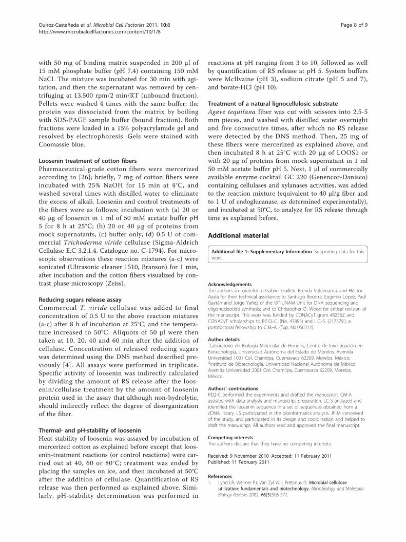
Loosenin exhibits disrupting-activity on celluloseLoosenin disruptive-activity on cotton fibers was theninvestigated. Cotton fibers incubated for 8 h with 20 μgLOOS1, mock supernatant proteins or acetate bufferonly were visualized by phase contrast microscopy. The
cotton fibers in the untreated controls had a homoge-neous structure with widths between 13 and 17 μmalong the fiber (Figure 5). On the contrary, LOOS1-treated fibers showed local disruption of the fiber struc-ture observed as enlarged “bubbles” (defined as thoseloosened structures measuring at least 40 μm width;Figure 5), 2.6 times wider with respect to the untreatedcontrols. The occurrence of these bubbles also dependedon the concentration of loosenin added, being observedat a frequency of 1 bubble/field at 20 μg (average width:52.25 μm ± 10.07), and 1.6 bubbles/field at 40 μg (aver-age width: 66.62 μm ± 10.62), compared to the absenceof evident bubbles in the untreated control (which occa-sionally showed wider regions of up to 30 μm, averagewidth: 26.60 μm ± 4.21) (Figure 5). Loosened areas ofcellulose have been previously reported for fungal swol-lenins that show expansin-like activity [26].
DiscussionCellulose recalcitrance remains a limiting factor forenzymatic hydrolysis in biofuels production. Increasingof the loosened cellulose surface area by the use of non-hydrolytic proteins (a process called amorphogenesis)would allow access to hydrolytic enzymes making thesaccharification process more efficient. We have clonedand characterized a new type of expansin-relatedprotein, loosenin, from the basidiomycete fungusBjerkandera adusta that seems to have a role in amor-phogenesis. This is the first report of a protein withexpansin-like activity for a basidiomycete. Indeed, Blastsimilarity searches recovered fungal sequences annotatedas members of the expansin superfamily. Despite thelow primary sequence similarity of LOOS1 to plantexpansins (approximately 20%), its three-dimensional
Figure 4 Loosenin treatment of Agave tequilana bagasse .Washed A. tequilana bagasse was cut into pieces and thenincubated with 50 mM acetate buffer, 50 mM acetate buffer plus20 μg of loosenin, or 20 μg of proteins from mock supernatantduring 8 h at room temperature. Then, one μl of GC 220 (equivalentto 1 U of endoglucanase) was added and samples taken at theindicated time points to analyze for RS release. Acetate buffer(diamonds); acetate buffer + GC 220 (squares); mock SN [20 μg] +GC 220 (open circles); loosenin [20 μg] (closed circles); loosenin[20 μg] + GC 220 (triangles). All experiments were performed intriplicate, and error bars indicate the standard deviations.
Figure 5 Representative micrographs of cotton fibersincubated with acetate buffer pH 5 (top panels), or loosenin(lower panels), at three different optical magnifications (20×left, 40× middle and 100× right panels). Bubble-like structuresare observed in the LOOS1-treated samples.
Quiroz-Castañeda et al. Microbial Cell Factories 2011, 10:8http://www.microbialcellfactories.com/content/10/1/8
Page 5 of 9

model fits domain I of the crystallized EXLX1 fromBacillus subtillis, Zea mays b-expansin and PHL P1homologue with the highest score possible, and con-serves those residues thought to be involved in bindingto the polysaccharide chain.Interestingly, the structure of loosenin consists of only
one domain with a DPBB fold, the first reported of thistype, suggesting that this structure might be sufficient toproduce cellulose-disrupting activity. Expansin domainII appears to bind carbohydrates via a number of hydro-phobic residues that tightly associate with the substrate,given the need of local (cell specific) cell wall disruption.For loosenin, both binding and disrupting activity residein domain I. On the other hand, expansin domain IIcould serve other purposes, such as providing higheractivity or stability. It was also noticed by Kerff et al,that the potential polysaccharide binding groove ofdomain I is extended by three aromatic residues on thesurface of D2, allowing the binding of four additionalsaccharide units, suggesting a stronger union to thepolysaccharide and possibly increasing its activitytowards it. Loosenin is a good model to answer thesequestions by making fusions to homologues of expansindomain II.Our work demonstrates that, in accordance with the
prediction of the three-dimensional modeling, LOOS1binds specifically to crystalline cellulose and chitin.Furthermore, loosenin is indeed a cellulose-disrupting pro-tein, and that cotton and Avicel (which behaved in thesame way as cotton when mercerized or not in the pre-sence of loosenin [Additional File 1, Figure S5b and S5c]),are modified by its action. Indeed, dose-dependent LOOS1treatment of cotton fibers induces local disruptions thatpossibly become a substrate for an endoglucanase enzymethat otherwise could not act upon them, supporting itsrole as a protein involved in amorphogenesis. It is impor-tant to remark that loosenin, contrary to plant expansins,was successfully heterologously expressed in yeast, giventhe industrial significance of cellulose-disrupting proteinsand cellulolytic enzymes. The putative secretion signalsequence of loosenin seems to be recognized by S. cerevi-siae because the protein was found in the culture superna-tants. The activity of loosenin purified from B. adustaitself remains to be analyzed and compared to thatreported here, given that it is known that S. cerevisiaehyperglycosylates secreted proteins in some cases (N-gly-cosylation). Indeed, loosenin molecular weight was higherthan the predicted by its primary sequence (15 versus 11.4kDa) suggesting a posttranslational modification. In thisrespect, three O-glycosylation sites were predicted at posi-tions 3, 105 and 107, but no N-glycosylation sites werefound for the loosenin of B. adusta. Further experimentsare needed to understand the role of glycosylation on loos-enin activity.
The biological function of loosenin in B. adustaremains to be analyzed. It is possible that the fungususes this protein to efficiently degrade lignocellulosetogether with a battery of extracellular hydrolyticenzymes (ligninases, cellulases, xylanases, etc). However,a role of loosenin in the physiology of the fungus cannotbe ruled out, as it could also participate in the remodel-ing of the fungal cell wall to allow hyphal growth assuggested for Aspergillus nidulans EglD with an expan-sin-like motif [25].Finally, loosenin represents a good candidate as an
additive to enhance sugar production from plant bio-mass. Loosenin activity was more efficient when the lig-nocellulosic materials were mercerized, as seen for othersaccharification processes. Agave tequilana is a cropextensively grown in some areas of Mexico, and theshredded fibrous waste is usually burnt or left to decom-pose. Indeed, A. tequilana fiber became a susceptiblesubstrate for a cocktail of commercial cellulases andxylanases in the presence of LOOS1. Loosenin showsoptimum activity at the same pH as most cellulolyticenzymes. And although it is not a thermostable protein,probably because of the temperate origin of B. adusta,similar sequences are present in a number of otherbasidiomycetes fungi, opening the possibility to findthem expressed also in thermophilic species, such asPycnopourus sanguineus [28], or others. It is importantto remark upon the low enzymatic activity needed incombination with loosenin to observe its effect, giventhe high costs of enzymes at industrial levels.
ConclusionsHere we describe a new type of protein with a role inamorphogenesis of cellulose that we have called loos-enin, from the basidiomycete Bjerkandera adusta. Loos-enin is distantly related to plant expansins, binds tocellulose and chitin, and has a disrupting activity oncellulose fibers. Treatment of lignocellulosic materials(cotton fibers and Agave tequilana bagasse) with loos-enin enhances sugar liberation after the addition ofcommercial cellulases, suggesting an interesting poten-tial use for the production of fermentable sugars fromlignocellulose.
MethodsStrains and growth conditionsBjerkandera adusta strain UAMH 8258 was kindly pro-vided by Dr. Rafael Vazquez-Duhalt. Mycelium was grownon PDA medium (2% potato, 2% dextrose and 1.5% agar)for its propagation and stocking. For the isolation of loos1gene, B. adusta was grown in 2% wheat straw liquid med-ium (mineral base medium: 7.8 mg/L CuSO4·5H2O,18 mg/L FeSO4·7H2O, 500 mg/L MgSO4·7H2O, 10 mg/LZnSO4, 50 mg/L KCl, 1 g/L K2HPO4 and 2 g/L NH4NO3,
Quiroz-Castañeda et al. Microbial Cell Factories 2011, 10:8http://www.microbialcellfactories.com/content/10/1/8
Page 6 of 9

1.5% agar; pH 5 (modified from [29]) supplemented withpowdered wheat straw (maximum and minimum particlesizes of 3 and 0.5 mm), for 5 days at 28°C and 200 rpm.Saccharomyces cerevisiae strain W303a (MATa
can1-100 ade2-1 his3-11, 15 leu2-3, 12 trp1-1 ura3-1)was grown in SC-Ura-minus medium [0.67% yeast nitro-gen base (Difco), 2% dextrose (Baker), adenine 20 mg/L,leucine 60 mg/L, tryptophan 20 mg/L and histidine20 mg/L (Sigma-Aldrich)]. For heterologous expression ofloosenin, SC-Ura-minus medium was supplemented with50 μM Cu2SO4 and cells were grown at 28°C for 1 day.E. coli strain DH5a was used for plasmid propagation
and manipulation according to [30].
Molecular cloning of loos1loos1 sequence was originally identified by BLAST ana-lysis (Additional File 1, Table S1) of clones obtainedfrom a subtracted cDNA library from B. adusta grownin the presence of crude oil (Cuervo, et al, manuscriptin preparation). The sequence was then amplified froma sample of total RNA of B. adusta grown in wheatstraw medium by Reverse Transcription coupled-PCRwith primers LOOS1fwd: 5’-CGGAATTCATGTTCTT-CAACG-3’ and LOOS1rev: 5’-CCTCGAGCTAATC-CAGGGT-3’. The primers sequences included EcoRIand XhoI sites (underlined) at the 5’- and 3’-ends,respectively, to facilitate subsequent cloning steps. The390 bp PCR fragment was purified and cloned into thepGEM-T (Promega) vector resulting in pGEM-LOOS1,and its sequence was confirmed (Macrogen, USA).
Genomic DNA extractionMycelium grown for 7 days in liquid wheat straw med-ium was collected, frozen and pulverized with liquidnitrogen. Genomic DNA was extracted with UltraCleanMegaprep Soil DNA kit (MoBio), and used as templatefor PCR amplification with primers LOOS1fwd andLOOS1rev. The PCR product was cloned in plasmidpGEM-T (Promega) resulting in vector pGEM-LOOS1g, and its sequence was determined (Macrogen USA).
Domain predictionConserved domains were identified using the CDD(Conserved Domain Database, http://www.ncbi.nlm.nih.gov/Structure/cdd/wrpsb.cgi) and InterProScan (http://www.ebi.ac.uk/Tools/InterProScan/) databases.
Structural Modeling of LooseninWe identified templates for Loosenin homology model-ing using the PHYRE Protein Fold Recognition Server[31]. This program identified two expansins and oneexpansin-homologue with the highest scores (100% esti-mated precision). We then modeled the Looseninsequence using the program modeller9v7 [32] using the
sequence alignments obtained by PHYRE simulta-neously generating 5 models with a molecular dynamicslevel of refine.very_slow. The four resulting modelswere compared using Pymol (DeLano Scientific LLC),first by fitting them together and then comparing thestructures.
Expression of recombinant LOOS1 in yeastPlasmid pGEM-loos1 was digested with EcoRI and XhoIand the 390 bp resulting fragment was subcloned in vec-tor pSAL3 [33], giving rise to plasmid pSAL3-loos1.Yeast strain W303a was transformed with plasmidpSAL3-loos1 or empty pSAL3 by the lithium acetatemethod [34], and selected on solid SC-Ura-minus med-ium. After 3 days of incubation, one colony of eachtransformation was grown in a pre-inocule of 20 ml ofSC-Ura-minus medium at 28°C and 250 rpm to midlog-phase (between 0.4-0.6 OD), at this point a dilutionwas made to 0.1 OD in 1 L of SC-Ura-minus medium,adding Cu2SO4 to a final concentration of 50 μM, andsupplementing the media with protease inhibitorsaccording to the manufacturer’s instructions (Complete®
Roche). Finally, cells were grown to 0.8 OD to recoversupernatant by centrifugation (1073 × g for 10 min at4°C), that was kept at 4°C until use.
Loosenin enrichmentSupernatant recovered from 1 L yeast cultures was con-centrated 50 times by ultrafiltration through a 10 kDacut-off membrane (Amicon stirred cell 8400, and Ultra-cell membranes, Millipore), and then filtered through a30 kDa cut-off membrane (Amicon Ultra 4 CentrifugalFilter, Millipore). The filtrate was recovered and concen-trated again through 10 kDa cut-off membranes (Ami-con Ultra 4 Centrifugal Filter, Millipore) to a finalvolume of 200 μl. Mock supernatant from empty pSAL3vector cultures was only concentrated by ultrafiltration(10 kDA cut-off). Protein concentration was measuredaccording to [35] using a bovine serum albumin (BSA)standard curve. Enrichment and molecular weight esti-mations of recombinant loosenin were performed resol-ving 20 μg of protein in 15% SDS-polyacrylamide gels.20 μg of mock supernatant were loaded as well. The PAgel was stained with 0.25% Coomassie Blue R-250(Sigma-Aldrich) and distained with a solution of metha-nol:water and acetic acid. Treatments of samples withloosenin were performed with these enriched prepara-tions throughout this work.
Polysaccharides binding assayThe assay was performed as reported by [36] withsome modifications. Avicel PH-101 (Fluka) and chitinfrom shrimp shells (Sigma-Aldrich) were used as bind-ing matrices. 40 μg of LOOS1 or BSA were mixed
Quiroz-Castañeda et al. Microbial Cell Factories 2011, 10:8http://www.microbialcellfactories.com/content/10/1/8
Page 7 of 9
with 50 mg of binding matrix suspended in 200 μl of15 mM phosphate buffer (pH 7.4) containing 150 mMNaCl. The mixture was incubated for 30 min with agi-tation, and then the supernatant was removed by cen-trifuging at 13,500 rpm/2 min/RT (unbound fraction).Pellets were washed 4 times with the same buffer; theprotein was dissociated from the matrix by boilingwith SDS-PAGE sample buffer (bound fraction). Bothfractions were loaded in a 15% polyacrylamide gel andresolved by electrophoresis. Gels were stained withCoomassie blue.
Loosenin treatment of cotton fibersPharmaceutical-grade cotton fibers were mercerizedaccording to [26]; briefly, 7 mg of cotton fibers wereincubated with 25% NaOH for 15 min at 4°C, andwashed several times with distilled water to eliminatethe excess of alkali. Loosenin and control treatments ofthe fibers were as follows: incubation with (a) 20 or40 μg of loosenin in 1 ml of 50 mM acetate buffer pH5 for 8 h at 25°C; (b) 20 or 40 μg of proteins frommock supernatants, (c) buffer only, (d) 0.5 U of com-mercial Trichoderma viride cellulase (Sigma-AldrichCellulase E.C 3.2.1.4, Catalogue no. C-1794). For micro-scopic observations these reaction mixtures (a-c) weresonicated (Ultrasonic cleaner 1510, Branson) for 1 min,after incubation and the cotton fibers visualized by con-trast phase microscopy (Zeiss).
Reducing sugars release assayCommercial T. viride cellulase was added to finalconcentration of 0.5 U to the above reaction mixtures(a-c) after 8 h of incubation at 25°C, and the tempera-ture increased to 50°C. Aliquots of 50 μl were thentaken at 10, 20, 40 and 60 min after the addition ofcellulase. Concentration of released reducing sugarswas determined using the DNS method described pre-viously [4]. All assays were performed in triplicate.Specific activity of loosenin was indirectly calculatedby dividing the amount of RS release after the loos-enin/cellulase treatment by the amount of looseninprotein used in the assay that although non-hydrolytic,should indirectly reflect the degree of disorganizationof the fiber.
Thermal- and pH-stability of looseninHeat-stability of loosenin was assayed by incubation ofmercerized cotton as explained before except that loos-enin-treatment reactions (or control reactions) were car-ried out at 40, 60 or 80°C; treatment was ended byplacing the samples on ice, and then incubated at 50°Cafter the addition of cellulase. Quantification of RSrelease was then performed as explained above. Simi-larly, pH-stability determination was performed in
reactions at pH ranging from 3 to 10, followed as wellby quantification of RS release at pH 5. System bufferswere McIlvaine (pH 3), sodium citrate (pH 5 and 7),and borate-HCl (pH 10).
Treatment of a natural lignocellulosic substrateAgave tequilana fiber was cut with scissors into 2.5-5mm pieces, and washed with distilled water overnightand five consecutive times, after which no RS releasewere detected by the DNS method. Then, 25 mg ofthese fibers were mercerized as explained above, andthen incubated 8 h at 25°C with 20 μg of LOOS1 orwith 20 μg of proteins from mock supernatant in 1 ml50 mM acetate buffer pH 5. Next, 1 μl of commerciallyavailable enzyme cocktail GC 220 (Genencor-Danisco)containing cellulases and xylanases activities, was addedto the reaction mixture (equivalent to 40 μl/g fiber andto 1 U of endoglucanase, as determined experimentally),and incubated at 50°C, to analyze for RS release throughtime as explained before.
Additional material
Additional file 1: Supplementary Information. Supporting data for thiswork.
AcknowledgementsThe authors are grateful to Gabriel Guillén, Brenda Valderrama, and HéctorAyala for their technical assistance; to Santiago Becerra, Eugenio López, PaúlGaytán and Jorge Yañez of the IBT-UNAM Unit for DNA sequencing andoligonucleotide synthesis; and to Christopher D. Wood for critical revision ofthe manuscript. This work was funded by CONACyT grant 48256Z andCONACyT scholarships to R.E.Q.-C. (No. 47895) and L.C.-S. (217379.); apostdoctoral fellowship to C.M.-A. (Exp. No.050272).
Author details1Laboratorio de Biología Molecular de Hongos, Centro de Investigación enBiotecnología, Universidad Autónoma del Estado de Morelos. AvenidaUniversidad 1001 Col. Chamilpa, Cuernavaca 62209, Morelos, México.2Instituto de Biotecnología. Universidad Nacional Autónoma de México.Avenida Universidad 2001 Col. Chamilpa, Cuernavaca 62209, Morelos,México.
Authors’ contributionsREQ-C performed the experiments and drafted the manuscript. CM-Aassisted with data analysis and manuscript preparation. LC-S analyzed andidentified the loosenin sequence in a set of sequences obtained from acDNA library. LS participated in the bioinformatics analysis. JF-M conceivedof the study, and participated in its design and coordination and helped todraft the manuscript. All authors read and approved the final manuscript.
Competing interestsThe authors declare that they have no competing interests.
Received: 9 November 2010 Accepted: 11 February 2011Published: 11 February 2011
References1. Lynd LR, Weimer PJ, Van Zyl WH, Pretorius IS: Microbial cellulose
utilization: fundamentals and biotechnology. Microbiology and MolecularBiology Reviews 2002, 66(3):506-577.
Quiroz-Castañeda et al. Microbial Cell Factories 2011, 10:8http://www.microbialcellfactories.com/content/10/1/8
Page 8 of 9

2. Aro N, Pakula T, Pentilla M: Transcriptional regulation of plant cell walldegradation by filamentous fungi. FEMS Microbiology Reviews 2005,29(4):719-739.
3. Wang Y, Vazquez-Duhalt R, Pickard MA: Manganese-lignin peroxidasehybrid from Bjerkandera adusta oxidizes polycyclic aromatichydrocarbons more actively in the absence of manganese. Can JMicrobiol 2003, 49(11):675-682.
4. Quiroz-Castaneda RE, Balcazar-Lopez E, Dantan-Gonzalez E, Martinez A,Folch-Mallol J, Martinez-Anaya C: Characterization of cellulolytic activitiesof Bjerkandera adusta and Pycnoporus sanguineus on solid wheat strawmedium. Electronic Journal of Biotechnology [online] 2009, 12(4) [http://ejbiotechnology.ucv.cl/content/vol12/issue4/full/3/index.html].
5. Quiroz-Castaneda RE, Perez-Mejia N, Martinez-Anaya C, Acosta-Urdapilleta L,Folch-Mallol J: Evaluation of different lignocellulosic substrates for theproduction of cellulases and xylanases by the basidiomycete fungiBjerkandera adusta and Pycnoporus sanguineus. Biodegradation 2010.
6. McQueen-Mason S, Durachko DM, Cosgrove DJ: Two endogenous proteinsthat induce cell wall extension in plants. Plant Cell 1992, 4:1425-1433.
7. Cho HT, Cosgrove DJ: Regulation of root hair initiation and expansingene expression in Arabidopsis. Plant Cell 2002, 14(12):3237-3253.
8. Cho HT, Cosgrove DJ: Altered expression of expansin modulates leafgrowth and pedicel abscission in Arabidopsis thaliana. Proc Natl Acad SciUSA 2000, 97(17):9783-9788.
9. Pien S, Wyrzykowska J, McQueen-Mason S, Smart C, Fleming A: Localexpression of expansin induces the entire process of leaf developmentand modifies leaf shape. Proceedings of the National Academy of Sciencesof the United States of America 2001, 98(20):11812-11817.
10. Civello PM, Powell AL, Sabehat A, Bennett AB: An expansin geneexpressed in ripening strawberry fruit. Plant Physiol 1999,121(4):1273-1280.
11. Rose JK, Lee HH, Bennett AB: Expression of a divergent expansin gene isfruit-specific and ripening-regulated. Proceedings of the National Academyof Sciences of the United States of America 1997, 94(11):5955-5960.
12. Cosgrove DJ, Li LC, Cho HT, Hoffmann-Benning S, Moore RC, Blecker D: Thegrowing world of expansins. Plant Cell Physiol 2002, 43(12):1436-1444.
13. Lee Y, Choi D, Kende H: Expansins: ever-expanding numbers andfunctions. Curr Opin Plant Biol 2001, 4(6):527-532.
14. Li Y, Jones L, McQueen-Mason S: Expansins and cell growth. Curr OpinPlant Biol 2003, 6(6):603-610.
15. McQueen-Mason S, Cosgrove DJ: Disruption of hydrogen bondingbetween plant cell wall polymers by proteins that induce wallextension. Proceedings of the National Academy of Sciences of the UnitedStates of America 1994, 91(14):6574-6578.
16. Cosgrove DJ, Bedinger P, Durachko DM: Group I allergens of grass pollenas cell wall-loosening agents. Proceedings of the National Academy ofSciences of the United States of America 1997, 94(12):6559-6564.
17. Sampedro J, Cosgrove DJ: The expansin superfamily. Genome biology 2005,6(12):242.
18. Dermatsev V, Weingarten-Baror C, Resnick N, Gadkar V, Wininger S,Kolotilin I, Mayzlish-Gati E, Zilberstein A, Koltai H, Kapulnik Y: Microarrayanalysis and functional tests suggest the involvement of expansins inthe early stages of symbiosis of the arbuscular mycorrhizal fungusGlomus intraradices on tomato (Solanum lycopersicum). Mol Plant Pathol2010, 11(1):121-135.
19. Cosgrove DJ: Relaxation in a high-stress environment: the molecularbases of extensible cell walls and cell enlargement. Plant Cell 1997,9(7):1031-1041.
20. Cosgrove DJ: Loosening of plant cell walls by expansins. Nature 2000,407(6802):321-326.
21. Kende H, Bradford K, Brummell D, Cho HT, Cosgrove D, Fleming A,Gehring C, Lee Y, McQueen-Mason S, Rose J, et al: Nomenclature formembers of the expansin superfamily of genes and proteins. Plant MolBiol 2004, 55(3):311-314.
22. Darley CP, Li Y, Schaap P, McQueen-Mason SJ: Expression of a family ofexpansin-like proteins during the development of Dictyosteliumdiscoideum. FEBS Lett 2003, 546(2-3):416-418.
23. Kim ES, Lee HJ, Bang WG, Choi IG, Kim KH: Functional characterization ofa bacterial expansin from Bacillus subtilis for enhanced enzymatichydrolysis of cellulose. Biotechnol Bioeng 2009, 102(5):1342-1353.
24. Laine MJ, Haapalainen M, Wahlroos T, Kankare K, Nissinen R, Kassuwi S,Metzler MC: The cellulase encoded by the native plasmid of Clavibacter
michiganensis ssp. sepedonicus plays a role in virulence and contains anexpansin-like domain. Physiological and Molecular Plant Pathology 2000,57(5):221-233.
25. Bouzarelou D, Billini M, Roumelioti K, Sophianopoulou V: EglD, a putativeendoglucanase, with an expansin like domain is localized in the conidialcell wall of Aspergillus nidulans. Fungal Genet Biol 2008, 45(6):839-850.
26. Saloheimo M, Paloheimo M, Hakola S, Pere J, Swanson B, Nyyssonen E,Bhatia A, Ward M, Penttila M: Swollenin, a Trichoderma reesei protein withsequence similarity to the plant expansins, exhibits disruption activityon cellulosic materials. Eur J Biochem 2002, 269(17):4202-4211.
27. Kerff F, Amoroso A, Herman R, Sauvage E, Petrella S, Filee P, Charlier P,Joris B, Tabuchi A, Nikolaidis N, et al: Crystal structure and activity ofBacillus subtilis YoaJ (EXLX1), a bacterial expansin that promotes rootcolonization. Proceedings of the National Academy of Sciences of the UnitedStates of America 2008, 105(44):16876-16881.
28. Dantan-Gonzalez E, Vite-Vallejo O, Martinez-Anaya C, Mendez-Sanchez M,Gonzalez MC, Palomares LA, Folch-Mallol J: Production of two novellaccase isoforms by a thermotolerant strain of Pycnoporus sanguineusisolated from an oil-polluted tropical habitat. Int Microbiol 2008,11(3):163-169.
29. Inglis GD, Popp AP, Selinger LB, Kawchuk LM, Gaudet DA, McAllister TA:Production of cellulases and xylanases by low-temperaturebasidiomycetes. Can J Microbiol 2000, 46(9):860-865.
30. Sambrook J, Fritsch EF, Maniatis T: Molecular Cloning. A LaboratoryManual. Cold Spring Harbor Laboratory Press;, Second 1989.
31. Kelley LA, Sternberg MJE: Protein structure prediction on the Web: a casestudy using the Phyre server. Nat Protocols 2009, 4(3):363-371.
32. Eswar N, Webb B, Marti-Renom MA, Madhusudhan MS, Eramian D,Shen MY, Pieper U, Sali A: Comparative protein structure modeling usingModeller. Curr Protoc Bioinformatics 2006, Chapter 5(Unit 5):6.
33. Mascorro-Gallardo JO, Covarrubias AA, Gaxiola R: Construction of a CUP1promoter-based vector to modulate gene expression in Saccharomycescerevisiae. Gene 1996, 172(1):169-170.
34. Gietz RD, Schiestl RH: Quick and easy yeast transformation using theLiAc/SS carrier DNA/PEG method. Nat Protoc 2007, 2(1):35-37.
35. Lowry OH, Rosebrough NJ, Farr AL, Randall RJ: Protein measurement withthe Folin phenol reagent. J Biol Chem 1951, 193(1):265-275.
36. Chen X-A, Ishida N, Todaka N, Nakamura R, Maruyama J-I, Takahashi H,Kitamoto K: Promotion of Efficient Saccharification with Aspergillusfumigatus AfSwo1 Towards Crystalline Cellulose. Appl Environ Microbiol2010, 76(8):2556-2561.
doi:10.1186/1475-2859-10-8Cite this article as: Quiroz-Castañeda et al.: Loosenin, a novel proteinwith cellulose-disrupting activity from Bjerkandera adusta. Microbial CellFactories 2011 10:8.
Submit your next manuscript to BioMed Centraland take full advantage of:
• Convenient online submission
• Thorough peer review
• No space constraints or color figure charges
• Immediate publication on acceptance
• Inclusion in PubMed, CAS, Scopus and Google Scholar
• Research which is freely available for redistribution
Submit your manuscript at www.biomedcentral.com/submit
Quiroz-Castañeda et al. Microbial Cell Factories 2011, 10:8http://www.microbialcellfactories.com/content/10/1/8
Page 9 of 9