Embed Size (px)
Citation preview
SHORT COMMUNICATION
Loss of Response of Carnitine Palmitoyltransferase I toOkadaic Acid in Transformed Hepatic Cells
Guillermo Velasco,* Patricia Passilly,† Manuel Guzman*‡ and Norbert Latruffe†*DEPARTMENT OF BIOCHEMISTRY AND MOLECULAR BIOLOGY I, FACULTY OF BIOLOGY, COMPLUTENSE UNIVERSITY,28040 MADRID, SPAIN; AND †LABORATOIRE DE BIOLOGIE MOLECULAIRE ET CELLULAIRE, FACULTE DES SCIENCES
MIRANDE, UNIVERSITE DE BOURGOGNE, BP 400, 21011 DIJON, FRANCE
ABSTRACT. The specific activity of carnitine palmitoyltransferase I (CPT-I) was similar in mitochondriaisolated from rat Fao and human HepG2 hepatoma cells and from rat hepatocytes, but almost twofold higher inpermeabilized hepatoma cells than in permeabilized hepatocytes. Short-term exposure to okadaic acid induceda ca. 80% stimulation of CPT-I in hepatocytes, whereas no significant response of the enzyme from hepatomacells was evident. Thus, the high CPT-I activity displayed by hepatoma cells may be reached by hepatocytesupon challenge to okadaic acid. Reconstitution experiments with purified mitochondrial and cytoskeletalfractions showed that the cytoskeleton of hepatocytes produced a more remarkable inhibition of CPT-I than thecytoskeleton of Fao cells. The present data may be explained by a disruption of interactions between CPT-I andcytoskeletal components in tumor cells that may be involved in the okadaic acid-induced activation of hepaticCPT-I as previously suggested. BIOCHEM PHARMACOL 56;11:1485–1488, 1998. © 1998 Elsevier Science Inc.
KEY WORDS. carnitine palmitoyltransferase I; hepatocytes; hepatoma cells; okadaic acid; cytoskeleton;mitochondria
Mitochondrial fatty acid oxidation provides a major sourceof energy in heart, skeletal muscle and liver (reviewed in[1–3]). Hepatic fatty acid oxidation also supplies extrahe-patic tissues with ketone bodies as a glucose-replacing fuel[1–3]. CPT-I,§ the outer mitochondrial membrane carni-tine palmitoyltransferase, catalyzes the pace-setting step oflong-chain fatty acid translocation into the mitochondrialmatrix [1–3]. Recent determination of flux control coeffi-cients of the enzymes involved in hepatic long-chain fattyacid oxidation shows that CPT-I plays a pivotal role incontrolling the flux through this pathway under differentsubstrate concentrations and pathophysiological states [4,5]. It is well established that long-term changes in hepaticCPT-I activity occur in response to alterations in thenutritional and hormonal status of the animal [1–3]. Inaddition, CPT-I is subject to allosteric inhibition by malo-nyl-CoA [1–3].
In recent years, a novel mechanism of control of hepaticCPT-I activity has been put forward. Several studies usingpermeabilized hepatocytes have shown that various agentsexert short-term effects on CPT-I activity in parallel withchanges in the rate of long-chain fatty acid oxidation(reviewed in [1]). Thus, cyclic 39:59-adenosine monophos-
phate analogues (e.g. dibutyryl-cAMP), effectors whichincrease intracellular cyclic 39:59-adenosine monophos-phate levels (e.g. glucagon, forskolin) and protein phospha-tase inhibitors (e.g. okadaic acid) are able to stimulatehepatic CPT-I [1]. This short-term activation of CPT-I isassumed to be mediated by a malonyl-CoA-independentmechanism [6] that may involve the phosphorylation ofcytoskeletal component(s) and the subsequent disruption ofinteractions between CPT-I and the cytoskeleton [7, 8].However, the significance of this putative mechanism ofcontrol of CPT-I activity is as yet unknown. In the contextof the aforementioned hypothesis, it is conceivable that theregulatory properties of CPT-I may change under patho-physiological situations in which the organization of thecytoskeleton is altered. Since it is well established that thecytoskeleton of transformed cells is disorganized, thepresent work was undertaken to study the regulation ofCPT-I activity by okadaic acid in hepatoma cells comparedto hepatocytes in primary cultures.
MATERIALS AND METHODSCell Culture
The rat hepatoma cell line Fao and the human hepatomacell line HepG2 were cultured as previously described [9].They were transferred to their respective serum-free cul-tured media [9] supplemented with 1% (w/v) defatted anddialyzed BSA 24 hr prior to the experiments. Hepatocyteswere isolated from male Wistar rats (250–300 g) which hadfree access to food and water by the collagenase perfusion
‡ Corresponding author: Dr. Manuel Guzman, Department of Biochemis-try and Molecular Biology I, School of Biology, Complutense University,28040-Madrid, Spain. Tel. 34–913944668; FAX 34–913944672; E-mail:[email protected]
§ Abbreviations: CPT-I, carnitine palmitoyltransferase I; GDH, gluta-mate dehydrogenase.
Received 17 July 1997; accepted 9 April 1998.
Biochemical Pharmacology, Vol. 56, pp. 1485–1488, 1998. ISSN 0006-2952/98/$19.00 1 0.00© 1998 Elsevier Science Inc. All rights reserved. PII S006-2952(98)00166-X
method described in [6]. They were inoculated in Dulbec-co’s modified Eagle’s medium containing 10% (v/v) fetalcalf serum (FCS). After cell attachment (ca. 6 hr), themedium was replaced with serum-free Dulbecco’s modifiedEagle’s medium containing 10 nM dexamethasone and 1%(w/v) defatted and dialyzed BSA, and the hepatocytes werecultured for 14–18 hr before the experiments were per-formed.
CPT-I Assay
CPT-I activity was determined in digitonin-permeabilizedcells as the tetradecylglycidate-sensitive incorporation ofradiolabeled L-carnitine into palmitoylcarnitine. Briefly,attached cells (plated in P6 plates) or cells in suspension(scraped from F75 flasks), as indicated in every case, werepreincubated for 45 min in the absence or presence of 20mM tetradecylglycidate (kindly donated by Dr. J.M. Lowen-stein, Brandeis University), a specific irreversible inhibitorof CPT-I (cf. [10]). Incubations were continued for anadditional 45-min period in the presence or absence ofvarying concentrations of okadaic acid. Subsequently,CPT-I activity was monitored in cell monolayers [11] orsuspensions [10].
For the determination of CPT-I activity in isolatedmitochondria, the culture medium from 10–15 F75 flaskswas aspirated, cells were washed in NaCl/Pi, scraped fromthe flasks, and homogenized in a medium containing 10mM of Tris-HCl, pH 7.4, 0.25 M of sucrose, and 1 mM ofEDTA. The resulting crude homogenates were directly usedfor isolation of mitochondria and determination of CPT-Iand GDH activities exactly as described [6]. In someexperiments, CPT-I activity in mitochondria was deter-mined in the presence of a total cytoskeleton fraction thatwas isolated and characterized as described [12].
Statistical Analysis
Results shown represent the means 6 SD of the number ofexperiments indicated in each case. Each experimentalcondition was always carried out at least in quadruplicate.Statistical analysis was performed by the Student’s t-test.
RESULTS AND DISCUSSION
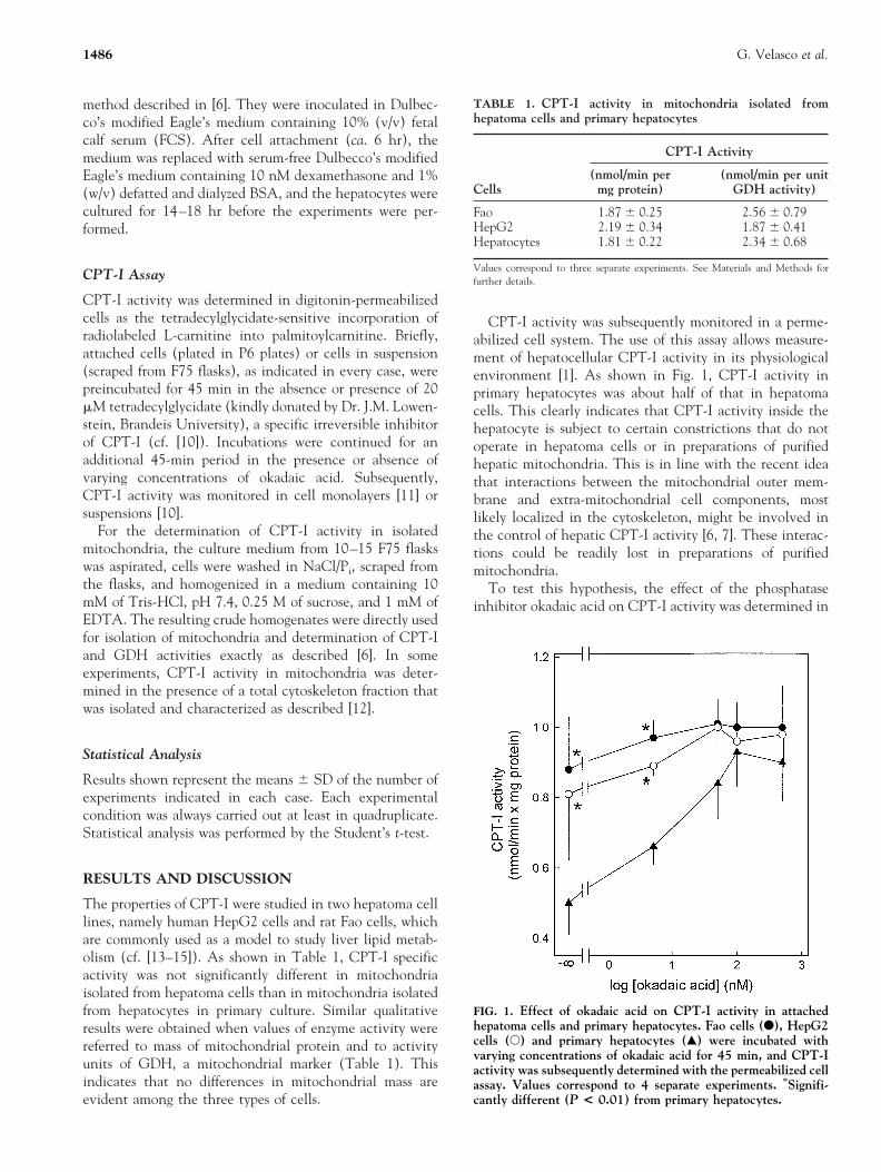
The properties of CPT-I were studied in two hepatoma celllines, namely human HepG2 cells and rat Fao cells, whichare commonly used as a model to study liver lipid metab-olism (cf. [13–15]). As shown in Table 1, CPT-I specificactivity was not significantly different in mitochondriaisolated from hepatoma cells than in mitochondria isolatedfrom hepatocytes in primary culture. Similar qualitativeresults were obtained when values of enzyme activity werereferred to mass of mitochondrial protein and to activityunits of GDH, a mitochondrial marker (Table 1). Thisindicates that no differences in mitochondrial mass areevident among the three types of cells.
CPT-I activity was subsequently monitored in a perme-abilized cell system. The use of this assay allows measure-ment of hepatocellular CPT-I activity in its physiologicalenvironment [1]. As shown in Fig. 1, CPT-I activity inprimary hepatocytes was about half of that in hepatomacells. This clearly indicates that CPT-I activity inside thehepatocyte is subject to certain constrictions that do notoperate in hepatoma cells or in preparations of purifiedhepatic mitochondria. This is in line with the recent ideathat interactions between the mitochondrial outer mem-brane and extra-mitochondrial cell components, mostlikely localized in the cytoskeleton, might be involved inthe control of hepatic CPT-I activity [6, 7]. These interac-tions could be readily lost in preparations of purifiedmitochondria.
To test this hypothesis, the effect of the phosphataseinhibitor okadaic acid on CPT-I activity was determined in
TABLE 1. CPT-I activity in mitochondria isolated fromhepatoma cells and primary hepatocytes
Cells
CPT-I Activity
(nmol/min permg protein)
(nmol/min per unitGDH activity)
Fao 1.87 6 0.25 2.56 6 0.79HepG2 2.19 6 0.34 1.87 6 0.41Hepatocytes 1.81 6 0.22 2.34 6 0.68
Values correspond to three separate experiments. See Materials and Methods forfurther details.
FIG. 1. Effect of okadaic acid on CPT-I activity in attachedhepatoma cells and primary hepatocytes. Fao cells (F), HepG2cells (E) and primary hepatocytes (Œ) were incubated withvarying concentrations of okadaic acid for 45 min, and CPT-Iactivity was subsequently determined with the permeabilized cellassay. Values correspond to 4 separate experiments. *Signifi-cantly different (P < 0.01) from primary hepatocytes.
1486 G. Velasco et al.

the three cells tested. One of the most remarkable effectselicited by okadaic acid in hepatocytes and other cell typesis the hyperphosphorylation and subsequent disruption ofthe cytoskeleton (e.g. [16, 17]). Figure 1 shows that aremarkable 80% stimulation of CPT-I ensued upon expo-sure of primary hepatocytes to okadaic acid. Fifty percentactivation of CPT-I occurred at ca. 10 nM okadaic acid,indicating that CPT-I stimulation is mediated by theinhibition of protein phosphatase 1 [7, 17]. This is inagreement with the observation that protein phosphatase 1seems to be the main phosphatase involved in the regula-tion of the phosphorylation state of the cytoskeleton, andin turn in the control of cytoskeletal integrity [17]. Incontrast to hepatocytes, a slight but not statistically signif-icant stimulation of CPT-I to okadaic acid was evident inhepatoma cells (Fig. 1). Therefore, the high CPT-I activitydisplayed by hepatoma cells may be reached by hepatocytesupon challenge to okadaic acid.
It might be argued that the distinct behavior of primaryhepatocytes and hepatoma cells may be a reflection ofdifferences in their attachment to the substrate, that may inturn involve different configurations of the cytoskeleton.Hence, CPT-I activity was monitored in cell suspensionsafter cell scraping from the flasks. As shown in Table 2,okadaic acid produced a significant stimulation of CPT-I inhepatocyte suspensions but not in Fao cell suspensions.
To further support the notion that cytoskeletal compo-nents may inhibit CPT-I, reconstitution experiments werecarried out with purified mitochondrial and cytoskeletalfractions. As shown in Fig. 2, the cytoskeletal fractionisolated from hepatocytes was able to markedly inhibitCPT-I. However, the decrease in CPT-I activity elicited bythe cytoskeletal fraction prepared from Fao cells was muchweaker, indicating that in hepatoma cells the cytoskeletonmay adopt a configuration that is less effective in inhibitingCPT-I.
In conclusion, the present data support the notion thatin hepatocytes okadaic acid liberates CPT-I from inhibitoryconstrictions imposed by cytoskeletal components that donot operate either in isolated mitochondria or in trans-
formed liver cells. In this respect, Paumen et al. haverecently put forward that cells that express high CPT-Iactivity are expected to withstand palmitate-induced apo-ptosis [18]. Whether liberation of CPT-I from the potentialconstrictions imposed by the cytoskeleton may help hepa-toma cells to escape from apoptosis is currently under studyin our laboratories. In addition, we have recently observed*that intermediate filaments are the components of thecytoskeleton most likely involved in the control of CPT-Iactivity. Interestingly, the interactions between intermedi-ate filaments and mitochondria become disrupted in a stresssituation such as heat shock [19]. Our results suggest thatthis might be the reason for the high CPT-I activity inhepatoma cells.
These investigations were supported by the Spanish Comision Intermin-isterial de Ciencia y Tecnologıa (SAF 96/0113), Fondo de Investiga-cion Sanitaria (FIS 97/0039) and Comunidad Autonoma de Madrid(CAM-6648), as well as by the French ARC (Association pour laRecherche sur le Cancer) and the Ligue Bourguignone contre leCancer.
References
1. Guzman M and Geelen MJH, Regulation of fatty acidoxidation in mammalian liver. Biochim Biophys Acta 1167:227–241, 1993.
*Velasco G, Geelen MJH, Gomez del Pulgar T and Guzman M, unpub-lished observations.
TABLE 2. Comparative effect of okadaic acid on CPT-Iactivity in hepatoma cells and hepatocytes, both in suspensionand in the attached state
CellsOkadaic acid(500 mM)
CPT-I Relative Activity (%)
Attachedcells
Cells insuspension
Fao No 100 6 17 100 6 22Yes 112 6 11 (5) 119 6 10 (3)
HepG2 No 100 6 23 n.d.Yes 120 6 7 (4) n.d.
Hepatocytes No 100 6 18 100 6 12Yes 180 6 12* (5) 171 6 18* (4)
Values correspond to the number of expeiments indicated in parentheses. SeeMaterials and Methods for further details. n.d. 5 not determined.
*Significantly different (P , 0.01) from the corresponding values with no okadaicacid.
FIG. 2. Effect of a cytoskeletal fraction on CPT-I activity.Mitochondria from hepatocytes (1.5–2.0 mg protein) wereincubated for 30 min in the absence (2) or presence ofcytoskeleton (CSK; 0.15–0.20 mg protein) from hepatocytes orFao cells, and CPT-I activity was subsequently determined asindicated in Materials and Methods. Values correspond to threeseparate experiments. Significantly different from incubationswith no additions: *P < 0.01; **P < 0.05. 1Significantlydifferent (P < 0.01) from incubations with cytoskeleton fromhepatocytes.
Regulation of Carnitine Palmitoyltransferase I 1487

2. Zammit VA, Regulation of ketone body metabolism. Acellular perspective. Diabetes Rev 2: 132–155, 1994.
3. McGarry JD and Brown NF, The mitochondrial carnitinepalmitoyltransferase system. From concept to molecular anal-ysis. Eur J Biochem 244: 1–14, 1997.
4. Drynan L, Quant PA and Zammit VA, Flux control exertedby mitochondrial outer membrane carnitine palmitoyltrans-ferase over b-oxidation, ketogenesis and tricarboxylic acidcycle activity in hepatocytes isolated from rats in differentmetabolic states. Biochem J 317: 791–795, 1996.
5. Spurway T, Sherrat HSA, Pogson CI and Agius L, The fluxcontrol coefficient of carnitine palmitoyltransferase I onpalmitate b-oxidation in rat hepatocyte cultures. Biochem J323: 119–122, 1997.
6. Guzman M, Kolodziej MP, Caldwell A, Costorphine CG andZammit VA, Evidence against direct involvement of phos-phorylation in the activation of carnitine palmitoyltransferaseby okadaic acid in rat hepatocytes. Biochem J 300: 693–699,1994.
7. Velasco G, Sanchez C, Geelen MJH and Guzman M, Arecytoskeletal components involved in the control of hepaticcarnitine palmitoyltransferase I activity? Biochem Biophys ResCommun 224: 754–759, 1996.
8. Velasco G, Guzman M, Zammit VA and Geelen MJH,Involvement of Ca21/calmodulin-dependent protein kinase IIin the activation of carnitine palmitoyltransferase I by oka-daic acid in rat hepatocytes. Biochem J 321: 211–216, 1997.
9. Passilly P, Jannin B and Latruffe N, Influence of peroxisomeproliferators on phosphoprotein levels in human and hepatic-derived cell lines. Eur J Biochem 230: 316–321, 1995.
10. Guzman M and Geelen MJH, Activity of carnitine palmitoyl-transferase in mitochondrial outer membranes and peroxi-somes in digitonin-permeabilized hepatocytes. Selective mod-ulation of mitochondrial enzyme activity by okadaic acid.Biochem J 287: 487–492, 1992.
11. Spurway TD, Agius L, Sherrat HSA and Pogson CI, Mea-surement of carnitine palmitoyltransferase I in hepatocytemonolayers. Biochem Soc Trans 22: 118S, 1994.
12. van Bergen en Henegouwen PMP, den Hartigh JC, RomeynP, Verkleij AJ and Boonstra J, The epidermal growth factorreceptor is associated with actin microfilaments. Exp Cell Res199: 90–97, 1992.
13. Prip-Buus C, Bouthillier-Voisin AC, Kohl C, Demaugre F,Girard J and Pegorier JP, Evidence for an impaired long-chainfatty acid oxidation and ketogenesis in Fao hepatoma cells.Eur J Biochem 209: 291–298, 1992.
14. Dixon JL and Ginsberg HN, Regulation of hepatic secretionof apolipoprotein B-containing lipoproteins: Information ob-tained from cultured liver cells. J Lipid Res 34: 167–179, 1993.
15. Duclos S, Bride J, Ramirez LC and Bournot P, Peroxisomeproliferation and b-oxidation in Fao and MH1C1 rat hepa-toma cells, HepG2 human hepatoblastoma cells and culturedhuman hepatocytes: Effect of cipofibrate. Eur J Cell Biol 72:314–323, 1997.
16. Holen I, Gordon PB and Seglen PO, Protein kinase-depen-dent effects of okadaic acid on hepatocytic autophagy andcytoskeletal integrity. Biochem J 284: 633–636, 1992.
17. Gurland G and Gundersen GG, Protein phosphatase inhibi-tors induce the selective breakdown of stable microtubules infibroblasts and epithelial cells. Proc Natl Acad Sci USA 90:8827–8831, 1993.
18. Paumen MP, Ishida Y, Muramatsu M, Yamamoto M andHonjo J, Inhibition of carnitine palmitoyltransferase I aug-ments sphingolipid synthesis and palmitate-induced apopto-sis. J Biol Chem 272: 3324–3329, 1997.
19. Collier NC, Sheetz MP and Schlesinger MJ, Concomitantchanges in mitochondria and intermediate filaments duringheat shock and recovery of chicken embryo fibroblasts. J CellBiochem 52: 297–307, 1993.
1488 G. Velasco et al.


![carnitine deficiency[1]](https://img.pdfslide.net/doc/110x75/577d20c11a28ab4e1e93ae46/carnitine-deficiency1.jpg)