Embed Size (px)
Citation preview
J. clin. Path. (1952), 5, 234.
LOWER NEPHRON NEPHROSIS AFTER*TRANSURETHRAL RESECTION OF THE PROSTATE
BY
JOHN SWINNEY AND B. E. TOMLINSONFrom the Departments of Urology and Pathology, Newcastle General Hospital
(RECEIVED FOR PUBLICATION MAY 28, 1952)
Oliguria and anuria due to lower nephronnephrosismay occur after crushing injuries (Bywatersand Dible, 1942), incompatible blood transfusion(Baker and Dodds, 1925; Foy, Altmann, Barnes,and Kondi, 1943), burns (Goodpastor, Levenson,Tagnon, Lund, and Taylor, 1946), sulphonamidetherapy (Hellwig and Reed, 1942; Lederer andRosenblatt, 1942), septic abortion (Bratton, 1941),concealed accidental haemorrhage (Young, 1942),and occasionally after operations, particularly theoperation of transurethral prostatic resection. Inthe past three years we have observed six cases,which have followed prostatic resection out of atotal of 742 such operations.
Creevy (1947) showed that when water is usedas the irrigating fluid during prostatic resection,haemoglobinaemia occurs, due to absorptionthrough the prostatic veins of lysed blood and wateritself. Haemoglobinaemia occurs in many caseswithout oliguria (Creevy, 1948; Biorn and Greene,1949), and its degree bears no relationship to theseverity of the renal failure. Whilst a number ofisotonic solutions have been employed as irrigatingfluids in prostatic resection (Ebert, 1949; Nesbitand Glickman, 1948; Garske, Phares, and Sweetser,1949) we have used isotonic glucose solution, whichdoes not cause haemoglobinaemia.We have found that the oliguric syndrome has
not been eliminated by the use of solutions whichavoid haemoglobinaemia, as has been asserted(Chapman and Sutherland, 1952). Further, insome of our cases, transfusion, sulphonamidetherapy, and clinical shock played no part, thoughthese factors have been present in many reportedcases of lower nephron nephrosis, and in most ofthose associated with transurethral prostatectomy.Our six cases all developed oliguria or anuria.
Water irrigation was employed in Cases 1 and 2;5% glucose in Cases 3 to 6 (though in Case 3, fortechnical reasons, water was substituted for glucoseduring the operation). Their clinical and patho-logical findings are briefly described.
Case ReportsCase 1.-J.N., aged 70 years, had benign prostatic
obstruction, for which prostatic resection was performedon November 10, 1949, when 25 g. of prostatic tissuewere resected. He had oliguria the following day, andwas completely anuric for 73 hours until his death inuraemic coma on November 16.
Necropsy was performed 36 hours after death.In addition to the renal lesion, there was extensive
bilateral bronchopneumonia, confluent in both lowerlobes. The kidneys together weighed 350 g. They showedconsiderable scarring, and on bisection appeared toconsist almost entirely of pale, swollen, cortical tissue,which bulged over the capsule.Histology.-The glomeruli showed no abnormality.
The cells of the first convoluted tubules were flattenedand the lumen of the tubules wide. No necrosis waspresent. A few small areas of complete tubular degener-ation were found in the outer medulla, the cells of the
7',
0.'~~~~~~~~~~0A
4~.¾' 40
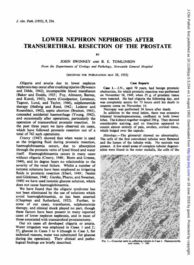
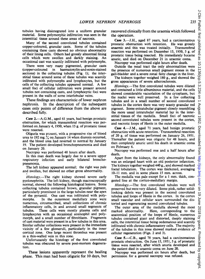
FIG. l.-Granular casts in collecting tubules in Case 1. Haematoxylinand eosin, x 100.
on 19 June 2018 by guest. Protected by copyright.
http://jcp.bmj.com
/J C
lin Pathol: first published as 10.1136/jcp.5.3.234 on 1 A
ugust 1952. Dow
nloaded from
LOWER NEPHRON NEPHROSIS
tubules having disintegrated into a uniform granularmaterial. Some polymorphic infiltration was seen in theinterstitial tissue around these areas of necrosis.
Many- of the second convoluted tubules containedcopper-coloured, granular casts. Some of the tubulescontaining these casts showed no obvious abnormalityof their lining cells. Others had grossly abnormal liningcells which were swollen and darkly staining. Anoccasional cast was scantily infiltrated with polymorphs.
There were very many pigmented, granular casts(copper-coloured in haematoxylin-and-eosin-stainedsections) in the collecting tubules (Fig. 1); the inter-stitial tissue around some of these tubules was scantilyinfiltrated with polymorphs and lymphocytes, but thecells of the collecting tubules appeared normal. A fewsmall foci of cellular infiltration were present aroundtubules not containing casts, and lymphocytic foci werepresent in the walls of some interlobular veins.
These findings are characteristic of lower nephronnephrosis. In the description of the subsequentcases only points of difference from the above aredescribed fully.
Case 2.-A.G.M., aged 61 years, had benign prostaticobstruction, for which transurethral resection was per-formed on January 4, 1950, when 15 g. of prostatic tissuewere resected.
Oliguria was present, with a progressive rise of bloodurea to 192 mg. % on January 14 when diuresis occurred,and the blood urea had reverted to normal by January19. The patient developed bronchopneumonia and diedon January 20.
Necropsy was performed 48 hours after death.In this man death was largely due to a severe upper
respiratory infection and early bilateral broncho-pneumonia.The left kidney appeared normal; the right was pale
and swollen, but showed no other gross abnormality.
Histology.-The right kidney showed severe earlypyelonephritis. The left kidney, though macroscopicallynormal, showed the following histological lesions. Somecollecting tubules contained brown, granular pigment,particularly prominent in the large tubules near the apexof the pyramid. One cast was infiltrated with poly-morphs. In the outermost medullary zone werenumerous, circumscribed, small collections of chronicinflammatory cells, in and around small segments oftubules. These cells were mostly plasma cells andlymphocytes with an occasional eosinophil and poly-morph, and a small number of fibroblasts. Fragmentsof cast material were present in a few tubules in this area.Similar cellular collections were present in the immediatevicinity of a few glomeruli, particularly in the innercortical zone. One large recent thrombus was presentin a thin-walled vein in this zone.
Unfortunately the histology of the first convolutedtubules was obscured by severe post-mortem degenera-tion.
These lesions apparently represent the healingphase. This man had been oliguric for 10 days, but
recovered clinically from the uraemia which followedthe operation.
Case 3.-J.H., aged 87 years, had a carcinomatousprostatic obstruction with retention. He was grosslyanaemic and this was treated initially. Transurethralresection was performed on December 15, 1950, 5 g. ofprostatic tissue being resected. He immediately bacameanuric, and died on December 21 in uraemic coma.Necropsy was performed eight hours after death.Outside the renal tract the only abnormalities were
the presence of numerous small pigment stones in thegall-bladder and a severe zonal fatty change in the liver.The kidneys together weighed 180 g., and showed the
gross appearances of severe atherosclerosis.Histology.-The first convoluted tubules were dilated
and contained a little albuminous material, and the cellsshowed considerable vacuolation of the cytoplasm, butthe nuclei were well preserved. In a few collectingtubules and in a small number of second convolutedtubules in the cortex there was very scanty granular redpigment. Some extratubular pigment closely resemblingthe more usual intratubular material lay in the inter-stitial tissues of the medulla. Small foci of necroticsecond convoluted tubules were present in the cortex,and necrotic loops of Henle in the outer medulla.Case 4.-J.M., aged 71 years, had benign prostatic
obstruction with acute retention., Transurethral resectionof 20 g. of tissue was performed on January 26, 1951.Thereafter the patient was oliguric for 48 hours, andthen completely anuric until his death in uraemic comaon February 6.Necropsy was performed one and a half hours after
death.Apart from the kidneys, the only abnormality found
was an enlarged heart with an old posterior infarction.The kidneys together weighed 440 g., and showed markedfoetal lobulation. The cortex was thickened, averaging10-11 mm. and in some places 15 mm. across.The medulla was pale except for a 1 mm. thick, con-
gested line at the cortico-medullary margin.Histology.-The first convoluted tubules were well
preserved but were very dilated. Some pink, rather solid-looking debris was present in the second convolutedtubules and loops of Henle in the cortex. In the cortexsmall vascular and cellular scars surrounded the dis-torted and regenerating second convoluted tubules.The outer area of the medulla showed the most
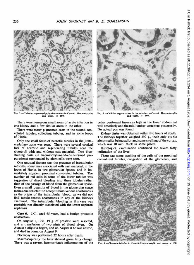
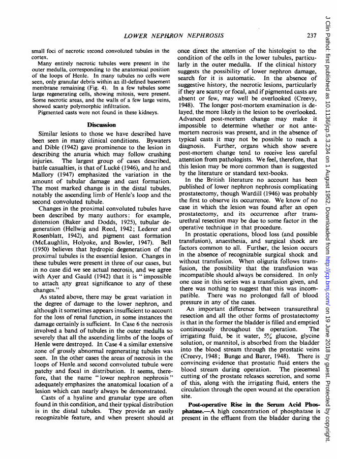
marked abnormality. Here, corresponding to theanatomical position of the loops of Henle, numeroustubules contained giant and distorted, deeply stainingcells, the interstitial tissue being oedematous and scantilyinfiltrated with chronic inflammatory cells. The majorityof the tubules in this zone showed marked evidence ofcellular regeneration (Figs. 2 and 3).
Case 5.-C.B., aged 78 years, had a carcinomatousprostatic obstruction. On June 15, 1951, 5 g. of prostatictissue were resected, after which anuria developed andthe patient died in uraemic coma on June 20.Necropsy was performed six hours after death, but
permission for a general necropsy was refused.
235
on 19 June 2018 by guest. Protected by copyright.
http://jcp.bmj.com
/J C
lin Pathol: first published as 10.1136/jcp.5.3.234 on 1 A
ugust 1952. Dow
nloaded from
JOHN SWINNEY and B. E. TOMLINSON
VA~~~~~~h
Alt f
4~~~~~~~ ,~~~~~
A \..'',,*t '_FIG. 2.-ellular regeneration in the tubules in Case 4. Haematoxylin
and eosin, x 160.
There were numerous small areas of acute infection inone kidney and a few similar areas in the other.There were many pigmented casts in the second con-
voluted tubules, collecting tubules, and in some loopsof Henle.Only one small focus of necrotic tubules in the juxta-
medullary zone was seen. There were several corticalfoci of necrotic and regenerating tubules near theglomeruli with and without cast material. Two blue-staining casts (in haematoxylin-and-eosin-stained pre-parations) surrounded by giant cells were seen.One unusual feature was the presence pf intratubular
red cells, sometimes associated with cast material, in theloops of Henle, in two glomerular spaces, and in im-mediately adjacent proximal convoluted tubules. Thenumber of red cells in some of the lower tubules wassuggestive of direct bleeding into these tubules ratherthan of the passage of blood from the glomerular space.Even a small quantity of blood in the glomerular spacemakes one reluctant to accept tubulo-venous anastomosisas the origin of the intratubular blood, as we did notfind tubulo-venous anastomosis in any of the kidneysexamined. The intratubular bleeding in this case wasprobably not directly associated with the lower nephronnephrosis.
Case 6.-J.C., aged 65 years, had a benign prostaticobstruction.On August 1, 1951, 19 g. of prostate were resected,
and a transfusion of two pints of blood given. OnAugust 4 oliguria began, and on August 8 he was anuric,and died in coma on August 9.Necropsy was performed 22 hours after death.Macroscopically the liver showed gross fatty change.
There was a severe, haemorrhagic inflammation of the
FIG. 3.-Cellular regeneration in the tubules in Case 4. Haematoxylinand eosin, x 560.
pelvic peritoneal tissues as high as the lower abdominalwall anteriorly and the mid-lumbar vertebrae posterorily.No actual pus was found.Kidney tisstie was obtained within five hours of death.
The kidneys together weighed 290 g., their only visibleabnormality being pallor and some swelling of the cortex,which was 10 mm. thick in some places.
Histological examination confirmed the severe fattyinfiltration of the liver.There was some swelling of the cells of the proximal
convoluted tubules, congestion of the glomeruli, and
236
on 19 June 2018 by guest. Protected by copyright.
http://jcp.bmj.com
/J C
lin Pathol: first published as 10.1136/jcp.5.3.234 on 1 A
ugust 1952. Dow
nloaded from
LOWER NEPILRON NEPHROSIS
small foci of necrotic second convoluted tubules in thecortex.Many entirely necrotic tubules were present in the
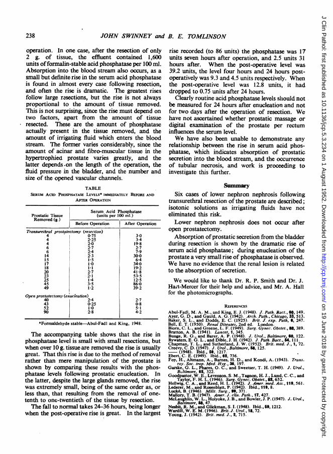
outer medulla, corresponding to the anatomical positionof the loops of Henle. In many tubules no cells wereseen, only granular debris within an ill-defined basementmembrane remaining (Fig. 4). In a few tubules somelarge regenerating cells, showing mitosis, were present.Some necrotic areas, and the walls of a few large veins,showed scanty polymorphic infiltration.Pigmented casts were not found in these kidneys.
DiscussionSimilar lesions to those we have described have
been seen in many clinical conditions. Bywatersand Dible (1942) gave prominence to the lesion indescribing the anuria which may follow crushinginjuries. The largest group of cases described,battle casualties, is that of Lucke (1946), and h_ andMallory (1947) emphasized the variation in theamount of tubular damage and cast formation.The most marked change is in the distal tubules,notably the ascending limb of Henle's loop and thesecond convoluted tubule.
Changes in the proximal convoluted tubules havebeen described by many authors: for example,distension (Baker and Dodds, 1925), tubular de-generation (Hellwig and Reed, 1942; Lederer andRosenblatt, 1942), and pigment cast formation(McLaughlin, Holyoke, and Bowler, 1947). Bell(1950) believes that hydropic degeneration of theproximal tubules is the essential lesion. Changes inthese tubules were present in three of our cases, butin no case did we see actual necrosis, and we agreewith Ayer and Gauld (1942) that it is " impossibleto attach any great significance to any of thesechanges."As stated above, there may be great variation in
the degree of damage to the lower nephron, andalthough it sometimes appears insufficient to accountfor the loss of renal function, in some instances thedamage certainly is sufficient. In Case 6 the necrosisinvolved a band of tubules in the outer medulla soseverely that all the ascending limbs of the loops ofHenle were destroyed. In Case 4 a similar extensivezone of grossly abnormal regenerating tubules wasseen. In the other cases the areas of necrosis in theloops of Henle and second convoluted tubule werepatchy and focal in distribution. It seems, there-fore, that the name "lower nephron nephrosis"adequately emphasizes the anatomical location of alesion which can nearly always be demonstrated.
Casts of a hyaline and granular type are oftenfound in this condition, and their typical distributionis in the distal tubules. They provide an easilyrecognizable feature, and when present should at
once direct the attention of the histologist to thecondition of the cells in the lower tubules, particu-larly in the outer medulla. If the clinical historysuggests the possibility of lower nephron damage,search for it is automatic. In the absence ofsuggestive history, the necrotic lesions, particularlyif they are scanty or focal, and if pigmented casts areabsent or few, may well be overlooked (Creevy,1948). The longer post-mortem examination is de-layed, the more likely is the lesion to be overlooked.Advanced post-mortem change may make itimpossible to determine whether or not ante-mortem necrosis was present, and in the absence oftypical casts it may not be possible to reach adiagnosis. Further, organs which show severepost-mortem change tend to receive less carefulattention from pathologists. We feel, therefore, thatthis lesion may be more common than is suggestedby the literature or standard text-books.
In the British literature no account has beenpublished of lower nephron nephrosis complicatingprostatectomy, though Wardill (1946) was probablythe first to observe its occurrence. We know of nocase in which the lesion was found after an openprostatectomy, and its occurrence after trans-urethral resection may be due to some factor in theoperative technique in that procedure.
In prostatic operations, blood loss (and possibletransfusion), anaesthesia, and surgical shock arefactors common to all. Further, the lesion occursin the absence of recognizable surgical shock andwithout transfusion. When oliguria follows trans-fusion, the possibility that the transfusion wasincompatible should always be considered. In onlyone case in this series was a transfusion given, andthere was nothing to suggest that this was incom-patible. There was no prolonged fall of bloodpressure in any of the cases.An important difference between transurethral
resection and all the other forms of prostatectomyis that in the former the bladder is filled and emptiedcontinuously throughout the operation. Theirrigating fluid, be it water, 5% glucose, glycinesolution, or mannitol, is absorbed from the bladderinto the blood stream through the prostatic veins(Creevy, 1948; Bunge and Barer, 1948). There isconvincing evidence that prostatic fluid enters theblood stream during operation. The piecemealcutting of the prostate releases secretion, and someof this, along with the irrigating fluid, enters thecirculation through the open wound at the operationsite.
Post-operative Rise in the Serum Acid Phos-phatase.-A high concentration of phosphatase ispresent in the effluent from the bladder during the
237
on 19 June 2018 by guest. Protected by copyright.
http://jcp.bmj.com
/J C
lin Pathol: first published as 10.1136/jcp.5.3.234 on 1 A
ugust 1952. Dow
nloaded from
JOHN SWINNEY and B. E. TOMLINSON
operation. In one case, after the resection of only2 g. of tissue, the effluent contained 1,600units of formalin-stable acid phosphatase per 100 ml.Absorption into the blood stream also occurs, as asmall but definite rise in the serum acid phosphataseis found in almost every case following resection,and often the rise is dramatic. The greatest risesfollow large resections, but the rise is not alwaysproportional to the amount of tissue removed.This is not surprising, since the rise must depend ontwo factors, apart from the amount of tissueresected. These are the amount of phosphataseactually present in the tissue removed, and theamount of irrigating fluid which enters the bloodstream. The former varies considerably, since theamount of acinar and fibro-muscular tissue in thehypertrophied prostate varies greatly, and thelatter depends .on the length of the operation, thefluid pressure in the bladder, and the number andsize of the opened vascular channels.
TABLESERUM AcID PHOSPHATASE LEVELS* IMMEDiATELY BEFORE AND
AFTER OPERATION
Serum Acid PhosphataseProstatic Tissue (units per 100 ml.)Removed (g.)
Before Operation After Operation
Transurethral prostf7tectomy (resection)4 075 304 2 25 3-44 2-0 1986 2-7 2-76 2.4 7914 2'3 30015 1 5 6-417 110 34-018 1.1 19 220 2-7 41 823 2-1 53 525 1.4 12-545 3.5 86-049 1-2 39-2
Open prostatectomy (enucleation)40 2.4 2-743 025 0-852 1-0 5 190 2-8 4-2
*Formaldehyde stable-Abul-Fadl and King, 1948.
The accompanying table shows that the rise inphosphatase level is small with small resections, butwhen over 10 g. tissue are removed the rise is usuallygreat. That this rise is due to the method ofremovalrather than mere manipulation of the prostate isshown by comparing these results with the phos-phatase levels following prostatic enucleation. Inthe latter, despite the large glands removed, the risewas extremely small, being of the same order as, orless than, that resulting from the removal of one-tenth to one-twentieth of the tissue by resection.The fall to normal takes 24-36 hours, being longer
when the post-operative rise is great. In the largest
rise recorded (to 86 units) the phosphatase was 17units seven hours after operation, and 2.5 units 31hours after. When the post-operative level was39.2 units, the level four hours and 24 hours post-operatively was 9.3 and 4.5 units respectively. Whenthe post-operative level was 12.8 units, it haddropped to 0.75 units after 24 hours.
Clearly routine acid phosphatase levels should notbe measured for 24 hours after enucleation and notfor two days after the operation of resection. Wehave not ascertained whether prostatic massage ordigital examination of the prostate per rectuminfluences the serum level.We have also been unable to demonstrate any
relationship between the rise in serum acid phos-phatase, which indicates absorption of prostaticsecretion into the blood stream, and the occurrenceof tubular necrosis, and work is proceeding toinvestigate this further.
SummarySix cases of lower nephron nephrosis following
transurethral resection of the prostate are described;isotonic solutions as irrigating fluids have noteliminated this risk.Lower nephron nephrosis does not occur after
open prostatectomy.Absorption ofprostatic secretion from the bladder
during resection is shown by the dramatic rise ofserum acid phosphatase; during enucleation of theprostate a very small rise of phosphatase is observed.We have no evidence that the renal lesion is relatedto the absorption of secretion.
We would like to thank Dr. R. P. Smith and Dr. J.Hart-Mercer for their help and advice, and Mr. A. Hallfor the photomicrographs.
REFERENCESAbul-Fadl, M. A. M., and King, E. J. (1948). J. Path. Bact., 60, 149.Ayer, G. D., and Gauld, A. G. (1942). Arch. Path., Chicago, 33, 513.Baker, S. L., and Dodds, E. C. (1925). Brit. J. exp. Path, 6, 247.Bell, E. T. (1950). Renal Diseases, 2nd ed. London.Biorn, C. L., and Greene, L. F. (1949). Surg. Gynec. Obstet., 88, 389.Bratton, A. B. (1941). Lancet, 1, 345.Bunge, R. G., and Barer, A. P. (1948). J. Urol., Baltimore, 60, 122.Bywaters, E. G. L., and Dible, J. H. (1942). J. Path. Bact., 54, 111.Chapman, T. L., and Sutherland, J. W. (1952). Brit. med. J., 1, 72.Creevy, C. D. (1947). J. Urol., Baltimore, 568, 125.- (1948). Ibid., 59, 1217.Ebert, C. E. (1949). Ibid., 62, 736.Foy, H., Altmann, A., Barnes, H. D., and Kondi, A. (1943). Trans.
roy. Soc. trop. Med. Hyg., 36, 197.Garske, G. L., Phares, 0. C., and Sweetser, T. H. (1949). J. Urol.,
B4ltimore, 62, 322.Goodpastor, W. E., Levenson, S. M., Tagnon, H. J., Lund, C. C., and
Taylor, F. H. L. (1946). Surg. Gynec. Obstet., 82,652.Hellwig, C. A., and Reed, H. L. (1942). J. Amer. med. Ass., 119, 561.Lederer, M., and Rosenblatt, P. (1942). Ibid., 119, 8.Lucke, B. (1946). Milit. Surg., "1, 371.Mallory, T. B. (1947). Amer. J. clin. Path., 17, 427.McLaughlin, W. L., Holyoke, J. B., and Bowler, J. P. (1947). J. Urol.,
Baltimore, 58, 47.Nesbit, R. M., and Glickman, S. I. (1948). Ibid., 59, 1212.Wardill, W. E. M. (1946). Brit. J. Urol., 18, 72.Young, J. (1942). Brit. med. J., 2, 715.
238
on 19 June 2018 by guest. Protected by copyright.
http://jcp.bmj.com
/J C
lin Pathol: first published as 10.1136/jcp.5.3.234 on 1 A
ugust 1952. Dow
nloaded from