Embed Size (px)
Citation preview
Cláudia Azevedo: New engineered
FcRn-targeting nanoplatforms for oral
delivery of biopharmaceuticals
D.ICBAS 2021
New engineered FcRn-targeting nanoplatforms
for oral delivery of biopharmaceuticals Cláudia Azevedo
INSTITUTO DE CIÊNCIAS BIOMÉDICAS ABEL SALAZAR
DO
UT
OR
AM
EN
TO
CIÊ
NC
IAS
BIO
MÉ
DIC
AS
New
en
gin
eere
d F
cR
n-t
arg
eti
ng
nan
op
latf
orm
s f
or
ora
l d
elivery
of
bio
ph
arm
aceu
ticals
Clá
udia
Azevedo
D
20
21
Cláudia Azevedo
NEW ENGINEERED FcRn-TARGETING NANOPLATFORMS FOR
ORAL DELIVERY OF BIOPHARMACEUTICALS
Tese de Candidatura ao grau de Doutor em Ciências
Biomédicas submetida ao Instituto de Ciências
Biomédicas Abel Salazar da Universidade do Porto
Orientador:
Doutor Bruno Filipe Carmelino Cardoso Sarmento
Categoria – Investigador Principal/Professor Auxiliar
Afiliação – i3S – Instituto de Investigação e Inovação em
Saúde & INEB - Instituto Nacional de Engenharia
Biomédica, Universidade do Porto / IUCS – Instituto
Universitário de Ciências da Saúde
Co-Orientador:
Doutor Jan Terje Andersen
Categoria – Investigador Principal
Afiliação – Oslo University Hospital - Department of
Biosciences, University of Oslo, Norway

DECLARAÇÃO DE HONRA
Eu, Cláudia Filipa Maia Azevedo, declaro que a presente tese é de minha autoria e não foi
utilizada previamente noutro curso ou unidade curricular, desta ou de outra instituição. As
referências a outros autores (afirmações, ideias, pensamentos) respeitam escrupulosamente
as regras da atribuição, e encontram-se devidamente indicadas no texto e nas referências
bibliográficas, de acordo com as normas de referenciação. Tenho consciência de que a
prática de plágio e auto-plágio constitui um ilícito académico.
Porto, 15 de Janeiro de 2021
____________________
Cláudia Azevedo

The work presented in this thesis was developed at:
Nanomedicines & Translational Drug Delivery Group
i3S - Instituto de Investigação e Inovação em Saúde and
INEB - Instituto Nacional de Engenharia Biomédica
Universidade do Porto, Porto, Portugal
Rua Alfredo Allen, 208
4200-135 Porto, Portugal
https://www.i3s.up.pt/ | www.ineb.up.pt
And
Oslo University Hospital and University of Oslo
Department of Immunology and department of Pharmacology
Centre for Immune Regulation
UiO FOCIS Center of Excellence
Sognsvannsveien 20, 0372 Oslo, Norway
https://www.ous-research.no/andersen/

And
Massachusetts Institute of Technology
Department of Mechanical Engineering and The Langer Lab
77 Massachusetts Ave., Room 76-661c, Cambridge, MA 02139
https://meche.mit.edu/ | https://langer-lab.mit.edu/
And
European Space Agency
European Space Research and Technology Centre
Keplerlaan 1, 2201 AZ Noordwijk, Netherlands
https://www.esa.int/About_Us/ESTEC
FINANCIAL SUPPORT
Cláudia Azevedo was supported by a national PhD grant (SFRH/BD/117598/2016) from
Fundação para a Ciência e Tecnologia (FCT), Portugal. Cláudia Azevedo was also supported
by a Fulbright (PS00293218) and FLAD (137/2019) grants during 6 months in 2019/2020.
This work is a result of the project NORTE-01-0145-FEDER-000012, supported by Norte
Portugal Regional Operational Programme (NORTE 2020), under the PORTUGAL 2020
Partnership Agreement, through the Europe and Regional Development Fund (ERDF). This
work was financed by FEDER - Fundo Europeu de Desenvolvimento Regional funds through
the COMPETE 2020 - Operacional Programme for Competitiveness and Internationalisation
(POCI), Portugal 2020, and by Portuguese funds through FCT- Fundação para a Ciência e a
Tecnologia/Ministério da Ciência, Tecnologia e Ensino Superior in the framework of the
project "Institute for Research and Innovation in Health Sciences" (UID/BIM/04293/2019),
and also Partnership Agreement PT2020 UID/QUI/50006/2013 -POCI/01/0145/FED
ER/007265. This work was also supported by the Research Council of Norway (Grant no.
274993), the Koch Institute/MIT, Merck, the Spin Your Thesis! 2020 program organized by
the European Space Agency (ESA) Education Office, Abbott, COST and by Instituto Ciências
Biomédicas de Abel Salazar (ICBAS).

PUBLICATIONS Ao abrigo do disposto do nº 2, alínea a) do artigo 31º do Decreto-Lei n.º 115/2013 de 7 de
Agosto, fazem parte integrante desta tese de doutoramento os seguintes trabalhos já
publicados ou submetidos para publicação:
• Azevedo C., Andersen J.T., Sarmento B., Traverso G., The potential of porcine ex vivo
platform for intestinal permeability screening of FcRn-targeted drugs, European Journal of
Pharmaceutics and Biopharmaceutics 2021, 162: 99-104.
• Azevedo C.*, Almeida A.*, Macedo M.H.*, Pinto S.*, van Loon J., Sarmento B, The effect
of hypergravity in intestinal permeability of nanoformulations and molecules, European
Journal of Pharmaceutics and Biopharmaceutics 2021, 163: 38-48. (*equal contribution)
• Azevedo C.*, Pinto S.*, Benjakul S., Santos H.A., Andersen J.T., Traverso G., Sarmento
S., Prevention of diabetes-associated fibrosis: Strategies to orally treat diabetes with FcRn-
targeted nanosystems, Advanced Drug Delivery Reviews 2021. (*equal contribution)
• Azevedo C., Nilsen J., Grevys A., Nunes R., Andersen J.T., Sarmento B., Engineered
albumin-functionalized nanoparticles for improved FcRn binding enhances oral delivery of
insulin, Journal of Controlled Release 2020, 327: 161-173.
o Outside Front Cover Page, Journal of Controlled Release 2020, Volume 327,
ISSN:0168-3659.
• Almeida A*, Azevedo C*, Macedo MH*, Sarmento B, 3D intestinal models towards a more
realistic permeability screening, in Santos H. and Martins J.P., Nanotechnology for Oral
Drug Delivery, Elsevier 2020, ISBN 9780128180389. (*equal contribution)
• Azevedo C., Pereira I., Sarmento B., Intestinal mucosal tissue models to validate
functionalized nanosystems, in Fahr A., et al., Characterization of Pharmaceutical Nano-
and Microsystems, Wiley 2020, ISBN: 978-1-119-41404-9.
• Azevedo C., Macedo M. H., Sarmento B., Strategies for enhanced intracellular delivery of
nanomaterials, Drug Discovery Today 2018, 23:944-959, DOI:
10.1016/j.drudis.2017.08.011.

ACKNOWLEDGEMENTS
Addressee unknown - retour à l'expéditeur
Many thanks for the clouds.
Many thanks for the well-tempered Clavier
and, why not, for the warm winter boots.
Many thanks for my strange brain
and for all manner of other hidden organs,
for the air, and, of course, for the claret.
Heartfelt thanks for my lighter
and my desire not running out of fuel,
as well as my regret, my deep regret.
Many thanks for the four seasons,
for the number e, for my dose of caffeine,
and, of course, for the strawberry dish
painted by Chardin, as well as for sleep,
for sleep quite especially,
and, last not least, for the beginning
and the end and the few minutes in between
fervent thanks, even, if you like,
the voles out there in the garden.
(Hans Magnus Enzensberger)
My PhD was painted, throughout four years, with the motivation and orientation of many
people. This canvas was a roller coaster of emotions, which sometimes was a bit hard to
manage. Fortunately, I have a lot of painters in my life that draw good lines on their unique
style and helped me to achieve one more goal. To all of them, I express my sincere
appreciation for their support on getting a great work and helping me to grow personally and
professionally. All these people and experiences will be with me for a lifetime and I am very
grateful for that! Particularly:
And first of all, to my supervisor, Professor Bruno Sarmento, for believing in my potential,
being always present and careful, the daily effort to provide easel, brushes and the canvas, to
take this painting to a good atelier. I am very thankful for his trust, guidance, encouragement
and friendship. For sure, he is an example of leader and mentor, who taught me to be more
assertive and confident, motivated me in more frustrated moments, challenged me and
supported my projects “outside the box”. You have my acknowledgement and respect!
To my co-supervisor, Jan Terje Andersen, for the warm welcome in his lab, at Norway. The
experience in Oslo was so productive and I feel so grateful for have learned so much. Jan is a
very dynamic and positive person, supports young generations and eagers for innovation.
Characteristics that inspire and motivate me. I am very thankful for the constructive
discussions, attentive readings and the feeling of “family” that I found in his group. It’s good to
be an “Albumin Girl”!
To Jeannette Nilsen, the best lab partner that I could have. Thank you very much for all the
scientific sharing about albumin and FcRn, smiles, adventures in and outside the lab and
friendship. I will never forget my norwegian sister and The scream (Edvard Munch)! Also, to
Julia Alopaeus, for helping me with some logistics and social meetings in Norway.
To Giovanni Traverso, for accepting me at MIT, Boston. It was both a blast and a challenging
experience. This was for sure the experience that I grew the most. Thank you for allowing me
to be in an environment full of opportunities, conducive to innovation, networking and
entrepreneurship. It was great to experience a bit of the american dream and know the
Nighthawks (Edward Hopper) style!
To Raquel Almeida, for giving me incentive to try new colors. To Jack van Loon, Alan
Downson and Nigel Savage, for warmly receiving and helping the team ARTEMIS at ESA,
The Netherlands. It was refreshing to see a Starry Night (Van Gogh) and the immensity that is
beyond our eyes! “The only limit is the one that you set yourself” and we raised the spark!

To Professor Vítor Seabra, for giving me access to CESPU - Instituto de Investigação e
Formação Avançada em Ciências e Tecnologias da Saúde, and to adequate equipment to
perform some assays of this work. And to Virgínia Gonçalves for helping me to solve some
troubleshooting with HPLC.
To the “Quarteto Fantástico”, the marvelous group that was born alongside with this project.
I am grateful for the surprise of our friendship, shared trips and craziness. To Flávia Sousa,
my trip partner, for sharing experiences, advices and motivation to be and do more and better;
to Andreia Almeida for having a relaxed way of being and a positive vibe and for sharing
adrenalin with me; and Helena Macedo, for being a conscious, critical and just person. This
group is always ready for a great adventure! Many thanks for supporting me throughout my
path. Following Afremov, you gave a bunch of colors to my PhD!
To all team members of different groups that I was in, especially from NTDD group, for all
the sharing of knowledge, fellowship, help and sweeties. I would like to highlight the important
role of José das Neves (general, sincere and critical input), Rute Nunes (animal assays),
Algirdas Grevys (transcytosis and recycling assays) and Ming Zhao (help in Boston) for their
motivation, wise advices through different moments of the painting.
To all the Portugueses that I met in Boston, specially to Ana Cadete and Ângelo Crespo for
helping me throughout this process and giving me a bit of familiar lines. Also, to Abhisheck
and Samip, my indian friends, for adding some joy, kindness and company during my stay.
They are all friends for life that were met in an unexpected way!
The last but not the least, to my Family:
Aos meus Pais, Paula e António, e aos meus Irmãos, Marco e Simão, pelo apoio
incondicional, por ficarem genuinamente felizes com as minhas conquistas, apesar de às
vezes não perceberem muito bem em que se traduzem. Aos meus Padrinhos, Olinda e
Joaquim, por se orgulharem de mim e darem sempre uma palavra de apreço e motivação. À
minha Avó por rezar por mim! Ao meu Namorado, melhor amigo e companheiro, João, por
me confortar, por me fazer sentir especial todos os dias, por me aceitar tal como sou, por ter
paciência para as minhas ansiedades, por o ter sentido perto apesar de estar 6 meses longe.
Com ele aprendi a redefinir prioridades e a tornar-me na minha melhor versão.
Agradeço profundamente a todos pela intimidade, confiança, apoio e cores cintilantes, bem
ao jeito d’ O Beijo (Gustav Klimt), que deram vida à minha tela!

“Questions you cannot answer are usually far better for you than answers you cannot question.”
(Yuval Noah Harari)
TABLE OF CONTENTS ABSTRACT .............................................................................................................................................. i
RESUMO .................................................................................................................................................iii
GRAPHICAL ABSTRACT........................................................................................................................ v
LIST OF FIGURES .................................................................................................................................vii
LIST OF TABLES .................................................................................................................................. xv
ACRONYMS AND ABBREVIATIONS LIST ......................................................................................... xvii
CHAPTER I: LITERATURE REVIEW ..................................................................................................... 1
1. Introduction .......................................................................................................................................... 5
1.1. Epidemiology and the biology behind the disease ....................................................................... 5
1.2. Oral insulin delivery for T1DM ...................................................................................................... 5
1.3. Biologic barriers of oral delivery drugs.......................................................................................... 7
2. Nanoparticles as a strategy for drug delivery ...................................................................................... 7
2.1. The evolution of Nanomedicine .................................................................................................... 7
2.2. Nanoparticles characteristics and their influence ......................................................................... 8
3. Functionalized nanoparticles for an enhanced bioavailability ........................................................... 11
3.1. Albumin as a ligand .................................................................................................................... 11
3.2. FcRn as a receptor ..................................................................................................................... 12
3.2.1. FcRn structure and interaction with albumin ........................................................................... 12
3.2.2. Biologic mechanisms mediated by FcRn ................................................................................. 14
3.3. Albumin- based drug delivery of insulin ...................................................................................... 16
3.4. Albumin-targeted nanosystems for oral delivery of insulin ......................................................... 17
4. Intestinal models for preclinical validation ......................................................................................... 18
4.1. Intestinal morphology .................................................................................................................. 18
4.2. In vitro models ............................................................................................................................. 21
4.2.1. Monoculture model .................................................................................................................. 22
4.2.2. Co-culture model...................................................................................................................... 23
4.2.3. 3D co-culture model ................................................................................................................. 25
4.2.4. Gut-on-a-chip model ................................................................................................................ 26
4.3. Ex vivo models ............................................................................................................................ 31
4.3.1. FcRn tissue expression and cross-species differences........................................................... 32
4.3.1.1. Human ............................................................................................................................... 32
4.3.1.2. Mouse ................................................................................................................................ 32
4.3.1.3. Porcine ............................................................................................................................... 33
4.3.2. Standard models ...................................................................................................................... 35
4.3.2.1. Diffusion chambers ............................................................................................................ 35
4.3.2.2. Everted intestinal sac model .............................................................................................. 36
4.3.2.3. Non-everted intestinal sac model ...................................................................................... 36
4.3.2.4. Everted intestinal ring ........................................................................................................ 37
4.4. In vivo FcRn model systems ....................................................................................................... 37
CHAPTER II: OVERVIEW AND AIMS .................................................................................................. 41
1. Overview ........................................................................................................................................ 43
2. Aims ............................................................................................................................................... 44
CHAPTER III: MATERIALS AND METHODS ...................................................................................... 45
1. Cell culture ..................................................................................................................................... 47
2. Protein production ......................................................................................................................... 47
3. Polymeric nanoparticles production .............................................................................................. 47
4. Physical chemical characterization ............................................................................................... 48
4.1. Particle size and zeta potential analysis ..................................................................................... 48
4.2. Morphological characterization and elementary analysis ........................................................... 48
4.3. Insulin association efficiency and drug loading .......................................................................... 49
4.4. Insulin release ............................................................................................................................. 49
4.5. Albumin conjugation efficiency ................................................................................................... 50
5. Protein structure ............................................................................................................................ 50
5.1. Determination of secondary structure ......................................................................................... 50
5.2. Confirmation of the conjugation .................................................................................................. 51
6. Binding to human FcRn ................................................................................................................. 51
7. In vitro experiments ....................................................................................................................... 52
7.1. Transcytosis assay ..................................................................................................................... 52
7.2. Uptake and recycling assays ...................................................................................................... 52
8. Ex vivo experiments ...................................................................................................................... 53
8.1. Tissue dissection and cultivation ................................................................................................ 53
8.2. Immunohistochemistry and hematoxylin and eosin staining ...................................................... 54
8.3. RT-PCR analysis ........................................................................................................................ 54
8.4. Transport experiments using the ex vivo platform ...................................................................... 55
9. In vivo experiments ........................................................................................................................ 55
9.1. Induction of T1DM....................................................................................................................... 55
9.2. Pharmacodynamics .................................................................................................................... 56
10. Statistical analysis ..................................................................................................................... 57
CHAPTER IV: RESULTS AND DISCUSSION ..................................................................................... 59
1. Rationale for the design of albumin decorated nanoparticles ....................................................... 63
2. Characterization of the produced nanoparticles ............................................................................ 64
2.1. Physical-chemical properties of albumin-decorated PLGA-PEG NPs ........................................ 64
2.2. Albumin conjugation to NPs ........................................................................................................ 64
2.3. Functional encapsulation of insulin in NPs ................................................................................. 67
2.4. Albumin decorated NPs bind FcRn in a pH dependent manner ................................................. 70
3. In vitro validation ............................................................................................................................ 71
3.1. Produced NPs show enhanced in vitro transcytosis in epithelial cells ....................................... 71
3.2. Produced NPs show enhanced in vitro uptake and recycling in endothelial cells ...................... 73
4. Ex vivo validation ........................................................................................................................... 76
4.1. The tissue integrity is maintained in the porcine ex vivo model ................................................. 76
4.2. FcRn is expressed across the gastrointestinal tract ................................................................... 78
4.3. Engineered albumin variant shows enhanced transcytosis in the porcine ex vivo platform....... 80
5. In vivo validation ............................................................................................................................ 83
CHAPTER V: CONCLUSIONS AND FUTURE PERSPECTIVES ....................................................... 89
1. Conclusions ................................................................................................................................... 91
2. Future Perspectives ....................................................................................................................... 93
CHAPTER VI: REFERENCES .............................................................................................................. 95
APPENDIX .......................................................................................................................................... 109
NMR RESULTS ................................................................................................................................... 111
THE EFFECT OF HYPERGRAVITY IN INTESTINAL PERMEABILITY ............................................ 117
1. Introduction .................................................................................................................................. 121
2. Materials and Methods ................................................................................................................ 122
2.1. Metabolic activity studies .......................................................................................................... 122
2.2. Expression of cellular components by immunocytochemistry .................................................. 123
2.3. Cell culture ................................................................................................................................ 124
2.4. Hypergravity exposure .............................................................................................................. 125
2.5. Cellular integrity by hematoxylin and eosin staining ................................................................. 125
2.6. Intestinal permeability study ..................................................................................................... 125
3. Results and Discussion ............................................................................................................... 127
3.1. No influence of hypergravity in cellular metabolic activity ........................................................ 127
3.2. Hypergravity effect might induce a tipping point on the expression of cellular components .... 128
4.3. Barrier integrity is maintained after hypergravity exposure ...................................................... 132
4.4. Hypergravity interfered differently among transport mechanisms ............................................ 133
4. References .................................................................................................................................. 135
THE ALBUMIN TRIPLE MUTANT ...................................................................................................... 137
1. Overview ...................................................................................................................................... 139
2. Materials and Methods ................................................................................................................ 139
2.1. Binding affinity to human FcRn ................................................................................................. 139
2.2. Assessment of FcRn expression in Caco-2 cells ..................................................................... 139
2.3. In vivo studies ........................................................................................................................... 140
2.3.1. Biodistribution .................................................................................................................. 140
2.3.1.1. Production of fluorescent nanoparticles .......................................................................... 140
2.3.1.2. Imaging ............................................................................................................................ 140
2.3.2. Pharmacokinetic .............................................................................................................. 141
3. Results and discussion ................................................................................................................ 141
3.1. Physical-chemical characterization ........................................................................................... 141
3.2. Binding affinity to human FcRn ................................................................................................. 141
3.3. FcRn expression in Caco-2 cells .............................................................................................. 143
3.4. In vitro experiments................................................................................................................... 144
3.5. NPs are widely maintained in the gut ....................................................................................... 145
3.6. The hypoglycemic effect ........................................................................................................... 147
4. References .................................................................................................................................. 148
PUBLISHED AND SUBMITTED PAPERS ......................................................................................... 149
Prevention of diabetes-associated fibrosis: Strategies in FcRn-targeted nanosystems for oral drug
delivery ................................................................................................................................................ 151
Strategies for enhanced intracellular delivery of nanomaterials ......................................................... 199

Engineered albumin-functionalized nanoparticles for improved FcRn binding enhance oral delivery of
insulin................................................................................................................................................... 241
The potential of porcine ex vivo platform for intestinal permeability screening of FcRn-targeted drugs
............................................................................................................................................................. 277
The effect of hypergravity on intestinal permeability of nanoformulations and molecules .................. 297
BIBLIOGRAPHIC NOTE ..................................................................................................................... 333
i
ABSTRACT
Diabetes mellitus (DM) is a widespread metabolic and chronic disease. Current management
of type 1 DM (T1DM) is based on exogenous subcutaneous administration of peptide drugs,
as insulin. However, the invasive and painful routes associated to their administration
frequently dictate poor patient compliance, contributing to a leading cause of mortality and
morbidity. To secure patients’ convenience and compliance, oral administration is the
preferred route for drug delivery. Though, the harsh condition of the gastrointestinal tract (GIT)
and selective epithelial barrier compromise efficient delivery of peptide drugs. The
developments in nanomedicine drug delivery have opened new perspectives to design and
synthesize efficient nanocarriers and multifunctional nanomaterials. Indeed, the potential of
nanomaterials to overwhelm the biologic barriers have led to the development of platforms
capable of improving the oral bioavailability of drugs. A strategy to overcome the intestinal
barrier is the modulation of nanomaterials size, charge and surface composition, in order to
dictate the internalization pathway, escape lysosomes and interact with the target. Particularly,
targeting of drugs to receptors expressed at the epithelial cell layers may enhance uptake and
transport across the mucosal barriers. One such receptor is the neonatal Fc receptor (FcRn),
which naturally transports immunoglobulin G and albumin bidirectically across polarized
epithelial cells. Comparing the endogenous FcRn-targeted ligands, albumin is reported to be
more efficiently transported through FcRn-expressing epithelial cells and produced in a cost-
effective manner, compared with IgG.
The main aim of this thesis was to develop biodegradable poly(lactic-co-glycolic)-poly(ethylene
glycol) (PLGA-PEG) mucodiffusive nanoparticles (NPs), surface-functionalized with albumin
as FcRn-targeting moieties, to increase the oral bioavailability of biopharmaceuticals, using
insulin as model drug. Initially, the focus was on the optimization of the physical-chemical
characteristics of NPs regarding particle size and surface charge, insulin association efficiency
(AE), albumin conjugation efficiency (CE), protein structure and insulin release. Specifically,
NPs were decorated with site-specific conjugated human albumin, engineered for improved
pH dependent binding to FcRn. The designed NPs of monodisperse 150 nm in size, had -9
mV of zeta potential, were 10% (w/w) loaded with insulin and their surface was further
functionalized in theoretical surface of 0.5% with engineered human albumin. Insulin structure
showed to be affected by the encapsulation method, while albumin structure was maintained
after conjugation. Furthermore, the developed nanosystem revealed a prolonged release, of
insulin, with only 2.5% being released within 8 hours. Importantly, the engineered albumin-
functionalized NPs bound human FcRn (hFcRn) favorably and showed enhanced transport
across epithelial polarized MDCK-hFcRn cell layers. After epithelial transcytosis, it was
demonstrated that NPs can be taken up and recycled by endothelial HMEC1-hFcRn cells, with
the same binding pattern.
ii
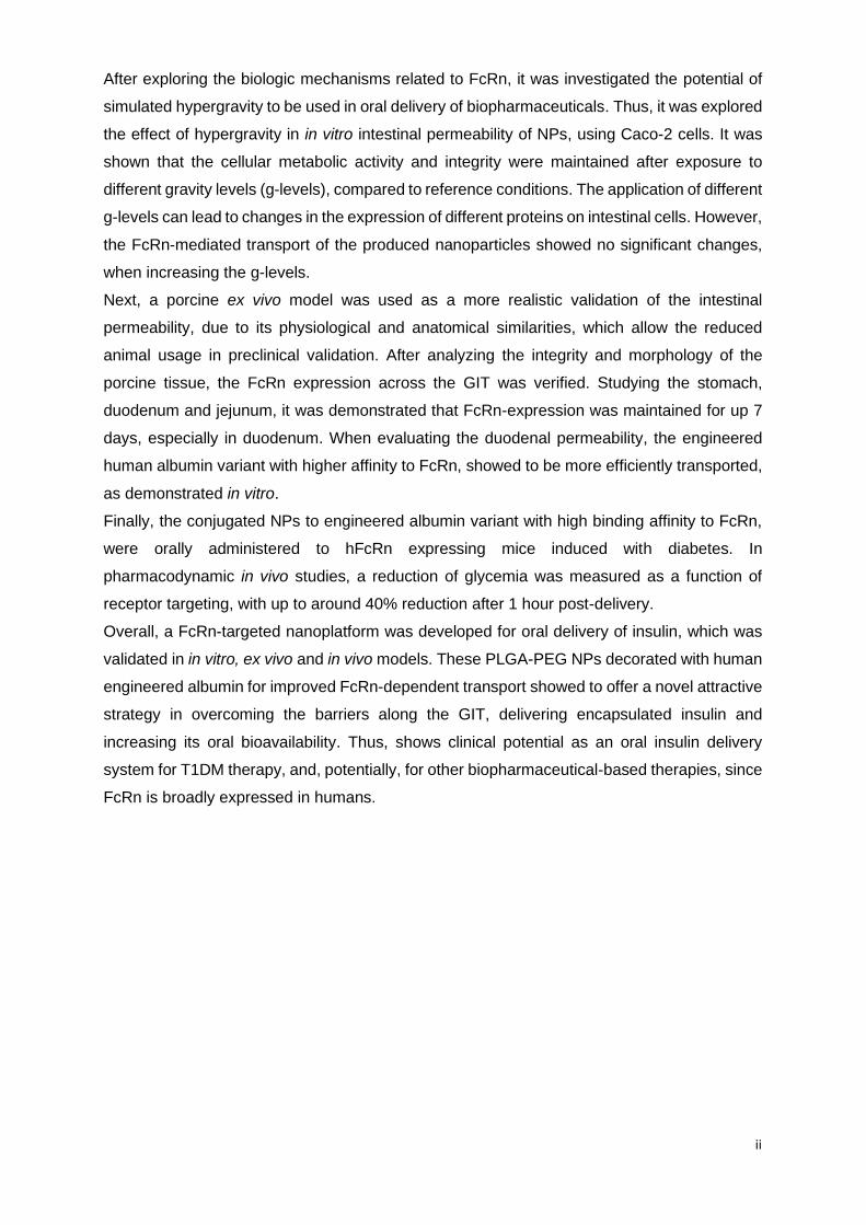
After exploring the biologic mechanisms related to FcRn, it was investigated the potential of
simulated hypergravity to be used in oral delivery of biopharmaceuticals. Thus, it was explored
the effect of hypergravity in in vitro intestinal permeability of NPs, using Caco-2 cells. It was
shown that the cellular metabolic activity and integrity were maintained after exposure to
different gravity levels (g-levels), compared to reference conditions. The application of different
g-levels can lead to changes in the expression of different proteins on intestinal cells. However,
the FcRn-mediated transport of the produced nanoparticles showed no significant changes,
when increasing the g-levels.
Next, a porcine ex vivo model was used as a more realistic validation of the intestinal
permeability, due to its physiological and anatomical similarities, which allow the reduced
animal usage in preclinical validation. After analyzing the integrity and morphology of the
porcine tissue, the FcRn expression across the GIT was verified. Studying the stomach,
duodenum and jejunum, it was demonstrated that FcRn-expression was maintained for up 7
days, especially in duodenum. When evaluating the duodenal permeability, the engineered
human albumin variant with higher affinity to FcRn, showed to be more efficiently transported,
as demonstrated in vitro.
Finally, the conjugated NPs to engineered albumin variant with high binding affinity to FcRn,
were orally administered to hFcRn expressing mice induced with diabetes. In
pharmacodynamic in vivo studies, a reduction of glycemia was measured as a function of
receptor targeting, with up to around 40% reduction after 1 hour post-delivery.
Overall, a FcRn-targeted nanoplatform was developed for oral delivery of insulin, which was
validated in in vitro, ex vivo and in vivo models. These PLGA-PEG NPs decorated with human
engineered albumin for improved FcRn-dependent transport showed to offer a novel attractive
strategy in overcoming the barriers along the GIT, delivering encapsulated insulin and
increasing its oral bioavailability. Thus, shows clinical potential as an oral insulin delivery
system for T1DM therapy, and, potentially, for other biopharmaceutical-based therapies, since
FcRn is broadly expressed in humans.
iii
RESUMO
Diabetes mellitus (DM) é uma doença metabólica e crônica prevalente. O tratamento atual da
DM tipo 1 (T1DM) é baseado na administração exógena subcutânea de fármacos peptídicos,
como a insulina. No entanto, a administração está associada a vias invasivas e dolorosas, o
que frequentemente ditam a baixa adesão do paciente, contribuindo para uma das principais
causas de mortalidade e morbidade. Para garantir a conveniência e conformidade dos
pacientes, a administração oral é a via preferida para a administração do medicamento.
Contudo, as condições adversas do trato gastrointestinal (GIT) e a barreira epitelial seletiva
comprometem a administração eficiente de fármacos peptídicos. Os desenvolvimentos da
nanomedicina, para a administração oral de fármacos, abriram novas perspetivas para
desenhar e sintetizar nanotransportadores eficientes e nanomateriais multifuncionais. Na
verdade, o potencial dos nanomateriais para ultrapassar as barreiras biológicas levou ao
desenvolvimento de plataformas capazes de melhorar a biodisponibilidade oral dos fármacos.
Uma estratégia para superar a barreira intestinal é a modulação das características físico-
químicas dos nanossistemas, como tamanho, carga e composição da superfície, a fim de ditar
a via de internalização, escapar aos lisossomas e interagir com o alvo. Particularmente, o
direcionamento de fármacos para recetores expressos nas células epiteliais, pode aumentar
a absorção e o transporte através das barreiras mucosas. Um desses recetores é o recetor
Fc neonatal (FcRn), que transporta naturalmente imunoglobulina G e albumina
bidirecionalmente através de células epiteliais polarizadas. Comparando os ligandos
endógenos direcionados ao FcRn, a albumina é descrita como sendo mais eficientemente
transportada através de células epiteliais que expressam FcRn e produzida de uma maneira
mais econômica, em comparação com a IgG.
O objetivo principal desta tese foi desenvolver nanopartículas (NPs) biodegradáveis e
mucodifusivas de poli(láctico-co-glicólico)-poli(etilenoglicol) (PLGA-PEG), funcionalizadas
com albumina como ligando do FcRn, para aumentar a biodisponibilidade oral de fármacos,
usando a insulina como fármaco modelo. Inicialmente, foi focada a otimização das
características físico-químicas dos NPs, em termos de tamanho de partícula e carga
superficial, eficiência de associação de insulina (EA), eficiência de conjugação de albumina
(CE), estrutura das proteínas e libertação do fármaco. Especificamente, as NPs foram
funcionalizadas com albumina humana, desenhada para melhorar a ligação dependente do
pH ao FcRn. As NPs produzidas tinham 150 nm de tamanho, eram monodispersas, tinham -
9 mV de zeta potencial, eram 10% (w/w) carregadas com insulina e a sua superfície teórica
foi 0.5% funcionalizada, com albumina humana. A estrutura da insulina foi afetada pelo
método de encapsulação, enquanto que a estrutura da albumina foi mantida após a
conjugação. Além disso, o nanossistema desenvolvido revelou uma libertação controlada e
prolongada, de insulina, com apenas 2.5% de libertação durante 8 horas. É importante
ressalvar que as NPs funcionalizadas com albumina ligaram-se favoravelmente ao hFcRn e

iv
mostraram um transporte aprimorado através das células epiteliais MCDK-hFcRn polarizadas.
Após a transcitose epitelial, foi demonstrado que as NPs podem ser absorvidas e recicladas
pelas células endoteliais HMEC1-hFcRn, com o mesmo padrão de ligação.
Após explorar os mecanismos biológicos relacionados com o FcRn, foi investigado o potencial
da hipergravidade simulada para ser utilizada na administração oral de fármacos. Assim, foi
explorado o efeito da hipergravidade na permeabilidade in vitro intestinal de NPs, usando as
células Caco-2. Foi demonstrado que a atividade metabólica e integridade celular foram
mantidas após exposição a diferentes níveis de gravidade (níveis de g), em comparação com
as condições de referência. A aplicação de diferentes níveis de g pode levar a alterações de
expressão de diferentes proteínas das células intestinais. Contudo, o transporte mediado por
FcRn das NPs produzidas, não apresentou alterações significativas, com o aumento dos
níveis de g.
Em seguida, um modelo ex vivo de porco foi utilizado como validação mais realista da
permeabilidade intestinal, devido às suas semelhanças fisiológicas e anatômicas, o que
permite diminuir o uso de animais na validação pré-clínica. Após análise da integridade e
morfologia do tecido suíno, foi verificada a expressão do FcRn no GIT. Estudando o
estômago, duodeno e jejuno, foi demonstrado que a expressão do FcRn foi mantida durante
7 dias, principalmente no duodeno. Ao avaliar a permeabilidade duodenal, a variante de
albumina humana com maior afinidade para FcRn, mostrou ser transportada de forma mais
eficiente, conforme demonstrado in vitro.
Finalmente, as NPs conjugadas à variante de albumina com maior afinidade para o FcRn,
foram administradas por via oral a ratinhos, induzidos com diabetes, que expressam hFcRn.
Em estudos de farmacodinâmica in vivo, uma redução da glicemia foi medida em função da
ligação ao recetor, com cerca de 40% de redução após 1 hora de administração.
De uma forma geral, uma nanoplataforma direcionada ao FcRn foi desenvolvida para a
administração oral de insulina, que foi validada em modelos in vitro, ex vivo e in vivo. Estas
NPs de PLGA-PEG decoradas com albumina humana modificada, para melhor transporte
dependente de FcRn, mostraram oferecer uma estratégia nova e atrativa para ultrapassar as
barreiras ao longo do GIT, entregando a insulina encapsulada e aumentando a sua
biodisponibilidade oral. Assim, o sistema de entrega de insulina oral mostra potencial clínico,
para terapia de DMT1 e potencialmente, para outras terapias, uma vez que FcRn é
amplamente expresso em humanos.
v
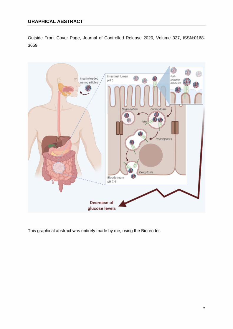
GRAPHICAL ABSTRACT
Outside Front Cover Page, Journal of Controlled Release 2020, Volume 327, ISSN:0168-
3659.
This graphical abstract was entirely made by me, using the Biorender.

vi
vii
LIST OF FIGURES
Figure 1 | Schematic representation of human insulin. It is formed by chain A with 21 a.a.,
chain B with 30 a.a., two disulfide bonds connecting the two chains and one disulfide linkage
within chain A.
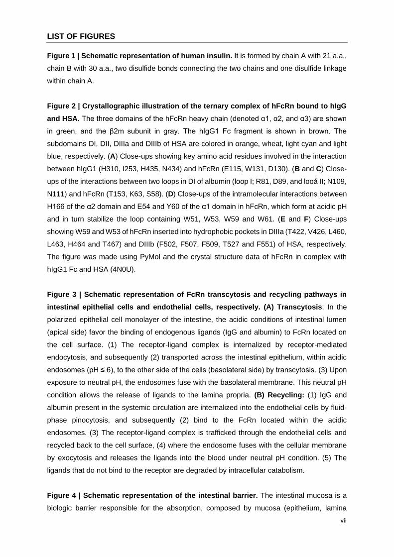
Figure 2 | Crystallographic illustration of the ternary complex of hFcRn bound to hIgG
and HSA. The three domains of the hFcRn heavy chain (denoted α1, α2, and α3) are shown
in green, and the β2m subunit in gray. The hIgG1 Fc fragment is shown in brown. The
subdomains DI, DII, DIIIa and DIIIb of HSA are colored in orange, wheat, light cyan and light
blue, respectively. (A) Close-ups showing key amino acid residues involved in the interaction
between hIgG1 (H310, I253, H435, N434) and hFcRn (E115, W131, D130). (B and C) Close-
ups of the interactions between two loops in DI of albumin (loop I; R81, D89, and looå II; N109,
N111) and hFcRn (T153, K63, S58). (D) Close-ups of the intramolecular interactions between
H166 of the α2 domain and E54 and Y60 of the α1 domain in hFcRn, which form at acidic pH
and in turn stabilize the loop containing W51, W53, W59 and W61. (E and F) Close-ups
showing W59 and W53 of hFcRn inserted into hydrophobic pockets in DIIIa (T422, V426, L460,
L463, H464 and T467) and DIIIb (F502, F507, F509, T527 and F551) of HSA, respectively.
The figure was made using PyMol and the crystal structure data of hFcRn in complex with
hIgG1 Fc and HSA (4N0U).
Figure 3 | Schematic representation of FcRn transcytosis and recycling pathways in
intestinal epithelial cells and endothelial cells, respectively. (A) Transcytosis: In the
polarized epithelial cell monolayer of the intestine, the acidic conditions of intestinal lumen
(apical side) favor the binding of endogenous ligands (IgG and albumin) to FcRn located on
the cell surface. (1) The receptor-ligand complex is internalized by receptor-mediated
endocytosis, and subsequently (2) transported across the intestinal epithelium, within acidic
endosomes (pH ≤ 6), to the other side of the cells (basolateral side) by transcytosis. (3) Upon
exposure to neutral pH, the endosomes fuse with the basolateral membrane. This neutral pH
condition allows the release of ligands to the lamina propria. (B) Recycling: (1) IgG and
albumin present in the systemic circulation are internalized into the endothelial cells by fluid-
phase pinocytosis, and subsequently (2) bind to the FcRn located within the acidic
endosomes. (3) The receptor-ligand complex is trafficked through the endothelial cells and
recycled back to the cell surface, (4) where the endosome fuses with the cellular membrane
by exocytosis and releases the ligands into the blood under neutral pH condition. (5) The
ligands that do not bind to the receptor are degraded by intracellular catabolism.
Figure 4 | Schematic representation of the intestinal barrier. The intestinal mucosa is a
biologic barrier responsible for the absorption, composed by mucosa (epithelium, lamina
viii
propria and muscularis mucosae), sub mucosa (lymph nodes, glands and blood vessels),
muscularis externa (smooth muscle) and serosa (mesothelium).
Figure 5 | Schematic illustration of in vitro models: (A) Caco-2 mono-culture model, (B)
Caco-2/HT29-MTX co-culture model, (C) Caco-2/ Raji B co-culture model, (D) Caco-2/HT29-
MTX/ Raji B co-culture model and (E) 3D co-culture model.
Figure 6 | The mechanically active human Gut Chip. (A) Human villus intestinal epithelium
and vascular endothelium are lined on opposite sides of a flexible porous membrane under
fluid flow and peristalsis-like strains. A zoom-in schematic shows the intestinal
microenvironment undergoing complex crosstalk between commensal gut microbiome,
bacterial pathogens, and immune cells in parenchymal and vascular channels, respectively.
(B) Villus morphogenesis of human Caco-2 intestinal epithelium in the Gut Chip under
physiologically controlled motions and flow. (C) An overlaid image of the coculture of green
fluorescent protein–labeled Escherichia coli and microengineered villi in the Gut Chip. Bars =
50 mm. Reprinted with permission from 137.
Figure 7 | Schematic representations of the four of the organ systems used for
functional coupling. (A) The intestinal module is constructed in Transwell® from primary
jejunum enteroids. Test agents are applied in the apical compartment (1). The media collected
in the basolateral compartment (2) is used to add to the liver. (B) Media from the jejunum
intestine basolateral compartment (2) is perfused as a 1:3 jejunum/naïve liver media into the
influx port of the SQL SAL liver model (3). Efflux media is collected (4) and used to add to two
downstream organ models. (C) The vascularized kidney proximal tubule module is a two
lumen, dual perfusion system. For the vascular compartment, jejunum/liver-conditioned media
(4) is diluted 1:2 or 1:4 with naïve EGM-2 media and then perfused into the influx port (5) to
collect effluent from the proximal tubule at (6). In parallel with perfusion through the vascular
compartment, the proximal tubule compartment is perfused with naïve DMEM/F12 PTEC
media (6) for effluent collection. (D) The blood-brain barrier with NVU is constructed in a
membrane-separated, two-chambered microfluidic device. The brain-derived endothelial
vascular compartment is perfused at the influx port (7) with jejunum/liver-conditioned media
(4) diluted 1:4 with naïve EGM-2 media. The effluent is collected at the efflux port (7). In parallel
with perfusion through the vascular compartment, the neuronal cell compartment is perfused
with naïve EBM-2 media at the influx port < 8> for effluent collection at (8). Reprinted with
permission from 152.
Figure 8 | Alignment of predicted porcine FcRn amino acid sequence (Sus scrofa,
NP_999362.2) with other species. The degree of homology is indicated by darker shading.
ix
Protein sequences are from GenBank (locus numbers: Human (Homo sapiens,
NP_001129491.1), Mouse (Mus musculus, NP_034319.2). Reprinted with permission from 171.
Figure 9 | Human FcRn transgenic mouse models for evaluation of IgG and albumin-
based therapeutics. Conventional C57BL/6J mice have been used to generate humanized
mouse strains that are transgenic for hFcRn. While (A) the Tg32 and Tg276 strains express
mIgG, (B) the Tg32-hFc strain expresses chimeric hIgG1 (chIgG1). In comparison to these
mouse strains, (C) Tg32 albumin knockout mice do not express MSA, while (D) the double
transgenic mouse model (AlbumusTM) have both hFcRn and HSA. The figure was created with
BioRender.
Figure 10 | Representation of the rationale of this study: (A) Illustration of the crystal
structure of human albumin where targeted residues are shown as spheres: the free cys34
(black) in the N-terminal domain I and K500 (orange), H510 (orange) and K573 (blue) in the
C-terminal domain III. The figure was made using Pymol and the crystal structure data of
human albumin (PDB 1AO6); (B) Schematic illustration of the designed NPs build up of PLGA
and PLGA-PEG-MAL with encapsulated soluble recombinant insulin and recombinant human
albumin on the NPs’ surface. Albumin was conjugated to the NP using maleimide chemistry,
which resulted in a thioether bond formed between the Cys34 of albumin and the maleimide
group of PLGA-PEG-MAL.
Figure 11 | Site-specific conjugation of albumin to NPs: (A) ELISA quantification of free
WT albumin detected after the washing step with Amicon® filter of 100 KDa; (B) Conjugation
efficiency (CE) with or without the presence of maleimide (MAL) after conjugation of 1 h or
overnight (ON) with WT albumin; (C) Conjugation efficiency (CE) of NPs conjugated with
reduced WT albumin (NP-WT) and non-reduced WT albumin (NP-WT without TCEP); (D)
Elementary analysis of native albumin, non-functionalized empty NPs (NPs-NF) and WT
albumin-conjugated empty NPs (NPs-WT); (E) Qualitative and (F) quantitative analysis
showing the similarity of the FTIR spectra between WT albumin and NPs-WT, using the area
overlap (AO) and spectral correlation coefficient (SCC) algorithms. Shown are the arithmetic
means ± SD. *P< 0.05, comparing with native albumin by Student’s t-test.
Figure 12 | Insulin NPs characterization: (A) Representative TEM microphotograph of WT
albumin decorated PLGA-PEG NPs encapsulated with insulin, lyophilized 25000x. Scale bar:
500 nm. (B) Insulin release profile of functionalized and non-functionalized NPs under SGF (2
h) and SIF (from 2 h to 6 h). (C) The qualitative and (D) quantitative effect of NPs encapsulation
on protein structure. Analysis showing area-normalized second-derivative amide I spectra of
native insulin vs insulin-encapsulated NPs using the area overlap (AO) and spectral correlation
x
coefficient (SCC) algorithms. Shown are the arithmetic means ± SD. **P<0.001, ***P<0.0001
comparing with native insulin by Student’s t-test.
Figure 13 | Albumin decorated NPs bind human FcRn. (A) A schematic illustration of the
ELISA set-up for measuring functional binding to human FcRn at pH 5.5. An engineered IgG1
antibody (MST/HN) was coated in wells, to which recombinant soluble human FcRn was
captured, before adding naked albumin or albumin-decorated NPs. Bound albumin and
albumin-decorated NPs were detected using ALP-conjugated polyclonal anti-human albumin
antibodies. (B) Binding of naked human albumin variants (WT, KAHQ or KP) to human FcRn
at pH 5.5. (C) Binding of NPs decorated with human albumin variants (WT, KAHQ or KP) to
human FcRn at pH 5.5.
Figure 14 | Albumin-decorated NPs showed enhanced transcytosis capacity. Relative
amounts of (A) naked albumin variants (WT, KAHQ and KP) and (B) albumin-decorated NPs
detected in media collected at the basolateral side of polarized epithelial MDCK-hFcRn cells
4 h after adding the variants to the apical side in the Transwell® system (n=5-9). The amounts
were quantified by two-way anti-albumin ELISA, where WT albumin or NPs decorated with WT
albumin were set to 1, as a control. Shown are the arithmetic means ± SD. **P<0.001,
***P<0.0001 comparing with WT or NPs-WT by one-way ANOVA Dunnett’s multiple
comparison test; n.a.: non applicable; ns: non-significant.
Figure 15 | HERA screening of engineered albumin variants and albumin-conjugated
NPs: (A) Relative uptake of naked albumin and albumin-conjugated NPs and (B) their
correspondent amount quantified by ELISA. (C) Relative recycling of naked albumin and
albumin-conjugated NPs and (D) their correspondent amount quantified by ELISA (n=9-11) in
HMEC1-hFcRn cells. Shown are the arithmetic means ± SD. *P<0.05, **P<0.005,
***P<0.0005, **** P< 0.0001 comparing with WT or NPs-WT, n.a.: non applicable, by one-way
ANOVA Dunnett’s multiple comparison test.
Figure 16 | Mean HERA score for naked albumin variants or conjugated to NPs. These
were calculated from the data shown in Figure 4.2 B and D.
Figure 17 | H&E stained sections of porcine GI tract. (A) Morphologic differences between
esophagus, stomach, duodenum, jejunum, ileum, caecum, colon and rectum (10X); (B)
Identification of duodenum sections, as mucosa, sub mucosa, muscularis externa and serosa
(20X); (C) Schematic representation of intestinal morphology and sections division (mucosa,
sub mucosa and muscularis externa).
xi
Figure 18 | Confirmation of FcRn gene expression across GI tract. Before performing the
RT-PCR, RNA was extracted from fresh tissue of different sections of GI tract and cDNA was
amplified. Results shown were performed with one tissue batch. Water and porcine blood were
included as negative and positive controls, respectively. Results shown were performed with
one tissue batch.
Figure 19 | Evaluation of FcRn expression over time, of stomach, duodenum and
jejunum. (A) Evaluation of FcRn expression by IHC of fresh porcine tissue. Control samples
were only treated with the secondary antibody, to check non-specific binding. Brown indicates
the antibody signal, while blue is the counterstain. (B) FcRn expression after 7 days of porcine
ex vivo incubation or freshly isolated at day zero. Before the RT-PCR, RNA was extracted from
fresh tissue and cDNA was amplified. Water and porcine blood were included as negative and
positive controls, respectively. Results shown were performed with one tissue batch.
Figure 20 | Engineered albumin showed enhanced transcytosis capacity in the porcine
ex vivo platform. (A) Representation showing porcine duodenum, as the tissue used to study
the intestinal permeability, using the porcine ex vivo platform. (B) Schematic illustration of the
cross-section of the interface design in a 96-well plate format. 100 µL of HBSS pH 7.4 was
added to the plate from the bottom, mimicking the basolateral side. Next, the tissue was put
on the plate, facing the apical side upwards. Finally, the magnetic plate from the top was
added, pressing the tissue to avoid leakage, and samples prepared in HBSS pH 6 were added.
(C) Relative amounts of naked albumin variants (WT, KAHQ, KP and PSA) detected in media
collected at the basolateral side, 4 h after adding the variants to the apical side in the porcine
ex vivo platform (n=6-10); (D) Relative amounts of NPs functionalized with albumin variants
(n=4-10). The amounts were quantified by two-way anti-albumin ELISA, where WT albumin
and NPs-WT were set to 1, as a control. These results were obtained from 3 different pigs.
Shown are the arithmetic means ± SD. ∗P < 0.05, ∗∗P < 0.005, and #P < 0.0001, comparing
with WT by one-way ANOVA Dunnett’s multiple comparison test.
Figure 21 | Glycemia in human FcRn expressing mice induced with diabetes. (A) T1DM-
induced Tg32 mice were i.p. administered with STZ (150 mg kg−1) to induce diabetes. Body
weight and glucose levels were followed up every 2 days, during 7 days. (B) At day 7,
formulations were administered and plasma glucose levels were analyzed at different time
points, during 8 h: PBS, oral insulin 50 IU/Kg, s.c. insulin 2.5 IU/Kg, oral insulin 50 IU/Kg loaded
and non-functionalized NPs (NP-NF) and oral insulin 50 IU/Kg loaded and albumin-
functionalized WT albumin and KP NPs (n = 3-6/ group). Shown are the arithmetic means ±
SEM. *P<0.05, ** P< 0.001 comparing with oral insulin by one-way ANOVA.
xii
Figure A1 | NMR analysis of all constituents from albumin-conjugated NPs loaded with
insulin: (A) HSA, (B) PLGA, (C) PLGA-PEG, (D) PLGA-PEG-MAL, (E) TCEP, (F) non-
functionalized NPs without maleimide, (G) non-functionalized NPs, (H) NPs functionalized with
non-reduced albumin, (I) albumin-functionalized NPs without maleimide and (J) albumin-
functionalized NPs.
Figure A2.1 | Experimental procedure of the intestinal permeability under different g-
levels; (A) Schematic illustration of cell culture and HG exposure process: Caco-2 cells were
cultured for 21 days, to allow their differentiation into enterocyte-like cells. After this, Ins-NPs-
Alb were incubated for 4 h, at different g-levels; (B) Schematic diagram for an example of HG
exposure at different time points (0.5, 1, 2, 3 and 4 h). During this rest period at 1 g, TEER
was measured and samples were removed from the basolateral side, for further quantification.
This set up was performed for 2.5 g, 5.0 g and 7.5 g using the LDC and 1.0 g using an orbital-
shaker, as control.
Figure A2.2 | Evaluation of metabolic activity of Caco-2 clone cells after HG exposure.
(A) Evaluation of cellular viability through resazurin assays; (B) Evaluation of cellular toxicity
through LDH assay. Shown are the arithmetic means ± SD. *P<0.05 comparing with 1 g, by
one-way analysis of variance (ANOVA) Dunnett’s multiple comparison test. N=6.
Figure A2.3 | Assessment and comparison of cellular integrity at different g-levels. (A)
Schematic illustration of the studied cellular components involved in different transport vias.
Visualization of (B) claudin-1 and MUC2; (C) ZO-1 and PEPT1, (D) actin and FcRn. Claudin-
1, ZO-1 and actin are presented in green. MUC2, PEPT1 and FcRn are presented in red.
Nuclei are blue.
Figure A2.4 | Overview sketch of the quantitative expression. Analysis of the expression
percentage of (A) transporters of influx and efflux; (B) components of tight junctions; (C)
cytoskeleton and (D) mucus production, after exposure to different g-levels. The quantitative
data are related to images presented at Figure 3. Quantification was done using ImageJ and
each sample was normalized to the reference condition.
Figure A2.5 | ORTO visualization of cellular components at different g-levels. Claudin-1,
ZO-1 and actin are presented in green. MUC2, PEPT1 and FcRn are presented in red. Nuclei
are blue.
Figure A2.6 | H&E staining to address the cellular integrity when exposing to different
g-levels. Ins-NPs-Alb were exposed to different g-levels (2.5 g, 5.0 g and 7.5 g) for 4 h. Cells
xiii
with no treatment and shaking at 1.0 g were included as controls. Cytoplasm stained with pink
and nucleus with purple. The Transwell® membrane is transparent and is right below the
cellular monolayer. Scale bar = 100 µm.
Figure A2.7 | Evaluation of the safety of Ins-NPs-Alb in Caco-2 clone cells. (A) Cellular
viability, assessed by the MTT assays and (B) cellular cytotoxicity assessed by the LDH assay,
of NPs loaded or not with insulin and conjugated or not with WT albumin. Free insulin was
included as a control.
Figure A2.8 | Intestinal permeability and respective TEER values of Ins-NPs-Alb after
HG exposure. (A) Schematic representation of FcRn-mediated transcytosis used by Ins-NPs-
Alb through intestinal epithelial cells; (B) Permeability percentage of Ins-NPs-Alb, quantified
by ELISA, and (C) respective TEER values during 4 h of incubation at different g-levels. Shown
are the arithmetic means ± SD. ∗P < 0.05 comparing with 1.0 g, by two-way analysis of
variance (ANOVA) Dunnett’s multiple comparison test. N=6 for all groups.
Figure A3.1 | Analysis of protein interaction using Bio-layer interferometry (BLI): (A)
screening of NPs concentrations; (B) regeneration test, (C) kinetic test of the three
formulations, against different FcRn concentrations, and (D) differences between shape and
intensity of the FcRn binding affinity with engineered albumin conjugated NPs. Relative binding
of (E) naked human albumin variants and (F) NPs decorated with human albumin variants to
human FcRn at pH 5.5.
Figure A3.2 | The relative transport of: (A) naked albumin variants (WT, KAHQ and
TNNEKP), and (B) albumin-decorated NPs detected in media collected at the basolateral side
of polarized epithelial MDCK-hFcRn cells 4 h after adding the variants to the apical side in the
Transwell® system (n=5-12). The amounts were quantified by ELISA, where WT albumin or
NPs decorated with WT albumin were set to 1, as a control. Shown are the arithmetic means
± SD. **P<0.005, ***P<0.0001 comparing with WT or NPs-WT by one-way ANOVA Dunnett’s
multiple comparison test.
Figure A3.3 | The relative uptake and recycling of: (A) naked albumin variants (WT, KAHQ
and TNNEKP), and (B) albumin-decorated NPs, and their correspondent amount quantified
by ELISA (n=9-15) in HMEC1-hFcRn cells. Shown are the arithmetic means ± SD.
***P<0.0005, **** P< 0.0001 comparing with WT or NPs-WT, by one-way ANOVA Dunnett’s
multiple comparison test.
Figure A3.4 | Biodistribution in vivo detection. (A) Qualitative analysis of the fluorescence,
xiv
after 30 min of administration of NPs with cy7.5 and respective blanks whithout cy7.5; and (B)
quantitative of the fluorescence present in mice, at different time points during 8 h. (C)
Qualitative and (D) quantitative analysis of the GIT after 2 h of administration. NPs without
cy7.5 were included as controls. All images were normalized by the control (same scale and
ROI window).
Figure A3.5 | Hypoglycemic effect of T1DM-induced mice following i.p. administration
of STZ (150 mg kg−1). (A) Plasma glucose levels and (B) Plasma insulin level following PBS,
oral insulin 50 IU/Kg, s.c. insulin 2.5 IU/Kg, non-functionalized NPs (NPs-NF) and
functionalized with KAHQ and TNNEKP (n = 5-6 animals).
xv
LIST OF TABLES
Table 1 | Oral insulin formulations for T1DM under clinical trials.
Table 2 | The influence of NPs characteristics on cellular uptake, internalization pathway and
their strategies.
Table 3 | Physical-chemical properties of developed PLGA-NPs.
Table 4 | Permeability percentage and apparent permeability coefficient calculated after 4 h
of incubation in MDCK-hFcRn cells.
Table 5 | Apparent permeability coefficient calculated after 4 h of incubation in ex vivo porcine,
based on the albumin amount quantified by ELISA. These results were obtained from 3
different pigs.
Table 6 | Pharmacodynamic parameters of formulations at different time points after
administration.
Table 7 | Pharmacodynamic parameters.
Table A3.1 | The pharmacokinetic parameters.

xvi
xvii
ACRONYMS AND ABBREVIATIONS LIST
1H NMR Proton nuclear magnetic resonance
a.a. Amino acid
AAC Area above the curve
ADMET Administration, distribution, metabolism, excretion and toxicity
AE Association efficiency
AO Area overlap
AUC Area under the curve
CE Conjugation efficiency
Cys Cystein
DAPI 4’, 6-diamidino-2-phenylindole, dihydrochloride
DL Drug loading
DLS Dynamic light scattering
DM Diabetes mellitus
DMEM Dulbecco's Modified Eagle’s Medium
EDS Elementary dispersive scanning
ELISA Enzyme-linked immunosorbent assay
ESA European Space Agency
ESTEC European Space Research and Technology Centre
FcRn Neonatal Fc receptor
FDA Food and Drug Administration
FITC Fluorescein isothiocyanate
FTIR Fourier Transform Infrared Spectroscopy
GIT Gastrointestinal tract
HSA Human serum albumin
HBSS Hank’s balanced salt solution
HD Hypoglycemic decrease
H&E Hematoxylin and eosin
HG Hypergravity
hFcRn Human FcRn
HPLC High performance liquid chromatography
i.p. Intraperitoneal
KAHQ K500A/H464Q
kDa kilodalton
KP K573P
LDC Large diameter centrifuge
xviii
LDH Lactate dehydrogenase
MAL Maleimide
MDCK-hFcRn Madin-Darby Canine Kidney cell line over-expressing human FcRn
MES 2-(N-morpholino) ethanesulfonic acid
mFcRn Mouse FcRn
MTT Triazolyl blue tetrazolium bromide
NMR Nuclear magnetic resonance
NPs Nanoparticles
PA Pharmacologic availability
Papp Apparent permeability
PB Phosphate buffer
PBSTM PB saline with 0.05% (v/v) of Tween 20 and 4% skimmed milk
PD Pharmacodynamics
PdI Polydispersity index
PEG Polyethylene glycol
PEPT-1 Peptide transporter 1
pFcRn Porcine FcRn
PK Pharmacokinetics
PLGA Poly(lactic-co-glycolic acid)
PSA Porcine serum albumin
RT Room temperature
RT-PCR Reverse transcription polymerase chain reaction
s.c. Subcutaneous
SCC Spectral correlation coefficient
SGF Simulated gastric fluid
SIF Simulated intestinal fluid
SD Standard deviation
TCEP Tris(2-carboxyethyl)phosphine hydrochloride
TEER Transepithelial electrical resistance
TEM Transmission electron microscopy
TJ Tight junction
T1DM Type 1 diabetes mellitus
STZ Streptozotocin
WT Wild type
ZO-1 Zona occludens 1
ZP Zeta potential

1
Chapter I
LITERATURE REVIEW

2
3
This chapter was based in the following publications:
• Azevedo C.*, Pinto S.*, Benjakul S.*, Santos H.A., Andersen J.T., Traverso G.,
Sarmento S., Prevention of diabetes-associated fibrosis: Strategies to orally treat
diabetes with FcRn-targeted nanosystems, Advanced Drug Delivery Reviews 2021.
(*equal contribution)
In this review paper, I was responsible for the conception and execution of half of the
contained information. The other half was complemented by Soraia Pinto. Sopisa
Benjakul was responsible for reorganizing the topics and improve the discussion and
images. The supervisors were responsible for reviewing and submit the manuscript. This
review was not previously included in other thesis and it is partially reproduced in this
section. The integral version of the review paper was included at the end of the thesis,
at Appendix IV.
• Almeida A.*, Azevedo C.*, Macedo M.H.*, Sarmento B., 3D intestinal models
towards a more realistic permeability screening, in Santos H. and Martins J.P.,
Nanotechnology for Oral Drug Delivery, 2020, Elsevier, ISBN 9780128180389.
(*equal contribution)
In this book chapter, I was responsible for the conception and execution of the gut-on-a-
chip’s section. Andreia Almeida was responsible for the organoids’ section and Helena
Macedo for the multilayered models’ section. The rest of the book chapter was written
and revised by all of us. The supervisor was responsible for reviewing and submit the
manuscript. This review was not previously included in other thesis and it is partially
reproduced in this section.
• Azevedo C., Pereira I., Sarmento B., Intestinal mucosal tissue models to validate
functionalized nanosystems, in Fahr A., et al., Characterization of Pharmaceutical
Nano- and Microsystems, Wiley 2020, ISBN: 978-1-119-41404-9.
In this book chapter, I was responsible for the conception, execution and revision of the
manuscript. Inês Pereira helped writing the ex vivo’s section and the supervisor was
4
responsible for reviewing and submit the manuscript. This review was not previously
included in other thesis and it is partially reproduced in this section.
• Azevedo C., Macedo M. H., Sarmento B., Strategies for enhanced intracellular
delivery of nanomaterials, Drug Discovery Today 2018, 23(5):944-959, DOI:
10.1016/j.drudis.2017.08.011.
In this review paper, I was responsible for the conception, execution and revision of the
manuscript. Helena Macedo revised the manuscript and the supervisor was responsible
for reviewing and submit the manuscript. This review was not previously included in other
thesis and it is partially reproduced in this section. The integral version of the review
paper was included at the end of the thesis, at Appendix IV.
5
1. Introduction
1.1. Epidemiology and the biology behind the disease
Diabetes mellitus (DM) is a major worldwide health burden. Around 463 million people
were living with diabetes worldwide by 2019, and this number will rise to 700 million by
2045 if current trends are maintained 1. In addition, it is estimated that the full global costs
of diabetes in adults will increase from U.S. $1.3 trillion in 2015 to $2.1 trillion by 2030 2.
There are three main types of DM: type I diabetes mellitus (T1DM), type II diabetes
mellitus (T2DM) and gestational diabetes. Also, intermediate conditions, such as
impaired glucose tolerance and impaired fasting glycemia can be considered, since they
can progress into diabetes 3. T1DM is caused by an autoimmune reaction, characterized
by the attack of the immune system to the pancreatic insulin-producing beta cells.
Commonly, it is a childhood-onset disease and patients are insulin dependent. In this
case, patients have to chronically administer multiple daily injections, which is painful,
invasive and reduces patient’s compliance 4. In T2DM, hyperglycemia is initially the result
of the inability of cells to fully respond to insulin and, over time, to inadequate production
of insulin as a result of failure of the pancreatic beta cells. T2DM, predominant in adults,
also implies recurrent administration of GLP-1 incretin analogs by invasive routes 5.
1.2. Oral insulin delivery for T1DM
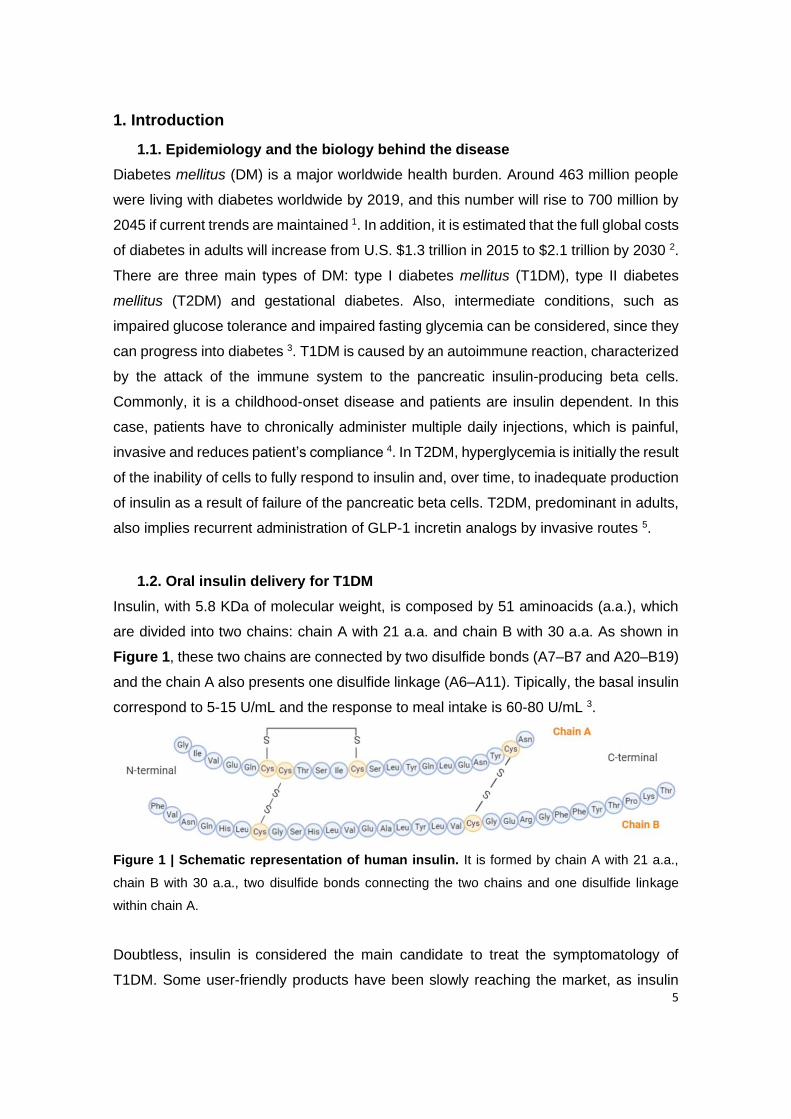
Insulin, with 5.8 KDa of molecular weight, is composed by 51 aminoacids (a.a.), which
are divided into two chains: chain A with 21 a.a. and chain B with 30 a.a. As shown in
Figure 1, these two chains are connected by two disulfide bonds (A7–B7 and A20–B19)
and the chain A also presents one disulfide linkage (A6–A11). Tipically, the basal insulin
correspond to 5-15 U/mL and the response to meal intake is 60-80 U/mL 3.
Figure 1 | Schematic representation of human insulin. It is formed by chain A with 21 a.a.,
chain B with 30 a.a., two disulfide bonds connecting the two chains and one disulfide linkage
within chain A.
Doubtless, insulin is considered the main candidate to treat the symptomatology of
T1DM. Some user-friendly products have been slowly reaching the market, as insulin
6
patch-pumps systems as well as inhaled and buccal insulin. However, these are currently
used for T2DM or do not have an advantageous bioavailability, compared to the current
subcutaneous (s.c.) administration. Lately, a lot of investigation has been focused on
oral delivery of insulin for T1DM, as the most preferable route of administration. In fact,
the s.c. insulin do not mimic a physiologic situation, since insulin is delivered to peripheral
circulation. In an oral delivery situation, insulin is absorbed in the intestinal lumen and
transported to the liver via portal circulation, where can avoid the hepatic glucose
production. This biodistribution was already studied elsewhere 6. The liver is the primary
site to regulate glucose homeostasis and it is known that 80% of the oral insulin is
retained in it and the rest reaches systemic circulation, creating a 3-fold higher insulin
concentration in portal vein than in systemic circulation. However, in case of s.c., insulin
is obviously more concentrated in peripheral circulation, which disrupt the liver’s balance
between glycogen storage and glucose output. This unbalanced state drives to
hiperglycemia, which can easily lead to hypoglycemia when treated with high insulin
doses 7. Insulin has a hydrophilic nature, so the paracellular route is the main way to
surpass the intestinal epithelium. However, as happens with other proteins, insulin can
be degraded throughout the gastrointestinal tract (GIT), reducing the oral bioavailability.
In this sense, absorption enhancers and protease inhibitors are currently used as
strategies to enhance insulin bioavailability. However, these can be a concern regarding
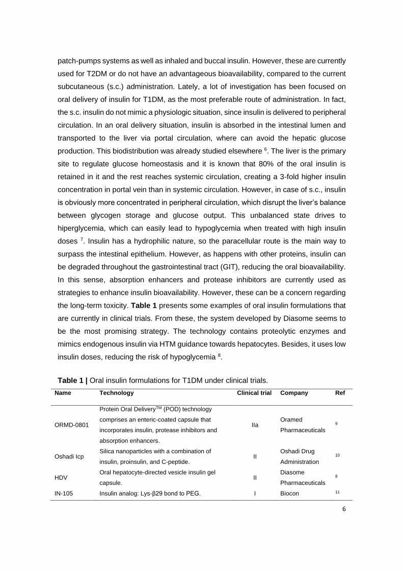
the long-term toxicity. Table 1 presents some examples of oral insulin formulations that
are currently in clinical trials. From these, the system developed by Diasome seems to
be the most promising strategy. The technology contains proteolytic enzymes and
mimics endogenous insulin via HTM guidance towards hepatocytes. Besides, it uses low
insulin doses, reducing the risk of hypoglycemia 8.
Table 1 | Oral insulin formulations for T1DM under clinical trials.
Name Technology Clinical trial Company Ref
ORMD-0801
Protein Oral DeliveryTM (POD) technology
comprises an enteric-coated capsule that
incorporates insulin, protease inhibitors and
absorption enhancers.
IIa Oramed
Pharmaceuticals
9
Oshadi Icp Silica nanoparticles with a combination of
insulin, proinsulin, and C-peptide. II
Oshadi Drug
Administration
10
HDV Oral hepatocyte-directed vesicle insulin gel
capsule. II
Diasome
Pharmaceuticals
8
IN-105 Insulin analog: Lys-β29 bond to PEG. I Biocon 11
7
1.3. Biologic barriers of oral delivery drugs
During drug design for drug delivery, some details should be taken into account: (1)
drugs must be able to surpass the harsh conditions of GIT; (2) once internalized, they
must reach the target; and (3) guarantee that pharmacokinetics is maintained.
Drugs can be administered by ocular, buccal, sublingual, oral, intravenous,
intramuscular, subcutaneous, transdermal, pulmonary/nasal or vaginal/rectal routes,
being the oral the most convenient 12. However, for orally delivering, the preferred route,
they have to pass through: the oral cavity coated by proteins, mucosal compounds and
bacterial flora; the stomach, and be attacked by acid bath; and the intestine and contact
with the alkaline environment, being subjected to the activity of different digestive
enzymes, the presence of a mucus layer (glycoprotein mucin) and tight junctions. After
being absorbed, drugs pass through endothelial cells, and reach the blood, where can
be taken up by a monocyte. Meanwhile, and in case of intracellular delivery, drugs travel
through endosomal and lysosomal environments, which might contribute for drug
degradation, until reach the target 13.
Taking all this in mind, it is mandatory that nanomaterials surpass the mononuclear
phagocyte system (MPS), avoid nonspecific distribution, drug resistance, escape to
lysosomes and reach the target with the original bioavailability 14.
2. Nanoparticles as a strategy for drug delivery
Nanoparticles (NPs) are seen as carriers that protect peptides from degradation and
deliver active payloads more efficiently to the intended sites within the GIT, ultimately
potentiating the oral bioavailability.
2.1. The evolution of Nanomedicine
The developments in nanomedicine drug delivery have opened new perspectives to
design and synthesize efficient nanocarriers and multifunctional nanomaterials. At the
beginning, the production was focused on biocompatibility and toxicity. The second-
generation of nanomaterials aimed for having an optimized surface, to give more
stability, stealth, and targeting capabilities. The most recent model support the “smart
nanomedicine” idea, improving targeting mechanisms and theranostic abilities 15. In fact,
nanomaterials provide a stable biocompatible environment to encapsulate drugs,
promote a controlled release and an efficient absorption 16, 17. It is also noted that they
8
improve the therapeutic effects duration and minimize unwanted effects by driving drugs
and increasing their concentration in the site of action 18, 19. Depending on the
accumulation of the delivery system in the tissue, cell or in a specific subcellular
compartment of interest, targeting approaches can be primary, secondary or tertiary,
respectively 20. This nanotechnological advance became very important since it allows
drugs to cross physiological barriers to reach target sites, safely and sustainably 21.
2.2. Nanoparticles characteristics and their influence
Internalization and intracellular transport mechanisms are affected by nanomaterials
properties. Different characteristics (size, charge, shape, elasticity) contribute to a
different interaction between the nanomaterial and the cell membrane, causing altered
signaling events and activation of different pathways of adhesion and internalization.
Thus, the desired result, for more effective and distinctly targeted delivery, can be
obtained controlling these parameters, which are important to attain the target and
specific activity at a predetermined rate and time 12, 15.
Size
Size is the priority parameter because it determines the endocytosis pathway and also
states which cells uptake the particle. Particles smaller than 10 nm are mainly removed
by renal clearance 22. Particles, when orally administered, with a size between 10-100
nm have higher cellular uptake efficiency, since they are taken up in Peyer’s patches,
and then absorbed into systemic circulation, avoiding rapid renal clearance. On the other
hand, larger particles are removed by MPS, and those within 200–300 nm cannot even
pass, being concentrated in the spleen 23, 24. In internalization, larger particles (>1µm)
enter the cell by macropinocytosis; with 120 nm by clathrin-dependent pathway; those
with 50–100 nm by caveolae-dependent pathway and finally smaller particles (<50 nm)
can be internalized through clathrin- and caveolae-independent pathway. So the
internalized nanomaterials are intimately connected with the size of vacuole formed 25.
Surface composition
Nanomaterials surface is also an important aspect, since its composition is in contact
with the cell, which can be affected by hydrophobicity or hydrophilicity. So, this parameter
determines the cellular internalization and intracellular transport and influences the
circulation time and nanocarrier association with cells and their recognition. Hydrophobic
particles can be delivered to immune cells and penetrate into the bilayer membrane,
9
being easily internalized 26, 27. Normally, polymers are added to nanomaterials to turn
their surface more inert, hydrophilic, avoid immune clearance and increase the blood
circulation time. PEGylation is a strategy to make nanomaterials invisible to
macrophages or phagocytes and to prolong half-life 28. In this case, polyethylene glycol
(PEG) chains are covalently conjugated to nanomaterials, conferring a hydrophilic nature
29. PEG can capture water molecules and form a natural barrier around the nanomaterial
hindering proteins from adsorption, usually known as the “stealth” effect 30.
Charge
Also surface charge can be manipulated. For example, intestinal cell membrane is
negatively charged, as plasmatic membrane, due to phospholipids, so positively charged
nanomaterials interact more strongly, enhancing internalization 31. A typical example is
polyethyleneimine (PEI), which is commonly used to permeate membranes and let
nanomaterials enter 32. In terms of transport, positively charged particles utilize clathrin
and macropinocytosis 33 while negatively charged particles tend to utilize caveolae
mediated pathway 34, 35. Neutral or slightly negatively charged particles are considered
the best for long circulation, since they are not susceptible to MPS. On the other hand,
charges highly positive or negative are taken up by macrophages 35-37.
Shape
Shape can also dictate the path of the particle and, inclusively, can change over time or
after receiving external stimuli for long circulation and surpass barriers. It is an important
factor in flow margination, avoidance of immune system and tissue accumulation.
Spherical particles are better internalized by macrophages, while asymmetrical
nanomaterials are more advantageous for sensing, self-assembly, tissue engineering,
immunoengineering, therapeutic and diagnostic delivery 15, 26. One of the strategies is to
create elliptical particles to escape macrophages and then, after a stimuli, change to a
sphere form for a better internalization 25, 38.
Elasticity and solubility
Finally, increasing elasticity also increases blood residence time and avoids clearance
by immune system. Soft nanomaterials use macropinocytosis while harder ones are
internalized through clathrin-dependent mechanisms 26, 39.
It is also popular chemically modify nanomaterials to change the fluidity and/or solubility
of mucus, regarding oral administration. According to literature it is possible to add fatty
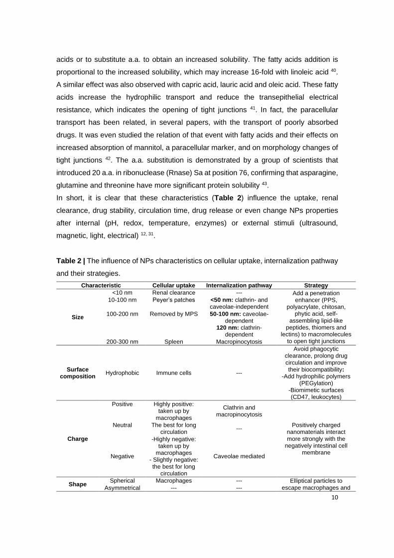
10
acids or to substitute a.a. to obtain an increased solubility. The fatty acids addition is
proportional to the increased solubility, which may increase 16-fold with linoleic acid 40.
A similar effect was also observed with capric acid, lauric acid and oleic acid. These fatty
acids increase the hydrophilic transport and reduce the transepithelial electrical
resistance, which indicates the opening of tight junctions 41. In fact, the paracellular
transport has been related, in several papers, with the transport of poorly absorbed
drugs. It was even studied the relation of that event with fatty acids and their effects on
increased absorption of mannitol, a paracellular marker, and on morphology changes of
tight junctions 42. The a.a. substitution is demonstrated by a group of scientists that
introduced 20 a.a. in ribonuclease (Rnase) Sa at position 76, confirming that asparagine,
glutamine and threonine have more significant protein solubility 43.
In short, it is clear that these characteristics (Table 2) influence the uptake, renal
clearance, drug stability, circulation time, drug release or even change NPs properties
after internal (pH, redox, temperature, enzymes) or external stimuli (ultrasound,
magnetic, light, electrical) 12, 31.
Table 2 | The influence of NPs characteristics on cellular uptake, internalization pathway
and their strategies.
Characteristic Cellular uptake Internalization pathway Strategy
Size
<10 nm Renal clearance --- Add a penetration enhancer (PPS,
polyacrylate, chitosan, phytic acid, self-
assembling lipid-like peptides, thiomers and
lectins) to macromolecules to open tight junctions
10-100 nm Peyer’s patches <50 nm: clathrin- and caveolae-independent
100-200 nm Removed by MPS 50-100 nm: caveolae-dependent
120 nm: clathrin-dependent
200-300 nm Spleen Macropinocytosis
Surface composition
Hydrophobic Immune cells ---
Avoid phagocytic clearance, prolong drug circulation and improve their biocompatibility:
-Add hydrophilic polymers (PEGylation)
-Biomimetic surfaces (CD47, leukocytes)
Charge
Positive Highly positive: taken up by
macrophages
Clathrin and macropinocytosis
Positively charged nanomaterials interact more strongly with the
negatively intestinal cell membrane
Neutral The best for long circulation
---
Negative
-Highly negative: taken up by
macrophages - Slightly negative: the best for long
circulation
Caveolae mediated
Shape Spherical Macrophages --- Elliptical particles to
escape macrophages and Asymmetrical --- ---
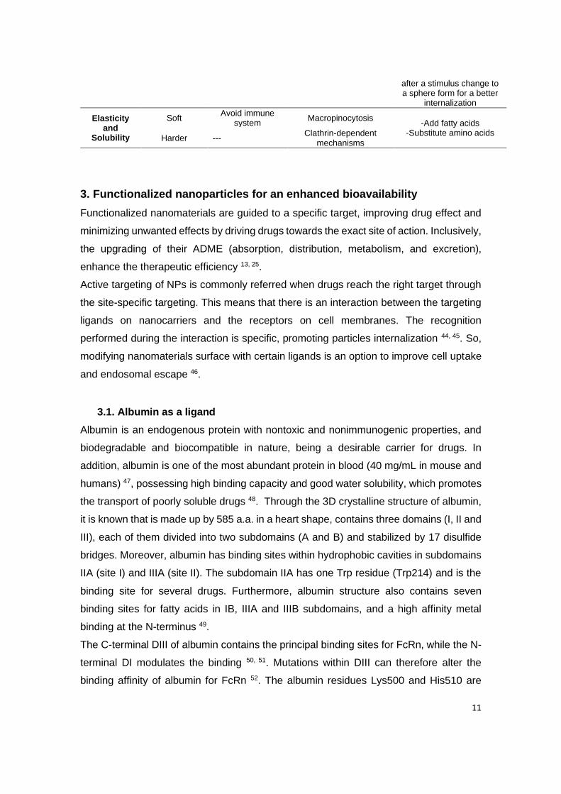
11
after a stimulus change to a sphere form for a better
internalization
Elasticity and
Solubility
Soft Avoid immune
system Macropinocytosis
-Add fatty acids -Substitute amino acids
Harder --- Clathrin-dependent
mechanisms
3. Functionalized nanoparticles for an enhanced bioavailability
Functionalized nanomaterials are guided to a specific target, improving drug effect and
minimizing unwanted effects by driving drugs towards the exact site of action. Inclusively,
the upgrading of their ADME (absorption, distribution, metabolism, and excretion),
enhance the therapeutic efficiency 13, 25.
Active targeting of NPs is commonly referred when drugs reach the right target through
the site-specific targeting. This means that there is an interaction between the targeting
ligands on nanocarriers and the receptors on cell membranes. The recognition
performed during the interaction is specific, promoting particles internalization 44, 45. So,
modifying nanomaterials surface with certain ligands is an option to improve cell uptake
and endosomal escape 46.
3.1. Albumin as a ligand
Albumin is an endogenous protein with nontoxic and nonimmunogenic properties, and
biodegradable and biocompatible in nature, being a desirable carrier for drugs. In
addition, albumin is one of the most abundant protein in blood (40 mg/mL in mouse and
humans) 47, possessing high binding capacity and good water solubility, which promotes
the transport of poorly soluble drugs 48. Through the 3D crystalline structure of albumin,
it is known that is made up by 585 a.a. in a heart shape, contains three domains (I, II and
III), each of them divided into two subdomains (A and B) and stabilized by 17 disulfide
bridges. Moreover, albumin has binding sites within hydrophobic cavities in subdomains
IIA (site I) and IIIA (site II). The subdomain IIA has one Trp residue (Trp214) and is the
binding site for several drugs. Furthermore, albumin structure also contains seven
binding sites for fatty acids in IB, IIIA and IIIB subdomains, and a high affinity metal
binding at the N-terminus 49.
The C-terminal DIII of albumin contains the principal binding sites for FcRn, while the N-
terminal DI modulates the binding 50, 51. Mutations within DIII can therefore alter the
binding affinity of albumin for FcRn 52. The albumin residues Lys500 and His510 are
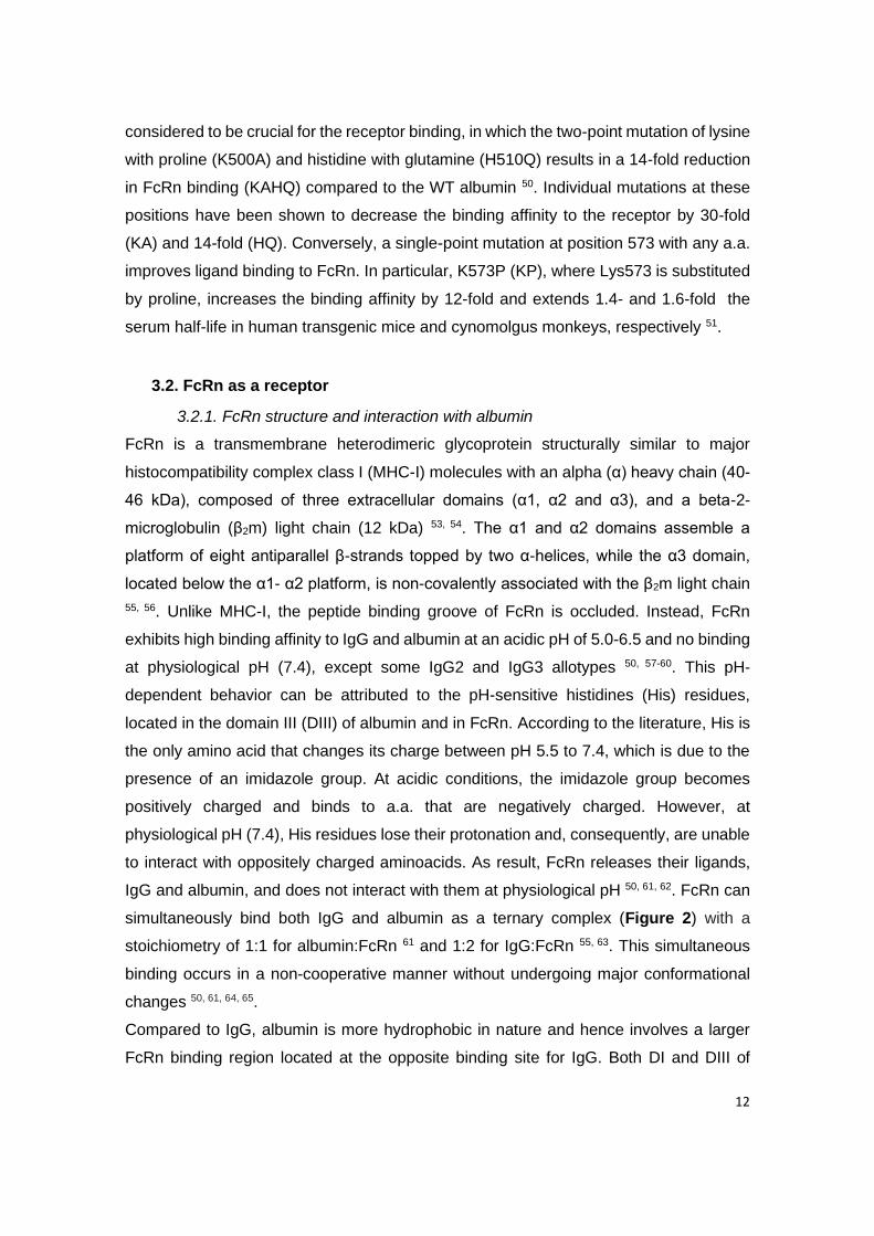
12
considered to be crucial for the receptor binding, in which the two-point mutation of lysine
with proline (K500A) and histidine with glutamine (H510Q) results in a 14-fold reduction
in FcRn binding (KAHQ) compared to the WT albumin 50. Individual mutations at these
positions have been shown to decrease the binding affinity to the receptor by 30-fold
(KA) and 14-fold (HQ). Conversely, a single-point mutation at position 573 with any a.a.
improves ligand binding to FcRn. In particular, K573P (KP), where Lys573 is substituted
by proline, increases the binding affinity by 12-fold and extends 1.4- and 1.6-fold the
serum half-life in human transgenic mice and cynomolgus monkeys, respectively 51.
3.2. FcRn as a receptor
3.2.1. FcRn structure and interaction with albumin
FcRn is a transmembrane heterodimeric glycoprotein structurally similar to major
histocompatibility complex class I (MHC-I) molecules with an alpha (α) heavy chain (40-
46 kDa), composed of three extracellular domains (α1, α2 and α3), and a beta-2-
microglobulin (β2m) light chain (12 kDa) 53, 54. The α1 and α2 domains assemble a
platform of eight antiparallel β-strands topped by two α-helices, while the α3 domain,
located below the α1- α2 platform, is non-covalently associated with the β2m light chain
55, 56. Unlike MHC-I, the peptide binding groove of FcRn is occluded. Instead, FcRn
exhibits high binding affinity to IgG and albumin at an acidic pH of 5.0-6.5 and no binding
at physiological pH (7.4), except some IgG2 and IgG3 allotypes 50, 57-60. This pH-
dependent behavior can be attributed to the pH-sensitive histidines (His) residues,
located in the domain III (DIII) of albumin and in FcRn. According to the literature, His is
the only amino acid that changes its charge between pH 5.5 to 7.4, which is due to the
presence of an imidazole group. At acidic conditions, the imidazole group becomes
positively charged and binds to a.a. that are negatively charged. However, at
physiological pH (7.4), His residues lose their protonation and, consequently, are unable
to interact with oppositely charged aminoacids. As result, FcRn releases their ligands,
IgG and albumin, and does not interact with them at physiological pH 50, 61, 62. FcRn can
simultaneously bind both IgG and albumin as a ternary complex (Figure 2) with a
stoichiometry of 1:1 for albumin:FcRn 61 and 1:2 for IgG:FcRn 55, 63. This simultaneous
binding occurs in a non-cooperative manner without undergoing major conformational
changes 50, 61, 64, 65.
Compared to IgG, albumin is more hydrophobic in nature and hence involves a larger
FcRn binding region located at the opposite binding site for IgG. Both DI and DIII of
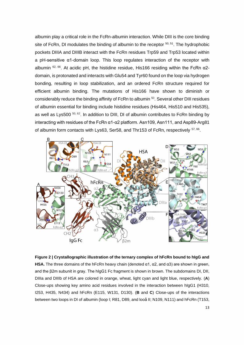
13
albumin play a critical role in the FcRn-albumin interaction. While DIII is the core binding
site of FcRn, DI modulates the binding of albumin to the receptor 50, 51. The hydrophobic
pockets DIIIA and DIIIB interact with the FcRn residues Trp59 and Trp53 located within
a pH-sensitive α1-domain loop. This loop regulates interaction of the receptor with
albumin 62, 66. At acidic pH, the histidine residue, His166 residing within the FcRn α2-
domain, is protonated and interacts with Glu54 and Tyr60 found on the loop via hydrogen
bonding, resulting in loop stabilization, and an ordered FcRn structure required for
efficient albumin binding. The mutations of His166 have shown to diminish or
considerably reduce the binding affinity of FcRn to albumin 50. Several other DIII residues
of albumin essential for binding include histidine residues (His464, His510 and His535),
as well as Lys500 50, 62. In addition to DIII, DI of albumin contributes to FcRn binding by
interacting with residues of the FcRn α1-α2 platform. Asn109, Asn111, and Asp89-Arg81
of albumin form contacts with Lys63, Ser58, and Thr153 of FcRn, respectively 57, 66.
Figure 2 | Crystallographic illustration of the ternary complex of hFcRn bound to hIgG and
HSA. The three domains of the hFcRn heavy chain (denoted α1, α2, and α3) are shown in green,
and the β2m subunit in gray. The hIgG1 Fc fragment is shown in brown. The subdomains DI, DII,
DIIIa and DIIIb of HSA are colored in orange, wheat, light cyan and light blue, respectively. (A)
Close-ups showing key amino acid residues involved in the interaction between hIgG1 (H310,
I253, H435, N434) and hFcRn (E115, W131, D130). (B and C) Close-ups of the interactions
between two loops in DI of albumin (loop I; R81, D89, and looå II; N109, N111) and hFcRn (T153,
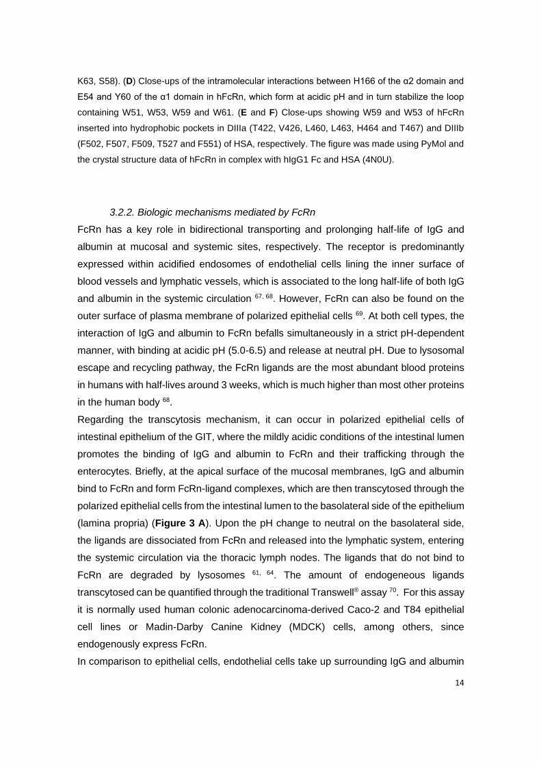
14
K63, S58). (D) Close-ups of the intramolecular interactions between H166 of the α2 domain and
E54 and Y60 of the α1 domain in hFcRn, which form at acidic pH and in turn stabilize the loop
containing W51, W53, W59 and W61. (E and F) Close-ups showing W59 and W53 of hFcRn
inserted into hydrophobic pockets in DIIIa (T422, V426, L460, L463, H464 and T467) and DIIIb
(F502, F507, F509, T527 and F551) of HSA, respectively. The figure was made using PyMol and
the crystal structure data of hFcRn in complex with hIgG1 Fc and HSA (4N0U).
3.2.2. Biologic mechanisms mediated by FcRn
FcRn has a key role in bidirectional transporting and prolonging half-life of IgG and
albumin at mucosal and systemic sites, respectively. The receptor is predominantly
expressed within acidified endosomes of endothelial cells lining the inner surface of
blood vessels and lymphatic vessels, which is associated to the long half-life of both IgG
and albumin in the systemic circulation 67, 68. However, FcRn can also be found on the
outer surface of plasma membrane of polarized epithelial cells 69. At both cell types, the
interaction of IgG and albumin to FcRn befalls simultaneously in a strict pH-dependent
manner, with binding at acidic pH (5.0-6.5) and release at neutral pH. Due to lysosomal
escape and recycling pathway, the FcRn ligands are the most abundant blood proteins
in humans with half-lives around 3 weeks, which is much higher than most other proteins
in the human body 68.
Regarding the transcytosis mechanism, it can occur in polarized epithelial cells of
intestinal epithelium of the GIT, where the mildly acidic conditions of the intestinal lumen
promotes the binding of IgG and albumin to FcRn and their trafficking through the
enterocytes. Briefly, at the apical surface of the mucosal membranes, IgG and albumin
bind to FcRn and form FcRn-ligand complexes, which are then transcytosed through the
polarized epithelial cells from the intestinal lumen to the basolateral side of the epithelium
(lamina propria) (Figure 3 A). Upon the pH change to neutral on the basolateral side,
the ligands are dissociated from FcRn and released into the lymphatic system, entering
the systemic circulation via the thoracic lymph nodes. The ligands that do not bind to
FcRn are degraded by lysosomes 61, 64. The amount of endogeneous ligands
transcytosed can be quantified through the traditional Transwell® assay 70. For this assay
it is normally used human colonic adenocarcinoma-derived Caco-2 and T84 epithelial
cell lines or Madin-Darby Canine Kidney (MDCK) cells, among others, since
endogenously express FcRn.
In comparison to epithelial cells, endothelial cells take up surrounding IgG and albumin
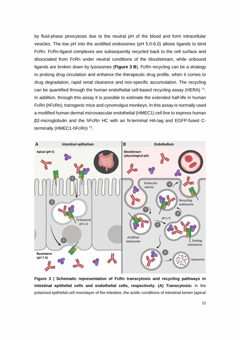
15
by fluid-phase pinocytosis due to the neutral pH of the blood and form intracellular
vesicles. The low pH into the acidified endosomes (pH 5.0-6.0) allows ligands to bind
FcRn. FcRn-ligand complexes are subsequently recycled back to the cell surface and
dissociated from FcRn under neutral conditions of the bloodstream, while unbound
ligands are broken down by lysosomes (Figure 3 B). FcRn recycling can be a strategy
to prolong drug circulation and enhance the therapeutic drug profile, when it comes to
drug degradation, rapid renal clearance and non-specific accumulation. The recycling
can be quantified through the human endothelial cell-based recycling assay (HERA) 71.
In addition, through this assay it is possible to estimate the extended half-life in human
FcRn (hFcRn), transgenic mice and cynomolgus monkeys. In this assay is normally used
a modified human dermal microvascular endothelial (HMEC1) cell line to express human
β2-microglobulin and the hFcRn HC with an N-terminal HA-tag and EGFP-fused C-
terminally (HMEC1-hFcRn) 72.
Figure 3 | Schematic representation of FcRn transcytosis and recycling pathways in
intestinal epithelial cells and endothelial cells, respectively. (A) Transcytosis: In the
polarized epithelial cell monolayer of the intestine, the acidic conditions of intestinal lumen (apical
16
side) favor the binding of endogenous ligands (IgG and albumin) to FcRn located on the cell
surface. (1) The receptor-ligand complex is internalized by receptor-mediated endocytosis, and
subsequently (2) transported across the intestinal epithelium, within acidic endosomes (pH ≤ 6),
to the other side of the cells (basolateral side) by transcytosis. (3) Upon exposure to neutral pH,
the endosomes fuse with the basolateral membrane. This neutral pH condition allows the release
of ligands to the lamina propria. (B) Recycling: (1) IgG and albumin present in the systemic
circulation are internalized into the endothelial cells by fluid-phase pinocytosis, and subsequently
(2) bind to the FcRn located within the acidic endosomes. (3) The receptor-ligand complex is
trafficked through the endothelial cells and recycled back to the cell surface, (4) where the
endosome fuses with the cellular membrane by exocytosis and releases the ligands into the blood
under neutral pH condition. (5) The ligands that do not bind to the receptor are degraded by
intracellular catabolism.
3.3. Albumin- based drug delivery of insulin
By harvesting recent knowledge about FcRn biology, it was possible to fuse receptor-
targeting molecules to the peptides, to provide a favorable binding and transport
properties. For the treatment of both T1DM and T2DM, fatty acid-based strategies have
been developed for insulin. One is insulin detemir (Levemir®) 73, 74 and another is insulin
degludec (Tresiba®) 75, 76. Both are recombinant human insulin analogs with a fatty acid
linked to a lysine residue, while insulin degludec also lacks a threonine residue. They
have distinct half-lives of 18-26 hours and 42 hours for insulin detemir and insulin
degludec, respectively 73-78. An insulin analog with an activity profile close to that of
insulin detemir is insulin glargine (Lantus®) with a half-life of about 24 hous 79. Instead of
modification of the fatty acid side chains, insulin glargine includes two additional arginine
residues in the beta chain and an amino acid replacement of asparagine at position 21
with glycine. Nevertheless, these products have gained market approval for once-daily
dosing. To further extend half-lives of insulin analogs, insulin icodec (LAI-287) has been
developed as a once-weekly insulin that is currently in phase I and phase III clinical trials
in patients with T1DM and T2DM, respectively 80, 81. As a result of three amino acid
substitutions and the addition of an icosane fatty diacid to this insulin analog, it shows an
extended half-life of 8 days in humans and improved stability and solubility.
Despite that these strategies secure prolonged blood persistence compared with native
peptides, treatment relies on s.c. injections. Currently, there are no oral insulin (OI)-
based drugs available for DM treatment. However, very recently, the first clinically tested
once-weekly OI analog, named OI338, demonstrated efficacy near subcutaneously
injected insulin glargine 82, which has led to the development of long-acting OI
17
formulations 83. Commonly, a tyrosine at position 14 and a phenylalanine at position 25
are replaced by glycine and histidine, respectively, in addition of a deletion of threonine
at position 30 83. These amino acid modifications make OI less susceptible to
degradation in the GIT upon oral administration 82, 83, in combination with the use of an
absorption enhancer, such as Gastro Intestinal Permeation Enhancer Tablet I
(GIPET®1) based on sodium caprate 84. To gain improved HSA binding, the lysine
residue at position 29 of the OI is coupled to a strong albumin binding molecule (C18
fatty diacid) via a hydrophilic linker. Moreover, OI molecules can be designed for
prolonged half-life by engineering for decreased insulin receptor affinity. This design
resulted in OI338 with a half-life of 70 hours in T2DM patients 82. In the context of FcRn,
it has been demonstrated that binding of fatty acids to the hydrophobic pockets of HSA
is, in fact, negatively affecting receptor binding, in which the effect depends on the length
of the fatty acid 66. As such, half-life extension upon conjugation to fatty acids will have
limitations. Regarding oral delivery, this means that fatty acid-based strategies may not
benefit from albumin binding to FcRn expressed at epithelial cell barriers. This is a highly
interesting topic that should be addressed in future studies.
Antidiabetic peptide strategies may be combined with nanosystems tailored for oral
administration to achieve more efficient delivery across the selective mucosal barriers.
Although, this requires a sophisticated design that allows the nanosystems to be taken
up and delivered to the circulatory system more efficiently than what has been
demonstrated to date. Receptor-targeting molecules may also be used to decorate
nanosystems, as will be discussed below.
3.4. Albumin-targeted nanosystems for oral delivery of insulin
Although IgG Fc have been explored as FcRn-targeting moieties, their use is impeded
due to the need for chemical modification for conjugation. As such, HSA may be used
for decoration of NPs owing to low immunogenicity and ease of production 85, 86.
Furthermore, it was very recently shown that HSA is transported more efficiently than
hIgG across mucosal lung barriers in vivo in hFcRn trangenic mice 87. On this basis, HSA
may be an attractive targeting molecule for favorable FcRn-mediated transepithelial
delivery of nanosystems for oral administration of antidiabetic peptides 88-90.
An example of nanosystems that have been combined with albumin is polymeric
alginate-dextran sulfate-based (ADS) NPs with an average hydrodynamic diameter of
233 nm, that were coated with chitosan 89. In contrast to synthetic PLA and PLGA, the
polymers alginate, dextran and chitosan used to synthesize NPs are natural product-
18
derived compounds that provide nanosystems with mucoadhesive characteristics 86. In
this study, bovine serum albumin (BSA) was non-specifically adsorbed onto the surface
of ADS NPs 89, in contrary to site-directed chemical coupling of Fc-decorated NPs 91-93.
In an in vitro model of polarized epithelial cells expressing hFcRn combined with mucus-
producing goblet cells, BSA-decorated NPs loaded with insulin showed an improved
permeability profile of the peptides compared to free insulin 89. However, cautious should
be taken when interpreting the result since the binding kinetics of BSA for hFcRn is not
identical to that of HSA 64. Aside from that, strategies for surface modification of NPs and
pH-dependent functionality of ligands are two other important parameters to address.
An attempt was made to decorate the surface of HPMC-treated undecylenic acid-coated
SiNPs with HSA via EDC/NHS reaction 90. The chemical surface modification of NPs with
HSA showed an increase in particle size from 162 nm to 238 nm after conjugation, with
high monodispersity. Interestingly, HSA-conjugated NPs (HSA-NPs) exhibited improved
cytocompatibility in human epithelial cell lines relative to non-conjugated NPs, which
suggests that decorating nanosystems with biocompatible compounds, such as HSA,
may reduce the toxicity of inorganic nanomaterials. Furthermore, the presence of HSA
on the NP surface demonstrated enhanced cellular uptake and internalization of
nanosystems in vitro with the aid of hFcRn expressed on the cell surface 90. Done in this
fashion, HSA-NPs encapsulating insulin showed enhanced transport of loaded peptides
across polarized epithelial cell monolayers in an in vitro transcytosis assays. The data
also suggested an improvement in the transepithelial delivery of encapsulated
antidiabetic peptides when comparing HSA-NPs with IgG Fc-NPs 91, which encourages
research on the use of engineered HSA variants tailored for improved FcRn binding and
transport properties as targeting ligands 87.
4. Intestinal models for preclinical validation
The intestinal epithelium is responsible for the control of drugs passage, namely
digestion, absorption of nutrients and water homeostasis. The intestine is thus important
for the permeability of NPs, which depends on intestinal morphology, biochemical factors
and dosage 94.
4.1. Intestinal morphology
Morphologically, the small intestine is divided into mucosa, sub mucosa, muscularis
19
propria and serosa 95. The mucosa is 90% covered by enterocytes and consequently by
microvilli, having an important impact on absorption 36. This area is composed by
epithelium, lamina propria and muscularis mucosae. In more detail, epithelium, the layer
in contact with lumen, is where cell proliferation, differentiation and apoptosis may occur,
due to the presence of proliferative crypts. These differentiated cells can be absorptive
enterocytes or secretory cells (goblet cells, enteroendocrine cells and Paneth cells). In
terms of function, enterocytes present the most important one, the absorption of
nutrients, since they are highly polarized cells and can transport them from the apical,
through the epithelium, to basolateral side. Other than those, goblet cells are responsible
for lubricate and protect the gut by secreting mucins, which produce mucus;
enteroendocrine cells can secrete hormones and coordinate the intestinal functioning;
finally, Paneth cells secrete proteins responsible for innate immunity 96. Away from
intestinal crypts there are isolated lymphoid follicles, Peyer’s patches, which contain the
specialized microfold cells (M cells). M cells are essential for the transepithelial transport
of macromolecules or particles and microorganisms. In addition, M cells have the
particularity of possessing sparse microvilli on their apical side, not possessing a
glycocalix layer and forming a pocket with lymphocytes or macrophages at the
basolateral side. These specific characteristics make these cells an ideal place for
antigen sampling, which facilitates the contact with the immune system 16, 97. The second
layer, lamina propria works as a connective tissue presenting a set of cells
(macrophages, dendritic cells, B and T cells and blood capillaries), while the third layer
is composed by a smooth muscle 96.
The sub mucosa is formed by a connective tissue with lymphatic and blood vessels,
ganglion and nerve cells. The muscularis propria layer is made by smooth muscles and
finally, the serosa layer by mesothelium, a continuous connective tissue with the
abdominal peritoneum 95 (Figure 4).
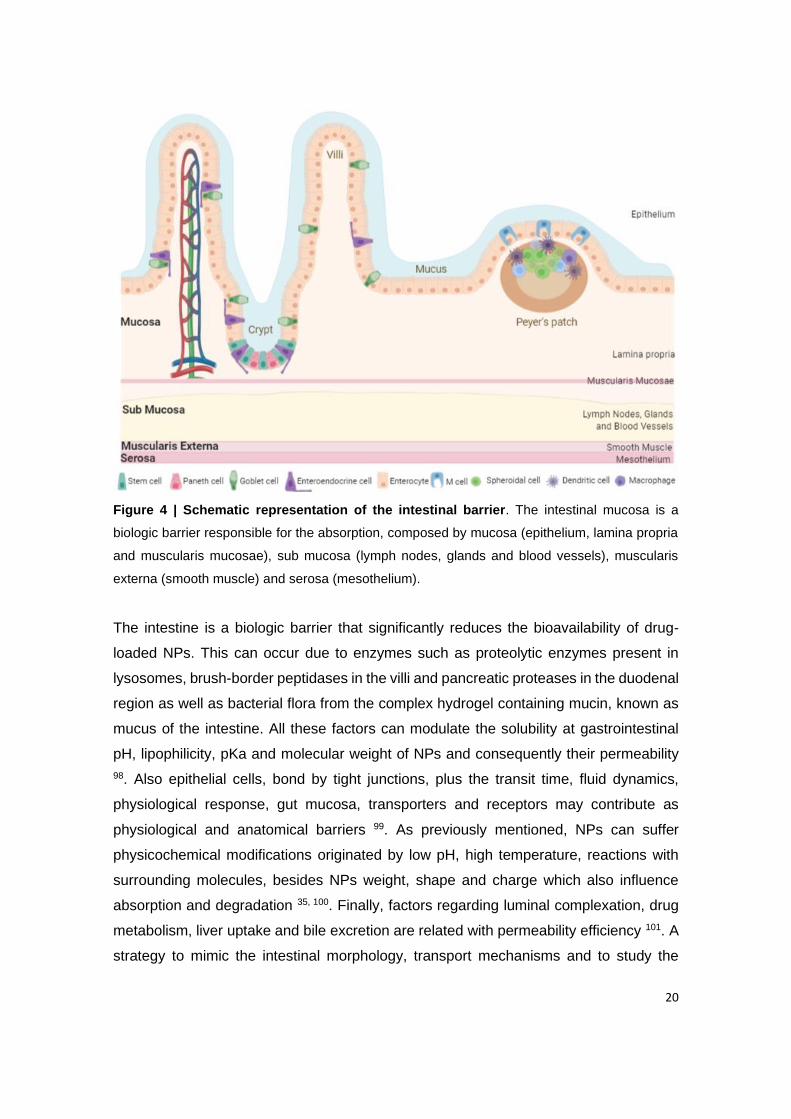
20
Figure 4 | Schematic representation of the intestinal barrier. The intestinal mucosa is a
biologic barrier responsible for the absorption, composed by mucosa (epithelium, lamina propria
and muscularis mucosae), sub mucosa (lymph nodes, glands and blood vessels), muscularis
externa (smooth muscle) and serosa (mesothelium).
The intestine is a biologic barrier that significantly reduces the bioavailability of drug-
loaded NPs. This can occur due to enzymes such as proteolytic enzymes present in
lysosomes, brush-border peptidases in the villi and pancreatic proteases in the duodenal
region as well as bacterial flora from the complex hydrogel containing mucin, known as
mucus of the intestine. All these factors can modulate the solubility at gastrointestinal
pH, lipophilicity, pKa and molecular weight of NPs and consequently their permeability
98. Also epithelial cells, bond by tight junctions, plus the transit time, fluid dynamics,
physiological response, gut mucosa, transporters and receptors may contribute as
physiological and anatomical barriers 99. As previously mentioned, NPs can suffer
physicochemical modifications originated by low pH, high temperature, reactions with
surrounding molecules, besides NPs weight, shape and charge which also influence
absorption and degradation 35, 100. Finally, factors regarding luminal complexation, drug
metabolism, liver uptake and bile excretion are related with permeability efficiency 101. A
strategy to mimic the intestinal morphology, transport mechanisms and to study the
21
permeability of NPs is using in vitro, ex vivo or in vivo models, which will be focused on
the following sections.
4.2. In vitro models
Despite the fact that in vitro models do not mimic a real in vivo situation, they can easily
simulate a multiple transport system and be reproduced in laboratory. Besides, this type
of studies are becoming more important to the pharmaceutical industry, since the
obtained results can influence the initial steps of drugs developing. This method is
characterized for being less labor-intensive and cost-intensive, and raise less ethical
considerations compared to in vivo studies. Plus, the use of in vitro models allows an
early evaluation of drugs, without using in vivo models and large amounts of drug to
analyze its ADMET 96. In this sense, it is possible to have in vitro models where gastric
and intestinal fluid can be simulated, and dialysis membrane system and Transwell® to
test the permeability of compounds.
The simulated gastric and intestinal fluid model can be used as a preliminary study to
analyse the reaction of the NPs to the GIT environment. The used medium has to be
representative of the stomach and intestine fluid, presenting characteristics as enzymes
and ionic strength, which influences the interaction and stability of NPs. According with
the United States Pharmacopeia (USP) the simulated gastric fluid (SGF) medium should
be at pH 1.2 and the simulated intestinal fluid (SIF) medium at pH 6.8. These mediums
are easily prepared and normally experiences are performed at 37 ºC with agitation (100
rpm). At different time points the supernatant can be removed by centrifugation and
quantified. It is known that these conditions reduce the number of NPs, besides changing
size and morphology and consequently bioavailability 102. On the other hand, the dialysis
membrane system test the transport of the nanosystems across the barriers of the gut.
In this method, nanosystems are added to a dialysis membrane with the same conditions
as the last technique, in order to represent the GIT characteristics and movements.
These two models can be done without using human cell lines, which can be a drawback
since do not represent the epithelial behaviour 103. Lastly, the Transwell® system is the
most used model to evaluate the permeability of drugs from lumen to blood circulation.
Briefly, this system is composed by a membrane with pores that separate the apical from
the basolateral side. The epithelial cells are seeded at the apical side and when confluent
with an ideal transepithelial electrical resistance (TEER), the experience can be
performed. Nowadays, this system can be performed with only one cell line (mono-
culture) or combined with other cell lines (co-culture) 96.
22
4.2.1. Monoculture model
In this type of model a single cell line is seeded forming a monolayer. There are several
cell lines being used in in vitro models of the intestine, but the most widely used to
recreate the human environment is the Caco-2 cell line. According to the Food and Drug
Administration (FDA), Caco-2 cell line integrates the Biopharmaceutics Classification
System (BCS), since it has the advantageous ability to screen and predict NPs solubility
and bioavailability, as well as their interaction with the gut 104. Caco-2 is a high-resistance
cell line from a human colorectal carcinoma and is considered the gold standard
permeability screen for compounds in drug discovery, due to its morphological and
functional characteristics as well as the presence of cytochrome P450 3A4 (CYP3A4)
enzymes, sucrase-isomaltase, mature enterocyte alkaline phosphatase, drug
transporters and efflux proteins. It was also previously confirmed the presence of FcRn
expression in Caco-2 cells. In addition, the albumin transport through Caco-2 monolayer
was considered a saturable process, above 40 µg/mL, indicating a receptor-mediated
uptake 105, 106. After culturing (3-4 days), cells are undifferentiated and do not present
microvilli, neither their indicator (sucrase immunoreactivity). When confluent, they form
a polarized monolayer with microvilli and connected by tight junctions (Figure 5 A). The
strenght and tightness of the monolayer, meaning confluence and viability, can be
identified by measuring the TEER. Higher TEER values indicate tighter membranes and
consequently the modulation of the paracellular transport. Also, an indicative of the
membrane integrity is the paracellular transport of small molecules like lucifer yellow,
FITC-dextran or mannitol 96. Although, Caco-2 cells present some drawbacks, namely
the low expression of the CYP3A4 metabolizing enzyme, high and variable expression
of P-glycoprotein (P-gp) and low paracellular transport of hydrophilic compounds,
because of the tigther junctions 107. Also, the permeability of drugs with slow absorption
is poor in this model, comparing with rates in humans, and may be due to the lack of a
3D architecture. Plus, this model lack the presence of mucus production, which exclude
the main barrier of NPs absorption.
Furthermore, there have been suggested some alternative cell lines to find a model that
better mimics the human system, being inclusively some of them used to analyze NPs
permeability. In this line of thought, human colonic adenocarcinoma cell line (T84) is a
possibility for permeability studies, specially regarding the transport of IgG and albumin
in a FcRn-dependent manner 70. Studies show that T84 cell line produces mucin in
culture and releases it in response to various secretagogues 108. It is known that the
23
absorption mainly occurs at the small intestine, so cells from colon have, per se, the
drawback of differing in the paracellular permeability since, they present a tighter
conformation and the absorption of compounds paracellularly is lower.
Despite having a nonintestinal origin, the Madin-Darby canine kidney (MDCK), from the
distal tubule and collecting duct, is highly correlated with Caco-2 cells, working as a
strong alternative to study drugs that are absorbed passively 109. Authors showed the
utility of MDCK cells in probing interactions between P-gp and their substrates.
Comparing the permeability of anti-malaria compounds in Caco-2 and MDCK cells, they
verified the overexpression of P-gp in MDCK-MDR1 cells, which allows high permeability
110. It was analysed the mecanisms of transport of Deoxynivalenol (DON), using
inhibitors, verifying the presence of organic anion and cation transporters. P-gp was also
identified as the major efflux protein, due to the higher basolateral-to-apical observed
transport 111. Regarding NPs permeability, it was also proved the transport of
nanoemulsions of self-nano-emulsifying drug delivery systems (SNEDDS) through
MDCK cells 112. Also, these cells only need 3-6 days to become confluent and form tight
junctions 109. Conversely, the fact that they are not intestinal neither human confer them
different levels of transporters expression as well as different metabolic activity 113.
4.2.2. Co-culture model
Caco-2/HT29-MTX model
HT29 cell line is originally from human colorectal adenocarcinoma and can mimic goblet
cells. Though, HT29 cells need a different medium, comparing with Caco-2 cells, so they
can differentiate and acquire a heterogeneous phenotype. To surpass this barrier,
authors verified that by adding 10-6 M methotrexate (MTX), HT29-MTX cell line is able to
form a polarized monolayer with goblet cells and differentiate into mature goblet cells,
able to produce mucus, after 21 days in culture 114. The differentiation character can be
approved by confirming MUC cell markers. Mucus is a visoelastic gel produced by mucin
glycoproteins and is responsible to prevent pathogens entrance, also acting as a barrier
of NPs absorption, which affects the retention time 95. In fact, authors already
demonstrated that mucus clearly decreases the uptake and transport of NPs 115. It is
known that NPs (with positive charges) can interact with mucus (negatively charged)
through electrostatic interactions, as well as hydrophobic or van der Waals interactions
116. The role of mucus in cell-based models is already well descibed elsewhere 117. So,
this parameter must be taken into consideration when evaluating NPs permeability.
When cultured with Caco-2 cells, there was the necessity to study different ratios to
24
prevent alterations in barrier properties caused by the overgrowth of HT29-MTX cells 118.
When 105 Caco-2 cells/cm2 are combined with HT29-MTX in a 9:1 proportion, the model
(Figure 5 B) gets closer to the human intestine characteristics, generating more
predictable results 119. Furthermore, HT29-MTX do not express P-gp and present tight
junction less tight than Caco-2 (decreasing TEER values), which facilitate the passive
permeability of nanosystems and represent a more physiological model 120.
Caco-2/Raji B model
It is also described that Raji B cells, from a human Burkitt’s lymphoma, when combined
in a 1:2 proportion with 105 Caco-2 cells/cm2 induce M cell phenotype (sparse microvilli
and high transcytosis activity) (Figure 5 C). This happens due to the interaction between
Caco-2 cells and lymphocytes and the presence of galectin-9 or SLAA, a signal of
differentiation. In this model, Raji B lymphocytes cells are only added at the basolateral
side after 14 days of seeding Caco-2 cells at the apical side. It was recently published
a protocol suggesting an inverted in vitro M-cell model, where it is possible to do transport
experiments every 10 h, after 3 weeks 121. Despite not being in contact with Caco-2 cells,
it is possible to observe cells with M cell characteristics. As mentioned before, M cells
serve as antigen-sampling cells and unlike enterocytes, they can translocate particles
without digesting them. Indeed, studies confirm that M cells play a major role in
translocation and permeability of NPs, claiming that these parameters are higher in this
type of co-culture comparing to Caco-2 mono-culture 122. All in all, as a drawback, this
model do not take into account the mucus, which is an important intestinal characteristic.
Caco-2/HT29-MTX/Raji B model
Taking all the above information into consideration, Sarmento’s group developed a triple
co-culture model, with the three cell lines: Caco-2, HT29-MTX and Raji B cells (Figure
5 D). This group tested the model with normal and inverted orientation, verifying higher
permeation with normal orientation 123. This is actually a more accurate and realistic
model due to its similarity with human intestine characteristics: enterocytes (Caco-2
cells), mucus production by goblet cells (HT29-MTX cells) and M-cells (Raji B cells). The
proportions mentioned before were based on physiological ratios and previous works 124.
In addition, it was already shown that Caco-2/HT29-MTX co-culture and the triple Caco-
2/HT29-MTX/Raji B co-cultyre models are useful to predict the intestinal transport of free
drugs and NPs and to classify them, acording to BCS, with low or high permeability.
Furthermore, they have a good correlation with ex vivo models, which will be explained

25
later 125.
4.2.3. 3D co-culture model
All the models mentioned before are based on a 2D-surface which, once again, do not
represent in vivo cells with a heterogenous environment containing several cellular
events 96. So, there was a necessity to create a 3D model with physiological structures
to reproduce more accurately intestinal functions, namely growth, differentiation,
interactions between cells themselves and with matrix and mechanical forces as well. In
addition, cells in a 3D arquitecture are easily differentiated and polarized when on top,
comparing with those that are near the villus base, which may be an advantage for NPs
that remais longer in the lumen (slow absorption) 126. Like atenolol, a hidrophilic and slow
absorbed drug, certain NPs can migrate to the bottom of cells and be transported through
cell-cell tight junctions 127. Furthermore, 3D models have tight junctions less tight,
comparing with Caco-2 monolayer, contributing for a better permeability which allies to
less expression of P-gp and is consistent with in vivo observations 128.
Some authors verified that MDCK cells in Matrigel can form a 3D arquitecture when the
viscosity is low and the cellular contractile forces are strong. The 3D structure can then
be changed in a viscosity-dependent manner. A way to play with it is to do a treatment
with genipin (GP), since it allows the cross-link between amino groups of the matrigel
peptide chains 129. The 3D model can also include macrophages, increasing the uptake
of particles, as well as other type of cells or even polymeric scaffolds to comprise the
intestinal microvilli architecture. For instance, a recent model was proposed, where 104
cells/cm2 of intestinal myofibroblasts (CCD18-Co cells) are entrapped in a thick layer of
Matrigel and then 105 cells/cm2 of Caco-2 cells are added over 130. The differentiation of
CCD18-Co cells can be confirmed by the observation of α-SMA or CD90 through
immunocytochemistry. It was verified that the matrix is important to maintain the
architecture and support the model. On the other hand, fibroblasts contribute for
epithelial cells growth (Figure 5 E) 130. There is, inclusively, a model using Caco-2, HT-
29 and T84 cell lines as well as proinflammatory stimuli, to mimic an inflamed intestinal
mucosa 131. Also, a model with the traditional Caco-2 cell line and human immune cell
lines (Human macrophages (THP-1) and human dendritic cells (MUTZ-3)) was
developed to evaluate the toxicity/safety of NPs 132. The 3D co-culture model presents,
in fact, additional complexity that may contribute for developing NPs to treat inflamatory
diseases or even to study the interaction of the NPs with an inflammated intestine. Some
authors already investigated the genetic and physiological properties of the 3D model,
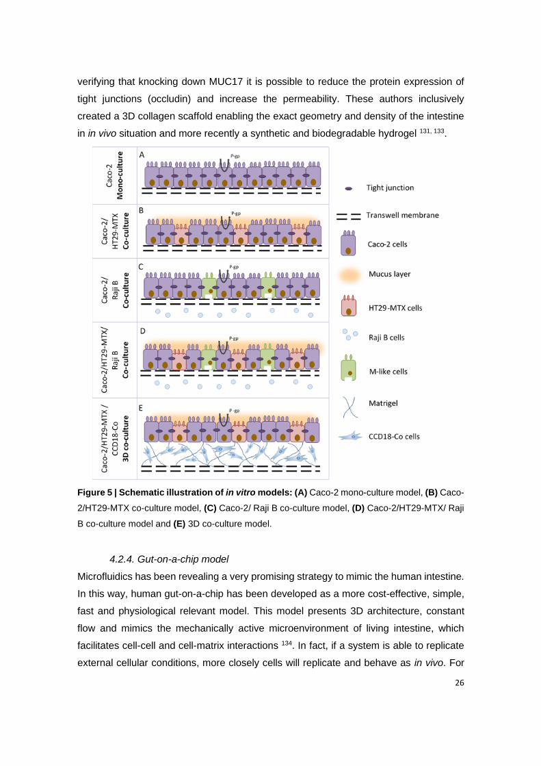
26
verifying that knocking down MUC17 it is possible to reduce the protein expression of
tight junctions (occludin) and increase the permeability. These authors inclusively
created a 3D collagen scaffold enabling the exact geometry and density of the intestine
in in vivo situation and more recently a synthetic and biodegradable hydrogel 131, 133.
Figure 5 | Schematic illustration of in vitro models: (A) Caco-2 mono-culture model, (B) Caco-
2/HT29-MTX co-culture model, (C) Caco-2/ Raji B co-culture model, (D) Caco-2/HT29-MTX/ Raji
B co-culture model and (E) 3D co-culture model.
4.2.4. Gut-on-a-chip model
Microfluidics has been revealing a very promising strategy to mimic the human intestine.
In this way, human gut-on-a-chip has been developed as a more cost-effective, simple,
fast and physiological relevant model. This model presents 3D architecture, constant
flow and mimics the mechanically active microenvironment of living intestine, which
facilitates cell-cell and cell-matrix interactions 134. In fact, if a system is able to replicate
external cellular conditions, more closely cells will replicate and behave as in vivo. For
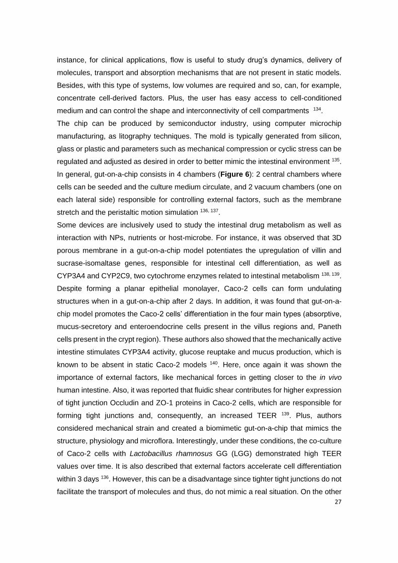
27
instance, for clinical applications, flow is useful to study drug’s dynamics, delivery of
molecules, transport and absorption mechanisms that are not present in static models.
Besides, with this type of systems, low volumes are required and so, can, for example,
concentrate cell-derived factors. Plus, the user has easy access to cell-conditioned
medium and can control the shape and interconnectivity of cell compartments 134.
The chip can be produced by semiconductor industry, using computer microchip
manufacturing, as litography techniques. The mold is typically generated from silicon,
glass or plastic and parameters such as mechanical compression or cyclic stress can be
regulated and adjusted as desired in order to better mimic the intestinal environment 135.
In general, gut-on-a-chip consists in 4 chambers (Figure 6): 2 central chambers where
cells can be seeded and the culture medium circulate, and 2 vacuum chambers (one on
each lateral side) responsible for controlling external factors, such as the membrane
stretch and the peristaltic motion simulation 136, 137.
Some devices are inclusively used to study the intestinal drug metabolism as well as
interaction with NPs, nutrients or host-microbe. For instance, it was observed that 3D
porous membrane in a gut-on-a-chip model potentiates the upregulation of villin and
sucrase-isomaltase genes, responsible for intestinal cell differentiation, as well as
CYP3A4 and CYP2C9, two cytochrome enzymes related to intestinal metabolism 138, 139.
Despite forming a planar epithelial monolayer, Caco-2 cells can form undulating
structures when in a gut-on-a-chip after 2 days. In addition, it was found that gut-on-a-
chip model promotes the Caco-2 cells’ differentiation in the four main types (absorptive,
mucus-secretory and enteroendocrine cells present in the villus regions and, Paneth
cells present in the crypt region). These authors also showed that the mechanically active
intestine stimulates CYP3A4 activity, glucose reuptake and mucus production, which is
known to be absent in static Caco-2 models 140. Here, once again it was shown the
importance of external factors, like mechanical forces in getting closer to the in vivo
human intestine. Also, it was reported that fluidic shear contributes for higher expression
of tight junction Occludin and ZO-1 proteins in Caco-2 cells, which are responsible for
forming tight junctions and, consequently, an increased TEER 139. Plus, authors
considered mechanical strain and created a biomimetic gut-on-a-chip that mimics the
structure, physiology and microflora. Interestingly, under these conditions, the co-culture
of Caco-2 cells with Lactobacillus rhamnosus GG (LGG) demonstrated high TEER
values over time. It is also described that external factors accelerate cell differentiation
within 3 days 136. However, this can be a disadvantage since tighter tight junctions do not
facilitate the transport of molecules and thus, do not mimic a real situation. On the other
28
hand, other authors verified that cyclic strain enhances the cell differentiation, formation
of 3D villi-like structures and paracellular permeability, without changing TEER values in
the monolayer, meaning that mechanical forces act directly in paracellular mechanisms
136. All in all, the TEER vales are not always concordant across literature and this might
be due to the current TEER measure devices being designed to fit into conventional
Transwell® culture plates and not into microfluidic device. As curiosity, embedding
electrodes inside microfluidic organ chips have already been designed. The creators
showed both in human lung airway chip and human gut chip the utility of this embedding
electrodes to assess formation and disruption of barrier function. In this way, the users
can have real-time results through a non-invasive approach 141.
Figure 6 | The mechanically active human Gut Chip. (A) Human villus intestinal epithelium and
vascular endothelium are lined on opposite sides of a flexible porous membrane under fluid flow
and peristalsis-like strains. A zoom-in schematic shows the intestinal microenvironment
undergoing complex crosstalk between commensal gut microbiome, bacterial pathogens, and
immune cells in parenchymal and vascular channels, respectively. (B) Villus morphogenesis of
human Caco-2 intestinal epithelium in the Gut Chip under physiologically controlled motions and
flow. (C) An overlaid image of the coculture of green fluorescent protein–labeled Escherichia coli
and microengineered villi in the Gut Chip. Bars = 50 mm. Reprinted with permission from 137.
Alternatively, some authors choose to incorporate human intestinal organoids (HIO)
through induced pluripotent stem cells (iPSCs) to an amenable model for study. To form
29
HIO, these authors supplemented iPSCs with Actividin A, CHIR99021, noggin, fibroblast
growth factor 4 and epidermal growth factor to induce endoderm and hindgut formation,
as to maintain the organoid formation 142. Besides having the four intestinal cell types of
diferentiation and the characteristic crypt-villus 3D architecture, this model has the
advantage of responding to exogenous stimuli 142. More recently, the same authors
aprimorated the intestine-on-a-chip to recapitulate the dynamic host-microbiota
interface. In this sense, they recreated a system with hipoxia gradient across an
endothelial-epithelial interface. The authors claim that the intestinal barrier integrity is
actually better under these conditions. This system includes microscale oxygen sensors,
Caco-2 cells or human organoid derived epithelial cells, housed in an anaerobic
chamber, and enables to keep different commensal aerobic and anaerobic
microorganisms in direct contact with patient-derived human intestinal epithelium for 5
days 143. Donald Ingber and colleagues also fabricated a human small intestine-on-a-
chip composed by 3D organoids of epithelial cells isolated from healthy regions of
intestinal biopsies. According to their transcriptomic analysis, this intestine chip is closer
to human duodenum than duodenal organoids 144. This can be a personalized strategy
for each individual, a way to understand specific mechanisms and therapies and get
more reliable results. These organoids are dissociated and cultured on a porous
membrane. This system is also characterized by having human intestinal microvascular
endothelium cultured in a parallel microchannel under flow and cyclic deformation. Plus,
as advantage, this intestine-on-a-chip is useful for metabolism, nutrition, infection and
drug pharmacokinetics studies, considering the easiness to quantify nutrient digestion,
mucus secretion and establish a intestinal barrier function 144. Meanwhile, there is a
commercial version available. Mimetas, the organ-on-a-chip company, created the
Organoplate that combine microfluidics to a standard well plate. This allows the user the
freedom to decide which extracellular matrix (ECM), cell types, co-culture and perfusion
to use, according to each interest. In addition, this company recently developed a
methodology to assess the barrier integrity using 40 leak-tight, polarized epithelial gut
tubes, exposed to an ECM. The method was recreated in a microfluidic environment,
expressing polarization and transporters. This can be a way to simulate a pathological
situation like dysfunction of epithelial barriers, or drug-induced toxicity, which can reduce
cellular life conditions and drug development. The epithelial barrier disruption condition
is characterized for having an increased paracellular permeability. The authors claim to
be a method of friendly-use, sensible, with real-time analysis 145.
Basically, it is possible to combine different approaches and aprimorate the intestinal
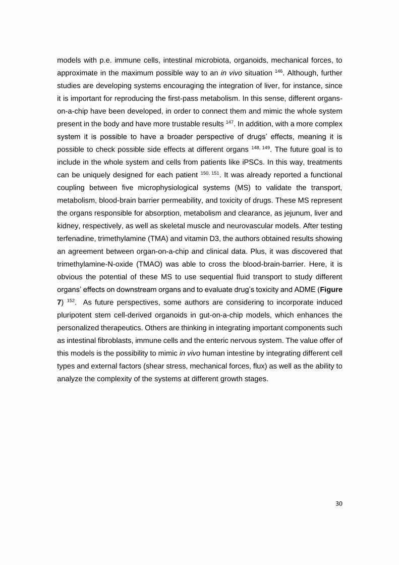
30
models with p.e. immune cells, intestinal microbiota, organoids, mechanical forces, to
approximate in the maximum possible way to an in vivo situation 146. Although, further
studies are developing systems encouraging the integration of liver, for instance, since
it is important for reproducing the first-pass metabolism. In this sense, different organs-
on-a-chip have been developed, in order to connect them and mimic the whole system
present in the body and have more trustable results 147. In addition, with a more complex
system it is possible to have a broader perspective of drugs’ effects, meaning it is
possible to check possible side effects at different organs 148, 149. The future goal is to
include in the whole system and cells from patients like iPSCs. In this way, treatments
can be uniquely designed for each patient 150, 151. It was already reported a functional
coupling between five microphysiological systems (MS) to validate the transport,
metabolism, blood-brain barrier permeability, and toxicity of drugs. These MS represent
the organs responsible for absorption, metabolism and clearance, as jejunum, liver and
kidney, respectively, as well as skeletal muscle and neurovascular models. After testing
terfenadine, trimethylamine (TMA) and vitamin D3, the authors obtained results showing
an agreement between organ-on-a-chip and clinical data. Plus, it was discovered that
trimethylamine-N-oxide (TMAO) was able to cross the blood-brain-barrier. Here, it is
obvious the potential of these MS to use sequential fluid transport to study different
organs’ effects on downstream organs and to evaluate drug’s toxicity and ADME (Figure
7) 152. As future perspectives, some authors are considering to incorporate induced
pluripotent stem cell-derived organoids in gut-on-a-chip models, which enhances the
personalized therapeutics. Others are thinking in integrating important components such
as intestinal fibroblasts, immune cells and the enteric nervous system. The value offer of
this models is the possibility to mimic in vivo human intestine by integrating different cell
types and external factors (shear stress, mechanical forces, flux) as well as the ability to
analyze the complexity of the systems at different growth stages.
31
Figure 7 | Schematic representations of the four of the organ systems used for functional
coupling. (A) The intestinal module is constructed in Transwell® from primary jejunum enteroids.
Test agents are applied in the apical compartment (1). The media collected in the basolateral
compartment (2) is used to add to the liver. (B) Media from the jejunum intestine basolateral
compartment (2) is perfused as a 1:3 jejunum/naïve liver media into the influx port of the SQL
SAL liver model (3). Efflux media is collected (4) and used to add to two downstream organ
models. (C) The vascularized kidney proximal tubule module is a two lumen, dual perfusion
system. For the vascular compartment, jejunum/liver-conditioned media (4) is diluted 1:2 or 1:4
with naïve EGM-2 media and then perfused into the influx port (5) to collect effluent from the
proximal tubule at (6) . In parallel with perfusion through the vascular compartment, the proximal
tubule compartment is perfused with naïve DMEM/F12 PTEC media (6) for effluent collection. (D)
The blood-brain barrier with NVU is constructed in a membrane-separated, two-chambered
microfluidic device. The brain-derived endothelial vascular compartment is perfused at the influx
port (7) with jejunum/liver-conditioned media (4) diluted 1:4 with naïve EGM-2 media. The effluent
is collected at the efflux port (7). In parallel with perfusion through the vascular compartment, the
neuronal cell compartment is perfused with naïve EBM-2 media at the influx port < 8> for effluent
collection at (8). Reprinted with permission from 152.
4.3. Ex vivo models
In vitro tissue-based models, also called ex vivo are used in intestinal permeability
studies in a way to move from in vitro to in vivo models. In this type of models, living
functional tissues or organs are cultivated in a controlled environment after being isolated
from organisms. These models present features that are normally not present in cell
32
models such as the adequate presence of mucus, paracellular permeability or the
expression of transport proteins and drug metabolism. The main drawbacks of this and
all in vitro models is the impossibility to recreate the gastric empty rate, GI pH or transit
rate.
4.3.1. FcRn tissue expression and cross-species differences
FcRn is expressed in nearly every organ in mammals, as blood-brain barrier, eye, lungs,
breast, kidneys, intestine, skin, endothelial cells, antigen-presenting cells (e.g.
macrophages, monocytes and dendritic cells), placenta, genitals and bone, which are
described elsewhere 153, 154. However, only intestine will be highlighted here, since this
work focus oral delivery of FcRn-targeted NPs for an enhanced intestinal permeability
with application in DM.
4.3.1.1. Human
FcRn has a special role in early life stages for transporting IgG from mothers to neonates.
However, the expression of FcRn is maintained along the adult stage in human intestine.
Through immunohistochemical studies, it was possible to verify that FcRn is mostly
located in the apical region of epithelial cells, in different sections of human intestine 155.
Moreover, FcRn is highly expressed in colon and ileum and less expressed in jejunum
and duodenum 93.
4.3.1.2. Mouse
FcRn is also expressed in several tissues of mice and has the same importance when
transporting the maternal IgG. It was previously reported that neonatal mice deficient in
β2m and FcRn HC were not able to absorb IgG from mother’s milk, which demonstrated
the highlight impact of FcRn in the IgG transport 155. In adults, these mice had low levels
of IgG in the bloodstream owing to the lysosomal degradation 156-158. Thus, the
expression of mFcRn is restricted to the suckling period, being down-regulated after
weaning 159. For this reason, it is recommended to use mice 6-16 weeks after weaning,
since FcRn levels are stable. Besides, the homology to the rodent FcRn is around 70%
compared to hFcRn, which is one more cause for the cross-species differences 160. Thus,
the transgenic mouse model mimics the tissue expression profile of hFcRn 157. Overall,
it is described that FcRn has higher expression in duodenum and jejunum in rat pup
intestine, which is ~10-fold higher than human proximal colon 106, 161.
33
Structurally, it was verified that some sequence variations may cause cross-species
binding differences. Among these variations are the residues 132-147, the nonconserved
residue 137 (Leu in hFcRn and Glu in mFcRn) 162 and residues 79-89, of which there is
a two-residue deletion in hFcRn and a possible glycosilation site in mFcRn 163. Regarding
the FcRn-albumin interaction, HSA has a 5-fold lower affinity to hFcRn compared to
mouse serum albumin (MSA) 60, 164, while MSA has a 25-fold stronger affinity to mFcRn
than HSA 51, 165. These cross-species differences are related to nonconserved amino
acid found in the conserved His166 within the α2 domain of FcRn 51, 60, 165, but also with
different amino acid structure of DI and DIII from both species 52, 165. The differences
associated to DI are represented by four mutations in loop I and II of its domain. However,
Arg114 (from loop II) as well as both loops of I are crucial role for the I-hFcRn interaction
52. Regarding the DIII, the major difference is the amino acid at position 573, which is
lysine in humans and proline in rodents 51. The replacement of Lys573 for Pro573
increases 1.5-fold the half-life of HSA in mice and augment 12-fold and 15-fold the affinity
for hFcRn and mFcRn, respectively 51. Thereby, the interaction between mFcRn and
MSA is stronger compared with the mFcRn interaction with HSA, which can contribute
to rapid clearance and interfere with in vivo preclinical evaluation in mice. In this case,
HSA competes with the higher amounts of endogenous MSA (40 mg/mL) for the binding
to mFcRn, resulting in an augment in the half-life of endogenous MSA and no alteration
in half-life of HSA 165.
4.3.1.3. Porcine
The pig has been confirmed as a non-primate model suitable for assessing PK of HSA,
since porcine serum albumin (PSA) minimally competes for binding to FcRn 160. In fact,
pigs have gained importance as a large animal in pharmaceutical research due to its
physiological and anatomical similarities to human. In addition, pigs are replacing dogs
as the preferred non-human primate model in preclinical studies. Stirling et al. 166
characterized the porcine FcRn (pFcRn) and tested its utility as a potential model for
transepithelial protein delivery 166. The sequence of the pFcRn gene is 80% similar to the
corresponding human gene and 69% similar to the rodent receptor gene (Figure 8).
Structurally, the similarity between the pFcRn protein and the human version is based
on the single N-linked glycosylation site (NVSV) in the α domain, as well as both di-
leucine- and tryptophan-based endocytosis signals present in the cytoplasmic tail. In
particular, the porcine receptor has a type 1 membrane protein with an N-terminal single
peptide and a single transmembrane domain. However, it does not seem to be an
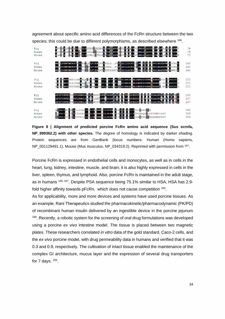
34
agreement about specific amino acid differences of the FcRn structure between the two
species; this could be due to different polymorphisms, as described elsewhere 166.
Figure 8 | Alignment of predicted porcine FcRn amino acid sequence (Sus scrofa,
NP_999362.2) with other species. The degree of homology is indicated by darker shading.
Protein sequences are from GenBank (locus numbers: Human (Homo sapiens,
NP_001129491.1), Mouse (Mus musculus, NP_034319.2). Reprinted with permission from 167.
Porcine FcRn is expressed in endothelial cells and monocytes, as well as in cells in the
heart, lung, kidney, intestine, muscle, and brain; it is also highly expressed in cells in the
liver, spleen, thymus, and lymphoid. Also, porcine FcRn is maintained in the adult stage,
as in humans 166, 167. Despite PSA sequence being 75.1% similar to HSA, HSA has 2.9-
fold higher affinity towards pFcRn, which does not cause competition 160.
As for applicability, more and more devices and systems have used porcine tissues. As
an example, Rani Therapeutics studied the pharmacokinetic/pharmacodynamic (PK/PD)
of recombinant human insulin delivered by an ingestible device in the porcine jejunum
168. Recently, a robotic system for the screening of oral drug formulations was developed
using a porcine ex vivo intestine model. The tissue is placed between two magnetic
plates. These researchers correlated in vitro data of the gold standard, Caco-2 cells, and
the ex vivo porcine model, with drug permeability data in humans and verified that it was
0.3 and 0.9, respectively. The cultivation of intact tissue enabled the maintenance of the
complex GI architecture, mucus layer and the expression of several drug transporters
for 7 days. 169.

35
4.3.2. Standard models
4.3.2.1. Diffusion chambers
The most performed method using intestinal tissues is diffusing chambers, namely
Ussing chamber or Franz cells. The main difference between these two is the direction
of the flux. In the first, is horizontal, or side-by-side, and in the second is vertical 170.
Ussing chamber
Ussing chamber was the first to be invented, in 1951, later using intestinal tissue in 1988
171, 172. The size, volume and area of exposed tissue are parameters that can vary. In this
methodology, a section of intestinal tissue is excised, separated in small fragments and
opened to form a flat surface. Then the tissue is placed in the chamber between two
halves. The chamber is filled with physiological buffer, gassed with carbogen and
warmed at 37°C. Two chambers, donor and receiver, are divided by the tissue, one of
them representing the serosal side and the other the mucosal side. The drug or NPs to
be tested in a permeability test can be placed in one of the chambers and samples should
be removed from the other chamber, replacing the volume took with fresh buffer 173, 174.
The TEER is again one of the parameters that can be measured by placing the
electrodes in each side of the membrane 175. The main advantage of this model is the
maintenance of the gut architecture, membrane transporters and enzymes when
compared with cell-based models. It is also possible to study regional differences in the
GIT by using different sections of it. Ussing chamber can also be used with human
tissues instead of animal tissues. Since tissues can be easily obtained from tumor
resections or gastric bypass surgeries, more recent studies have been using this type of
tissue 176. One disadvantage, though, is the fact that the experiments should be
performed near a surgery clinic since the tissues can quickly deteriorate outside the body
177.
Franz cell
On the other hand, Franz cell is mostly used for skin permeation studies than intestinal
permeability, but used nevertheless. This model is not much different than Ussing
chamber, since the tissue membrane is still placed between the donor and receptor
chamber. The main significant difference, besides the direction of the flow, is the size of
the chambers, since in the Ussing chamber the sizes are similar and in the Franz cell
the donor compartment is smaller than the receptor 178. This difference in volumes can
induce discrepancies in the permeability when compared to the Ussing chamber, but can
36
be resolved by using a magnetic stirrer in the receptor chamber in order to reduce the
unstirred water layer. It is important to use the tissue as soon as harvested and ensure
sink conditions are maintained. Few work have been done using intestine tissue in Franz
cells to evaluate nanoparticle permeability.
4.3.2.2. Everted intestinal sac model
Another method used in ex vivo intestinal permeability studies is the everted intestinal
sac. This model can be applied in the study of mechanisms and kinetics of drug
absorption and metabolism including pro-drug conversion in the GIT, efflux transport,
drug resistance and interactions 179. A section of the intestine of the animal is removed,
washed, inverted with a rod and one of the ends is tied. The inside of the sac is filled with
oxygenated buffer and the other end closed after. Then it is placed in the container. This
container has the sample to be tested, and its presence in the interior of the sac is then
evaluated, within the determined time-points. There are two different techniques that can
be applied in this method, one where both sides are closed, creating a closed sac and
the other where the end of the tissue is cannulated in the tubes. The main advantages
of this model is the low cost, quickness, the possibility of test several drugs, the presence
of the mucus layer, large surface area and the possibility of study passive and active
transport. The main disadvantages are the possibility of damaging the tissue, the release
of proteins and/or enzymes as well as the muscularis mucosa that mislead the results,
and the time which the tissue remains viable, that is only two hours 180, 181. The everted
sac is the main method used to test NPs permeability.
4.3.2.3. Non-everted intestinal sac model
In order to avoid tissue damaging, this model does not evert the gut sac after excision.
The intestinal sac is removed from the animal, washed properly and one of the ends of
the sac is tied. The sample solution is placed inside the sac and the other end is closed.
Afterwards, the sac is placed in the release medium. This release medium should be
able to mimic the conditions existing in the GIT. This method is more limited than the
everted sac in terms of information that can be accessed, but it is simple and rapid to
perform, while still capable of test drug absorption. It is not only simpler but also requires
low amounts of the drug tested and samples are easier to quantify an can be collected
at various time points 178, 182. In terms of disadvantages, this method is quite similar to
the everted sac, where the viability of the tissue is the main issue to carefully handle.
37
4.3.2.4. Everted intestinal ring
Similar to the everted intestinal sac, the everted intestinal ring is also used in ex vivo
intestinal permeability studies. Most used to assess the uptake and metabolism of the
tested compounds, in this model, the intestinal tissue is harvested, everted and cut into
small rings. Then it is put in an incubator with proper incubation media and agitation. The
simplicity is the main advantage of this method allied with the ability of testing several
compounds 183. However, this technique also presents disadvantages. Since both
connective tissue and muscular layer are exposed in the media, the diffusion of the
tested compounds could not occur on the mucosal side. Also, the time of incubation is
quite reduced, around 10 min 174, 183.
4.4. In vivo FcRn model systems
Primates are considered the best animal model to mimic the human PK parameters 184,
but the high costs and ethical subjects make their use unapproachable 164, 185. In this
sense, rodents have been considered the preferred models to assess the bioavailability
of biopharmaceuticals 184, due to the convenience, cost-effective and defined genetic
background 164, 185. Nevertheless, the expression of FcRn and the affinity of its ligands
differ between humans and rodents, which can interfere with the FcRn therapeutics.
Taking into account all the differences reported between humans and rodents, as
previously discussed, the conventional rodent models do not reflect an adequate PK
profile of FcRn-targeted ligands. To solve this preclinical challenge, several genetically
engineered mouse strains have been developed where the mFcRn heavy chain has
been replaced with the human counterpart, either under the control of the mouse or
human promoter elements 64, 157, 186-192. These strains are the current gold standards for
the evaluation of hFcRn-targeting strategies addressing PK/PD and delivery across
selective mucosal barriers prior to studies in non-human primates.
The most commonly used hFcRn-expressing strains are generated from conventional
C57BL/6J mice, named Tg32 and Tg276 (Figure 9 A), which are extensively used to
assess the half-life of hIgG and HSA variants 64, 157, 186, 189, 191, 192. Both strains have
substituted the mouse heavy chain with that of the human form, which can pair non-
covalently with the endogenous mouse β2m 64, 157, 186, 191, 192. A distinct difference between
these strains is that the Tg32 mice express the heavy chain under the control of its
human regulatory elements 157, 186, 189, 191, 192, while the expression in Tg276 mice is
regulated by a constitutive CAG promoter, a human cytomegalovirus enhancer fused to
chicken β-actin/rabbit β-globin hybrid 64, 186. This results in a much more widespread
38
expression profile of hFcRn in Tg276, whereas the FcRn expression profile in Tg32 mice
has been mapped to be similar to that in humans 193, 194. There is also a modified version
of Tg32 that expresses human β2m, but no difference in half-life of the ligands has been
reported that can be related to whether the mice carry a mouse or human form of β2m
191. In general, the half-life of IgG is shorter in Tg276 homozygous mice relative to the
Tg32 strain, and longer in the Tg32 hemizygous than the homozygous strain 191. Since
the expression profile of the Tg32 strains resembles more closely to that of humans,
studies of FcRn in mucosal immunity and receptor targeting for delivery across the
epithelial barriers have been mainly studied in these mice, as recently reviewed 195.
Therefore, the Tg32 strains should be used for studies addressing the mucosal delivery
of FcRn-targeted molecules or nanosystems.
In humans, injected hIgG and HSA variants as well as fusions will compete for binding
to FcRn in the presence of high concentrations of IgG (10 mg/mL) and albumin (40
mg/mL), produced by the plasma cells and liver, respectively. This is in stark contrast to
what has been found in mice kept under pathogen-free housing, where the
concentrations of endogenous mIgG are low (roughly 1.0-0.2 mg/mL) compared to those
found in nature, while the concentrations of MSA are high due to constitutive production
by the liver (20-50 mg/mL) 64, 196. These conditions must be taken into consideration in
the preclinical evaluation of FcRn-targeted strategies, since the outcome can be greatly
affected by a lack of competition for receptor binding, as in the case of the IgG binding
site, which is in contrast to strong competition for the albumin-binding site. To address
how competition for the IgG binding site of FcRn affects targeting approaches, mice may
be pre-loaded with excess amounts of hIgG prior to administration, which has been
shown to modulate the half-life of injected IgG variants 185, 186, 188. Interestingly, a modified
version of the Tg32 strain was recently reported that is engineered to produce hIgG1 Fc
combined with mIgG Fab arms, which is rescued by hFcRn (Figure 9 B) 188. The levels
of endogenous chimeric mouse-human IgG1 were shown to be greatly increased upon
immunization, which modulated the half-life of injected hIgG. Thus, this Tg32-hFc mouse
model may be attractive for studies addressing competition for the IgG binding sites. A
similar mouse model that also expresses the human classical Fcγ receptors have been
reported 197.
Regarding studies of HSA-based strategies, a version of Tg32 that lacks expression of
MSA has been developed 190. This model allows studies in the absence of competition,
but also in the presence HSA following preloading (Figure 9 C). An alternative to this is
a mouse strain, named AlbumusTM, that has been genetically modified to express both

39
hFcRn and HSA 187. However, the expression of the heavy chain of hFcRn is here not
under control of the human promoter (Figure 9 D). Despite this, studies of engineered
HSA variants in Tg32 with competition and in the double transgenic mouse model have
revealed similar half-lives 51, 187, 192. Due to differences in the construction of these strain,
they may vary regarding the expression of the receptor in different mucosal tissues,
which has not yet been studied in detail. Importantly, it has recently been shown that
competition for the albumin-binding site strongly affects the half-life of injected HSA 87,
190. While the half-life in Tg32 mice lacking expression of albumin was measured to be
around 3 weeks for injected WT HSA, it was only a few days in the presence of
competition 87, 190. This demonstrates that the competitive pressure is high for the
albumin-binding site of FcRn. As such, only minor alterations to the binding kinetics of
WT HSA fusions may affect their half-life. To compensate for this, HSA engineered for
improved hFcRn binding may be used.
Figure 9 | Human FcRn transgenic mouse models for evaluation of IgG and albumin-based
therapeutics. Conventional C57BL/6J mice have been used to generate humanized mouse
strains that are transgenic for hFcRn. While (A) the Tg32 and Tg276 strains express mIgG, (B)
the Tg32-hFc strain expresses chimeric hIgG1 (chIgG1). In comparison to these mouse strains,
40
(C) Tg32 albumin knockout mice do not express MSA, while (D) the double transgenic mouse
model (AlbumusTM) have both hFcRn and HSA. The figure was created with BioRender.

41
Chapter II
OVERVIEW AND AIMS

42
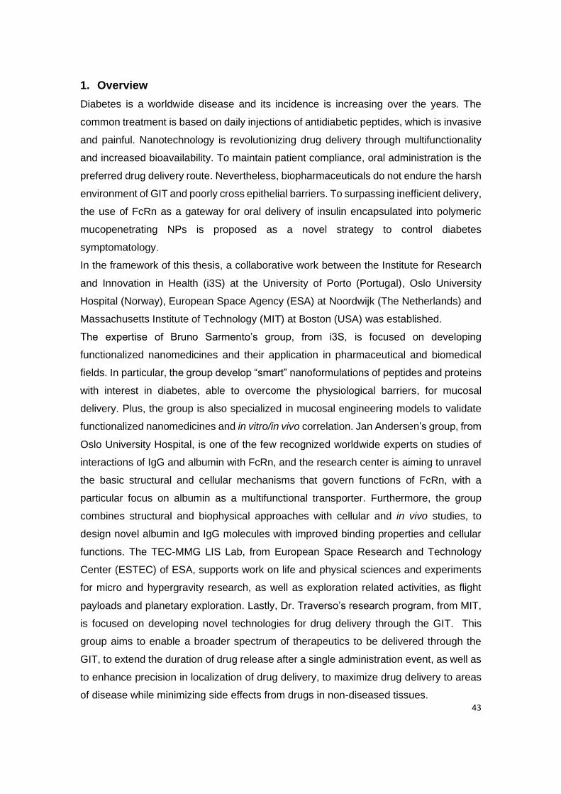
43
1. Overview
Diabetes is a worldwide disease and its incidence is increasing over the years. The
common treatment is based on daily injections of antidiabetic peptides, which is invasive
and painful. Nanotechnology is revolutionizing drug delivery through multifunctionality
and increased bioavailability. To maintain patient compliance, oral administration is the
preferred drug delivery route. Nevertheless, biopharmaceuticals do not endure the harsh
environment of GIT and poorly cross epithelial barriers. To surpassing inefficient delivery,
the use of FcRn as a gateway for oral delivery of insulin encapsulated into polymeric
mucopenetrating NPs is proposed as a novel strategy to control diabetes
symptomatology.
In the framework of this thesis, a collaborative work between the Institute for Research
and Innovation in Health (i3S) at the University of Porto (Portugal), Oslo University
Hospital (Norway), European Space Agency (ESA) at Noordwijk (The Netherlands) and
Massachusetts Institute of Technology (MIT) at Boston (USA) was established.
The expertise of Bruno Sarmento’s group, from i3S, is focused on developing
functionalized nanomedicines and their application in pharmaceutical and biomedical
fields. In particular, the group develop “smart” nanoformulations of peptides and proteins
with interest in diabetes, able to overcome the physiological barriers, for mucosal
delivery. Plus, the group is also specialized in mucosal engineering models to validate
functionalized nanomedicines and in vitro/in vivo correlation. Jan Andersen’s group, from
Oslo University Hospital, is one of the few recognized worldwide experts on studies of
interactions of IgG and albumin with FcRn, and the research center is aiming to unravel
the basic structural and cellular mechanisms that govern functions of FcRn, with a
particular focus on albumin as a multifunctional transporter. Furthermore, the group
combines structural and biophysical approaches with cellular and in vivo studies, to
design novel albumin and IgG molecules with improved binding properties and cellular
functions. The TEC-MMG LIS Lab, from European Space Research and Technology
Center (ESTEC) of ESA, supports work on life and physical sciences and experiments
for micro and hypergravity research, as well as exploration related activities, as flight
payloads and planetary exploration. Lastly, Dr. Traverso’s research program, from MIT,
is focused on developing novel technologies for drug delivery through the GIT. This
group aims to enable a broader spectrum of therapeutics to be delivered through the
GIT, to extend the duration of drug release after a single administration event, as well as
to enhance precision in localization of drug delivery, to maximize drug delivery to areas
of disease while minimizing side effects from drugs in non-diseased tissues.
44
These collaborations and combined expertise between groups allowed the performance
of this thesis and benefit my personal and professional skills, throughout the
development of FcRn-targeted nanoplatform for oral delivery of antidiabetic peptides.
2. Aims
The main aim of this thesis project was to develop nanoengineered platforms for delivery
of biopharmaceuticals through the intestine, with impact on diabetes management. The
main hypothesis is that surface-functionalized NPs with engineered albumin variants with
improved pH dependent binding to FcRn will enhance transcellular transport across
mucosal epithelial barriers and thus provide drug stability, controlled release and improve
the PK/PD profile of insulin, as a biopharmaceutical model. The ultimate objective was
to use the developed system as a therapy for T1DM.
The specific objectives of this thesis were:
i. To produce and characterize the efficacy of engineered albumin variants
conjugated to insulin-loaded NPs in a site-oriented manner (Chapter IV and
Appendix I);
ii. To analyze the FcRn binding and transport properties of the designed insulin
encapsulated NPs using in vitro model systems (Chapter IV and Appendix III);
iii. To explore the potential of simulated hypergravity in oral delivery of
biopharmaceuticals (Appendix II);
iv. To evaluate the intestinal permeability in a porcine ex vivo model, as a more
realistic platform to screen oral FcRn-targeted drug formulations (Chapter IV);
v. To assess in vivo the ability of the developed polymeric NPs to reduce glucose
levels in a hFcRn transgenic mice model, induced with T1DM (Chapter IV).
45
Chapter III
MATERIALS AND METHODS

46
47
1. Cell culture
HEK293E (human embryonic kidney) cells were acquired from ATCC (Manassas, VA,
USA) and maintained in RDMI 1640 (Sigma-Aldrich) with 10% heat-inactivated fetal calf
serum (FCS) (Sigma-Aldrich). A MDCK (Madin-Darby Canine Kidney) cell line
overexpressing human FcRn (MDCK-hFcRn) was generated by Drs. Alex Haas and Jens
Fischer (Roche Pharma Research and Early Development, pRED, Roche Innovation
Center Munich, Germany) and maintained until passage 20. MDCK-hFcRn cells were
cultured in DMEM (Gibco) with 15% fetal calf serum (FCS) and 200 μg/mL of G418
(Sigma-Aldrich). The expression level of FcRn-Enhanced Green Fluorescent Protein
(EGFP) was stably maintained by G418 and regularly checked by Thermofisher cell
counter countless II equipped with EVOS Light Cubes for GFP detection, being always
above 80%. Human microvascular endothelial cells (HMEC1) stably expressing HA-
hFcRn-EGFP (HMEC1-hFcRn) 198 were grown in MCDB 131 (Gibco) supplemented with
10% FCS, 2 mM L-glutamine, 10 ng/mL mouse epidermal growth factor (PeproTech)
and 1 µg/mL hydrocortisone (Sigma). Per flask was also added 5 µg/mL blasticidin S
(InvivoGen) and 100 µg/mL G418 (Sigma) to maintain stable the expression of hFcRn.
All the culture medium was supplied with 25 μg/mL streptomycin and 25 U/mL penicillin
(BioWhittaker), and cells were incubated at 37°C in a humidified 5% CO2, 95% air
incubator. All the cells were passaged twice per week.
2. Protein production
WT albumin and engineered albumin variants, namely K500A/K510Q (KAHQ) and
K573P (KP), were produced by following the procedures previously described, using
HEK293E cells 199. Purification of albumin variants was performed using a Capture select
albumin affinity column (GE Healthcare) and followed by gel filtration with a Superdex
200 increased column (GE Healthcare) connected to an Avant25 instrument (GE
Healthcare). Human IgG1-MST/HN and recombinant soluble human FcRn were
produced as previously described 71.
3. Polymeric nanoparticles production
Engineered albumin-conjugated and insulin encapsulated poly(lactic-co-glycolic)-
poly(ethylene glycol) (PLGA-PEG) NPs were prepared through a modified solvent
emulsification-evaporation method based on a w/o/w double emulsion technique 200, 201.
Briefly, 100 mg of polymer, in which 95% was PLGA (PURASORB® PDLG 5004A, in a
48
50/50 molar ratio of DL-lactide and Glycolide, kindly provided by Corbion) and 5% PLGA-
PEG-MAL (Akina, PolyScitech, AI110), was overnight dissolved in 2 mL of ethyl acetate.
Then, 0.2 mL of 75 mg/mL insulin solution (Sigma-Aldrich) in 0.1 M of HCl was added to
the polymeric solution and homogenized using a Bioblock vibracell 75186 sonicator
(Fischer Bioblock Scientific), for 1 min with 70% of amplitude. This primary emulsion
(w/o) was poured into 8 mL of 2% (w/v) Pluronic F-127 (Sigma) aqueous solution and
homogenized for 1 min using the same equipment, forming the second emulsion (w/o/w).
To this, it was added 15 mL of 2% Pluronic F-127 and the organic solvent was then
removed by evaporation during 3 h under magnetic stirring (300 rpm). The NPs were
washed twice with 0.25 mL of ultrapure water by centrifugation using Amicon® filters of
100 KDa (Millipore Corporation, Bedford, MA, USA) at 1000 x g for 40 min, and
redisposed in 0.1 M phosphate buffer (Na2HPO4, NaH2PO4) at pH 6.
For conjugation, the amount of albumin was set to the molar ratio 1:0.1 of PLGA-PEG-
MAL:Albumin. For each albumin variant, 500 µL of NPs (20 mg/mL) were transferred to
an eppendorf Amicon® filter of 100 KDa and centrifuged at 10,000 x g for 5 min at 4°C.
The supernatant was discarded, and NPs were resuspended in 250 µL of 0.1 M
phosphate buffer. Thereafter, it was added 250 µL of 5.71 µM of albumin-TCEP solution.
Each variant was diluted and incubated with 5.71 µM tris(2-carboxyethyl)phosphine
hydrochloride (TCEP) for 1 h at room temperature (RT), in order to have a 5.71 µM of
albumin-TCEP solution. The reaction was incubated overnight at 4°C. The following day,
samples were washed three times with 0.1 M Phosphate Buffer at pH 7 by centrifugation
using Amicon® filters of 100 KDa at 10,000 x g for 5 min at 4°C. The same procedure
was followed to unloaded PLGA-PEG NPs.
4. Physical chemical characterization
4.1. Particle size and zeta potential analysis
Three µL of NPs were characterized for hydrodynamic diameter, polydispersity index
(PdI) and zeta potential (ZP) upon dispersion in 1 mL of 10 mM sodium chloride by
dynamic light scattering (DLS) and laser Doppler electrophoresis (LDE) using a Zetasizer
Nano ZS (Malvern Instruments, Malvern, UK).
4.2. Morphological characterization and elementary analysis
The morphology of NPs was measured by transmission electron microscopy (TEM) and
the elementary analysis by elementary dispersive scanning (EDS). The samples were
49
placed on a grid and observed in a JEOL JEM-1400 electron microscope (JEOL Ltd,
Tokyo, Japan). During the preparation, samples were mounted onto metal stubs and
vacuum-coated with a layer of gold/palladium.
4.3. Insulin association efficiency and drug loading
The association efficiency (AE) and drug loading (DL) were determined using the
following formulas:
Equation (1) AE (%) = total amount of insulin−free insulin in supernatant
total amount of insulin∗ 100
Equation (2) DL (%) = total amount of insulin−free insulin in supernatant
total amount of NPs∗ 100
The free insulin in supernatant was isolated during the first washing step by
centrifugation with Amicon® filters at 1000 x g for 40 min at 4 °C. The insulin that did not
associate to NPs and remained in the aqueous phase was then collected and quantified
by a high-performance liquid chromatography (HPLC) method previously validated 202.
Briefly, insulin quantification was performed using a Merck-Hitachi LaChrom HPLC
instrument (Merck, Whitehouse Station, NJ, U.S.A.) equipped with a XTerra RP 18
column, 5 μm particle size, 4.6 mm internal diameter, ×250 mm length from Waters
(Milford, MA, U.S.A.), and a LiChrospher 100 RP-18, 5 μm particle size guard column
(Merk, Whitehouse Station, NJ, U.S.A.). Samples were run in triplicate.
4.4. Insulin release
Insulin-loaded NPs (1 mL) were dispersed in 10 mL of pH 1.2 buffer (50 mM KCl) to
simulate the gastric fluid (SGF) and incubated at 37°C under magnetic stirring at 100
rpm. After 2 h, the 10 mL suspension was ultracentrifuged at 14,000 x g for 20 min to
remove SGF and the NPs were then ressuspended with 10 mL of pH 6.0 buffer (50 mM
KH2PO4, 15 mM NaOH) to simulate the intestinal fluid (SIF) and incubated at 37°C under
magnetic stirring at 100 rpm. Aliquots of 0.2 mL were taken at predetermined time points
(15, 30, 45, 60, 120, 180, 240, 360 and 480 min) and the samples were replaced with a
respective pre-heated fresh buffer, keeping the volume constant. With the purpose to
calculate the released insulin, each aliquot was ultracentrifuged, and the supernatant
was used for HPLC analysis in order to quantify the insulin released from the NPs over
time. Samples were run in triplicate.
50
4.5. Albumin conjugation efficiency
The conjugation efficiency (CE) of albumin was determined using the following formula:
Equation (3) CE (%) = total amount of albumin−free albumin in supernatant
total amount of albumin∗ 100
The quantification was done using a two-way anti-albumin enzyme-linked
immunosorbent assay (ELISA). Briefly, 96 wells plates (Costar) were coated with 100 µL
of a polyclonal anti-albumin antibody produced in goat (Sigma Aldrich, ref A1151) diluted
1:2000 in PBS by incubation overnight at 4ºC. The wells were blocked with 200 µL
PBSTM solution, composed by PBS, 0.005% Tween 20 and 4% skimmed milk
(Acumedia), for 1 h at RT and then washed three times with 200 µL PBST (PBS, 0.005%
Tween 20). Dilution series of the albumin-conjugated NPs samples were prepared (1
mg/mL), starting from diluting the samples 100x and then titrated 1:2 in PBSTM. 100 µL
of each dilution was subsequently added in triplicates to wells and incubated for 1 h at
RT. The wells were washed as before. Alkaline phosphatase (AP)-conjugated polyclonal
anti-albumin from goat (Bethyl Laboratories; A80229AP) was diluted 1:4000 in PBSTM
and 100 µL was added per well and incubated for 1 h at RT. After washing as before,
visualization was performed by adding 100 µL of AP substrate (4-nitrophenyl phosphate
disodium salt hexahydrate substrate) (Sigma-Aldrich) diluted in diethanolamine buffer.
The absorbance was measured at 405 nm using the sunrise spectrophotometer
(TECAN).
5. Protein structure
5.1. Determination of secondary structure
The secondary structure elements of insulin loaded and albumin conjugated PLGA-PEG
NPs were assessed by Fourier Transform Infrared Spectroscopy (FTIR). The spectra
analysis was performed in ABB MB3000 FTIR spectrometer from ABB (Zurich,
Switzerland) equipped with a MIRacle single reflection attenuated total reflectance (ATR)
accessory from PIKE Technologies (Madison, WI, U.S.A.). Samples were freeze-dried
before analysis. The spectra were made with 256 scans and a 4 cm−1 resolution in the
region of 600-4000 cm−1, and each formulation was analyzed in triplicate. In order to
obtain the insulin and albumin spectra, a double subtraction procedure, followed by a 15
51
points Savitsky-Golay second derivative, and a baseline correction using a 3-4 point
adjustment at the amide I region (1710-1590 cm−1) 203. This spectral treatment was
executed using the HorizonMB FTIR software from ABB (Zurich, Switzerland). Lastly, all
spectra were area-normalized for comparison using Origin 8 software (OriginLab
Corporation, Northampton, MA, USA).
The quantitative comparison of the similarity of the FTIR spectra between insulin-loaded
NPs with that of native insulin, as well as between functionalized NPs and native albumin,
were determined using the area overlap (AO) and spectral correlation coefficient (SCC)
algorithms 204. Both algorithms were used considering the area normalized second-
derivative amide I spectra of native insulin or albumin. All the results are displayed in
percentage, and the percentage is proportionally correlated with the similarity of spectra.
The reference chosen was the insulin or albumin solutions because it is known that the
stability of a solid protein formulation increases with the rise of similarity to the FTIR
spectra in solution 204.
5.2. Confirmation of the conjugation
The 1H NMR spectra was conducted to confirm the chemical structure of albumin and it
conjugation to maleimide of NPs. The spectra were acquired at 80 ºC by using a Bruker
AVANCE III spectrometer (400 MHz). For these analyses, samples were freeze-dried
and then dissolved in DMSO-d6 (1/1 v/v) to result in 13 mg/mL. The available thiol
content in HSA and HSA-conjugated NPs was also determined by Ellman’s assay, as
described eslsewhere 205.
6. Binding to human FcRn
Microtiter wells were coated with a human IgG1 mutant variant
(M252Y/S254T/T256E/H433K/N434F) (10 µg/mL) in 100 µL PBS pH 7.4 by incubation
overnight at 4ºC. The wells were blocked with 200 µL PBSTM for 1 h at RT and then
washed three times with 200 µL PBST pH 5.5. Recombinant soluble human FcRn (10
µg/mL) in 100 µL PBSTM pH 5.5 was added per well and incubated for 1 h at RT, before
repeating the washing step above. Dilution series of the albumin-conjugated NPs
samples were prepared, starting from diluting the samples 20x in PBSTM pH 5.5,
followed by 1:2 titrations. 100 µL each dilution was subsequently added in triplicates to
wells and incubated for 1 h at RT. The wells were washed as before. AP-conjugated anti-
albumin from goat was diluted 1:4000 in PBSTM and 100 µL was added per well and
52
incubated for 1 h at RT. After washing as above, visualization was done by adding 100
µL of AP substrate (Sigma-Aldrich) diluted in diethanolamine buffer. The absorbance
was measured at 405 nm after 1h using the Sunrise spectrophotometer (TECAN).
7. In vitro experiments
7.1. Transcytosis assay
MDCK-hFcRn were seeded at a density of 1.2-1.4 x 10⁶ cells per well onto Transwell®
inserts (1.12 cm2) with collagen coated polytetrafluoroethylene (PTFE) membranes of
0.4 μm pore size (Corning Costar, Durham, NC, USA). Cells with a passage number
from 9 to 13 were used for all experiments. Cells form a monolayer with a transepithelial
electrical resistance (TEER) value of 600–900 Ω·cm2 on the day after seeding. TEER
was monitored using a MILLICELL-ERS-2 V-ohm meter (MILLIPORE). Prior to
experiments, the monolayers were washed twice then starved for 1 h in Hank's Balanced
Salt Solution (HBSS). For transport experiments at acidic pH, HBSS was buffered to pH
6.0 by addition of MES buffer (Sigma-Aldrich). Then, 200 µL of 500 nM albumin variants
or albumin-conjugated NPs were added to the apical chamber and 500 µL of HBSS at
the basolateral side. Samples were collected from the basolateral chambers at time 0
and 4 h post adding to the apical side. Results were expressed in relative transport and
apparent permeability (Papp), using the following formulas:
Equation (4) Relative Transport = Amount of sample
Mean amount of control
Equation (5) 𝑃𝑎𝑝𝑝 = ∆Q
A ×C0 × ∆t∗ 100
where C0 is the initial concentration in the apical compartment (mg/mL), A is the surface
area of the insert (cm2), ∆t is the time during which the experiment occurred (seconds)
and ∆Q is the amount of compound detected in the basolateral side (mg).
7.2. Uptake and recycling assays
The human endothelial cell-based recycling assay (HERA) was performed as previously
described 71. Briefly, 3.0 × 10⁵ HMEC1 cells per mL (p.13-16) expressing HA-hFcRn-
EGFP were seeded into 24-well plates per well (Costar) and cultured for 2 days in growth
medium. The cells were washed twice and starved for 1 h in Hank’s balanced salt
solution (HBSS) (Life Technologies). Then, 500 nM of naked albumin and the
53
correspondent of NPs conjugated with 500 nM of albumin were diluted in 250 μL HBSS
(pH 7.4) and added to the cells followed by 4 h incubation. The medium was removed
and the cells were washed four times with ice cold HBSS (pH 7.4), before fresh warm
HBSS (pH 7.4) or growth medium without FCS and supplemented with MEM non-
essential amino acids (ThermoFisher) was added. Samples were collected at 4 h or ON.
The amounts of albumin variants were quantified using ELISA as described above. In
parallel, total protein lysates were obtained using RIPA lysis buffer (ThermoFisher)
completed with protease inhibitor tablets (Roche). The mixture was incubated with the
cells on ice and a shaker for 10 min followed by centrifugation for 15 min at 10,000 × g
to remove cellular debris. Quantification of the amounts of HSA variants present in the
lysates was done by ELISA.
The relative recycling was calculated by following the equation:
Equation (6) Relative Recycling = Amount of sample
Mean amount of control
The derived values for recycling and residual amount for each of the albumin variants
were used to calculate the HERA score. The following formula was used:
Equation (7) Mean HERA score = Mean R sample
Mean R control
Mean RA sample
Mean RA control
The parameter R (in ng) is recycling at a given time while RA (in ng) is the residual
amount. The sample is the albumin variant naked or conjugated to NPs, while the control
is the WT or WT-conjugated NPs, used to standardize results.
8. Ex vivo experiments
8.1. Tissue dissection and cultivation
All animal procedures were conducted in accordance with protocols approved by the MIT
Committee on Animal Care in compliance with federal, state, local and institutional
regulations, including the Animal Welfare Act and Public Health Service Policy. Small
sections of intestinal tissue were isolated from freshly procured intact GI tracts from pigs
from selected local slaughterhouses. A section of the tissue was cut out of the GI tract,
dissected longitudinally and the outer muscle layer and serosa were removed. For
standard transport experiments, duodenum tissue was used. The tissue was washed in
a series of saline solutions under sterile conditions. The tissue was then either mounted
on the porcine ex vivo platform or maintained in culture. For cultivation, the media
advanced DMEM/F-12 (Life Technologies, 12634028) was used and supplemented with
54
10% fetal bovine serum (FBS) and 5% antibiotic–antimycotic solution (Thermo Fisher
Scientific, 15240062). Tissue was cultured in this cell culture medium in a sealed
polystyrene container that was then stored in an incubator kept at 37°C (no supplement
gas was provided). A set of experiments was conducted with fixed time points as well as
longer-term experiments monitoring the tissue daily, with analyses performed at varying
time points.
8.2. Immunohistochemistry and hematoxylin and eosin staining
Sections of the porcine GI tract were put into cassettes, fixed with 10% (v/w) formalin for
24 h at 4ºC and then storage in 70% ethanol at 4ºC. Paraffin sections (5-µm) on glass
slides were dried at 70°C for 30 min, deparaffinized and heat induced epitope retrieve
(HIER) at 97ºC for 20 min in citrate buffer pH 6. These procedures were completely
automated by using a ThermoFisher IHC Autostainer 360. Briefly, for
immunohistochemistry (IHC), endogenous peroxidase blocking was done for 10 min
followed by the protein block for 30 min. Primary antibody (1:100 anti-FCGRT, abcam,
ab193148) was incubated for 1 h and the secondary antibody (1:2000 goat anti-rabbit
IgG, abcam, ab205718) for 30 min. Both antibodies diluted with Tris Buffered Saline with
Tween 20 (TBST) and incubated at room temperature. Finally, an incubation of 5 min
with 3,3′-Diaminobenzidine (DAB) was done. For hematoxylin and eosin (H&E) staining,
paraffin sections were counterstained with hematoxylin and eosin, according to standard
protocols, using an automatic self-contained slide stainer (ThermoShandon Varistain
Gemini). The stained cells were then mounted on a cover slide using ConsulMount
(Thermo Fisher, 9990440). Representative images using Koehler illumination were
obtained with an Olympus Microfire digital camera (M/N S97809) attached to an Olympus
BX60 microscope.
8.3. RT-PCR analysis
The FcRn expression of ex vivo cultured porcine-derived intestinal tissue was
determined by reverse transcription polymerase chain reaction (RT–PCR) analysis and
detected by agarose electrophoresis. Initially, the tissue was removed from fresh GI,
frozen in liquid nitrogen and storage at -80ºC. Then, total RNA from each tissue sample
was extracted and purified with Quick-RNA plus (Zymo Research) followed by reverse
transcription into complementary DNA using a High-Capacity cDNA reverse transcription
kit (ThermoFisher Scientific). Target genes were amplified using the reported primers
55
(GAATTGGGCCCCGACAAT and CTGCTCACCCTTCCACTCC) 167 and Platinum™
SuperFi™ DNA Polymerase (ThermoFisher Scientific,12351010). The PCR program
was optimized and consisted of an enzyme activation step (98ºC, 10min), forty cycles of
denaturation (98ºC, 30 sec), annealing extension (62ºC, 1 min) and extension (72ºC, 45
sec), followed by an extension at 72ºC for 10 min. The expression level of FcRn was
measured with 1.5% agarose gel electrophoreses, with SYBR Safe DNA gel stain
(Thermofisher, S33102) and 100 bp DNA Ladder (New England BioLab, N3231S) and
detected using the Bio-Rad Image suite.
8.4. Transport experiments using the ex vivo platform
For assembly of the ex vivo platform, freshly isolated intestinal tissue from the duodenum
was prepared according to the above-described tissue dissection procedure and
mounted on the manufactured interface design with a generic 96-well plate (96-well
plates, clear bottom, Corning), as previously described 169. To the plate from the bottom,
the basolateral side, it was added 100 µL of Hanks' Balanced Salt solution (HBSS) pH
7.4. The duodenum was identified as the region of the small intestine approximately 1 m
that followed the stomach. Duodenum tissue was then carefully moved over the bottom
magnetic plate with the apical side facing upwards. Afterwards, the top magnetic plate
was mounted, and 90 µL of 189.83 µg/mL (500 nM) of albumin or albumin-conjugated to
NPs was added, at the apical side. Samples were prepared at acidic pH, using HBSS
buffered to pH 6.0 by addition of 2-(N-morpholino) ethanesulfonic acid (MES) buffer
(Sigma Aldrich). After 4 h of incubation, samples were collected from the basolateral
side, with a syringe, at the bottom magnetic plate and quantified by ELISA. All
experiments, including sample incubation, were performed at room temperature. Results
were expressed in relative transport and apparent permeability (Papp), as described in
section 7.1.
9. In vivo experiments
9.1. Induction of T1DM
Male homozygous human FcRn Tg32 mice (The Jackson Laboratory, B6.Cg-
Fcgrttm1DcrTg(FCGRT)32Dcr/DcrJ) 157, age 7-11 weeks and weighing between 16-25 g,
were used to determine the glycemic decrease after oral administration of albumin
decorated insulin-loaded NPs. Experiments were conducted in accordance with the
provisions of FELASA and EU (Council Directive 86/609/EEC), after approval by local
56
Ethic Committee. The mice were maintained under standard laboratory conditions (12 h
light/dark cycles, temperature of 21 ± 2°C and relative humidity of 35% to 60%) and fed
with standard pellets and water added libitum. Diabetes induction was done in 12 h
fasting mice by intraperitoneal injection of 150 mg/kg streptozotocin (STZ) (Sigma-
Aldrich). STZ with a concentration of 20 mg/mL was freshly dissolved in citrate buffer 0.1
M at pH 4.5 201, 206. STZ destroy pancreatic β-cells, possible leading to a massive insulin
release and consequent fatal hypoglycemia. To prevent this, water supplemented with
10% sucrose was provided to animals over 48 h after STZ induction. After this period, it
was substituted by tap water and glucose levels and body weight were measured daily.
9.2. Pharmacodynamics
One week after STZ induction, 12 h fasted animals with glucose levels above 250 mg/dL
were randomly divided into 6 groups: (1) PBS, (2) 50 IU/kg of insulin-loaded NPs-NF,
(3) 50 IU/kg of insulin-loaded NPs-WT, (4) 50 IU/kg of insulin-loaded NPs-KP, (5) 50
IU/kg of insulin in solution, all diluted in PBS and administered by oral gavage and (6)
2.5 IU/Kg of s.c. insulin diluted in PBS. The dose was chosen based on previous studies
201, 206. Blood glucose levels were measured by tail puncture at different time points (0,
0.5, 1, 2, 4, 6 and 8 h) after administration. Plasma glucose levels were plotted against
time to evaluate the cumulative hypoglycemic effect (HD) over time, quantified by the
area under the curve (AUC), determined using the trapezoidal method. Pharmacological
availability (PA), quantified by the area above the curve (AAC), of insulin-loaded NPs
and in solution were determined as the relative measure of the cumulative reduction in
glucose blood levels. Both parameters were compared to 0% of decrease or 100%
availability, respectively, of the control insulin administered subcutaneously at a dose of
2.5 IU/kg. PA and HD in serum glucose levels were calculated as follows 201, 206:
Equation (8) PA (%) = AAC test
Dose testAAC control
Dose control
× 100
Equation (9) HD (%) =AUC (control 0−480min)−AUC (group 0−480min)
AUC (control 0−480min)× 100
Plasma glucose levels were determined using the Medisense Precision Xceed Kit
(Abbot, range 20-500 mg/dL) and expressed as the percentage of the baseline plasma
glucose level.
57
10. Statistical analysis
Statistical analysis was performed using the GraphPad Prism Software vs. 6.0
(GraphPad Software Inc). Statistical significance of the difference between two groups
was evaluated by the Student’s t-test. Differences between groups were compared using
one-way analysis of variance (ANOVA) Tukey’s multiple comparisons test or Dunnett’s
multiple comparison test. Results are expressed as mean ± standard deviation and
geometric means with 95% confidence intervals.

58
59
Chapter IV
RESULTS AND DISCUSSION

60
61
This chapter was based in the following publications:
• Azevedo C., Andersen J.T., Sarmento B., Traverso G., The potential of porcine ex
vivo platform for intestinal permeability screening of FcRn-targeted drugs, European
Journal of Pharmaceutics and Biopharmaceutics 2021, 162: 99-104.
In this original paper, I was responsible for the design and execution of the experimental
work, as well as the data analysis, their interpretation and discussion and conception
and revision of the manuscript. The supervisors were responsible for reviewing and
submit the manuscript. This paper was not previously included in other thesis and it is
partially reproduced in this section. The integral version of the original paper was
included at the end of the thesis, at Appendix IV.
• Azevedo C, Nilsen J, Grevys A, Nunes R, Andersen JT, Sarmento B, Engineered
albumin-functionalized nanoparticles for improved FcRn binding enhances oral
delivery of insulin, Journal of Controlled Release 2020, 327: 161-173.
In this original paper, I was responsible for the design and execution of the experimental
work, as well as data analysis, their interpretation and discussion and conception and
revision of the manuscript. Jeannette Nilsen taught and helped me to produce
engineered albumin variants. Algirdas Grevys taught me how to handle with MDCK-
hFcRn and HMEC1-hFcRn cells and to perform permeability and recycling assays. Rute
Nunes helped me with the in vivo assays. The supervisors were responsible for reviewing
and submit the manuscript. This paper was not previously included in other thesis and it
is partially reproduced in this section. The integral version of the original paper was
included at the end of the thesis, at Appendix IV.

62

63
1. Rationale for the design of albumin decorated nanoparticles
To design biodegradable insulin-loaded PLGA-PEG NPs decorated with albumin for
targeting of FcRn, a recombinant wild-type (WT) human albumin and an engineered
variant with improved human FcRn binding properties were used. The engineered variant
contains a single amino acid substitution in the C-terminal domain III (K573P, KP), which
has been shown to extend serum half-life in human FcRn expressing mice and non-
human primates 207, 208. In addition, a human albumin variant engineered to abolish
binding to the receptor (K500A/H464Q, KAHQ) was included 207, 208. These mutations are
also located in the C-terminal domain III of albumin, which harbors the principle binding
site for FcRn (Figure 10 A).
To ensure directional attachment of the albumin variants to the NPs, an exposed and
free cysteine residue within the N-terminal domain I of albumin (Cys34) (Figure 10 A),
which is located distally from the core FcRn binding site, was explored 207, 208. Maleimide
(MAL) chemistry was used to promote formation of a thioether bond between Cys34 of
albumin and the maleimide group of PLGA-PEG-MAL on the surface of the NP (Figure
10 B). As such, this approach secures directional and site-specific conjugation, as
opposed to random conjugation.
Figure 10 | Representation of the rationale of this study: (A) Illustration of the crystal structure
of human albumin where targeted residues are shown as spheres: the free cys34 (black) in the
N-terminal domain I and K500 (orange), H510 (orange) and K573 (blue) in the C-terminal domain
III. The figure was made using Pymol and the crystal structure data of human albumin (PDB
1AO6); (B) Schematic illustration of the designed NPs build up of PLGA and PLGA-PEG-MAL
with encapsulated soluble recombinant insulin and recombinant human albumin on the NPs’
surface. Albumin was conjugated to the NP using maleimide chemistry, which resulted in a
thioether bond formed between the Cys34 of albumin and the maleimide group of PLGA-PEG-
MAL.
64
2. Characterization of the produced nanoparticles
2.1. Physical-chemical properties of albumin-decorated PLGA-PEG NPs
To establish a factorial production strategy, a well-established solvent emulsification-
evaporation method based on a w/o/w double emulsion technique was used 200, 201.
PLGA, PLGA-PEG-MAL and WT albumin were mixed in different ratios following
determination of parameters such as size, polydispersity index (PdI), zeta potential (ZP)
and conjugation efficiency (CE). Based on previous works 209, 210, and corroborated by
JMP software analysis, we selected the ratio 1:0.05:0.005 of PLGA: PLGA-PEG-MAL:
albumin, due to favorable CE, lowest PdI, more negative ZP and size around 150 nm
(not shown). In fact, the NPs composition is of upmost importance to surpass mucosal
barriers. In the particular case of PEGylated nanoparticles, PLGA-PEG is responsible by
forming a hydrophilic corona and PEG is regularly used to prevent NPs from aggregation
and enzymatic degradation, contributing for their stability. PEG is the “gold standard” to
promote mucus penetration, since interactions with mucins by its hydrophilic and net-
neutrally charged character are reduced 211, 212. It has been demonstrated that a minimal
surface coverage of 5% PEG in NPs is crucial for a rapid mucus penetration 213. These
features were important to modulate and facilitate mucus penetration, epithelial
absorption and transcytosis of NPs.
2.2. Albumin conjugation to NPs
To ensure that Cys34 was accessible for covalent conjugation to PLGA-PEG-MAL
containing NPs, human albumin was reduced by tris(2-carboxyethyl)phosphine
hydrochloride (TCEP). Albumin combined with NPs without maleimide (MAL) (Figure 11
B) or non-reduced albumin with PLGA-PEG-MAL NPs (Figure 11 C) were included as
controls. To remove unconjugated albumin, NPs were washed three times with
phosphate buffer (PB) using an Amicon® filter with 100 kDa cut off. Initially, the washing
procedure was performed with free albumin, which confirmed that albumin passes
through the filter (Figure 11 A). To indirectly determine the conjugation efficiency, the
unbound albumin fraction was quantified using a two-way anti-human albumin ELISA.
The results revealed that only 10% of the total albumin incubated with the NPs was
detected in the wash solution, supporting that 90% was bound to the NPs (Figure 11 B).
Interestingly, this was also the case in the absence of reactive MAL groups on the NPs
or a reduced Cys34 of albumin, both necessary for the formation of a covalent thioether
bond, which suggest that albumin also bound nonspecifically to the NPs. To reduce the
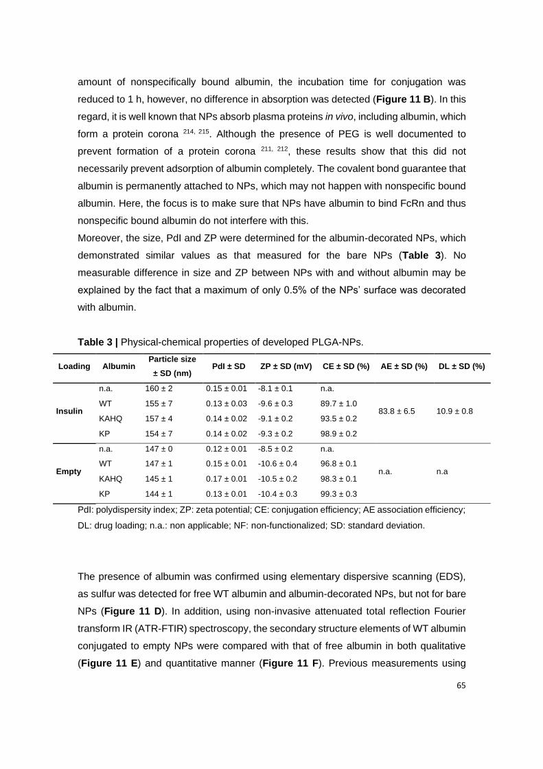
65
amount of nonspecifically bound albumin, the incubation time for conjugation was
reduced to 1 h, however, no difference in absorption was detected (Figure 11 B). In this
regard, it is well known that NPs absorb plasma proteins in vivo, including albumin, which
form a protein corona 214, 215. Although the presence of PEG is well documented to
prevent formation of a protein corona 211, 212, these results show that this did not
necessarily prevent adsorption of albumin completely. The covalent bond guarantee that
albumin is permanently attached to NPs, which may not happen with nonspecific bound
albumin. Here, the focus is to make sure that NPs have albumin to bind FcRn and thus
nonspecific bound albumin do not interfere with this.
Moreover, the size, PdI and ZP were determined for the albumin-decorated NPs, which
demonstrated similar values as that measured for the bare NPs (Table 3). No
measurable difference in size and ZP between NPs with and without albumin may be
explained by the fact that a maximum of only 0.5% of the NPs’ surface was decorated
with albumin.
Table 3 | Physical-chemical properties of developed PLGA-NPs.
Loading Albumin Particle size
± SD (nm) PdI ± SD ZP ± SD (mV) CE ± SD (%) AE ± SD (%) DL ± SD (%)
Insulin
n.a. 160 ± 2 0.15 ± 0.01 -8.1 ± 0.1 n.a.
83.8 ± 6.5 10.9 ± 0.8 WT 155 ± 7 0.13 ± 0.03 -9.6 ± 0.3 89.7 ± 1.0
KAHQ 157 ± 4 0.14 ± 0.02 -9.1 ± 0.2 93.5 ± 0.2
KP 154 ± 7 0.14 ± 0.02 -9.3 ± 0.2 98.9 ± 0.2
Empty
n.a. 147 ± 0 0.12 ± 0.01 -8.5 ± 0.2 n.a.
n.a. n.a WT 147 ± 1 0.15 ± 0.01 -10.6 ± 0.4 96.8 ± 0.1
KAHQ 145 ± 1 0.17 ± 0.01 -10.5 ± 0.2 98.3 ± 0.1
KP 144 ± 1 0.13 ± 0.01 -10.4 ± 0.3 99.3 ± 0.3
PdI: polydispersity index; ZP: zeta potential; CE: conjugation efficiency; AE association efficiency;
DL: drug loading; n.a.: non applicable; NF: non-functionalized; SD: standard deviation.
The presence of albumin was confirmed using elementary dispersive scanning (EDS),
as sulfur was detected for free WT albumin and albumin-decorated NPs, but not for bare
NPs (Figure 11 D). In addition, using non-invasive attenuated total reflection Fourier
transform IR (ATR-FTIR) spectroscopy, the secondary structure elements of WT albumin
conjugated to empty NPs were compared with that of free albumin in both qualitative
(Figure 11 E) and quantitative manner (Figure 11 F). Previous measurements using
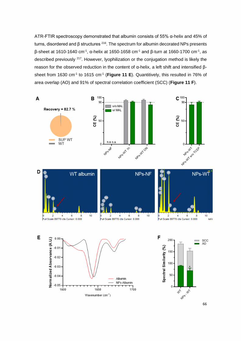
66
ATR-FTIR spectroscopy demonstrated that albumin consists of 55% α-helix and 45% of
turns, disordered and β structures 216. The spectrum for albumin decorated NPs presents
β-sheet at 1610-1640 cm-1, α-helix at 1650-1658 cm-1 and β-turn at 1660-1700 cm-1, as
described previously 217. However, lyophilization or the conjugation method is likely the
reason for the observed reduction in the content of α-helix, a left shift and intensified β-
sheet from 1630 cm-1 to 1615 cm-1 (Figure 11 E). Quantitively, this resulted in 76% of
area overlap (AO) and 91% of spectral correlation coefficient (SCC) (Figure 11 F).
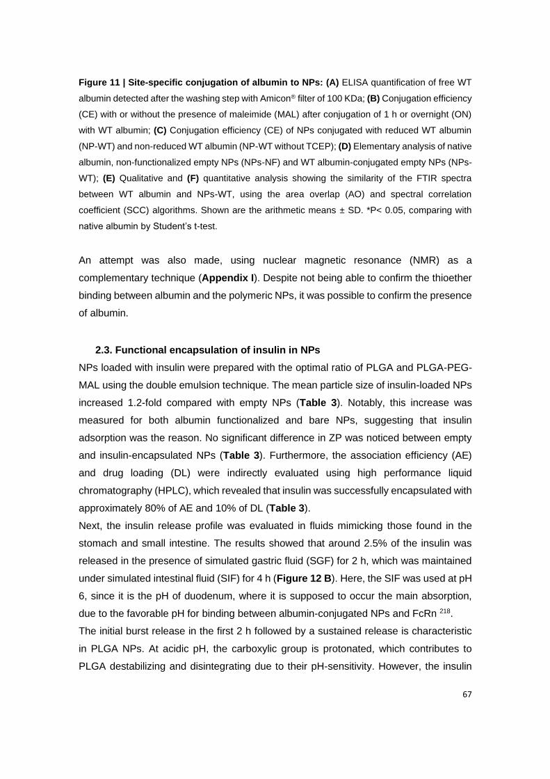
67
Figure 11 | Site-specific conjugation of albumin to NPs: (A) ELISA quantification of free WT
albumin detected after the washing step with Amicon® filter of 100 KDa; (B) Conjugation efficiency
(CE) with or without the presence of maleimide (MAL) after conjugation of 1 h or overnight (ON)
with WT albumin; (C) Conjugation efficiency (CE) of NPs conjugated with reduced WT albumin
(NP-WT) and non-reduced WT albumin (NP-WT without TCEP); (D) Elementary analysis of native
albumin, non-functionalized empty NPs (NPs-NF) and WT albumin-conjugated empty NPs (NPs-
WT); (E) Qualitative and (F) quantitative analysis showing the similarity of the FTIR spectra
between WT albumin and NPs-WT, using the area overlap (AO) and spectral correlation
coefficient (SCC) algorithms. Shown are the arithmetic means ± SD. *P< 0.05, comparing with
native albumin by Student’s t-test.
An attempt was also made, using nuclear magnetic resonance (NMR) as a
complementary technique (Appendix I). Despite not being able to confirm the thioether
binding between albumin and the polymeric NPs, it was possible to confirm the presence
of albumin.
2.3. Functional encapsulation of insulin in NPs
NPs loaded with insulin were prepared with the optimal ratio of PLGA and PLGA-PEG-
MAL using the double emulsion technique. The mean particle size of insulin-loaded NPs
increased 1.2-fold compared with empty NPs (Table 3). Notably, this increase was
measured for both albumin functionalized and bare NPs, suggesting that insulin
adsorption was the reason. No significant difference in ZP was noticed between empty
and insulin-encapsulated NPs (Table 3). Furthermore, the association efficiency (AE)
and drug loading (DL) were indirectly evaluated using high performance liquid
chromatography (HPLC), which revealed that insulin was successfully encapsulated with
approximately 80% of AE and 10% of DL (Table 3).
Next, the insulin release profile was evaluated in fluids mimicking those found in the
stomach and small intestine. The results showed that around 2.5% of the insulin was
released in the presence of simulated gastric fluid (SGF) for 2 h, which was maintained
under simulated intestinal fluid (SIF) for 4 h (Figure 12 B). Here, the SIF was used at pH
6, since it is the pH of duodenum, where it is supposed to occur the main absorption,
due to the favorable pH for binding between albumin-conjugated NPs and FcRn 218.
The initial burst release in the first 2 h followed by a sustained release is characteristic
in PLGA NPs. At acidic pH, the carboxylic group is protonated, which contributes to
PLGA destabilizing and disintegrating due to their pH-sensitivity. However, the insulin
68
released could also be some adsorbed on the NPs’ surface. Either way, it is not
significant, considering the 2.5% of the released profile. Besides, insulin has an
isoelectric point of 5.3, meaning that above this value, insulin is negatively charged. As
PLGA-PEG NPs have a negative charge, at pH lower than 5.3, there is a strong
electrostatic attraction between insulin and NPs, decreasing the release rate. It is
described that PLGA is strongly degraded at the alkaline and acidic conditions due to
the ester hydrolysis caused by acid-base catalysis 219. Thus, we expect that insulin can
be sustainly released at neutral pH, as blood circulation. The results show that NPs are
able to sustain low release of insulin under gastrointestinal conditions and thus, prevent
the premature release of insulin, which suggest that it will be protected against
degradation in the GIT. The slow release is a good omen to reduce the frequency of
administrations and reduce the cost of therapy. In line with this, a previous report
demonstrated that peptides were protected from low gastric pH value and enzymatic
hydrolysis when encapsulated in anionic polymeric NPs 220, 221. In fact, it was described
the reduction in drug release from PLGA microparticles due to the surface crosslinking
of PLGA microparticles 222. Plus, albumin-based NPs can also protect the encapsulated
drug from enzymatic degradation, due to the level of chemical crosslinking 223. Albumin
adsorbed to alginate/dextran sulfate (ADS)-NPs have shown a 30% of insulin released
in SGF and a slow release following insulin passage to intestinal conditions. Plus, these
authors claimed that chitosan and albumin coating augmented intestinal interactions
between glycocalix and the NPs, which translated in enhanced intestinal permeability 89.
Moreover, to determine the morphology of insulin-loaded NPs decorated with WT
albumin, transmission electron microscopy (TEM) was performed, which revealed a
spherical shape and smooth surface (Figure 12 A). This feature is in line with expected
morphology based on the encapsulation methodology used 200, 201. The size of the NPs
was calculated to be in the range of 150 nm, in agreement with the DLS analysis (Table
3).
To address if the double emulsion technique affected the integrity of insulin, ATR-FTIR
spectroscopy was used to determine the secondary structure elements in both a
qualitative (Figure 12 C) and a quantitative manner (Figure 12 D). When proteins are
associated with PLGA NPs, the C=O stretching vibrations of the ester group of PLGA
appear at 1750 cm-1 and structural modifications can be determined following
normalization of the area of the second derivative amide I spectra 203. In addition,
qualitatively assessment can be done by comparing the spectra or quantitatively by
calculating the AO and SCC where AO indicates modifications in structure elements,
69
while SCC represents the changes in the band positions 200. Notably, the normalized
area of the second-derivative of insulin spectrum is characterized by β-sheets bands at
1610-1604, α-helix at 1650-1658 cm-1, a random coil at 1640-1650 cm-1 and a β-turn
structure 1660-1700 cm-1 224. The obtained spectra for insulin in solution and
encapsulated in NPs were similar. However, some bands were modified, indicating
alterations in secondary structure (Figure 12 A). This was evident from the spectrum on
the insulin-loaded NPs with a shift of α-helix from ~1645 cm-1 to ~1655 cm-1 and
increased intensity, in addition to a left shift and higher intensity in β-turn, which is in
agreement with previous work 225. Moreover, the 68% of AO and 79% of SCC (Figure
12 B) support the visualization of the spectrum, which can be justified by the
encapsulation method or by lyophilization of the samples, suggesting the necessity of
adding lyoprotectants before performing ATR-FTIR spectroscopy. Notably, it has been
shown that lyoprotectants stabilize the protein by protecting it from denaturation caused
by pore formation on NPs 201, 226. It has also been reported that lyophilization leads to
protein aggregation with intermolecular β-sheets formation (caused by intermolecular
hydrogen bonds) that increases protein–protein interactions while decreasing the α-helix
content and stability 227. In addition, the helical structure of protein is destabilized during
evaporation, since water molecules compete for the hydrogen bonds with amino and
carboxyl groups of peptide bonds 228. Regarding the tertiary structure, related to insulin
bioactivity, we have previously analyzed it by fluorescence spectroscopy, after extracting
insulin from NPs, confirming similarity with FTIR results after encapsulation 201, 225.

70
Figure 12 | Insulin NPs characterization: (A) Representative TEM microphotograph of WT
albumin decorated PLGA-PEG NPs encapsulated with insulin, lyophilized 25000x. Scale bar: 500
nm. (B) Insulin release profile of functionalized and non-functionalized NPs under SGF (2 h) and
SIF (from 2 h to 6 h). (C) The qualitative and (D) quantitative effect of NPs encapsulation on
protein structure. Analysis showing area-normalized second-derivative amide I spectra of native
insulin vs insulin-encapsulated NPs using the area overlap (AO) and spectral correlation
coefficient (SCC) algorithms. Shown are the arithmetic means ± SD. **P<0.001, ***P<0.0001
comparing with native insulin by Student’s t-test.
2.4. Albumin decorated NPs bind FcRn in a pH dependent manner
To address whether albumin is functional post conjugation to insulin-loaded NPs, binding
to FcRn at pH 5.5 was tested by ELISA. Equal amounts of the NPs formulations or the
free human albumin variants were added to wells where recombinant human FcRn had
been captured on a coated engineered IgG molecule that strongly binds the receptor

71
(Figure 13 A), which were subsequently detected using a polyclonal anti-human albumin
antibody. The three albumin variants bound as expected, as KAHQ did not bind while
the KP variant bound more strongly than the WT (Figure 13 B). These results are in line
with the reported in literature 207, 208. Importantly, the same binding hierarchy was
detected for the three NPs formulations (Figure 13 C). Specifically, KP decorated NPs
bound more strongly to human FcRn than WT-decorated NPs, while the KAHQ-
containing NPs ignored the receptor. As expected, no signal was detected for non-
functionalized NPs. Thus, PLGA-PEG NPs decorated with albumin retain the ability to
interact with human FcRn.
Figure 13 | Albumin decorated NPs bind human FcRn. (A) A schematic illustration of the
ELISA set-up for measuring functional binding to human FcRn at pH 5.5. An engineered IgG1
antibody (MST/HN) was coated in wells, to which recombinant soluble human FcRn was captured,
before adding naked albumin or albumin-decorated NPs. Bound albumin and albumin-decorated
NPs were detected using ALP-conjugated polyclonal anti-human albumin antibodies. (B) Binding
of naked human albumin variants (WT, KAHQ or KP) to human FcRn at pH 5.5. (C) Binding of
NPs decorated with human albumin variants (WT, KAHQ or KP) to human FcRn at pH 5.5.
3. In vitro validation
3.1. Produced NPs show enhanced in vitro transcytosis in epithelial cells
The finding that albumin decorated NPs bound human FcRn favorably, motivated me to
investigate if this property could translate into enhanced transcellular transport. To test
this, a Transwell® assay was run using a polarizing Madin-Darby Canine Kidney cell line
over-expressing human FcRn (MDCK-hFcRn). To verify that the system could be
adopted to study transcytosis of human albumin, equal amounts of the WT, KAHQ and
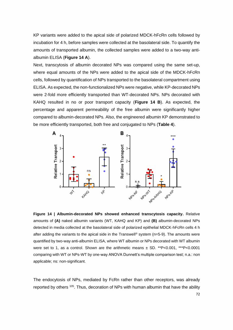
72
KP variants were added to the apical side of polarized MDCK-hFcRn cells followed by
incubation for 4 h, before samples were collected at the basolateral side. To quantify the
amounts of transported albumin, the collected samples were added to a two-way anti-
albumin ELISA (Figure 14 A).
Next, transcytosis of albumin decorated NPs was compared using the same set-up,
where equal amounts of the NPs were added to the apical side of the MDCK-hFcRn
cells, followed by quantification of NPs transported to the basolateral compartment using
ELISA. As expected, the non-functionalized NPs were negative, while KP-decorated NPs
were 2-fold more efficiently transported than WT-decorated NPs. NPs decorated with
KAHQ resulted in no or poor transport capacity (Figure 14 B). As expected, the
percentage and apparent permeability of the free albumin were significantly higher
compared to albumin-decorated NPs. Also, the engineered albumin KP demonstrated to
be more efficiently transported, both free and conjugated to NPs (Table 4).
Figure 14 | Albumin-decorated NPs showed enhanced transcytosis capacity. Relative
amounts of (A) naked albumin variants (WT, KAHQ and KP) and (B) albumin-decorated NPs
detected in media collected at the basolateral side of polarized epithelial MDCK-hFcRn cells 4 h
after adding the variants to the apical side in the Transwell® system (n=5-9). The amounts were
quantified by two-way anti-albumin ELISA, where WT albumin or NPs decorated with WT albumin
were set to 1, as a control. Shown are the arithmetic means ± SD. **P<0.001, ***P<0.0001
comparing with WT or NPs-WT by one-way ANOVA Dunnett’s multiple comparison test; n.a.: non
applicable; ns: non-significant.
The endocytosis of NPs, mediated by FcRn rather than other receptors, was already
reported by others 105. Thus, decoration of NPs with human albumin that have the ability
73
to engage human FcRn resulted in enhanced apical to basolateral transport across
polarized epithelial cells. Importantly, transport could be further enhanced by taking
advantage of a human albumin variant (KP) engineered for improved pH dependent
FcRn binding.
Table 4 | Permeability percentage and apparent permeability coefficient calculated
after 4 h of incubation in MDCK-hFcRn cells.
Formulation Permeability ± SD (%) Papp ± SD (x 10-6)
WT 11.4 ± 6.9 1.44 ± 0.87
KAHQ 5.2 ± 2.8 0.66 ± 0.36
KP 28.7 ± 4.9 3.63 ± 0.62
NPs-NF 0.31 ± 0.28 0.04 ± 0.04
NPs-WT 0.84 ± 0.36 0.11 ± 0.05
NPs-KAHQ 0.72 ± 0.57 0.09 ± 0.07
NPs- KP 1.86 ± 0.60 0.25 ± 0.08
Considering the NPs size (~150 nm) is relatively easy to obtain good cellular uptake
outputs. As described in the literature, NPs are passivly translocated across epithelium
through enterocytes and specifically M cells, within Peyer’s patches 229. It was also
reported that NPs are able to transverse the lymphatic system and by lymphatic drainage
through thoracic duct reach the systemic circulation 230. On top of this, receptor targeting
is likely to increase the chance of absorption, since FcRn is expressed in epithelial cells
231. Notably, a significant increase of insulin permeability by using chitosan-based NPs
with adsorbed albumin has been shown in cell-based engineered intestinal models 89.
A similar assay was performed with Caco-2 cells, using insulin-loaded NPs conjugated
with WT albumin. This was done within the scope of a pioneer project, to investigate the
potential of simulated hypergravity to be used in oral delivery of biopharmaceuticals
(Appendix II).
3.2. Produced NPs show enhanced in vitro uptake and recycling in
endothelial cells
Next, it was investigated what happens to NPs after being transported through epithelial
cells. There is no reported data related to FcRn-mediated recycling of NPs, thus, to clarify
this, a recycling assay was performed. For this assay it was used a modified human
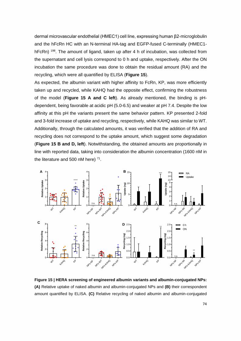
74
dermal microvascular endothelial (HMEC1) cell line, expressing human β2-microglobulin
and the hFcRn HC with an N-terminal HA-tag and EGFP-fused C-terminally (HMEC1-
hFcRn) 198. The amount of ligand, taken up after 4 h of incubation, was collected from
the supernatant and cell lysis correspond to 0 h and uptake, respectively. After the ON
incubation the same procedure was done to obtain the residual amount (RA) and the
recycling, which were all quantified by ELISA (Figure 15).
As expected, the albumin variant with higher affinity to FcRn, KP, was more efficiently
taken up and recycled, while KAHQ had the opposite effect, confirming the robustness
of the model (Figure 15 A and C left). As already mentioned, the binding is pH-
dependent, being favorable at acidic pH (5.0-6.5) and weaker at pH 7.4. Despite the low
affinity at this pH the variants present the same behavior pattern. KP presented 2-fold
and 3-fold increase of uptake and recycling, respectively, while KAHQ was similar to WT.
Additionally, through the calculated amounts, it was verified that the addition of RA and
recycling does not correspond to the uptake amount, which suggest some degradation
(Figure 15 B and D, left). Notwithstanding, the obtained amounts are proportionally in
line with reported data, taking into consideration the albumin concentration (1600 nM in
the literature and 500 nM here) 71.
NPs-
NF
NPs-
WT
NPs-
KAHQ
NPs-
KP
0
1
2
3
4
Rela
tive U
pta
ke
n.a.
WT
KAHQ KP
0.0
0.5
1.0
1.5
2.0
2.5
Recyclin
g (
ng
)
**
NPs-
NF
NPs-
Wt
NPs-
KAHQ
NPs-
KP
0.0
0.5
1.0
1.5
2.0
2.5
Recyclin
g (
ng
)
**
**
n.a.
WT
KAHQ KP
0
1
2
3
4
Rela
tive U
pta
ke ****
A
WT
KAHQ KP
0
5
10
15
Up
tak
e (
ng
)
***
NPs-
NF
NPs-
Wt
NPs-
KAHQ
NPs-
KP
0
1
2
3
4
513
14
15
Up
tak
e (
ng
)
n.a.
WT
KAHQ KP
0
2
4
6
8
Rela
tive R
ecyclin
g
**
NPs-
NF
NPs-
WT
NPs-
KAHQ
NPs-
KP
0
2
4
6
8
Rela
tive R
ecyclin
g
n.a.
B
DC
0 h
ON
RA
Uptake
Figure 15 | HERA screening of engineered albumin variants and albumin-conjugated NPs:
(A) Relative uptake of naked albumin and albumin-conjugated NPs and (B) their correspondent
amount quantified by ELISA. (C) Relative recycling of naked albumin and albumin-conjugated
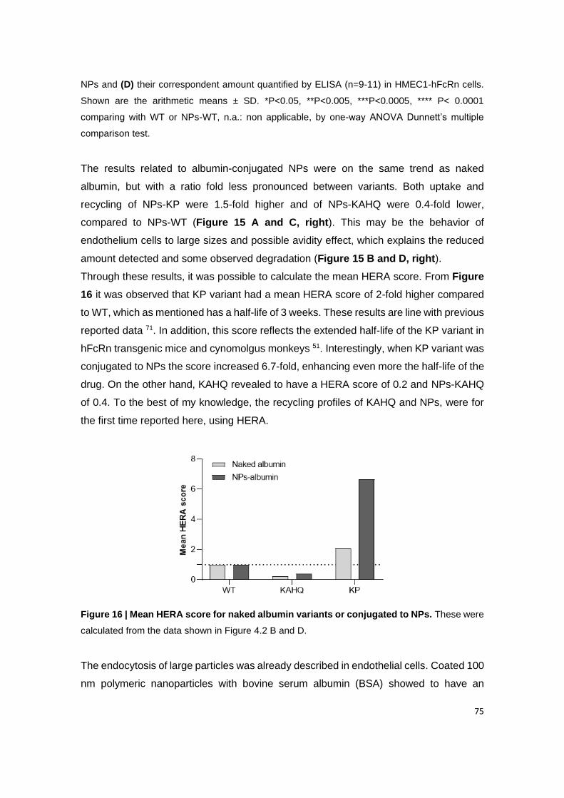
75
NPs and (D) their correspondent amount quantified by ELISA (n=9-11) in HMEC1-hFcRn cells.
Shown are the arithmetic means ± SD. *P<0.05, **P<0.005, ***P<0.0005, **** P< 0.0001
comparing with WT or NPs-WT, n.a.: non applicable, by one-way ANOVA Dunnett’s multiple
comparison test.
The results related to albumin-conjugated NPs were on the same trend as naked
albumin, but with a ratio fold less pronounced between variants. Both uptake and
recycling of NPs-KP were 1.5-fold higher and of NPs-KAHQ were 0.4-fold lower,
compared to NPs-WT (Figure 15 A and C, right). This may be the behavior of
endothelium cells to large sizes and possible avidity effect, which explains the reduced
amount detected and some observed degradation (Figure 15 B and D, right).
Through these results, it was possible to calculate the mean HERA score. From Figure
16 it was observed that KP variant had a mean HERA score of 2-fold higher compared
to WT, which as mentioned has a half-life of 3 weeks. These results are line with previous
reported data 71. In addition, this score reflects the extended half-life of the KP variant in
hFcRn transgenic mice and cynomolgus monkeys 51. Interestingly, when KP variant was
conjugated to NPs the score increased 6.7-fold, enhancing even more the half-life of the
drug. On the other hand, KAHQ revealed to have a HERA score of 0.2 and NPs-KAHQ
of 0.4. To the best of my knowledge, the recycling profiles of KAHQ and NPs, were for
the first time reported here, using HERA.
Figure 16 | Mean HERA score for naked albumin variants or conjugated to NPs. These were
calculated from the data shown in Figure 4.2 B and D.
The endocytosis of large particles was already described in endothelial cells. Coated 100
nm polymeric nanoparticles with bovine serum albumin (BSA) showed to have an
76
improved NPs’ internalization, via caveolae, in bovine lung microvessel endothelial cells
(BLMVEC), due to the presence of albumin 232. Furthermore, by using human umbilical
vein endothelial cells (HUVECs), it was shown that microparticles can also be
internalized. This process occurs in a few hours, involving anionic phospholipids,
lactadherin and avb3 integrin 233. Inclusively, it is reported that larger-sized hydrogel
nanodiscs and nanorods are more efficiently internalized than their smallest counterparts
234. This synergic effect can possibly explain the favorable HERA score of the produced
NPs.
4. Ex vivo validation
Due to the physiological and anatomical similarities to human, the porcine has gaining
importance and attention from investigators. In fact, the pig is considered a suitable
model to perform PK studies of HSA, since PSA provides minimal competition for binding
to FcRn 160. Plus, it has been used as a potential model for transepithelial delivery of
FcRn-targeted proteins 166.
4.1. The tissue integrity is maintained in the porcine ex vivo model
Before studying the FcRn expression and FcRn-targeted ligands transcytosis, it was
crucial to understand how the porcine GI tract is organized. Initially, it was evaluated the
integrity and morphology of the GI tract from the porcine ex vivo model, using H&E
staining. According to Figure 17 A, it was possible to see morphologic differences
throughout intestinal sections. The porcine duodenum has a light-staining with a
branched and tubular structure with Brunner’s glands in its sub mucosa, which has a
protective role against the acidic chyme. Ileum is highlighted by the presence of
lymphatic aggregates, as Peyer’s patches, with phagocytic M-cells in the sub mucosa
and lamina propria regions. On the other hand, jejunum is marked by the lack of
Brunner’s glands and Peyer’s patches. In general, the absence of villi and Paneth cells
and reduction of lymph nodes aggregates distinguishes the large from the small intestine.
However, crypts and goblet cells are more abundant in the colon. By contrast, esophagus
and stomach have a different composition and cell types since they are not responsible
for the absorption function. For instance, the mucosa of the stomach is in a columnar
format and is covered with a protective layer of alkaline mucus. In its epithelium it is
possible to find gastric glands, mucus neck cells to produce mucus, parietal cells to
secrete acid, and chief cells to release pepsinogen. These evidences are in line with
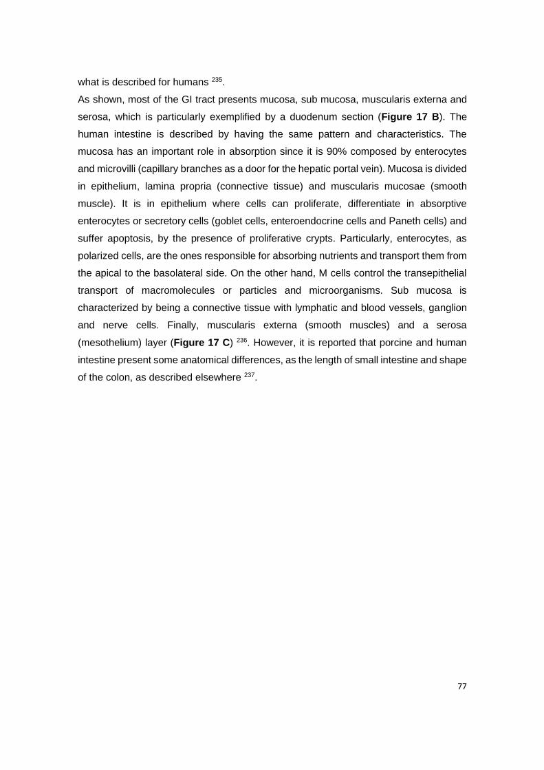
77
what is described for humans 235.
As shown, most of the GI tract presents mucosa, sub mucosa, muscularis externa and
serosa, which is particularly exemplified by a duodenum section (Figure 17 B). The
human intestine is described by having the same pattern and characteristics. The
mucosa has an important role in absorption since it is 90% composed by enterocytes
and microvilli (capillary branches as a door for the hepatic portal vein). Mucosa is divided
in epithelium, lamina propria (connective tissue) and muscularis mucosae (smooth
muscle). It is in epithelium where cells can proliferate, differentiate in absorptive
enterocytes or secretory cells (goblet cells, enteroendocrine cells and Paneth cells) and
suffer apoptosis, by the presence of proliferative crypts. Particularly, enterocytes, as
polarized cells, are the ones responsible for absorbing nutrients and transport them from
the apical to the basolateral side. On the other hand, M cells control the transepithelial
transport of macromolecules or particles and microorganisms. Sub mucosa is
characterized by being a connective tissue with lymphatic and blood vessels, ganglion
and nerve cells. Finally, muscularis externa (smooth muscles) and a serosa
(mesothelium) layer (Figure 17 C) 236. However, it is reported that porcine and human
intestine present some anatomical differences, as the length of small intestine and shape
of the colon, as described elsewhere 237.
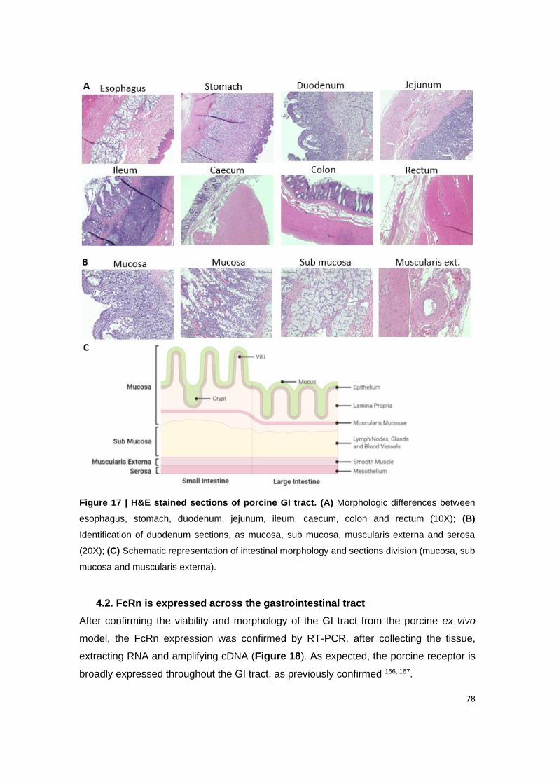
78
Figure 17 | H&E stained sections of porcine GI tract. (A) Morphologic differences between
esophagus, stomach, duodenum, jejunum, ileum, caecum, colon and rectum (10X); (B)
Identification of duodenum sections, as mucosa, sub mucosa, muscularis externa and serosa
(20X); (C) Schematic representation of intestinal morphology and sections division (mucosa, sub
mucosa and muscularis externa).
4.2. FcRn is expressed across the gastrointestinal tract
After confirming the viability and morphology of the GI tract from the porcine ex vivo
model, the FcRn expression was confirmed by RT-PCR, after collecting the tissue,
extracting RNA and amplifying cDNA (Figure 18). As expected, the porcine receptor is
broadly expressed throughout the GI tract, as previously confirmed 166, 167.
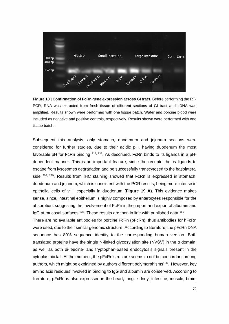
79
Figure 18 | Confirmation of FcRn gene expression across GI tract. Before performing the RT-
PCR, RNA was extracted from fresh tissue of different sections of GI tract and cDNA was
amplified. Results shown were performed with one tissue batch. Water and porcine blood were
included as negative and positive controls, respectively. Results shown were performed with one
tissue batch.
Subsequent this analysis, only stomach, duodenum and jejunum sections were
considered for further studies, due to their acidic pH, having duodenum the most
favorable pH for FcRn binding 218, 238. As described, FcRn binds to its ligands in a pH-
dependent manner. This is an important feature, since the receptor helps ligands to
escape from lysosomes degradation and be successfully transcytosed to the basolateral
side 208, 239. Results from IHC staining showed that FcRn is expressed in stomach,
duodenum and jejunum, which is consistent with the PCR results, being more intense in
epithelial cells of villi, especially in duodenum (Figure 19 A). This evidence makes
sense, since, intestinal epithelium is highly composed by enterocytes responsible for the
absorption, suggesting the involvement of FcRn in the import and export of albumin and
IgG at mucosal surfaces 236. These results are then in line with published data 166.
There are no available antibodies for porcine FcRn (pFcRn), thus antibodies for hFcRn
were used, due to their similar genomic structure. According to literature, the pFcRn DNA
sequence has 80% sequence identity to the corresponding human version. Both
translated proteins have the single N-linked glycosylation site (NVSV) in the α domain,
as well as both di-leucine- and tryptophan-based endocytosis signals present in the
cytoplasmic tail. At the moment, the pFcRn structure seems to not be concordant among
authors, which might be explained by authors different polymorphisms166. However, key
amino acid residues involved in binding to IgG and albumin are conserved. According to
literature, pFcRn is also expressed in the heart, lung, kidney, intestine, muscle, brain,
80
endothelial cells and monocytes, and highly expressed in liver, spleen, thymus,
lymphoid, being also maintained in the adult stage, similar to humans 166, 167.
Studying the durability of FcRn expression of tissue in culture, it was observed that it is
maintained for up, at least, 7 days, especially in duodenum (Figure 19 B). These results
are in line with previous characterization of the ex vivo platform, when evaluating other
receptors’ expression. Authors verified that it is possible to guarantee the complex GI
architecture, mucus layer and the expression of several drug transporter for 7 days, when
the tissue is intact in culture 169.
Figure 19 | Evaluation of FcRn expression over time, of stomach, duodenum and jejunum.
(A) Evaluation of FcRn expression by IHC of fresh porcine tissue. Control samples were only
treated with the secondary antibody, to check non-specific binding. Brown indicates the antibody
signal, while blue is the counterstain. (B) FcRn expression after 7 days of porcine ex vivo
incubation or freshly isolated at day zero. Before the RT-PCR, RNA was extracted from fresh
tissue and cDNA was amplified. Water and porcine blood were included as negative and positive
controls, respectively. Results shown were performed with one tissue batch.
4.3. Engineered albumin variant shows enhanced transcytosis in the porcine
ex vivo platform
Next, the evaluation of the ex vivo platform, using porcine duodenum (Figure 20 A), for
the screening of the intestinal permeability of FcRn-targeted drugs was done. For this
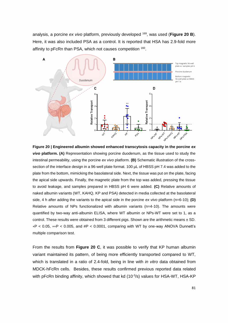
81
analysis, a porcine ex vivo platform, previously developed 169, was used (Figure 20 B).
Here, it was also included PSA as a control. It is reported that HSA has 2.9-fold more
affinity to pFcRn than PSA, which not causes competition 160.
Figure 20 | Engineered albumin showed enhanced transcytosis capacity in the porcine ex
vivo platform. (A) Representation showing porcine duodenum, as the tissue used to study the
intestinal permeability, using the porcine ex vivo platform. (B) Schematic illustration of the cross-
section of the interface design in a 96-well plate format. 100 µL of HBSS pH 7.4 was added to the
plate from the bottom, mimicking the basolateral side. Next, the tissue was put on the plate, facing
the apical side upwards. Finally, the magnetic plate from the top was added, pressing the tissue
to avoid leakage, and samples prepared in HBSS pH 6 were added. (C) Relative amounts of
naked albumin variants (WT, KAHQ, KP and PSA) detected in media collected at the basolateral
side, 4 h after adding the variants to the apical side in the porcine ex vivo platform (n=6-10); (D)
Relative amounts of NPs functionalized with albumin variants (n=4-10). The amounts were
quantified by two-way anti-albumin ELISA, where WT albumin or NPs-WT were set to 1, as a
control. These results were obtained from 3 different pigs. Shown are the arithmetic means ± SD.
∗P < 0.05, ∗∗P < 0.005, and #P < 0.0001, comparing with WT by one-way ANOVA Dunnett’s
multiple comparison test.
From the results from Figure 20 C, it was possible to verify that KP human albumin
variant maintained its pattern, of being more efficiently transported compared to WT,
which is translated in a ratio of 2.4-fold, being in line with in vitro data obtained from
MDCK-hFcRn cells. Besides, these results confirmed previous reported data related
with pFcRn binding affinity, which showed that kd (10-3/s) values for HSA-WT, HSA-KP
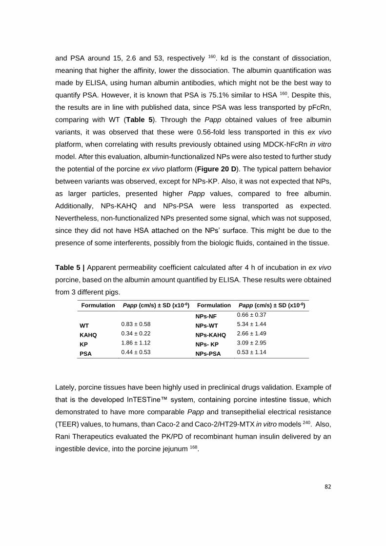
82
and PSA around 15, 2.6 and 53, respectively 160. kd is the constant of dissociation,
meaning that higher the affinity, lower the dissociation. The albumin quantification was
made by ELISA, using human albumin antibodies, which might not be the best way to
quantify PSA. However, it is known that PSA is 75.1% similar to HSA 160. Despite this,
the results are in line with published data, since PSA was less transported by pFcRn,
comparing with WT (Table 5). Through the Papp obtained values of free albumin
variants, it was observed that these were 0.56-fold less transported in this ex vivo
platform, when correlating with results previously obtained using MDCK-hFcRn in vitro
model. After this evaluation, albumin-functionalized NPs were also tested to further study
the potential of the porcine ex vivo platform (Figure 20 D). The typical pattern behavior
between variants was observed, except for NPs-KP. Also, it was not expected that NPs,
as larger particles, presented higher Papp values, compared to free albumin.
Additionally, NPs-KAHQ and NPs-PSA were less transported as expected.
Nevertheless, non-functionalized NPs presented some signal, which was not supposed,
since they did not have HSA attached on the NPs’ surface. This might be due to the
presence of some interferents, possibly from the biologic fluids, contained in the tissue.
Table 5 | Apparent permeability coefficient calculated after 4 h of incubation in ex vivo
porcine, based on the albumin amount quantified by ELISA. These results were obtained
from 3 different pigs.
Formulation Papp (cm/s) ± SD (x10-6) Formulation Papp (cm/s) ± SD (x10-6)
NPs-NF 0.66 ± 0.37
WT 0.83 ± 0.58 NPs-WT 5.34 ± 1.44
KAHQ 0.34 ± 0.22 NPs-KAHQ 2.66 ± 1.49
KP 1.86 ± 1.12 NPs- KP 3.09 ± 2.95
PSA 0.44 ± 0.53 NPs-PSA 0.53 ± 1.14
Lately, porcine tissues have been highly used in preclinical drugs validation. Example of
that is the developed InTESTine™ system, containing porcine intestine tissue, which
demonstrated to have more comparable Papp and transepithelial electrical resistance
(TEER) values, to humans, than Caco-2 and Caco-2/HT29-MTX in vitro models 240. Also,
Rani Therapeutics evaluated the PK/PD of recombinant human insulin delivered by an
ingestible device, into the porcine jejunum 168.

83
5. In vivo validation
As the in vitro and ex vivo data strongly suggested that NPs decorated site-specifically
with human albumin could lead to enhanced delivery across barriers, this concept was
subsequently addressed in vivo using the designed insulin encapsulated NPs. A state-
of-the-art mouse model that is transgenic for hFcRn and knockout for mFcRn (FcRn-/-
hFcRn Tg32 mice) 157 was chosen for this study, as conventional mice are unreliable
models for evaluation of human albumin-based therapeutics due to large cross-species
binding differences 52, 60, 241. Specifically, HSA binds poorly to mFcRn, and as such will
be competed out for binding to the murine receptor in the presence of large amounts of
endogenous MSA 52, 60. The hFcRn transgenic mice indeed have MSA but in this case
mouse and human albumin bind equally well to the receptor 52, 60.
To study the effect of albumin decorated NPs encapsulated with insulin, the transgenic
mice were treated with streptozotocin (STZ) to induce diabetes. One week later, the mice
were fasted for 12 h, before being divided into six groups based on their blood glucose
level. After administering different formulations, pharmacodynamic parameters were
evaluated over 8 h (Figure 21 A). This time frame was not extended, since it is not
recommended starving animals for long periods. Besides, if the animal is in a starvation
state it is hard to objectively discuss the results, since they could be due to the drug
effect or due to organism failure. In fact, it was observed some deaths during the
experiments, which were not considered for data analysis. Knowing that mice consume
primarily at night, overnight fasting leads to a catabolic state, reducing 15% of body mass
in lean mice 242. Overnight fasting deletes liver glycogen stores, which contributes for
reducing the variability in baseline blood glucose. Besides, contrary to what happens in
humans, a prolonged fast in mice enhances the insulin-stimulated glucose utilization 242.
In this sense, it is recommended that mice used for metabolic studies, should be fasted
for 14–18 h (overnight fast) 243.
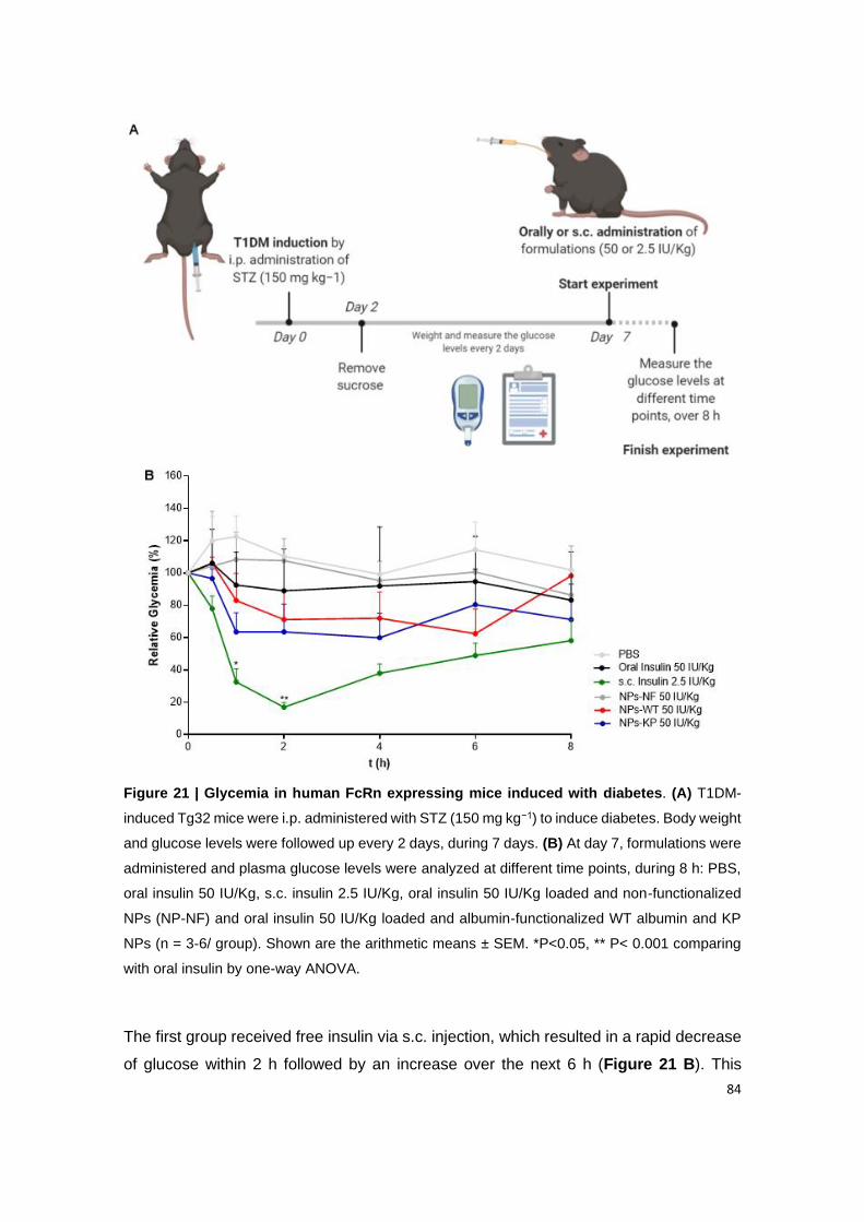
84
Figure 21 | Glycemia in human FcRn expressing mice induced with diabetes. (A) T1DM-
induced Tg32 mice were i.p. administered with STZ (150 mg kg−1) to induce diabetes. Body weight
and glucose levels were followed up every 2 days, during 7 days. (B) At day 7, formulations were
administered and plasma glucose levels were analyzed at different time points, during 8 h: PBS,
oral insulin 50 IU/Kg, s.c. insulin 2.5 IU/Kg, oral insulin 50 IU/Kg loaded and non-functionalized
NPs (NP-NF) and oral insulin 50 IU/Kg loaded and albumin-functionalized WT albumin and KP
NPs (n = 3-6/ group). Shown are the arithmetic means ± SEM. *P<0.05, ** P< 0.001 comparing
with oral insulin by one-way ANOVA.
The first group received free insulin via s.c. injection, which resulted in a rapid decrease
of glucose within 2 h followed by an increase over the next 6 h (Figure 21 B). This
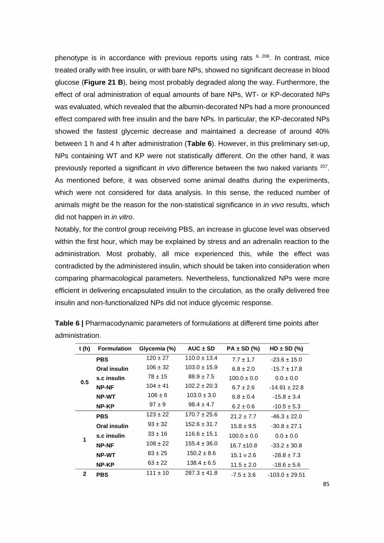
85
phenotype is in accordance with previous reports using rats 6, 206. In contrast, mice
treated orally with free insulin, or with bare NPs, showed no significant decrease in blood
glucose (Figure 21 B), being most probably degraded along the way. Furthermore, the
effect of oral administration of equal amounts of bare NPs, WT- or KP-decorated NPs
was evaluated, which revealed that the albumin-decorated NPs had a more pronounced
effect compared with free insulin and the bare NPs. In particular, the KP-decorated NPs
showed the fastest glycemic decrease and maintained a decrease of around 40%
between 1 h and 4 h after administration (Table 6). However, in this preliminary set-up,
NPs containing WT and KP were not statistically different. On the other hand, it was
previously reported a significant in vivo difference between the two naked variants 207.
As mentioned before, it was observed some animal deaths during the experiments,
which were not considered for data analysis. In this sense, the reduced number of
animals might be the reason for the non-statistical significance in in vivo results, which
did not happen in in vitro.
Notably, for the control group receiving PBS, an increase in glucose level was observed
within the first hour, which may be explained by stress and an adrenalin reaction to the
administration. Most probably, all mice experienced this, while the effect was
contradicted by the administered insulin, which should be taken into consideration when
comparing pharmacological parameters. Nevertheless, functionalized NPs were more
efficient in delivering encapsulated insulin to the circulation, as the orally delivered free
insulin and non-functionalized NPs did not induce glycemic response.
Table 6 | Pharmacodynamic parameters of formulations at different time points after
administration.
t (h) Formulation Glycemia (%) AUC ± SD PA ± SD (%) HD ± SD (%)
0.5
PBS 120 ± 27 110.0 ± 13.4 7.7 ± 1.7 -23.6 ± 15.0
Oral insulin 106 ± 32 103.0 ± 15.9 6.8 ± 2.0 -15.7 ± 17.8
s.c insulin 78 ± 15 88.9 ± 7.5 100.0 ± 0.0 0.0 ± 0.0
NP-NF 104 ± 41 102.2 ± 20.3 6.7 ± 2.6 -14.91 ± 22.8
NP-WT 106 ± 6 103.0 ± 3.0 6.8 ± 0.4 -15.8 ± 3.4
NP-KP 97 ± 9 98.4 ± 4.7 6.2 ± 0.6 -10.5 ± 5.3
1
PBS 123 ± 22 170.7 ± 25.6 21.2 ± 7.7 -46.3 ± 22.0
Oral insulin 93 ± 32 152.6 ± 31.7 15.8 ± 9.5 -30.8 ± 27.1
s.c insulin 33 ± 16 116.6 ± 15.1 100.0 ± 0.0 0.0 ± 0.0
NP-NF 108 ± 22 155.4 ± 36.0 16.7 ±10.8 -33.2 ± 30.8
NP-WT 83 ± 25 150.2 ± 8.6 15.1 v 2.6 -28.8 ± 7.3
NP-KP 63 ± 22 138.4 ± 6.5 11.5 ± 2.0 -18.6 ± 5.6
2 PBS 111 ± 10 287.3 ± 41.8 -7.5 ± 3.6 -103.0 ± 29.51
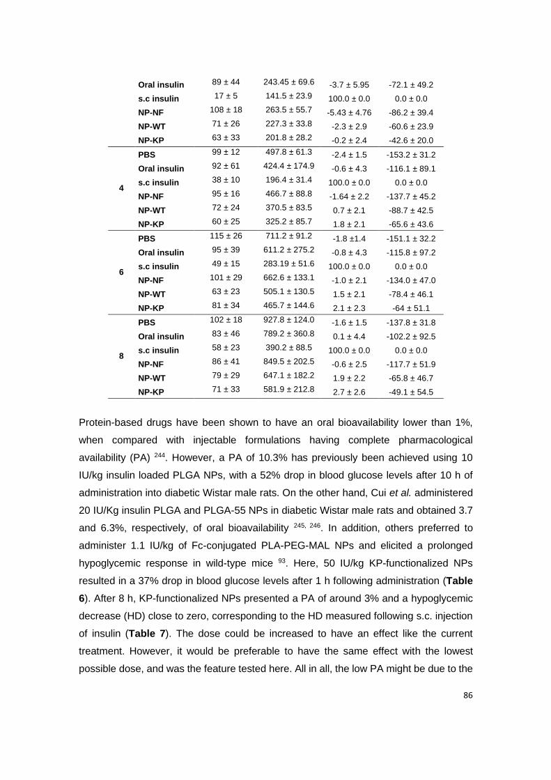
86
Oral insulin 89 ± 44 243.45 ± 69.6 -3.7 ± 5.95 -72.1 ± 49.2
s.c insulin 17 ± 5 141.5 ± 23.9 100.0 ± 0.0 0.0 ± 0.0
NP-NF 108 ± 18 263.5 ± 55.7 -5.43 ± 4.76 -86.2 ± 39.4
NP-WT 71 ± 26 227.3 ± 33.8 -2.3 ± 2.9 -60.6 ± 23.9
NP-KP 63 ± 33 201.8 ± 28.2 -0.2 ± 2.4 -42.6 ± 20.0
4
PBS 99 ± 12 497.8 ± 61.3 -2.4 ± 1.5 -153.2 ± 31.2
Oral insulin 92 ± 61 424.4 ± 174.9 -0.6 ± 4.3 -116.1 ± 89.1
s.c insulin 38 ± 10 196.4 ± 31.4 100.0 ± 0.0 0.0 ± 0.0
NP-NF 95 ± 16 466.7 ± 88.8 -1.64 ± 2.2 -137.7 ± 45.2
NP-WT 72 ± 24 370.5 ± 83.5 0.7 ± 2.1 -88.7 ± 42.5
NP-KP 60 ± 25 325.2 ± 85.7 1.8 ± 2.1 -65.6 ± 43.6
6
PBS 115 ± 26 711.2 ± 91.2 -1.8 ±1.4 -151.1 ± 32.2
Oral insulin 95 ± 39 611.2 ± 275.2 -0.8 ± 4.3 -115.8 ± 97.2
s.c insulin 49 ± 15 283.19 ± 51.6 100.0 ± 0.0 0.0 ± 0.0
NP-NF 101 ± 29 662.6 ± 133.1 -1.0 ± 2.1 -134.0 ± 47.0
NP-WT 63 ± 23 505.1 ± 130.5 1.5 ± 2.1 -78.4 ± 46.1
NP-KP 81 ± 34 465.7 ± 144.6 2.1 ± 2.3 -64 ± 51.1
8
PBS 102 ± 18 927.8 ± 124.0 -1.6 ± 1.5 -137.8 ± 31.8
Oral insulin 83 ± 46 789.2 ± 360.8 0.1 ± 4.4 -102.2 ± 92.5
s.c insulin 58 ± 23 390.2 ± 88.5 100.0 ± 0.0 0.0 ± 0.0
NP-NF 86 ± 41 849.5 ± 202.5 -0.6 ± 2.5 -117.7 ± 51.9
NP-WT 79 ± 29 647.1 ± 182.2 1.9 ± 2.2 -65.8 ± 46.7
NP-KP 71 ± 33 581.9 ± 212.8 2.7 ± 2.6 -49.1 ± 54.5
Protein-based drugs have been shown to have an oral bioavailability lower than 1%,
when compared with injectable formulations having complete pharmacological
availability (PA) 244. However, a PA of 10.3% has previously been achieved using 10
IU/kg insulin loaded PLGA NPs, with a 52% drop in blood glucose levels after 10 h of
administration into diabetic Wistar male rats. On the other hand, Cui et al. administered
20 IU/Kg insulin PLGA and PLGA-55 NPs in diabetic Wistar male rats and obtained 3.7
and 6.3%, respectively, of oral bioavailability 245, 246. In addition, others preferred to
administer 1.1 IU/kg of Fc-conjugated PLA-PEG-MAL NPs and elicited a prolonged
hypoglycemic response in wild-type mice 93. Here, 50 IU/kg KP-functionalized NPs
resulted in a 37% drop in blood glucose levels after 1 h following administration (Table
6). After 8 h, KP-functionalized NPs presented a PA of around 3% and a hypoglycemic
decrease (HD) close to zero, corresponding to the HD measured following s.c. injection
of insulin (Table 7). The dose could be increased to have an effect like the current
treatment. However, it would be preferable to have the same effect with the lowest
possible dose, and was the feature tested here. All in all, the low PA might be due to the

87
incomplete release of insulin, which may be increased with increasing the study time.
Table 7 | Pharmacodynamic parameters.
Dose (IU/Kg) tmin (h) Cmin ± SD (mg/dL) AUC 8 h PA 8 h ± SD (%) HD 8 h ± SD (%)
PBS 0 4 374.20 ± 58.64 927.8 ± 124.0 -1.6 ± 1.5 -137.8 ± 31.8
Oral insulin 50 8 256.75 ± 102.88 789.2 ± 360.8 0.1 ± 4.4 -102.2 ± 92.5
s.c insulin 2.5 2 72.50 ± 25.67 390.2 ± 88.5 100.0 ± 0.0 0.0 ± 0.0
NP-NF 50 8 328.00 ± 65.33 849.5 ± 202.5 -0.6 ± 2.5 -117.7 ± 51.9
NP-WT 50 6 290.50 ± 103.75 768.6 ± 24.7 0.4 ± 0.3 -97.0 ± 6.3
NP-KP 50 4 282.40 ± 137.68 581.9 ± 212.8 2.7 ± 2.6 -49.1 ± 54.5
AUC: area under the curve; PA: pharmacological availability; HD: hypoglycemic decrease; SD:
standard deviation; tmin: time with minimal concentration of glucose after administration of different
formulations.
Taken together, the in vivo glycemic decrease obtained using the KP variant is a clear
improvement of the therapeutic efficacy. In fact, the obtained in vivo results agree with
in vitro data, supporting that albumin decorated NPs increase the effect as a result of
improved FcRn-binding affinity. It is reported in the literature the use of FcRn knock out
mouse, confirming the FcRn-mediated transcytosis of FcRn-targeted NPs 93.
Interestingly, while the intestinal expression of FcRn in rodents has been demonstrated
to be developmentally down-regulated after weaning 247, this is not the case in humans
or in the hFcRn expressing mice, as the transgene is expressed under the human
promoter 248. Thus, the transgenic mouse model mimics the tissue expression profile of
human FcRn, by not being down-regulated after weaning 157.
Additionally, biodistribution and pharmacokinetic preliminary studies were done, using
NPs-KAHQ and an albumin triple mutant, which can give an insight of the behavior of
the studied variants (Appendix III).

88

89
Chapter V
CONCLUSIONS AND FUTURE PERSPECTIVES

90
91
1. Conclusions
The oral delivery of antidiabetic drugs is more convenient, safe and cost-effective.
Notwithstanding, biopharmaceuticals may benefit with the encapsulation in
multifunctional nanomaterials. Functionalized nanomaterials are definitely the best way
to reach a specific target and have reduced side effects and increased efficiency. Lately,
the potential of FcRn-targeted NPs, as an attractive gateway for enhanced delivery of
antidiabetic drugs across mucosal barrier, has been under investigation. Moreover, the
use of engineered FcRn-targeted ligands have revealed to be a promising strategy to
ameliorate the effects on binding, transport and recycling. These parameters can be
crutial on modulating cell uptake, long circulation time, renal clearance, and drug stability,
by surpassing extracellular barriers, select internalization via and overcome intracellular
obstacles, as lysosomal degradation.
In this thesis, a FcRn-targeted nanoplatform was developed, as an advanced delivery
system for the improved oral bioavailability of insulin. The developed PLGA-PEG NPs
were characterized as being monodisperse with 150 nm in size and -9 mV in zeta
potential, having a drug loading over 10% and around 90% of albumin conjugation
efficiency. Insulin structure showed to be affected by the encapsulation method, while
albumin structure was maintained after conjugation. Furthermore, the developed
nanosystem revealed a prolonged and controlled release of insulin, which contributes for
the reduced insulin dosing. NPs were decorated with human albumin variants, which
presented different binding affinities to FcRn. Particularly, the engineered albumin
variant KP, with a substitution of Lys573 by proline on DIII, showed the highest
improvement on binding to hFcRn. Moreover, KP showed enhanced transcytosis in 2.5-
fold across polarized MDCK-hFcRn epithelia, and 2-fold increase when conjugated to
NPs, compared with WT albumin and WT albumin decorated NPs, respectively.
Additionally, after being transported throughout epithelial cells, these NPs exhibited to
be taken up and recycled by endothelial HMEC1-hFcRn cells, through the FcRn-
recycling pathway. Again, the KP variant showed a more pronounced effect, presenting
2-fold and 3-fold increase of uptake and recycling, respectively, compared to WT
albumin. After conjugation, NPs-KP maintained the marked effect, with both uptake and
recycling 1.5-fold higher than NPs-WT. These results were reflected in the half-life
projection, demonstrating a score of 2-fold higher for KP and 6.7-fold higher for NPs-KP,
compared to WT, which has a mean half-life of 3 weeks. Through both FcRn-related
biologic mechanisms, it was thus possible to show how FcRn-binding therapeutics are
rescued from intracellular degradation. These advantages can play a key role in doses
92
management, revolutionizing the design of new materials. Furthermore, when exploring
the potential of simulated hypergravity in oral delivery of biopharmaceuticals, it was
shown that different hypergravity levels (2.5 g, 5.0 g and 7.5 g) did not affect the
metabolic activity and cellular integrity of intestinal epithelial Caco-2 cells. Nevertheless,
the expression of tight junctions and transporters of Caco-2 cells was apparently
affected, which might interfere with paracellular and passive transport. However, the
FcRn-mediated transport of the produced NPs revealed itself constant and not different
from reference conditions. This evidence can thus open doors for the development of
new strategies for oral delivery of biopharmaceuticals.
Next, to evaluate the intestinal permeability in a more reliable predictive manner, a
porcine ex vivo model was used. The porcine has gaining importance and attention from
investigators, due to its physiological and anatomical similarities. However, the potential
of this platform on the screening of FcRn-targeted nanosystems was never investigated.
In this sense, the FcRn expression through the GI tract, which was maintained for up 7
days, was assessed. Through the intestinal permeability assay it was shown the
characteristic hierarchy of albumin variants. Expectadely, KP demonstrated to be 2.4-
fold more efficiently transported, compared to WT, which is in line with in vitro data. The
consistent results in different models, showed a robustness of the developed NPs and a
correlation between models. Therefore, this porcine ex vivo platform revealed to be a
potential model for the screening of FcRn-targeted oral drug formulations, which may
have a positive and significant impact on saving time and money, during their
development.
Finally, as a proof-of-concept, the ability of the developed polymeric NPs to reduce
glucose levels was assessed by using hFcRn transgenic mice, to avoid cross-species
differences, induced with diabetes. After oral delivery, it was observed that NPs have a
controlled and prolonged effect, compared to s.c. insulin, which had only a peak effect
after 2 h. In addition, encapsulated insulin showed a better effect, when comparing with
oral free insulin, suggesting a drug protection. Also, funcionalized NPs were more
efficient than non-functionalized NPs, due to FcRn advantageous characteristics.
Particularly, NPs-KP were able to reduce glycemia up to around 40% after 1 h, which is
maintained between 1 h and 4 h, and enhance pharmacologic availability. The novel
concept described should pave the way for further engineering of biodegradable
albumin-decorated NP formulations for tailored targeting of FcRn for enhanced intestinal
delivery.
Overall, the developed nanosystem holds a tremendous promise to address the unmet
93
needs, as poor oral bioavailability of biopharmaceuticals and specially diabetic patient
compliance. Plus, it is a versatile carrier candidate for oral delivery of
biopharmaceuticals, playing a positive and significant impact in welfare of diabetic
patients.
2. Future Perspectives
As future perspectives a panel of studies could be done, to improve the quality of this
work, by clarifying some details.
Regarding the conjugation, since it was not possible to concretely demonstrate the
thioether covalent bond, it would be interesting to conjugate albumin to the polymer,
before producing NPs. The approach will probably facilitate the detection by p.e. NMR.
Also, the potential of a recent albumin variant, E505Q/T527M/K573P (QMP), published
by J.T. Andersen, could be explored. As previously mentioned, it is reported that the
binding affinity of this triple mutant gains impressively more than 180-fold increase in
hFcRn without affecting binding at neutral pH. Plus, QMP translated into 4-fold enhanced
transcellular transport in vitro, compared with WT albumin 87.
In general, ex vivo and especially in vivo assays need a higher number of replicates in
different tissues/animals, to clarify the statistical significance of the results. Further
investigation into the implication of obtained differences on the PD/PK is also required.
In addition, here it was used insulin as a model drug, using diabetes as example.
However, it would be interesting try to encapsulate other antidiabetic peptides, as insulin
glargine, degludec, icodec or the oral i338, for the treatment of both diabetes types. To
answer to some particularities of diabetes, NPs could be microencapsulated into enteric
capsules, in parallel with free insulin, to ensure a fast effect (after meals, p.e.), while,
NPs will assure a prolonged effect. To take advantage of the FcRn-recycling pathways
and half-life extension, both free insulin and encapsulated insulin could be conjugated to
simpler and smaller FcRn-targeted ligands, as FcRn binding peptides (FcBP) 249, 250.
The potential of the developed technology can also be explored in diabetes-related
disorders, as vasculopathy or fibrosis, or even in other diseases since FcRn is broadly
expressed thoughout the body, which may pave the way for using FcRn-targeting NPs
for delivery to different body sites.

94
95
Chapter VI
REFERENCES

96
97
1. Saeedi P, Petersohn I, Salpea P, et al. Global and regional diabetes prevalence estimates for 2019 and projections for 2030 and 2045: Results from the International Diabetes Federation Diabetes Atlas, 9(th) edition. Diabetes Res Clin Pract 2019;157:107843.
2. Bommer C, Sagalova V, Heesemann E, et al. Global Economic Burden of Diabetes in Adults: Projections From 2015 to 2030. Diabetes Care 2018;41:963-970.
3. Easa N, Alany RG, Carew M, et al. A review of non-invasive insulin delivery systems for diabetes therapy in clinical trials over the past decade. Drug Discovery Today 2019;24:440-451.
4. Cutfield WS, Derraik JGB, Gunn AJ, et al. Non-Compliance with Growth Hormone Treatment in Children Is Common and Impairs Linear Growth. PLoS ONE 2011;6:e16223.
5. Davies MJ, Gagliardino JJ, Gray LJ, et al. Real-world factors affecting adherence to insulin therapy in patients with Type 1 or Type 2 diabetes mellitus: a systematic review. Diabetic Medicine 2013;30:512-524.
6. Sonaje K, Lin KJ, Wey SP, et al. Biodistribution, pharmacodynamics and pharmacokinetics of insulin analogues in a rat model: Oral delivery using pH-responsive nanoparticles vs. subcutaneous injection. Biomaterials 2010;31:6849-58.
7. Arbit E, Kidron M. Oral Insulin Delivery in a Physiologic Context: Review. J Diabetes Sci Technol 2017;11:825-832.
8. Geho WB, Geho HC, Lau JR, et al. Hepatic-Directed Vesicle Insulin: A Review of Formulation Development and Preclinical Evaluation. Journal of Diabetes Science and Technology 2009;3:1451-1459.
9. Eldor R, Arbit E, Corcos A, et al. Glucose-Reducing Effect of the ORMD-0801 Oral Insulin Preparation in Patients with Uncontrolled Type 1 Diabetes: A Pilot Study. PLOS ONE 2013;8:e59524.
10. Alexander Vol, Gribova O. Methods and compositions for oral administration of insulin. In: Administration OD, ed. Volume US20100278922A1, 2014.
11. Khedkar A, Iyer H, Anand A, et al. A dose range finding study of novel oral insulin (IN-105) under fed conditions in type 2 diabetes mellitus subjects. Diabetes Obes Metab 2010;12:659-64.
12. Bennet D, Kim S. Polymer Nanoparticles for Smart Drug Delivery. In: Sezer AD, ed. Application of Nanotechnology in Drug Delivery: Nanotechnology and Nanomaterials, 2014:257-309.
13. Duschl A. Nanomedicine. In: Boraschi D, Penton-Rol G, eds. Immune Rebalancing - The Future of Immunosuppression: Elsevier, 2016:251-274.
14. Stewart MP, Sharei A, Ding X, et al. In vitro and ex vivo strategies for intracellular delivery. Nature 2016;538:183-192.
15. Banik BL, Fattahi P, Brown JL. Polymeric nanoparticles: the future of nanomedicine. WIREs Nanomedicine and Nanobiotechnology 2016;8:271–299.
16. Pereira C, Araújo F, Granja PL, et al. Targeting membrane transporters and receptors as a mean to optimize orally delivered biotecnhological based drugs through nanoparticle delivery systems. Current Pharmaceutical Biotechnology 2014;15:650-658.
17. Araujo F, Shrestha N, Granja PL, et al. Antihyperglycemic potential of incretins orally delivered via nano and microsystems and subsequent glucoregulatory effects. Current Pharmaceutical Biotechnology 2014;15:609-19.
18. Moulari B, Beduneau A, Pellequer Y, et al. Nanoparticle targeting to inflamed tissues of gastrointestinal tract Current Drug Delivery 2013;10:9-17.
19. Sarmento B, Ferreira D, Vasconcelos T. Polymer-Based Delivery Systems for Oral Delivery of Peptides and Proteins. In: Jorgensen L, Nielsen HM, eds. Delivery Technologies for Biopharmaceuticals: Peptides, Proteins, Nucleic Acids and Vaccines: John Wiley & Sons, Ltd, 2009:207-227.
20. Parodi A, Corbo C, Cevenini A, et al. Enabling cytoplasmic delivery and organelle targeting by surface modification of nanocarriers. Nanomedicine 2015;10:1923–1940.
21. Rekha MR, Sharma CP. Oral delivery of therapeutic protein/peptide for diabetes – Future perspectives. International Journal of Pharmaceutics 2013;440:48-62.
22. Soo Choi H, Liu W, Misra P, et al. Renal clearance of quantum dots. Nat Biotech 2007;25:1165-1170.
98
23. Wahajuddin, Arora S. Superparamagnetic iron oxide nanoparticles: magnetic nanoplatforms as drug carriers. International Journal of Nanomedicine 2012;7:3445–3471.
24. Howe SE, Lickteig DJ, Plunkett KN, et al. The Uptake of Soluble and Particulate Antigens by Epithelial Cells in the Mouse Small Intestine. PLOS ONE 2014;9:e86656.
25. Sun W, Hu Q, Ji W, et al. Leveraging physiology for precision drug delivery. Physiological Reviews 2017;97:189–225.
26. Agarwal R, Roy K. Intracellular delivery of polymeric nanocarriers: a matter of size, shape, charge, elasticity and surface composition. Therapeutic delivery 2013;4:705–723.
27. Blanco E, Shen H, Ferrari M. Principles of nanoparticle design for overcoming biological barriers to drug delivery. Nature Biotechnology 2015;9:941-951.
28. Huang H, Hernandez R, Geng J, et al. A porphyrin-PEG polymer with rapid renal clearance. Biomaterials 2016;76:25-32.
29. Roberts MJ, Bentley MD, Harris JM. Chemistry for peptide and protein PEGylation. Advanced Drug Delivery Reviews 2012;64, Supplement:116-127.
30. Schöttler S, Becker G, Winzen S, et al. Protein adsorption is required for stealth effect of poly(ethylene glycol)- and poly(phosphoester)-coated nanocarriers. Nat Nano 2016;11:372-377.
31. Jhaveri A, Torchilin V. Intracellular delivery of nanocarriers and targeting to subcellular organelles. Expert Opinion on Drug Delivery 2015;13.
32. Bera A, Jewrajka SK. Tailoring polyamide thin film composite nanofiltration membranes by polyethyleneimine and its conjugates for the enhancement of selectivity and antifouling property. RSC Advances 2016;6:4521-4530.
33. Noh SM, Kim W-K, Kim SJ, et al. Enhanced cellular delivery and transfection efficiency of plasmid DNA using positively charged biocompatible colloidal gold nanoparticles. Biochimica et Biophysica Acta (BBA) - General Subjects 2007;1770:747-752.
34. Chanthick C, Kanlaya R, Kiatbumrung R, et al. Caveolae-mediated albumin transcytosis is enhanced in dengueinfected human endothelial cells: A model of vascular leakage in dengue hemorrhagic fever. Scientific Reports 2016;6.
35. Alai MS, Lin WJ, Pingale SS. Application of polymeric nanoparticles and micelles in insulin oral delivery. Journal of food and drug analysis 2015;23:351-358.
36. Pawar VK, Meher JG, Singh Y, et al. Targeting of gastrointestinal tract for amended delivery of 3Q4 protein/peptide therapeutics: Strategies and industrial perspectives. Journal of Controlled Release 2014;196:168-83.
37. Xiao K, Li Y, Luo J, et al. The effect of surface charge on in vivo biodistribution of PEG-oligocholic acid based micellar nanoparticles. Biomaterials 2011;32:3435-3446.
38. Yoo JW, Mitragotri S. Polymer particles that switch shape in response to a stimulus. Proc Natl Acad Sci U S A 2010;107:11205-10.
39. Banquy X, Suarez F, Argaw A, et al. Effect of mechanical properties of hydrogel nanoparticles on macrophage cell uptake. Soft Matter 2009;5:3984-3991.
40. O'Reilly JR, Corrigan OI, O'Driscoll CM. The effect of mixed micellar systems, bile salt/fatty acids, on the solubility and intestinal absorption of clofazimine (B663) in the anaesthetised rat. International Journal of Pharmaceutics 1994;109:147-154.
41. Pabla D, Akhlaghi F, Zia H. Intestinal permeability enhancement of levothyroxine sodium by straight chain fatty acids studied in MDCK epithelial cell line. European Journal of Pharmaceutical Sciences 2010;40:466-472.
42. Aspenström-Fagerlund B, Sundström B, Tallkvist J, et al. Fatty acids increase paracellular absorption of aluminium across Caco-2 cell monolayers. Chemico-Biological Interactions 2009;181:272-278.
43. Trevino SR, Scholtz JM, Pace CN. Amino acid contribution to protein solubility: Asp, Glu, and Ser contribute more favorably than the other hydrophilic amino acids in RNase Sa. J Mol Biol 2007;366:449-60.
44. Li N, Huang C, Luan Y, et al. Active targeting co-delivery system based on pH-sensitive methoxy-poly (ethylene glycol)2K-poly(e-caprolactone)4K-poly(glutamic acid)1K for enhanced cancer therapy. Journal of Colloid and Interface Science 2016;472:90-98.
99
45. Li X, Yu M, Fan W, et al. Orally active-targeted drug delivery systems for proteins and peptides. Expert Opinion Drug Delivery 2014;11:1435-1447.
46. Steinbach JM, Seo Y-E, Saltzman WM. Cell penetrating peptide-modified poly(lactic-co-glycolic acid) nanoparticles with enhanced cell internalization. Acta Biomaterialia 2016;30:49-61.
47. Ghuman J, Zunszain PA, Petitpas I, et al. Structural basis of the drug-binding specificity of human serum albumin. J Mol Biol 2005;353:38-52.
48. Tan YL, Ho HK. Navigating albumin-based nanoparticles through various drug delivery routes. Drug Discovery Today 2018.
49. Sand KMK, Bern M, Nilsen J, et al. Unraveling the Interaction between FcRn and Albumin: Opportunities for Design of Albumin-Based Therapeutics. Frontiers in Immunology 2015;5.
50. Andersen JT, Dalhus B, Cameron J, et al. Structure-based mutagenesis reveals the albumin-binding site of the neonatal Fc receptor. Nat Commun 2012;3:610.
51. Andersen JT, Dalhus B, Viuff D, et al. Extending Serum Half-life of Albumin by Engineering Neonatal Fc Receptor (FcRn) Binding. Journal of Biological Chemistry 2014;289:13492-13502.
52. Nilsen J, Bern M, Sand KMK, et al. Human and mouse albumin bind their respective neonatal Fc receptors differently. Scientific Reports 2018;8:14648.
53. Simister NE, Rees AR. Isolation and characterization of an Fc receptor from neonatal rat small intestine. Eur J Immunol 1985;15:733-8.
54. Simister NE, Mostov KE. An Fc receptor structurally related to MHC class I antigens. Nature 1989;337:184-7.
55. West AP, Jr., Bjorkman PJ. Crystal structure and immunoglobulin G binding properties of the human major histocompatibility complex-related Fc receptor(,). Biochemistry 2000;39:9698-708.
56. Burmeister WP, Huber AH, Bjorkman PJ. Crystal structure of the complex of rat neonatal Fc receptor with Fc. Nature 1994;372:379-83.
57. Oganesyan V, Damschroder MM, Cook KE, et al. Structural insights into neonatal Fc receptor-based recycling mechanisms. The Journal of biological chemistry 2014;289:7812-7824.
58. Raghavan M, Bonagura VR, Morrison SL, et al. Analysis of the pH Dependence of the Neonatal Fc Receptor/Immunoglobulin G Interaction Using Antibody and Receptor Variants. Biochemistry 1995;34:14649-14657.
59. Stapleton NM, Andersen JT, Stemerding AM, et al. Competition for FcRn-mediated transport gives rise to short half-life of human IgG3 and offers therapeutic potential. Nat Commun 2011;2:599.
60. Andersen JT, Daba MB, Berntzen G, et al. Cross-species binding analyses of mouse and human neonatal Fc receptor show dramatic differences in immunoglobulin G and albumin binding. J Biol Chem 2010;285:4826-36.
61. Chaudhury C, Brooks CL, Carter DC, et al. Albumin binding to FcRn: distinct from the FcRn-IgG interaction. Biochemistry 2006;45:4983-90.
62. Sand KM, Dalhus B, Christianson GJ, et al. Dissection of the neonatal Fc receptor (FcRn)-albumin interface using mutagenesis and anti-FcRn albumin-blocking antibodies. J Biol Chem 2014;289:17228-39.
63. Huber AH, Kelley RF, Gastinel LN, et al. Crystallization and stoichiometry of binding of a complex between a rat intestinal Fc receptor and Fc. J Mol Biol 1993;230:1077-83.
64. Chaudhury C, Mehnaz S, Robinson JM, et al. The major histocompatibility complex-related Fc receptor for IgG (FcRn) binds albumin and prolongs its lifespan. J Exp Med 2003;197:315-22.
65. Vaughn DE, Bjorkman PJ. Structural basis of pH-dependent antibody binding by the neonatal Fc receptor. Structure 1998;6:63-73.
66. Schmidt MM, Townson SA, Andreucci AJ, et al. Crystal structure of an HSA/FcRn complex reveals recycling by competitive mimicry of HSA ligands at a pH-dependent hydrophobic interface. Structure 2013;21:1966-78.
67. Ober RJ, Martinez C, Vaccaro C, et al. Visualizing the site and dynamics of IgG salvage by the MHC class I-related receptor, FcRn. J Immunol 2004;172:2021-9.
68. Anderson CL, Chaudhury C, Kim J, et al. Perspective - FcRn transports albumin: relevance to immunology and medicine. Trends Immunol 2006;27:343-8.
100
69. Pyzik M, Rath T, Kuo TT, et al. Hepatic FcRn regulates albumin homeostasis and susceptibility to liver injury. Proc Natl Acad Sci U S A 2017;114:E2862-e2871.
70. Foss S, Grevys A, Sand KMK, et al. Enhanced FcRn-dependent transepithelial delivery of IgG by Fc-engineering and polymerization. J Control Release 2016;223:42-52.
71. Grevys A, Nilsen J, Sand KMK, et al. A human endothelial cell-based recycling assay for screening of FcRn targeted molecules. Nature Communications 2018;9:621.
72. Weflen AW, Baier N, Tang QJ, et al. Multivalent immune complexes divert FcRn to lysosomes by exclusion from recycling sorting tubules. Mol Biol Cell 2013;24:2398-405.
73. EMA. Levemir (insulin detemir). Available on: https://ec.europa.eu/health/documents/community-register/2010/2010033077668/anx_77668_en.pdf [accessed 24/11/2020]. 2004.
74. Home P, Kurtzhals P. Insulin detemir: from concept to clinical experience. Expert Opin Pharmacother 2006;7:325-43.
75. Rendell M. Insulin degludec: A long-acting modern insulin analogue with a predictable pharmacokinetic/ pharmacodynamic profile. Drugs of Today 2013;49:387-397.
76. NovoNordisk. Tresiba®FlexTouch®100 U/mL. Available on: https://www.novonordisk.com.br/content/dam/brazil/affiliate/www-novonordisk-br/Bulas/Profissionais/Tresiba-FlexTouch_bula-profissional.pdf [accessed on 24/11/2020]. 2016.
77. Elsadek B, Kratz F. Impact of albumin on drug delivery--new applications on the horizon. J Control Release 2012;157:4-28.
78. Bern M, Sand KM, Nilsen J, et al. The role of albumin receptors in regulation of albumin homeostasis: Implications for drug delivery. J Control Release 2015;211:144-62.
79. Wang F, Carabino JM, Vergara CM. Insulin glargine: A systematic review of a long-acting insulin analogue. Clinical Therapeutics 2003;25:1541-1577.
80. Rosenstock J, Bajaj HS, Janez A, et al. Once-Weekly Insulin for Type 2 Diabetes without Previous Insulin Treatment. N Engl J Med 2020;383:2107-2116.
81. HÖvelmann U, Brøndsted L, Kristensen NR, et al. 237-OR: Insulin Icodec: An Insulin Analog Suited for Once-Weekly Dosing in Type 2 Diabetes. Diabetes 2020;69.
82. Halberg IB, Lyby K, Wassermann K, et al. Efficacy and safety of oral basal insulin versus subcutaneous insulin glargine in type 2 diabetes: a randomised, double-blind, phase 2 trial. Lancet Diabetes Endocrinol 2019;7:179-188.
83. Hubalek F, Refsgaard HHF, Gram-Nielsen S, et al. Molecular engineering of safe and efficacious oral basal insulin. Nat Commun 2020;11:3746.
84. Leonard TW, Lynch J, McKenna MJ, et al. Promoting absorption of drugs in humans using medium-chain fatty acid-based solid dosage forms: GIPET. Expert Opin Drug Deliv 2006;3:685-92.
85. Nag OK, Delehanty JB. Active Cellular and Subcellular Targeting of Nanoparticles for Drug Delivery. Pharmaceutics 2019;11.
86. Mansoor S, Kondiah PPD, Choonara YE, et al. Polymer-Based Nanoparticle Strategies for Insulin Delivery. Polymers (Basel) 2019;11.
87. Bern M, Nilsen J, Ferrarese M, et al. An engineered human albumin enhances half-life and transmucosal delivery when fused to protein-based biologics. Science Translational Medicine 2020;12:eabb0580.
88. Azevedo C, Nilsen J, Grevys A, et al. Engineered albumin-functionalized nanoparticles for improved FcRn binding enhance oral delivery of insulin. Journal of Controlled Release 2020;327:161-173.
89. Lopes M, Derenne A, Pereira C, et al. Impact of the in vitro gastrointestinal passage of biopolymer-based nanoparticles on insulin absorption. RSC Advances 2016;6:20155-20165.
90. Martins JP, D'Auria R, Liu D, et al. Engineered Multifunctional Albumin-Decorated Porous Silicon Nanoparticles for FcRn Translocation of Insulin. Small 2018;14:e1800462.
91. Martins JP, Liu D, Fontana F, et al. Microfluidic Nanoassembly of Bioengineered Chitosan-Modified FcRn-Targeted Porous Silicon Nanoparticles @ Hypromellose Acetate Succinate for Oral Delivery of Antidiabetic Peptides. ACS Applied Materials & Interfaces 2018;10:44354-44367.
101
92. Shi Y, Sun X, Zhang L, et al. Fc-modified exenatide-loaded nanoparticles for oral delivery to improve hypoglycemic effects in mice. Scientific Reports 2018;8:726.
93. Pridgen EM, Alexis F, Kuo TT, et al. Transepithelial transport of Fc-targeted nanoparticles by the neonatal fc receptor for oral delivery. Sci Transl Med 2013;5:213ra167.
94. Nunes R, Silva C, Chaves L. 4.2 - Tissue-based in vitro and ex vivo models for intestinal permeability studies A2 - Sarmento, Bruno. Concepts and Models for Drug Permeability Studies: Woodhead Publishing, 2016:203-236.
95. Braakhuis HM, Kloet SK, Kezic S, et al. Progress and future of in vitro models to study translocation of nanoparticles. Arch Toxicol 2015;89:1469-95.
96. Pereira C, Costa J, Sarmento B, et al. 3.3 - Cell-based in vitro models for intestinal permeability studies. Concepts and Models for Drug Permeability Studies: Woodhead Publishing, 2016:57-81.
97. Lopes MA, Abrahim BA, Cabral LM, et al. Intestinal absorption of insulin nanoparticles: Contribution of M cells. Nanomedicine: Nanotechnology, Biology, and Medicine 2014;10:1139–1151.
98. Hu M, Li X. Oral Bioavailability: Basic Principles, Advanced Concepts, and Applications: Wiley, 2011.
99. El-Kattan A, Varma M. Oral Absorption, Intestinal Metabolism and Human Oral Bioavailability. In: Paxton J, ed. Topics on Drug Metabolism. Rijeka: InTech, 2012:Ch. 1.
100. Banerjee A, Qi J, Gogoi R, et al. Role of nanoparticle size, shape and surface chemistry in oral drug delivery. Journal of Controlled Release 2016;238:176-185.
101. Imai S, Morishita Y, Hata T, et al. Cellular internalization, transcellular transport, and cellular effects of silver nanoparticles in polarized Caco-2 cells following apical or basolateral exposure. Biochemical and Biophysical Research Communications 2017;484:543-549.
102. Pinďáková L, Kašpárková V, Kejlová K, et al. Behaviour of silver nanoparticles in simulated saliva and gastrointestinal fluids. International Journal of Pharmaceutics 2017;527:12-20.
103. Modi S, Anderson BD. Determination of drug release kinetics from nanoparticles: overcoming pitfalls of the dynamic dialysis method. Mol Pharm 2013;10:3076-89.
104. Kim JS, Mitchell S, Kijek P, et al. The suitability of an in situ perfusion model for permeability determinations: utility for BCS class I biowaiver requests. Mol Pharm 2006;3:686-94.
105. Hashem L, Swedrowska M, Vllasaliu D. Intestinal uptake and transport of albumin nanoparticles: potential for oral delivery. Nanomedicine (Lond) 2018;13:1255-1265.
106. Hornby PJ, Cooper PR, Kliwinski C, et al. Human and non-human primate intestinal FcRn expression and immunoglobulin G transcytosis. Pharmaceutical research 2014;31:908-922.
107. Gamboa JM, Leong KW. In vitro and in vivo models for the study of oral delivery of nanoparticles. Adv Drug Deliv Rev 2013;65:800-10.
108. McCool DJ, Marcon MA, Forstner JF, et al. The T84 human colonic adenocarcinoma cell line produces mucin in culture and releases it in response to various secretagogues. Biochem J 1990;267:491-500.
109. Irvine JD, Takahashi L, Lockhart K, et al. MDCK (Madin-Darby canine kidney) cells: A tool for membrane permeability screening. J Pharm Sci 1999;88:28-33.
110. Jin X, Luong T-L, Reese N, et al. Comparison of MDCK-MDR1 and Caco-2 cell based permeability assays for anti-malarial drug screening and drug investigations. Journal of Pharmacological and Toxicological Methods 2014;70:188-194.
111. Li X, Mu P, Wen J, et al. Carrier-Mediated and Energy-Dependent Uptake and Efflux of Deoxynivalenol in Mammalian Cells. Scientific Reports 2017;7:5889.
112. Khatri P, Shao J. Transport of lipid nano-droplets through MDCK epithelial cell monolayer. Colloids and Surfaces B: Biointerfaces 2017;153:237-243.
113. Braun A, Hämmerle S, Suda K, et al. Cell cultures as tools in biopharmacy. European Journal of Pharmaceutical Sciences 2000;11:S51-S60.
114. Lesuffleur T, Barbat A, Dussaulx E, et al. Growth adaptation to methotrexate of HT-29 human colon carcinoma cells is associated with their ability to differentiate into columnar absorptive and mucus-secreting cells. Cancer Res 1990;50.
115. Akbari A, Lavasanifar A, Wu J. Interaction of cruciferin-based nanoparticles with Caco-2 cells and Caco-2/HT29-MTX co-cultures. Acta Biomaterialia 2017.

102
116. Araujo F, Martins C, Azevedo C, et al. Chemical modification of drug molecules as strategy to reduce interactions with mucus. Adv Drug Deliv Rev 2017.
117. Lechanteur A, das Neves J, Sarmento B. The role of mucus in cell-based models used to screen mucosal drug delivery. Advanced Drug Delivery Reviews 2017.
118. Mahler GJ, Esch MB, Glahn RP, et al. Characterization of a gastrointestinal tract microscale cell culture analog used to predict drug toxicity. Biotechnol Bioeng 2009;104:193-205.
119. Chen X-M, Elisia I, Kitts DD. Defining conditions for the co-culture of Caco-2 and HT29-MTX cells using Taguchi design. Journal of Pharmacological and Toxicological Methods 2010;61:334-342.
120. Pontier C, Pachot J, Botham R, et al. HT29‐MTX and Caco‐2/TC7 Monolayers as Predictive Models for Human Intestinal Absorption: Role of the Mucus Layer. Journal of Pharmaceutical Sciences 2001;90:1608-1619.
121. Beloqui A, Brayden DJ, Artursson P, et al. A human intestinal M-cell-like model for investigating particle, antigen and microorganism translocation. Nat. Protocols 2017;12:1387-1399.
122. Cabellos J, Delpivo C, Fernández-Rosas E, et al. Contribution of M-cells and other experimental variables in the translocation of TiO2 nanoparticles across in vitro intestinal models. NanoImpact 2017;5:51-60.
123. Antunes F, Andrade F, Araújo F, et al. Establishment of a triple co-culture in vitro cell models to study intestinal absorption of peptide drugs. European Journal of Pharmaceutics and Biopharmaceutics 2013;83:427-435.
124. Araújo F, Sarmento B. Towards the characterization of an in vitro triple co-culture intestine cell model for permeability studies. International Journal of Pharmaceutics 2013;458:128-134.
125. Lozoya-Agullo I, Araujo F, Gonzalez-Alvarez I, et al. Usefulness of Caco-2/HT29-MTX and Caco-2/HT29-MTX/Raji B Coculture Models To Predict Intestinal and Colonic Permeability Compared to Caco-2 Monoculture. Mol Pharm 2017;14:1264-1270.
126. Yu J, Peng S, Luo D, et al. In vitro 3D human small intestinal villous model for drug permeability determination. Biotechnol Bioeng 2012;109:2173-8.
127. Artursson P. Cell cultures as models for drug absorption across the intestinal mucosa. Crit Rev Ther Drug Carrier Syst 1991;8:305-30.
128. Hidalgo IJ. Assessing the absorption of new pharmaceuticals. Curr Top Med Chem 2001;1:385-401.
129. Imai M, Furusawa K, Mizutani T, et al. Three-dimensional morphogenesis of MDCK cells induced by cellular contractile forces on a viscous substrate. Scientific Reports 2015;5:14208.
130. Pereira C, Araujo F, Barrias CC, et al. Dissecting stromal-epithelial interactions in a 3D in vitro cellularized intestinal model for permeability studies. Biomaterials 2015;56:36-45.
131. Leonard F, Collnot E-M, Lehr C-M. A Three-Dimensional Coculture of Enterocytes, Monocytes and Dendritic Cells To Model Inflamed Intestinal Mucosa in Vitro. Molecular Pharmaceutics 2010;7:2103-2119.
132. Susewind J, Carvalho Wodarz C, Repnik U, et al. A 3D co-culture of three human cell lines to model the inflamed intestinal mucosa for safety testing of nanomaterials, 2015.
133. Sung JH, Yu J, Luo D, et al. Microscale 3-D hydrogel scaffold for biomimetic gastrointestinal (GI) tract model. Lab Chip 2011;11:389-92.
134. Gupta N, Liu JR, Patel B, et al. Microfluidics-based 3D cell culture models: Utility in novel drug discovery and delivery research. Bioeng Transl Med 2016;1:63-81.
135. Balijepalli A, Sivaramakrishan V. Organs-on-chips: research and commercial perspectives. Drug Discovery Today 2017;22:397-403.
136. Kim HJ, Huh D, Hamilton G, et al. Human gut-on-a-chip inhabited by microbial flora that experiences intestinal peristalsis-like motions and flow. Lab Chip 2012;12:2165-74.
137. Bein A, Shin W, Jalili-Firoozinezhad S, et al. Microfluidic Organ-on-a-Chip Models of Human Intestine. Cellular and Molecular Gastroenterology and Hepatology 2018;5:659-668.
138. Guo Y, Li Z, Su W, et al. A Biomimetic Human Gut-on-a-Chip for Modeling Drug Metabolism in Intestine. Artif Organs 2018;42:1196-1205.
139. Shim KY, Lee D, Han J, et al. Microfluidic gut-on-a-chip with three-dimensional villi structure. Biomed Microdevices 2017;19:37.
103
140. Kim HJ, Ingber DE. Gut-on-a-Chip microenvironment induces human intestinal cells to undergo villus differentiation. Integr Biol (Camb) 2013;5:1130-40.
141. Henry OYF, Villenave R, Cronce MJ, et al. Organs-on-chips with integrated electrodes for trans-epithelial electrical resistance (TEER) measurements of human epithelial barrier function. Lab Chip 2017;17:2264-2271.
142. Workman MJ, Gleeson JP, Troisi EJ, et al. Enhanced Utilization of Induced Pluripotent Stem Cell–Derived Human Intestinal Organoids Using Microengineered Chips. Cellular and Molecular Gastroenterology and Hepatology 2017.
143. Jalili-Firoozinezhad S, Gazzaniga FS, Calamari EL, et al. A complex human gut microbiome cultured in an anaerobic intestine-on-a-chip. Nature Biomedical Engineering 2019.
144. Kasendra M, Tovaglieri A, Sontheimer-Phelps A, et al. Development of a primary human Small Intestine-on-a-Chip using biopsy-derived organoids. Scientific Reports 2018;8:2871.
145. Trietsch SJ, Naumovska E, Kurek D, et al. Membrane-free culture and real-time barrier integrity assessment of perfused intestinal epithelium tubes. Nature Communications 2017;8:262.
146. Huh D, Torisawa YS, Hamilton GA, et al. Microengineered physiological biomimicry: organs-on-chips. Lab Chip 2012;12:2156-64.
147. Maschmeyer I, Hasenberg T, Jaenicke A, et al. Chip-based human liver-intestine and liver-skin co-cultures--A first step toward systemic repeated dose substance testing in vitro. Eur J Pharm Biopharm 2015;95:77-87.
148. Esch MB, Mahler GJ, Stokol T, et al. Body-on-a-chip simulation with gastrointestinal tract and liver tissues suggests that ingested nanoparticles have the potential to cause liver injury. Lab on a Chip 2014;14:3081-3092.
149. Tottey W, Denonfoux J, Jaziri F, et al. The human gut chip "HuGChip", an explorative phylogenetic microarray for determining gut microbiome diversity at family level. PLoS One 2013;8:e62544.
150. Bhatia SN, Ingber DE. Microfluidic organs-on-chips. Nature Biotechnology 2014;32:760. 151. Ingber DE. Reverse Engineering Human Pathophysiology with Organs-on-Chips. Cell
2016;164:1105-1109. 152. Vernetti L, Gough A, Baetz N, et al. Functional Coupling of Human Microphysiology Systems:
Intestine, Liver, Kidney Proximal Tubule, Blood-Brain Barrier and Skeletal Muscle. Sci Rep 2017;7:42296.
153. Martins JP, Kennedy PJ, Santos HA, et al. A comprehensive review of the neonatal Fc receptor and its application in drug delivery. Pharmacology & Therapeutics 2016;161:22-39.
154. Latvala S, Jacobsen B, Otteneder M, et al. Distribution of FcRn Across Species and Tissues. Journal of Histochemistry and Cytochemistry 2017;65:321-333.
155. Israel EJ, Patel VK, Taylor SF, et al. Requirement for a beta 2-microglobulin-associated Fc receptor for acquisition of maternal IgG by fetal and neonatal mice. J Immunol 1995;154:6246-51.
156. Junghans RP, Anderson CL. The protection receptor for IgG catabolism is the beta2-microglobulin-containing neonatal intestinal transport receptor. Proc Natl Acad Sci U S A 1996;93:5512-6.
157. Roopenian DC, Christianson GJ, Sproule TJ, et al. The MHC class I-like IgG receptor controls perinatal IgG transport, IgG homeostasis, and fate of IgG-Fc-coupled drugs. J Immunol 2003;170:3528-33.
158. Ghetie V, Hubbard JG, Kim JK, et al. Abnormally short serum half-lives of IgG in beta 2-microglobulin-deficient mice. Eur J Immunol 1996;26:690-6.
159. Martin M, Wu SV, Walsh J. Ontogenetic Development and Distribution of Antibody Transport and Fc Receptor mRNA Expression in Rat Intestine. Digestive Diseases and Sciences 1997;42:1062-1069.
160. Jason Cameron DS, Jan Terje Andersen, Inger Sandlie. Pharmacokinetic animal model 2018. 161. Cooper PR, Kliwinski CM, Perkinson RA, et al. The contribution of cell surface FcRn in monoclonal
antibody serum uptake from the intestine in suckling rat pups. Frontiers in pharmacology 2014;5:225-225.
162. Zhou J, Johnson JE, Ghetie V, et al. Generation of mutated variants of the human form of the MHC class I-related receptor, FcRn, with increased affinity for mouse immunoglobulin G. J Mol Biol 2003;332:901-13.
104
163. Zhou J, Mateos F, Ober RJ, et al. Conferring the Binding Properties of the Mouse MHC Class I-related Receptor, FcRn, onto the Human Ortholog by Sequential Rounds of Site-directed Mutagenesis. Journal of Molecular Biology 2005;345:1071-1081.
164. Viuff D, Antunes F, Evans L, et al. Generation of a double transgenic humanized neonatal Fc receptor (FcRn)/albumin mouse to study the pharmacokinetics of albumin-linked drugs. Journal of Controlled Release 2016;223:22-30.
165. Andersen JT, Cameron J, Plumridge A, et al. Single-chain variable fragment albumin fusions bind the neonatal Fc receptor (FcRn) in a species-dependent manner: implications for in vivo half-life evaluation of albumin fusion therapeutics. J Biol Chem 2013;288:24277-85.
166. Stirling CMA, Charleston B, Takamatsu H, et al. Characterization of the porcine neonatal Fc receptor--potential use for trans-epithelial protein delivery. Immunology 2005;114:542-553.
167. Yang L, Chonglong W, Zhengzhu L, et al. Tissues Expression, Polymorphisms Identification of FcRn Gene and Its Relationship with Serum Classical Swine Fever Virus Antibody Level in Pigs. Asian-Australasian journal of animal sciences 2012;25:1089-1095.
168. Hashim M, Korupolu R, Syed B, et al. Jejunal wall delivery of insulin via an ingestible capsule in anesthetized swine-A pharmacokinetic and pharmacodynamic study. Pharmacol Res Perspect 2019;7:e00522.
169. von Erlach T, Saxton S, Shi Y, et al. Robotically handled whole-tissue culture system for the screening of oral drug formulations. Nature Biomedical Engineering 2020.
170. Fröhlich E, Roblegg E. Oral uptake of nanoparticles: human relevance and the role of in vitro systems. Archives of Toxicology 2016;90:2297-2314.
171. Ussing HH, Zerahn K. Active Transport of Sodium as the Source of Electric Current in the Short‐circuited Isolated Frog Skin. Acta Physiologica 1951;23:110-127.
172. Grass GM, Sweetana SA. In vitro measurement of gastrointestinal tissue permeability using a new diffusion cell. Pharmaceutical research 1988;5:372-376.
173. Barthe L, Woodley J, Houin G. Gastrointestinal absorption of drugs: methods and studies. Fundamental & clinical pharmacology 1999;13:154-168.
174. Nunes R, atia Silva C, Chaves L. Tissue-based in vitro and ex vivo models for intestinal permeability studies. Concepts and Models for Drug Permeability Studies: Cell and Tissue based In Vitro Culture Models 2015:203.
175. Gitter AH, Fromm M, Schulzke J-D. Impedance analysis for the determination of epithelial and subepithelial resistance in intestinal tissues. Journal of Biochemical and Biophysical Methods 1998;37:35-46.
176. Lundquist P, Artursson P. Oral absorption of peptides and nanoparticles across the human intestine: Opportunities, limitations and studies in human tissues. Advanced drug delivery reviews 2016;106:256-276.
177. Sjöberg Å, Lutz M, Tannergren C, et al. Comprehensive study on regional human intestinal permeability and prediction of fraction absorbed of drugs using the Ussing chamber technique. European Journal of Pharmaceutical Sciences 2013;48:166-180.
178. Jain AS, Dhawan VV, Sarmento B, et al. In Vitro and Ex Vivo Evaluations of Lipid Anti-Cancer Nanoformulations: Insights and Assessment of Bioavailability Enhancement. AAPS PharmSciTech 2016;17:553-571.
179. Alam MA, Al‐Jenoobi FI, Al‐mohizea AM. Everted gut sac model as a tool in pharmaceutical research: limitations and applications. Journal of Pharmacy and Pharmacology 2012;64:326-336.
180. Barthe L, Woodley J, Kenworthy S, et al. An improved everted gut sac as a simple and accurate technique to measure paracellular transport across the small intestine. European journal of drug metabolism and pharmacokinetics 1998;23:313-323.
181. Luo Z, Liu Y, Zhao B, et al. Ex vivo and in situ approaches used to study intestinal absorption. Journal of Pharmacological and Toxicological Methods 2013;68:208-216.
182. Ruan LP, Chen S, Yu BY, et al. Prediction of human absorption of natural compounds by the non-everted rat intestinal sac model. European Journal of Medicinal Chemistry 2006;41:605-610.
183. Leppert PS, Fix JA. Use of everted intestinal rings for in vitro examination of oral absorption potential. Journal of pharmaceutical sciences 1994;83:976-981.
105
184. Roopenian DC, Akilesh S. FcRn: the neonatal Fc receptor comes of age. Nat Rev Immunol 2007;7:715-25.
185. Tam SH, McCarthy SG, Brosnan K, et al. Correlations between pharmacokinetics of IgG antibodies in primates vs. FcRn-transgenic mice reveal a rodent model with predictive capabilities. MAbs 2013;5:397-405.
186. Petkova SB, Akilesh S, Sproule TJ, et al. Enhanced half-life of genetically engineered human IgG1 antibodies in a humanized FcRn mouse model: potential application in humorally mediated autoimmune disease. Int Immunol 2006;18:1759-69.
187. Viuff D, Antunes F, Evans L, et al. Generation of a double transgenic humanized neonatal Fc receptor (FcRn)/albumin mouse to study the pharmacokinetics of albumin-linked drugs. J Control Release 2016;223:22-30.
188. Low BE, Christianson GJ, Lowell E, et al. Functional humanization of immunoglobulin heavy constant gamma 1 Fc domain human FCGRT transgenic mice. MAbs 2020;12:1829334.
189. Roopenian DC, Christianson GJ, Proetzel G, et al. Human FcRn Transgenic Mice for Pharmacokinetic Evaluation of Therapeutic Antibodies. Methods Mol Biol 2016;1438:103-14.
190. Roopenian DC, Low BE, Christianson GJ, et al. Albumin-deficient mouse models for studying metabolism of human albumin and pharmacokinetics of albumin-based drugs. MAbs 2015;7:344-51.
191. Proetzel G, Roopenian DC. Humanized FcRn mouse models for evaluating pharmacokinetics of human IgG antibodies. Methods 2014;65:148-53.
192. Nilsen J, Sandlie I, Roopenian DC, et al. Animal models for evaluation of albumin-based therapeutics. Current Opinion in Chemical Engineering 2018;19:68-76.
193. Fan YY, Neubert H. Quantitative Analysis of Human Neonatal Fc Receptor (FcRn) Tissue Expression in Transgenic Mice by Online Peptide Immuno-Affinity LC-HRMS. Anal Chem 2016;88:4239-47.
194. Latvala S, Jacobsen B, Otteneder MB, et al. Distribution of FcRn Across Species and Tissues. J Histochem Cytochem 2017;65:321-333.
195. Aaen KH, Anthi AK, Sandlie I, et al. The neonatal Fc receptor in mucosal immune regulation. Scand J Immunol 2020:e13017.
196. Stein C, Kling L, Proetzel G, et al. Clinical chemistry of human FcRn transgenic mice. Mamm Genome 2012;23:259-69.
197. Lee CH, Kang TH, Godon O, et al. An engineered human Fc domain that behaves like a pH-toggle switch for ultra-long circulation persistence. Nat Commun 2019;10:5031.
198. Weflen AW, Baier N, Tang Q-J, et al. Multivalent immune complexes divert FcRn to lysosomes by exclusion from recycling sorting tubules. Molecular biology of the cell 2013;24:2398-2405.
199. Nilsen J, Trabjerg E, Grevys A, et al. An intact C-terminal end of albumin is required for its long half-life in humans. Communications Biology 2020;3:181.
200. Fonte P, Soares S, Sousa F, et al. Stability study perspective of the effect of freeze-drying using cryoprotectants on the structure of insulin loaded into PLGA nanoparticles. Biomacromolecules 2014;15:3753-65.
201. Fonte P, Andrade F, Azevedo C, et al. Effect of the Freezing Step in the Stability and Bioactivity of Protein-Loaded PLGA Nanoparticles Upon Lyophilization. Pharm Res 2016;33:2777-93.
202. Sarmento B, Ribeiro A, Veiga F, et al. Development and validation of a rapid reversed-phase HPLC method for the determination of insulin from nanoparticulate systems. Biomedical Chromatography 2006;20:898-903.
203. Sousa F, Cruz A, Fonte P, et al. A new paradigm for antiangiogenic therapy through controlled release of bevacizumab from PLGA nanoparticles. Scientific Reports 2017;7:3736.
204. Maltesen MJ, Bjerregaard S, Hovgaard L, et al. Analysis of insulin allostery in solution and solid state with FTIR. Journal of Pharmaceutical Sciences 2009;98:3265-77.
205. Petersen SS, Kläning E, Ebbesen MF, et al. Neonatal Fc Receptor Binding Tolerance toward the Covalent Conjugation of Payloads to Cysteine 34 of Human Albumin Variants. Molecular Pharmaceutics 2016;13:677-682.
206. Sarmento B, Ribeiro A, Veiga F, et al. Oral bioavailability of insulin contained in polysaccharide nanoparticles. Biomacromolecules 2007;8:3054-60.
106
207. Andersen JT, Dalhus B, Viuff D, et al. Extending serum half-life of albumin by engineering neonatal Fc receptor (FcRn) binding. J Biol Chem 2014;289:13492-502.
208. Andersen JT, Dalhus B, Cameron J, et al. Structure-based mutagenesis reveals the albumin-binding site of the neonatal Fc receptor. Nature Communications 2012;3:610.
209. Kennedy PJ, Perreira I, Ferreira D, et al. Impact of surfactants on the target recognition of Fab-conjugated PLGA nanoparticles. Eur J Pharm Biopharm 2018;127:366-370.
210. Pereira I, Sousa F, Kennedy P, et al. Carcinoembryonic antigen-targeted nanoparticles potentiate the delivery of anticancer drugs to colorectal cancer cells. International Journal of Pharmaceutics 2018;549:397-403.
211. Suk JS, Xu Q, Kim N, et al. PEGylation as a strategy for improving nanoparticle-based drug and gene delivery. Adv Drug Deliv Rev 2016;99:28-51.
212. Huckaby JT, Lai SK. PEGylation for enhancing nanoparticle diffusion in mucus. Adv Drug Deliv Rev 2018;124:125-139.
213. Bertrand N, Grenier P, Mahmoudi M, et al. Mechanistic understanding of in vivo protein corona formation on polymeric nanoparticles and impact on pharmacokinetics. Nat Commun 2017;8:777.
214. Sempf K, Arrey T, Gelperina S, et al. Adsorption of plasma proteins on uncoated PLGA nanoparticles. European journal of pharmaceutics and biopharmaceutics : official journal of Arbeitsgemeinschaft fur Pharmazeutische Verfahrenstechnik e.V 2013;85:53-60.
215. Sobczynski DJ, Charoenphol P, Heslinga MJ, et al. Plasma protein corona modulates the vascular wall interaction of drug carriers in a material and donor specific manner. PLoS One 2014;9:e107408.
216. Charbonneau D, Beauregard M, Tajmir-Riahi HA. Structural analysis of human serum albumin complexes with cationic lipids. J Phys Chem B 2009;113:1777-84.
217. Tang J, Luan F, Chen X. Binding analysis of glycyrrhetinic acid to human serum albumin: Fluorescence spectroscopy, FTIR, and molecular modeling. Bioorganic & Medicinal Chemistry 2006;14:3210-3217.
218. Fallingborg J. Intraluminal pH of the human gastrointestinal tract. Dan Med Bull 1999;46:183-96. 219. Dinarvand R, Sepehri N, Manoochehri S, et al. Polylactide-co-glycolide nanoparticles for
controlled delivery of anticancer agents. Int J Nanomedicine 2011;6:877-95. 220. Czuba E, Diop M, Mura C, et al. Oral insulin delivery, the challenge to increase insulin
bioavailability: Influence of surface charge in nanoparticle system. Int J Pharm 2018;542:47-55. 221. McClements DJ. Encapsulation, protection, and delivery of bioactive proteins and peptides using
nanoparticle and microparticle systems: A review. Adv Colloid Interface Sci 2018;253:1-22. 222. Thote AJ, Chappell JT, Kumar R, et al. Reduction in the Initial-Burst Release by Surface Crosslinking
of PLGA Microparticles Containing Hydrophilic or Hydrophobic Drugs. Drug Development and Industrial Pharmacy 2005;31:43-57.
223. Langer K, Anhorn MG, Steinhauser I, et al. Human serum albumin (HSA) nanoparticles: Reproducibility of preparation process and kinetics of enzymatic degradation. International Journal of Pharmaceutics 2008;347:109-117.
224. Fonte P, Araujo F, Seabra V, et al. Co-encapsulation of lyoprotectants improves the stability of protein-loaded PLGA nanoparticles upon lyophilization. Int J Pharm 2015;496:850-62.
225. Fonte P, Lino PR, Seabra V, et al. Annealing as a tool for the optimization of lyophilization and ensuring of the stability of protein-loaded PLGA nanoparticles. Int J Pharm 2016;503:163-73.
226. Fonte P, Soares S, Costa A, et al. Effect of cryoprotectants on the porosity and stability of insulin-loaded PLGA nanoparticles after freeze-drying. Biomatter 2012;2:329-39.
227. Costantino HR, Griebenow K, Mishra P, et al. Fourier-transform infrared spectroscopic investigation of protein stability in the lyophilized form. Biochim Biophys Acta 1995;1253:69-74.
228. Abrosimova KV, Shulenina OV, Paston SV. FTIR study of secondary structure of bovine serum albumin and ovalbumin. Journal of Physics: Conference Series 2016;769:012016.
229. Woitiski CB, Carvalho RA, Ribeiro AJ, et al. Strategies toward the improved oral delivery of insulin nanoparticles via gastrointestinal uptake and translocation. BioDrugs 2008;22:223-37.
230. Attili-Qadri S, Karra N, Nemirovski A, et al. Oral delivery system prolongs blood circulation of docetaxel nanocapsules via lymphatic absorption. Proc Natl Acad Sci U S A 2013;110:17498-503.

107
231. Goldberg M, Gomez-Orellana I. Challenges for the oral delivery of macromolecules. Nature Reviews Drug Discovery 2003;2:289.
232. Wang Z, Tiruppathi C, Minshall RD, et al. Size and Dynamics of Caveolae Studied Using Nanoparticles in Living Endothelial Cells. ACS Nano 2009;3:4110-4116.
233. Terrisse AD, Puech N, Allart S, et al. Internalization of microparticles by endothelial cells promotes platelet/endothelial cell interaction under flow. J Thromb Haemost 2010;8:2810-9.
234. Agarwal R, Singh V, Jurney P, et al. Mammalian cells preferentially internalize hydrogel nanodiscs over nanorods and use shape-specific uptake mechanisms. Proceedings of the National Academy of Sciences 2013;110:17247-17252.
235. Kenhub. Histology of the lower digestive tract: want to learn more about it? Volume 2020. https://www.kenhub.com/en/library/anatomy/histology-of-the-lower-digestive-tract, 2020.
236. Azevedo C, Pereira I, Sarmento B. Intestinal mucosal models to validate functionalized nanosystems In: Wiley, ed. Characterization of Pharmaceutical Nano- and Microsystems, 2020.
237. Gonzalez LM, Moeser AJ, Blikslager AT. Porcine models of digestive disease: the future of large animal translational research. Translational research : the journal of laboratory and clinical medicine 2015;166:12-27.
238. Ovesen L, Bendtsen F, Tage-Jensen U, et al. Intraluminal pH in the stomach, duodenum, and proximal jejunum in normal subjects and patients with exocrine pancreatic insufficiency. Gastroenterology 1986;90:958-62.
239. Roopenian DC, Akilesh S. FcRn: the neonatal Fc receptor comes of age. Nature Reviews Immunology 2007;7:715.
240. Westerhout J, Steeg Evd, Grossouw D, et al. A new approach to predict human intestinal absorption using porcine intestinal tissue and biorelevant matrices. European Journal of Pharmaceutical Sciences 2014;63:167-177.
241. Sand KM, Bern M, Nilsen J, et al. Interaction with both domain I and III of albumin is required for optimal pH-dependent binding to the neonatal Fc receptor (FcRn). J Biol Chem 2014;289:34583-94.
242. Ayala JE, Bracy DP, McGuinness OP, et al. Considerations in the design of hyperinsulinemic-euglycemic clamps in the conscious mouse. Diabetes 2006;55:390-7.
243. Ayala JE, Samuel VT, Morton GJ, et al. Standard operating procedures for describing and performing metabolic tests of glucose homeostasis in mice. Disease models & mechanisms 2010;3:525-534.
244. Shaji J, Patole V. Protein and Peptide drug delivery: oral approaches. Indian J Pharm Sci 2008;70:269-77.
245. Pan Y, Xu H, Zhao HY, et al. Study on preparation and oral efficacy of insulin-loaded poly(lactic-co-glycolic acid) nanoparticles. Yao Xue Xue Bao 2002;37:374-7.
246. Cui FD, Tao AJ, Cun DM, et al. Preparation of insulin loaded PLGA-Hp55 nanoparticles for oral delivery. J Pharm Sci 2007;96:421-7.
247. Martin MG, Wu SV, Walsh JH. Ontogenetic Development and Distribution of Antibody Transport and Fc Receptor mRNA Expression in Rat Intestine. Digestive Diseases and Sciences 1997;42:1062-1069.
248. Pyzik M, Rath T, Lencer WI, et al. FcRn: The Architect Behind the Immune and Nonimmune Functions of IgG and Albumin. J Immunol 2015;194:4595-603.
249. Mezo AR, McDonnell KA, Castro A, et al. Structure-activity relationships of a peptide inhibitor of the human FcRn:human IgG interaction. Bioorg Med Chem 2008;16:6394-405.
250. Mezo AR, McDonnell KA, Hehir CA, et al. Reduction of IgG in nonhuman primates by a peptide antagonist of the neonatal Fc receptor FcRn. Proc Natl Acad Sci U S A 2008;105:2337-42.

108

109
APPENDIX

110
111
Appendix I
NMR RESULTS

112
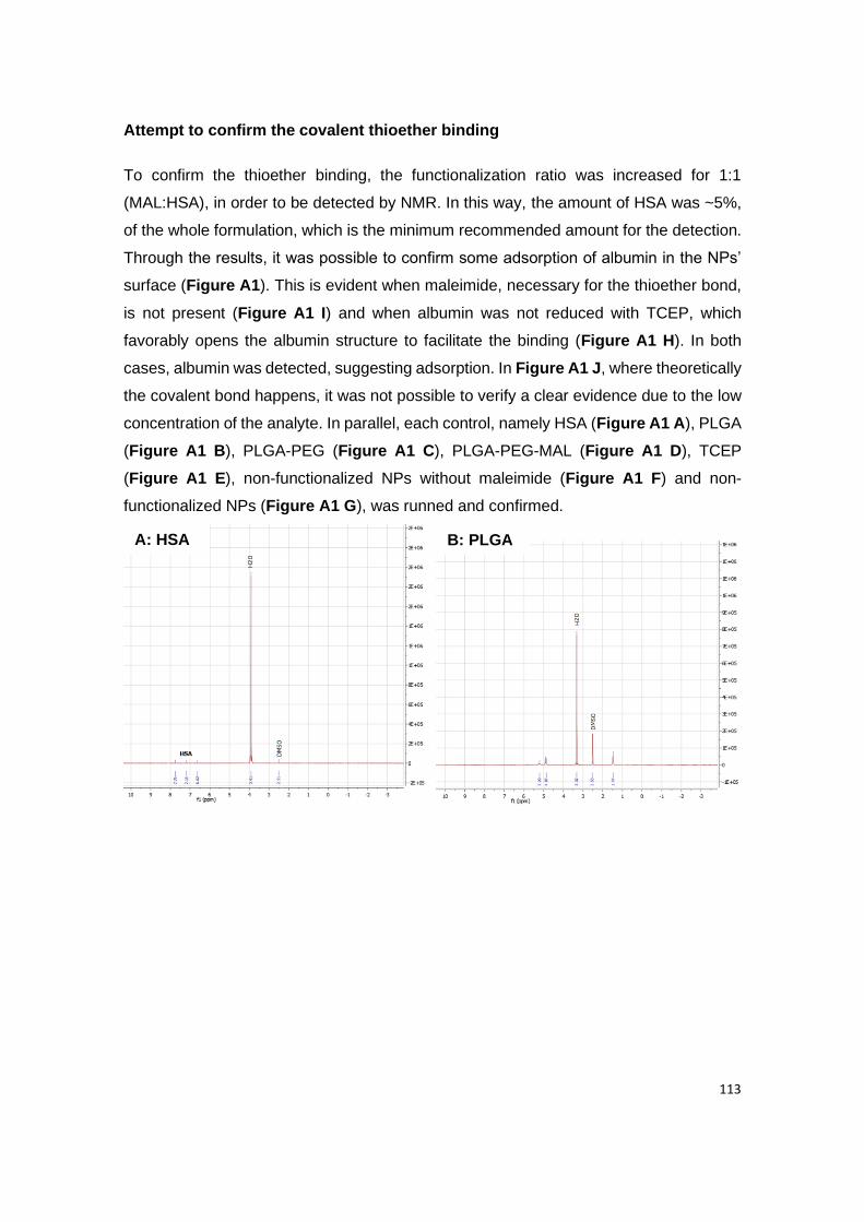
113
Attempt to confirm the covalent thioether binding
To confirm the thioether binding, the functionalization ratio was increased for 1:1
(MAL:HSA), in order to be detected by NMR. In this way, the amount of HSA was ~5%,
of the whole formulation, which is the minimum recommended amount for the detection.
Through the results, it was possible to confirm some adsorption of albumin in the NPs’
surface (Figure A1). This is evident when maleimide, necessary for the thioether bond,
is not present (Figure A1 I) and when albumin was not reduced with TCEP, which
favorably opens the albumin structure to facilitate the binding (Figure A1 H). In both
cases, albumin was detected, suggesting adsorption. In Figure A1 J, where theoretically
the covalent bond happens, it was not possible to verify a clear evidence due to the low
concentration of the analyte. In parallel, each control, namely HSA (Figure A1 A), PLGA
(Figure A1 B), PLGA-PEG (Figure A1 C), PLGA-PEG-MAL (Figure A1 D), TCEP
(Figure A1 E), non-functionalized NPs without maleimide (Figure A1 F) and non-
functionalized NPs (Figure A1 G), was runned and confirmed.
A: HSA B: PLGA
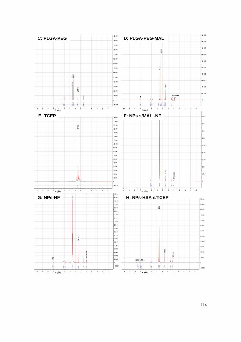
114
C: PLGA-PEG D: PLGA-PEG-MAL
E: TCEP F: NPs s/MAL -NF
G: NPs-NF H: NPs-HSA s/TCEP
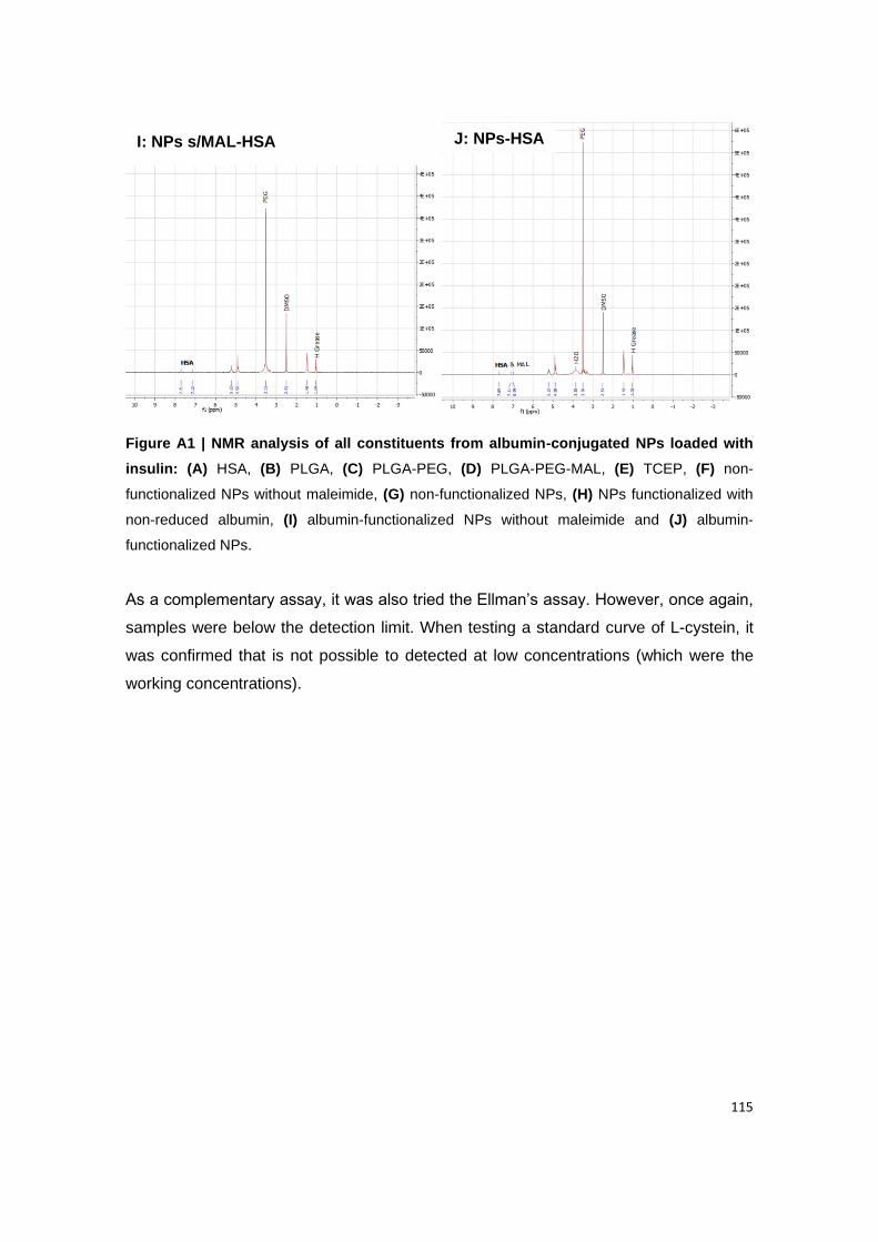
115
Figure A1 | NMR analysis of all constituents from albumin-conjugated NPs loaded with
insulin: (A) HSA, (B) PLGA, (C) PLGA-PEG, (D) PLGA-PEG-MAL, (E) TCEP, (F) non-
functionalized NPs without maleimide, (G) non-functionalized NPs, (H) NPs functionalized with
non-reduced albumin, (I) albumin-functionalized NPs without maleimide and (J) albumin-
functionalized NPs.
As a complementary assay, it was also tried the Ellman’s assay. However, once again,
samples were below the detection limit. When testing a standard curve of L-cystein, it
was confirmed that is not possible to detected at low concentrations (which were the
working concentrations).
I: NPs s/MAL-HSA J: NPs-HSA

116
117
Appendix II
THE EFFECT OF HYPERGRAVITY IN INTESTINAL
PERMEABILITY

118

119
This chapter was based in the following paper:
• Azevedo C.*, Almeida A.*, Macedo M.H.*, Pinto S.*, Sarmento B, van Loon J., The
effect of hypergravity in intestinal permeability of nanoformulations and molecules,
European Journal of Pharmaceutics and Biopharmaceutics 2021, 163: 38-48.
(*equal contribution)
As leader of this project, I idealized it and managed people, funding and sources. In this
original paper, I actively participated in the conception and execution of the experimental
work, as well as, data analyzes, their interpretation and discussion and in the writing,
formatting and revision of the manuscript. These tasks were equally done by the co-
authors. As leader, I had the role of validating each task at the end. The supervisors
were responsible for reviewing and submit the manuscript. This paper was not previously
included in other thesis and it is partially reproduced in this section. The integral version
of the original paper was included at the end of the thesis, at Appendix IV.

120
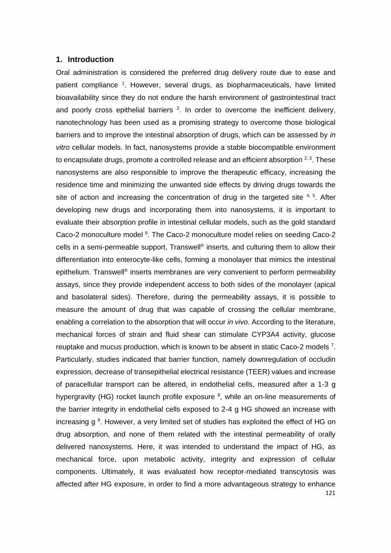
121
1. Introduction
Oral administration is considered the preferred drug delivery route due to ease and
patient compliance 1. However, several drugs, as biopharmaceuticals, have limited
bioavailability since they do not endure the harsh environment of gastrointestinal tract
and poorly cross epithelial barriers 2. In order to overcome the inefficient delivery,
nanotechnology has been used as a promising strategy to overcome those biological
barriers and to improve the intestinal absorption of drugs, which can be assessed by in
vitro cellular models. In fact, nanosystems provide a stable biocompatible environment
to encapsulate drugs, promote a controlled release and an efficient absorption 2, 3. These
nanosystems are also responsible to improve the therapeutic efficacy, increasing the
residence time and minimizing the unwanted side effects by driving drugs towards the
site of action and increasing the concentration of drug in the targeted site 4, 5. After
developing new drugs and incorporating them into nanosystems, it is important to
evaluate their absorption profile in intestinal cellular models, such as the gold standard
Caco-2 monoculture model 6. The Caco-2 monoculture model relies on seeding Caco-2
cells in a semi-permeable support, Transwell® inserts, and culturing them to allow their
differentiation into enterocyte-like cells, forming a monolayer that mimics the intestinal
epithelium. Transwell® inserts membranes are very convenient to perform permeability
assays, since they provide independent access to both sides of the monolayer (apical
and basolateral sides). Therefore, during the permeability assays, it is possible to
measure the amount of drug that was capable of crossing the cellular membrane,
enabling a correlation to the absorption that will occur in vivo. According to the literature,
mechanical forces of strain and fluid shear can stimulate CYP3A4 activity, glucose
reuptake and mucus production, which is known to be absent in static Caco-2 models 7.
Particularly, studies indicated that barrier function, namely downregulation of occludin
expression, decrease of transepithelial electrical resistance (TEER) values and increase
of paracellular transport can be altered, in endothelial cells, measured after a 1-3 g
hypergravity (HG) rocket launch profile exposure 8, while an on-line measurements of
the barrier integrity in endothelial cells exposed to 2-4 g HG showed an increase with
increasing g 9. However, a very limited set of studies has exploited the effect of HG on
drug absorption, and none of them related with the intestinal permeability of orally
delivered nanosystems. Here, it was intended to understand the impact of HG, as
mechanical force, upon metabolic activity, integrity and expression of cellular
components. Ultimately, it was evaluated how receptor-mediated transcytosis was
affected after HG exposure, in order to find a more advantageous strategy to enhance
122
drug delivery. This innovative study will help researchers and pharmaceutical industries
in oral drug development areas, since it may correlate more precisely the obtained
results with the permeability in the human intestine.
2. Materials and Methods
2.1. Metabolic activity studies
The influence of HG on the metabolic activity of Caco-2 cells was evaluated by a
resazurin and lactate dehydrogenase (LDH) assays. Caco-2 cells (4.0 x 10⁴ cells per
well) were seeded in 96-well plates in 200 μL DMEM complete medium and were
incubated for 24 h inside an incubator as described before. After that, medium was
removed and cells were washed twice with 200 μL of fresh pre-heated Phosphate-
Buffered Saline (PBS) 1X to remove cellular debris and 200 μL of fresh pre-heated
DMEM without FBS was added to each well. Plates were then incubated for 4 h at 1.0 g,
37°C in an orbital shaker or hypergravity in LDC at 2.5 g, 5.0 g and 7.5 g at 37°C.
Afterwards, the 96-well plates were centrifuged at 250 g for 10 min at RT to remove dead
cells, and the supernatant was used to perform the LDH assay, while the remaining
Caco-2 cells attached at the bottom of the plate were used for resazurin assay. For this,
cells were washed twice with 200 μL PBS 1X and then treated with DMEM complete
medium containing 20% resazurin (0.1 mg/mL; Merck) during 2 h at 37°C, 5% CO2 and
95% relative humidity, protected from light. After 2 h of incubation, the 150 μL of each
well was transferred to a black 96-well plate and the fluorescence was measured at
530/590 nm (excitation/emission wavelengths, respectively), using a plate reader (Victor
NivoTM, PerkinElmer). The negative control consisted in cells incubated with 1% (v/v) of
Triton X-100 in complete medium at 1.0 g and the positive control consisted in cells
incubated with complete medium at 1.0 g, while shaking. A number of six replicates were
performed for each group. The metabolic activity was expressed according to the
following equation:
Equation (1)
𝐶𝑒𝑙𝑙𝑢𝑙𝑎𝑟 𝑣𝑖𝑎𝑏𝑖𝑙𝑖𝑡𝑦 (%) =𝐴𝑏𝑠𝑜𝑟𝑏𝑎𝑛𝑐𝑒 (𝐸𝑥𝑝𝑒𝑟𝑖𝑚𝑒𝑛𝑡𝑎𝑙 𝑣𝑎𝑙𝑢𝑒) − 𝐴𝑏𝑠𝑜𝑟𝑏𝑎𝑛𝑐𝑒 (𝑃𝑜𝑠𝑖𝑡𝑖𝑣𝑒 𝑐𝑜𝑛𝑡𝑟𝑜𝑙)
𝐴𝑏𝑠𝑜𝑟𝑏𝑎𝑛𝑐𝑒 (𝑁𝑒𝑔𝑎𝑡𝑖𝑣𝑒 𝑐𝑜𝑛𝑡𝑟𝑜𝑙) − 𝐴𝑏𝑠𝑜𝑟𝑏𝑎𝑛𝑐𝑒 (𝑃𝑜𝑠𝑖𝑡𝑖𝑣𝑒 𝑐𝑜𝑛𝑡𝑟𝑜𝑙)× 100
To the 100 μL of supernatant collected after centrifugation from the 96-well plates, 100
μL of the reaction mixture composed by the catalyst solution and the dye solution (LDH
Cytotoxicity Detection Kit, Takara Bio) were added to each well and incubated for 10 min
at RT in the dark. The absorbance was measured at 492 nm and 690 nm, using a plate
123
reader (Victor NivoTM, PerkinElmer, Inc.). The negative control was composed by cells
incubated with complete medium only at 1.0 g and the positive control was composed
by cells incubated with 1% (v/v) of Triton X-100 in complete medium at 1.0 g, while
shaking. A number of six replicates were performed for each group. The cellular
cytotoxicity was expressed according to the following equation:
Equation (2)
𝐶𝑒𝑙𝑙𝑢𝑙𝑎𝑟 𝐶𝑦𝑡𝑜𝑡𝑜𝑥𝑖𝑐𝑖𝑡𝑦 (%) =𝐴𝑏𝑠𝑜𝑟𝑏𝑎𝑛𝑐𝑒 (𝐸𝑥𝑝𝑒𝑟𝑖𝑚𝑒𝑛𝑡𝑎𝑙 𝑣𝑎𝑙𝑢𝑒) − 𝐴𝑏𝑠𝑜𝑟𝑏𝑎𝑛𝑐𝑒 (𝑁𝑒𝑔𝑎𝑡𝑖𝑣𝑒 𝑐𝑜𝑛𝑡𝑟𝑜𝑙)
𝐹𝑙𝑢𝑜𝑟𝑒𝑠𝑐𝑒𝑛𝑐𝑒 (𝑃𝑜𝑠𝑖𝑡𝑖𝑣𝑒 𝑐𝑜𝑛𝑡𝑟𝑜𝑙) − 𝐹𝑙𝑢𝑜𝑟𝑒𝑠𝑐𝑒𝑛𝑐𝑒 (𝑁𝑒𝑔𝑎𝑡𝑖𝑣𝑒 𝑐𝑜𝑛𝑡𝑟𝑜𝑙)× 100
To evaluate the effect of insulin-loaded NPs conjugated to WT albumin (Ins-NPs-Alb)
upon metabolic activity of Caco-2 cells, a MTT assays was performed. In this case, Caco-
2 monoculture was seeded at a density of 10 000 cells/well in 200 µL per well and
incubated for 4 hours. After that, the medium was aspirated, and the wells were washed
twice with 100 mL of fresh pre-heated HBSS buffer (pH 7.4). After washing, 0.2 mL of
the nanoparticle solutions corresponding to insulin amounts of 25, 50, 100 or 150 μg/mL
of insulin and the correspondent that was encapsulated in NPs were added to each well
and the plates were incubated for a period of 4 and 24 h at 37°C. After the incubation
period, the media was removed, and 150 μL of 0.5 mg/mL MTT solution was added at
each well, followed by incubation of the plates at 37°C during 4 h. The reaction was
terminated by removal of the media and addition of 150 μL of dimethyl sulfoxide. The
levels of reduced MTT were determined by measuring the absorbance at 470 nm and
630 nm using a microtiter plate reader (Biotek-Synergy H1 Hibrid reader, using the Gen5
2.01 program). The % of MTT reduction was calculated as follows:
Equation (3) 𝑀𝑇𝑇 𝑟𝑒𝑑𝑢𝑐𝑡𝑖𝑜𝑛 (%) = 𝑎𝑏𝑠𝑜𝑟𝑏𝑎𝑛𝑐𝑒 𝑜𝑓 𝑡𝑟𝑒𝑎𝑡𝑒𝑑 𝑐𝑒𝑙𝑙𝑠
𝑎𝑏𝑠𝑜𝑟𝑏𝑎𝑛𝑐𝑒 𝑜𝑓 𝑢𝑛𝑡𝑟𝑒𝑎𝑡𝑒𝑑 𝑐𝑒𝑙𝑙𝑠 × 100
The final results of MTT reduction, are expressed as the percentage of the absorbance
of control cells.
2.2. Expression of cellular components by immunocytochemistry
To understand if HG could have an impact in tight junctions (TJs) and drug transporters
expression of cells, the models were subjected to the different g-levels, without any
treatment. The TJ presence was assessed using claudin-1, which is an integral
transmembrane protein, and zona occludens 1 (ZO-1), a peripheral TJ protein. The
presence of the drug transporters peptide transporter 1 (PEPT-1) and nenonatal Fc
receptor (FcRn) was also evaluated. Mucin 2 (MUC2) was chosen to evaluate mucin
124
expression, since it is the mostly expressed mucin in the small intestine 10. After 4 h of
exposure to HG, samples were washed once with PBS 1X and fixed with 2% PFA for 20
min. Then, cells were washed three times with PBS 1X. To stain with different antibodies,
a permeabilization step using a solution of 0.2% (v/v) Triton X-100 in PBS 1X for 5 min
at RT was performed. Afterward, cells were washed thrice with PBS 1X for 5 min. A
blocking step was performed using a solution containing 1.5% BSA and 5% FBS in PBS
1X for 1 h at RT. Inserts were incubated with the primary antibodies’ solutions [rabbit
anti-human Claudin-1 (1:200; SAB5500083-100, Sigma Aldrich) + mouse anti-human
MUC2 primary antibody (1:50; ab118964, Abcam)], [rabbit anti-human ZO-1 primary
antibody conjugated with Alexa Fluor 488 (1:50, SC-33725, Santa Cruz Biotechnology)
+ mouse anti-human PEPT1 primary antibody (1:100; ab203043, Abcam)] and [rabbit
anti-human FcRn primary antibody (1:100; ab193148, Abcam) + Phalloidin (1:50;
A22287, Thermo Fisher Scientific)] overnight at 4°C in a humidified chamber protected
from light. Inserts were washed three times for 5 min with PBST and incubated with the
secondary antibodies and DAPI to counterstain the nuclei (500 ng/mL; Sigma Aldrich).
Depending on the primary antibodies used, Goat anti-Mouse Alexa Fluor 594 F(ab’)2
fragment of IgG (H+L) (1:500; A11020, Invitrogen), Goat anti-Rabbit Alexa Fluor 488
F(ab’) fragment of IgG (H+L) (1:500; A11070, Invitrogen) and Donkey anti-Rabbit IgG
H&L Alexa Fluor 594 preadsorbed (1:500; Ab150064, Abcam) were used. Secondary
antibodies were added to samples for 2 h at 4°C in a humidified chamber protected from
light. Cells were then washed twice with PBST and once with PBS 1X, all for 5 min.
Inserts were mounted in slices using fluorescent mounting medium (Dako). Samples
were left to dry protected from light and were maintained at 4°C until visualization by a
spectral confocal laser scanning microscope TCS-SP5 AOBS (Leica). Since the aim was
to compare the amount and localization of tight and adherens junctions between samples
and also the expression of drug transporters, laser power and gain of each laser were
maintained for all samples regarding each antibody.
2.3. Cell culture
C2BBe1 [Caco-2 clone] (human colorectal adenocarcinoma) cells were acquired from
ATCC and maintained in a complete medium consisting of Dulbecco’s Modified Eagle’s
Medium (DMEM, ATCC) supplemented with 10% (v/v) fetal bovine serum (FBS,
Biochrom) and 1% (v/v) antibiotic-antimitotic mixture (final concentration of 100 U/mL
penicillin and 100 U/mL streptomycin) in 75 cm2 T-flasks, inside an incubator at 37°C
125
and 5% CO2 and 95% relative humidity. DMEM medium was changed three times per
week. Caco-2 cells were used at passages 77 to 83. When confluence reached about
70% to 80%, cells were detached from the flasks by treating it with trypsin-EDTA at 37°C
and transferred to another 75 cm2 T-flasks or Transwell® and multiwell plates.
2.4. Hypergravity exposure
Cells were exposed to hypergravity in the Large Diameter Centrifuge (LDC) 12 at the
TEC-MMG LIS Lab from the Technology Center (ESTEC) of the European Space
Agency, ESA, in Noordwijk, The Netherlands. The LDC, with a diameter of 8 meters,
allows for the application of up to 20 g. The four arms of the centrifuge hold at maximum
six gondolas, plus one gondola in the center for a 1 g rotational control, if required
(Figure A2.1 A). The gondolas of the centrifuge swing out, resulting in an acceleration
vector that is always perpendicular to the samples surface. Because of this and its size,
the systems avoid inertial shear forces common in smaller centrifuges 13. The Transwell®
multiwell plates were placed in a 37°C incubator (Thermo Scientific Heratherm IMC-18
mini incubator) that were accommodated in one of the gondolas. Since multi g-levels can
be created during one run the experiment was performed during 2 rounds.
2.5. Cellular integrity by hematoxylin and eosin staining
To assess cellular integrity after exposure to different g-levels, hematoxylin and eosin
(H&E) staining was performed. For this, inserts with the cells were washed once with
PBS 1X for 5 min and fixed using 2% paraformaldehyde (PFA) in PBS 1X for 20 min at
RT. Then, inserts were washed thrice with PBS 1X for 5 min and kept in PBS 1X at 4oC
until further processing for paraffin embedding. Sections of 3 mm were obtained using
an RM2255 microtome (Leica) and then stained for H&E. Briefly, sections were
deparaffinized and rehydrated, stained for 3 min in Gil’s Hematoxylin (Thermo Scientific),
6 min in running water, dehydrated, stained for 1 min in Eosin Y (Thermo Scientific),
cleared, and mounted in Entellan (Merck). Representative images of each condition were
obtained with a Light microscope Olympus DP 25 Camera Software Cell B.
2.6. Intestinal permeability study
Caco-2 cells were seeded at a density of 1.0 x 10⁵ cells per cm2 onto 12-Transwell® cell
culture inserts (surface area of 1.1 cm2, inner diameter of 12 mm and pore size of 1 μm;
Millicell®) and were allowed to grow and differentiate for 21 days inside an incubator at
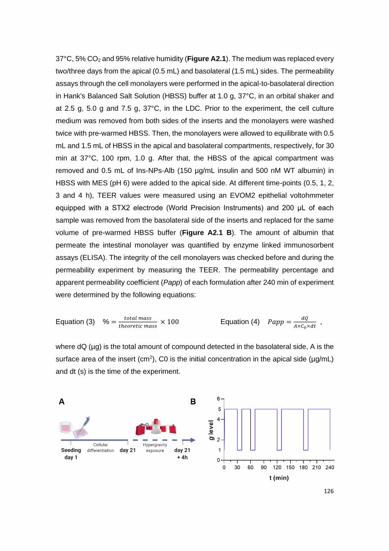
126
37°C, 5% CO2 and 95% relative humidity (Figure A2.1). The medium was replaced every
two/three days from the apical (0.5 mL) and basolateral (1.5 mL) sides. The permeability
assays through the cell monolayers were performed in the apical-to-basolateral direction
in Hank's Balanced Salt Solution (HBSS) buffer at 1.0 g, 37°C, in an orbital shaker and
at 2.5 g, 5.0 g and 7.5 g, 37°C, in the LDC. Prior to the experiment, the cell culture
medium was removed from both sides of the inserts and the monolayers were washed
twice with pre-warmed HBSS. Then, the monolayers were allowed to equilibrate with 0.5
mL and 1.5 mL of HBSS in the apical and basolateral compartments, respectively, for 30
min at 37°C, 100 rpm, 1.0 g. After that, the HBSS of the apical compartment was
removed and 0.5 mL of Ins-NPs-Alb (150 µg/mL insulin and 500 nM WT albumin) in
HBSS with MES (pH 6) were added to the apical side. At different time-points (0.5, 1, 2,
3 and 4 h), TEER values were measured using an EVOM2 epithelial voltohmmeter
equipped with a STX2 electrode (World Precision Instruments) and 200 μL of each
sample was removed from the basolateral side of the inserts and replaced for the same
volume of pre-warmed HBSS buffer (Figure A2.1 B). The amount of albumin that
permeate the intestinal monolayer was quantified by enzyme linked immunosorbent
assays (ELISA). The integrity of the cell monolayers was checked before and during the
permeability experiment by measuring the TEER. The permeability percentage and
apparent permeability coefficient (Papp) of each formulation after 240 min of experiment
were determined by the following equations:
Equation (3) % =𝑡𝑜𝑡𝑎𝑙 𝑚𝑎𝑠𝑠
𝑡ℎ𝑒𝑜𝑟𝑒𝑡𝑖𝑐 𝑚𝑎𝑠𝑠 × 100 Equation (4) 𝑃𝑎𝑝𝑝 =
𝑑𝑄
𝐴×𝐶0×𝑑𝑡 ,
where dQ (µg) is the total amount of compound detected in the basolateral side, A is the
surface area of the insert (cm2), C0 is the initial concentration in the apical side (µg/mL)
and dt (s) is the time of the experiment.
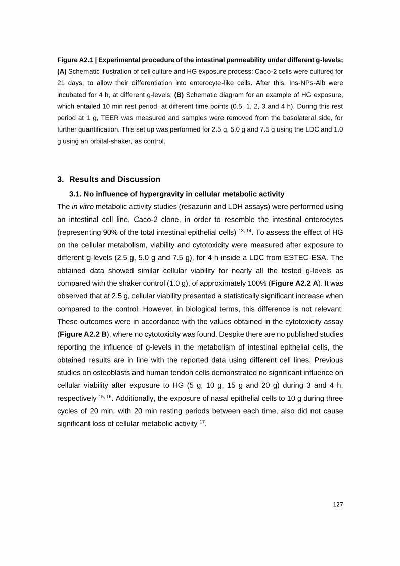
127
Figure A2.1 | Experimental procedure of the intestinal permeability under different g-levels;
(A) Schematic illustration of cell culture and HG exposure process: Caco-2 cells were cultured for
21 days, to allow their differentiation into enterocyte-like cells. After this, Ins-NPs-Alb were
incubated for 4 h, at different g-levels; (B) Schematic diagram for an example of HG exposure,
which entailed 10 min rest period, at different time points (0.5, 1, 2, 3 and 4 h). During this rest
period at 1 g, TEER was measured and samples were removed from the basolateral side, for
further quantification. This set up was performed for 2.5 g, 5.0 g and 7.5 g using the LDC and 1.0
g using an orbital-shaker, as control.
3. Results and Discussion
3.1. No influence of hypergravity in cellular metabolic activity
The in vitro metabolic activity studies (resazurin and LDH assays) were performed using
an intestinal cell line, Caco-2 clone, in order to resemble the intestinal enterocytes
(representing 90% of the total intestinal epithelial cells) 13, 14. To assess the effect of HG
on the cellular metabolism, viability and cytotoxicity were measured after exposure to
different g-levels (2.5 g, 5.0 g and 7.5 g), for 4 h inside a LDC from ESTEC-ESA. The
obtained data showed similar cellular viability for nearly all the tested g-levels as
compared with the shaker control (1.0 g), of approximately 100% (Figure A2.2 A). It was
observed that at 2.5 g, cellular viability presented a statistically significant increase when
compared to the control. However, in biological terms, this difference is not relevant.
These outcomes were in accordance with the values obtained in the cytotoxicity assay
(Figure A2.2 B), where no cytotoxicity was found. Despite there are no published studies
reporting the influence of g-levels in the metabolism of intestinal epithelial cells, the
obtained results are in line with the reported data using different cell lines. Previous
studies on osteoblasts and human tendon cells demonstrated no significant influence on
cellular viability after exposure to HG (5 g, 10 g, 15 g and 20 g) during 3 and 4 h,
respectively 15, 16. Additionally, the exposure of nasal epithelial cells to 10 g during three
cycles of 20 min, with 20 min resting periods between each time, also did not cause
significant loss of cellular metabolic activity 17.
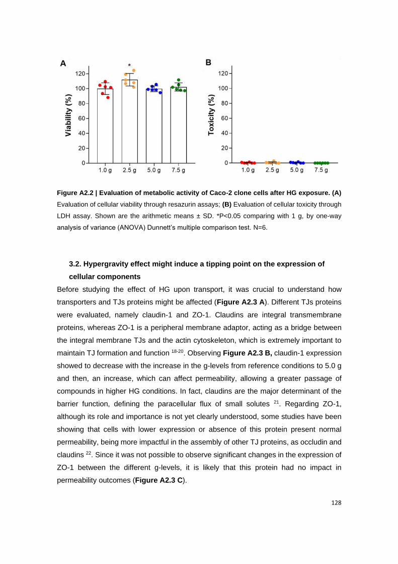
128
Figure A2.2 | Evaluation of metabolic activity of Caco-2 clone cells after HG exposure. (A)
Evaluation of cellular viability through resazurin assays; (B) Evaluation of cellular toxicity through
LDH assay. Shown are the arithmetic means ± SD. *P<0.05 comparing with 1 g, by one-way
analysis of variance (ANOVA) Dunnett’s multiple comparison test. N=6.
3.2. Hypergravity effect might induce a tipping point on the expression of
cellular components
Before studying the effect of HG upon transport, it was crucial to understand how
transporters and TJs proteins might be affected (Figure A2.3 A). Different TJs proteins
were evaluated, namely claudin-1 and ZO-1. Claudins are integral transmembrane
proteins, whereas ZO-1 is a peripheral membrane adaptor, acting as a bridge between
the integral membrane TJs and the actin cytoskeleton, which is extremely important to
maintain TJ formation and function 18-20. Observing Figure A2.3 B, claudin-1 expression
showed to decrease with the increase in the g-levels from reference conditions to 5.0 g
and then, an increase, which can affect permeability, allowing a greater passage of
compounds in higher HG conditions. In fact, claudins are the major determinant of the
barrier function, defining the paracellular flux of small solutes 21. Regarding ZO-1,
although its role and importance is not yet clearly understood, some studies have been
showing that cells with lower expression or absence of this protein present normal
permeability, being more impactful in the assembly of other TJ proteins, as occludin and
claudins 22. Since it was not possible to observe significant changes in the expression of
ZO-1 between the different g-levels, it is likely that this protein had no impact in
permeability outcomes (Figure A2.3 C).
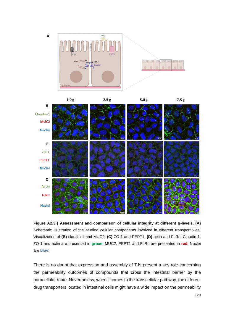
129
Figure A2.3 | Assessment and comparison of cellular integrity at different g-levels. (A)
Schematic illustration of the studied cellular components involved in different transport vias.
Visualization of (B) claudin-1 and MUC2; (C) ZO-1 and PEPT1, (D) actin and FcRn. Claudin-1,
ZO-1 and actin are presented in green. MUC2, PEPT1 and FcRn are presented in red. Nuclei
are blue.
There is no doubt that expression and assembly of TJs present a key role concerning
the permeability outcomes of compounds that cross the intestinal barrier by the
paracellular route. Nevertheless, when it comes to the transcellular pathway, the different
drug transporters located in intestinal cells might have a wide impact on the permeability

130
of compounds, mostly in the ones that are substrates of these transporters. Taking into
consideration that FcRn mediates the uptake of Ins-NPs-Alb, the expression of this
transporter after the exposure to HG was evaluated. Despite PEPT-1 not being directly
linked to the drugs tested in this study, its expression was evaluated, since this is an
important uptake transporter present in intestinal cells and it is considered highly
atractive as a drug target 23. When looking at the expression of PEPT1 (Figure A2.3 C),
its expression was increased from reference to 2.5 g and then a decrease was observed
for the higher g-levels. When looking at the expression of FcRn, the behavior was
different, where a decrease in the expression was observed from reference to 5.0 g, and
a steep increase was observed at from 5.0 g to 7.5 g (Figure A2.3 D). In the case of
actin, there was a decrease from reference to 2.5 g, a stable signal between 2.5 g and
5.0 g and then an increase at 7.5 g (Figure A2.3 D). MUC2 was evaluated because this
is the most expressed mucin in the small intestine and it is known that mechanical forces
can interfere with the production of mucus, as previously stated 7, 10. In this sense, it could
be interesting to understand if HG might have an impact on its secretion. However, the
change of MUC2 with the increase in g-levels seems to be negligible (Figure A2.3 B).
Considering all these results, it was possible to conclude that HG affects protein
expression in different ways. It is interesting to see that there seems to be a tipping point
at 2.5 g or 5.0 g and proteins revert their behavior when reaching that point. For PEPT-
1 this turning point occurs at 2.5 g, whether for FcRn and Claudin-1, this occurs at 5.0 g.
Other interesting fact is that this change in behavior can occur in both directions,
meaning protein expression can first increase and then decrease or the other way
around, depending on the protein (Figure A2.4).
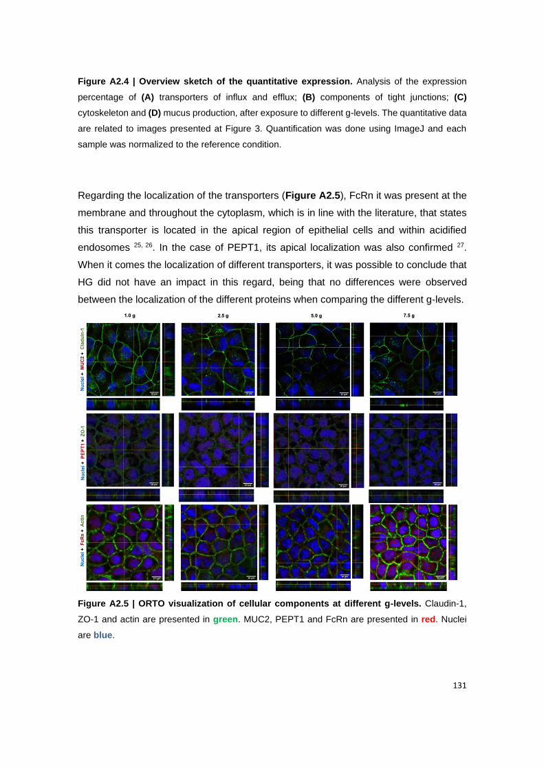
131
Figure A2.4 | Overview sketch of the quantitative expression. Analysis of the expression
percentage of (A) transporters of influx and efflux; (B) components of tight junctions; (C)
cytoskeleton and (D) mucus production, after exposure to different g-levels. The quantitative data
are related to images presented at Figure 3. Quantification was done using ImageJ and each
sample was normalized to the reference condition.
Regarding the localization of the transporters (Figure A2.5), FcRn it was present at the
membrane and throughout the cytoplasm, which is in line with the literature, that states
this transporter is located in the apical region of epithelial cells and within acidified
endosomes 25, 26. In the case of PEPT1, its apical localization was also confirmed 27.
When it comes the localization of different transporters, it was possible to conclude that
HG did not have an impact in this regard, being that no differences were observed
between the localization of the different proteins when comparing the different g-levels.
Figure A2.5 | ORTO visualization of cellular components at different g-levels. Claudin-1,
ZO-1 and actin are presented in green. MUC2, PEPT1 and FcRn are presented in red. Nuclei
are blue.
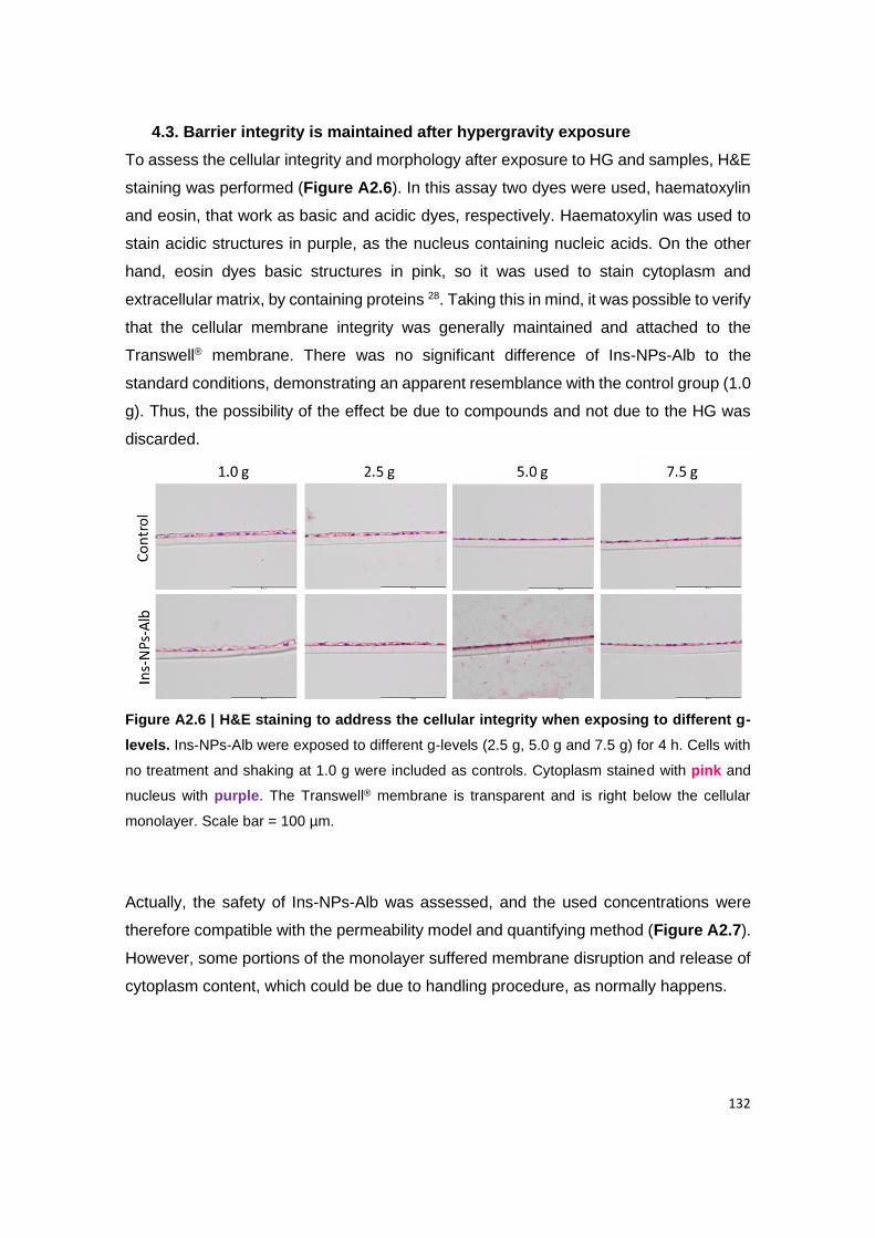
132
4.3. Barrier integrity is maintained after hypergravity exposure
To assess the cellular integrity and morphology after exposure to HG and samples, H&E
staining was performed (Figure A2.6). In this assay two dyes were used, haematoxylin
and eosin, that work as basic and acidic dyes, respectively. Haematoxylin was used to
stain acidic structures in purple, as the nucleus containing nucleic acids. On the other
hand, eosin dyes basic structures in pink, so it was used to stain cytoplasm and
extracellular matrix, by containing proteins 28. Taking this in mind, it was possible to verify
that the cellular membrane integrity was generally maintained and attached to the
Transwell® membrane. There was no significant difference of Ins-NPs-Alb to the
standard conditions, demonstrating an apparent resemblance with the control group (1.0
g). Thus, the possibility of the effect be due to compounds and not due to the HG was
discarded.
Figure A2.6 | H&E staining to address the cellular integrity when exposing to different g-
levels. Ins-NPs-Alb were exposed to different g-levels (2.5 g, 5.0 g and 7.5 g) for 4 h. Cells with
no treatment and shaking at 1.0 g were included as controls. Cytoplasm stained with pink and
nucleus with purple. The Transwell® membrane is transparent and is right below the cellular
monolayer. Scale bar = 100 µm.
Actually, the safety of Ins-NPs-Alb was assessed, and the used concentrations were
therefore compatible with the permeability model and quantifying method (Figure A2.7).
However, some portions of the monolayer suffered membrane disruption and release of
cytoplasm content, which could be due to handling procedure, as normally happens.
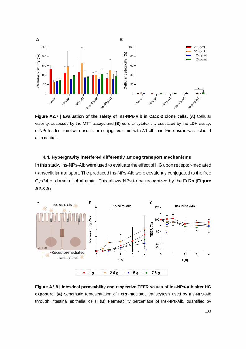
133
Figure A2.7 | Evaluation of the safety of Ins-NPs-Alb in Caco-2 clone cells. (A) Cellular
viability, assessed by the MTT assays and (B) cellular cytotoxicity assessed by the LDH assay,
of NPs loaded or not with insulin and conjugated or not with WT albumin. Free insulin was included
as a control.
4.4. Hypergravity interfered differently among transport mechanisms
In this study, Ins-NPs-Alb were used to evaluate the effect of HG upon receptor-mediated
transcellular transport. The produced Ins-NPs-Alb were covalently conjugated to the free
Cys34 of domain I of albumin. This allows NPs to be recognized by the FcRn (Figure
A2.8 A).
Figure A2.8 | Intestinal permeability and respective TEER values of Ins-NPs-Alb after HG
exposure. (A) Schematic representation of FcRn-mediated transcytosis used by Ins-NPs-Alb
through intestinal epithelial cells; (B) Permeability percentage of Ins-NPs-Alb, quantified by
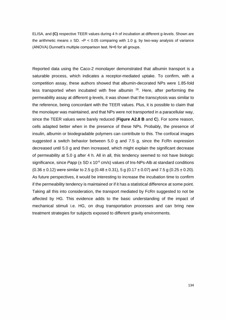
134
ELISA, and (C) respective TEER values during 4 h of incubation at different g-levels. Shown are
the arithmetic means ± SD. ∗P < 0.05 comparing with 1.0 g, by two-way analysis of variance
(ANOVA) Dunnett’s multiple comparison test. N=6 for all groups.
Reported data using the Caco-2 monolayer demonstrated that albumin transport is a
saturable process, which indicates a receptor-mediated uptake. To confirm, with a
competition assay, these authors showed that albumin-decorated NPs were 1.85-fold
less transported when incubated with free albumin 28. Here, after performing the
permeability assay at different g-levels, it was shown that the transcytosis was similar to
the reference, being concordant with the TEER values. Plus, it is possible to claim that
the monolayer was maintained, and that NPs were not transported in a paracellular way,
since the TEER values were barely reduced (Figure A2.8 B and C). For some reason,
cells adapted better when in the presence of these NPs. Probably, the presence of
insulin, albumin or biodegradable polymers can contribute to this. The confocal images
suggested a switch behavior between 5.0 g and 7.5 g, since the FcRn expression
decreased until 5.0 g and then increased, which might explain the significant decrease
of permeability at 5.0 g after 4 h. All in all, this tendency seemed to not have biologic
significance, since Papp (± SD x 10-6 cm/s) values of Ins-NPs-Alb at standard conditions
(0.36 ± 0.12) were similar to 2.5 g (0.48 ± 0.31), 5 g (0.17 ± 0.07) and 7.5 g (0.25 ± 0.20).
As future perspectives, it would be interesting to increase the incubation time to confirm
if the permeability tendency is maintained or if it has a statistical difference at some point.
Taking all this into consideration, the transport mediated by FcRn suggested to not be
affected by HG. This evidence adds to the basic understanding of the impact of
mechanical stimuli i.e. HG, on drug transportation processes and can bring new
treatment strategies for subjects exposed to different gravity environments.

135
4. References
1. B. Sarmento DF, T. Vasconcelos (Eds: L. Jorgensen, H. M. Nielsen), John Wiley & Sons, Ltd
Polymer-based delivery systems for oral delivery of peptides and proteins, in: L. Jorgensen
HMN, ed. Delivery Technologies for Biopharmaceuticals: Peptides, Proteins, Nucleic Acids
and Vaccines: John Wiley & Sons, Ltd 2009:207.
2. Araujo F, Shrestha N, Granja PL, et al. Antihyperglycemic potential of incretins orally
delivered via nano and microsystems and subsequent glucoregulatory effects. Current
Pharmaceutical Biotechnology 2014;15:609-19.
3. Pereira C, Araújo F, Granja PL, et al. Targeting membrane transporters and receptors as a
mean to optimize orally delivered biotecnhological based drugs through nanoparticle delivery
systems. Current Pharmaceutical Biotechnology 2014;15:650-658.
4. Moulari B, Beduneau A, Pellequer Y, et al. Nanoparticle targeting to inflamed tissues of
gastrointestinal tract Current Drug Delivery 2013;10:9-17.
5. Sarmento B, Ferreira D, Vasconcelos T. Polymer-Based Delivery Systems for Oral Delivery
of Peptides and Proteins. In: Jorgensen L, Nielsen HM, eds. Delivery Technologies for
Biopharmaceuticals: Peptides, Proteins, Nucleic Acids and Vaccines: John Wiley & Sons, Ltd,
2009:207-227.
6. Hidalgo IJ, Raub TJ, Borchardt RT. Characterization of the human colon carcinoma cell line
(Caco-2) as a model system for intestinal epithelial permeability. Gastroenterology
1989;96:736-49.
7. Kim HJ, Ingber DE. Gut-on-a-Chip microenvironment induces human intestinal cells to
undergo villus differentiation. Integr Biol (Camb) 2013;5:1130-40.
8. Sumanasekera WK, Sumanasekera GU, Mattingly KA, et al. Estradiol and
dihydrotestosterone regulate endothelial cell barrier function after hypergravity-induced
alterations in MAPK activity. Am J Physiol Cell Physiol 2007;293:C566-73.
9. Szulcek R, van Bezu J, Boonstra J, et al. Transient Intervals of Hyper-Gravity Enhance
Endothelial Barrier Integrity: Impact of Mechanical and Gravitational Forces Measured
Electrically. PLoS One 2015;10:e0144269.
10. Schneider H, Pelaseyed T, Svensson F, et al. Study of mucin turnover in the small intestine
by in vivo labeling. Scientific Reports 2018;8:5760.
11. Loon JJWAv, Krausse J, Cunha H, et al. The Large Diameter Centrifuge, LDC, for Life and
Physical Sciences and Technology, 2008.
12. van Loon JJWA, Folgering EHTE, Bouten CVC, et al. Inertial Shear Forces and the Use of
Centrifuges in Gravity Research. What is the Proper Control? Journal of Biomechanical
Engineering 2003;125:342-346.
13. Araújo F, Sarmento B. Towards the characterization of an in vitro triple co-culture intestine
cell model for permeability studies. Int J Pharm 2013;458:128-34.
14. Pereira C, Costa J, Sarmento B, et al. 3.3 - Cell-based in vitro models for intestinal
136
permeability studies. In: Sarmento B, ed. Concepts and Models for Drug Permeability Studies:
Woodhead Publishing, 2016:57-81.
15. Searby ND, Steele CR, Globus RK. Influence of increased mechanical loading by
hypergravity on the microtubule cytoskeleton and prostaglandin E2 release in primary
osteoblasts. Am J Physiol Cell Physiol 2005;289:C148-58.
16. Costa-Almeida R, Carvalho DTO, Ferreira MJS, et al. Continuous Exposure to Simulated
Hypergravity-Induced Changes in Proliferation, Morphology, and Gene Expression of Human
Tendon Cells. Stem Cells Dev 2018;27:858-869.
17. Kim D, Kim YH, Kwon S. Enhanced nasal drug delivery efficiency by increasing mechanical
loading using hypergravity. Scientific Reports 2018;8:168.
18. Gonzalez-Mariscal L, Betanzos A, Nava P, et al. Tight junction proteins. Prog Biophys Mol
Biol 2003;81:1-44.
19. Lee B, Moon KM, Kim CY. Tight Junction in the Intestinal Epithelium: Its Association with
Diseases and Regulation by Phytochemicals. J Immunol Res 2018;2018:2645465.
20. Lee SH. Intestinal permeability regulation by tight junction: implication on inflammatory bowel
diseases. Intest Res 2015;13:11-8.
21. Gunzel D, Yu AS. Claudins and the modulation of tight junction permeability. Physiol Rev
2013;93:525-69.
22. Umeda K, Matsui T, Nakayama M, et al. Establishment and characterization of cultured
epithelial cells lacking expression of ZO-1. J Biol Chem 2004;279:44785-94.
23. Estudante M, Morais JG, Soveral G, et al. Intestinal drug transporters: an overview. Adv Drug
Deliv Rev 2013;65:1340-56.
24. Ober RJ, Martinez C, Vaccaro C, et al. Visualizing the site and dynamics of IgG salvage by
the MHC class I-related receptor, FcRn. J Immunol 2004;172:2021-9.
25. Pyzik M, Rath T, Kuo TT, et al. Hepatic FcRn regulates albumin homeostasis and
susceptibility to liver injury. Proc Natl Acad Sci U S A 2017;114:E2862-e2871.
26. Shugarts S, Benet LZ. The role of transporters in the pharmacokinetics of orally administered
drugs. Pharm Res 2009;26:2039-54.
27. Fischer AH, Jacobson KA, Rose J, et al. Hematoxylin and eosin staining of tissue and cell
sections. CSH Protoc 2008;2008:pdb.prot4986.
28. Hashem L, Swedrowska M, Vllasaliu D. Intestinal uptake and transport of albumin
nanoparticles: potential for oral delivery. Nanomedicine (Lond) 2018;13:1255-1265.
137
Appendix III
THE ALBUMIN TRIPLE MUTANT

138
139
1. Overview
In this section, it will be presented all data related to the albumin triple mutant. Besides
the K573P mutation, the triple mutant also has T83N and N111E mutations (TNNEKP).
These additional mutations confere a higher binding affinity to the receptor 1. However,
due to a conflict of interests, all these results were not allowed to be published yet.
Despite this, these were techniques that I learned, work that I did and time that I spent.
2. Materials and Methods
2.1. Binding affinity to human FcRn
Biolayer Interferometry (BLI) was used to investigate the hFcRn affinity of engineered
albumin variants comjugated to NPs. Measurements were performed on Octet RED96e
system (ForteBio, Pall Life Sciences, USA). Biotinylated engineered albumin variants
were conjugated to NPs and attached to the streptavidin sensor by biotinstreptavidin
interactions. The Dip and Read™ High Precision Streptavidin (SAX) Biosensors were
dipped in PBS (Sigma-Aldrich, USA) pH 7.4 containing 0.01% Tween® 20 (PBST)
(Sigma-Aldrich, USA) before coating with biotinylated albumin-conjugated NPs in PBST
pH 7.4. The sensor was then washed in PBST pH 7.4. Kinetic measurements were
performed at increasing FcRn concentrations at room temperature in association buffer
(25mM Na Acetate, 25mM NaH2PO4, and 150mM NaCl 0.01% Tween® 20. pH 5.5). Two
baselines were made for 30 s using PBST pH 7.4 and 60 s using association buffer pH
5.5. Inicially, different NPs concentrations (0.78 μg/mL, 1.56 μg/mL, 3.10 μg/mL, 6.25
μg/mL, 12.50 μg/mL, 25.00 μg/mL and 50.00 μg/mL) were tested in order to find the
desired loading curve. Kinetic measurements were performed from titrating 1 μM of
FcRn. The association phase lasted for 200 s and the dissociation phase 300 sec
followed by a regeneration step for 50 sec in PBST pH 7.4. Shaker speed for all
measurements were 2200 rpm. All values were zero-adjusted, and the buffer reference
value was subtracted. The three binding profiles were fitted to a 1:1 binding model after
intercept correction using global fit.
2.2. Assessment of FcRn expression in Caco-2 cells
The FcRn expression in Caco-2 cells was assessed by Fluorescence-activated cell
sorting (FACS) assay. Cells were detached from culture flask with versene and 4 × 10⁶
cells/mL were ressuspended in FACS buffer (PBS with 1% BSA and 0.1% NaN3) for 30
140
min at 4ºC. 100 µL of cells suspension was aliquot, in triplicate, into each well of a round-
bottom 96-well plate. The plate was then centrifuged at 300 x g for 3 min at 4ºC. After
discard the supernatant, the blocked cells were ressuspended in 12.5 µg/mL of mouse
IgG2a antibody anti-FcRn, previousy prepared in FACS buffer for 1 h at 4ºC. Cells were
washed three times with 100 µL of FACS buffer following centrifugation. Supernantant
was discard and cells were ressuspended in gost anti-mouse Alexa 594, diluted 1:500 in
FACS buffer, for 1 h at 4ºC. Repeat the washing step and at the end, cells were prepared
in PBS and transfered into FACS tubes.
2.3. In vivo studies
2.3.1. Biodistribution
2.3.1.1. Production of fluorescent nanoparticles
The synthesis of fluorescently labeled PLGA with Cyanine7.5 amine (Cy7.5; acquired
from Lumiprobe, Hunt Valley, MD, USA) was conducted using click chemistry. The
protocol used in this work was adapted from das Neves et al. 2. Briefly, PLGA-COOH
(0.012 mmol, 528 mg) and Cy7.5 (0.018 mmol, 14.75 mg) were combined to PLGA-
Cy7.5 in DCM, in the presence of excess EDC (0.06 mmol, 11.50 mg) and NHS (0.06
mmol, 6.90 mg) under stirring for 2h. PLGA-Cy7.5 was then precipitated with 30 mL of
ice-cold ethyl ether/methanol (1:1) and collected by 5min of centrifugation at 1,500 x g
(Eppendorf 5810R Refrigerated Centrifuge). The polymer was re-dissolved in 1 mL of
DCM and the precipitation process repeated twice as described, with 6 mL of ice-cold
ethyl ether/methanol in order to remove residual EDC and NHS. Then the solution was
dry under vacuum at room temperature for 48 h. The NPs production was conducted
according to was mentioned before on Chapter III - Section 3.3.1, including 0.02% (w/w)
of PLGA-Cy7.5.
2.3.1.2. Imaging
Fresh fluorescent NPs were oraly administered as previously mentioned. Whole body
near infrared (NIR) imaging of animals treated with NPs-Cy7.5 was performed at pre-
established time points by using an IVIS Lumina LT system (Perkin Elmer, Waltham, MA,
USA) after brief anesthetization with inhalational isoflurane. Abdominal fur was removed
with a hair clipper before imaging. Background signal from non-treated animals was used
to establish relevant radiance signal (≥ 0.4×10⁸ p cm2 s−1 μW−1).
Animals were sacrificed at 2 h or 8 h by isoflurane overdose followed by cervical
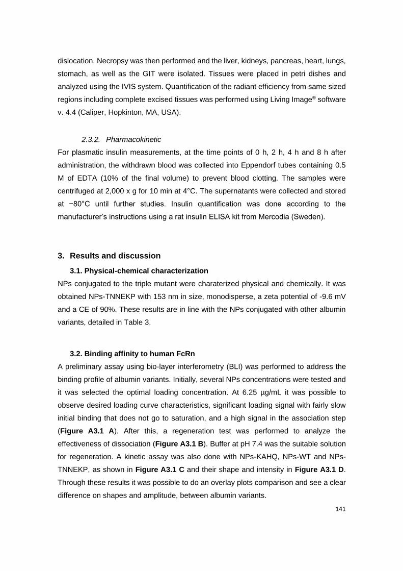
141
dislocation. Necropsy was then performed and the liver, kidneys, pancreas, heart, lungs,
stomach, as well as the GIT were isolated. Tissues were placed in petri dishes and
analyzed using the IVIS system. Quantification of the radiant efficiency from same sized
regions including complete excised tissues was performed using Living Image® software
v. 4.4 (Caliper, Hopkinton, MA, USA).
2.3.2. Pharmacokinetic
For plasmatic insulin measurements, at the time points of 0 h, 2 h, 4 h and 8 h after
administration, the withdrawn blood was collected into Eppendorf tubes containing 0.5
M of EDTA (10% of the final volume) to prevent blood clotting. The samples were
centrifuged at 2,000 x g for 10 min at 4°C. The supernatants were collected and stored
at −80°C until further studies. Insulin quantification was done according to the
manufacturer’s instructions using a rat insulin ELISA kit from Mercodia (Sweden).
3. Results and discussion
3.1. Physical-chemical characterization
NPs conjugated to the triple mutant were charaterized physical and chemically. It was
obtained NPs-TNNEKP with 153 nm in size, monodisperse, a zeta potential of -9.6 mV
and a CE of 90%. These results are in line with the NPs conjugated with other albumin
variants, detailed in Table 3.
3.2. Binding affinity to human FcRn
A preliminary assay using bio-layer interferometry (BLI) was performed to address the
binding profile of albumin variants. Initially, several NPs concentrations were tested and
it was selected the optimal loading concentration. At 6.25 µg/mL it was possible to
observe desired loading curve characteristics, significant loading signal with fairly slow
initial binding that does not go to saturation, and a high signal in the association step
(Figure A3.1 A). After this, a regeneration test was performed to analyze the
effectiveness of dissociation (Figure A3.1 B). Buffer at pH 7.4 was the suitable solution
for regeneration. A kinetic assay was also done with NPs-KAHQ, NPs-WT and NPs-
TNNEKP, as shown in Figure A3.1 C and their shape and intensity in Figure A3.1 D.
Through these results it was possible to do an overlay plots comparison and see a clear
difference on shapes and amplitude, between albumin variants.
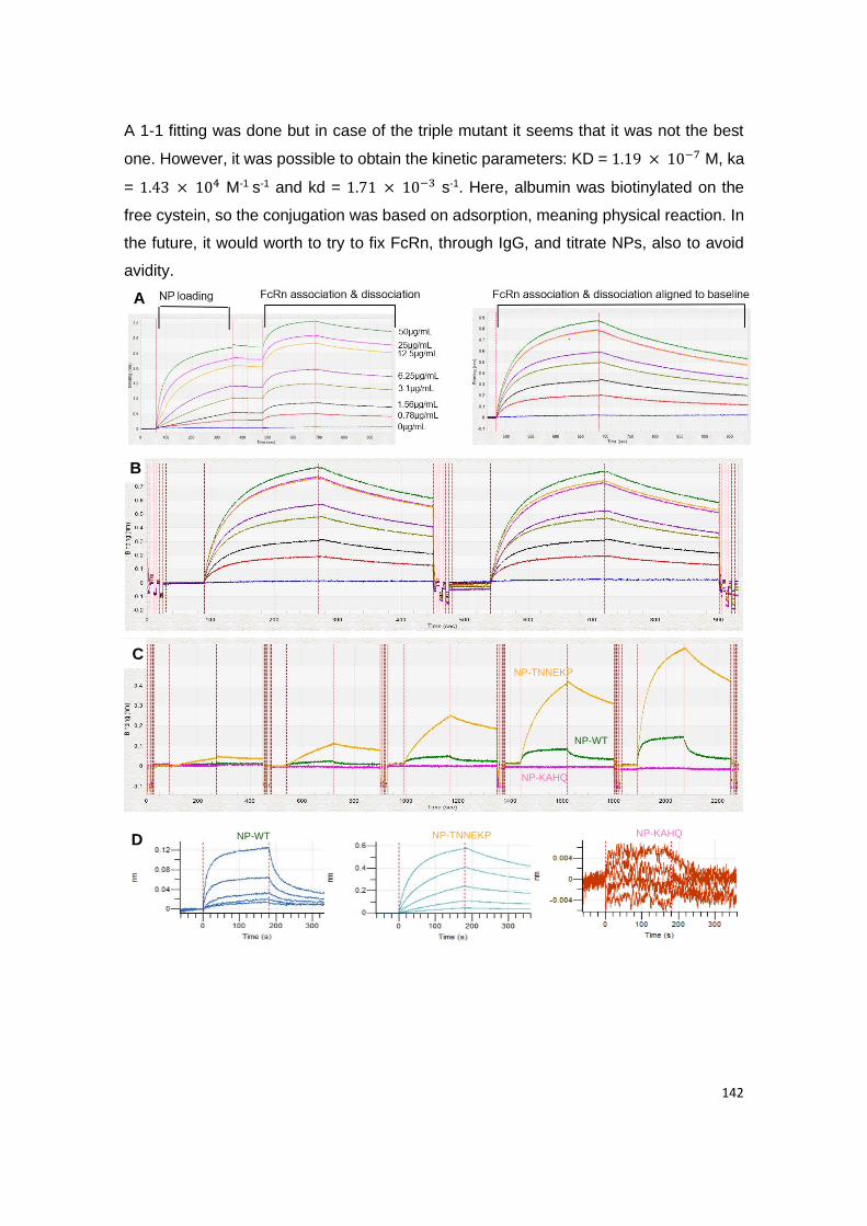
142
A 1-1 fitting was done but in case of the triple mutant it seems that it was not the best
one. However, it was possible to obtain the kinetic parameters: KD = 1.19 × 10⁻⁷ M, ka
= 1.43 × 10⁴ M-1 s-1 and kd = 1.71 × 10⁻³ s-1. Here, albumin was biotinylated on the
free cystein, so the conjugation was based on adsorption, meaning physical reaction. In
the future, it would worth to try to fix FcRn, through IgG, and titrate NPs, also to avoid
avidity.
NP-WT NP-KAHQ NP-TNNEKP
NP-TNNEKP
NP-WT
NP-KAHQ
A
B
C
D
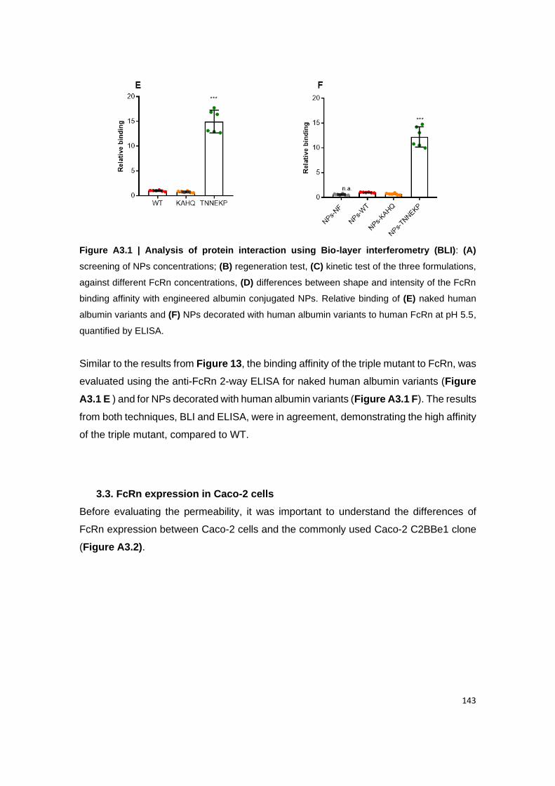
143
Figure A3.1 | Analysis of protein interaction using Bio-layer interferometry (BLI): (A)
screening of NPs concentrations; (B) regeneration test, (C) kinetic test of the three formulations,
against different FcRn concentrations, (D) differences between shape and intensity of the FcRn
binding affinity with engineered albumin conjugated NPs. Relative binding of (E) naked human
albumin variants and (F) NPs decorated with human albumin variants to human FcRn at pH 5.5,
quantified by ELISA.
Similar to the results from Figure 13, the binding affinity of the triple mutant to FcRn, was
evaluated using the anti-FcRn 2-way ELISA for naked human albumin variants (Figure
A3.1 E ) and for NPs decorated with human albumin variants (Figure A3.1 F). The results
from both techniques, BLI and ELISA, were in agreement, demonstrating the high affinity
of the triple mutant, compared to WT.
3.3. FcRn expression in Caco-2 cells
Before evaluating the permeability, it was important to understand the differences of
FcRn expression between Caco-2 cells and the commonly used Caco-2 C2BBe1 clone
(Figure A3.2).
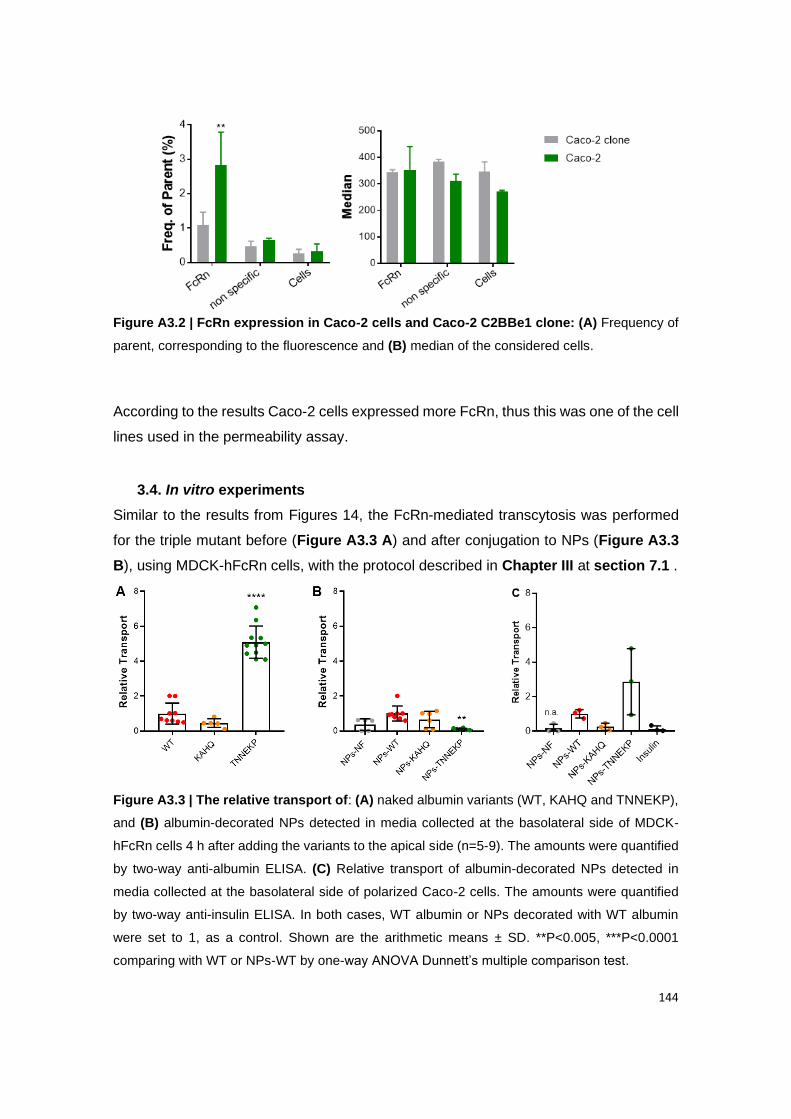
144
Figure A3.2 | FcRn expression in Caco-2 cells and Caco-2 C2BBe1 clone: (A) Frequency of
parent, corresponding to the fluorescence and (B) median of the considered cells.
According to the results Caco-2 cells expressed more FcRn, thus this was one of the cell
lines used in the permeability assay.
3.4. In vitro experiments
Similar to the results from Figures 14, the FcRn-mediated transcytosis was performed
for the triple mutant before (Figure A3.3 A) and after conjugation to NPs (Figure A3.3
B), using MDCK-hFcRn cells, with the protocol described in Chapter III at section 7.1 .
Figure A3.3 | The relative transport of: (A) naked albumin variants (WT, KAHQ and TNNEKP),
and (B) albumin-decorated NPs detected in media collected at the basolateral side of MDCK-
hFcRn cells 4 h after adding the variants to the apical side (n=5-9). The amounts were quantified
by two-way anti-albumin ELISA. (C) Relative transport of albumin-decorated NPs detected in
media collected at the basolateral side of polarized Caco-2 cells. The amounts were quantified
by two-way anti-insulin ELISA. In both cases, WT albumin or NPs decorated with WT albumin
were set to 1, as a control. Shown are the arithmetic means ± SD. **P<0.005, ***P<0.0001
comparing with WT or NPs-WT by one-way ANOVA Dunnett’s multiple comparison test.
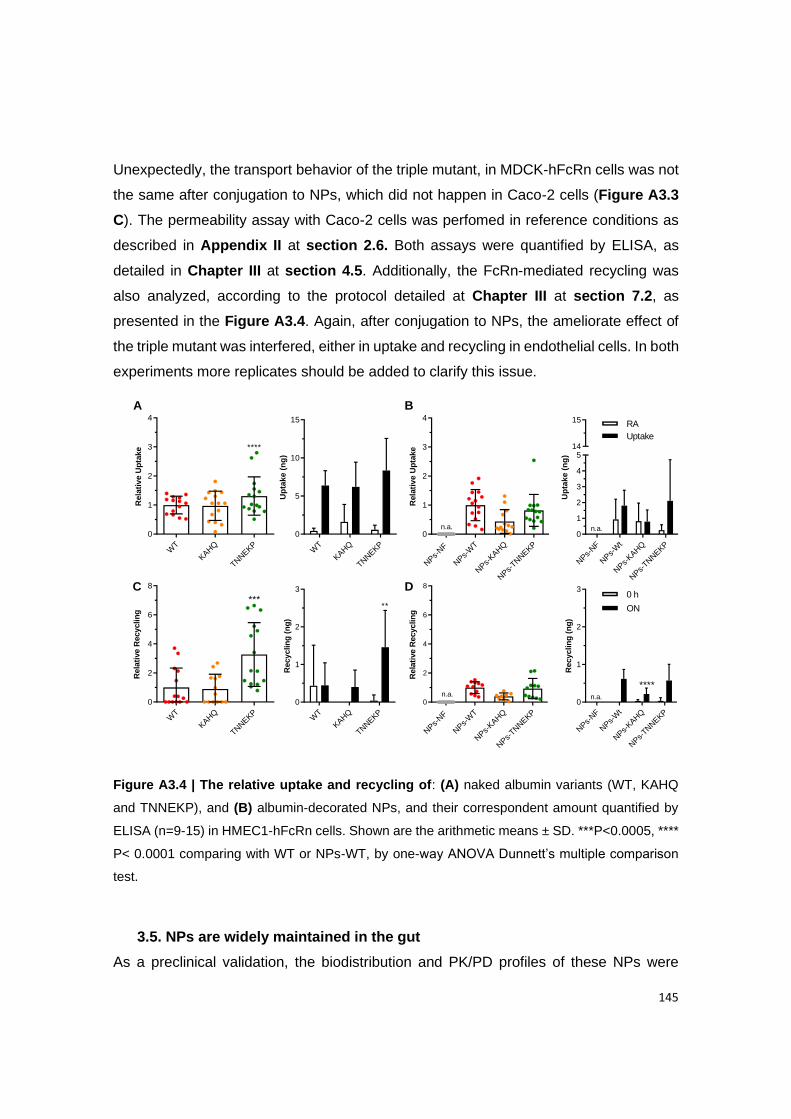
145
Unexpectedly, the transport behavior of the triple mutant, in MDCK-hFcRn cells was not
the same after conjugation to NPs, which did not happen in Caco-2 cells (Figure A3.3
C). The permeability assay with Caco-2 cells was perfomed in reference conditions as
described in Appendix II at section 2.6. Both assays were quantified by ELISA, as
detailed in Chapter III at section 4.5. Additionally, the FcRn-mediated recycling was
also analyzed, according to the protocol detailed at Chapter III at section 7.2, as
presented in the Figure A3.4. Again, after conjugation to NPs, the ameliorate effect of
the triple mutant was interfered, either in uptake and recycling in endothelial cells. In both
experiments more replicates should be added to clarify this issue.
NPs-
NF
NPs-
WT
NPs-
KAHQ
NPs-
TNNEKP
0
1
2
3
4R
ela
tive U
pta
ke
n.a.
WT
KAHQ
TNNEKP
0
1
2
3
Recyclin
g (
ng
)
**
NPs-
NF
NPs-
Wt
NPs-
KAHQ
NPs-
TNNEKP
0
1
2
3
Recyclin
g (
ng
)
n.a. *
****
WT
KAHQ
TNNEKP
0
1
2
3
4
Rela
tive U
pta
ke ****
A
WT
KAHQ
TNNEKP
0
5
10
15
Up
tak
e (
ng
)
NPs-
NF
NPs-
Wt
NPs-
KAHQ
NPs-
TNNEKP
0
1
2
3
4
514
15
Up
tak
e (
ng
)
n.a.
WT
KAHQ
TNNEKP
0
2
4
6
8
Rela
tive R
ecyclin
g
***
NPs-
NF
NPs-
WT
NPs-
KAHQ
NPs-
TNNEKP
0
2
4
6
8
Rela
tive R
ecyclin
g
n.a.
B
DC0 h
ON
RA
Uptake
Figure A3.4 | The relative uptake and recycling of: (A) naked albumin variants (WT, KAHQ
and TNNEKP), and (B) albumin-decorated NPs, and their correspondent amount quantified by
ELISA (n=9-15) in HMEC1-hFcRn cells. Shown are the arithmetic means ± SD. ***P<0.0005, ****
P< 0.0001 comparing with WT or NPs-WT, by one-way ANOVA Dunnett’s multiple comparison
test.
3.5. NPs are widely maintained in the gut
As a preclinical validation, the biodistribution and PK/PD profiles of these NPs were
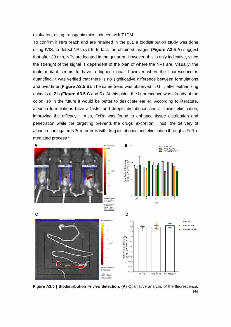
146
evaluated, using transgenic mice induced with T1DM.
To confirm if NPs reach and are retained in the gut, a biodistribution study was done
using IVIS, to detect NPs-cy7.5. In fact, the obtained images (Figure A3.5 A) suggest
that after 30 min, NPs are located in the gut area. However, this is only indicative, since
the strenght of the signal is dependent of the plan of where the NPs are. Visually, the
triple mutant seems to have a higher signal, however when the fluorescence is
quantified, it was verified that there is no significative difference between formulations
and over time (Figure A3.5 B). The same trend was observed in GIT, after euthanizing
animals at 2 h (Figure A3.5 C and D). At this point, the fluorescence was already at the
colon, so in the future it would be better to desiccate earlier. According to literature,
albumin formulations have a faster and deeper distribution and a slower elimination,
improving the efficacy 3. Also, FcRn was found to enhance tissue distribution and
penetration while the targeting prevents the drugs’ excretion. Thus, the delivery of
albumin-conjugated NPs interferes with drug distribution and elimination through a FcRn-
mediated process 4.
Figure A3.5 | Biodistribution in vivo detection. (A) Qualitative analysis of the fluorescence,

147
after 30 min of administration of NPs with cy7.5 and respective blanks whithout cy7.5; and (B)
quantitative of the fluorescence present in mice, at different time points during 8 h. (C) Qualitative
and (D) quantitative analysis of the GIT after 2 h of administration. NPs without cy7.5 were
included as controls. All images were normalized by the control (same scale and ROI window).
3.6. The hypoglycemic effect
Also the PK/PD profiles were assessed, using hFcRn transgenic mice induced with
diabetes. These animals were fasted for a shorter time (4-6 h), due to the animal well-
being, which might lead to a less evidenced effect. In fact, results related to glycemia
levels (Figure A3.6 A) are not in line with the plasmatic insulin levels (Figure A3.6 B).
Looking to the PD profile, it seems that NPs-TNNEKP has the behavior that should be
from oral insulin. Notwithstanding, the PK profile is in line with the in vitro results, since
the triple presented higher levels. As expected, on the opposite side, it is NPs-KAHQ,
oral insulin and NPs-NF, with lower insulin levels. NPs present a controlled and
prolonged effect, being enhanced with the triple mutant.
Figure A3.6 | Hypoglycemic effect of T1DM-induced mice following i.p. administration of
STZ (150 mg kg−1). (A) Plasma glucose levels and (B) Plasma insulin level following PBS, oral
insulin 50 IU/Kg, s.c. insulin 2.5 IU/Kg, non-functionalized NPs (NPs-NF) and functionalized with
KAHQ and TNNEKP (n = 5-6 animals).
From the obtained results it was possible to calculate the PK parameters, as presented
in the following table:
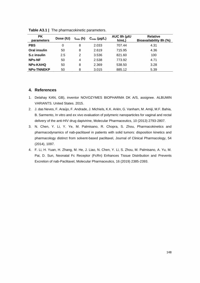
148
Table A3.1 | The pharmacokinetic parameters.
PK parameters
Dose (IU) tmáx (h) Cmáx (µg/L) AUC 8h (µIU
h/mL) Relative
Bioavailability 8h (%)
PBS 0 8 2.033 707.44 4.31
Oral insulin 50 8 2.619 715.95 4.36
S.c insulin 2.5 2 3.536 821.60 100
NPs-NF 50 4 2.538 773.92 4.71
NPs-KAHQ 50 8 2.369 538.50 3.28
NPs-TNNEKP 50 8 3.015 885.12 5.39
4. References
1. Delahay KAN, GB), inventor NOVOZYMES BIOPHARMA DK A/S, assignee. ALBUMIN
VARIANTS. United States. 2015.
2. J. das Neves, F. Araújo, F. Andrade, J. Michiels, K.K. Ariën, G. Vanham, M. Amiji, M.F. Bahia,
B. Sarmento, In vitro and ex vivo evaluation of polymeric nanoparticles for vaginal and rectal
delivery of the anti-HIV drug dapivirine, Molecular Pharmaceutics, 10 (2013) 2793-2807.
3. N. Chen, Y. Li, Y. Ye, M. Palmisano, R. Chopra, S. Zhou, Pharmacokinetics and
pharmacodynamics of nab-paclitaxel in patients with solid tumors: disposition kinetics and
pharmacology distinct from solvent-based paclitaxel, Journal of Clinical Pharmacology, 54
(2014), 1097.
4. F. Li, H. Yuan, H. Zhang, M. He, J. Liao, N. Chen, Y. Li, S. Zhou, M. Palmisano, A. Yu, M.
Pai, D. Sun, Neonatal Fc Receptor (FcRn) Enhances Tissue Distribution and Prevents
Excretion of nab-Paclitaxel, Molecular Pharmaceutics, 16 (2019) 2385-2393.
149
Appendix IV
PUBLISHED AND SUBMITTED PAPERS

150
151
Prevention of diabetes-associated fibrosis: Strategies in FcRn-targeted
nanosystems for oral drug delivery
Cláudia Azevedo1,2,#, Soraia Pinto1,2,#, Sopisa Benjakul3,4,#, Jeannette Nilsen3,4, Helder A.
Santos5,6, Giovanni Traverso7,8,9, Jan Terje Andersen3,4, Bruno Sarmento1,10,*
1 Instituto de Investigação e Inovação em Saúde (i3S), University of Porto, Rua Alfredo Allen, 208, 4200-
135 Porto, Portugal.
2 Instituto de Ciências Biomédicas Abel Salazar (ICBAS), University of Porto, Rua Jorge Viterbo Ferreira,
228, 4150-180 Porto, Portugal.
3 Department of Immunology, Oslo University Hospital Rikshospitalet and University of Oslo, N-0424 Oslo,
Norway.
4 Institute of Clinical Medicine and Department of Pharmacology, University of Oslo and Oslo University
Hospital, 0372 Oslo, Norway.
5 Drug Research Program, Division of Pharmaceutical Chemistry and Technology, Faculty of Pharmacy,
University of Helsinki, FI-00014 Helsinki, Finland.
6 Helsinki Institute of Life Science (HiLIFE), University of Helsinki, FI-00014 Helsinki, Finland.
7 Department of Chemical Engineering and Koch Institute for Integrative Cancer Research, Massachusetts
Institute of Technology, 02139 Cambridge, MA, USA.
8 Division of Gastroenterology, Brigham and Women’s Hospital, Harvard Medical School, 02115 Boston,
MA, USA.
9 Department of Mechanical Engineering, Massachusetts Institute of Technology, 02139 Cambridge, MA,
USA.
10 CESPU, Instituto de Investigação e Formação Avançada em Ciências e Tecnologias da Saúde & Instituto
Universitário de Ciências da Saúde, 4585-116 Gandra, Portugal.
# Cláudia Azevedo, Soraia Pinto and Sopisa Benjakul contributed equally.
* Corresponding authors: [email protected]
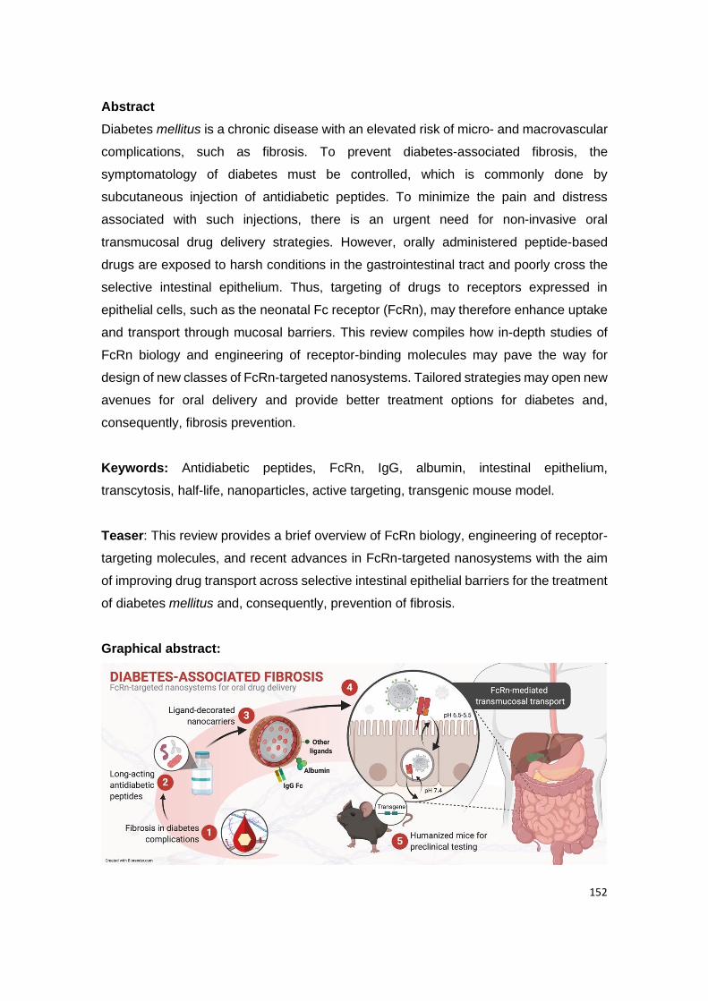
152
Abstract
Diabetes mellitus is a chronic disease with an elevated risk of micro- and macrovascular
complications, such as fibrosis. To prevent diabetes-associated fibrosis, the
symptomatology of diabetes must be controlled, which is commonly done by
subcutaneous injection of antidiabetic peptides. To minimize the pain and distress
associated with such injections, there is an urgent need for non-invasive oral
transmucosal drug delivery strategies. However, orally administered peptide-based
drugs are exposed to harsh conditions in the gastrointestinal tract and poorly cross the
selective intestinal epithelium. Thus, targeting of drugs to receptors expressed in
epithelial cells, such as the neonatal Fc receptor (FcRn), may therefore enhance uptake
and transport through mucosal barriers. This review compiles how in-depth studies of
FcRn biology and engineering of receptor-binding molecules may pave the way for
design of new classes of FcRn-targeted nanosystems. Tailored strategies may open new
avenues for oral delivery and provide better treatment options for diabetes and,
consequently, fibrosis prevention.
Keywords: Antidiabetic peptides, FcRn, IgG, albumin, intestinal epithelium,
transcytosis, half-life, nanoparticles, active targeting, transgenic mouse model.
Teaser: This review provides a brief overview of FcRn biology, engineering of receptor-
targeting molecules, and recent advances in FcRn-targeted nanosystems with the aim
of improving drug transport across selective intestinal epithelial barriers for the treatment
of diabetes mellitus and, consequently, prevention of fibrosis.
Graphical abstract:
153
Introduction
Diabetes mellitus (DM) is a chronic metabolic disease that develops when the pancreas
does not produce insulin or when the body does not respond fully to insulin, leading to
impaired glucose tolerance. More than 460 million people worldwide are suffering from
DM, and the number of cases is predicted to rise to 700 million by 2045 1, 2. Chronic
exposure to hyperglycemia may cause tissue injury and lead to fibrosis, which increases
the risk of micro- and macrovascular complications in several organs, such as the eyes
(diabetic retinopathy), kidneys (diabetic nephropathy), skin, heart, liver, lungs, nerves
(neuropathy) and blood vessels (angiopathy) 3, 4. Fibrosis is characterized by abnormal
deposition of the extracellular matrix (ECM), and there is currently no specific treatment
available 5. As such, there is an urgent need for strategies that could delay the
progression or prevent diabetes-associated fibrosis.
Conventional treatment of DM requires subcutaneous injection of peptide-based
antidiabetic drugs for glycemic control, which is considered a painful and invasive
approach. The preferred route of administration is oral delivery via mucosal surfaces,
due to its ease of administration and high patient compliance, without the need for
syringes, needles, and trained health workers 6, 7. Notwithstanding, orally administered
antidiabetic peptides are susceptible to the harsh environment of the gastrointestinal
tract (GIT) and the selective barrier of the intestinal epithelium 8, 9. Hence, there is a
demand for innovative strategies to overcome these limitations.
Biodegradable and biocompatible nanoparticles (NPs) have emerged as promising oral
drug delivery systems that protect encapsulated drugs from acidic pH and enzymatic
degradation in the GIT, as well as providing controlled release and enhanced
bioavailability of drugs to target sites. However, the transport of NPs across intestinal
mucosa is restricted by polarized epithelial cell layers 8, 10. Several recent studies support
that targeting of the neonatal Fc receptor (FcRn), which is expressed by such epithelial
cells and mediates bidirectional transport and recycling of immunoglobulin G (IgG) and
albumin, may be used as a gateway for more efficient transcellular transport of NPs 11-
15. Furthermore, in light of successes in the development of engineered ligands and
alternative scaffolds with improved FcRn binding and transport properties, researchers
may now harvest from these advances in the design of new classes of NPs decorated
with FcRn-targeted molecules, with the aim of enhancing transmucosal delivery upon
oral administration. This review provides a brief overview of FcRn biology and discusses
how receptor-binding molecules may be incorporated into nanosystems for improved
FcRn-mediated oral delivery of antidiabetic peptides for the treatment of DM and the
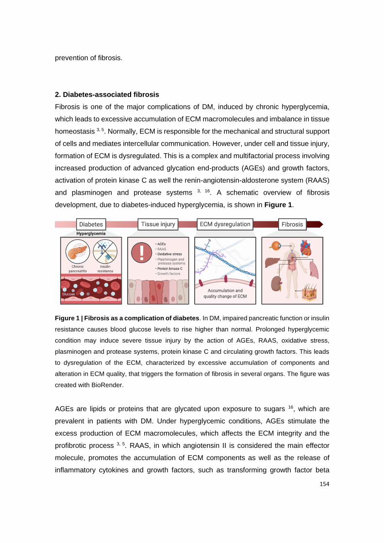
154
prevention of fibrosis.
2. Diabetes-associated fibrosis
Fibrosis is one of the major complications of DM, induced by chronic hyperglycemia,
which leads to excessive accumulation of ECM macromolecules and imbalance in tissue
homeostasis 3, 5. Normally, ECM is responsible for the mechanical and structural support
of cells and mediates intercellular communication. However, under cell and tissue injury,
formation of ECM is dysregulated. This is a complex and multifactorial process involving
increased production of advanced glycation end-products (AGEs) and growth factors,
activation of protein kinase C as well the renin-angiotensin-aldosterone system (RAAS)
and plasminogen and protease systems 3, 16. A schematic overview of fibrosis
development, due to diabetes-induced hyperglycemia, is shown in Figure 1.
Figure 1 | Fibrosis as a complication of diabetes. In DM, impaired pancreatic function or insulin
resistance causes blood glucose levels to rise higher than normal. Prolonged hyperglycemic
condition may induce severe tissue injury by the action of AGEs, RAAS, oxidative stress,
plasminogen and protease systems, protein kinase C and circulating growth factors. This leads
to dysregulation of the ECM, characterized by excessive accumulation of components and
alteration in ECM quality, that triggers the formation of fibrosis in several organs. The figure was
created with BioRender.
AGEs are lipids or proteins that are glycated upon exposure to sugars 16, which are
prevalent in patients with DM. Under hyperglycemic conditions, AGEs stimulate the
excess production of ECM macromolecules, which affects the ECM integrity and the
profibrotic process 3, 5. RAAS, in which angiotensin II is considered the main effector
molecule, promotes the accumulation of ECM components as well as the release of
inflammatory cytokines and growth factors, such as transforming growth factor beta
155
(TGF-β), vascular endothelial growth factor, platelet-derived growth factor, and
connective tissue growth factor 3, 5. In addition, oxidative stress can trigger the
progression of fibrosis [2]. In particular, the increase in TGF-β stimulates the epithelial-
to-mesenchymal transition, which contributes to the accumulation of ECM components,
culminating in tissue fibrosis 17, 18.
Interestingly, antidiabetic peptides have great potential in the treatment of fibrosis. For
instance, dipeptidyl peptidase-4 (DDP-4) inhibitors, known as gliptins, and glucagon-like
peptide 1 (GLP-1) agonists, have demonstrated mild antifibrotic properties in models of
liver fibrosis by reducing oxidative stress 19. Owning to the antidiabetic activity of gliptins,
they are used for treatment of type 2 diabetes (T2DM) 20. Moreover, GLP-1 analogs,
such as exendin-4, liraglutide and semaglutide, are also used to treat T2DM, which have
antifibrotic effects by reducing inflammation and oxidative stress 21-23. As such, the
encapsulation of these peptide drugs into nanosystems targeting FcRn may lead to
enhanced transmucosal delivery. In addition, these drugs may be engineered for half-life
extension by taking advantage of FcRn-binding molecules. Notably, FcRn is expressed
by cells of the lungs, eyes and kidneys 24-26, where fibrosis typically occurs.
3. The biology and molecular mechanisms of FcRn
3.1. Structure and binding of ligands
FcRn is a heterodimeric glycoprotein that is structurally similar to major histocompatibility
complex class I (MHC-I) molecules, consisting of a unique transmembrane heavy chain
(40-46 kDa) that is non-covalently associated with the common beta-2-microglobulin
(β2m) light chain (12 kDa) 27, 28. The heavy chain contains three extracellular domains,
denoted α1, α2 and α3, where the α1 and α2 domains assemble a platform of eight
antiparallel β-strands topped by two α-helices followed by the α3 domain, which is folded
up on the soluble β2m 29, 30.
Unlike MHC-I molecules that present antigenic peptides via their peptide binding groove
to cytotoxic T cells, the corresponding binding groove of FcRn is closed 29-32. Instead,
FcRn has evolved to bind IgG and albumin, two structurally and functionally unrelated
proteins. Both ligands bind in a pH-dependent manner, binding at acidic pH (6.5-5-0) and
no binding or release at physiological pH (7.4) 33-38. The strict pH-dependent mode of
action is mediated by protonation of the imidazole side chains of conserved histidine
residues found on both the receptor and the ligands. When the pH increases towards
neutral, protonation of the histidine residues is gradually lost, and the ligands are
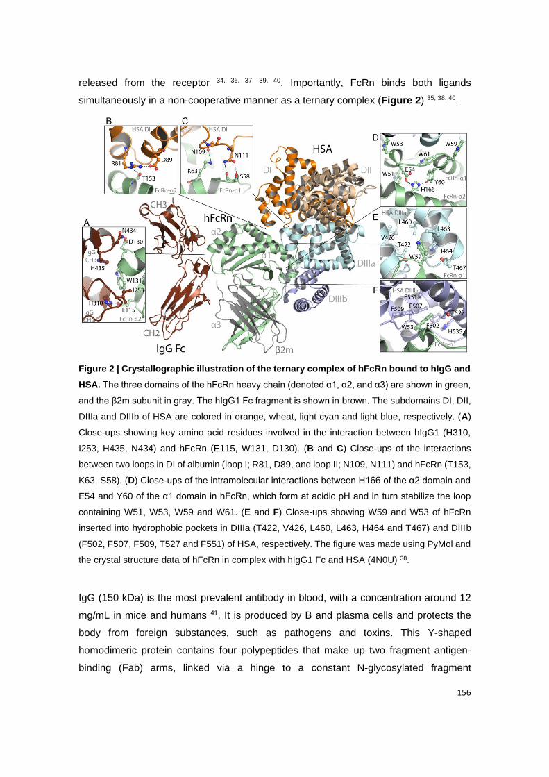
156
released from the receptor 34, 36, 37, 39, 40. Importantly, FcRn binds both ligands
simultaneously in a non-cooperative manner as a ternary complex (Figure 2) 35, 38, 40.
Figure 2 | Crystallographic illustration of the ternary complex of hFcRn bound to hIgG and
HSA. The three domains of the hFcRn heavy chain (denoted α1, α2, and α3) are shown in green,
and the β2m subunit in gray. The hIgG1 Fc fragment is shown in brown. The subdomains DI, DII,
DIIIa and DIIIb of HSA are colored in orange, wheat, light cyan and light blue, respectively. (A)
Close-ups showing key amino acid residues involved in the interaction between hIgG1 (H310,
I253, H435, N434) and hFcRn (E115, W131, D130). (B and C) Close-ups of the interactions
between two loops in DI of albumin (loop I; R81, D89, and loop II; N109, N111) and hFcRn (T153,
K63, S58). (D) Close-ups of the intramolecular interactions between H166 of the α2 domain and
E54 and Y60 of the α1 domain in hFcRn, which form at acidic pH and in turn stabilize the loop
containing W51, W53, W59 and W61. (E and F) Close-ups showing W59 and W53 of hFcRn
inserted into hydrophobic pockets in DIIIa (T422, V426, L460, L463, H464 and T467) and DIIIb
(F502, F507, F509, T527 and F551) of HSA, respectively. The figure was made using PyMol and
the crystal structure data of hFcRn in complex with hIgG1 Fc and HSA (4N0U) 38.
IgG (150 kDa) is the most prevalent antibody in blood, with a concentration around 12
mg/mL in mice and humans 41. It is produced by B and plasma cells and protects the
body from foreign substances, such as pathogens and toxins. This Y-shaped
homodimeric protein contains four polypeptides that make up two fragment antigen-
binding (Fab) arms, linked via a hinge to a constant N-glycosylated fragment
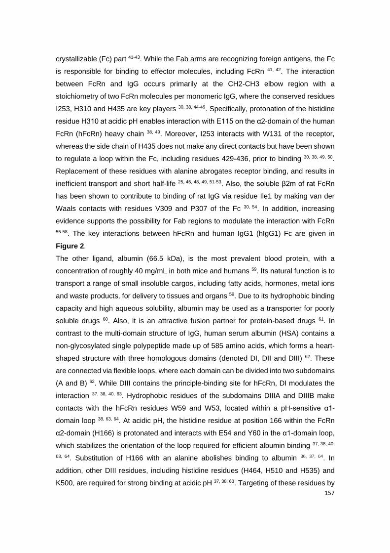
157
crystallizable (Fc) part 41-43. While the Fab arms are recognizing foreign antigens, the Fc
is responsible for binding to effector molecules, including FcRn 41, 42. The interaction
between FcRn and IgG occurs primarily at the CH2-CH3 elbow region with a
stoichiometry of two FcRn molecules per monomeric IgG, where the conserved residues
I253, H310 and H435 are key players 30, 38, 44-49. Specifically, protonation of the histidine
residue H310 at acidic pH enables interaction with E115 on the α2-domain of the human
FcRn (hFcRn) heavy chain 38, 49. Moreover, I253 interacts with W131 of the receptor,
whereas the side chain of H435 does not make any direct contacts but have been shown
to regulate a loop within the Fc, including residues 429-436, prior to binding 30, 38, 49, 50.
Replacement of these residues with alanine abrogates receptor binding, and results in
inefficient transport and short half-life 25, 45, 48, 49, 51-53. Also, the soluble β2m of rat FcRn
has been shown to contribute to binding of rat IgG via residue Ile1 by making van der
Waals contacts with residues V309 and P307 of the Fc 30, 54. In addition, increasing
evidence supports the possibility for Fab regions to modulate the interaction with FcRn
55-58. The key interactions between hFcRn and human IgG1 (hIgG1) Fc are given in
Figure 2.
The other ligand, albumin (66.5 kDa), is the most prevalent blood protein, with a
concentration of roughly 40 mg/mL in both mice and humans 59. Its natural function is to
transport a range of small insoluble cargos, including fatty acids, hormones, metal ions
and waste products, for delivery to tissues and organs 59. Due to its hydrophobic binding
capacity and high aqueous solubility, albumin may be used as a transporter for poorly
soluble drugs 60. Also, it is an attractive fusion partner for protein-based drugs 61. In
contrast to the multi-domain structure of IgG, human serum albumin (HSA) contains a
non-glycosylated single polypeptide made up of 585 amino acids, which forms a heart-
shaped structure with three homologous domains (denoted DI, DII and DIII) 62. These
are connected via flexible loops, where each domain can be divided into two subdomains
(A and B) 62. While DIII contains the principle-binding site for hFcRn, DI modulates the
interaction 37, 38, 40, 63. Hydrophobic residues of the subdomains DIIIA and DIIIB make
contacts with the hFcRn residues W59 and W53, located within a pH-sensitive α1-
domain loop 38, 63, 64. At acidic pH, the histidine residue at position 166 within the FcRn
α2-domain (H166) is protonated and interacts with E54 and Y60 in the α1-domain loop,
which stabilizes the orientation of the loop required for efficient albumin binding 37, 38, 40,
63, 64. Substitution of H166 with an alanine abolishes binding to albumin 36, 37, 64. In
addition, other DIII residues, including histidine residues (H464, H510 and H535) and
K500, are required for strong binding at acidic pH 37, 38, 63. Targeting of these residues by
158
mutagenesis reduces receptor binding considerably, which results in short half-lives in
hFcRn transgenic mice 37. The contribution from DI is minor but important for optimal
hFcRn binding. Notably, a recombinant form of HSA DIII binds 10-fold weaker to hFcRn
than the full-length version 65, 66. Structurally, the DI residues involved are N109, N111
and R81-D89 that make contacts with K63, S58, and T153 of hFcRn, respectively 38, 63,
65. Close-ups of the interactions between hFcRn and HSA are given in Figure 2.
3.2. Recycling and transcytosis of ligands
IgG and HSA share a half-life of 3 weeks in humans on average 67-69. This results from
their molecular sizes above the renal clearance threshold and FcRn-mediated rescue
from intracellular degradation 35, 70-73. The astonishing role of FcRn in half-life regulation
is evident in patients suffering from rare human disease hypercatabolic hypoproteinemia
that have remarkably low blood amounts of both ligands, which is due to the deficient
FcRn expression 74, 75. In addition, mice lacking FcRn expression have about 4-fold and
2-fold lower levels of IgG and albumin than normal mice, respectively 35, 71, 72. As such,
FcRn is a key homeostatic regulator of IgG and albumin by transporting the ligands within
and across cells via two cellular pathways, recycling and transcytosis. Both processes
are regulated by strict pH-dependent binding-and-release interactions between FcRn
and its ligands 66, 70, 73, 76-78.
Insights into how FcRn mediates cellular recycling have been extensively studied by in
vitro cellular transport assays combined with advanced imaging technologies, mainly
with a focus on IgG 70, 73, 76, 79-85. However, recent studies have also revealed knowledge
about how FcRn transports both ligands simultaneously 70, 78, 86. Together, the studies
demonstrate that the ligands are taken up via fluid-phase pinocytosis at neutral pH,
followed by entering of acidified endosomal compartments where FcRn is predominantly
located. The mildly acidic pH (6.0-6.5) herein facilitates protonation of the histidine
residues and engagement of the receptor. Bound ligands are then rescued from
intracellular lysosomal degradation by sorting of the FcRn-ligand complexes to the cell
surface, where exposure to neutral pH triggers ligand release from FcRn into the
extracellular space. Proteins that do not bind the receptor will be rapidly degraded in
lysosomes 70, 76. Studies in mice have shown that both non-hematopoietic and
hematopoietic cells are equally important for FcRn-mediated half-life regulation 77, 87-93.
Furthermore, FcRn can transport the ligands in both directions across polarized cellular
barriers, such as the epithelial cell layers of the lungs and the intestine 25, 51, 66, 77, 94-102.
Regarding the polarized intestinal epithelium of the GIT, IgG and albumin are taken up
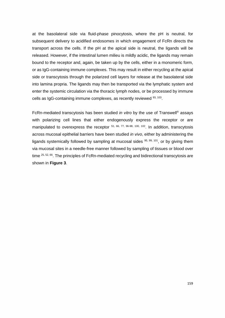
159
at the basolateral side via fluid-phase pinocytosis, where the pH is neutral, for
subsequent delivery to acidified endosomes in which engagement of FcRn directs the
transport across the cells. If the pH at the apical side is neutral, the ligands will be
released. However, if the intestinal lumen milieu is mildly acidic, the ligands may remain
bound to the receptor and, again, be taken up by the cells, either in a monomeric form,
or as IgG-containing immune complexes. This may result in either recycling at the apical
side or transcytosis through the polarized cell layers for release at the basolateral side
into lamina propria. The ligands may then be transported via the lymphatic system and
enter the systemic circulation via the thoracic lymph nodes, or be processed by immune
cells as IgG-containing immune complexes, as recently reviewed 93, 103.
FcRn-mediated transcytosis has been studied in vitro by the use of Transwell® assays
with polarizing cell lines that either endogenously express the receptor or are
manipulated to overexpress the receptor 51, 66, 77, 96-98, 100, 102. In addition, transcytosis
across mucosal epithelial barriers have been studied in vivo, either by administering the
ligands systemically followed by sampling at mucosal sides 95, 99, 101, or by giving them
via mucosal sites in a needle-free manner followed by sampling of tissues or blood over
time 25, 52, 66. The principles of FcRn-mediated recycling and bidirectional transcytosis are
shown in Figure 3.
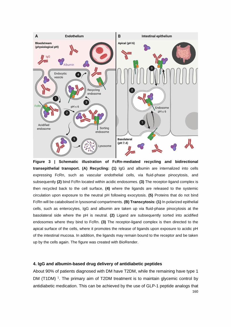
160
Figure 3 | Schematic illustration of FcRn-mediated recycling and bidirectional
transepithelial transport. (A) Recycling: (1) IgG and albumin are internalized into cells
expressing FcRn, such as vascular endothelial cells, via fluid-phase pinocytosis, and
subsequently (2) bind FcRn located within acidic endosomes. (3) The receptor-ligand complex is
then recycled back to the cell surface, (4) where the ligands are released to the systemic
circulation upon exposure to the neutral pH following exocytosis. (5) Proteins that do not bind
FcRn will be catabolised in lysosomal compartments. (B) Transcytosis: (1) In polarized epithelial
cells, such as enterocytes, IgG and albumin are taken up via fluid-phase pinocytosis at the
basolateral side where the pH is neutral. (2) Ligand are subsequently sorted into acidified
endosomes where they bind to FcRn. (3) The receptor-ligand complex is then directed to the
apical surface of the cells, where it promotes the release of ligands upon exposure to acidic pH
of the intestinal mucosa. In addition, the ligands may remain bound to the receptor and be taken
up by the cells again. The figure was created with BioRender.
4. IgG and albumin-based drug delivery of antidiabetic peptides
About 90% of patients diagnosed with DM have T2DM, while the remaining have type 1
DM (T1DM) 1. The primary aim of T2DM treatment is to maintain glycemic control by
antidiabetic medication. This can be achieved by the use of GLP-1 peptide analogs that
161
stimulate insulin secretion by pancreatic β cells, or insulin analogs that promote efficient
glucose disposal. However, such peptides have a very short half-life of about a few
minutes, due to their small size and rapid clearance from the circulatory system via
kidney filtration. Hence, efficient treatment would require regular monitoring and frequent
dosing schedules 1. To improve the efficacy and patient compliance of DM therapy, the
natural long half-life of hIgG4 Fc and HSA may be used in drug design, which have
already been explored for a range of antidiabetic peptides (Table 1).
The first GLP-1 analog to reach the market was liraglutide (Victoza®), which is linked to
a fatty acid (palmitic acid) that binds reversibly to hydrophobic pockets of HSA 104. As
such, it associates non-covalently with endogenous HSA within the subcutaneous tissue
and bloodstream. This strategy extends the half-life to 11-15 hours and allows for once-
daily dosing 104-106. Another example is the zinc-based taspoglutide, where
aminoisobutyric acid is covalently linked to two positions of GLP-1. This was shown to
extend the half-life up to 2-3 days in humans 107, 108. However, clinical testing was halted
due to severe gastrointestinal side effects and hypersensitivity reactions 109.
Furthermore, for the treatment of not only T2DM but also T1DM, other fatty acid-based
strategies have been developed for insulin. One is insulin detemir (Levemir®) 110, 111 and
another is insulin degludec (Tresiba®) 112, 113. Both are recombinant human insulin
analogs with a fatty acid linked to a lysine residue, while insulin degludec also lacks a
threonine residue. They have distinct half-lives of 18-26 hours and 42 hours for insulin
detemir and insulin degludec, respectively 61, 110-114. An insulin analog with an activity
profile close to that of insulin detemir is insulin glargine (Lantus®) with a half-life of about
24 hous 115. Instead of modification of the fatty acid side chains, insulin glargine includes
two additional arginine residues in the beta chain and an amino acid replacement of
asparagine at position 21 with glycine. Nevertheless, these products have gained market
approval for once-daily dosing. To further extend half-lives of insulin analogs, insulin
icodec has been developed as a once-weekly insulin that is currently in phase I and
phase III clinical trials in patients with T1DM and T2DM, respectively 116, 117. As a result
of three amino acid substitutions and the addition of an icosane fatty diacid to this insulin
analog, it shows an extended half-life of 8 days in humans and improved stability and
solubility.
Despite that these strategies secure prolonged blood persistence compared with native
peptides, treatment relies on subcutaneous injections. To allow oral administration,
semaglutide has been developed 118. It is a modified GLP-1 where alanine and lysine
residues at positions 8 and 34 have been replaced with 2-aminoisobutyric acid and
162
arginine, respectively, to prevent degradation by DPP-4. In addition, lysine at position 26
is engineered to contain a spacer and a fatty acid that can bind endogenous HSA upon
injection. This formulation provides a half-life of about 7 days in humans 119, 120. When
semaglutide is co-formulated with an absorption enhancer, namely sodium N-(8-[2-
hydroxybenzoyl]amino) caprylate, which neutralizes the pH of the gastric fluid and
hinders enzymatic activity, it is protected from degradation in the GIT upon oral
administration 118. The injected version of semaglutide (Ozempic®) was clinically
approved in the US and Europe in 2017-2018, while the oral formulation (Rybelsus®)
was approved last year 118, 121-123. The latter is the first GLP-1 receptor agonist to be
approved for oral delivery. Notably, there may be room for further optimization as its oral
bioavailability is limited (≤ 1.0 %) 124, 125. Currently, there are no oral insulin (OI)-based
drugs available for DM treatment. However, very recently, the first clinically tested once-
weekly OI analog, named OI338, demonstrated efficacy near subcutaneously injected
insulin glargine 126, which has led to the development of long-acting OI formulations 127.
Commonly, a tyrosine at position 14 and a phenylalanine at position 25 are replaced by
glycine and histidine, respectively, in addition of a deletion of threonine at position 30 127.
These amino acid modifications make OI less susceptible to degradation in the GIT upon
oral administration 126, 127, in combination with the use of an absorption enhancer, such
as Gastro Intestinal Permeation Enhancer Tablet I (GIPET®1) based on sodium caprate
128. To gain improved HSA binding, the lysine residue at position 29 of the OI is coupled
to a strong albumin binding molecule (C18 fatty diacid) via a hydrophilic linker. Moreover,
OI molecules can be designed for prolonged half-life by engineering for decreased insulin
receptor affinity. This design resulted in OI338 with a half-life of 70 hours in T2DM
patients 126. In the context of FcRn, it has been demonstrated that binding of fatty acids
to the hydrophobic pockets of HSA is in fact negatively affecting receptor binding, in
which the effect depends on the length of the fatty acid 63. As such, half-life extension
upon conjugation to fatty acids will have limitations. Regarding oral delivery, this means
that fatty acid-based strategies may not benefit from albumin binding to FcRn expressed
at epithelial cell barriers. This is a highly interesting topic that should be addressed in
future studies.
An alternative strategy to non-covalent association with HSA is site-specific conjugation.
One example is conjugation of the GLP-1 receptor agonist exendin-4 129-131. Notably,
exendin-4 increases insulin secretion in a glucose-dependent manner at the same time
as it stimulates β-cell proliferation and restores sensitivity to glucose 132. When injected
in the absence of HSA, it has a half-life below 1 hour 132, 133, however, when linked to
163
HSA (CJC-1134-PC), it has been shown to gain a half-life of about 8 days in a clinical
phase II trial 105, 130, 134. This allows for once-a-week dosing. Specifically, exendin-4 is
linked to HSA via the PC-DAC™ technology that utilizes maleimide (MAL) chemistry for
stable conjugation to the thiol group of a free cysteine residue (C34) in DI 106, 133. The
same technology has been used for conjugation of insulin (PC-DACTM:Insulin), which is
under preclinical development, where it has demonstrated 30 times longer half-life
compared with native insulin in rats 135. Importantly, C34 is distant from the hFcRn
interaction site in HSA DI 63, 136. However, it has been demonstrated that conjugation to
C34 may compromise pH-dependent FcRn binding 137-139. How pronounced this effect
could be will depend on the biophysical properties of the conjugated drug and, as such,
needs to be addressed in a case-to-case scenario. Notably, the loss of receptor binding
may be restored by taking advantage of engineered HSA variants with improved hFcRn
binding, as will be dicussed in Section 6. Another alternative strategy is to genetically
fuse the peptide of interest to the N- or C-terminal end of HSA. In the context of DM, a
di-peptide of GLP-1 has been fused to wild-type (WT) HSA (albiglutide) 140-142, a product
known as Tanzeum® in the US and Eperzan® in Europe, which was recently withdrawn
from the market due to economic reasons. Nevertheless, the design gave rise to a half-
life of 5 days in humans, allowing for weekly subcutaneous injection 140-142. This fusion
was based on the addition of the di-peptide to the N-terminal of HSA, which leaves the
C-terminal end free. Interestingly, the last leucine residue of the C-terminal end of HSA
may be enzymatically cleaved in blood, which has been demonstrated to reduce hFcRn
binding and lead to a short half-life in mice and humans 143. Hence, this biological
phenomenon should be considered when designing HSA fusions. A way to compensate
for loss of receptor binding is to take advantage of engineered HSA variants with
improved receptor affinity, as recently demonstrated 143.
The last example is the use of hIgG Fc fusion technology, where an GLP-1 receptor
agonist, again engineered to be resistant to enzymatic degradation, is fused to an IgG4
Fc fragment via a short linker (dulaglutide, Trulicity®) 144, 145. IgG4 is likely to be chosen
instead of IgG1 due to safety concerns, as IgG4 Fc is less potent in induction of effector
functions 42, without altering the ability to engage hFcRn. Dulaglutide has a half-life of
nearly 5 days in humans, and the product is approved for once-weekly subcutaneous
administration 144, 145.
Hitherto, only one of these strategies has been approved for oral delivery. However, in
principle, all the antidiabetic peptide strategies may be combined with nanosystems
tailored for oral administration to achieve more efficient delivery across the selective

164
mucosal barriers. Although, this will require a sophisticated design that allows the
nanosystems to be taken up and delivered to the circulatory system more efficiently than
what has been demonstrated to date. This may be achieved by harvesting from recent
knowledge about FcRn biology and advances in the engineering of receptor-targeting
molecules with favorable binding and transport properties. Such molecules may be
directly fused to the peptides, but they may also be used to decorate nanosystems, as
will be discussed below.
Table 1 | IgG Fc and albumin as platform for half-life extention of antidiabetic peptides
in clinic.
Analog Name Ligand Drug type Clinical status
t1/2 ROA Company Reference
GLP-1 Victoza® HSA Liraglutide Marketed 11-15 h SC Novo Nordisk 104, 105
Rybelsus® HSA Semaglutide Marketed 6-7 d Oral Novo Nordisk 118, 122, 123
(T2DM) Ozempic® HSA Semaglutide Marketed 7 d SC Novo Nordisk 121
Trulicity® IgG4 Fc
Dulaglutide Marketed 4.7-5 d SC Eli Lilly and Company
144, 145
CJC-1134 HSA Exendin-4 Phase II 8 d SC ConjuChem 129, 130, 133
R1538 HSA Taspoglutide Halted phase III
2-3 d SC Roche/Ipsen 107-109
Tanzeum®/ Eperzan®
HSA Albiglutide Withdrawn 5-8 d SC GlaxoSmithKline 140-142
Insulin Lantus® HSA Insulin glargine
Marketed 24 h SC Sanofi 115
(T1DM, T2DM) Levemir® HSA Insulin detemir Marketed 18-26 h SC Novo Nordisk 110, 111
Tresiba® HSA Insulin degludec
Marketed 42 h SC Novo Nordisk 112, 113
LAI-287 HSA Insulin icodec Phase I/III > 8 d SC Novo Nordisk 116, 117
OI-338 HSA Insulin-338 Phase II 70 h Oral Novo Nordisk 126, 127
PC-DAC™ HSA Insulin Preclinical 5-15 h SC ConjuChem 135
t1/2: half-life; ROA: route of administration; SC: subcutaneous administration.
5. Ligand engineering for improved FcRn binding
The therapeutic efficacy of many protein-based drug candidates is hampered by their
short half-life, and those that have reached the market require frequent administration,
which is a burden for the patients. As hIgG and HSA have a naturally long half-life, they
are extensively used in therapy, either as full-length hIgG antibodies, or as Fc or HSA
fused to therapeutic peptides or proteins of interest 61, 146, 147.
165
The in-depth knowledge gained from dissecting of the relationship between FcRn and its
ligands at molecular and cellular level has been used in the design of hIgG and HSA
variants with improved hFcRn binding and transport properties 41, 61, 66, 147-149. One
motivation for this is the clinical approval rate of monoclonal hIgG antibodies and HSA
fusions used to treat a range of different diseases. In fact, hIgG antibodies are the fastest
growing class of protein-based drugs with more than 100 products on the market, and
impressively over 800 antibodies under clinical testing 150, 151. One example is
otelixizumab (TRX4), a hIgG1 antibody targeting CD3 to suppress pathogenic T cells,
which is used to treat T1DM caused by antigen-specific T cells that infiltrate pancreatic
beta cells and lead to loss of insulin secretion 152. While many of the early developed
antibodies are based on WT hIgG1, next-generation of monoclonal antibodies will be
engineered for either lack or improved effector functions, including optimal hFcRn-
mediated transport properties 149. In addition, recent research has also resulted in
engineering of HSA variants with improved half-life 63, 66, 153. Moreover, several alternative
scaffolds with FcRn-binding properties have been reported 154-158, but a major challenge
for such engineering is fine-tuning of strict pH-dependent receptor binding in order to
gain favorable recycling and transcytosis capacity. Improvements in FcRn engagement
and transport may result in more favorable pharmacokinetic profiles, and consequently
less frequent administration and higher patient compliance.
5.1. IgG Fc-engineering
Scrutinizing the FcRn-IgG interaction has led to the development of Fc-engineered
variants where amino acid substitutions within the CH2 and CH3 domains have been
modified to gain improved pH-dependent binding to hFcRn (Table 2). Importantly, a
prerequisite for successful engineering is to augment receptor binding at acidic pH while,
at the same time, avoiding increase in receptor affinity at neutral pH 41, 148, 159, 160. The first
proof-of-concept was given in 1997 by isolation of mouse IgG1 (mIgG1) Fc fragments by
phage display that gave 3.5-fold improved binding towards mouse FcRn (mFcRn), which
resulted in about 1.6-fold extended half-life in conventional mice 161. This was followed
by a generation of hIgG Fc-engineered variants with increased affinity for hFcRn, which
provided a several fold extended half-life in mice, non-human primates and humans. One
example is the YTE triple mutant (M252Y/S254T/T256E) with 10-fold improved binding
at pH 6.0, which resulted in about 3.5-fold extended half-life in non-human primates and
humans 148, 162, 163. Another well-studied hIgG1 variant, harboring two amino acid
substitutions within the CH3 domains (M428L/N434S: LS variant), with 11-fold increased
166
binding to hFcRn at acidic pH, and negligible binding at neutral pH, gave more than 3-
fold extended half-life in hFcRn transgenic mice and non-human primates 164.
Importantly, such engineering has shown to improve therapeutic efficacy in mouse tumor
models when combined with antibodies targeting tumor antigens 164. In addition,
extended half-life improves antibody-based prophylaxis, as shown in non-human
primates upon viral challenge 165, 166. Clinical data on the use of the LS substitutions have
revealed that the antibody can reach a half-life of over 100 days compared to 20 days
for the WT counterpart 167, 168. A range of additional hIgG Fc-engineered variants have
been reported with increased persistence over the WT antibody when tested in hFcRn
transgenic mice or non-human primates 159, 160, 169-176, but these do not reach the same
half-life values as that of YTE and LS. However, a few recent examples are of interest.
In one report, three double mutant variants (M252Y/T256D (YD), T256D/T307Q (DQ)
and T256D/T307W (DW)) with enhanced pH-dependent binding to hFcRn resulted in
similar extended half-lives in hFcRn expressing mice and non-human primates as that
of LS used for benchmarking 177. Another recent Fc-engineered variant with three Fc
substitutions (L309D/Q311H/N434S (DHS)) showed a half-life of 2 to 3-fold beyond that
of the clinical stage YTE and LS variants in hFcRn expressing mice [89-91, 95]. This is
interesting as it has only 5-fold improved affinity to hFcRn at acidic pH with no detectable
binding at pH 7.4 178. The authors speculate that this may be explained by compete lack
of binding at neutral pH, which warrants further studies on how binding throughout the
pH gradient is regulating FcRn-mediated transport within and across cells. These Fc-
engineered variants may be used as fusion partners for therapeutic proteins, such as
antidiabetic peptides, to achieve extended half-life and improved bioavailability.
Preclinically examples are the combination of Fc-containing YTE or LS variants fused to
cytotoxic T lymphocyte-associated protein 4, where both Fc fusion products yielded more
than 2-fold extended half-life in non-human primates 179, 180. In addition, studies on
transcytosis have demonstrated that Fc-engineering can enhance delivery across
polarized cells, as shown for hIgG1 antibodies containing LS or YTE substitutions that
were transported almost twice as efficiently across human epithelial cells in vitro as the
WT counterpart [43]. Interestingly, the capacity to be transported in an hFcRn-dependent
manner could be further enhanced when the Fc-engineered antibodies were
administered as multivalent immune complexes 51. As such, FcRn-directed
transepithelial transport may be enhanced by Fc engineering for improved pH-dependent
receptor binding combined with increased avidity, factors that should inspire the design
of nanosystems aiming for enhanced mucosal delivery.

167
5.2. HSA-engineering
IgG antibodies and Fc fusions have the ability to engage effector molecules, such as the
complement factor C1q and the classical Fcγ receptors, and as such induce a range of
effector functions, like antibody-mediated cellular cytotoxicity or complement dependent
cytotoxicity 42, 151. Although these immune responses are therapeutically attractive, they
may also cause undesirable negative side effects 151, 181. To avoid the latter, a solution
may be to use the effector-negative HSA as a fusion partner. As the C-terminal DIII of
HSA contains the principal binding sites for hFcRn 37, 38, 40, 63, 182, engineering for improved
binding has so far focused on this domain (Table 2). Among the first examples to be
published was a single amino acid substitution introduced in the last α-helix (K573P
(KP)), which gave a 12-fold stronger binding to hFcRn at acidic pH with negligible binding
at neutral pH 153. This resulted in about 1.5-fold extended half-life in hFcRn transgenic
mice and non-human primates compared with WT HSA 153. This variant has been shown
to be transported up to 2.5 times more efficiently across polarized epithelial cell lines 15,
66. Similarly, another single amino acid substitution (V547A (VA)) showed 13-fold
stronger binding to hFcRn at acidic pH, and 1.5-fold longer half-life in hFcRn transgenic
mice 63. However, in an unrelated study, VA gave comparable improved binding to hFcRn
as that of KP, but was transcytosed less well across polarized human epithelial cells 66.
Very recently, a triple mutant variant (E505Q/T527M/K573P (QMP)) was reported with
impressively more than 180-fold increased hFcRn binding affinity without affecting
binding at neutral pH 66. QMP translated into 4-fold enhanced transcellular transport in
vitro, and upon intranasal delivery, 2-fold more of the engineered variant reached the
blood compared with WT HSA. Importantly, following intravenous injection, QMP gained
2-fold longer half-life in hFcRn transgenic mice in the presence of strong competition with
endogenous albumin 66. The QMP variant has been benchmarked toward a panel of
engineered HSA variants and shown to display more favorable binding and transport
properties 66. As such, QMP should be an attractive fusion partner for antidiabetic
peptides to achieve prolonged half-life upon subcutaneous or mucosal delivery. The
encouraging data on enhanced delivery across polarized lung epithelial barriers should
inspire research exploring if QMP can be used for oral drug delivery to facilitate more
efficient uptake and delivery across the selective intestinal epithelial barriers of the GIT.
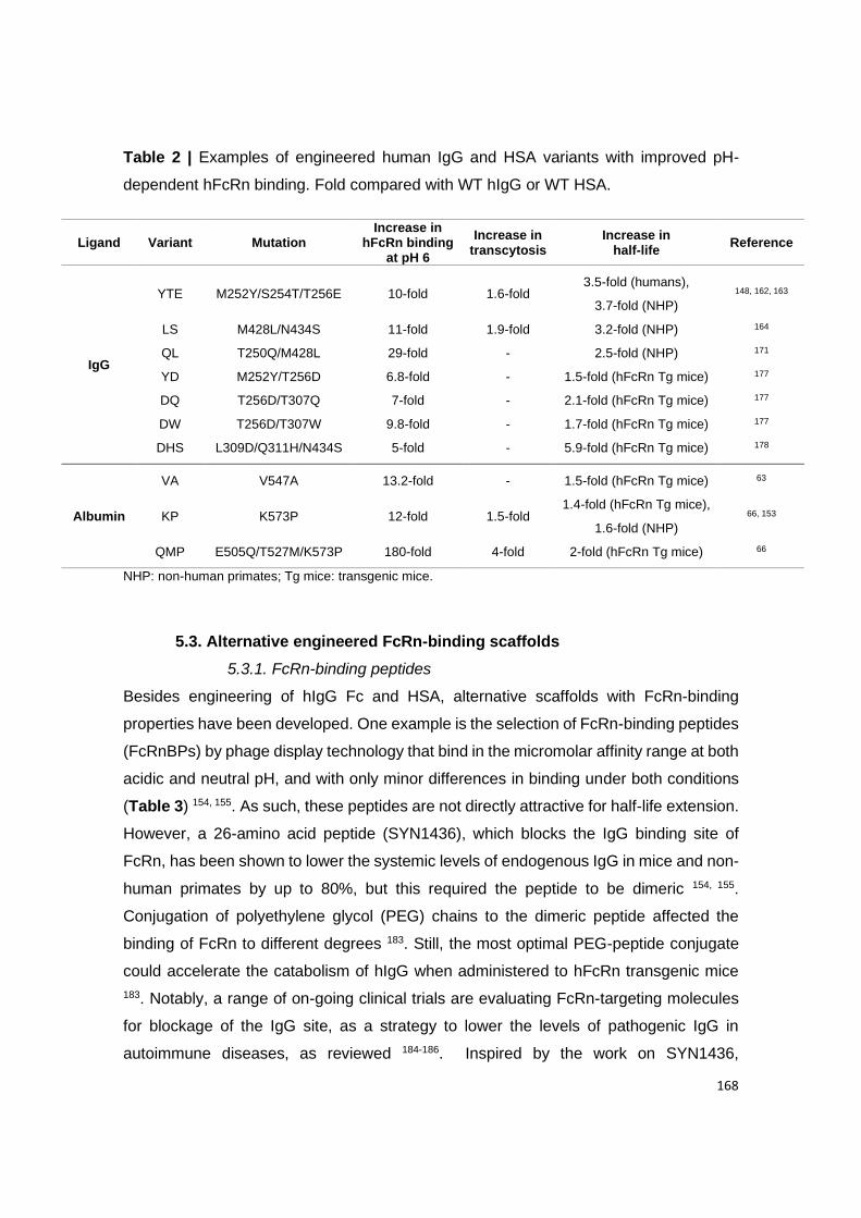
168
Table 2 | Examples of engineered human IgG and HSA variants with improved pH-
dependent hFcRn binding. Fold compared with WT hIgG or WT HSA.
Ligand Variant Mutation Increase in
hFcRn binding at pH 6
Increase in transcytosis
Increase in half-life
Reference
IgG
YTE M252Y/S254T/T256E 10-fold 1.6-fold 3.5-fold (humans),
3.7-fold (NHP)
148, 162, 163
LS M428L/N434S 11-fold 1.9-fold 3.2-fold (NHP) 164
QL T250Q/M428L 29-fold - 2.5-fold (NHP) 171
YD M252Y/T256D 6.8-fold - 1.5-fold (hFcRn Tg mice) 177
DQ T256D/T307Q 7-fold - 2.1-fold (hFcRn Tg mice) 177
DW T256D/T307W 9.8-fold - 1.7-fold (hFcRn Tg mice) 177
DHS L309D/Q311H/N434S 5-fold - 5.9-fold (hFcRn Tg mice) 178
Albumin
VA V547A 13.2-fold - 1.5-fold (hFcRn Tg mice) 63
KP K573P 12-fold 1.5-fold 1.4-fold (hFcRn Tg mice),
1.6-fold (NHP)
66, 153
QMP E505Q/T527M/K573P 180-fold 4-fold 2-fold (hFcRn Tg mice) 66
NHP: non-human primates; Tg mice: transgenic mice.
5.3. Alternative engineered FcRn-binding scaffolds
5.3.1. FcRn-binding peptides
Besides engineering of hIgG Fc and HSA, alternative scaffolds with FcRn-binding
properties have been developed. One example is the selection of FcRn-binding peptides
(FcRnBPs) by phage display technology that bind in the micromolar affinity range at both
acidic and neutral pH, and with only minor differences in binding under both conditions
(Table 3) 154, 155. As such, these peptides are not directly attractive for half-life extension.
However, a 26-amino acid peptide (SYN1436), which blocks the IgG binding site of
FcRn, has been shown to lower the systemic levels of endogenous IgG in mice and non-
human primates by up to 80%, but this required the peptide to be dimeric 154, 155.
Conjugation of polyethylene glycol (PEG) chains to the dimeric peptide affected the
binding of FcRn to different degrees 183. Still, the most optimal PEG-peptide conjugate
could accelerate the catabolism of hIgG when administered to hFcRn transgenic mice
183. Notably, a range of on-going clinical trials are evaluating FcRn-targeting molecules
for blockage of the IgG site, as a strategy to lower the levels of pathogenic IgG in
autoimmune diseases, as reviewed 184-186. Inspired by the work on SYN1436,
169
Sockolosky and colleagues made an engineered version by introducing a single point
substitution (Y14H) to make it binds more pH-dependently to the receptor 187. When
fused to a model fusion partner, it bound hFcRn without affecting the biological function
of the fusion partner. Notably, the fusion protein bound hFcRn more strongly at acidic pH
than at neutral pH, and still better at neutral pH than hIgG1. Further, one of the fusion
proteins was shown to be transcytosed 14-fold more efficiently than the non-fused
counterpart across hFcRn-expressing polarized cell monolayers, but not to the same
extend as that to hIgG1 187, 188. The ability of the fused FcRnBPs to extend the half-life or
deliver the fusion partner across mucosal barriers in vivo was not addressed in this study.
However, in another study, Y14H-containing FcRnBPs were used to build a panel of
antibody Fab fusions where linear and cyclic versions of the peptides that were fused to
the light chain or heavy chain of the Fab at either the N- or C-terminal end, or both, via
a glycine-serine-linker 188. Screening for hFcRn binding followed by transcytosis
experiments, showed that the fusion proteins had varying degree of receptor binding
activity and ability to be transported across polarized human cells. In addition,
determination of the half-life in non-human primates revealed that a fusion with four linear
FcRnBPs fused to the N- and C-terminal ends of both light and heavy chains gave the
greatest systemic exposure 188. While the half-life of a Fab fragment was measured to
be only about 3-5 hours due to its molecular weight below the renal clearance threshold,
the fused FcRnBPs could extend the half-life to 67 hours, which is far from that of IgG
and albumin with a half-life of about 5 days in non-human primates 153, 162. In spite of this,
the study pinpoints the importance of avidity for efficient receptor engagement and
transport.
5.3.2. Engineered protein domains
Protein domains (16kDa) with FcRn-binding properties have been generated by
combining the CH2 domain of an IgG1 Fc (m01s) with the grafting of a stretch of five
amino acids derived from the CH3 domain, containing the key FcRn interaction residues
H433 and H435 (Table 3) 157. Although this fusion bound hFcRn, further engineering was
done by the use of phage display to select mutant versions with increased and strict pH-
dependent binding to the receptor. One such variant (2C10) showed the capacity to be
transcytosed across polarized cells in a Transwell® system but did not perform as well
as the hIgG1 Fc fragment. When tested in non-human primates, the half-life was
measured to be 40-44 hours, more than 3-fold longer than that of m01s 157. While this
example shows that protein domains with a size below the renal clearance threshold
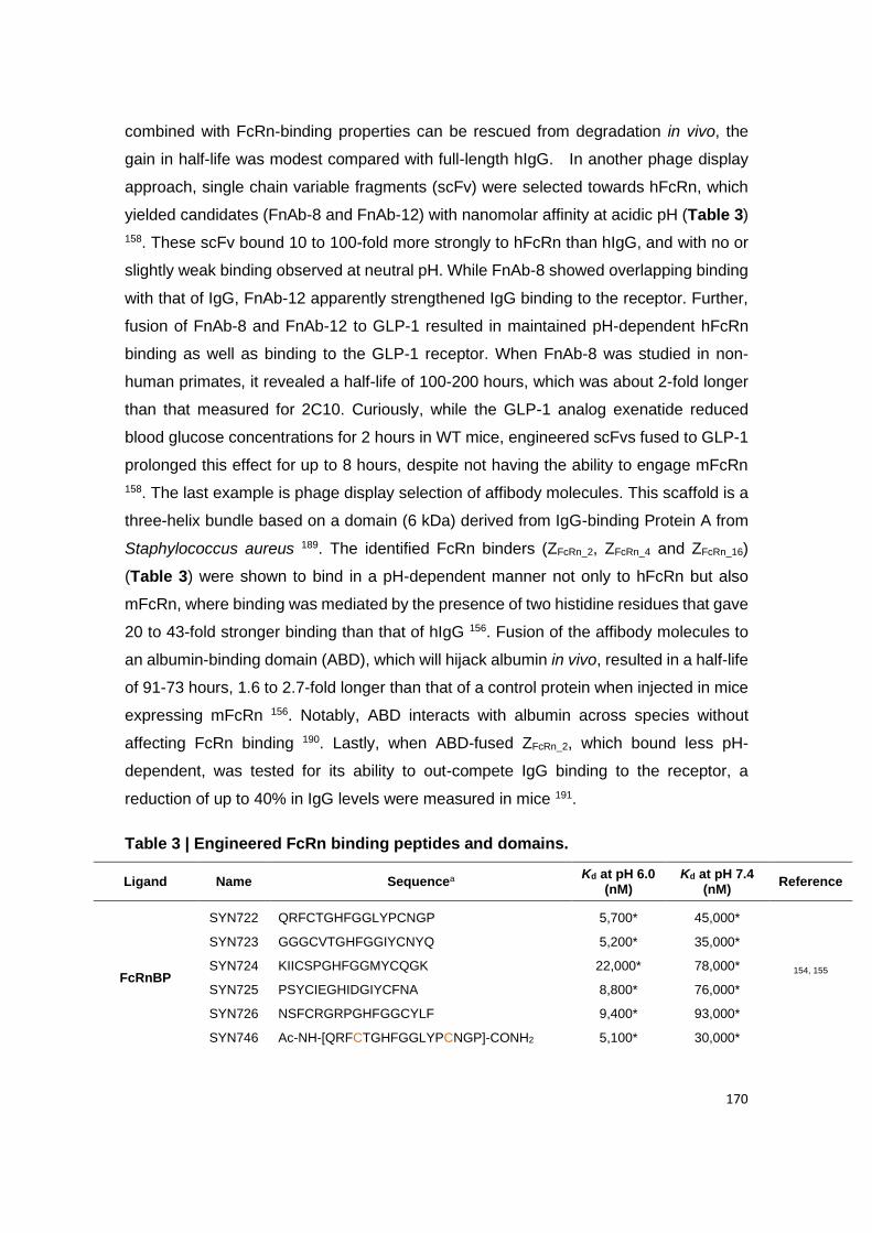
170
combined with FcRn-binding properties can be rescued from degradation in vivo, the
gain in half-life was modest compared with full-length hIgG. In another phage display
approach, single chain variable fragments (scFv) were selected towards hFcRn, which
yielded candidates (FnAb-8 and FnAb-12) with nanomolar affinity at acidic pH (Table 3)
158. These scFv bound 10 to 100-fold more strongly to hFcRn than hIgG, and with no or
slightly weak binding observed at neutral pH. While FnAb-8 showed overlapping binding
with that of IgG, FnAb-12 apparently strengthened IgG binding to the receptor. Further,
fusion of FnAb-8 and FnAb-12 to GLP-1 resulted in maintained pH-dependent hFcRn
binding as well as binding to the GLP-1 receptor. When FnAb-8 was studied in non-
human primates, it revealed a half-life of 100-200 hours, which was about 2-fold longer
than that measured for 2C10. Curiously, while the GLP-1 analog exenatide reduced
blood glucose concentrations for 2 hours in WT mice, engineered scFvs fused to GLP-1
prolonged this effect for up to 8 hours, despite not having the ability to engage mFcRn
158. The last example is phage display selection of affibody molecules. This scaffold is a
three-helix bundle based on a domain (6 kDa) derived from IgG-binding Protein A from
Staphylococcus aureus 189. The identified FcRn binders (ZFcRn_2, ZFcRn_4 and ZFcRn_16)
(Table 3) were shown to bind in a pH-dependent manner not only to hFcRn but also
mFcRn, where binding was mediated by the presence of two histidine residues that gave
20 to 43-fold stronger binding than that of hIgG 156. Fusion of the affibody molecules to
an albumin-binding domain (ABD), which will hijack albumin in vivo, resulted in a half-life
of 91-73 hours, 1.6 to 2.7-fold longer than that of a control protein when injected in mice
expressing mFcRn 156. Notably, ABD interacts with albumin across species without
affecting FcRn binding 190. Lastly, when ABD-fused ZFcRn_2, which bound less pH-
dependent, was tested for its ability to out-compete IgG binding to the receptor, a
reduction of up to 40% in IgG levels were measured in mice 191.
Table 3 | Engineered FcRn binding peptides and domains.
Ligand Name Sequencea Kd at pH 6.0
(nM) Kd at pH 7.4
(nM) Reference
FcRnBP
SYN722 QRFCTGHFGGLYPCNGP 5,700* 45,000*
154, 155
SYN723 GGGCVTGHFGGIYCNYQ 5,200* 35,000*
SYN724 KIICSPGHFGGMYCQGK 22,000* 78,000*
SYN725 PSYCIEGHIDGIYCFNA 8,800* 76,000*
SYN726 NSFCRGRPGHFGGCYLF 9,400* 93,000*
SYN746 Ac-NH-[QRFCTGHFGGLYPCNGP]-CONH2 5,100* 30,000*
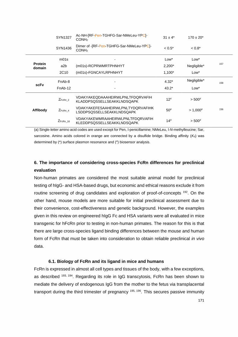
171
SYN1327 Ac-NH-[RF-Pen-TGHFG-Sar-NMeLeu-YPC]-CONH2
31 ± 4* 170 ± 20*
SYN1436 Dimer of -[RF-Pen-TGHFG-Sar-NMeLeu-YPC]-CONH2
< 0.5* < 0.8*
Protein domain
m01s - Low* Low* 157 a2b (m01s)-RCPRWMRTPHNHYT 2,200* Negligible*
2C10 (m01s)-FGNCAYLRPHNHYT 1,100* Low*
scFv FnAb-8 - 4.32* Negligible*
158
FnAb-12 - 43.2* Low*
Affibody
ZFcRn_2 VDAKYAKEQDAAAHEIRWLPNLTFDQRVAFIHKLADDPSQSSELLSEAKKLNDSQAPK
12# > 500#
ZFcRn_4 VDAKYAKEFESAAHEIRWLPNLTYDQRVAFIHKLSDDPSQSSELLSEAKKLNDSQAPK
50# > 1,000# 156
ZFcRn_16 VDAKYAKEWMRAAHEIRWLPNLTFDQRVAFIHKLEDDPSQSSELLSEAKKLNDSQAPK
14# > 500#
(a) Single-letter amino acid codes are used except for Pen, l-penicillamine; NMeLeu, l-N-methylleucine; Sar,
sarcosine. Amino acids colored in orange are connected by a disulfide bridge. Binding affinity (Kd) was
determined by (*) surface plasmon resonance and (#) biosensor analysis.
6. The importance of considering cross-species FcRn differences for preclinical
evaluation
Non-human primates are considered the most suitable animal model for preclinical
testing of hIgG- and HSA-based drugs, but economic and ethical reasons exclude it from
routine screening of drug candidates and exploration of proof-of-concepts 192. On the
other hand, mouse models are more suitable for initial preclinical assessment due to
their convenience, cost-effectiveness and genetic background. However, the examples
given in this review on engineered hIgG Fc and HSA variants were all evaluated in mice
transgenic for hFcRn prior to testing in non-human primates. The reason for this is that
there are large cross-species ligand binding differences between the mouse and human
form of FcRn that must be taken into consideration to obtain reliable preclinical in vivo
data.
6.1. Biology of FcRn and its ligand in mice and humans
FcRn is expressed in almost all cell types and tissues of the body, with a few exceptions,
as described 193, 194. Regarding its role in IgG transcytosis, FcRn has been shown to
mediate the delivery of endogenous IgG from the mother to the fetus via transplacental
transport during the third trimester of pregnancy 195, 196. This secures passive immunity
172
of the newborn at a critical time of life. In mice, such transport occurs via the placenta,
but mostly via intestinal uptake of neonatal mice during the suckling period, when large
amounts of IgG are taken up from the mother’s milk via pH-dependent FcRn-directed
transcytosis 197. Interestingly, while the intestinal tissue expression of FcRn in humans
are maintained throughout life, it is developmentally downregulated in conventional mice
87, 198. For this reason, caution should be taken when addressing FcRn-targeting
approaches and, due to this, it has been suggested that such studies should be
performed no later than 6-16 weeks after weaning in mice expressing mFcRn 199.
Notably, mFcRn is expressed in high levels in duodenum and jejunum during neonatal
life 198, 200, 201. In humans, the receptor is expressed in different sections of the human
intestine, including the colon and ileum, but somewhat less in jejunum and duodenum
102, 193, 199, 202. Although the FcRn expression of intestinal tissues is down-regulated in
mice, it persists throughout life in other tissues and, as such, regulates IgG and albumin
half-life and biodistribution.
Despite the fact that the mouse and human forms of the FcRn heavy chain share nearly
70% similarity in the amino acid composition, and that the key interacting residues
responsible for pH-dependent binding are conserved 29, 203, there are distinct variations
that give rise to large differences in cross-species binding. Specifically, this results in no
or very poor binding of mIgG to the human receptor, while hIgG binds more strongly to
the mouse receptor than to the human form 204, 205. As such, hIgG Fc-engineering for
improved binding to hFcRn does not translate into extended half-life in conventional
mice, but rather shorter half-life than the WT counterpart, as engineering for more
favorable pH-dependent binding is not preserved 70, 206. This is the case for all hIgG Fc-
engineered variants reported to date, which means that they cannot be used in mouse
models expressing mFcRn. In contrast to hIgG, HSA binds very poorly to mFcRn, with
more than 20-fold weaker binding than mouse serum albumin (MSA), whereas MSA
binds slightly stronger than HSA to hFcRn 37, 192, 204, 207, 208. Thus, HSA injected into
conventional mice is out-competed for binding to mFcRn in the presence of high amounts
of natural endogenous MSA, produced continuously by the liver. This means that HSA
is not rescued from intracellular degradation in mice expressing mFcRn, as illustrated by
the fact that WT HSA has nearly identical half-life as HSA engineered for considerably
reduced binding to hFcRn 153. Therefore, studies conducted under such conditions do
not take into consideration the contribution of FcRn and, as such, this preclinical setting
is non-physiological. The molecular explanations for the large cross-species differences
have been described in depth elsewhere 73, 153, 192, 207, 209, 210.

173
6.2. Human FcRn transgenic mouse models
To solve this preclinical challenge, several genetically engineered mouse strains have
been developed where the mFcRn heavy chain has been replaced with the human
counterpart, either under the control of the mouse or human promoter elements 35, 72, 175,
208, 211-215. These strains are the current gold standards for the evaluation of hFcRn-
targeting strategies addressing pharmacokinetics, pharmacodynamics and delivery
across selective mucosal barriers prior to studies in non-human primates. The most
commonly used hFcRn-expressing strains are generated from conventional C57BL/6J
mice, named Tg32 and Tg276 (Figure 4 A), which are extensively used to assess the
half-life of hIgG and HSA variants 35, 72, 175, 212, 214, 215. Both strains have substituted the
mouse heavy chain with that of the human form, which can pair non-covalently with the
endogenous mouse β2m 35, 72, 175, 214, 215. A distinct difference between these strains is
that the Tg32 mice express the heavy chain under the control of its human regulatory
elements 72, 175, 212, 214, 215, while the expression in Tg276 mice is regulated by a
constitutive CAG promoter, a human cytomegalovirus enhancer fused to chicken β-
actin/rabbit β-globin hybrid 35, 175. This results in a much more widespread expression
profile of hFcRn in Tg276, whereas the FcRn expression profile in Tg32 mice has been
mapped to be similar to that in humans 193, 216. There is also a modified version of Tg32
that expresses human β2m, but no difference in half-life of the ligands has been reported
that can be related to whether the mice carry a mouse or human form of β2m 214. In
general, the half-life of IgG is shorter in Tg276 homozygous mice relative to the Tg32
strain, and longer in the Tg32 hemizygous than the homozygous strain 214. Since the
expression profile of the Tg32 strains resembles more closely to that of humans, studies
of FcRn in mucosal immunity and receptor targeting for delivery across the epithelial
barriers have been mainly studied in these mice, as recently reviewed 93. Therefore, the
Tg32 strains should be used for studies addressing the mucosal delivery of FcRn-
targeted molecules or nanosystems.
In humans, injected hIgG and HSA variants as well as fusions will compete for binding
to FcRn in the presence of high concentrations of IgG (10 mg/mL) and albumin (40
mg/mL), produced by the plasma cells and liver, respectively. This is in stark contrast to
what has been found in mice kept under pathogen-free housing, where the
concentrations of endogenous mIgG are low (roughly 1.0-0.2 mg/mL) compared to those
found in nature, while the concentrations of MSA are high due to constitutive production
by the liver (20-50 mg/mL) 35, 217. These conditions must be taken into consideration in
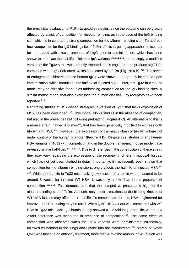
174
the preclinical evaluation of FcRn-targeted strategies, since the outcome can be greatly
affected by a lack of competition for receptor binding, as in the case of the IgG binding
site, which is in contrast to strong competition for the albumin-binding site. To address
how competition for the IgG binding site of FcRn affects targeting approaches, mice may
be pre-loaded with excess amounts of hIgG prior to administration, which has been
shown to modulate the half-life of injected IgG variants 175, 211, 218. Interestingly, a modified
version of the Tg32 strain was recently reported that is engineered to produce hIgG1 Fc
combined with mIgG Fab arms, which is rescued by hFcRn (Figure 4 B) 211. The levels
of endogenous chimeric mouse-human IgG1 were shown to be greatly increased upon
immunization, which modulated the half-life of injected hIgG. Thus, this Tg32-hFc mouse
model may be attractive for studies addressing competition for the IgG binding sites. A
similar mouse model that also expresses the human classical Fcγ receptors have been
reported 219.
Regarding studies of HSA-based strategies, a version of Tg32 that lacks expression of
MSA has been developed 213. This model allows studies in the absence of competition,
but also in the presence HSA following preloading (Figure 4 C). An alternative to this is
a mouse strain, named AlbumusTM, that has been genetically modified to express both
hFcRn and HSA 208. However, the expression of the heavy chain of hFcRn is here not
under control of the human promoter (Figure 4 D). Despite this, studies of engineered
HSA variants in Tg32 with competition and in the double transgenic mouse model have
revealed similar half-lives 153, 208, 215. Due to differences in the construction of these strain,
they may vary regarding the expression of the receptor in different mucosal tissues,
which has not yet been studied in detail. Importantly, it has recently been shown that
competition for the albumin-binding site strongly affects the half-life of injected HSA 66,
213. While the half-life in Tg32 mice lacking expression of albumin was measured to be
around 3 weeks for injected WT HSA, it was only a few days in the presence of
competition 66, 213. This demonstrates that the competitive pressure is high for the
albumin-binding site of FcRn. As such, only minor alterations to the binding kinetics of
WT HSA fusions may affect their half-life. To compensate for this, HSA engineered for
improved hFcRn binding may be used. When QMP HSA variant was compared with WT
HSA in Tg32 mice lacking albumin, it only showed a 1.2-fold longer half-life, whereas a
2-fold difference was measured in presence of competition 66. The same effect of
competition was observed when the HSA variants were administered intranasally,
followed by homing to the lungs and uptake into the bloodstream 66. Moreover, when
QMP was fused to an antibody fragment, more than 4-fold the amount of WT fusion was
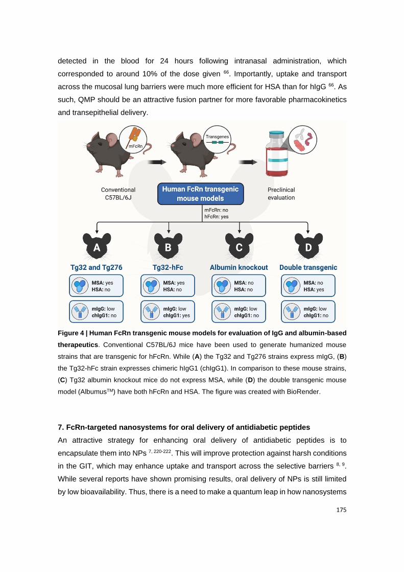
175
detected in the blood for 24 hours following intranasal administration, which
corresponded to around 10% of the dose given 66. Importantly, uptake and transport
across the mucosal lung barriers were much more efficient for HSA than for hIgG 66. As
such, QMP should be an attractive fusion partner for more favorable pharmacokinetics
and transepithelial delivery.
Figure 4 | Human FcRn transgenic mouse models for evaluation of IgG and albumin-based
therapeutics. Conventional C57BL/6J mice have been used to generate humanized mouse
strains that are transgenic for hFcRn. While (A) the Tg32 and Tg276 strains express mIgG, (B)
the Tg32-hFc strain expresses chimeric hIgG1 (chIgG1). In comparison to these mouse strains,
(C) Tg32 albumin knockout mice do not express MSA, while (D) the double transgenic mouse
model (AlbumusTM) have both hFcRn and HSA. The figure was created with BioRender.
7. FcRn-targeted nanosystems for oral delivery of antidiabetic peptides
An attractive strategy for enhancing oral delivery of antidiabetic peptides is to
encapsulate them into NPs 7, 220-222. This will improve protection against harsh conditions
in the GIT, which may enhance uptake and transport across the selective barriers 8, 9.
While several reports have shown promising results, oral delivery of NPs is still limited
by low bioavailability. Thus, there is a need to make a quantum leap in how nanosystems
176
are designed to make oral delivery far more efficient. One way to achieve this may be to
combine the biotechnological advances of NP design with strategies to target FcRn at
mucosal surfaces 11-15. This may allow active transport across the polarized intestinal
epithelium for delivery to the systemic circulation 11, 12, 15. The NP design must then be
done in a way that favors FcRn engagement and transcellular transport, parameters that
are not yet fully defined. However, while NPs can be decorated with FcRn-targeting
molecules, the encapsulated peptides may also be engineered for efficient FcRn-
mediated recycling, as discussed in Section 4, which can improve half-life and
bioavailability upon release from the NPs. Next, a brief overview of parameters that are
relevant to be considered in the context of NP design and FcRn-targeting will be
discussed.
7.1. Design of targeting nanosystems
The stability and characteristics of nanosystems, including their size, charge, shape,
material and coating are parameters that dictate their internalization and transport
properties (Figure 5), as extensively reviewed 7, 8, 223-226. However, for the NPs to be
targeted to FcRn, they must be engineered to display receptor-targeting molecules on
their surface in such a manner that will allow for efficient binding-and-release and stability
throughout the endosomal pH gradient. Relevant parameters to be considered are how
the pH-dependent binding kinetics of the FcRn-binding molecules affect the ability of the
NPs to be transported, and in this context, how the number and density of the targeting
molecules and, as such, avidity and flexibility are affecting the outcome 227. Decoration
of NPs with FcRn-targeting molecules could be achieved in a non-covalent manner by
adsorption, but preferably covalently to secure stability 228. To ensure the latter,
conjugation should be irreversible and site-specific to fix the orientation of the molecules
in such a way that guarantees availability and functional display. One strategy is to use
MAL-thiol or carbodiimide chemistry for site-specific conjugation. Regarding the MAL-
thiol reaction, it requires coupling of sulfhydryl reactive MAL on the surface of NPs to
thiol groups present on hIgG or HSA, which will result in irreversible thioether bonds 229.
As hIgG or the constant Fc does not contain any free cysteine residues, functional groups
using primary amine reactive reagents or crosslinkers must be used 11, 12. However, when
it comes to HSA, C34 in DI is an attractive site for conjugation 15. On the other hand,
carbodiimide crosslinking can be achieved using 1-ethyl-3-(3-dimethylaminopropyl)-
carbodiimide/N-hydroxysuccinimide (EDC/NHS) reaction to form an amide bond
between the carboxylic groups of hIgG Fc or HSA and the functionalized aminated NPs
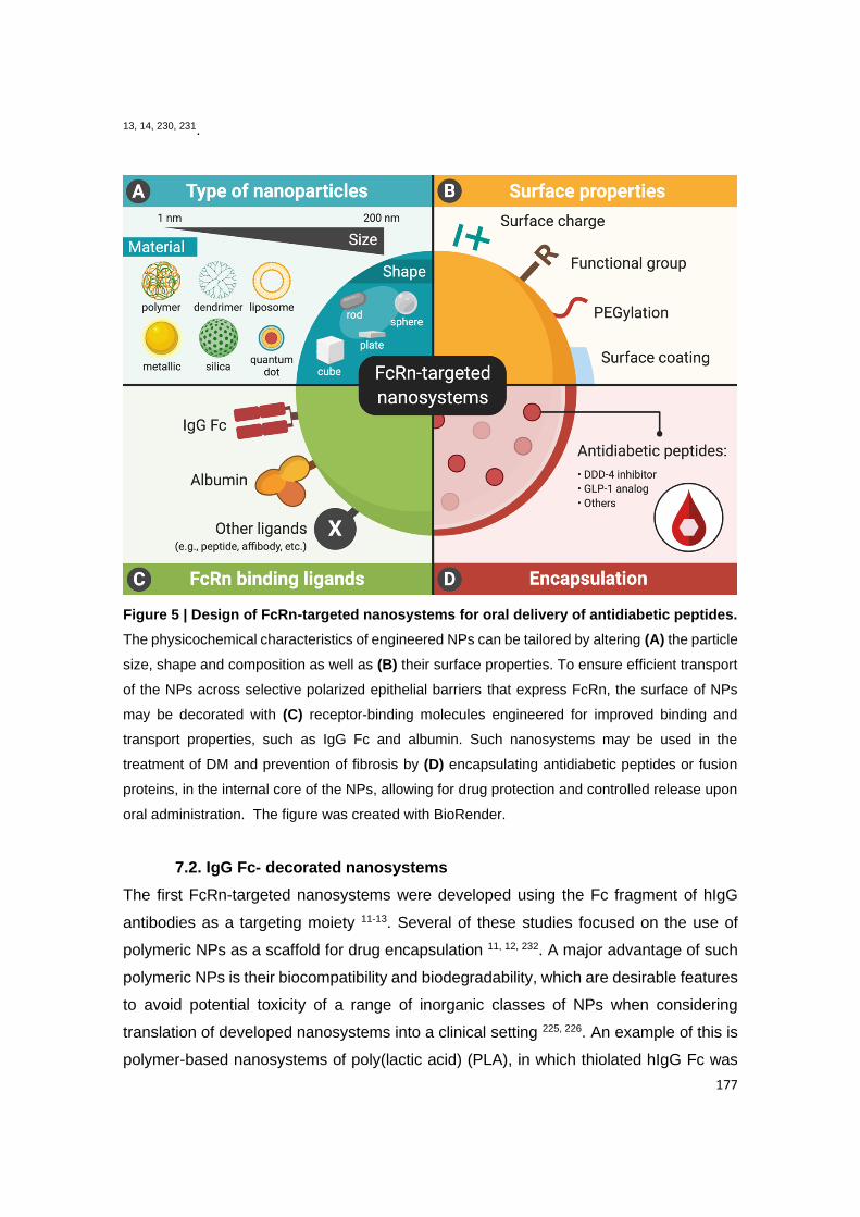
177
13, 14, 230, 231.
Figure 5 | Design of FcRn-targeted nanosystems for oral delivery of antidiabetic peptides.
The physicochemical characteristics of engineered NPs can be tailored by altering (A) the particle
size, shape and composition as well as (B) their surface properties. To ensure efficient transport
of the NPs across selective polarized epithelial barriers that express FcRn, the surface of NPs
may be decorated with (C) receptor-binding molecules engineered for improved binding and
transport properties, such as IgG Fc and albumin. Such nanosystems may be used in the
treatment of DM and prevention of fibrosis by (D) encapsulating antidiabetic peptides or fusion
proteins, in the internal core of the NPs, allowing for drug protection and controlled release upon
oral administration. The figure was created with BioRender.
7.2. IgG Fc- decorated nanosystems
The first FcRn-targeted nanosystems were developed using the Fc fragment of hIgG
antibodies as a targeting moiety 11-13. Several of these studies focused on the use of
polymeric NPs as a scaffold for drug encapsulation 11, 12, 232. A major advantage of such
polymeric NPs is their biocompatibility and biodegradability, which are desirable features
to avoid potential toxicity of a range of inorganic classes of NPs when considering
translation of developed nanosystems into a clinical setting 225, 226. An example of this is
polymer-based nanosystems of poly(lactic acid) (PLA), in which thiolated hIgG Fc was
178
covalently conjugated to the MAL-functionalized NPs, with an average hydrodynamic
diameter of about 55 nm, via MAL-thiol reaction 11. The conjugation of Fc resulted in a
slight increase in the particle size and showed a maintained monodispersity of the NPs
before and after coupling. Since IgG antibodies generally carry a weakly negative charge
under physiological conditions 233, the surface charge of the Fc-decorated NPs (Fc-NPs)
became slightly more negatively charged when coupled to the Fc fragments.
Interestingly, Fc-NPs gave a 2-fold rise in transcytosis in vitro compared with non-
targeted NPs across a polarized epithelial cell line that endogenously expresses hFcRn,
while transport was reduced in the presence of free IgG Fc competing for receptor
binding, suggesting that the increase in uptake and transcytosis was FcRn-dependent
11. In conventional WT BALB/c mice, which express mFcRn throughout the small
intestine and colon, Fc-targeting of FcRn enhanced in vivo biodistribution of the NPs in
several organs, as well as the absorption efficiency by 11.5-fold following oral
administration. Importantly, insulin-loaded Fc-NPs showed prolonged hypoglycemic
responses in fasted mice when administered orally at a clinically relevant insulin dose
compared with non-targeted NPs and to injected insulin, while free OI solution did not
generate a glucose response. This effect was not observed in mice lacking FcRn
expression, which may indicate FcRn-induced transcytosis of Fc-NPs in the GIT and, as
such, the release of insulin from the NP matrix in blood circulation. While these studies
were done in WT mice, further studies on molecules engineered for improved hFcRn
should be performed in mice transgenic for the human receptor 212, 215.
A similar strategy has been explored to improve oral efficacy of exenatide, a GLP-1
receptor agonist for T2DM treatment, in which the peptides were loaded into poly(lactic-
co-glycolic acid) (PLGA) NPs using the same coupling chemistry as PLA-based
nanosystems 12. The particle size of native PLGA NPs was about 109 nm, which was
larger than that of the PLA NPs 11. The difference in hydrodynamic diameters observed
can be explained by the methods used to synthesize NPs and the chemical composition
of NP formulations. Similar increase in particle size and surface charge of human Fc-
decorated PLGA NPs as that of PLA NPs was observed. In this study, relative to non-
targeted nanosystems, Fc-NPs showed to be more efficiently taken up and transcytosed
in a polarized epithelial cell line expressing endogenous hFcRn 12. Notably, it would be
of importance to compare to what degree the particle size may affect the internalization
of NPs 234, 235. On the other hand, a mouse model of human TD2M (db/db) was used to
study the hypoglycemic response of the peptide-loaded NPs. Upon oral administration,
the Fc-NPs showed a higher reduction in blood glucose levels and particle retention in
179
the GIT compared with non-decorated NPs 12. Furthermore, FcRn-targeting of Fc-
decorated nanosystems exhibited a prolonged hypoglycemic response relative to
injected exenatide, although the reduction effect of the injection solution was shown to
be more efficient in the first few hours following subcutaneous injection.
In addition to polymer-based NPs, nanosystems may be designed with inorganic
components having unique electrical, magnetic and optical properties 225, 226. Such
inorganic NPs allow for fine-tuning of the size, structure and geometry for enhanced
performance that may benefit oral delivery across epithelial barriers of the tailored-made
nanosystems. Nevertheless, due to the toxicity and solubility limitations of such NPs, the
use of inorganic materials in humans must be carefully examined 236, 237. An example of
inorganic nanosystems explored for oral delivery of antidiabetic peptides is undecylenic
acid-coated silicon NPs (SiNPs) that were loaded with GLP-1 13. In contrast to the
examples of polymeric NP formulations given 11, 12, SiNPs were functionalized with
amine-modified Fc via EDC/NHS reaction. The NPs were then treated with
mucoadhesive chitosan and eventually entrapped into a pH-responsive polymeric matrix
of hypromellose acetate succinate (HPMC) to improve drug adsorption at mucosal sites
and prevent premature release of encapsulated drugs under strong acidic conditions in
the GIT 13. The Fc-NPs showed enhanced transcytosis in vitro in polarized epithelial cell
lines expressing hFcRn compared with non-targeted NPs, which resulted in higher
amounts of GLP-1 released from the NP matrix at the basolateral side of Transwell®
system 13.
Overall, these findings have demonstrated that decoration with hIgG Fc fragments may
enable the oral delivery of peptide-loaded NPs across epithelial barriers for delivery to
the systemic circulation 11-13. However, in addition to the lack of use of hFcRn expressing
mouse models, pH-dependent binding affinity and functional avidity for the receptor of
the Fc ligands decorating the surface NPs have yet to be examined to ensure optimal
receptor-mediated binding and transepithelial transport of nanosystems. Furthermore,
the mechanisms of action of the FcRn-targeted NPs should be studied in detail.
7.3. Albumin- decorated nanosystems
Although IgG Fc have been explored as FcRn-targeting moieties, their use is impeded
due to the need for chemical modification for conjugation. As such, HSA may be used
for decoration of NPs owing to low immunogenicity and ease of production 238, 239.
Furthermore, it was very recently shown that HSA is transported more efficiently than
hIgG across mucosal lung barriers in vivo in hFcRn trangenic mice 66. On this basis, HSA
180
may be an attractive targeting molecule for favorable FcRn-mediated transepithelial
delivery of nanosystems for oral administration of antidiabetic peptides 14, 15, 232.
An example of nanosystems that have been combined with albumin is polymeric
alginate-dextran sulfate-based (ADS) NPs with an average hydrodynamic diameter of
233 nm, that were coated with chitosan 232. In contrast to synthetic PLA and PLGA, the
polymers alginate, dextran and chitosan used to synthesize NPs are natural product-
derived compounds that provide nanosystems with mucoadhesive characteristics 239. In
this study, bovine serum albumin (BSA) was non-specifically adsorbed onto the surface
of ADS NPs 232, in contrary to site-directed chemical coupling of Fc-decorated NPs 11-13.
In an in vitro model of polarized epithelial cells expressing hFcRn combined with mucus-
producing goblet cells, BSA-decorated NPs loaded with insulin showed an improved
permeability profile of the peptides compared to free insulin 232. However, cautious
should be taken when interpreting the result since the binding kinetics of BSA for hFcRn
is not identical to that of HSA 35. Aside from that, strategies for surface modification of
NPs and pH-dependent functionality of ligands are two other important parameters to
address.
An attempt was made to decorate the surface of HPMC-treated undecylenic acid-coated
SiNPs with HSA via EDC/NHS reaction 14. The chemical surface modification of NPs with
HSA showed an increase in particle size from 162 nm to 238 nm after conjugation, with
high monodispersity. Interestingly, HSA-conjugated NPs (HSA-NPs) exhibited improved
cytocompatibility in human epithelial cell lines relative to non-conjugated NPs, which
suggests that decorating nanosystems with biocompatible compounds, such as HSA,
may reduce the toxicity of inorganic nanomaterials. Furthermore, the presence of HSA
on the NP surface demonstrated enhanced cellular uptake and internalization of
nanosystems in vitro with the aid of hFcRn expressed on the cell surface 14. Done in this
fashion, HSA-NPs encapsulating insulin showed enhanced transport of loaded peptides
across polarized epithelial cell monolayers in an in vitro transcytosis assays. The data
also suggested an improvement in the transepithelial delivery of encapsulated
antidiabetic peptides when comparing HSA-NPs with IgG Fc-NPs 13, which encourages
research on the use of engineered HSA variants tailored for improved FcRn binding and
transport properties as targeting ligands, as discussed in Section 5 66.
Recently, the KP variant of HSA, which shows a 12-fold improved hFcRn binding
compared to WT HSA 153, has been explored in an NP formulation of polymeric PLGA
with an average hydrodynamic diameter of about 150 nm 15. In this study, the surface of
PLGA NPs was functionalized with HSA through MAL-thiol conjugation chemistry, in

181
which no or slight increase in particle size was observed as a result of protein coupling.
Most importantly, the pH-dependent binding characteristics for hFcRn of HSA-NPs were
in line with reported studies for free/uncoupled HSA 66, 153. Compared to WT-decorated
NPs, KP-decorated NPs showed enhanced binding to hFcRn at mildly acidic pH (5.5),
while receptor binding was not observed for NPs decorated with an engineered HSA
variant that does not bind hFcRn 15. Similar results were demonstrated in an in vitro
transcytosis assays, where KP-decorated NPs were transported 2-fold more efficiently
than WT-decorated NPs across polarized epithelial cells expressing hFcRn 15.
Intriguingly, when studying the effect of HSA functionalization on the hypoglycemic
response of insulin-loaded PLGA NPs in T1DM-induced mice transgenic for hFcRn, KP-
decorated NPs showed the most efficient glycemic reduction following oral
administration compared with other HSA-NPs and to OI solution.
Altogether, engineered HSA shows promise in the next-generation design of FcRn-
targeted nanosystems for oral delivery of antidiabetic peptides. The ultimate aim now will
be to tailor-design such strategies for further improvement in receptor engangment and
transport propertes ta favor efficient transepithelial transport of NPs across the GIT upon
oral administration. The QMP HSA variant, should be an attractive FcRn targeting
partner for decoration of NPs to achieve not only enhanced mucosal delivery but also
prolonged half-life of fused peptides 66.
8. Conclusions
Nanosystems for encapsulation of drugs have the potential to make a significant impact
on the treatment of many diseases, including diabetes and fibrosis. While a number of
NP-based strategies have entered clinical trials or have been approved for use, they can
only be administered by parenteral delivery methods. An alternative and preferable
approach would be oral administration, but this remains a challenge because uptake and
delivery across selective epithelial barriers is very limited. Furthermore, frequent
administration is often required. As such, design of nanosystems tailored for oral
administration is preferable due to patient convenience and compliance. In this review,
we have highlighted how in-depth insights into the biology of FcRn may be used to
engineer NPs decorated with receptor-binding molecules with the aim to overcome the
highly inefficient uptake at the intestinal epithelial barriers. If this major challenge is
solved, it will allow for more cost-effective self-administered methods. In addition, the
encapsulated drugs, such as antidiabetic peptides, may also be engineered for extended
182
half-life and increased bioavailability by taking advantage of pH-dependent FcRn
engagement. For preclinical testing of such strategies, considerable cross-species FcRn
binding differences must be taken into account to secure reliable results from mouse
model systems.
Conflit of interest
The authors declare no conflict of interest.
Acknowledgements
This paper was financed by Portuguese funds through FCT - Fundação para a Ciência
e a Tecnologia/Ministério da Ciência, Tecnologia e Ensino Superior in the framework of
the project "Institute for Research and Innovation in Health Sciences"
UID/BIM/04293/2019. Cláudia Azevedo (SFRH/BD/117598/2016) and Soraia Pinto
(SFRH/BD/144719/2019) would like to thank to Fundação para a Ciência e a Tecnologia
(FCT), Portugal for financial support. S.B., J.N. and J.T.A. were supported by the
Research Council of Norway (Grant no. 274993; 287927), and J.T.A. by the South-
Eastern Norway Regional Health Authority (Grant no. 2018052; 2019084).

183
References
1. Zheng Y, Ley SH, Hu FB. Global aetiology and epidemiology of type 2 diabetes mellitus
and its complications. Nature Reviews Endocrinology 2018;14:88-98.
2. WHO. Classification of diabetes mellitus 2019. Volume 2020, 2019.
3. Basson R, Bayat A. Chapter 7 - Fibrosis and diabetes: Chronic hyperglycemia triggers
organ-specific fibrotic mechanisms. In: Bagchi D, Das A, Roy S, eds. Wound Healing,
Tissue Repair, and Regeneration in Diabetes: Academic Press, 2020:121-147.
4. Fowler MJ. Microvascular and Macrovascular Complications of Diabetes. Clinical
Diabetes 2008;26:77-82.
5. Ban CR, Twigg SM. Fibrosis in diabetes complications: pathogenic mechanisms and
circulating and urinary markers. Vascular health and risk management 2008;4:575-596.
6. Sosnik A, das Neves J, Sarmento B. Mucoadhesive polymers in the design of nano-drug
delivery systems for administration by non-parenteral routes: A review. Progress in
Polymer Science 2014;39:2030-2075.
7. Thwala LN, Préat V, Csaba NS. Emerging delivery platforms for mucosal administration
of biopharmaceuticals: a critical update on nasal, pulmonary and oral routes. Expert Opin
Drug Deliv 2017;14:23-36.
8. Araújo F, das Neves J, Martins JP, et al. Functionalized materials for multistage platforms
in the oral delivery of biopharmaceuticals. Progress in Materials Science 2017;89:306-
344.
9. Goldberg M, Gomez-Orellana I. Challenges for the oral delivery of macromolecules. Nat
Rev Drug Discov 2003;2:289-95.
10. Patra JK, Das G, Fraceto LF, et al. Nano based drug delivery systems: recent
developments and future prospects. J Nanobiotechnology 2018;16:71.
11. Pridgen EM, Alexis F, Kuo TT, et al. Transepithelial transport of Fc-targeted nanoparticles
by the neonatal fc receptor for oral delivery. Sci Transl Med 2013;5:213ra167.
12. Shi Y, Sun X, Zhang L, et al. Fc-modified exenatide-loaded nanoparticles for oral delivery
to improve hypoglycemic effects in mice. Scientific Reports 2018;8:726.
13. Martins JP, Liu D, Fontana F, et al. Microfluidic Nanoassembly of Bioengineered
Chitosan-Modified FcRn-Targeted Porous Silicon Nanoparticles @ Hypromellose
Acetate Succinate for Oral Delivery of Antidiabetic Peptides. ACS Applied Materials &
Interfaces 2018;10:44354-44367.
14. Martins JP, D'Auria R, Liu D, et al. Engineered Multifunctional Albumin-Decorated Porous
Silicon Nanoparticles for FcRn Translocation of Insulin. Small 2018;14:e1800462.
15. Azevedo C, Nilsen J, Grevys A, et al. Engineered albumin-functionalized nanoparticles
for improved FcRn binding enhance oral delivery of insulin. Journal of Controlled Release
2020;327:161-173.
184
16. Goldin A, Beckman JA, Schmidt AM, et al. Advanced glycation end products: sparking
the development of diabetic vascular injury. Circulation 2006;114:597-605.
17. Lamouille S, Xu J, Derynck R. Molecular mechanisms of epithelial-mesenchymal
transition. Nat Rev Mol Cell Biol 2014;15:178-96.
18. Yu MA, Shin KS, Kim JH, et al. HGF and BMP-7 ameliorate high glucose-induced
epithelial-to-mesenchymal transition of peritoneal mesothelium. J Am Soc Nephrol
2009;20:567-81.
19. Zeisberg M, Zeisberg EM. Evidence for antifibrotic incretin-independent effects of the
DPP-4 inhibitor linagliptin. Kidney Int 2015;88:429-31.
20. Wang X, Hausding M, Weng SY, et al. Gliptins Suppress Inflammatory Macrophage
Activation to Mitigate Inflammation, Fibrosis, Oxidative Stress, and Vascular Dysfunction
in Models of Nonalcoholic Steatohepatitis and Liver Fibrosis. Antioxid Redox Signal
2018;28:87-109.
21. Jensterle M, Janez A, Fliers E, et al. The role of glucagon-like peptide-1 in reproduction:
from physiology to therapeutic perspective. Hum Reprod Update 2019;25:504-517.
22. de Mesquita FC, Guixé-Muntet S, Fernández-Iglesias A, et al. Liraglutide improves liver
microvascular dysfunction in cirrhosis: Evidence from translational studies. Scientific
Reports 2017;7:3255.
23. Seghieri M, Christensen AS, Andersen A, et al. Future Perspectives on GLP-1 Receptor
Agonists and GLP-1/glucagon Receptor Co-agonists in the Treatment of NAFLD.
Frontiers in Endocrinology 2018;9.
24. Haymann JP, Levraud JP, Bouet S, et al. Characterization and localization of the neonatal
Fc receptor in adult human kidney. J Am Soc Nephrol 2000;11:632-9.
25. Spiekermann GM, Finn PW, Ward ES, et al. Receptor-mediated immunoglobulin G
transport across mucosal barriers in adult life: functional expression of FcRn in the
mammalian lung. J Exp Med 2002;196:303-10.
26. Akilesh S, Huber TB, Wu H, et al. Podocytes use FcRn to clear IgG from the glomerular
basement membrane. Proc Natl Acad Sci U S A 2008;105:967-72.
27. Simister NE, Rees AR. Isolation and characterization of an Fc receptor from neonatal rat
small intestine. Eur J Immunol 1985;15:733-8.
28. Simister NE, Mostov KE. An Fc receptor structurally related to MHC class I antigens.
Nature 1989;337:184-7.
29. West AP, Jr., Bjorkman PJ. Crystal structure and immunoglobulin G binding properties of
the human major histocompatibility complex-related Fc receptor(,). Biochemistry
2000;39:9698-708.
30. Burmeister WP, Huber AH, Bjorkman PJ. Crystal structure of the complex of rat neonatal
Fc receptor with Fc. Nature 1994;372:379-83.
185
31. Bjorkman PJ, Saper MA, Samraoui B, et al. The foreign antigen binding site and T cell
recognition regions of class I histocompatibility antigens. Nature 1987;329:512-8.
32. Burmeister WP, Gastinel LN, Simister NE, et al. Crystal structure at 2.2 A resolution of
the MHC-related neonatal Fc receptor. Nature 1994;372:336-43.
33. Raghavan M, Bonagura VR, Morrison SL, et al. Analysis of the pH Dependence of the
Neonatal Fc Receptor/Immunoglobulin G Interaction Using Antibody and Receptor
Variants. Biochemistry 1995;34:14649-14657.
34. Vaughn DE, Bjorkman PJ. Structural basis of pH-dependent antibody binding by the
neonatal Fc receptor. Structure 1998;6:63-73.
35. Chaudhury C, Mehnaz S, Robinson JM, et al. The major histocompatibility complex-
related Fc receptor for IgG (FcRn) binds albumin and prolongs its lifespan. J Exp Med
2003;197:315-22.
36. Andersen JT, Dee Qian J, Sandlie I. The conserved histidine 166 residue of the human
neonatal Fc receptor heavy chain is critical for the pH-dependent binding to albumin. Eur
J Immunol 2006;36:3044-51.
37. Andersen JT, Dalhus B, Cameron J, et al. Structure-based mutagenesis reveals the
albumin-binding site of the neonatal Fc receptor. Nat Commun 2012;3:610.
38. Oganesyan V, Damschroder MM, Cook KE, et al. Structural insights into neonatal Fc
receptor-based recycling mechanisms. J Biol Chem 2014;289:7812-24.
39. Raghavan M, Bonagura VR, Morrison SL, et al. Analysis of the pH dependence of the
neonatal Fc receptor/immunoglobulin G interaction using antibody and receptor variants.
Biochemistry 1995;34:14649-57.
40. Chaudhury C, Brooks CL, Carter DC, et al. Albumin binding to FcRn: distinct from the
FcRn-IgG interaction. Biochemistry 2006;45:4983-90.
41. Roopenian DC, Akilesh S. FcRn: the neonatal Fc receptor comes of age. Nature Reviews
Immunology 2007;7:715.
42. de Taeye SW, Rispens T, Vidarsson G. The Ligands for Human IgG and Their Effector
Functions. Antibodies (Basel) 2019;8.
43. Saphire EO, Parren PW, Pantophlet R, et al. Crystal structure of a neutralizing human
IGG against HIV-1: a template for vaccine design. Science 2001;293:1155-9.
44. Kim JK, Tsen MF, Ghetie V, et al. Localization of the site of the murine IgG1 molecule
that is involved in binding to the murine intestinal Fc receptor. Eur J Immunol
1994;24:2429-34.
45. Kim JK, Tsen MF, Ghetie V, et al. Identifying amino acid residues that influence plasma
clearance of murine IgG1 fragments by site-directed mutagenesis. Eur J Immunol
1994;24:542-8.
186
46. Raghavan M, Chen MY, Gastinel LN, et al. Investigation of the interaction between the
class I MHC-related Fc receptor and its immunoglobulin G ligand. Immunity 1994;1:303-
315.
47. Medesan C, Matesoi D, Radu C, et al. Delineation of the amino acid residues involved in
transcytosis and catabolism of mouse IgG1. The Journal of Immunology 1997;158:2211-
2217.
48. Kim JK, Firan M, Radu CG, et al. Mapping the site on human IgG for binding of the MHC
class I-related receptor, FcRn. Eur J Immunol 1999;29:2819-25.
49. Martin WL, West AP, Gan L, et al. Crystal Structure at 2.8 Å of an FcRn/Heterodimeric
Fc Complex. Molecular Cell 2001;7:867-877.
50. Walters BT, Jensen PF, Larraillet V, et al. Conformational Destabilization of
Immunoglobulin G Increases the Low pH Binding Affinity with the Neonatal Fc Receptor.
J Biol Chem 2016;291:1817-25.
51. Foss S, Grevys A, Sand KMK, et al. Enhanced FcRn-dependent transepithelial delivery
of IgG by Fc-engineering and polymerization. J Control Release 2016;223:42-52.
52. Bitonti AJ, Dumont JA, Low SC, et al. Pulmonary delivery of an erythropoietin Fc fusion
protein in non-human primates through an immunoglobulin transport pathway. Proc Natl
Acad Sci U S A 2004;101:9763-8.
53. Medesan C, Matesoi D, Radu C, et al. Delineation of the amino acid residues involved in
transcytosis and catabolism of mouse IgG1. J Immunol 1997;158:2211-7.
54. Vaughn DE, Milburn CM, Penny DM, et al. Identification of critical IgG binding epitopes
on the neonatal Fc receptor. J Mol Biol 1997;274:597-607.
55. Piche-Nicholas NM, Avery LB, King AC, et al. Changes in complementarity-determining
regions significantly alter IgG binding to the neonatal Fc receptor (FcRn) and
pharmacokinetics. mAbs 2018;10:81-94.
56. Wang W, Lu P, Fang Y, et al. Monoclonal antibodies with identical Fc sequences can
bind to FcRn differentially with pharmacokinetic consequences. Drug Metab Dispos
2011;39:1469-77.
57. Schoch A, Kettenberger H, Mundigl O, et al. Charge-mediated influence of the antibody
variable domain on FcRn-dependent pharmacokinetics. Proc Natl Acad Sci U S A
2015;112:5997-6002.
58. Jensen PF, Larraillet V, Schlothauer T, et al. Investigating the interaction between the
neonatal Fc receptor and monoclonal antibody variants by hydrogen/deuterium exchange
mass spectrometry. Mol Cell Proteomics 2015;14:148-61.
59. Theodore Peters J. All About Albumin: Biochemistry, Genetics, and Medical Applications,
1995.
60. Ghuman J, Zunszain PA, Petitpas I, et al. Structural basis of the drug-binding specificity
of human serum albumin. J Mol Biol 2005;353:38-52.

187
61. Bern M, Sand KM, Nilsen J, et al. The role of albumin receptors in regulation of albumin
homeostasis: Implications for drug delivery. J Control Release 2015;211:144-62.
62. Curry S, Mandelkow H, Brick P, et al. Crystal structure of human serum albumin
complexed with fatty acid reveals an asymmetric distribution of binding sites. Nat Struct
Biol 1998;5:827-35.
63. Schmidt MM, Townson SA, Andreucci AJ, et al. Crystal structure of an HSA/FcRn
complex reveals recycling by competitive mimicry of HSA ligands at a pH-dependent
hydrophobic interface. Structure 2013;21:1966-78.
64. Sand KM, Dalhus B, Christianson GJ, et al. Dissection of the neonatal Fc receptor
(FcRn)-albumin interface using mutagenesis and anti-FcRn albumin-blocking antibodies.
J Biol Chem 2014;289:17228-39.
65. Sand KM, Bern M, Nilsen J, et al. Interaction with both domain I and III of albumin is
required for optimal pH-dependent binding to the neonatal Fc receptor (FcRn). J Biol
Chem 2014;289:34583-94.
66. Bern M, Nilsen J, Ferrarese M, et al. An engineered human albumin enhances half-life
and transmucosal delivery when fused to protein-based biologics. Science Translational
Medicine 2020;12:eabb0580.
67. Peters T, Jr. Serum albumin. Adv Protein Chem 1985;37:161-245.
68. Spiegelberg HL, Fishkin BG. The catabolism of human G immunoglobulins of different
heavy chain subclasses. 3. The catabolism of heavy chain disease proteins and of Fc
fragments of myeloma proteins. Clinical and experimental immunology 1972;10:599-607.
69. Waldmann TA, Strober W. Metabolism of immunoglobulins. Prog Allergy 1969;13:1-110.
70. Grevys A, Nilsen J, Sand KMK, et al. A human endothelial cell-based recycling assay for
screening of FcRn targeted molecules. Nature Communications 2018;9:621.
71. Israel EJ, Wilsker DF, Hayes KC, et al. Increased clearance of IgG in mice that lack beta
2-microglobulin: possible protective role of FcRn. Immunology 1996;89:573-8.
72. Roopenian DC, Christianson GJ, Sproule TJ, et al. The MHC class I-like IgG receptor
controls perinatal IgG transport, IgG homeostasis, and fate of IgG-Fc-coupled drugs. J
Immunol 2003;170:3528-33.
73. Ward ES, Ober RJ. Chapter 4: Multitasking by exploitation of intracellular transport
functions the many faces of FcRn. Adv Immunol 2009;103:77-115.
74. Wani MA, Haynes LD, Kim J, et al. Familial hypercatabolic hypoproteinemia caused by
deficiency of the neonatal Fc receptor, FcRn, due to a mutant beta2-microglobulin gene.
Proc Natl Acad Sci U S A 2006;103:5084-9.
75. Waldmann TA, Terry WD. Familial hypercatabolic hypoproteinemia. A disorder of
endogenous catabolism of albumin and immunoglobulin. J Clin Invest 1990;86:2093-8.
76. Ober RJ, Martinez C, Vaccaro C, et al. Visualizing the site and dynamics of IgG salvage
by the MHC class I-related receptor, FcRn. J Immunol 2004;172:2021-9.
188
77. Pyzik M, Rath T, Kuo TT, et al. Hepatic FcRn regulates albumin homeostasis and
susceptibility to liver injury. Proc Natl Acad Sci U S A 2017;114:E2862-e2871.
78. Schmidt EGW, Hvam ML, Antunes F, et al. Direct demonstration of a neonatal Fc receptor
(FcRn)-driven endosomal sorting pathway for cellular recycling of albumin. J Biol Chem
2017;292:13312-13322.
79. Gan Z, Ram S, Ober RJ, et al. Using multifocal plane microscopy to reveal novel
trafficking processes in the recycling pathway. J Cell Sci 2013;126:1176-88.
80. Gan Z, Ram S, Vaccaro C, et al. Analyses of the recycling receptor, FcRn, in live cells
reveal novel pathways for lysosomal delivery. Traffic 2009;10:600-14.
81. Ober RJ, Martinez C, Lai X, et al. Exocytosis of IgG as mediated by the receptor, FcRn:
an analysis at the single-molecule level. Proc Natl Acad Sci U S A 2004;101:11076-81.
82. Prabhat P, Gan Z, Chao J, et al. Elucidation of intracellular recycling pathways leading to
exocytosis of the Fc receptor, FcRn, by using multifocal plane microscopy. Proc Natl Acad
Sci U S A 2007;104:5889-94.
83. Ward ES, Martinez C, Vaccaro C, et al. From sorting endosomes to exocytosis:
association of Rab4 and Rab11 GTPases with the Fc receptor, FcRn, during recycling.
Mol Biol Cell 2005;16:2028-38.
84. Ward ES, Zhou J, Ghetie V, et al. Evidence to support the cellular mechanism involved
in serum IgG homeostasis in humans. Int Immunol 2003;15:187-95.
85. Weflen AW, Baier N, Tang QJ, et al. Multivalent immune complexes divert FcRn to
lysosomes by exclusion from recycling sorting tubules. Mol Biol Cell 2013;24:2398-405.
86. Chia J, Louber J, Glauser I, et al. Half-life-extended recombinant coagulation factor IX-
albumin fusion protein is recycled via the FcRn-mediated pathway. J Biol Chem
2018;293:6363-6373.
87. Akilesh S, Christianson GJ, Roopenian DC, et al. Neonatal FcR expression in bone
marrow-derived cells functions to protect serum IgG from catabolism. J Immunol
2007;179:4580-8.
88. Montoyo HP, Vaccaro C, Hafner M, et al. Conditional deletion of the MHC class I-related
receptor FcRn reveals the sites of IgG homeostasis in mice. Proc Natl Acad Sci U S A
2009;106:2788-93.
89. Sarav M, Wang Y, Hack BK, et al. Renal FcRn reclaims albumin but facilitates elimination
of IgG. J Am Soc Nephrol 2009;20:1941-52.
90. Kobayashi K, Qiao SW, Yoshida M, et al. An FcRn-dependent role for anti-flagellin
immunoglobulin G in pathogenesis of colitis in mice. Gastroenterology 2009;137:1746-
56 e1.
91. Tenten V, Menzel S, Kunter U, et al. Albumin is recycled from the primary urine by tubular
transcytosis. J Am Soc Nephrol 2013;24:1966-80.
189
92. Challa DK, Wang X, Montoyo HP, et al. Neonatal Fc receptor expression in macrophages
is indispensable for IgG homeostasis. MAbs 2019;11:848-860.
93. Aaen KH, Anthi AK, Sandlie I, et al. The neonatal Fc receptor in mucosal immune
regulation. Scand J Immunol 2020:e13017.
94. Kuo TT, Baker K, Yoshida M, et al. Neonatal Fc Receptor: From Immunity to
Therapeutics. Journal of Clinical Immunology 2010;30:777-789.
95. Yoshida M, Claypool SM, Wagner JS, et al. Human neonatal Fc receptor mediates
transport of IgG into luminal secretions for delivery of antigens to mucosal dendritic cells.
Immunity 2004;20:769-83.
96. Dickinson BL, Badizadegan K, Wu Z, et al. Bidirectional FcRn-dependent IgG transport
in a polarized human intestinal epithelial cell line. J Clin Invest 1999;104:903-11.
97. McCarthy KM, Yoong Y, Simister NE. Bidirectional transcytosis of IgG by the rat neonatal
Fc receptor expressed in a rat kidney cell line: a system to study protein transport across
epithelia. J Cell Sci 2000;113 ( Pt 7):1277-85.
98. Claypool SM, Dickinson BL, Yoshida M, et al. Functional reconstitution of human FcRn
in Madin-Darby canine kidney cells requires co-expressed human beta 2-microglobulin.
J Biol Chem 2002;277:28038-50.
99. Yoshida M, Kobayashi K, Kuo TT, et al. Neonatal Fc receptor for IgG regulates mucosal
immune responses to luminal bacteria. J Clin Invest 2006;116:2142-2151.
100. Tzaban S, Massol RH, Yen E, et al. The recycling and transcytotic pathways for IgG
transport by FcRn are distinct and display an inherent polarity. J Cell Biol 2009;185:673-
84.
101. Li Z, Palaniyandi S, Zeng R, et al. Transfer of IgG in the female genital tract by MHC class
I-related neonatal Fc receptor (FcRn) confers protective immunity to vaginal infection.
Proc Natl Acad Sci U S A 2011;108:4388-93.
102. Hornby PJ, Cooper PR, Kliwinski C, et al. Human and non-human primate intestinal FcRn
expression and immunoglobulin G transcytosis. Pharm Res 2014;31:908-22.
103. Rath T, Kuo TT, Baker K, et al. The immunologic functions of the neonatal Fc receptor
for IgG. J Clin Immunol 2013;33 Suppl 1:S9-17.
104. Drucker DJ, Dritselis A, Kirkpatrick P. Liraglutide. Nat Rev Drug Discov 2010;9:267-8.
105. Elsadek B, Kratz F. Impact of albumin on drug delivery — New applications on the
horizon. Journal of Controlled Release 2012;157:4-28.
106. Alavi SE, Cabot PJ, Moyle PM. Glucagon-Like Peptide-1 Receptor Agonists and
Strategies To Improve Their Efficiency. Molecular Pharmaceutics 2019;16:2278-2295.
107. Sebokova E, Christ AD, Wang H, et al. Taspoglutide, an analog of human glucagon-like
Peptide-1 with enhanced stability and in vivo potency. Endocrinology 2010;151:2474-82.
190
108. Kapitza C, Heise T, Birman P, et al. Pharmacokinetic and pharmacodynamic properties
of taspoglutide, a once-weekly, human GLP-1 analogue, after single-dose administration
in patients with Type 2 diabetes. Diabet Med 2009;26:1156-64.
109. Rosenstock J, Balas B, Charbonnel B, et al. The fate of taspoglutide, a weekly GLP-1
receptor agonist, versus twice-daily exenatide for type 2 diabetes: the T-emerge 2 trial.
Diabetes Care 2013;36:498-504.
110. EMA. Levemir (insulin detemir). Available on:
https://ec.europa.eu/health/documents/community-
register/2010/2010033077668/anx_77668_en.pdf [accessed 24/11/2020]. 2004.
111. Home P, Kurtzhals P. Insulin detemir: from concept to clinical experience. Expert Opin
Pharmacother 2006;7:325-43.
112. Rendell M. Insulin degludec: A long-acting modern insulin analogue with a predictable
pharmacokinetic/ pharmacodynamic profile. Drugs of Today 2013;49:387-397.
113. NovoNordisk. Tresiba®FlexTouch®100 U/mL. Available on:
https://www.novonordisk.com.br/content/dam/brazil/affiliate/www-novonordisk-
br/Bulas/Profissionais/Tresiba-FlexTouch_bula-profissional.pdf [accessed on
24/11/2020]. 2016.
114. Elsadek B, Kratz F. Impact of albumin on drug delivery--new applications on the horizon.
J Control Release 2012;157:4-28.
115. Wang F, Carabino JM, Vergara CM. Insulin glargine: A systematic review of a long-acting
insulin analogue. Clinical Therapeutics 2003;25:1541-1577.
116. Rosenstock J, Bajaj HS, Janez A, et al. Once-Weekly Insulin for Type 2 Diabetes without
Previous Insulin Treatment. N Engl J Med 2020;383:2107-2116.
117. HÖvelmann U, Brøndsted L, Kristensen NR, et al. 237-OR: Insulin Icodec: An Insulin
Analog Suited for Once-Weekly Dosing in Type 2 Diabetes. Diabetes 2020;69.
118. Anderson SL, Beutel TR, Trujillo JM. Oral semaglutide in type 2 diabetes. J Diabetes
Complications 2020;34:107520.
119. Baekdal TA, Thomsen M, Kupcova V, et al. Pharmacokinetics, Safety, and Tolerability of
Oral Semaglutide in Subjects With Hepatic Impairment. J Clin Pharmacol 2018;58:1314-
1323.
120. Granhall C, Sondergaard FL, Thomsen M, et al. Pharmacokinetics, Safety and
Tolerability of Oral Semaglutide in Subjects with Renal Impairment. Clin Pharmacokinet
2018;57:1571-1580.
121. Dhillon S. Semaglutide: First Global Approval. Drugs 2018;78:275-284.
122. Blakely KK, Weaver K. Semaglutide Is a New Once-Daily Oral Medication to Treat Type
2 Diabetes. Nurs Womens Health 2020;24:370-376.
123. Semenya AM, Wilson SA. Oral Semaglutide (Rybelsus) for the Treatment of Type 2
Diabetes Mellitus. Am Fam Physician 2020;102:627-628.
191
124. Husain M, Birkenfeld AL, Donsmark M, et al. Oral Semaglutide and Cardiovascular
Outcomes in Patients with Type 2 Diabetes. N Engl J Med 2019;381:841-851.
125. Mosenzon O, Miller EM, Warren ML. Oral semaglutide in patients with type 2 diabetes
and cardiovascular disease, renal impairment, or other comorbidities, and in older
patients. Postgrad Med 2020;132:37-47.
126. Halberg IB, Lyby K, Wassermann K, et al. Efficacy and safety of oral basal insulin versus
subcutaneous insulin glargine in type 2 diabetes: a randomised, double-blind, phase 2
trial. Lancet Diabetes Endocrinol 2019;7:179-188.
127. Hubalek F, Refsgaard HHF, Gram-Nielsen S, et al. Molecular engineering of safe and
efficacious oral basal insulin. Nat Commun 2020;11:3746.
128. Leonard TW, Lynch J, McKenna MJ, et al. Promoting absorption of drugs in humans using
medium-chain fatty acid-based solid dosage forms: GIPET. Expert Opin Drug Deliv
2006;3:685-92.
129. Leger R, Thibaudeau K, Robitaille M, et al. Identification of CJC-1131-albumin
bioconjugate as a stable and bioactive GLP-1(7-36) analog. Bioorg Med Chem Lett
2004;14:4395-8.
130. Baggio LL, Huang Q, Cao X, et al. An albumin-exendin-4 conjugate engages central and
peripheral circuits regulating murine energy and glucose homeostasis. Gastroenterology
2008;134:1137-47.
131. Kim JG, Baggio LL, Bridon DP, et al. Development and characterization of a glucagon-
like peptide 1-albumin conjugate: the ability to activate the glucagon-like peptide 1
receptor in vivo. Diabetes 2003;52:751-9.
132. Fan R, Kang Z, He L, et al. Exendin-4 improves blood glucose control in both young and
aging normal non-diabetic mice, possible contribution of beta cell independent effects.
PLoS One 2011;6:e20443.
133. Giannoukakis N. CJC-1131. ConjuChem. Curr Opin Investig Drugs 2003;4:1245-9.
134. Pilati D, Howard KA. Albumin-based drug designs for pharmacokinetic modulation.
Expert Opin Drug Metab Toxicol 2020;16:783-795.
135. ConjuChem. PC-DAC™: Insulin, 2020.
136. Oganesyan V, Damschroder MM, Cook KE, et al. Structural insights into neonatal Fc
receptor-based recycling mechanisms. The Journal of biological chemistry
2014;289:7812-7824.
137. Bukrinski JT, Sonderby P, Antunes F, et al. Glucagon-like Peptide 1 Conjugated to
Recombinant Human Serum Albumin Variants with Modified Neonatal Fc Receptor
Binding Properties. Impact on Molecular Structure and Half-Life. Biochemistry
2017;56:4860-4870.
192
138. Schmokel J, Voldum A, Tsakiridou G, et al. Site-selective conjugation of an anticoagulant
aptamer to recombinant albumins and maintenance of neonatal Fc receptor binding.
Nanotechnology 2017;28:204004.
139. Petersen SS, Klaning E, Ebbesen MF, et al. Neonatal Fc Receptor Binding Tolerance
toward the Covalent Conjugation of Payloads to Cysteine 34 of Human Albumin Variants.
Mol Pharm 2016;13:677-82.
140. Rendell MS. Albiglutide: a unique GLP-1 receptor agonist. Expert Opin Biol Ther
2016;16:1557-1569.
141. Poole RM, Nowlan ML. Albiglutide: first global approval. Drugs 2014;74:929-38.
142. Baggio LL, Huang Q, Brown TJ, et al. A recombinant human glucagon-like peptide (GLP)-
1-albumin protein (albugon) mimics peptidergic activation of GLP-1 receptor-dependent
pathways coupled with satiety, gastrointestinal motility, and glucose homeostasis.
Diabetes 2004;53:2492-500.
143. Nilsen J, Trabjerg E, Grevys A, et al. An intact C-terminal end of albumin is required for
its long half-life in humans. Commun Biol 2020;3:181.
144. Jimenez-Solem E, Rasmussen MH, Christensen M, et al. Dulaglutide, a long-acting GLP-
1 analog fused with an Fc antibody fragment for the potential treatment of type 2 diabetes.
Curr Opin Mol Ther 2010;12:790-7.
145. Sanford M. Dulaglutide: first global approval. Drugs 2014;74:2097-103.
146. Lu RM, Hwang YC, Liu IJ, et al. Development of therapeutic antibodies for the treatment
of diseases. J Biomed Sci 2020;27:1.
147. Rath T, Baker K, Dumont JA, et al. Fc-fusion proteins and FcRn: structural insights for
longer-lasting and more effective therapeutics. Crit Rev Biotechnol 2015;35:235-54.
148. Acqua WFD, Woods RM, Ward ES, et al. Increasing the Affinity of a Human IgG1 for the
Neonatal Fc Receptor: Biological Consequences. The Journal of Immunology
2002;169:5171.
149. Ward ES, Ober RJ. Targeting FcRn to Generate Antibody-Based Therapeutics. Trends
Pharmacol Sci 2018;39:892-904.
150. Lu R-M, Hwang Y-C, Liu IJ, et al. Development of therapeutic antibodies for the treatment
of diseases. Journal of Biomedical Science 2020;27:1.
151. Carter PJ, Lazar GA. Next generation antibody drugs: pursuit of the 'high-hanging fruit'.
Nat Rev Drug Discov 2018;17:197-223.
152. Keymeulen B, van Maurik A, Inman D, et al. A randomised, single-blind, placebo-
controlled, dose-finding safety and tolerability study of the anti-CD3 monoclonal antibody
otelixizumab in new-onset type 1 diabetes. Diabetologia 2021;64:313-324.
153. Andersen JT, Dalhus B, Viuff D, et al. Extending Serum Half-life of Albumin by
Engineering Neonatal Fc Receptor (FcRn) Binding. Journal of Biological Chemistry
2014;289:13492-13502.
193
154. Mezo AR, McDonnell KA, Castro A, et al. Structure-activity relationships of a peptide
inhibitor of the human FcRn:human IgG interaction. Bioorg Med Chem 2008;16:6394-
405.
155. Mezo AR, McDonnell KA, Hehir CA, et al. Reduction of IgG in nonhuman primates by a
peptide antagonist of the neonatal Fc receptor FcRn. Proc Natl Acad Sci U S A
2008;105:2337-42.
156. Seijsing J, Lindborg M, Höidén-Guthenberg I, et al. An engineered affibody molecule with
pH-dependent binding to FcRn mediates extended circulatory half-life of a fusion protein.
Proc Natl Acad Sci U S A 2014;111:17110-5.
157. Ying T, Wang Y, Feng Y, et al. Engineered antibody domains with significantly increased
transcytosis and half-life in macaques mediated by FcRn. MAbs 2015;7:922-30.
158. Qiu Y, Lv W, Xu M, et al. Single chain antibody fragments with pH dependent binding to
FcRn enabled prolonged circulation of therapeutic peptide in vivo. J Control Release
2016;229:37-47.
159. Borrok MJ, Wu Y, Beyaz N, et al. pH-dependent binding engineering reveals an FcRn
affinity threshold that governs IgG recycling. J Biol Chem 2015;290:4282-90.
160. Yeung YA, Leabman MK, Marvin JS, et al. Engineering human IgG1 affinity to human
neonatal Fc receptor: impact of affinity improvement on pharmacokinetics in primates. J
Immunol 2009;182:7663-71.
161. Ghetie V, Popov S, Borvak J, et al. Increasing the serum persistence of an IgG fragment
by random mutagenesis. Nat Biotechnol 1997;15:637-40.
162. Dall'Acqua WF, Kiener PA, Wu H. Properties of human IgG1s engineered for enhanced
binding to the neonatal Fc receptor (FcRn). J Biol Chem 2006;281:23514-24.
163. Robbie GJ, Criste R, Dall'acqua WF, et al. A novel investigational Fc-modified humanized
monoclonal antibody, motavizumab-YTE, has an extended half-life in healthy adults.
Antimicrob Agents Chemother 2013;57:6147-53.
164. Zalevsky J, Chamberlain AK, Horton HM, et al. Enhanced antibody half-life improves in
vivo activity. Nature Biotechnology 2010;28:157-159.
165. Gautam R, Nishimura Y, Pegu A, et al. A single injection of anti-HIV-1 antibodies protects
against repeated SHIV challenges. Nature 2016;533:105-109.
166. Ko SY, Pegu A, Rudicell RS, et al. Enhanced neonatal Fc receptor function improves
protection against primate SHIV infection. Nature 2014;514:642-5.
167. Gaudinski MR, Coates EE, Houser KV, et al. Safety and pharmacokinetics of the Fc-
modified HIV-1 human monoclonal antibody VRC01LS: A Phase 1 open-label clinical trial
in healthy adults. PLoS Med 2018;15:e1002493.
168. Gaudinski MR, Houser KV, Doria-Rose NA, et al. Safety and pharmacokinetics of broadly
neutralising human monoclonal antibody VRC07-523LS in healthy adults: a phase 1
dose-escalation clinical trial. Lancet HIV 2019;6:e667-e679.
194
169. Deng R, Loyet KM, Lien S, et al. Pharmacokinetics of Humanized Monoclonal Anti-Tumor
Necrosis Factor-α Antibody and Its Neonatal Fc Receptor Variants in Mice and
Cynomolgus Monkeys. Drug Metabolism and Disposition 2010;38:600-605.
170. Borrok MJ, Mody N, Lu X, et al. An "Fc-Silenced" IgG1 Format With Extended Half-Life
Designed for Improved Stability. J Pharm Sci 2017;106:1008-1017.
171. Hinton PR, Xiong JM, Johlfs MG, et al. An Engineered Human IgG1 Antibody with Longer
Serum Half-Life. The Journal of Immunology 2006;176:346.
172. Datta-Mannan A, Chow CK, Dickinson C, et al. FcRn affinity-pharmacokinetic relationship
of five human IgG4 antibodies engineered for improved in vitro FcRn binding properties
in cynomolgus monkeys. Drug Metab Dispos 2012;40:1545-55.
173. Hinton PR, Johlfs MG, Xiong JM, et al. Engineered human IgG antibodies with longer
serum half-lives in primates. J Biol Chem 2004;279:6213-6.
174. Monnet C, Jorieux S, Souyris N, et al. Combined glyco- and protein-Fc engineering
simultaneously enhance cytotoxicity and half-life of a therapeutic antibody. MAbs
2014;6:422-36.
175. Petkova SB, Akilesh S, Sproule TJ, et al. Enhanced half-life of genetically engineered
human IgG1 antibodies in a humanized FcRn mouse model: potential application in
humorally mediated autoimmune disease. Int Immunol 2006;18:1759-69.
176. Van de Walle I, Silence K, Budding K, et al. ARGX-117, a therapeutic complement
inhibiting antibody targeting C2. J Allergy Clin Immunol 2020.
177. Mackness BC, Jaworski JA, Boudanova E, et al. Antibody Fc engineering for enhanced
neonatal Fc receptor binding and prolonged circulation half-life. MAbs 2019;11:1276-
1288.
178. Lee C-H, Kang TH, Godon O, et al. An engineered human Fc domain that behaves like
a pH-toggle switch for ultra-long circulation persistence. Nature Communications
2019;10:5031.
179. Bernett MJ, Chu SY, Leung I, et al. Immune suppression in cynomolgus monkeys by
XPro9523: an improved CTLA4-Ig fusion with enhanced binding to CD80, CD86 and
neonatal Fc receptor FcRn. MAbs 2013;5:384-96.
180. Douthwaite J, Moisan J, Privezentzev C, et al. Correction: A CD80-Biased CTLA4-Ig
Fusion Protein with Superior In Vivo Efficacy by Simultaneous Engineering of Affinity,
Selectivity, Stability, and FcRn Binding. J Immunol 2017;199:1943.
181. Lagasse HAD, Hengel H, Golding B, et al. Fc-Fusion Drugs Have FcgammaR/C1q
Binding and Signaling Properties That May Affect Their Immunogenicity. AAPS J
2019;21:62.
182. Andersen JT, Daba MB, Sandlie I. FcRn binding properties of an abnormal truncated
analbuminemic albumin variant. Clin Biochem 2010;43:367-72.
195
183. Mezo AR, Low SC, Hoehn T, et al. PEGylation enhances the therapeutic potential of
peptide antagonists of the neonatal Fc receptor, FcRn. Bioorg Med Chem Lett
2011;21:6332-5.
184. Peter HH, Ochs HD, Cunningham-Rundles C, et al. Targeting FcRn for
immunomodulation: Benefits, risks, and practical considerations. J Allergy Clin Immunol
2020;146:479-491 e5.
185. Zuercher AW, Spirig R, Baz Morelli A, et al. Next-generation Fc receptor-targeting
biologics for autoimmune diseases. Autoimmun Rev 2019;18:102366.
186. Gable KL, Guptill JT. Antagonism of the Neonatal Fc Receptor as an Emerging Treatment
for Myasthenia Gravis. Front Immunol 2019;10:3052.
187. Sockolosky JT, Tiffany MR, Szoka FC. Engineering neonatal Fc receptor-mediated
recycling and transcytosis in recombinant proteins by short terminal peptide extensions.
Proc Natl Acad Sci U S A 2012;109:16095-100.
188. Datta-Mannan A, Boyles J, Huang L, et al. Engineered FcRn Binding Fusion Peptides
Significantly Enhance the Half-Life of a Fab Domain in Cynomolgus Monkeys. Biotechnol
J 2019;14:e1800007.
189. Nygren PA. Alternative binding proteins: affibody binding proteins developed from a small
three-helix bundle scaffold. FEBS J 2008;275:2668-76.
190. Andersen JT, Pehrson R, Tolmachev V, et al. Extending half-life by indirect targeting of
the neonatal Fc receptor (FcRn) using a minimal albumin binding domain. J Biol Chem
2011;286:5234-41.
191. Seijsing J, Yu S, Frejd FY, et al. In vivo depletion of serum IgG by an affibody molecule
binding the neonatal Fc receptor. Sci Rep 2018;8:5141.
192. Nilsen J, Bern M, Sand KMK, et al. Human and mouse albumin bind their respective
neonatal Fc receptors differently. Sci Rep 2018;8:14648.
193. Latvala S, Jacobsen B, Otteneder MB, et al. Distribution of FcRn Across Species and
Tissues. J Histochem Cytochem 2017;65:321-333.
194. Martins JP, Kennedy PJ, Santos HA, et al. A comprehensive review of the neonatal Fc
receptor and its application in drug delivery. Pharmacol Ther 2016;161:22-39.
195. Firan M, Bawdon R, Radu C, et al. The MHC class I-related receptor, FcRn, plays an
essential role in the maternofetal transfer of gamma-globulin in humans. Int Immunol
2001;13:993-1002.
196. Roy S, Nanovskaya T, Patrikeeva S, et al. M281, an anti-FcRn antibody, inhibits IgG
transfer in a human ex vivo placental perfusion model. Am J Obstet Gynecol
2019;220:498 e1-498 e9.
197. Israel EJ, Patel VK, Taylor SF, et al. Requirement for a beta 2-microglobulin-associated
Fc receptor for acquisition of maternal IgG by fetal and neonatal mice. J Immunol
1995;154:6246-51.
196
198. Martin MG, Wu SV, Walsh JH. Ontogenetic development and distribution of antibody
transport and Fc receptor mRNA expression in rat intestine. Dig Dis Sci 1997;42:1062-9.
199. Israel EJ, Taylor S, Wu Z, et al. Expression of the neonatal Fc receptor, FcRn, on human
intestinal epithelial cells. Immunology 1997;92:69-74.
200. Cooper PR, Kliwinski CM, Perkinson RA, et al. The contribution of cell surface FcRn in
monoclonal antibody serum uptake from the intestine in suckling rat pups. Front
Pharmacol 2014;5:225.
201. Kliwinski C, Cooper PR, Perkinson R, et al. Contribution of FcRn binding to intestinal
uptake of IgG in suckling rat pups and human FcRn-transgenic mice. Am J Physiol
Gastrointest Liver Physiol 2013;304:G262-70.
202. Shah U, Dickinson BL, Blumberg RS, et al. Distribution of the IgG Fc receptor, FcRn, in
the human fetal intestine. Pediatr Res 2003;53:295-301.
203. Story CM, Mikulska JE, Simister NE. A major histocompatibility complex class I-like Fc
receptor cloned from human placenta: possible role in transfer of immunoglobulin G from
mother to fetus. J Exp Med 1994;180:2377-81.
204. Andersen JT, Daba MB, Berntzen G, et al. Cross-species binding analyses of mouse and
human neonatal Fc receptor show dramatic differences in immunoglobulin G and albumin
binding. J Biol Chem 2010;285:4826-36.
205. Ober RJ, Radu CG, Ghetie V, et al. Differences in promiscuity for antibody-FcRn
interactions across species: implications for therapeutic antibodies. Int Immunol
2001;13:1551-9.
206. Vaccaro C, Bawdon R, Wanjie S, et al. Divergent activities of an engineered antibody in
murine and human systems have implications for therapeutic antibodies. Proc Natl Acad
Sci U S A 2006;103:18709-14.
207. Andersen JT, Cameron J, Plumridge A, et al. Single-chain variable fragment albumin
fusions bind the neonatal Fc receptor (FcRn) in a species-dependent manner:
implications for in vivo half-life evaluation of albumin fusion therapeutics. J Biol Chem
2013;288:24277-85.
208. Viuff D, Antunes F, Evans L, et al. Generation of a double transgenic humanized neonatal
Fc receptor (FcRn)/albumin mouse to study the pharmacokinetics of albumin-linked
drugs. J Control Release 2016;223:22-30.
209. Zhou J, Johnson JE, Ghetie V, et al. Generation of Mutated Variants of the Human Form
of the MHC Class I-related Receptor, FcRn, with Increased Affinity for Mouse
Immunoglobulin G. Journal of Molecular Biology 2003;332:901-913.
210. Zhou J, Mateos F, Ober RJ, et al. Conferring the binding properties of the mouse MHC
class I-related receptor, FcRn, onto the human ortholog by sequential rounds of site-
directed mutagenesis. J Mol Biol 2005;345:1071-81.
197
211. Low BE, Christianson GJ, Lowell E, et al. Functional humanization of immunoglobulin
heavy constant gamma 1 Fc domain human FCGRT transgenic mice. MAbs
2020;12:1829334.
212. Roopenian DC, Christianson GJ, Proetzel G, et al. Human FcRn Transgenic Mice for
Pharmacokinetic Evaluation of Therapeutic Antibodies. Methods Mol Biol 2016;1438:103-
14.
213. Roopenian DC, Low BE, Christianson GJ, et al. Albumin-deficient mouse models for
studying metabolism of human albumin and pharmacokinetics of albumin-based drugs.
MAbs 2015;7:344-51.
214. Proetzel G, Roopenian DC. Humanized FcRn mouse models for evaluating
pharmacokinetics of human IgG antibodies. Methods 2014;65:148-53.
215. Nilsen J, Sandlie I, Roopenian DC, et al. Animal models for evaluation of albumin-based
therapeutics. Current Opinion in Chemical Engineering 2018;19:68-76.
216. Fan YY, Neubert H. Quantitative Analysis of Human Neonatal Fc Receptor (FcRn) Tissue
Expression in Transgenic Mice by Online Peptide Immuno-Affinity LC-HRMS. Anal Chem
2016;88:4239-47.
217. Stein C, Kling L, Proetzel G, et al. Clinical chemistry of human FcRn transgenic mice.
Mamm Genome 2012;23:259-69.
218. Tam SH, McCarthy SG, Brosnan K, et al. Correlations between pharmacokinetics of IgG
antibodies in primates vs. FcRn-transgenic mice reveal a rodent model with predictive
capabilities. MAbs 2013;5:397-405.
219. Lee CH, Kang TH, Godon O, et al. An engineered human Fc domain that behaves like a
pH-toggle switch for ultra-long circulation persistence. Nat Commun 2019;10:5031.
220. Shrestha N, Araujo F, Shahbazi MA, et al. Oral hypoglycaemic effect of GLP-1 and DPP4
inhibitor based nanocomposites in a diabetic animal model. J Control Release
2016;232:113-9.
221. Zhang Z, Shan H, Chen L, et al. Synthesis of pH-responsive starch nanoparticles grafted
poly (l-glutamic acid) for insulin controlled release. European Polymer Journal
2013;49:2082-2091.
222. Cui M, Wu W, Hovgaard L, et al. Liposomes containing cholesterol analogues of botanical
origin as drug delivery systems to enhance the oral absorption of insulin. Int J Pharm
2015;489:277-84.
223. Azevedo C, Macedo MH, Sarmento B. Strategies for the enhanced intracellular delivery
of nanomaterials. Drug Discovery Today 2017.
224. Xu Q, Ensign LM, Boylan NJ, et al. Impact of Surface Polyethylene Glycol (PEG) Density
on Biodegradable Nanoparticle Transport in Mucus ex Vivo and Distribution in Vivo. ACS
Nano 2015;9:9217-27.
198
225. Mitchell MJ, Billingsley MM, Haley RM, et al. Engineering precision nanoparticles for drug
delivery. Nat Rev Drug Discov 2020.
226. Khan I, Saeed K, Khan I. Nanoparticles: Properties, applications and toxicities. Arabian
Journal of Chemistry 2019;12:908-931.
227. Oliveira JP, Prado AR, Keijok WJ, et al. Impact of conjugation strategies for targeting of
antibodies in gold nanoparticles for ultrasensitive detection of 17beta-estradiol. Sci Rep
2019;9:13859.
228. Werengowska-Ciećwierz K, Wiśniewski M, Terzyk AP, et al. The Chemistry of
Bioconjugation in Nanoparticles-Based Drug Delivery System. Advances in Condensed
Matter Physics 2015;2015:1-27.
229. Ravasco J, Faustino H, Trindade A, et al. Bioconjugation with Maleimides: A Useful Tool
for Chemical Biology. Chemistry 2019;25:43-59.
230. Bartczak D, Kanaras AG. Preparation of Peptide-Functionalized Gold Nanoparticles
Using One Pot EDC/Sulfo-NHS Coupling. Langmuir 2011;27:10119-10123.
231. Keleştemur S, Altunbek M, Culha M. Influence of EDC/NHS coupling chemistry on
stability and cytotoxicity of ZnO nanoparticles modified with proteins. Applied Surface
Science 2017;403:455-463.
232. Lopes M, Derenne A, Pereira C, et al. Impact of the in vitro gastrointestinal passage of
biopolymer-based nanoparticles on insulin absorption. RSC Advances 2016;6:20155-
20165.
233. Bremer MGEG, Duval J, Norde W, et al. Electrostatic interactions between
immunoglobulin (IgG) molecules and a charged sorbent. Colloids and Surfaces A:
Physicochemical and Engineering Aspects 2004;250:29-42.
234. Awaad A, Nakamura M, Ishimura K. Imaging of size-dependent uptake and identification
of novel pathways in mouse Peyer's patches using fluorescent organosilica particles.
Nanomedicine 2012;8:627-36.
235. Desai MP, Labhasetwar V, Amidon GL, et al. Gastrointestinal uptake of biodegradable
microparticles: effect of particle size. Pharm Res 1996;13:1838-45.
236. Arias LS, Pessan JP, Vieira APM, et al. Iron Oxide Nanoparticles for Biomedical
Applications: A Perspective on Synthesis, Drugs, Antimicrobial Activity, and Toxicity.
Antibiotics (Basel) 2018;7.
237. Manshian BB, Jimenez J, Himmelreich U, et al. Personalized medicine and follow-up of
therapeutic delivery through exploitation of quantum dot toxicity. Biomaterials
2017;127:1-12.
238. Nag OK, Delehanty JB. Active Cellular and Subcellular Targeting of Nanoparticles for
Drug Delivery. Pharmaceutics 2019;11.
239. Mansoor S, Kondiah PPD, Choonara YE, et al. Polymer-Based Nanoparticle Strategies
for Insulin Delivery. Polymers (Basel) 2019;11.
199
Strategies for enhanced intracellular delivery of nanomaterials
Cláudia Azevedo1,2, Maria Helena Macedo1, Bruno Sarmento*,1,3
1 i3S – Instituto de Investigação e Inovação em Saúde, Universidade do Porto, Rua Alfredo Allen, 208,
4200-135 Porto, Portugal
2 Instituto de Ciências Biomédicas Abel Salazar, Universidade do Porto, Porto, Portugal
3 CESPU, Instituto de Investigação e Formação Avançada em Ciências e Tecnologias da Saúde & Instituto
Universitário de Ciências da Saúde, Gandra, Portugal
*Corresponding author:
Tel: +351 22 040 8800
E-mail: [email protected]
200
Abstract
Intracellular delivery of nanomaterials and drugs has been attracting aggregate interest,
mostly due to their effect and important functions in several organelles. Targeting specific
organelles can in fact treat or decrease symptoms of diabetes, cancer, infectious and
autoimmune diseases. Tuning biological and chemical properties allows creating
functionalized nanomaterials with enhanced intracellular uptake, ability to escape to
premature lysosomes degradation and reach a specific target. The overall aim of this
review is to update recent advances of intracellular delivery events and report
mechanisms that may help reach the target efficiently.
Keywords: Nanomaterials; Intracellular delivery; Drugs; Intracellular targets; Endosomal
escape
Teaser: This review updates recent advances of intracellular delivery events and report
mechanisms that may help reach the target efficiently, in order to create smarter drugs
that reach the target with the original bioavailability.
201
1. Introduction
The developments in nanomedicine drug delivery have opened up new perspectives to
design and synthesize efficient nanocarriers and multifunctional nanomaterials. At the
beginning, the production was focused on biocompatibility and toxicity. The second-
generation of nanomaterials aimed for having an optimized surface, to give more
stability, stealth, and targeting capabilities. The most recent model support the “smart
nanomedicine” idea, improving targeting mechanisms and theranostic abilities 1. This
nanotechnological advance became very important since it allows drugs to cross
physiological barriers to reach target sites, safely and sustainably 2. In fact,
nanomaterials provide a stable biocompatible environment to encapsulate drugs,
promote a controlled release and an efficient absorption 3, 4. It is also noted that they
improve the therapeutic effects duration and minimize unwanted effects by driving drugs
toward the site of action and increasing the concentration of a drug in the area of
pathology in a specific way 5, 6. Depending on the accumulation of the delivery system in
the tissue, cell or in a specific subcellular compartment of interest, targeting approaches
can be primary, secondary or tertiary, respectively 7. This nanotechnologic advance
became very important since it allows drugs to cross physiological barriers to reach
target sites, safely and sustainably 2.
Now more than ever, the subcellular targeting is more important for an efficient and
specific treatment, making also known specific barriers that must be overcome. The
importance of organelle targeting increases when drugs efficiently treat or decreases
symptoms of several diseases like cancer, Alzheimer, diabetes, infectious and
autoimmune diseases 8. Specifically, intracellular environment possesses important
compounds, responsible for cell growth, proliferation, differentiation, and death that can
be promising targets. Therefore, target sites can be distributed by cytoplasm, nucleus,
mitochondria, endoplasmic reticulum and Golgi complex 9. However, there are also
factors that can lead to drug degradation or nonspecific distribution, endosomes and
lysosomes with low pH and rich in enzymes 10, 11. So a strategy to overcome this problem
is the modulation of nanomaterials size, charge and surface composition, in order to
dictate the internalization pathway, escape lysosomes and interact with the target
organelle 12. Indeed, the potential of nanomaterials to overwhelmed the barriers have
led to the development of platforms capable of improving their bioavailability 13.
In short, it is crucial taking into consideration pathways of nanomaterials uptake and
understanding intracellular mechanisms in order to create smarter drugs that reach the
target with the original bioavailability 14. In this sense, this review attempts to focus on
202
every step until reaching the target, in order to identify all the challenges that must be
surpassed. Facing the presented challenges, it will be described the recent research
advancements related with intracellular delivery approaches. So, it begins with the
preferred intracellular transport process and the influence of certain nanomaterials
characteristics. Then unmodified nanomaterials are addressed as well as functionalized
nanomaterials and their mechanisms of molecular recognition, as an advantage. Next, it
will be focused the intracellular transport, along with their obstacles, and the most
important target organelles. This overview will contribute to a more efficient design,
guaranteeing a better nanomaterials uptake.
2. Physical and chemical barriers
There are three fundamental steps, during nanomaterials production for drug delivery
that must be taken into account: (1) nanomaterials must be able to surpass the harsh
conditions of gastrointestinal tract GIT; (2) once internalized, they must reach the target;
and (3) guarantee that pharmacokinetics is maintained.
Nanomaterials can be administrated by ocular, buccal, sublingual, oral, intravenous,
intramuscular, subcutaneous, transdermal, pulmonary/nasal or vaginal/rectal routes,
being the oral the most convenient 15. However, for orally delivering they pass through:
oral cavity - coated by proteins, mucosal compounds and bacterial flora; stomach -
attacked by acid bath; intestine - contacting with the alkaline environment, being
subjected to the activity of different digestive enzymes, the presence of a mucus layer
(glycoprotein mucin) and tight junctions; blood - if nanomaterials are absorbed, they pass
through endothelial cells, reach the blood and then can be taken up by a monocyte; and
target - after travel through endosomal and lysosomal environments, nanomaterials
could arrive in the cytoplasm where they may find their intended drug target 16. Normally,
in intracellular drug delivery, target sites can be distributed by cytoplasm, nucleus,
mitochondria and lysosomes 9.
It is already known that the harsh conditions from GIT are the main barrier, specially
contributing for drug degradation, due to enzymes such as proteolytic enzymes in
lysosomes, brush-border peptidases in the villi, and pancreatic proteases in the duodenal
region; and bacterial flora from the complex hydrogel containing mucin, known as mucus
of intestine. Also, epithelial cells contribute as a physiological barrier, since they are bond
by tight junctions. Nanomaterials can also suffer physicochemical modifications
203
originated by low pH, high temperature, reactions with surrounding molecules, besides
nanoparticle weight and charge which also influence poor absorption and degradation.
The second main barrier is the intestinal epithelium which is responsible for the control
of drugs passage It surface area, which is 99% covered by enterocytes and consequently
by microvilli, has an important impact on absorption 17. Morphologically, intestinal crypts
are constituted by absorptive enterocytes and secretory cells (goblet cells,
enteroendocrine cells and Paneth cells). Away from intestinal crypts there are isolated
lymphoid follicles, Peyer's patches, which contain specialized M cells 3, 18.
So, it is mandatory that nanomaterials surpass the mononuclear phagocyte system,
avoid nonspecific distribution, drug resistance, escape to endosomes and reach the
target with the original bioavailability 19. As is described in literature by Blanco et al, the
mononuclear phagocyte system can be surpassed by modulating certain characteristics
of nanomaterials, the nonspecific distribution can be resolved with functionalization or
adding biomimetic surfaces, the drug efflux pumps and consequent drug resistance can
be avoid with CD44 targeting and drugs can escape endosomes by disrupt their
membrane 11. All these issues will be more deeply addressed over the whole manuscript.
3. Cellular internalization
After administration via different routes and reaching the target cell, the nanomaterials
internalization gathers a set of processes, such as pre-absorption, uptake, translocation
and reaching the final target 3, 20. On the first phase, nanomaterials activate the transport
into the cell. After ligands have recognized their receptors, in case of that, nanomaterials
enter the cell.
3.1. Transport mechanisms
According to literature there are several possible pathways for nanomaterials
internalization. However, this review will only address paracellular pathway and
endocytosis which can be through or not a receptor, since they are the most used
pathways to non-functionalized and functionalized nanomaterials,
Nanomaterials with a particular nature, size or absence of receptors frequently preferred
the paracellular route as internalization pathway. So, nanomaterials can be transported
through two juxtaposed cells where there are aquaporin and also tight junctions.
Aquaporins are proteins that facilitate water transport across the luminal space to the
basolateral membrane 21, while tight junctions are responsible to maintain structural
integrity. Tight junctions (TJ) are gaps between cells with occludin and claudin-2 proteins, which
204
thanks to their permeability nature dictates the paracellular transport 22. The presence of TJ
and low surface area limit the transport of nanomaterials, since they cannot pass through
the intestinal barrier through this via. So, TJ must be reversibly opened by chemically
modifying nanomaterials using permeation enhancers such as chitosan, polyacrylate,
and thiolated polymers (anionic and cationicpolymers) 23. Hence, the paracellular
pathway occurs to one side of the cell and mainly transports hydrophilic drugs thanks to
the diffusion through these channels and its deficiency? in proteolytic activity 3, 6. A recent
study exemplified this situation, using a cationic cyclodextrin-polyethylenimine 2k
conjugate complexed with anionic mRNA encoding HIV gp120. With this approach it is
possible to open tight junctions and enhance the paracellular transport 24.
For instance, thiolated polymers, known as thiomers, are hydrophilic polymers and
currently conjugated with hydrophilic macromolecules for a better uptake. Thiomers have
the ability to mimic the mechanism of secreted mucus glycoproteins, covalently binding
through disulphide bonds with cysteine from mucus, enhancing mucoadhesion and
inhibit enzyme action 25. This allows thiomers to stay concentrated at absorption
membrane, through the protection of thiol groups and the inhibition of efflux, which can
be beneficial for certain treatments 26. Briefly, thiomers can reduce oxidized glutathione
(GSSG) to GSH, at the same time that thiol groups are oxidized to disulfide bonds,
attaching to the mucosa glycoproteins 27. Then, GSH can bind to cystein active site of
protein tyrosine phosphatase (PTP) via disulfide bond, preventing PTP action of
removing phosphate group from tyrosine residues. Consequently, the tyrosine
phosphorylation permits the opening of tight junctions, improving permeation properties
and facilitating the diffusion 28. An early study shows protein tyrosine phosphatase
inhibition, with sodium orthovanadate, which leads to tyrosine phosphorylation at tight
junctions and inducing permeability in bovine corneal epithelial cells 29. In fact, TJ are
responsible for hydrophilic nanomaterials uptake throughout paracellular route and their
association with thiomers increases increases the apparent permeability (Papp)30, 31.
On the other hand, functionalized and some of the non-functionalized nanomaterials are
typically endocytosed. Endocytosis involves engulfment in membrane invaginations and
formation of intracellular vesicles 9, 11. Actually, this pathway represents the classic
mechanism of nanomaterials uptake, which can be divided into five types. Even when
the nanomaterial is already inside the cell, drugs can be pumped out through drug efflux
pumps, creating drug resistance 32.
Phagocytosis is a receptor-mediated process, where large particles are engulfed by the
cellular membrane. It is limited to macrophages, dendritic cells and M cells 33, 34. On the
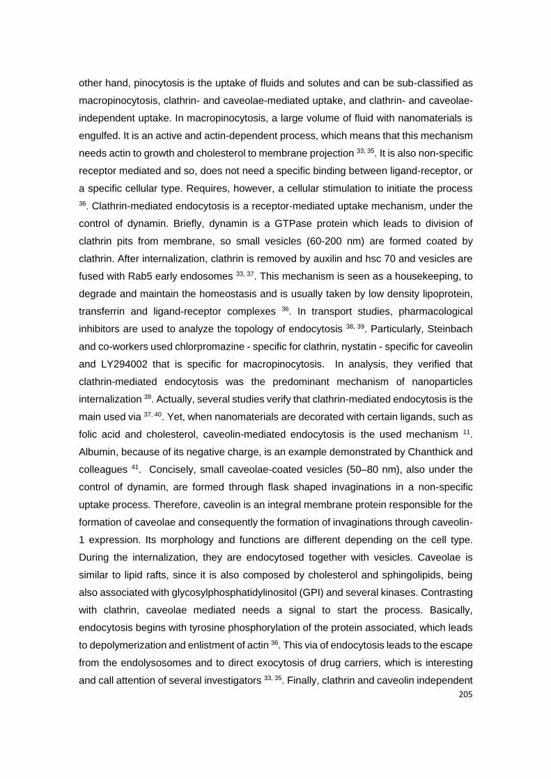
205
other hand, pinocytosis is the uptake of fluids and solutes and can be sub-classified as
macropinocytosis, clathrin- and caveolae-mediated uptake, and clathrin- and caveolae-
independent uptake. In macropinocytosis, a large volume of fluid with nanomaterials is
engulfed. It is an active and actin-dependent process, which means that this mechanism
needs actin to growth and cholesterol to membrane projection 33, 35. It is also non-specific
receptor mediated and so, does not need a specific binding between ligand-receptor, or
a specific cellular type. Requires, however, a cellular stimulation to initiate the process
36. Clathrin-mediated endocytosis is a receptor-mediated uptake mechanism, under the
control of dynamin. Briefly, dynamin is a GTPase protein which leads to division of
clathrin pits from membrane, so small vesicles (60-200 nm) are formed coated by
clathrin. After internalization, clathrin is removed by auxilin and hsc 70 and vesicles are
fused with Rab5 early endosomes 33, 37. This mechanism is seen as a housekeeping, to
degrade and maintain the homeostasis and is usually taken by low density lipoprotein,
transferrin and ligand-receptor complexes 36. In transport studies, pharmacological
inhibitors are used to analyze the topology of endocytosis 38, 39. Particularly, Steinbach
and co-workers used chlorpromazine - specific for clathrin, nystatin - specific for caveolin
and LY294002 that is specific for macropinocytosis. In analysis, they verified that
clathrin-mediated endocytosis was the predominant mechanism of nanoparticles
internalization 39. Actually, several studies verify that clathrin-mediated endocytosis is the
main used via 37, 40. Yet, when nanomaterials are decorated with certain ligands, such as
folic acid and cholesterol, caveolin-mediated endocytosis is the used mechanism 11.
Albumin, because of its negative charge, is an example demonstrated by Chanthick and
colleagues 41. Concisely, small caveolae-coated vesicles (50–80 nm), also under the
control of dynamin, are formed through flask shaped invaginations in a non-specific
uptake process. Therefore, caveolin is an integral membrane protein responsible for the
formation of caveolae and consequently the formation of invaginations through caveolin-
1 expression. Its morphology and functions are different depending on the cell type.
During the internalization, they are endocytosed together with vesicles. Caveolae is
similar to lipid rafts, since it is also composed by cholesterol and sphingolipids, being
also associated with glycosylphosphatidylinositol (GPI) and several kinases. Contrasting
with clathrin, caveolae mediated needs a signal to start the process. Basically,
endocytosis begins with tyrosine phosphorylation of the protein associated, which leads
to depolymerization and enlistment of actin 36. This via of endocytosis leads to the escape
from the endolysosomes and to direct exocytosis of drug carriers, which is interesting
and call attention of several investigators 33, 35. Finally, clathrin and caveolin independent

206
transport occurs via non-specific endocytosis, not needing a receptor and also
characterized as dynamin in- and dependent pathway 3, 36. In this case, EIPA is normally
used as transport inhibitor 42.
A recent and very promising advance biomanufacturing for intracellular delivery of
nanomaterials is microfluidics, a membrane disruption method. Microfluidics is a
technology that improves the quality of the delivery system and establishes a relationship
between structure-function of nanomaterials, presenting also as advantages the uniform-
sized emulsion droplet and the understanding of the intracellular delivery barriers.
However, it is not clear yet if it is possible to scale-up 43. The microfluidic device allows
passing single cells, through parallel channels, into a thin constriction. This approach
allows creating transient pores in the plasmatic membrane, which enhance the
intracellular delivery via mechanoporation 44. Authors already demonstrated it efficiency
in intracellular delivery of NPs loaded with dexamethasone, and also studied the effect
of NPs design in stem cells fate 45. Others choose this strategy to prevent the rapid
clearance and consequent loss of microbubbles, stable producing them, with flow-
focusing microfluidic devices, in real time and near to the therapeutic site 46.
3.2. Nanomaterials characteristcs and their influence
Internalization and intracellular transport mechanisms are affected by nanomaterials
properties (Figure 1). Different characteristics (size, charge, shape, elasticity) contribute
to a different interaction between the nanomaterial and the cell membrane, causing
altered signaling events and activation of different pathways of adhesion and
internalization. Thus, the desired result, for more effective and distinctly targeted
delivery, can be obtained controlling these parameters, which are important to attain the
target and specific activity at a predetermined rate and time 1, 15.
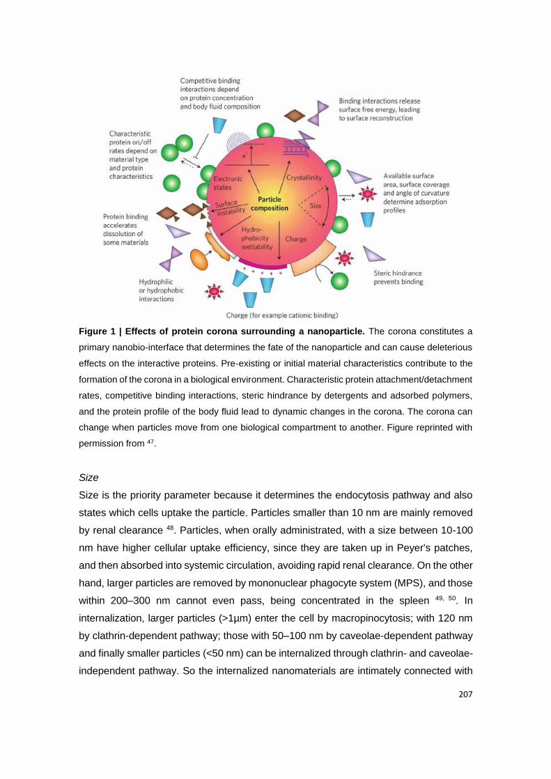
207
Figure 1 | Effects of protein corona surrounding a nanoparticle. The corona constitutes a
primary nanobio-interface that determines the fate of the nanoparticle and can cause deleterious
effects on the interactive proteins. Pre‑existing or initial material characteristics contribute to the
formation of the corona in a biological environment. Characteristic protein attachment/detachment
rates, competitive binding interactions, steric hindrance by detergents and adsorbed polymers,
and the protein profile of the body fluid lead to dynamic changes in the corona. The corona can
change when particles move from one biological compartment to another. Figure reprinted with
permission from 47.
Size
Size is the priority parameter because it determines the endocytosis pathway and also
states which cells uptake the particle. Particles smaller than 10 nm are mainly removed
by renal clearance 48. Particles, when orally administrated, with a size between 10-100
nm have higher cellular uptake efficiency, since they are taken up in Peyer's patches,
and then absorbed into systemic circulation, avoiding rapid renal clearance. On the other
hand, larger particles are removed by mononuclear phagocyte system (MPS), and those
within 200–300 nm cannot even pass, being concentrated in the spleen 49, 50. In
internalization, larger particles (>1µm) enter the cell by macropinocytosis; with 120 nm
by clathrin-dependent pathway; those with 50–100 nm by caveolae-dependent pathway
and finally smaller particles (<50 nm) can be internalized through clathrin- and caveolae-
independent pathway. So the internalized nanomaterials are intimately connected with
208
the size of vacuole formed 20.
A penetration enhancer can be added to the macromolecules with high molecular weight,
such as dimethyl palmitoyl ammonio propanesulfonate (PPS), a zwitterionic surfactant.
According to microscopy studies, PPS allows macromolecules to diffuse through
paracellular and transcellular pathway, improving 45-fold of their bioavailability 51. Also,
polyacrylate, chitosan 52, phytic acid 53 and self-assembling lipid-like peptides 54 work as
enhancers, as well as thiomers and lectins 55. These enhancers have the ability to
reversibly open tight junctions, increasing the paracellular transport of macromolecular
drugs. This is due to the down-regulation of occludin and claudin, TJ proteins, besides
the synergism of reductive effects of Ca2+ 56, 57. On the last case, the cations bind with
more affinity to the polyacrylate derivatives, causing an inhibition of lumen enzymes (p.e.
trypsin). Thus the complex polyacrylate-Ca2+ trigger the TJ opening 58, 59. It was also
demonstrated that cyclodextrin improves the macromolecular absorption by the
pulmonary delivery 60. Finally, an alternative strategy is the use of palmitoyl glycol
chitosan hydrogels 61 or the development of liposomes with enhancers to delivery
hydrophilic macromolecules 62.
Surface composition
Nanomaterials surface is also an important aspect, since its composition is in contact
with the cell, which can be affected by hydrophobicity or hydrophilicity. So, this parameter
determines the cellular internalization and intracellular transport and influences the
circulation time and nanocarrier association with cells and their recognition. Hydrophobic
particles can be delivered to immune cells and penetrate into the bilayer membrane,
being easily internalized 11, 34. Normally, polymers are added to nanomaterials to turn
their surface more inert, hydrophilic, avoid immune clearance and increase the blood
circulation time. PEGylation is a strategy to make nanomaterials invisible to
macrophages or phagocytes and to prolong half-life 63. In this case, ethylene glycol
(PEG) chains are covalently conjugated added to nanomaterials, conferring a hydrophilic
nature 64. According to Schottler S. et al., PEG can capture water molecules and form a
natural barrier around the nanomaterial hindering proteins from adsorption, usually
known as the “stealth” effect 65.
Other strategy is to use biomimetic surfaces, where the nanomaterials surface is
modified with natural membrane coating to avoid phagocytic clearance, prolong drug
circulation and improve their biocompatibility. In this case, markers as CD47 66 or even
from leukocytes are attached to the surface to camouflage nanomaterials, being
209
recognized as ‘self’ 67.
Charge
Also surface charge can be manipulated. For example, intestinal cell membrane is
negatively charged, as plasmatic membrane, due to phospholipids, so positively charged
nanomaterials interact more strongly, enhancing internalization 9. A typical example is
polyethyleneimine (PEI), which is commonly used to permeate membranes and let
nanomaterials enter 68. In terms of transport, positively charged particles utilize clathrin
and macropinocytosis 69 while negatively charged particles tend to utilize caveolae
mediated pathway 23, 41. Neutral or slightly negatively charged particles are considered
the best for long circulation, since they are not susceptible to MPS. On the other hand,
charges highly positive or negative are taken up by macrophages 17, 23, 70.
Shape
Shape can also dictate the path of the particle and, inclusively, can change over time or
after receiving external stimuli for long circulation and surpass barriers. It is an important
factor in flow margination, avoidance of immune system and tissue accumulation.
Spherical particles are better internalized by macrophages, while asymmetrical
nanomaterials are more advantageous for sensing, self-assembly, tissue engineering,
immunoengineering, therapeutic and diagnostic delivery 1, 34. So, one of the strategies,
as mentioned before, is to create elliptical particles to escape macrophages and then,
after a stimuli, change to a sphere form for a better internalization 20, 71.
Elasticity and solubility
Finally, increasing elasticity also increases blood residence time and avoids clearance
by immune system. Soft nanomaterials use macropinocytosis while harder ones are
internalized through clathrin-dependent mechanisms 34, 72.
It is also popular chemically modify nanomaterials to change the fluidity and/or solubility
of mucus, regarding orally administration. According to literature it is possible to add fatty
acids or to substitute amino acids to obtain an increased solubility. The fatty acids
addition is proportional to the increased solubility, which may increase 16-fold with
linoleic acid 73. A similar effect was also observed with capric acid, lauric acid and oleic
acid. These fatty acids increase the hydrophilic transport and reduce the transepithelial
electrical resistance, which indicates the opening of TJ 74. In fact, the paracellular
transport has been related, in several papers, with the transport of poorly absorbed

210
drugs. It was even studied the relation of that event with fatty acids and their effects on
increased absorption of mannitol, a paracellular marker, and on morphology changes of
TJ 75. The amino acid substitution is demonstrated by a group of scientists that introduced
20 amino acids in ribonuclease (RNase) Sa at position 76, confirming that asparagine,
glutamine and threonine have more significantly for protein solubility 76.
In short, it is clear that these characteristics (Table 1) influence the uptake, renal
clearance, drug stability, circulation time, and make nanomaterials release the drug or
even change properties by internal (pH, redox, temperature, enzymes) and external
stimuli (ultrasound, magnetic, light, electrical) 9, 15. Stimuli-responsive nanocarriers,
already reviewed elsewhere 77, are definitely a smart approach for intracellular drug
delivery. Recently, authors inclusively created porous silica nanocarriers sensitive to
three stimulis (protease/redox/pH) for drug delivery 78.
Blanco E and co-workers summarized the main effects of nanoparticle size, shape and
surface charge among different organs including the lungs, liver, spleen and kidneys 11.
Besides the importance of intracellular delivery/targeting, the drug or nanomaterials is
equally significant as well. So, it is crucial imaging and analyze the nanomaterials in
intracellular organs. Nowadays, that is possible using methods like magnetic resonance,
ultrasound, fluorescence PET, phosphorescence, X‑ray, optical coherence tomography,
photoacoustic and Raman, fluorescence and photoacoustic. On the other hand
intracellular delivering can be sensed by electrochemistry or electrophysiology methods,
reviewed elsewhere 79, 80.
In fact, the characteristics manipulation helps non- and functionalized nanomaterials to
reach the target. These two types of nanomaterials will be discussed on the following
section.
Table 1 | Synthesis of the influence of nanomaterials characteristics on cellular uptake
and internalization pathway and their strategies
Characteristic Cellular uptake Internalization
pathway Strategy
Size
<10 nm Renal clearance --- Add a penetration enhancer (PPS, polyacrylate, chitosan, phytic acid, self-assembling lipid-like peptides, thiomers and lectins) to
10-10 nm Peyer’s patches <50 nm: clathrin- and caveolae-independent
100-200 nm Removed by MPS
50-100 nm: caveolae-dependent 120 nm: clathrin-dependent
200-300 nm Spleen Macropinocytosis
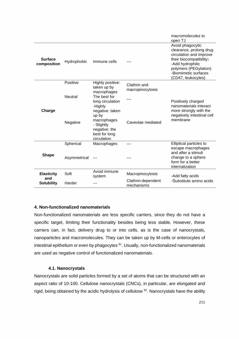
211
macromolecules to open TJ
Surface composition
Hydrophobic Immune cells ---
Avoid phagocytic clearance, prolong drug circulation and improve their biocompatibility: -Add hydrophilic polymers (PEGylation) -Biomimetic surfaces (CD47, leukocytes)
Charge
Positive Highly positive: taken up by macrophages
Clathrin and macropinocytosis
Positively charged nanomaterials interact more strongly with the negatively intestinal cell membrane
Neutral The best for long circulation
---
Negative
-Highly negative: taken up by macrophages - Slightly negative: the best for long circulation
Caveolae mediated
Shape
Spherical Macrophages --- Elliptical particles to escape macrophages and after a stimuli change to a sphere form for a better internalization
Asymmetrical --- ---
Elasticity and
Solubility
Soft Avoid immune system
Macropinocytosis -Add fatty acids
-Substitute amino acids Harder ---
Clathrin-dependent mechanisms
4. Non-functionalized nanomaterials
Non-functionalized nanomaterials are less specific carriers, since they do not have a
specific target, limiting their functionality besides being less stable. However, these
carriers can, in fact, delivery drug to or into cells, as is the case of nanocrystals,
nanoparticles and macromolecules. They can be taken up by M-cells or enterocytes of
intestinal epithelium or even by phagocytes 81. Usually, non-functionalized nanomaterials
are used as negative control of functionalized nanomaterials.
4.1. Nanocrystals
Nanocrystals are solid particles formed by a set of atoms that can be structured with an
aspect ratio of 10-100. Cellulose nanocrystals (CNCs), in particular, are elongated and
rigid, being obtained by the acidic hydrolysis of cellulose 82. Nanocrystals have the ability
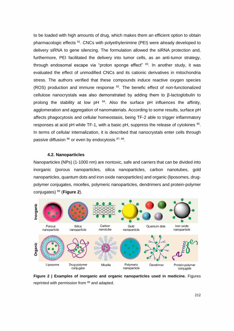
212
to be loaded with high amounts of drug, which makes them an efficient option to obtain
pharmacologic effects 81. CNCs with polyethylenimine (PEI) were already developed to
delivery siRNA to gene silencing. The formulation allowed the siRNA protection and,
furthermore, PEI facilitated the delivery into tumor cells, as an anti-tumor strategy,
through endosomal escape via “proton sponge effect” 83. In another study, it was
evaluated the effect of unmodified CNCs and its cationic derivatives in mitochondria
stress. The authors verified that these compounds induce reactive oxygen species
(ROS) production and immune response 82. The benefic effect of non-functionalized
cellulose nanocrystals was also demonstrated by adding them to β-lactoglobulin to
prolong the stability at low pH 84. Also the surface pH influences the affinity,
agglomeration and aggregation of nanomaterials. According to some results, surface pH
affects phagocytosis and cellular homeostasis, being TF-2 able to trigger inflammatory
responses at acid pH while TF-1, with a basic pH, suppress the release of cytokines 85.
In terms of cellular internalization, it is described that nanocrystals enter cells through
passive diffusion 86 or even by endocytosis 87, 88.
4.2. Nanoparticles
Nanoparticles (NPs) (1-1000 nm) are nontoxic, safe and carriers that can be divided into
inorganic (porous nanoparticles, silica nanoparticles, carbon nanotubes, gold
nanoparticles, quantum dots and iron oxide nanoparticles) and organic (liposomes, drug-
polymer conjugates, micelles, polymeric nanoparticles, dendrimers and protein-polymer
conjugates) 89 (Figure 2).
Figure 2 | Examples of inorganic and organic nanoparticles used in medicine. Figures
reprinted with permission from 89 and adapted.
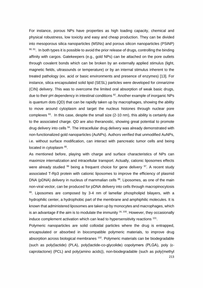
213
For instance, porous NPs have properties as high loading capacity, chemical and
physical robustness, low toxicity and easy and cheap production. They can be divided
into mesoporous silica nanoparticles (MSNs) and porous silicon nanoparticles (PSiNP)
90, 91. In both types it is possible to avoid the prior release of drugs, controlling the binding
affinity with cargos. Gatekeepers (e.g., gold NPs) can be attached on the pore outlets
through covalent bonds which can be broken by an externally applied stimulus (light,
magnetic fields, ultrasounds or temperature) or by an internal stimulus inherent to the
treated pathology (ex. acid or basic environments and presence of enzymes) [13]. For
instance, silica encapsulated solid lipid (SESL) particles were developed for cinnarizine
(CIN) delivery. This was to overcome the limited oral absorption of weak basic drugs,
due to their pH dependency in intestinal conditions 92. Another example of inorganic NPs
is quantum dots (QD) that can be rapidly taken up by macrophages, showing the ability
to move around cytoplasm and target the nucleus histones through nuclear pore
complexes 93. In this case, despite the small size (2-10 nm), this ability is certainly due
to the associated charge. QD are also theranostic, showing great potential to promote
drug delivery into cells 94. The intracellular drug delivery was already demonstrated with
non-functionalized gold nanoparticles (AuNPs). Authors verified that unmodified AuNPs,
i.e. without surface modification, can interact with pancreatic tumor cells and being
located in cytoplasm 95.
As mentioned before, playing with charge and surface characteristics of NPs can
maximize internalization and intracellular transport. Actually, cationic liposomes effects
were already studied 96 being a frequent choice for gene delivery 97. A recent study
associated T-Rp3 protein with cationic liposomes to improve the efficiency of plasmid
DNA (pDNA) delivery in nucleus of mammalian cells 98. Liposomes, as one of the main
non-viral vector, can be produced for pDNA delivery into cells through macropinocytosis
99. Liposomes are composed by 3-4 nm of lamellar phospholipid bilayers, with a
hydrophilic center, a hydrophobic part of the membrane and amphiphilic molecules. It is
known that administered liposomes are taken up by monocytes and macrophages, which
is an advantage if the aim is to modulate the immunity 16, 100. However, they occasionally
induce complement activation which can lead to hypersensitivity reactions 101.
Polymeric nanoparticles are solid colloidal particles where the drug is entrapped,
encapsulated or absorbed in biocompatible polymeric materials, to improve drug
absorption across biological membranes 102. Polymeric materials can be biodegradable
(such as poly(lactide) (PLA), poly(lactide-co-glycolide) copolymers (PLGA), poly (ε-
caprolactone) (PCL) and poly(amino acids)), non-biodegradable (such as poly(methyl
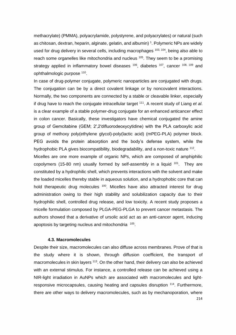
214
methacrylate) (PMMA), polyacrylamide, polystyrene, and polyacrylates) or natural (such
as chitosan, dextran, heparin, alginate, gelatin, and albumin) 1. Polymeric NPs are widely
used for drug delivery in several cells, including macrophages 103, 104, being also able to
reach some organelles like mitochondria and nucleus 105. They seem to be a promising
strategy applied in inflammatory bowel diseases 106, diabetes 107, cancer 108, 109 and
ophthalmologic purpose 110.
In case of drug-polymer conjugate, polymeric nanoparticles are conjugated with drugs.
The conjugation can be by a direct covalent linkage or by noncovalent interactions.
Normally, the two components are connected by a stable or cleavable linker, especially
if drug have to reach the conjugate intracellular target 111. A recent study of Liang et al.
is a clear example of a stable polymer-drug conjugate for an enhanced anticancer effect
in colon cancer. Basically, these investigators have chemical conjugated the amine
group of Gemcitabine (GEM; 2′,2′difluorodeoxycytidine) with the PLA carboxylic acid
group of methoxy poly(ethylene glycol)-poly(lactic acid) (mPEG-PLA) polymer block.
PEG avoids the protein absorption and the body’s defense system, while the
hydrophobic PLA gives biocompatibility, biodegradability, and a non-toxic nature 112.
Micelles are one more example of organic NPs, which are composed of amphiphilic
copolymers (15-80 nm) usually formed by self-assembly in a liquid 101. They are
constituted by a hydrophilic shell, which prevents interactions with the solvent and make
the loaded micelles thereby stable in aqueous solution, and a hydrophobic core that can
hold therapeutic drug molecules 100. Micelles have also attracted interest for drug
administration owing to their high stability and solubilization capacity due to their
hydrophilic shell, controlled drug release, and low toxicity. A recent study proposes a
micelle formulation composed by PLGA-PEG-PLGA to prevent cancer metastasis. The
authors showed that a derivative of ursolic acid act as an anti-cancer agent, inducing
apoptosis by targeting nucleus and mitochondria 105.
4.3. Macromolecules
Despite their size, macromolecules can also diffuse across membranes. Prove of that is
the study where it is shown, through diffusion coefficient, the transport of
macromolecules in skin layers 113. On the other hand, their delivery can also be achieved
with an external stimulus. For instance, a controlled release can be achieved using a
NIR-light irradiation in AuNPs which are associated with macromolecules and light-
responsive microcapsules, causing heating and capsules disruption 114. Furthermore,
there are other ways to delivery macromolecules, such as by mechanoporation, where
215
pores are formed to allow the access 115. A common strategy is the conjugation of
macromolecules to mucoadhesive polymers, such as chitosan, to open the tight
junctions, which facilitates the paracellular transport 116.
The mechanisms of non-functionalized nanomaterials internalization are not yet well
established, so more investigation is needed. Indeed, reports within this topic are scarce
due to the limitations of non-functionalization, being almost mandatory an improvement
in this sense. Thus, functionalization is fundamental in several biomedical applications,
guaranteeing more specificity.
5. Functionalized nanomaterials
Functionalized nanomaterials are directed to a specific target, improving their effect and
minimizing unwanted effects by driving drugs towards the exact site of action. Inclusively,
the upgrading of their ADME (absorption, distribution, metabolism, and excretion),
enhance the therapeutic efficiency 16, 20.
Active targeting of nanomaterials is commonly referred when drugs reach the right target
through the site-specific targeting. This means that there is an interaction between the
targeting ligands on nanocarriers and the receptors on cell membranes. The recognition
performed during the interaction is specific, promoting particles internalization 117, 118. So,
modifying nanomaterials surface with certain ligands is an option to improve cell uptake
and endosomal escape 39. Table 2 resumes some receptors mentioned in recent studies,
such as CD31, integrin β3 and transferrin 119, TLR2, TLR3 and TLR9 120, αvβ3 integrin
121, transferrin 122, EGFR 123, CD44 124, IGFR 125,FcRn 126, CD163 127, biotin 128, folate 129
and vitamin B12 130.
Cell penetrating peptides (CPPs) are a group of peptides (10-30 a.a.) with the ability to
promote the molecular transport and are important to enhance the intracellular uptake.
CPPs can be conjugated via covalent binding to protein drugs and it is believed that
endocytosis is involved on their translocation. Since they are typically positively charged,
CPPs easily have electrostatic interactions with negatively charged cells, such as
glycosaminoglycans (GAGs) and sialic acids, at the beginning of translocation 131.
Authors already developed a CPP-adaptor fusion protein, TAT-calmodulin (TAT-CaM),
which leads to an efficient intracellular delivery and endosomal escape. In this case there
is a reversible and high affinity noncovalent binding that delivery cargo into cytoplasm of
eukaryotic cells 132. Others simply used analogs of the CPP transportan10 conjugated
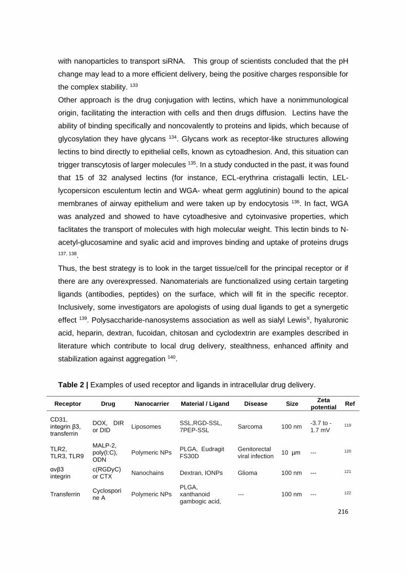
216
with nanoparticles to transport siRNA. This group of scientists concluded that the pH
change may lead to a more efficient delivery, being the positive charges responsible for
the complex stability. 133
Other approach is the drug conjugation with lectins, which have a nonimmunological
origin, facilitating the interaction with cells and then drugs diffusion. Lectins have the
ability of binding specifically and noncovalently to proteins and lipids, which because of
glycosylation they have glycans 134. Glycans work as receptor-like structures allowing
lectins to bind directly to epithelial cells, known as cytoadhesion. And, this situation can
trigger transcytosis of larger molecules 135. In a study conducted in the past, it was found
that 15 of 32 analysed lectins (for instance, ECL-erythrina cristagalli lectin, LEL-
lycopersicon esculentum lectin and WGA- wheat germ agglutinin) bound to the apical
membranes of airway epithelium and were taken up by endocytosis 136. In fact, WGA
was analyzed and showed to have cytoadhesive and cytoinvasive properties, which
faclitates the transport of molecules with high molecular weight. This lectin binds to N-
acetyl-glucosamine and syalic acid and improves binding and uptake of proteins drugs
137, 138.
Thus, the best strategy is to look in the target tissue/cell for the principal receptor or if
there are any overexpressed. Nanomaterials are functionalized using certain targeting
ligands (antibodies, peptides) on the surface, which will fit in the specific receptor.
Inclusively, some investigators are apologists of using dual ligands to get a synergetic
effect 139. Polysaccharide-nanosystems association as well as sialyl LewisX, hyaluronic
acid, heparin, dextran, fucoidan, chitosan and cyclodextrin are examples described in
literature which contribute to local drug delivery, stealthness, enhanced affinity and
stabilization against aggregation 140.
Table 2 | Examples of used receptor and ligands in intracellular drug delivery.
Receptor Drug Nanocarrier Material / Ligand Disease Size Zeta
potential Ref
CD31, integrin β3, transferrin
DOX, DIR or DID
Liposomes SSL,RGD-SSL, 7PEP-SSL
Sarcoma 100 nm -3.7 to -1.7 mV
119
TLR2, TLR3, TLR9
MALP-2, poly(I:C), ODN
Polymeric NPs PLGA, Eudragit FS30D
Genitorectal viral infection
10 µm --- 120
αvβ3 integrin
c(RGDyC) or CTX
Nanochains Dextran, IONPs Glioma 100 nm --- 121
Transferrin Cyclosporine A
Polymeric NPs PLGA, xanthanoid gambogic acid,
--- 100 nm --- 122
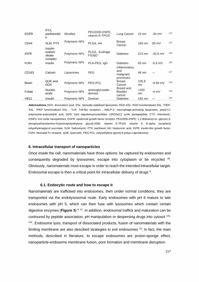
217
Abbreviations DOX: doxorubicin acid, SSL: sterically-stabilized liposomes, RGD-SSL: RGD functionalized SSL, 7PEP-
SSL: 7PEP functionalized SSL , TLR: Toll-like receptors , MALP-2: macrophage-activating lipoprotein, poly(I:C):
polyinosine-polycytidylic acid, ODN: CpG oligodeoxynucleotides, c(RGDyC): cyclic pentapeptide, CTX: chlorotoxin,
IONPs: iron oxide nanoparticles, EGFR: epidermal growth factor receptor, PEG2000-DSPE: 1,2-distearoyl-sn -glycero-3-
phosphoethanolamine-N-[amino(polyethylene glycol)-2000, vitamin E-TPGS: vitamin E D-alpha tocopheryl
polyethyleneglycol succinate, SLM: Salinomycin, PTX: paclitaxel, HA: Hyaluronic acid, IGFR: insulin-like growth factor,
FcRn: Neonatal Fc receptor, QUE: Quercetin, PEG-PCL: poly(ethylene glycol)-b-poly(ε-caprolactone).
6. Intracellular transport of nanoparticles
Once inside the cell, nanomaterials have three options: be captured by endosomes and
consequently degraded by lysosomes; escape into cytoplasm or be recycled 39.
Obviously, nanomaterials must escape in order to reach the intended intracellular target.
Endosomal escape is then a critical point for intracellular delivery of drugs 9.
6.1. Endocytic route and how to escape it
Nanomaterials are trafficked into endosomes, then under normal conditions, they are
transported via the endolysosomal route. Early endosomes with pH 6 mature to late
endosomes with pH 5, which can then fuse with lysosomes which contain certain
digestive enzymes (Figure 3) 9, 12. In addition, endosomal traffick and maturation can be
contoured by peptide association, pH manipulation or despersing drugs into cytosol 141,
142. Endosome lysis, transport of dissociated products, fusion of nanomaterials with the
limiting membrane are also descibed strategies to exit endosomes 19. In fact, the main
methods, described in literature, to escape endosomes are proton-sponge effect,
nanoparticle-endosome membrane fusion, pore formation and membrane disruption.
EGFR PTX, parthenolide
Micelles PEG2000-DSPE, vitamin E-TPGS
Lung Cancer 15 nm -34 mV 123
CD44 SLM, PTX Polymeric NPs
PLGA, HA Breast Cancer
150 nm 50 mV 124
IGFR
Insulin- sodium oleate complex
Polymeric NPs PLGA, Eudragit FS30D
Diabetes 213 nm -32.6 mV 125
FcRn Insulin Polymeric NPs PLA-PEG, IgG Diabetes 63 nm -5.6 mV 126
CD163 Calcein Liposomes PEG
Inflammatory and malignant processes
46 nm --- 127
Biotin QUE and DOX
Polymeric NPs PEG-PCL Breast Cancer
105.8 nm
-9.56 mV 128
Folate Nucleic acids
Polymeric NPs aminoglycoside-derived
Breast and Bladder cancer
<200 nm
-4 mV 129
VB12 Insulin Polymeric NPs Dextran Diabetes 192 nm --- 130
218
Specifically, the proton-sponge effect causes acidification, through translocation of
protons into the endosomes, causing osmosis, lysis and release of content. Polyamine-
based polymers present a pKa around 5-7 that leads to the increased ions influx by
inflow of water and consequently endosomal membrane rupture. The most
representative polymer is polyethylenimine (PEI) 20, 36. In nanoparticle-endosome
membrane fusion, fusogenic lipids are incorporated in the carriers. Fusogenic lipids are
essentialy constituted by alanine, glycine, as well as certain hydrophobic residues. For
instance 1,2-dioleoyl-sn-glycero-3-phosphoethanolamine (DOPE) is acidresponsive, so
the acid pH drives to glutamate and aspartate protonation, resulting in a new hexagonal
conformational. This means that the reposition facilitates the insertion of fusogenic lipids
between juxtaposed membranes and distroys the lipid packing 20, 36. In pore formation,
after peptides binding in α-helical structure, they are obliquely inserted into the
hydrophobic core of endosomal membrane, which stabilizes the transmembrane pore,
and then oriented by their hydrophilic part. On the other hand, that does not happen in
membrane disruption process. In this case, peptides bind to the hydrophilic part of
phospholipids; suffer a reorientation of the hydrophobic residues, gaining an amphipathic
α-helical structure, which allows peptides to cover the membrane like a carpet; and then
there is a disintegration by micellization of membrane 36.
As a practical example, Berguig and colleagues created antibody–peptide drug
conjugates to suppress tumor growth and prolong halflife with low-dose cytotoxicity. The
polymer was constituted with two blocks: the first one to improve pharmacokinetics and
the second to allow endosomal escape, containing a pH-responsive composition. With
the second block they were able to form the micelle at physiological pH and disrupt the
membrane at endosomal pH, promoting endosomal escape and intracellular delivery of
the first block 143. Recently the hydrophobic amino acid R groups in viral escape from
endosomes were also used 144; Ca2+-siRNA nanocomplexes, where electrotastic
interactions lead to siRNA release into the cytoplasm 145; and TMab4 variant with 3-fold
improved endosomal escape through membrane pores 142. An intersting study about
disulfide-bonded dimer of TAT (dfTAT), found at cytosol and nucleus, shows that it is
essential to cell penetration but also to membrane leakage. In this study, Erazo-Oliveras
and co-workers demonstrated that dfTAT interacting with
bis(monoacylglycero)phosphate (BMP), present in late endosomes, mediates the
endosomal leakage 111.
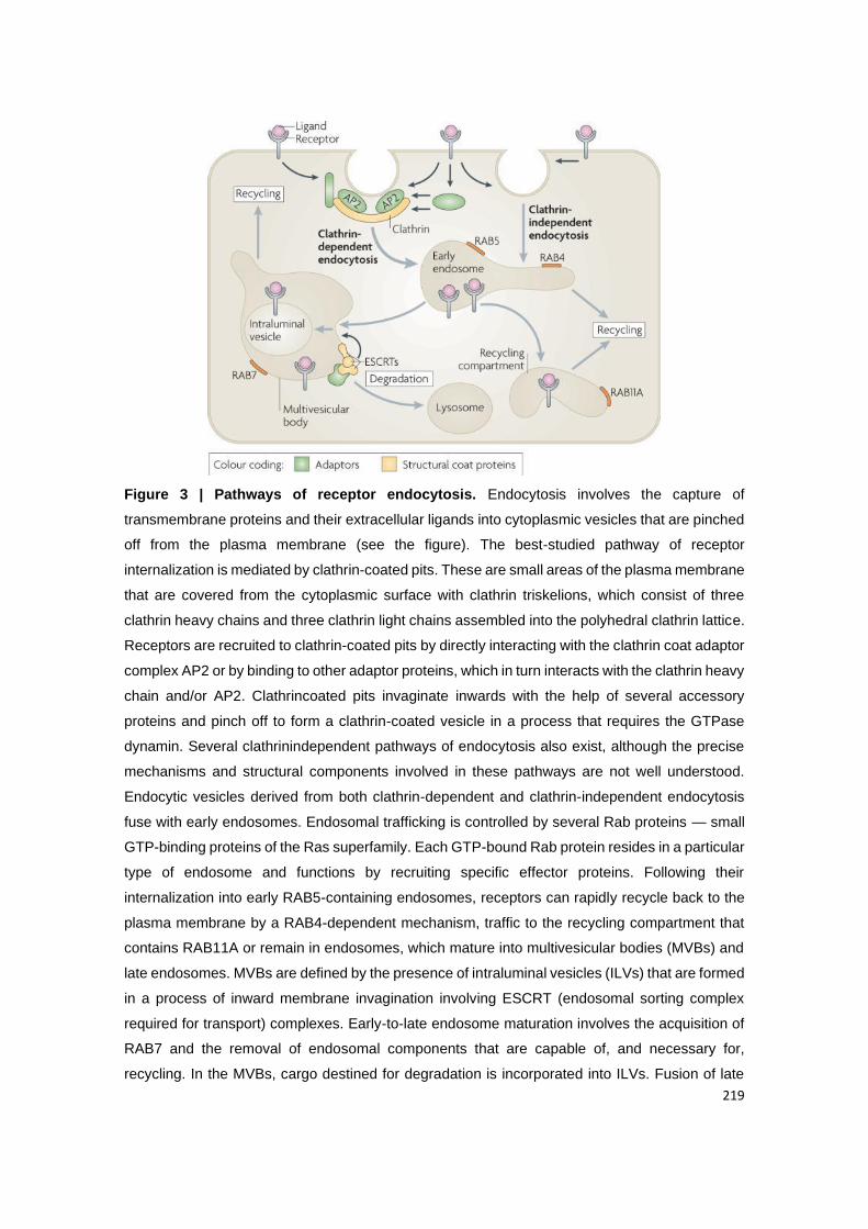
219
Figure 3 | Pathways of receptor endocytosis. Endocytosis involves the capture of
transmembrane proteins and their extracellular ligands into cytoplasmic vesicles that are pinched
off from the plasma membrane (see the figure). The best-studied pathway of receptor
internalization is mediated by clathrin-coated pits. These are small areas of the plasma membrane
that are covered from the cytoplasmic surface with clathrin triskelions, which consist of three
clathrin heavy chains and three clathrin light chains assembled into the polyhedral clathrin lattice.
Receptors are recruited to clathrin-coated pits by directly interacting with the clathrin coat adaptor
complex AP2 or by binding to other adaptor proteins, which in turn interacts with the clathrin heavy
chain and/or AP2. Clathrincoated pits invaginate inwards with the help of several accessory
proteins and pinch off to form a clathrin-coated vesicle in a process that requires the GTPase
dynamin. Several clathrinindependent pathways of endocytosis also exist, although the precise
mechanisms and structural components involved in these pathways are not well understood.
Endocytic vesicles derived from both clathrin-dependent and clathrin-independent endocytosis
fuse with early endosomes. Endosomal trafficking is controlled by several Rab proteins — small
GTP-binding proteins of the Ras superfamily. Each GTP-bound Rab protein resides in a particular
type of endosome and functions by recruiting specific effector proteins. Following their
internalization into early RAB5-containing endosomes, receptors can rapidly recycle back to the
plasma membrane by a RAB4-dependent mechanism, traffic to the recycling compartment that
contains RAB11A or remain in endosomes, which mature into multivesicular bodies (MVBs) and
late endosomes. MVBs are defined by the presence of intraluminal vesicles (ILVs) that are formed
in a process of inward membrane invagination involving ESCRT (endosomal sorting complex
required for transport) complexes. Early-to-late endosome maturation involves the acquisition of
RAB7 and the removal of endosomal components that are capable of, and necessary for,
recycling. In the MVBs, cargo destined for degradation is incorporated into ILVs. Fusion of late
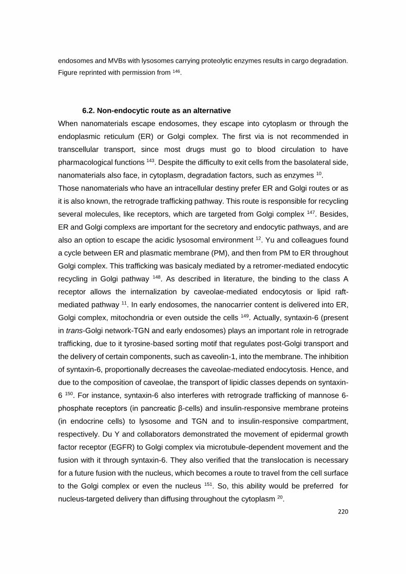
220
endosomes and MVBs with lysosomes carrying proteolytic enzymes results in cargo degradation.
Figure reprinted with permission from 146.
6.2. Non-endocytic route as an alternative
When nanomaterials escape endosomes, they escape into cytoplasm or through the
endoplasmic reticulum (ER) or Golgi complex. The first via is not recommended in
transcellular transport, since most drugs must go to blood circulation to have
pharmacological functions 143. Despite the difficulty to exit cells from the basolateral side,
nanomaterials also face, in cytoplasm, degradation factors, such as enzymes 10.
Those nanomaterials who have an intracellular destiny prefer ER and Golgi routes or as
it is also known, the retrograde trafficking pathway. This route is responsible for recycling
several molecules, like receptors, which are targeted from Golgi complex 147. Besides,
ER and Golgi complexs are important for the secretory and endocytic pathways, and are
also an option to escape the acidic lysosomal environment 12. Yu and colleagues found
a cycle between ER and plasmatic membrane (PM), and then from PM to ER throughout
Golgi complex. This trafficking was basicaly mediated by a retromer-mediated endocytic
recycling in Golgi pathway 148. As described in literature, the binding to the class A
receptor allows the internalization by caveolae-mediated endocytosis or lipid raft-
mediated pathway 11. In early endosomes, the nanocarrier content is delivered into ER,
Golgi complex, mitochondria or even outside the cells 149. Actually, syntaxin-6 (present
in trans-Golgi network-TGN and early endosomes) plays an important role in retrograde
trafficking, due to it tyrosine-based sorting motif that regulates post-Golgi transport and
the delivery of certain components, such as caveolin-1, into the membrane. The inhibition
of syntaxin-6, proportionally decreases the caveolae-mediated endocytosis. Hence, and
due to the composition of caveolae, the transport of lipidic classes depends on syntaxin-
6 150. For instance, syntaxin-6 also interferes with retrograde trafficking of mannose 6-
phosphate receptors (in pancreatic β-cells) and insulin-responsive membrane proteins
(in endocrine cells) to lysosome and TGN and to insulin-responsive compartment,
respectively. Du Y and collaborators demonstrated the movement of epidermal growth
factor receptor (EGFR) to Golgi complex via microtubule-dependent movement and the
fusion with it through syntaxin-6. They also verified that the translocation is necessary
for a future fusion with the nucleus, which becomes a route to travel from the cell surface
to the Golgi complex or even the nucleus 151. So, this ability would be preferred for
nucleus-targeted delivery than diffusing throughout the cytoplasm 20.
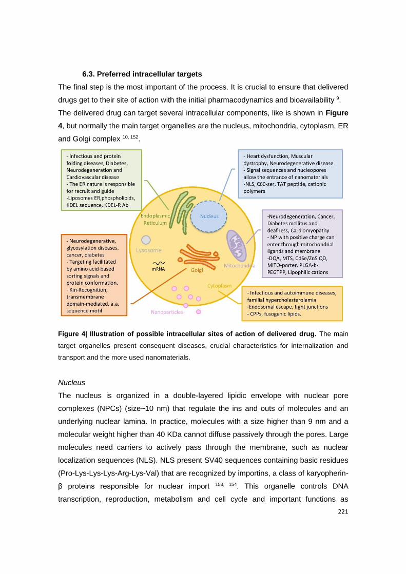
221
6.3. Preferred intracellular targets
The final step is the most important of the process. It is crucial to ensure that delivered
drugs get to their site of action with the initial pharmacodynamics and bioavailability 9.
The delivered drug can target several intracellular components, like is shown in Figure
4, but normally the main target organelles are the nucleus, mitochondria, cytoplasm, ER
and Golgi complex 10, 152.
Figure 4| Illustration of possible intracellular sites of action of delivered drug. The main
target organelles present consequent diseases, crucial characteristics for internalization and
transport and the more used nanomaterials.
Nucleus
The nucleus is organized in a double-layered lipidic envelope with nuclear pore
complexes (NPCs) (size~10 nm) that regulate the ins and outs of molecules and an
underlying nuclear lamina. In practice, molecules with a size higher than 9 nm and a
molecular weight higher than 40 KDa cannot diffuse passively through the pores. Large
molecules need carriers to actively pass through the membrane, such as nuclear
localization sequences (NLS). NLS present SV40 sequences containing basic residues
(Pro-Lys-Lys-Lys-Arg-Lys-Val) that are recognized by importins, a class of karyopherin-
β proteins responsible for nuclear import 153, 154. This organelle controls DNA
transcription, reproduction, metabolism and cell cycle and important functions as
222
electron transport, ATP synthesis, ROS generation, genetic programming and cellular
signaling regulation may also be affected. According to literature, it is possible to use
nanomaterials smaller than nucleus pores, apply a positive charge to the nuclear
membrane, interact with nuclear signals or even try molecules with high affinity for DNA,
like C60-ser, TAT peptide 7. Being the most important organelle, it has a crucial role in
the treatment of heart diseases, dystrophy, neurodegenerative diseases, cancer and
infection, where occurred gene mutation. A strategy to overcome nuclear membrane is
to use positive charge in nanomaterials surface, as mentioned before 10, 20. Induce
permeabilization or even use nanomaterials coated with certain molecules that are
recognized by nuclear receptors present in cytoplasm, like dexamethasone which is
identified by glucocorticoid receptors, may be a way to interfere with DNA transcription
and genetic programming 154. Actually, there are various systems for gene delivery,
which is the most common way for nucleus targeting 155. For instance, micelles with two
synergetic anti-cancer drugs co-encapsulated into hyaluronic acid-vitamin E succinate
were already produced. The nanoformulation was internalized via caveolae-mediated
endocytosis and accumulated in the nucleus. Then cell apoptosis was confirmed and
reverse multidrug resistance via CD44 targeting 156. Some scientists also created PLGA
nanoparticles coated with AS1411 aptamer (Apt) and loaded them with
superparamagnetic iron oxide nanoparticles (SPIONs) with DOX. The secret, in this
case, is Apt that targets to nucleolin. Nucleolin proteins are present in the nucleus and
have an important role in angiogenesis and cell proliferation and adhesion. This fact is
very useful in cancer, especially when it is associated with DOX, a DNA intercalating
agent 157. Izeddin et al. developed a technique to track protein molecules in the nucleus
and determine transcription factors (TFs) dynamics. They found that c-Myc and P-TEFb
are a global and local explorer of the nucleus, respectively. As the authors say, the
control of TFs influence gene regulation, proteins reaction and function in the nucleus,
which may be an advantage 158. Another interesting approach is NPs of PEG-benzoic
imine-oligo-L-lysine/iridium (III) metallodrug complex with pH-activated size reduction.
Meaning, at pH 7 they are large and protected by PEG shell. But at acid pH of
endosomes, NPs stay smaller due to the dissociation and PEG detachment. After this,
smaller NPs present oligo-L-lysine that works as a nuclear localization, favoring
translocation into the nucleus 159.
Mitochondria
This organelle is characterized by an external membrane, intermembrane space, an
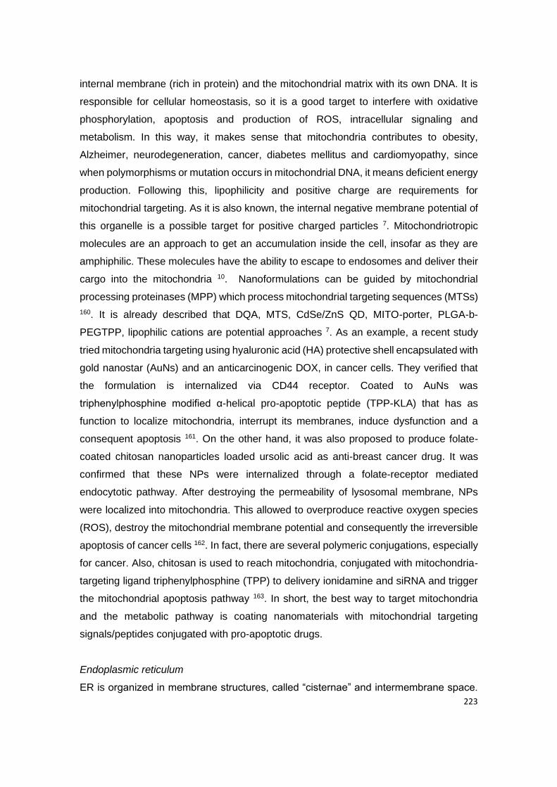
223
internal membrane (rich in protein) and the mitochondrial matrix with its own DNA. It is
responsible for cellular homeostasis, so it is a good target to interfere with oxidative
phosphorylation, apoptosis and production of ROS, intracellular signaling and
metabolism. In this way, it makes sense that mitochondria contributes to obesity,
Alzheimer, neurodegeneration, cancer, diabetes mellitus and cardiomyopathy, since
when polymorphisms or mutation occurs in mitochondrial DNA, it means deficient energy
production. Following this, lipophilicity and positive charge are requirements for
mitochondrial targeting. As it is also known, the internal negative membrane potential of
this organelle is a possible target for positive charged particles 7. Mitochondriotropic
molecules are an approach to get an accumulation inside the cell, insofar as they are
amphiphilic. These molecules have the ability to escape to endosomes and deliver their
cargo into the mitochondria 10. Nanoformulations can be guided by mitochondrial
processing proteinases (MPP) which process mitochondrial targeting sequences (MTSs)
160. It is already described that DQA, MTS, CdSe/ZnS QD, MITO-porter, PLGA-b-
PEGTPP, lipophilic cations are potential approaches 7. As an example, a recent study
tried mitochondria targeting using hyaluronic acid (HA) protective shell encapsulated with
gold nanostar (AuNs) and an anticarcinogenic DOX, in cancer cells. They verified that
the formulation is internalized via CD44 receptor. Coated to AuNs was
triphenylphosphine modified α-helical pro-apoptotic peptide (TPP-KLA) that has as
function to localize mitochondria, interrupt its membranes, induce dysfunction and a
consequent apoptosis 161. On the other hand, it was also proposed to produce folate-
coated chitosan nanoparticles loaded ursolic acid as anti-breast cancer drug. It was
confirmed that these NPs were internalized through a folate-receptor mediated
endocytotic pathway. After destroying the permeability of lysosomal membrane, NPs
were localized into mitochondria. This allowed to overproduce reactive oxygen species
(ROS), destroy the mitochondrial membrane potential and consequently the irreversible
apoptosis of cancer cells 162. In fact, there are several polymeric conjugations, especially
for cancer. Also, chitosan is used to reach mitochondria, conjugated with mitochondria-
targeting ligand triphenylphosphine (TPP) to delivery ionidamine and siRNA and trigger
the mitochondrial apoptosis pathway 163. In short, the best way to target mitochondria
and the metabolic pathway is coating nanomaterials with mitochondrial targeting
signals/peptides conjugated with pro-apoptotic drugs.
Endoplasmic reticulum
ER is organized in membrane structures, called “cisternae” and intermembrane space.
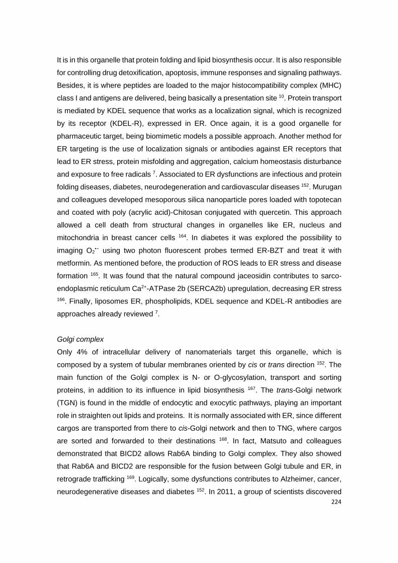
224
It is in this organelle that protein folding and lipid biosynthesis occur. It is also responsible
for controlling drug detoxification, apoptosis, immune responses and signaling pathways.
Besides, it is where peptides are loaded to the major histocompatibility complex (MHC)
class I and antigens are delivered, being basically a presentation site 10. Protein transport
is mediated by KDEL sequence that works as a localization signal, which is recognized
by its receptor (KDEL-R), expressed in ER. Once again, it is a good organelle for
pharmaceutic target, being biomimetic models a possible approach. Another method for
ER targeting is the use of localization signals or antibodies against ER receptors that
lead to ER stress, protein misfolding and aggregation, calcium homeostasis disturbance
and exposure to free radicals 7. Associated to ER dysfunctions are infectious and protein
folding diseases, diabetes, neurodegeneration and cardiovascular diseases 152. Murugan
and colleagues developed mesoporous silica nanoparticle pores loaded with topotecan
and coated with poly (acrylic acid)-Chitosan conjugated with quercetin. This approach
allowed a cell death from structural changes in organelles like ER, nucleus and
mitochondria in breast cancer cells 164. In diabetes it was explored the possibility to
imaging O2•− using two photon fluorescent probes termed ER-BZT and treat it with
metformin. As mentioned before, the production of ROS leads to ER stress and disease
formation 165. It was found that the natural compound jaceosidin contributes to sarco-
endoplasmic reticulum Ca2+-ATPase 2b (SERCA2b) upregulation, decreasing ER stress
166. Finally, liposomes ER, phospholipids, KDEL sequence and KDEL-R antibodies are
approaches already reviewed 7.
Golgi complex
Only 4% of intracellular delivery of nanomaterials target this organelle, which is
composed by a system of tubular membranes oriented by cis or trans direction 152. The
main function of the Golgi complex is N- or O-glycosylation, transport and sorting
proteins, in addition to its influence in lipid biosynthesis 167. The trans-Golgi network
(TGN) is found in the middle of endocytic and exocytic pathways, playing an important
role in straighten out lipids and proteins. It is normally associated with ER, since different
cargos are transported from there to cis-Golgi network and then to TNG, where cargos
are sorted and forwarded to their destinations 168. In fact, Matsuto and colleagues
demonstrated that BICD2 allows Rab6A binding to Golgi complex. They also showed
that Rab6A and BICD2 are responsible for the fusion between Golgi tubule and ER, in
retrograde trafficking 169. Logically, some dysfunctions contributes to Alzheimer, cancer,
neurodegenerative diseases and diabetes 152. In 2011, a group of scientists discovered
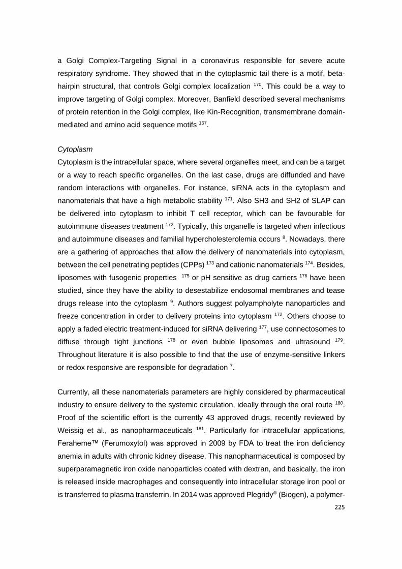
225
a Golgi Complex-Targeting Signal in a coronavirus responsible for severe acute
respiratory syndrome. They showed that in the cytoplasmic tail there is a motif, beta-
hairpin structural, that controls Golgi complex localization 170. This could be a way to
improve targeting of Golgi complex. Moreover, Banfield described several mechanisms
of protein retention in the Golgi complex, like Kin-Recognition, transmembrane domain-
mediated and amino acid sequence motifs 167.
Cytoplasm
Cytoplasm is the intracellular space, where several organelles meet, and can be a target
or a way to reach specific organelles. On the last case, drugs are diffunded and have
random interactions with organelles. For instance, siRNA acts in the cytoplasm and
nanomaterials that have a high metabolic stability 171. Also SH3 and SH2 of SLAP can
be delivered into cytoplasm to inhibit T cell receptor, which can be favourable for
autoimmune diseases treatment 172. Typically, this organelle is targeted when infectious
and autoimmune diseases and familial hypercholesterolemia occurs 8. Nowadays, there
are a gathering of approaches that allow the delivery of nanomaterials into cytoplasm,
between the cell penetrating peptides (CPPs) 173 and cationic nanomaterials 174. Besides,
liposomes with fusogenic properties 175 or pH sensitive as drug carriers 176 have been
studied, since they have the ability to desestabilize endosomal membranes and tease
drugs release into the cytoplasm 9. Authors suggest polyampholyte nanoparticles and
freeze concentration in order to delivery proteins into cytoplasm 172. Others choose to
apply a faded electric treatment-induced for siRNA delivering 177, use connectosomes to
diffuse through tight junctions 178 or even bubble liposomes and ultrasound 179.
Throughout literature it is also possible to find that the use of enzyme-sensitive linkers
or redox responsive are responsible for degradation 7.
Currently, all these nanomaterials parameters are highly considered by pharmaceutical
industry to ensure delivery to the systemic circulation, ideally through the oral route 180.
Proof of the scientific effort is the currently 43 approved drugs, recently reviewed by
Weissig et al., as nanopharmaceuticals 181. Particularly for intracellular applications,
Feraheme™ (Ferumoxytol) was approved in 2009 by FDA to treat the iron deficiency
anemia in adults with chronic kidney disease. This nanopharmaceutical is composed by
superparamagnetic iron oxide nanoparticles coated with dextran, and basically, the iron
is released inside macrophages and consequently into intracellular storage iron pool or
is transferred to plasma transferrin. In 2014 was approved Plegridy® (Biogen), a polymer-
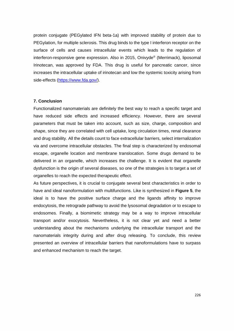
226
protein conjugate (PEGylated IFN beta-1a) with improved stability of protein due to
PEGylation, for multiple sclerosis. This drug binds to the type I interferon receptor on the
surface of cells and causes intracellular events which leads to the regulation of
interferon-responsive gene expression. Also in 2015, Onivyde® (Merrimack), liposomal
Irinotecan, was approved by FDA. This drug is useful for pancreatic cancer, since
increases the intracellular uptake of irinotecan and low the systemic toxicity arising from
side-effects (https://www.fda.gov/).
7. Conclusion
Functionalized nanomaterials are definitely the best way to reach a specific target and
have reduced side effects and increased efficiency. However, there are several
parameters that must be taken into account, such as size, charge, composition and
shape, since they are correlated with cell uptake, long circulation times, renal clearance
and drug stability. All the details count to face extracellular barriers, select internalization
via and overcome intracellular obstacles. The final step is characterized by endosomal
escape, organelle location and membrane translocation. Some drugs demand to be
delivered in an organelle, which increases the challenge. It is evident that organelle
dysfunction is the origin of several diseases, so one of the strategies is to target a set of
organelles to reach the expected therapeutic effect.
As future perspectives, it is crucial to conjugate several best characteristics in order to
have and ideal nanoformulation with multifunctions. Like is synthesized in Figure 5, the
ideal is to have the positive surface charge and the ligands affinity to improve
endocytosis, the retrograde pathway to avoid the lysosomal degradation or to escape to
endosomes. Finally, a biomimetic strategy may be a way to improve intracellular
transport and/or exocytosis. Nevertheless, it is not clear yet and need a better
understanding about the mechanisms underlying the intracellular transport and the
nanomaterials integrity during and after drug releasing. To conclude, this review
presented an overview of intracellular barriers that nanoformulations have to surpass
and enhanced mechanism to reach the target.
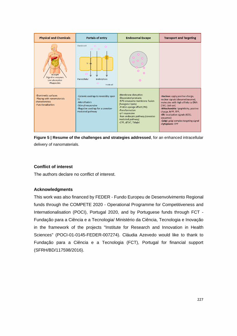
227
Figure 5 | Resume of the challenges and strategies addressed, for an enhanced intracellular
delivery of nanomaterials.
Conflict of interest
The authors declare no conflict of interest.
Acknowledgments
This work was also financed by FEDER - Fundo Europeu de Desenvolvimento Regional
funds through the COMPETE 2020 - Operational Programme for Competitiveness and
Internationalisation (POCI), Portugal 2020, and by Portuguese funds through FCT -
Fundação para a Ciência e a Tecnologia/ Ministério da Ciência, Tecnologia e Inovação
in the framework of the projects "Institute for Research and Innovation in Health
Sciences" (POCI-01-0145-FEDER-007274). Cláudia Azevedo would like to thank to
Fundação para a Ciência e a Tecnologia (FCT), Portugal for financial support
(SFRH/BD/117598/2016).
228
References
1. Banik BL, Fattahi P, Brown JL. Polymeric nanoparticles: the future of nanomedicine.
WIREs Nanomedicine and Nanobiotechnology 2016;8:271–299.
2. Rekha MR, Sharma CP. Oral delivery of therapeutic protein/peptide for diabetes – Future
perspectives. International Journal of Pharmaceutics 2013;440:48-62.
3. Pereira C, Araújo F, Granja PL, et al. Targeting membrane transporters and receptors as
a mean to optimize orally delivered biotecnhological based drugs through nanoparticle
delivery systems. Current Pharmaceutical Biotechnology 2014;15:650-658.
4. Araujo F, Shrestha N, Granja PL, et al. Antihyperglycemic potential of incretins orally
delivered via nano and microsystems and subsequent glucoregulatory effects. Current
Pharmaceutical Biotechnology 2014;15:609-19.
5. Moulari B, Beduneau A, Pellequer Y, et al. Nanoparticle targeting to inflamed tissues of
gastrointestinal tract Current Drug Delivery 2013;10:9-17.
6. Sarmento B, Ferreira D, Vasconcelos T. Polymer-Based Delivery Systems for Oral
Delivery of Peptides and Proteins. In: Jorgensen L, Nielsen HM, eds. Delivery
Technologies for Biopharmaceuticals: Peptides, Proteins, Nucleic Acids and Vaccines:
John Wiley & Sons, Ltd, 2009:207-227.
7. Parodi A, Corbo C, Cevenini A, et al. Enabling cytoplasmic delivery and organelle
targeting by surface modification of nanocarriers. Nanomedicine 2015;10:1923–1940.
8. Sakhrani NM, Padh H. Organelle targeting: third level of drug targeting. Drug Des Devel
Ther 2013;7:585-99.
9. Jhaveri A, Torchilin V. Intracellular delivery of nanocarriers and targeting to subcellular
organelles. Expert Opinion on Drug Delivery 2015;13.
10. Ma X, Gong N, Zhong L, et al. Future of nanotherapeutics: Targeting the cellular sub-
organelles. Biomaterials 2016;97:10-21.
11. Blanco E, Shen H, Ferrari M. Principles of nanoparticle design for overcoming biological
barriers to drug delivery. Nature Biotechnology 2015;9:941-951.
12. Fan W, Xia D, Zhu Q, et al. Intracellular transport of nanocarriers across the intestinal
epithelium. Drug Discovery Today 2016;21:856-863.
13. Lopes M, Shrestha N, Correia A, et al. Dual chitosan/albumin-coated alginate/dextran
sulfate nanoparticles for enhanced oral delivery of insulin. Journal of Controlled Release
2016;232:29–41.
14. Mann SK, Czuba E, Selby LI, et al. Quantifying Nanoparticle Internalization Using a High
Throughput Internalization Assay. Pharmaceutical Research 2016;33:2421-2432.
15. Bennet D, Kim S. Polymer Nanoparticles for Smart Drug Delivery. In: Sezer AD, ed.
Application of Nanotechnology in Drug Delivery: Nanotechnology and Nanomaterials,
2014:257-309.
229
16. Duschl A. Nanomedicine. In: Boraschi D, Penton-Rol G, eds. Immune Rebalancing - The
Future of Immunosuppression: Elsevier, 2016:251-274.
17. Pawar VK, Meher JG, Singh Y, et al. Targeting of gastrointestinal tract for amended
delivery of 3Q4 protein/peptide therapeutics: Strategies and industrial perspectives.
Journal of Controlled Release 2014;196:168-83.
18. Lopes MA, Abrahim BA, Cabral LM, et al. Intestinal absorption of insulin nanoparticles:
Contribution of M cells. Nanomedicine: Nanotechnology, Biology, and Medicine
2014;10:1139–1151.
19. Stewart MP, Sharei A, Ding X, et al. In vitro and ex vivo strategies for intracellular delivery.
Nature 2016;538:183-192.
20. Sun W, Hu Q, Ji W, et al. Leveraging physiology for precision drug delivery. Physiological
Reviews 2017;97:189–225.
21. Agarwal SK, Gupta A. Aquaporins: The renal water channels. Indian J Nephrol
2008;18:95-100.
22. DiMarco RL, Hunt DR, Dewi RE, et al. Improvement of paracellular transport in the Caco-
2 drug screening model using protein-engineered substrates. Biomaterials 2017;129:152-
162.
23. Alai MS, Lin WJ, Pingale SS. Application of polymeric nanoparticles and micelles in
insulin oral delivery. Journal of food and drug analysis 2015;23:351-358.
24. Li M, Zhao M, Fu Y, et al. Enhanced intranasal delivery of mRNA vaccine by overcoming
the nasal epithelial barrier via intra- and paracellular pathways. Journal of Controlled
Release 2016;228:9-19.
25. Bernkop-Schnürch A. Oral Delivery of Macromolecular Drugs: Barriers, Strategies and
Future Trends: Springer New York, 2009.
26. Rahmat D, Sakloetsakun D, Shahnaz G, et al. HEC-cysteamine conjugates: Influence of
degree of thiolation on efflux pump inhibitory and permeation enhancing properties.
International Journal of Pharmaceutics 2012;422:40-46.
27. Clausen AE, Kast CE, Bernkop-Schnürch A. The Role of Glutathione in the Permeation
Enhancing Effect of Thiolated Polymers. Pharmaceutical Research 2002;19:602-608.
28. Bernkop-Schnürch A. Thiomers: A new generation of mucoadhesive polymers. Advanced
Drug Delivery Reviews 2005;57:1569-1582.
29. Chen Y-M, Tu IH, Hu F-R, et al. The role of protein tyrosine phosphorylation in the cell–
cell junctions and intercellular permeability of post-confluent bovine corneal epithelial
cells. Taiwan Journal of Ophthalmology 2013;3:37-41.
30. Palazzo C, Trapani G, Ponchel G, et al. Mucoadhesive properties of low molecular weight
chitosan- or glycol chitosan- and corresponding thiomer-coated
poly(isobutylcyanoacrylate) core-shell nanoparticles. European Journal of
Pharmaceutics and Biopharmaceutics 2017;117:315-323.
230
31. Mahmood A, Bonengel S, Laffleur F, et al. An in-vitro exploration of permeation
enhancement by novel polysulfonate thiomers. International Journal of Pharmaceutics
2015;496:304-313.
32. Dartier J, Lemaitre E, Chourpa I, et al. ATP-dependent activity and mitochondrial
localization of drug efflux pumps in doxorubicin-resistant breast cancer cells. Biochimica
et Biophysica Acta (BBA) - General Subjects 2017;1861:1075-1084.
33. Fonte P, Araújo F, Silva C, et al. Polymer-based nanoparticles for oral insulin delivery:
Revisited approaches. Biotechnology Advances 2015;33:1342–1354.
34. Agarwal R, Roy K. Intracellular delivery of polymeric nanocarriers: a matter of size, shape,
charge, elasticity and surface composition. Therapeutic delivery 2013;4:705–723.
35. Woitiski CB, Carvalho RA, Ribeiro AJ, et al. Strategies Toward the Improved Oral Delivery
of Insulin Nanoparticles via Gastrointestinal Uptake and Translocation. Biodrugs
2008;22:223-237.
36. Mundra V, Mahato RI. Design of nanocarriers for efficient cellular uptake and endosomal
release of small molecule and nucleic acid drugs: learning from virus. Frontiers of
Chemical Science and Engineering 2014;8:387–404.
37. Forte M, Iachetta G, Tussellino M, et al. Polystyrene nanoparticles internalization in
human gastric adenocarcinoma cells. Toxicology in Vitro 2016;31:126-136.
38. Iversen T-G, Skotland T, Sandvig K. Endocytosis and intracellular transport of
nanoparticles: Present knowledge and need for future studies. Nano Today 2011;6:176—
185.
39. Steinbach JM, Seo Y-E, Saltzman WM. Cell penetrating peptide-modified poly(lactic-co-
glycolic acid) nanoparticles with enhanced cell internalization. Acta Biomaterialia
2016;30:49-61.
40. Surassmo S, Saengkrit N, Ruktanonchai UR, et al. Surface modification of PLGA
nanoparticles by carbopol to enhancemucoadhesion and cell internalization. Colloids and
Surfaces B: Biointerfaces 2015;130:229-236.
41. Chanthick C, Kanlaya R, Kiatbumrung R, et al. Caveolae-mediated albumin transcytosis
is enhanced in dengueinfected human endothelial cells: A model of vascular leakage in
dengue hemorrhagic fever. Scientific Reports 2016;6.
42. Forte M, Iachetta G, Tussellino M, et al. Polystyrene nanoparticles internalization in
human gastric adenocarcinoma cells. Toxicology in Vitro 2016;31:126-136.
43. Chan HF, Ma S, Leong KW. Can microfluidics address biomanufacturing challenges in
drug/gene/cell therapies? Regen Biomater 2016;3:87-98.
44. Szeto GL, Van Egeren D, Worku H, et al. Microfluidic squeezing for intracellular antigen
loading in polyclonal B-cells as cellular vaccines. 2015;5:10276.
231
45. Hasani-Sadrabadi MM, Pour Hajrezaei S, Hojjati Emami S, et al. Enhanced osteogenic
differentiation of stem cells via microfluidics synthesized nanoparticles. Nanomedicine:
Nanotechnology, Biology and Medicine 2015;11:1809-1819.
46. Dixon AJ, Dhanaliwala AH, Chen JL, et al. Enhanced Intracellular Delivery of a Model
Drug Using Microbubbles Produced by a Microfluidic Device. Ultrasound in Medicine &
Biology 2013;39:1267-1276.
47. Nel AE, Madler L, Velegol D, et al. Understanding biophysicochemical interactions at the
nano-bio interface. Nat Mater 2009;8:543-557.
48. Soo Choi H, Liu W, Misra P, et al. Renal clearance of quantum dots. Nat Biotech
2007;25:1165-1170.
49. Wahajuddin, Arora S. Superparamagnetic iron oxide nanoparticles: magnetic
nanoplatforms as drug carriers. International Journal of Nanomedicine 2012;7:3445–
3471.
50. Howe SE, Lickteig DJ, Plunkett KN, et al. The Uptake of Soluble and Particulate Antigens
by Epithelial Cells in the Mouse Small Intestine. PLOS ONE 2014;9:e86656.
51. Gupta V, Hwang BH, Doshi N, et al. A permeation enhancer for increasing transport of
therapeutic macromolecules across the intestine. Journal of Controlled Release
2013;172:541-549.
52. Wang J, Kong M, Zhou Z, et al. Mechanism of surface charge triggered intestinal
epithelial tight junction opening upon chitosan nanoparticles for insulin oral delivery.
Carbohydrate Polymers 2017;157:596-602.
53. Fu Q, Wang H, Xia M, et al. The effect of phytic acid on tight junctions in the human
intestinal Caco-2 cell line and its mechanism. European Journal of Pharmaceutical
Sciences 2015;80:1-8.
54. Karavasili C, Spanakis M, Papagiannopoulou D, et al. Bioactive Self-Assembling Lipid-
Like Peptides as Permeation Enhancers for Oral Drug Delivery. Journal of
Pharmaceutical Sciences 2015;104:2304-2311.
55. Dünnhaupt S, Barthelmes J, Köllner S, et al. Thiolated nanocarriers for oral delivery of
hydrophilic macromolecular drugs. Carbohydrate Polymers 2015;117:577-584.
56. Yeh T-H, Hsu L-W, Tseng MT, et al. Mechanism and consequence of chitosan-mediated
reversible epithelial tight junction opening. Biomaterials 2011;32:6164-6173.
57. Chi Y, Zhang X, zhang Z, et al. Connexin43 hemichannels contributes to the disassembly
of cell junctions through modulation of intracellular oxidative status. Redox Biology
2016;9:198-209.
58. Lueßen HL, Rentel CO, Kotzé AF, et al. Mucoadhesive polymers in peroral peptide drug
delivery. IV. Polycarbophil and chitosan are potent enhancers of peptide transport across
intestinal mucosae in vitro. Journal of Controlled Release 1997;45:15-23.
232
59. Borchard G, Lueβen HL, de Boer AG, et al. The potential of mucoadhesive polymers in
enhancing intestinal peptide drug absorption. III: Effects of chitosan-glutamate and
carbomer on epithelial tight junctions in vitro. Journal of Controlled Release 1996;39:131-
138.
60. Zhang H, Huang X, Sun Y, et al. Improvement of pulmonary absorption of poorly
absorbable macromolecules by hydroxypropyl-β-cyclodextrin grafted polyethylenimine
(HP-β-CD-PEI) in rats. International Journal of Pharmaceutics 2015;489:294-303.
61. Martin L, Wilson CG, Koosha F, et al. The release of model macromolecules may be
controlled by the hydrophobicity of palmitoyl glycol chitosan hydrogels. Journal of
Controlled Release 2002;80:87-100.
62. Parmentier J, Hartmann FJ, Fricker G. In vitro evaluation of liposomes containing bio-
enhancers for the oral delivery of macromolecules. European Journal of Pharmaceutics
and Biopharmaceutics 2010;76:394-403.
63. Huang H, Hernandez R, Geng J, et al. A porphyrin-PEG polymer with rapid renal
clearance. Biomaterials 2016;76:25-32.
64. Roberts MJ, Bentley MD, Harris JM. Chemistry for peptide and protein PEGylation.
Advanced Drug Delivery Reviews 2012;64, Supplement:116-127.
65. Schöttler S, Becker G, Winzen S, et al. Protein adsorption is required for stealth effect of
poly(ethylene glycol)- and poly(phosphoester)-coated nanocarriers. Nat Nano
2016;11:372-377.
66. Qie Y, Yuan H, von Roemeling CA, et al. Surface modification of nanoparticles enables
selective evasion of phagocytic clearance by distinct macrophage phenotypes. Scientific
Reports 2016;6:26269.
67. Evangelopoulos M, Parodi A, Martinez JO, et al. Cell source determines the
immunological impact of biomimetic nanoparticles. Biomaterials 2016;82:168-177.
68. Bera A, Jewrajka SK. Tailoring polyamide thin film composite nanofiltration membranes
by polyethyleneimine and its conjugates for the enhancement of selectivity and antifouling
property. RSC Advances 2016;6:4521-4530.
69. Noh SM, Kim W-K, Kim SJ, et al. Enhanced cellular delivery and transfection efficiency
of plasmid DNA using positively charged biocompatible colloidal gold nanoparticles.
Biochimica et Biophysica Acta (BBA) - General Subjects 2007;1770:747-752.
70. Xiao K, Li Y, Luo J, et al. The effect of surface charge on in vivo biodistribution of PEG-
oligocholic acid based micellar nanoparticles. Biomaterials 2011;32:3435-3446.
71. Yoo JW, Mitragotri S. Polymer particles that switch shape in response to a stimulus. Proc
Natl Acad Sci U S A 2010;107:11205-10.
72. Banquy X, Suarez F, Argaw A, et al. Effect of mechanical properties of hydrogel
nanoparticles on macrophage cell uptake. Soft Matter 2009;5:3984-3991.

233
73. O'Reilly JR, Corrigan OI, O'Driscoll CM. The effect of mixed micellar systems, bile
salt/fatty acids, on the solubility and intestinal absorption of clofazimine (B663) in the
anaesthetised rat. International Journal of Pharmaceutics 1994;109:147-154.
74. Pabla D, Akhlaghi F, Zia H. Intestinal permeability enhancement of levothyroxine sodium
by straight chain fatty acids studied in MDCK epithelial cell line. European Journal of
Pharmaceutical Sciences 2010;40:466-472.
75. Aspenström-Fagerlund B, Sundström B, Tallkvist J, et al. Fatty acids increase
paracellular absorption of aluminium across Caco-2 cell monolayers. Chemico-Biological
Interactions 2009;181:272-278.
76. Trevino SR, Scholtz JM, Pace CN. Amino acid contribution to protein solubility: Asp, Glu,
and Ser contribute more favorably than the other hydrophilic amino acids in RNase Sa. J
Mol Biol 2007;366:449-60.
77. Mura S, Nicolas J, Couvreur P. Stimuli-responsive nanocarriers for drug delivery. Nat
Mater 2013;12:991-1003.
78. Qiu L, Zhao Y, Li B, et al. Triple-stimuli (protease/redox/pH) sensitive porous silica
nanocarriers for drug delivery. Sensors and Actuators B: Chemical 2017;240:1066-1074.
79. Park S-m, Aalipour A, Vermesh O, et al. Towards clinically translatable in vivo
nanodiagnostics. Nature Reviews Materials 2017;2:17014.
80. Wang D, Zhao X, Gu Z. Advanced optoelectronic nanodevices and nanomaterials for
sensing inside single living cell. Optics Communications 2017;395:3-15.
81. Müller RH, Gohla S, Keck CM. State of the art of nanocrystals – Special features,
production, nanotoxicology aspects and intracellular delivery. European Journal of
Pharmaceutics and Biopharmaceutics 2011;78:1-9.
82. Sunasee R, Araoye E, Pyram D, et al. Cellulose nanocrystal cationic derivative induces
NLRP3 inflammasome-dependent IL-1β secretion associated with mitochondrial ROS
production. Biochemistry and Biophysics Reports 2015;4:1-9.
83. Ndong Ntoutoume GMA, Grassot V, Brégier F, et al. PEI-cellulose nanocrystal hybrids
as efficient siRNA delivery agents—Synthesis, physicochemical characterization and in
vitro evaluation. Carbohydrate Polymers 2017;164:258-267.
84. Scheuble N, Geue T, Windhab EJ, et al. Tailored Interfacial Rheology for Gastric Stable
Adsorption Layers. Biomacromolecules 2014;15:3139-3145.
85. Boonrungsiman S, Suchaoin W, Chetprayoon P, et al. Shape and surface properties of
titanate nanomaterials influence differential cellular uptake behavior and biological
responses in THP-1 cells. Biochemistry and Biophysics Reports 2017;9:203-210.
86. Junyaprasert VB, Morakul B. Nanocrystals for enhancement of oral bioavailability of
poorly water-soluble drugs. Asian Journal of Pharmaceutical Sciences 2015;10:13-23.
234
87. Li J, Qiu J, Guo W, et al. Cellular internalization of LiNbO3 nanocrystals for second
harmonic imaging and the effects on stem cell differentiation. Nanoscale 2016;8:7416-
7422.
88. Murugan K, Choonara YE, Kumar P, et al. Cellular internalisation kinetics and cytotoxic
properties of statistically designed and optimised neo-geometric copper nanocrystals.
Materials Science and Engineering: C 2017;78:376-388.
89. Tang L, Cheng J. Nonporous silica nanoparticles for nanomedicine application. Nano
Today 2013;8:290—312.
90. Baeza A, Ruiz-Molina D, Vallet-Regí M. Recent advances in porous nanoparticles for
drug delivery in antitumoral applications: inorganic nanoparticles and nanoscale metal-
organic frameworks. Expert Opinion on Drug Delivery 2016:1-14.
91. Santos HA, Mäkilä E, Airaksinen AJ, et al. Porous silicon nanoparticles for nanomedicine:
preparation and biomedical applications. Nanomedicine 2014;9:535–554.
92. Yasmin R, Rao S, Bremmell K, et al. Synergistic role of solid lipid and porous silica in
improving the oral delivery of weakly basic poorly water soluble drugs. European Journal
of Pharmaceutical Sciences 2017;96:508-514.
93. Nabiev I, Mitchell S, Davies A, et al. Nonfunctionalized Nanocrystals Can Exploit a Cell's
Active Transport Machinery Delivering Them to Specific Nuclear and Cytoplasmic
Compartments. Nano Letters 2007;7:3452-3461.
94. Volkov Y. Quantum dots in nanomedicine: recent trends, advances and unresolved
issues. Biochemical and Biophysical Research Communications 2015;468:419-427.
95. Sobhan MA, Sreenivasan VKA, Withford MJ, et al. Non-specific internalization of laser
ablated pure gold nanoparticles in pancreatic tumor cell. Colloids and Surfaces B:
Biointerfaces 2012;92:190-195.
96. Li Y, Wang J, Gao Y, et al. Relationships between Liposome Properties, Cell Membrane
Binding, Intracellular Processing, and Intracellular Bioavailability. Aaps j 2011;13:585-97.
97. Ran R, Liu Y, Gao H, et al. Enhanced gene delivery efficiency of cationic liposomes
coated with PEGylated hyaluronic acid for anti P-glycoprotein siRNA: A potential
candidate for overcoming multi-drug resistance. International Journal of Pharmaceutics
2014;477:590-600.
98. Alves RF, Favaro MTP, Balbino TA, et al. Recombinant protein-based nanocarriers and
their association with cationic liposomes: Characterization and in vitro evaluation.
Colloids and Surfaces A: Physicochemical and Engineering Aspects 2017;513:1-10.
99. Kim BK, Seu YB, Bae YU, et al. Efficient delivery of plasmid DNA using cholesterol-based
cationic lipids containing polyamines and ether linkages. Int J Mol Sci 2014;15:7293-312.
100. Wang Y, Chen L. Quantum dots, lighting up the research and development of
nanomedicine. Nanomedicine: Nanotechnology, Biology, and Medicine 2011;7:385–402.
235
101. KB K, H N, PE K, et al. In vivo toxicity of cationic micelles and liposomes. Nanomedicine
2015;11:467-477.
102. Desai PP, Date AA, Patravale VB. Overcoming poor oral bioavailability using nanoparticle
formulations – opportunities and limitations. Drug Discovery Today: Technologies 2012;9:
e87–e95.
103. Lee SY, Cho H-J. Dopamine-conjugated poly(lactic-co-glycolic acid) nanoparticles for
protein delivery to macrophages. Journal of Colloid and Interface Science 2017;490:391-
400.
104. Vene E, Barouti G, Jarnouen K, et al. Opsonisation of nanoparticles prepared from poly(β-
hydroxybutyrate) and poly(trimethylene carbonate)-b-poly(malic acid) amphiphilic diblock
copolymers: Impact on the in vitro cell uptake by primary human macrophages and
HepaRG hepatoma cells. International Journal of Pharmaceutics 2016;513:438-452.
105. Chen X, Chen J, Li B, et al. PLGA-PEG-PLGA triblock copolymeric micelles as oral drug
delivery system: In vitro drug release and in vivo pharmacokinetics assessment. Journal
of Colloid and Interface Science 2017;490:542–552.
106. Lee S, Kimb Y-C, Parkc J-H. Zein-alginate based oral drug delivery systems: Protection
and release of therapeutic proteins. International Journal of Pharmaceutics
2016;515:300–306.
107. Lin Y-H, Chen C-T, Liang H-F, et al. Novel nanoparticles for oral insulin delivery via the
paracellular pathway. Nanotechnology 2007;18:1-11.
108. Quinteros DA, Ferreira LM, Schaffazick SR, et al. Novel Polymeric Nanoparticles
Intended for Ophthalmic Administration of Acetazolamide. Journal of Pharmaceutical
Sciences 2016;105:3183-3190.
109. Lv Y, Hao L, Hu W, et al. Novel multifunctional pH-sensitive nanoparticles loaded into
microbubbles as drug delivery vehicles for enhanced tumor targeting. Scientific Reports
2016;6.
110. Yousry C, Elkheshen SA, El-laithy HM, et al. Studying the influence of formulation and
process variables on Vancomycin-loaded polymeric nanoparticles as potential carrier for
enhanced ophthalmic delivery. European Journal of Pharmaceutical Sciences
2017;100:142-154.
111. Erazo-Oliveras A, Najjar K, Truong D, et al. The Late Endosome and Its Lipid BMP Act
as Gateways for Efficient Cytosolic Access of the Delivery Agent dfTAT and Its
Macromolecular Cargos. Cell Chemical Biology 2016;23:1-10.
112. Lianga T-J, Zhoub Z-M, Caoc Y-Q, et al. Gemcitabine-based polymer-drug conjugate for
enhanced anticancer effect in colon cancer. International Journal of Pharmaceutics
2016;513:564–571.
236
113. Römgens AM, Bader DL, Bouwstra JA, et al. Diffusion profile of macromolecules within
and between human skin layers for (trans)dermal drug delivery. Journal of the Mechanical
Behavior of Biomedical Materials 2015;50:215-222.
114. Carregal-Romero S, Ochs M, Rivera-Gil P, et al. NIR-light triggered delivery of
macromolecules into the cytosol. Journal of Controlled Release 2012;159:120-127.
115. Matsumoto D, Yamagishi A, Saito M, et al. Mechanoporation of living cells for delivery of
macromolecules using nanoneedle array. Journal of Bioscience and Bioengineering
2016;122:748-752.
116. Sheng J, He H, Han L, et al. Enhancing Insulin Oral Absorption by Using Mucoadhesive
Nanoparticles Loaded with LMWP-linked Insulin Conjugates. Journal of Controlled
Release 2016;233:181- 190.
117. Li N, Huang C, Luan Y, et al. Active targeting co-delivery system based on pH-sensitive
methoxy-poly (ethylene glycol)2K-poly(e-caprolactone)4K-poly(glutamic acid)1K for
enhanced cancer therapy. Journal of Colloid and Interface Science 2016;472:90-98.
118. Li X, Yu M, Fan W, et al. Orally active-targeted drug delivery systems for proteins and
peptides. Expert Opinion Drug Delivery 2014;11:1435-1447.
119. Wang D, Fu J, Shi Y, et al. The modulation of tumor vessel permeability by thalidomide
and its impacts on different types of targeted drug delivery systems in a sarcoma mouse
model. Journal of Controlled Release 2016;238:186–196.
120. Zhu Q, Talton J, Zhang G, et al. Large intestine–targeted, nanoparticle-releasing oral
vaccine to control genitorectal viral infection. Nature Medicine 2012;18:1291-1297.
121. Zhang Y, Huang Z, Wu Z, et al. Functionalized magnetic nanochains with enhanced MR
imaging: a novel nanosystem for targeting and inhibition of early glioma. Colloids Surf B
Biointerfaces 2016;140:437-445.
122. Saini P, Ganugula R, Arora M, et al. The Next Generation Noncompetitive Active
Polyester Nanosystems for Transferrin Receptor-mediated Peroral Transport Utilizing
Gambogic Acid as a Ligand. Scientific Reports 2016;6.
123. Gill KK, Kamal MM, Kaddoumi A, et al. EGFR targeted delivery of paclitaxel and
parthenolide co-loaded in PEG-Phospholipid micelles enhance cytotoxicity and cellular
uptake in non-small cell lung cancer cells. Journal of Drug Delivery Science and
Technology 2016;36:150-155.
124. Muntimadugua E, Kumarb R, Saladic S, et al. CD44 targeted chemotherapy for co-
eradication of breast cancer stem cells and cancer cells using polymeric nanoparticles of
salinomycin and paclitaxel. Colloids and Surfaces B: Biointerfaces 2016;143:532-546.
125. Sun S, Liang N, Yamamoto H, et al. pH-sensitive poly(lactide-co-glycolide) nanoparticle
composite microcapsules for oral delivery of insulin. International Journal of
Nanomedicine 2015;10:3489–3498.
237
126. Pridgen EM, Alexis F, Kuo TT, et al. Transepithelial Transport of Fc -Targeted
Nanoparticles by the Neonatal Fc Receptor for Oral Delivery. Science Translational
Medicine 2013;5:213ra167.
127. Etzerodt A, Maniecki MB, Graversen JH, et al. Efficient intracellular drug-targeting of
macrophages using stealth liposomes directed to the hemoglobin scavenger receptor
CD163. Journal of Controlled Release 2012;160:72-80.
128. Lv L, Liu C, Chen C, et al. Quercetin and doxorubicin co-encapsulated biotin receptor-
targeting nanoparticles for minimizing drug resistance in breast cancer. Oncotarget
2016;7:32184-99.
129. Godeshala S, Nitiyanandan R, Thompson B, et al. Folate receptor-targeted
aminoglycoside-derived polymers for transgene expression in cancer cells.
Bioengineering & Translational Medicine 2016;1:220-231.
130. Chalasani KB, Russell-Jones GJ, Jain AK, et al. Effective oral delivery of insulin in animal
models using vitamin B12-coated dextran nanoparticles. J Control Release
2007;122:141-50.
131. Kristensen M, Birch D, Mørck Nielsen H. Applications and Challenges for Use of Cell-
Penetrating Peptides as Delivery Vectors for Peptide and Protein Cargos. Int J Mol Sci
2016;17.
132. Salerno JC, Ngwa VM, Nowak SJ, et al. Novel cell penetrating peptide-adaptors effect
intracellular delivery and endosomal escape of protein cargos. Journal of Cell Science
2016.
133. Pärnaste L, Arukuusk P, Langel K, et al. The Formation of Nanoparticles between Small
Interfering RNA and Amphipathic Cell-Penetrating Peptides. Molecular Therapy - Nucleic
Acids 2017;7:1-10.
134. Gavrovic-Jankulovic M, Prodanovic R. Drug Delivery: Plant Lectins as Bioadhesive Drug
Delivery Systems. Journal of Biomaterials and Nanobiotechnology 2011;2:614-621.
135. Lehr C-M. Lectin-mediated drug delivery:: The second generation of bioadhesives.
Journal of Controlled Release 2000;65:19-29.
136. Yi SM, Harson RE, Zabner J, et al. Lectin binding and endocytosis at the apical surface
of human airway epithelia. Gene Ther 2001;8:1826-32.
137. Gabor F, Schwarzbauer A, Wirth M. Lectin-mediated drug delivery: binding and uptake
of BSA-WGA conjugates using the Caco-2 model. International Journal of Pharmaceutics
2002;237:227-239.
138. Wirth M, Gerhardt K, Wurm C, et al. Lectin-mediated drug delivery: influence of mucin on
cytoadhesion of plant lectins in vitro. J Control Release 2002;79:183-91.
139. Bayyurt B, Tincer G, Almacioglu K, et al. Encapsulation of two different TLR ligands into
liposomes confer protective immunity and prevent tumor development Journal of
Controlled Release 2017:in press.
238
140. Silva AKA, Letourneur D, Chauvierre C. Polysaccharide Nanosystems for Future
Progress in Cardiovascular Pathologies. Theranostic 2014;4:579-591.
141. Torchilin V. Intracellular delivery of protein and peptide therapeutics. Drug Discovery
Today: Technologies 2008;5:95-103.
142. Kim J-S, Choi D-K, Shin J-Y, et al. Endosomal acidic pH-induced conformational changes
of a cytosol-penetrating antibody mediate endosomal escape. Journal of Controlled
Release 2016;235:165-175.
143. Berguig GY, Convertine AJ, Frayo S, et al. Intracellular Delivery System for Antibody–
Peptide Drug Conjugates. Molecular Therapy 2015;23:907–917.
144. Lönn P, Kacsinta AD, Cui X-S, et al. Enhancing Endosomal Escape for Intracellular
Delivery of Macromolecular Biologic Therapeutics. Scientific Reports 2016;6:32301.
145. Goldshtein M, Forti E, Ruvinov E, et al. Mechanisms of cellular uptake and endosomal
escape of calcium-siRNA nanocomplexes. International Journal of Pharmaceutics
2016;515:46-56.
146. Sorkin A, Zastrow Mv. Endocytosis and signalling: intertwining molecular networks.
Nature Reviews | Molecular Cell Biology 2009;10:609-622.
147. Carney DW, Nelson CDS, Ferris BD, et al. Structural optimization of a retrograde
trafficking inhibitor that protects cells from infections by human polyoma- and
papillomaviruses. Bioorganic & Medicinal Chemistry 2014;22:4836–4847.
148. Yu J, Chia J, Canning CA, et al. WLS Retrograde Transport to the Endoplasmic Reticulum
during Wnt Secretion. Developmental Cell 2014;29:277-291.
149. Kou L, Sun J, Zhai Y, et al. The endocytosis and intracellular fate of nanomedicines:
Implication for rational design. Asian Journal of Pharmaceutical Sciences 2013;8:1-10.
150. Jung J-J, Inamdar SM, Tiwari A, et al. Regulation of intracellular membrane trafficking
and cell dynamics by syntaxin-6. Bioscience Reports 2012;32:383–391.
151. Du Y, Shen J, Hsu J, et al. Syntaxin 6-mediated Golgi translocation plays an important
role in nuclear functions of EGFR through microtubule-dependent trafficking. Oncogene
2014;33:756–770.
152. Maity AR, Stepensky D. Delivery of drugs to intracellular organelles using drug delivery
systems: Analysis of research trends and targeting efficiencies. International Journal of
Pharmaceutics 2015;496:268–274.
153. Sun Q, Chen X, Zhou Q, et al. Inhibiting cancer cell hallmark features through nuclear
export inhibition. Signal Transduction and Targeted Therapy 2016;1.
154. Tammam SN, Azzazy HME, Lamprecht A. How successful is nuclear targeting by
nanocarriers? Journal of Controlled Release 2016;229:140-153.
155. Chen K, Guo L, Zhang J, et al. A gene delivery system containing nuclear localization
signal: Increased nucleus import and transfection efficiency with the assistance of
RanGAP1. Acta Biomaterialia 2017;48:215-226.
239
156. Ma W, Guo Q, Li Y, et al. Co-assembly of doxorubicin and curcumin targeted micelles for
synergistic delivery and improving anti-tumor efficacy. European Journal of
Pharmaceutics and Biopharmaceutics 2017;112:209-223.
157. Mosafer J, Abnous K, Tafaghodi M, et al. In vitro and in vivo evaluation of anti-nucleolin-
targeted magnetic PLGA nanoparticles loaded with doxorubicin as a theranostic agent
for enhanced targeted cancer imaging and therapy. European Journal of Pharmaceutics
and Biopharmaceutics 2017;113:60-74.
158. Izeddin I, Récamier V, Bosanac L, et al. Single-molecule tracking in live cells reveals
distinct target-search strategies of transcription factors in the nucleus. Elife 2014;3.
159. Fan Y, Li C, Li F, et al. pH-activated size reduction of large compound nanoparticles for
in vivo nucleus-targeted drug delivery. Biomaterials 2016;85:30-39.
160. Marcus D, Lichtenstein M, Cohen N, et al. Heterologous mitochondrial targeting
sequences can deliver functional proteins into mitochondria. The International Journal of
Biochemistry & Cell Biology 2016;81:48-56.
161. Chen S, Lei Q, Qiu W-X, et al. Mitochondria-targeting “Nanoheater” for enhanced
photothermal/ chemo-therapy. Biomaterials 2017;17:92-104.
162. Jin H, Pi J, Yang F, et al. Folate-Chitosan Nanoparticles Loaded with Ursolic Acid Confer
Anti-Breast Cancer Activities in vitro and in vivo. Scientific Reports 2016;6.
163. Zhang B-F, Xing L, Cui P-F, et al. Mitochondria apoptosis pathway synergistically
activated by hierarchical targeted nanoparticles co-delivering siRNA and lonidamine.
Biomaterials 2015;61:178-189.
164. Murugan C, Rayappan K, Thangam R, et al. Combinatorial nanocarrier based drug
delivery approach for amalgamation of anti-tumor agents in bresat cancer cells: an
improved nanomedicine strategies. Scientific Reports 2016;6.
165. Xiao H, Liu X, Wu C, et al. A new endoplasmic reticulum-targeted two-photon fluorescent
probe for imaging of superoxide anion in diabetic mice. Biosensors and Bioelectronics
2017;91:449-455.
166. Ouyang Z, Li W, Meng Q, et al. A natural compound jaceosidin ameliorates endoplasmic
reticulum stress and insulin resistance via upregulation of SERCA2b. Biomedicine &
Pharmacotherapy 2017;89:1286-1296.
167. Banfield DK. Mechanisms of Protein Retention in the Golgi. Cold Spring Harbor
Perspectives in Biology 2011;3:a005264.
168. Matteis MAD, Luini A. Exiting the Golgi complex. Nature Reviews | Molecular Cell Biology
2008;9:273-284.
169. Matsuto M, Kano F, MasayukiMurata. Reconstitution of the targeting of Rab6A to the
Golgi apparatus in semi-intact HeLa cells: A role of BICD2 in stabilizing Rab6A on Golgi
membranes and a concerted role of Rab6A/BICD2 interactions in Golgi-to-ER retrograde
transport. Biochimica et Biophysica Acta 2015;1853:2592-2609.

240
170. Cohen JR, Lin LD, Machamer CE. Identification of a Golgi Complex-Targeting Signal in
the Cytoplasmic Tail of the Severe Acute Respiratory Syndrome Coronavirus Envelope
Protein. Journal of Virology 2011;85:5794–5803.
171. Tanaka K, Kanazawa T, Horiuchi S, et al. Cytoplasm-responsive nanocarriers conjugated
with a functional cell-penetrating peptide for systemic siRNA delivery. International
Journal of Pharmaceutics 2013;455:40-47.
172. Kim J-H, Moon J-S, Yu J, et al. Intracellular cytoplasm-specific delivery of SH3 and SH2
domains of SLAP inhibits TcR-mediated signaling. Biochemical and Biophysical
Research Communications 2015;460:603-608.
173. Zou X, Rajendran M, Magda D, et al. Cytoplasmic delivery and selective, multicomponent
labeling with oligoarginine-linked protein tags. Bioconjug Chem 2015;26:460-5.
174. Kozielski KL, Tzeng SY, Hurtado De Mendoza BA, et al. Bioreducible Cationic Polymer-
Based Nanoparticles for Efficient and Environmentally Triggered Cytoplasmic siRNA
Delivery to Primary Human Brain Cancer Cells. ACS Nano 2014;8:3232-41.
175. Atif SM, Hasan I, Ahmad N, et al. Fusogenic potential of sperm membrane lipids: Nature’s
wisdom to accomplish targeted gene delivery. FEBS Letters 2006;580:2183-2190.
176. Kim SK, Foote MB, Huang L. Targeted delivery of EV peptide to tumor cell cytoplasm
using lipid coated calcium carbonate nanoparticles. Cancer Letters 2013;334:311-318.
177. Hasan M, Nishimoto A, Ohgita T, et al. Faint electric treatment-induced rapid and efficient
delivery of extraneous hydrophilic molecules into the cytoplasm. Journal of Controlled
Release 2016;228:20-25.
178. Gadok AK, Busch DJ, Ferrati S, et al. Connectosomes for Direct Molecular Delivery to
the Cellular Cytoplasm. Journal of the American Chemical Society 2016;138:12833-
12840.
179. Negishi Y, Endo Y, Fukuyama T, et al. Delivery of siRNA into the cytoplasm by liposomal
bubbles and ultrasound. Journal of Controlled Release 2008;132:124-130.
180. Rajendran L, Knölker H-J, Simons K. Subcellular targeting strategies for drug design and
delivery. Nature Reviews | Drug Discovery 2010;9:29-42.
181. Weissig V, Pettinger TK, Murdock N. Nanopharmaceuticals (part 1): products on the
market. International Journal of Nanomedicine 2014;9:4357–4373.
241
Engineered albumin-functionalized nanoparticles for improved FcRn
binding enhance oral delivery of insulin
Cláudia Azevedo1,2,3, Jeannette Nilsen4,5, Algirdas Grevys4,5, Rute Nunes1,2, Jan Terje
Andersen4,5, Bruno Sarmento1, 2,6 *
1 Instituto de Investigação e Inovação em Saúde (i3S), University of Porto, Rua Alfredo Allen,
208, 4200-135, Porto, Portugal;
2 Instituto de Engenharia Biomédica (INEB), University of Porto, Rua Alfredo Allen, 208 4200-
135, Porto, Portugal;
3 Instituto Ciências Biomédicas Abel Salazar (ICBAS), University of Porto, Rua Jorge Viterbo 228,
4150-180, Porto, Portugal;
4 Department of Immunology, Centre for Immune Regulation (CIR), Oslo University Hospital
Rikshospitalet and University of Oslo, N-0424 Oslo, Norway;
5 Department of Pharmacology, Institute of Clinical Medicine, University of Oslo and Oslo
University Hospital, N-0424 Oslo, Norway
6 CESPU, Instituto de Investigação e Formação Avançada em Ciências e Tecnologias da Saúde
& Instituto Universitário de Ciências da Saúde, 4585-116, Gandra, Portugal
* Corresponding Author: Tel: +351 22 040 8800; E-mail: [email protected]

242
Abstract
Oral delivery of biopharmaceuticals, as insulin, is hampered by rapid degradation and
inefficient absorption in the gastrointestinal tract (GIT). To solve this, a new class of
biodegradable poly(lactic-co-glycolic)-poly(ethylene glycol) (PLGA-PEG) mucodiffusive
nanoparticles (NPs) was designed. Specifically, these were decorated with site-specific
conjugated human albumin, engineered for improved pH dependent binding to the
neonatal Fc receptor (FcRn), which naturally mediates transport of albumin across the
intestinal epithelium. The designed NPs of monodisperse 150 nm in size were 10%
loaded with insulin and their surface was successfully functionalized with human
albumin. Importantly, the engineered albumin-functionalized NPs bound human FcRn
favorably in a pH dependent manner and showed enhanced transport across polarized
cell layers. When orally administered to human FcRn expressing mice induced with
diabetes, a reduction of glycemia was measured as a function of receptor targeting, with
up to around 40% reduction after 1 hour post-delivery. Thus, biodegradable PLGA-PEG
NPs decorated with human albumin for improved FcRn-dependent transport offer a novel
attractive strategy for delivery of encapsulated biopharmaceuticals across intestinal
barriers.
Keywords: Engineered albumin variants; FcRn binding; glycemic decrease; intestinal
permeability; PLGA-PEG nanoparticles; type 1 diabetes mellitus.
Teaser: This study intends to analyze the potential of engineered albumin variants
conjugated to mucodiffusive NPs, for enhanced intestinal permeability through the
binding to FcRn.
Graphical abstract
243
1. Introduction
Diabetes mellitus is a metabolic and chronic disease and according to the latest data,
around 463 million people were living with diabetes worldwide by 2019, and this number
will rise up to 700 million by 2045 if current trends were maintained [1]. In addition, it is
estimated that the full global costs of diabetes in adults will increase from U.S. $1.3 trillion
in 2015 to $2.1 trillion by 2030 [2]. Specifically, type 1 diabetes mellitus (T1DM) is caused
by an autoimmune reaction, characterized by the attack of the immune system to the
pancreatic insulin-producing beta cells. Commonly a childhood-onset disease, requires
daily injections of recombinant insulin, which is invasive and painful and suffer from poor
patient compliance, which contributes to a leading cause of mortality and morbidity [3].
To secure patients’ convenience and compliance, oral administration is the preferred
route for drug delivery [4, 5]. However, biopharmaceuticals do not endure the harsh
environment of the gastrointestinal tract (GIT) and uptake and transport across mucosal
epithelial barriers is inefficient [6, 7]. Thus, there is a need for strategies that protect
biopharmaceuticals from degradation and secure efficient delivery across the GIT.
One possible solution is the use of biodegradable nanoparticles (NPs) that can promote
local absorption and controlled release of biopharmaceuticals [6, 7]. In fact, it has been
demonstrated that NPs encapsulated with biopharmaceuticals improve the duration of
the therapeutic effect, decrease side effects and increase the drug concentration at the
site of action [5, 8]. However, delivery across the selective polarized intestinal cell layer
is still poor [9]. Novel strategies that can enhance the delivery of NPs across epithelial
barriers in an efficient and cost-effective manner are therefore desired.
In this paper, the design of biodegradable poly(lactic-co-glycolic)-poly(ethylene glycol)
(PLGA-PEG) NPs decorated with human albumin variants, recently developed by us,
with district ability to interact with the human form of neonatal Fc receptor (FcRn), is
described [10]. The rational is based on targeting of albumin to FcRn, which is expressed
by polarized intestinal epithelial cells, where it mediates bidirectional transcytosis of its
ligands [11, 12]. FcRn does not only bind albumin but also IgG in a non-cooperative
manner to distal binding sites and in a pH dependent manner [13, 14]. The receptor binds
its ligands at acidic pH (6.5-5.0) and not at neutral pH, which is crucial for the transcytosis
process [13, 14] (Figure 1A). Interestingly, PEG-PLGA and PLA-PEG-MAL NPs
functionalized with human Fc fragments have been shown to transport encapsulated
exenatide and insulin, respectively, across intestinal barriers [15, 16]. However, a
drawback with using the Fc fragment of IgG is its ability to bind other Fc receptors, which
could affect the biodistribution of the functionalized NPs or possibly lead to an immune

244
reaction. The scenario is more complicated when NPs are not functionalized. In this
case, they have to (1) be taken up by fluid-phase pinocytosis, instead by receptor-
mediated, before being excreted; (2) they are not sensitive to pH and so are poorly taken
up and transcytosed or do not escape to endosomes, being later degraded; (3) finally,
they do not take advantage of the recycling pathway and consequent extended half-life.
In our study, it was hypothesized that functionalization of NPs with engineered human
albumin would result in higher affinity to FcRn, more efficient transport across the
intestinal barriers and enhanced delivery of NPs encapsulated with biopharmaceuticals
via FcRn-mediated transcytosis (Figure 1B), compared to wild-type (WT) albumin.
Albumin constitutes more than 60% of the total protein in the blood, where it functions
as a transporter of various endogenous molecules, such as fatty acids, thyroxine and
bilirubin [10]. In addition, many small chemical drugs bind to albumin once administered.
The fact that it is nontoxic, nonimmunogenic and biodegradable makes it a desirable
carrier of drugs. Indeed, the use of albumin to improve the efficacy of biotherapeutics
and chemical drugs is continuously growing. Albumin is also exploited in the context of
NPs for drug delivery. Abraxane® was the first human albumin-based NP to enter the
market when it was approved for treatment of breast cancer in 2005. It consists of
albumin molecules enclosing a hydrophobic core with the lipophilic anticancer drug,
paclitaxel. Importantly, Abraxane® is injected, more quickly effective and able to be
administered with higher doses while causing less toxicity and side effects compared to
other formulations of paclitaxel [17]. In recent years, increased interest has been given
to design albumin-NPs for oral delivery, but none of these strategies have so far reached
the clinic. However, these studies have shown in vitro that albumin-NPs have higher
epithelial transport capacity [18, 19]. To the best of our knowledge, none of the strategies
have aimed for decoration of biodegradable NPs with engineered human albumin with a
particular focus on favorable pH dependent binding to FcRn for enhanced transepithelial
transport capacity, applicable for diabetes or oral route.
Thus, in this study, the aim was to design insulin-loaded PLGA-PEG NPs decorated with
engineered human albumin for altered FcRn binding and transport properties. The
results demonstrated favorable receptor binding and enhanced transcellular transport of
NPs decorated with albumin engineered for improved FcRn binding, which further
translated into enhanced glycemic control upon oral administration into diabetic human
FcRn transgenic mice.
245
2. Experimental section
2.1. Cell culture
HEK293E (human embryonic kidney) cells were acquired from ATCC (Manassas, VA,
USA) and maintained in RDMI 1640 (Sigma-Aldrich) with 10% heat-inactivated fetal calf
serum (FCS) (Sigma-Aldrich). A MDCK (Madin-Darby Canine Kidney) cell line
overexpressing human FcRn (MDCK-hFcRn) was generated by Drs. Alex Haas and Jens
Fischer (Roche Pharma Research and Early Development, pRED, Roche Innovation
Center Munich, Germany) and maintained until passage 20. MDCK-hFcRn cells were
cultured in DMEM (Gibco) with 15% fetal calf serum (FCS) and 200 μg/mL of G418
(Sigma-Aldrich). The expression level of FcRn- Enhanced Green Fluorescent Protein
(EGFP) was stably maintained by G418 and regularly checked by Thermofisher cell
counter countless II equipped with EVOS Light Cubes for GFP detection, being always
above 80%. The culture medium was supplied with 25 μg/mL streptomycin and 25 U/mL
penicillin (BioWhittaker), and the cells were incubated at 37°C in a humidified 5% CO2,
95% air incubator. All the cells were passaged twice per week.
2.2. Protein production
WT albumin and engineered albumin variants, namely K500A/K510Q (KAHQ) and
K573P (KP), were produced by following the procedures previously described, using
HEK293E cells [20]. Purification of albumin variants was performed using a Capture
select albumin affinity column (GE Healthcare) and followed by gel filtration with a
Superdex 200 increased column (GE Healthcare) connected to an Avant25 instrument
(GE Healthcare). Human IgG1-MST/HN and recombinant soluble human FcRn were
produced as previously described [21].
2.3. PLGA-PEG NPs production
2.3.1. Insulin encapsulation
Engineered albumin-conjugated and insulin encapsulated poly(lactic-co-glycolic)-
poly(ethylene glycol) (PLGA-PEG) NPs were prepared through a modified solvent
emulsification-evaporation method based on a w/o/w double emulsion technique [22,
23]. Briefly, 100 mg of polymer, in which 95% was PLGA (PURASORB® PDLG 5004A,
in a 50/50 molar ratio of DL-lactide and Glycolide, kindly provided by Corbion) and 5%
PLGA-PEG-MAL (Akina, PolyScitech, Ref AI110), was overnight dissolved in 2 mL of
ethyl acetate. Then, 0.2 mL of 75 mg/mL insulin solution (Sigma-Aldrich) in 0.1 M of HCl
was added to the polymeric solution and homogenized using a Bioblock vibracell 75186
246
sonicator (Fischer Bioblock Scientific), for 1 min with 70% of amplitude. This primary
emulsion (w/o) was poured into 8 mL of 2% (w/v) Pluronic F-127 (Sigma) aqueous
solution and homogenized for 1 min using the same equipment, forming the second
emulsion (w/o/w). To this, it was added 15 mL of 2% Pluronic F-127 and the organic
solvent was then removed by evaporation during 3 h under magnetic stirring (300 rpm).
The NPs were washed twice with 0.25 mL of ultrapure water by centrifugation using
Amicon® filters of 100 KDa (Millipore Corporation, Bedford, MA, USA) at 1000 x g for 40
min, and redisposed in 0.1 M phosphate buffer (Na2HPO4, NaH2PO4) at pH 6.
2.3.2. Albumin conjugation
The amount of albumin was set to the molar ratio 1:0.1 of PLGA-PEG-MAL:Albumin. For
each albumin variant, 500 µL of NPs (20 mg/mL) were transferred to an eppendorf
Amicon® filter of 100 KDa and centrifuged at 10,000 x g for 5 min at 4°C. The supernatant
was discarded, and NPs were resuspended in 250 µL of 0.1 M phosphate buffer.
Thereafter, it was added 250 µL of 5.71 µM of albumin-TCEP solution. Each variant was
diluted and incubated with 5.71 µM tris(2-carboxyethyl)phosphine hydrochloride (TCEP)
for 1 h at room temperature (RT), in order to have a 5.71 µM of albumin-TCEP solution.
The reaction was incubated overnight at 4°C. The following day, samples were washed
three times with 0.1 M Phosphate Buffer at pH 7 by centrifugation using Amicon® filters
of 100 KDa at 10,000 x g for 5 min at 4°C. The same procedure was followed to unloaded
PLGA-PEG NPs.
2.5. Physical-chemical characterization
2.5.1. Particle size and zeta potential analysis
Three µL of NPs were characterized for hydrodynamic diameter, polydispersity index
(PdI) and zeta potential (ZP) upon dispersion in 10 mM sodium chloride by dynamic light
scattering (DLS) and laser Doppler electrophoresis (LDE) using a Zetasizer Nano ZS
(Malvern Instruments, Malvern, UK).
2.5.2. Insulin release
Insulin-loaded NPs (1 mL) were dispersed in 10 mL of pH 1.2 buffer (50 mM KCl) to
simulate the gastric fluid (SGF) and incubated at 37 °C under magnetic stirring at 100
rpm. After 2h, the 10 mL suspension was ultracentrifuged at 14,000 x g for 20 min to
remove SGF and the NPs were then resuspended with 10 mL of pH 6.0 buffer (50 mM
KH2PO4, 15 mM NaOH) to simulate the intestinal fluid (SIF) and incubated at 37°C under
247
magnetic stirring at 100 rpm. Aliquots of 0.2 mL were taken at predetermined time points
(15, 30, 45, 60, 120, 180, 240, 360 and 480 min) and the samples were replaced with a
respective pre-heated fresh buffer, keeping the volume constant. With the purpose to
calculate the released insulin, each aliquot was ultracentrifuged, and the supernatant
was used for HPLC analysis in order to quantify the insulin released from the NPs over
time. Samples were run in triplicate.
2.3.3. Morphological characterization and elementary analysis
The morphology of NPs was measured by transmission electron microscopy (TEM) and
the elementary analysis by elementary dispersive scanning (EDS). The samples were
placed on a grid and observed in a JEOL JEM-1400 electron microscope (JEOL Ltd,
Tokyo, Japan). During the preparation, samples were mounted onto metal stubs and
vacuum-coated with a layer of gold/palladium.
2.3.4. Albumin conjugation efficiency
The conjugation efficiency (CE) of albumin was determined using the following formula:
Equation (1) CE (%) = total amount of albumin−free albumin in supernatant
total amount of albumin∗ 100
The quantification was done using a two-way anti-albumin enzyme-linked
immunosorbent assay (ELISA). Briefly, 96 wells plates (Costar) were coated with 100 µL
of a polyclonal anti-albumin antibody produced in goat (Sigma Aldrich, ref A1151) diluted
1:2000 in PBS by incubation overnight at 4ºC. The wells were blocked with 200 µL
PBSTM solution, composed by PBS, 0.005% Tween 20 and 4% skimmed milk
(Acumedia), for 1 h at RT and then washed three times with 200 µL PBST (PBS, 0.005%
Tween 20). Dilution series of the albumin-conjugated NPs samples were prepared (1
mg/mL), starting from diluting the samples 100x and then titrated 1:2 in PBSTM. 100 µL
of each dilution was subsequently added in triplicates to wells and incubated for 1 h at
RT. The wells were washed as before. Alkaline phosphatase (AP)-conjugated polyclonal
anti-albumin from goat (Bethyl Laboratories, Inc.; A80229AP) was diluted 1:4000 in
PBSTM and 100 µL was added per well and incubated for 1 h at RT. After washing as
before, visualization was performed by adding 100 µL of AP substrate (4-nitrophenyl
phosphate disodium salt hexahydrate substrate) (Sigma-Aldrich) diluted in
248
diethanolamine buffer. The absorbance was measured at 405 nm using the sunrise
spectrophotometer (TECAN).
2.3.5. Insulin association efficiency and drug loading
The association efficiency (AE) and drug loading (DL) were determined using the
following formulas:
Equation (2) AE (%) = total amount of insulin−free insulin in supernatant
total amount of insulin∗ 100
Equation (3) DL (%) = total amount of insulin−free insulin in supernatant
total amount of NPs∗ 100
The free insulin in supernatant was isolated during the first washing step by
centrifugation with Amicon® filters at 1000 x g for 40 min at 4 °C. The insulin that did not
associate to NPs and remained in the aqueous phase was then collected and quantified
by a high-performance liquid chromatography (HPLC) method previously validated [24].
Briefly, insulin quantification was performed using a Merck-Hitachi LaChrom HPLC
instrument (Merck, Whitehouse Station, NJ, U.S.A.) equipped with a XTerra RP 18
column, 5 μm particle size, 4.6 mm internal diameter, ×250 mm length from Waters
(Milford, MA, U.S.A.), and a LiChrospher 100 RP-18, 5 μm particle size guard column
(Merk, Whitehouse Station, NJ, U.S.A.). Samples were run in triplicate.
2.6. Protein structure
2.6.1. Determination of secondary structure
The secondary structure elements of insulin loaded and albumin conjugated PLGA-PEG
NPs were assessed by Fourier Transform Infrared Spectroscopy (FTIR). The spectra
analysis was performed in ABB MB3000 FTIR spectrometer from ABB (Zurich,
Switzerland) equipped with a MIRacle single reflection attenuated total reflectance (ATR)
accessory from PIKE Technologies (Madison, WI, U.S.A.). Samples were freeze-dried
before analysis. The spectra were made with 256 scans and a 4 cm−1 resolution in the
region of 600-4000 cm−1, and each formulation was analyzed in triplicate. In order to
obtain the insulin and albumin spectra, a double subtraction procedure, followed by a 15
points Savitsky-Golay second derivative, and a baseline correction using a 3-4 point
adjustment at the amide I region (1710-1590 cm−1) [25]. This spectral treatment was
executed using the HorizonMB FTIR software from ABB (Zurich, Switzerland). Lastly, all
spectra were area-normalized for comparison using Origin 8 software (OriginLab
Corporation, Northampton, MA, USA).
249
2.6.2. Spectral similarity analysis
The quantitative comparison of the similarity of the FTIR spectra between insulin-loaded
NPs with that of native insulin, as well as between functionalized NPs and native albumin,
were determined using the area overlap (AO) and spectral correlation coefficient (SCC)
algorithms [26]. Both algorithms were used considering the area normalized second-
derivative amide I spectra of native insulin or albumin. All the results are displayed in
percentage, and the percentage is proportionally correlated with the similarity of spectra.
The reference chosen was the insulin or albumin solutions because it is known that the
stability of a solid protein formulation increases with the rise of similarity to the FTIR
spectra in solution [26].
2.7. Binding to human FcRn
Microtiter wells were coated with a human IgG1 mutant variant
(M252Y/S254T/T256E/H433K/N434F) (10 µg/mL) in 100 µL PBS pH 7.4 by incubation
overnight at 4ºC. The wells were blocked with 200 µL PBSTM for 1 h at RT and then
washed three times with 200 µL PBST pH 5.5. Recombinant soluble human FcRn (10
µg/mL) in 100 µL PBSTM pH 5.5 was added per well and incubated for 1 h at RT, before
repeating the washing step above. Dilution series of the albumin-conjugated NPs
samples were prepared, starting from diluting the samples 20x in PBSTM pH 5.5,
followed by 1:2 titrations. 100 µL each dilution was subsequently added in triplicates to
wells and incubated for 1 h at RT. The wells were washed as before. AP-conjugated anti-
albumin from goat was diluted 1:4000 in PBSTM and 100 µL was added per well and
incubated for 1 h at RT. After washing as above, visualization was done by adding 100
µL of AP substrate (Sigma-Aldrich) diluted in diethanolamine buffer. The absorbance
was measured at 405 nm after 1h using the Sunrise spectrophotometer (TECAN).
2.8. Transcytosis assay
MDCK-hFcRn were seeded at a density of 1.2-1.4 x 10⁶ cells per well onto Transwell®
inserts (1.12 cm2) with collagen coated polytetrafluoroethylene (PTFE) membranes of
0.4 μm pore size (Corning Costar, Durham, NC, USA). Cells with a passage number
from 9 to 13 were used for all experiments. Cells form a monolayer with a transepithelial
electrical resistance (TEER) value of 6000–900 Ω·cm2 on the day after seeding. TEER
was monitored using a MILLICELL-ERS-2 V-ohm meter (MILLIPORE). Prior to
experiments, the monolayers were washed twice then starved for 1 h in Hank's Balanced
250
Salt Solution (HBSS). For transport experiments at acidic pH, HBSS was buffered to pH
6.0 by addition of MES buffer (Sigma-Aldrich). Then, 200 µL of 500 nM albumin variants
or albumin-conjugated NPs were added to the apical chamber and 500 µL of HBSS at
the basolateral side. Samples were collected from the basolateral chambers at time 0
and 4 h post adding to the apical side. Results were expressed in relative transport and
apparent permeability (Papp), using the following formulas:
Equation (4) Relative Transport = Amount of sample
Mean amount of control
Equation (5) 𝑃app = ∆Q
A ×C0 × ∆t∗ 100
where C0 is the initial concentration in the apical compartment (mg/mL), A is the surface
area of the insert (cm2), ∆t is the time during which the experiment occurred (seconds)
and ∆Q is the amount of compound detected in the basolateral side (mg).
2.9. In vivo experiment
2.9.1. Induction of T1DM
Male homozygous human FcRn Tg32 mice (The Jackson Laboratory, B6.Cg-
Fcgrttm1DcrTg(FCGRT)32Dcr/DcrJ) [27], age 7-11 weeks and weighing between 16-25 g,
were used to determine the glycemic decrease after oral administration of albumin
decorated insulin-loaded NPs. Experiments were conducted in accordance with the
provisions of FELASA and EU (Council Directive 86/609/EEC), after approval by local
Ethic Committee. The mice were maintained under standard laboratory conditions (12 h
light/dark cycles, temperature of 21 ± 2°C and relative humidity of 35% to 60%) and fed
with standard pellets and water added libitum. Diabetes induction was done in 12 h
fasting mice by intraperitoneal injection of 150 mg/kg streptozotocin (STZ) (Sigma-
Aldrich). STZ with a concentration of 20 mg/mL was freshly dissolved in citrate buffer 0.1
M at pH 4.5 [23, 28]. STZ destroy pancreatic β-cells, possible leading to a massive insulin
release and consequent fatal hypoglycemia. To prevent this, water supplemented with
10% sucrose was provided to animals over 48 h after STZ induction. After this period, it
was substituted by tap water and glucose levels and body weight were measured daily.
2.9.2. Pharmacodynamics
One week after STZ induction, 12 h fasted animals with glucose levels above 250 mg/dL
were randomly divided into 6 groups: (1) PBS, (2) 50 IU/kg of insulin-loaded NPs-NF,
(3) 50 IU/kg of insulin-loaded NPs-WT, (4) 50 IU/kg of insulin-loaded NPs-KP, (5) 50
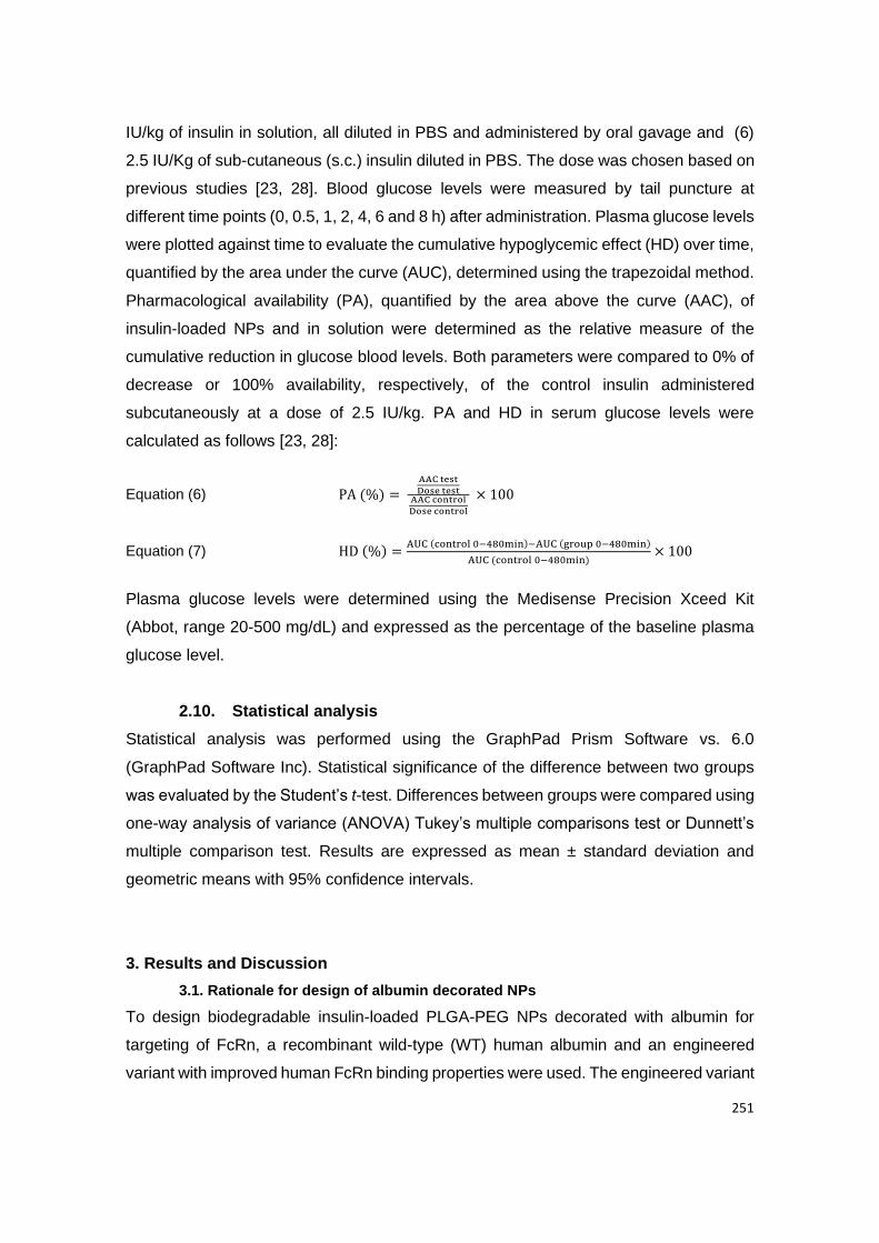
251
IU/kg of insulin in solution, all diluted in PBS and administered by oral gavage and (6)
2.5 IU/Kg of sub-cutaneous (s.c.) insulin diluted in PBS. The dose was chosen based on
previous studies [23, 28]. Blood glucose levels were measured by tail puncture at
different time points (0, 0.5, 1, 2, 4, 6 and 8 h) after administration. Plasma glucose levels
were plotted against time to evaluate the cumulative hypoglycemic effect (HD) over time,
quantified by the area under the curve (AUC), determined using the trapezoidal method.
Pharmacological availability (PA), quantified by the area above the curve (AAC), of
insulin-loaded NPs and in solution were determined as the relative measure of the
cumulative reduction in glucose blood levels. Both parameters were compared to 0% of
decrease or 100% availability, respectively, of the control insulin administered
subcutaneously at a dose of 2.5 IU/kg. PA and HD in serum glucose levels were
calculated as follows [23, 28]:
Equation (6) PA (%) = AAC test
Dose testAAC control
Dose control
× 100
Equation (7) HD (%) =AUC (control 0−480min)−AUC (group 0−480min)
AUC (control 0−480min)× 100
Plasma glucose levels were determined using the Medisense Precision Xceed Kit
(Abbot, range 20-500 mg/dL) and expressed as the percentage of the baseline plasma
glucose level.
2.10. Statistical analysis
Statistical analysis was performed using the GraphPad Prism Software vs. 6.0
(GraphPad Software Inc). Statistical significance of the difference between two groups
was evaluated by the Student’s t-test. Differences between groups were compared using
one-way analysis of variance (ANOVA) Tukey’s multiple comparisons test or Dunnett’s
multiple comparison test. Results are expressed as mean ± standard deviation and
geometric means with 95% confidence intervals.
3. Results and Discussion
3.1. Rationale for design of albumin decorated NPs
To design biodegradable insulin-loaded PLGA-PEG NPs decorated with albumin for
targeting of FcRn, a recombinant wild-type (WT) human albumin and an engineered
variant with improved human FcRn binding properties were used. The engineered variant

252
contains a single amino acid substitution in the C-terminal domain III (K573P, KP), which
has been shown to extend serum half-life in human FcRn expressing mice and non-
human primates [14, 29]. In addition, a human albumin variant engineered to abolish
binding to the receptor (K500A/H464Q, KAHQ) was included [14, 29]. These mutations
are also located in the C-terminal domain III of albumin, which harbors the principle
binding site for FcRn (Figure 1C).
To ensure directional attachment of the albumin variants to the NPs, an exposed and
free cysteine residue within the N-terminal domain I of albumin (Cys34) (Figure 1C),
which is located distally from the core FcRn binding site, was explored [14, 29].
Maleimide (MAL) chemistry was used to promote formation of a thioether bond between
Cys34 of albumin and the maleimide group of PLGA-PEG-MAL on the surface of the NP
(Figure 1D). As such, this approach secures directional and site-specific conjugation, as
opposed to random conjugation.
Figure 1 | Representation of the rationale of this study: (A) A schematic illustration of the pH
dependent FcRn-mediated transcytosis in polarized intestinal epithelial cells: (1) Albumin may be
taken up by fluid-phase pinocytosis or by receptor-mediated uptake, as the slightly acidic pH at
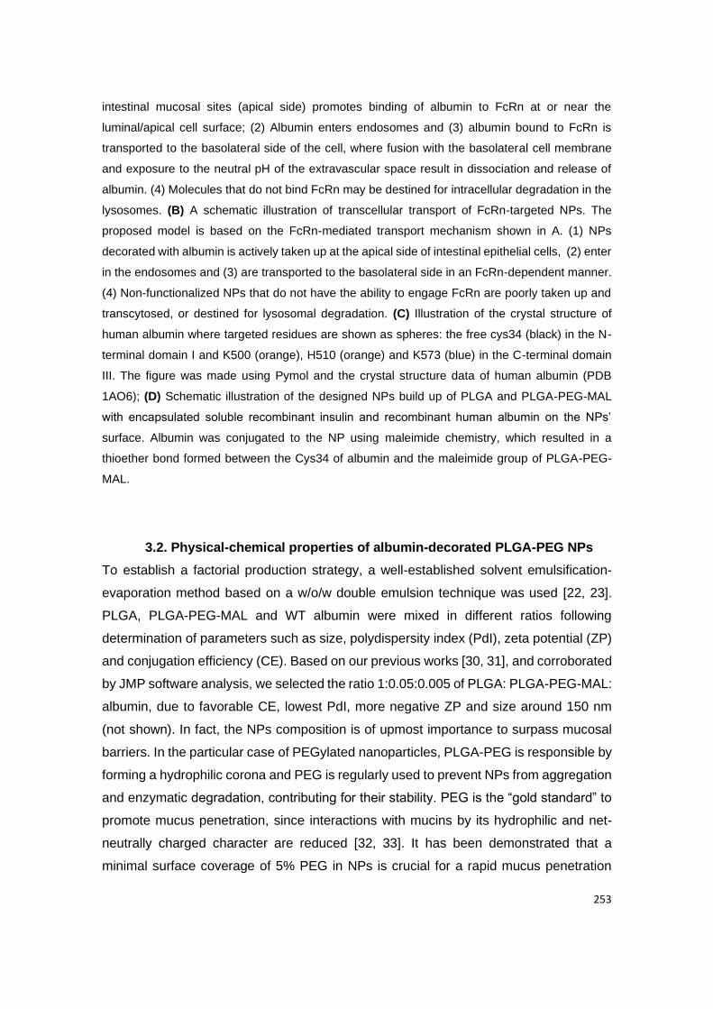
253
intestinal mucosal sites (apical side) promotes binding of albumin to FcRn at or near the
luminal/apical cell surface; (2) Albumin enters endosomes and (3) albumin bound to FcRn is
transported to the basolateral side of the cell, where fusion with the basolateral cell membrane
and exposure to the neutral pH of the extravascular space result in dissociation and release of
albumin. (4) Molecules that do not bind FcRn may be destined for intracellular degradation in the
lysosomes. (B) A schematic illustration of transcellular transport of FcRn-targeted NPs. The
proposed model is based on the FcRn-mediated transport mechanism shown in A. (1) NPs
decorated with albumin is actively taken up at the apical side of intestinal epithelial cells, (2) enter
in the endosomes and (3) are transported to the basolateral side in an FcRn-dependent manner.
(4) Non-functionalized NPs that do not have the ability to engage FcRn are poorly taken up and
transcytosed, or destined for lysosomal degradation. (C) Illustration of the crystal structure of
human albumin where targeted residues are shown as spheres: the free cys34 (black) in the N-
terminal domain I and K500 (orange), H510 (orange) and K573 (blue) in the C-terminal domain
III. The figure was made using Pymol and the crystal structure data of human albumin (PDB
1AO6); (D) Schematic illustration of the designed NPs build up of PLGA and PLGA-PEG-MAL
with encapsulated soluble recombinant insulin and recombinant human albumin on the NPs’
surface. Albumin was conjugated to the NP using maleimide chemistry, which resulted in a
thioether bond formed between the Cys34 of albumin and the maleimide group of PLGA-PEG-
MAL.
3.2. Physical-chemical properties of albumin-decorated PLGA-PEG NPs
To establish a factorial production strategy, a well-established solvent emulsification-
evaporation method based on a w/o/w double emulsion technique was used [22, 23].
PLGA, PLGA-PEG-MAL and WT albumin were mixed in different ratios following
determination of parameters such as size, polydispersity index (PdI), zeta potential (ZP)
and conjugation efficiency (CE). Based on our previous works [30, 31], and corroborated
by JMP software analysis, we selected the ratio 1:0.05:0.005 of PLGA: PLGA-PEG-MAL:
albumin, due to favorable CE, lowest PdI, more negative ZP and size around 150 nm
(not shown). In fact, the NPs composition is of upmost importance to surpass mucosal
barriers. In the particular case of PEGylated nanoparticles, PLGA-PEG is responsible by
forming a hydrophilic corona and PEG is regularly used to prevent NPs from aggregation
and enzymatic degradation, contributing for their stability. PEG is the “gold standard” to
promote mucus penetration, since interactions with mucins by its hydrophilic and net-
neutrally charged character are reduced [32, 33]. It has been demonstrated that a
minimal surface coverage of 5% PEG in NPs is crucial for a rapid mucus penetration
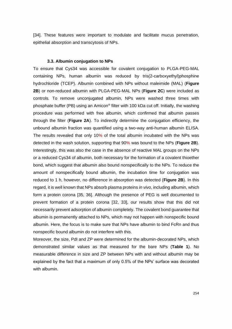
254
[34]. These features were important to modulate and facilitate mucus penetration,
epithelial absorption and transcytosis of NPs.
3.3. Albumin conjugation to NPs
To ensure that Cys34 was accessible for covalent conjugation to PLGA-PEG-MAL
containing NPs, human albumin was reduced by tris(2-carboxyethyl)phosphine
hydrochloride (TCEP). Albumin combined with NPs without maleimide (MAL) (Figure
2B) or non-reduced albumin with PLGA-PEG-MAL NPs (Figure 2C) were included as
controls. To remove unconjugated albumin, NPs were washed three times with
phosphate buffer (PB) using an Amicon® filter with 100 kDa cut off. Initially, the washing
procedure was performed with free albumin, which confirmed that albumin passes
through the filter (Figure 2A). To indirectly determine the conjugation efficiency, the
unbound albumin fraction was quantified using a two-way anti-human albumin ELISA.
The results revealed that only 10% of the total albumin incubated with the NPs was
detected in the wash solution, supporting that 90% was bound to the NPs (Figure 2B).
Interestingly, this was also the case in the absence of reactive MAL groups on the NPs
or a reduced Cys34 of albumin, both necessary for the formation of a covalent thioether
bond, which suggest that albumin also bound nonspecifically to the NPs. To reduce the
amount of nonspecifically bound albumin, the incubation time for conjugation was
reduced to 1 h, however, no difference in absorption was detected (Figure 2B). In this
regard, it is well known that NPs absorb plasma proteins in vivo, including albumin, which
form a protein corona [35, 36]. Although the presence of PEG is well documented to
prevent formation of a protein corona [32, 33], our results show that this did not
necessarily prevent adsorption of albumin completely. The covalent bond guarantee that
albumin is permanently attached to NPs, which may not happen with nonspecific bound
albumin. Here, the focus is to make sure that NPs have albumin to bind FcRn and thus
nonspecific bound albumin do not interfere with this.
Moreover, the size, PdI and ZP were determined for the albumin-decorated NPs, which
demonstrated similar values as that measured for the bare NPs (Table 1). No
measurable difference in size and ZP between NPs with and without albumin may be
explained by the fact that a maximum of only 0.5% of the NPs’ surface was decorated
with albumin.
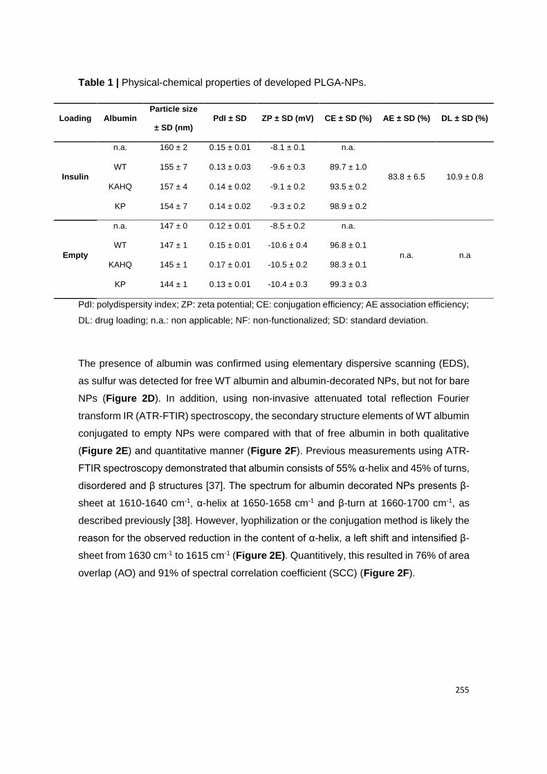
255
Table 1 | Physical-chemical properties of developed PLGA-NPs.
Loading Albumin Particle size
± SD (nm) PdI ± SD ZP ± SD (mV) CE ± SD (%) AE ± SD (%) DL ± SD (%)
Insulin
n.a. 160 ± 2 0.15 ± 0.01 -8.1 ± 0.1 n.a.
83.8 ± 6.5 10.9 ± 0.8
WT 155 ± 7 0.13 ± 0.03 -9.6 ± 0.3 89.7 ± 1.0
KAHQ 157 ± 4 0.14 ± 0.02 -9.1 ± 0.2 93.5 ± 0.2
KP 154 ± 7 0.14 ± 0.02 -9.3 ± 0.2 98.9 ± 0.2
Empty
n.a. 147 ± 0 0.12 ± 0.01 -8.5 ± 0.2 n.a.
n.a. n.a
WT 147 ± 1 0.15 ± 0.01 -10.6 ± 0.4 96.8 ± 0.1
KAHQ 145 ± 1 0.17 ± 0.01 -10.5 ± 0.2 98.3 ± 0.1
KP 144 ± 1 0.13 ± 0.01 -10.4 ± 0.3 99.3 ± 0.3
PdI: polydispersity index; ZP: zeta potential; CE: conjugation efficiency; AE association efficiency;
DL: drug loading; n.a.: non applicable; NF: non-functionalized; SD: standard deviation.
The presence of albumin was confirmed using elementary dispersive scanning (EDS),
as sulfur was detected for free WT albumin and albumin-decorated NPs, but not for bare
NPs (Figure 2D). In addition, using non-invasive attenuated total reflection Fourier
transform IR (ATR-FTIR) spectroscopy, the secondary structure elements of WT albumin
conjugated to empty NPs were compared with that of free albumin in both qualitative
(Figure 2E) and quantitative manner (Figure 2F). Previous measurements using ATR-
FTIR spectroscopy demonstrated that albumin consists of 55% α-helix and 45% of turns,
disordered and β structures [37]. The spectrum for albumin decorated NPs presents β-
sheet at 1610-1640 cm-1, α-helix at 1650-1658 cm-1 and β-turn at 1660-1700 cm-1, as
described previously [38]. However, lyophilization or the conjugation method is likely the
reason for the observed reduction in the content of α-helix, a left shift and intensified β-
sheet from 1630 cm-1 to 1615 cm-1 (Figure 2E). Quantitively, this resulted in 76% of area
overlap (AO) and 91% of spectral correlation coefficient (SCC) (Figure 2F).
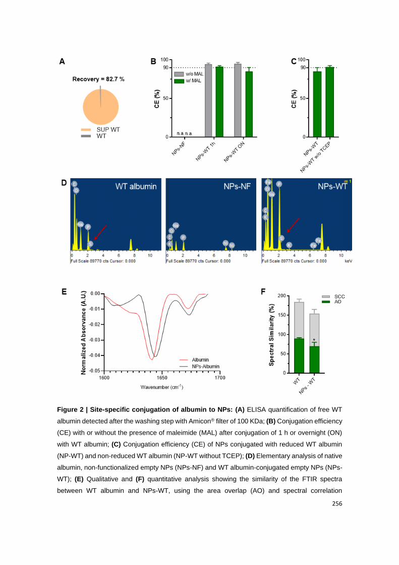
256
Figure 2 | Site-specific conjugation of albumin to NPs: (A) ELISA quantification of free WT
albumin detected after the washing step with Amicon® filter of 100 KDa; (B) Conjugation efficiency
(CE) with or without the presence of maleimide (MAL) after conjugation of 1 h or overnight (ON)
with WT albumin; (C) Conjugation efficiency (CE) of NPs conjugated with reduced WT albumin
(NP-WT) and non-reduced WT albumin (NP-WT without TCEP); (D) Elementary analysis of native
albumin, non-functionalized empty NPs (NPs-NF) and WT albumin-conjugated empty NPs (NPs-
WT); (E) Qualitative and (F) quantitative analysis showing the similarity of the FTIR spectra
between WT albumin and NPs-WT, using the area overlap (AO) and spectral correlation
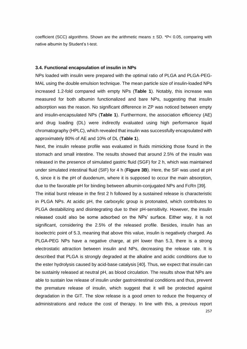
257
coefficient (SCC) algorithms. Shown are the arithmetic means ± SD. *P< 0.05, comparing with
native albumin by Student’s t-test.
3.4. Functional encapsulation of insulin in NPs
NPs loaded with insulin were prepared with the optimal ratio of PLGA and PLGA-PEG-
MAL using the double emulsion technique. The mean particle size of insulin-loaded NPs
increased 1.2-fold compared with empty NPs (Table 1). Notably, this increase was
measured for both albumin functionalized and bare NPs, suggesting that insulin
adsorption was the reason. No significant difference in ZP was noticed between empty
and insulin-encapsulated NPs (Table 1). Furthermore, the association efficiency (AE)
and drug loading (DL) were indirectly evaluated using high performance liquid
chromatography (HPLC), which revealed that insulin was successfully encapsulated with
approximately 80% of AE and 10% of DL (Table 1).
Next, the insulin release profile was evaluated in fluids mimicking those found in the
stomach and small intestine. The results showed that around 2.5% of the insulin was
released in the presence of simulated gastric fluid (SGF) for 2 h, which was maintained
under simulated intestinal fluid (SIF) for 4 h (Figure 3B). Here, the SIF was used at pH
6, since it is the pH of duodenum, where it is supposed to occur the main absorption,
due to the favorable pH for binding between albumin-conjugated NPs and FcRn [39].
The initial burst release in the first 2 h followed by a sustained release is characteristic
in PLGA NPs. At acidic pH, the carboxylic group is protonated, which contributes to
PLGA destabilizing and disintegrating due to their pH-sensitivity. However, the insulin
released could also be some adsorbed on the NPs’ surface. Either way, it is not
significant, considering the 2.5% of the released profile. Besides, insulin has an
isoelectric point of 5.3, meaning that above this value, insulin is negatively charged. As
PLGA-PEG NPs have a negative charge, at pH lower than 5.3, there is a strong
electrostatic attraction between insulin and NPs, decreasing the release rate. It is
described that PLGA is strongly degraded at the alkaline and acidic conditions due to
the ester hydrolysis caused by acid-base catalysis [40]. Thus, we expect that insulin can
be sustainly released at neutral pH, as blood circulation. The results show that NPs are
able to sustain low release of insulin under gastrointestinal conditions and thus, prevent
the premature release of insulin, which suggest that it will be protected against
degradation in the GIT. The slow release is a good omen to reduce the frequency of
administrations and reduce the cost of therapy. In line with this, a previous report
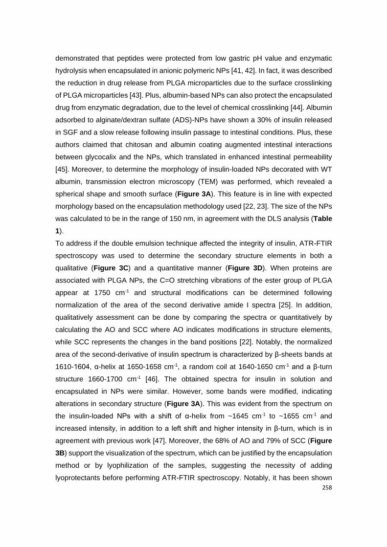
258
demonstrated that peptides were protected from low gastric pH value and enzymatic
hydrolysis when encapsulated in anionic polymeric NPs [41, 42]. In fact, it was described
the reduction in drug release from PLGA microparticles due to the surface crosslinking
of PLGA microparticles [43]. Plus, albumin-based NPs can also protect the encapsulated
drug from enzymatic degradation, due to the level of chemical crosslinking [44]. Albumin
adsorbed to alginate/dextran sulfate (ADS)-NPs have shown a 30% of insulin released
in SGF and a slow release following insulin passage to intestinal conditions. Plus, these
authors claimed that chitosan and albumin coating augmented intestinal interactions
between glycocalix and the NPs, which translated in enhanced intestinal permeability
[45]. Moreover, to determine the morphology of insulin-loaded NPs decorated with WT
albumin, transmission electron microscopy (TEM) was performed, which revealed a
spherical shape and smooth surface (Figure 3A). This feature is in line with expected
morphology based on the encapsulation methodology used [22, 23]. The size of the NPs
was calculated to be in the range of 150 nm, in agreement with the DLS analysis (Table
1).
To address if the double emulsion technique affected the integrity of insulin, ATR-FTIR
spectroscopy was used to determine the secondary structure elements in both a
qualitative (Figure 3C) and a quantitative manner (Figure 3D). When proteins are
associated with PLGA NPs, the C=O stretching vibrations of the ester group of PLGA
appear at 1750 cm-1 and structural modifications can be determined following
normalization of the area of the second derivative amide I spectra [25]. In addition,
qualitatively assessment can be done by comparing the spectra or quantitatively by
calculating the AO and SCC where AO indicates modifications in structure elements,
while SCC represents the changes in the band positions [22]. Notably, the normalized
area of the second-derivative of insulin spectrum is characterized by β-sheets bands at
1610-1604, α-helix at 1650-1658 cm-1, a random coil at 1640-1650 cm-1 and a β-turn
structure 1660-1700 cm-1 [46]. The obtained spectra for insulin in solution and
encapsulated in NPs were similar. However, some bands were modified, indicating
alterations in secondary structure (Figure 3A). This was evident from the spectrum on
the insulin-loaded NPs with a shift of α-helix from ~1645 cm-1 to ~1655 cm-1 and
increased intensity, in addition to a left shift and higher intensity in β-turn, which is in
agreement with previous work [47]. Moreover, the 68% of AO and 79% of SCC (Figure
3B) support the visualization of the spectrum, which can be justified by the encapsulation
method or by lyophilization of the samples, suggesting the necessity of adding
lyoprotectants before performing ATR-FTIR spectroscopy. Notably, it has been shown
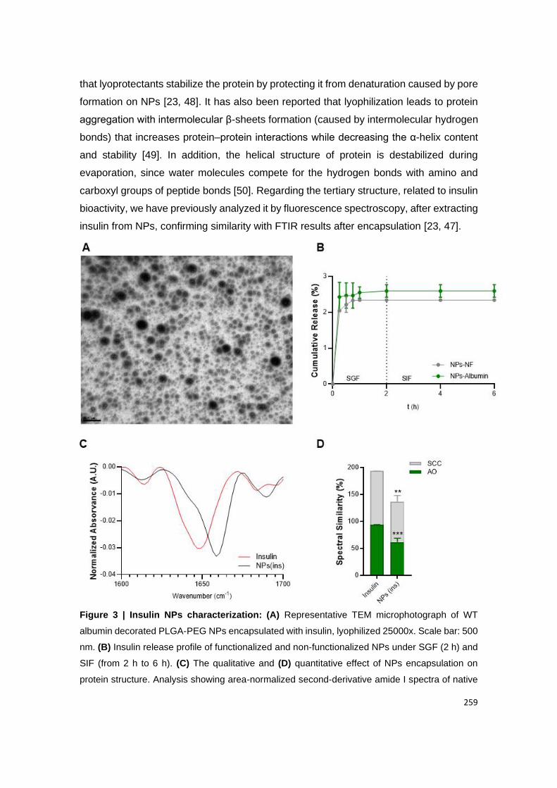
259
that lyoprotectants stabilize the protein by protecting it from denaturation caused by pore
formation on NPs [23, 48]. It has also been reported that lyophilization leads to protein
aggregation with intermolecular β-sheets formation (caused by intermolecular hydrogen
bonds) that increases protein–protein interactions while decreasing the α-helix content
and stability [49]. In addition, the helical structure of protein is destabilized during
evaporation, since water molecules compete for the hydrogen bonds with amino and
carboxyl groups of peptide bonds [50]. Regarding the tertiary structure, related to insulin
bioactivity, we have previously analyzed it by fluorescence spectroscopy, after extracting
insulin from NPs, confirming similarity with FTIR results after encapsulation [23, 47].
Figure 3 | Insulin NPs characterization: (A) Representative TEM microphotograph of WT
albumin decorated PLGA-PEG NPs encapsulated with insulin, lyophilized 25000x. Scale bar: 500
nm. (B) Insulin release profile of functionalized and non-functionalized NPs under SGF (2 h) and
SIF (from 2 h to 6 h). (C) The qualitative and (D) quantitative effect of NPs encapsulation on
protein structure. Analysis showing area-normalized second-derivative amide I spectra of native
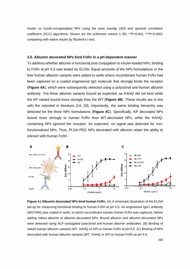
260
insulin vs insulin-encapsulated NPs using the area overlap (AO) and spectral correlation
coefficient (SCC) algorithms. Shown are the arithmetic means ± SD. **P<0.001, ***P<0.0001
comparing with native insulin by Student’s t-test.
3.5. Albumin decorated NPs bind FcRn in a pH dependent manner
To address whether albumin is functional post conjugation to insulin-loaded NPs, binding
to FcRn at pH 5.5 was tested by ELISA. Equal amounts of the NPs formulations or the
free human albumin variants were added to wells where recombinant human FcRn had
been captured on a coated engineered IgG molecule that strongly binds the receptor
(Figure 4A), which were subsequently detected using a polyclonal anti-human albumin
antibody. The three albumin variants bound as expected, as KAHQ did not bind while
the KP variant bound more strongly than the WT (Figure 4B). These results are in line
with the reported in literature [14, 29]. Importantly, the same binding hierarchy was
detected for the three NPs formulations (Figure 4C). Specifically, KP decorated NPs
bound more strongly to human FcRn than WT-decorated NPs, while the KAHQ-
containing NPs ignored the receptor. As expected, no signal was detected for non-
functionalized NPs. Thus, PLGA-PEG NPs decorated with albumin retain the ability to
interact with human FcRn.
Figure 4 | Albumin decorated NPs bind human FcRn. (A) A schematic illustration of the ELISA
set-up for measuring functional binding to human FcRn at pH 5.5. An engineered IgG1 antibody
(MST/HN) was coated in wells, to which recombinant soluble human FcRn was captured, before
adding naked albumin or albumin-decorated NPs. Bound albumin and albumin-decorated NPs
were detected using ALP-conjugated polyclonal anti-human albumin antibodies. (B) Binding of
naked human albumin variants (WT, KAHQ or KP) to human FcRn at pH 5.5. (C) Binding of NPs
decorated with human albumin variants (WT, KAHQ or KP) to human FcRn at pH 5.5.
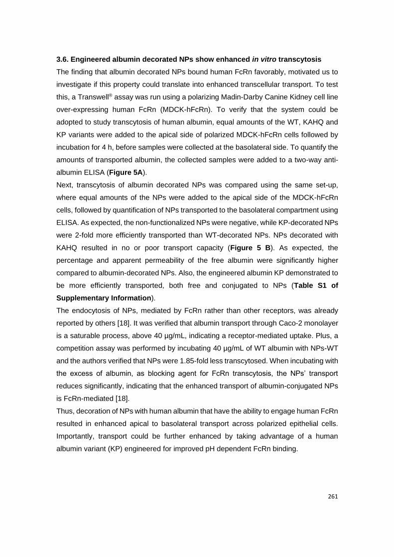
261
3.6. Engineered albumin decorated NPs show enhanced in vitro transcytosis
The finding that albumin decorated NPs bound human FcRn favorably, motivated us to
investigate if this property could translate into enhanced transcellular transport. To test
this, a Transwell® assay was run using a polarizing Madin-Darby Canine Kidney cell line
over-expressing human FcRn (MDCK-hFcRn). To verify that the system could be
adopted to study transcytosis of human albumin, equal amounts of the WT, KAHQ and
KP variants were added to the apical side of polarized MDCK-hFcRn cells followed by
incubation for 4 h, before samples were collected at the basolateral side. To quantify the
amounts of transported albumin, the collected samples were added to a two-way anti-
albumin ELISA (Figure 5A).
Next, transcytosis of albumin decorated NPs was compared using the same set-up,
where equal amounts of the NPs were added to the apical side of the MDCK-hFcRn
cells, followed by quantification of NPs transported to the basolateral compartment using
ELISA. As expected, the non-functionalized NPs were negative, while KP-decorated NPs
were 2-fold more efficiently transported than WT-decorated NPs. NPs decorated with
KAHQ resulted in no or poor transport capacity (Figure 5 B). As expected, the
percentage and apparent permeability of the free albumin were significantly higher
compared to albumin-decorated NPs. Also, the engineered albumin KP demonstrated to
be more efficiently transported, both free and conjugated to NPs (Table S1 of
Supplementary Information).
The endocytosis of NPs, mediated by FcRn rather than other receptors, was already
reported by others [18]. It was verified that albumin transport through Caco-2 monolayer
is a saturable process, above 40 µg/mL, indicating a receptor-mediated uptake. Plus, a
competition assay was performed by incubating 40 µg/mL of WT albumin with NPs-WT
and the authors verified that NPs were 1.85-fold less transcytosed. When incubating with
the excess of albumin, as blocking agent for FcRn transcytosis, the NPs’ transport
reduces significantly, indicating that the enhanced transport of albumin-conjugated NPs
is FcRn-mediated [18].
Thus, decoration of NPs with human albumin that have the ability to engage human FcRn
resulted in enhanced apical to basolateral transport across polarized epithelial cells.
Importantly, transport could be further enhanced by taking advantage of a human
albumin variant (KP) engineered for improved pH dependent FcRn binding.
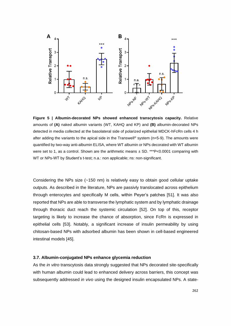
262
Figure 5 | Albumin-decorated NPs showed enhanced transcytosis capacity. Relative
amounts of (A) naked albumin variants (WT, KAHQ and KP) and (B) albumin-decorated NPs
detected in media collected at the basolateral side of polarized epithelial MDCK-hFcRn cells 4 h
after adding the variants to the apical side in the Transwell® system (n=5-9). The amounts were
quantified by two-way anti-albumin ELISA, where WT albumin or NPs decorated with WT albumin
were set to 1, as a control. Shown are the arithmetic means ± SD. ***P<0.0001 comparing with
WT or NPs-WT by Student’s t-test; n.a.: non applicable; ns: non-significant.
Considering the NPs size (~150 nm) is relatively easy to obtain good cellular uptake
outputs. As described in the literature, NPs are passivly translocated across epithelium
through enterocytes and specifically M cells, within Peyer’s patches [51]. It was also
reported that NPs are able to transverse the lymphatic system and by lymphatic drainage
through thoracic duct reach the systemic circulation [52]. On top of this, receptor
targeting is likely to increase the chance of absorption, since FcRn is expressed in
epithelial cells [53]. Notably, a significant increase of insulin permeability by using
chitosan-based NPs with adsorbed albumin has been shown in cell-based engineered
intestinal models [45].
3.7. Albumin-conjugated NPs enhance glycemia reduction
As the in vitro transcytosis data strongly suggested that NPs decorated site-specifically
with human albumin could lead to enhanced delivery across barriers, this concept was
subsequently addressed in vivo using the designed insulin encapsulated NPs. A state-

263
of-the-art mouse model that is transgenic for human FcRn and knockout for mouse FcRn
(FcRn-/- hFcRn Tg32 mice) [27] was chosen for this study, as conventional mice are
unreliable models for evaluation of human albumin-based therapeutics due to large
cross-species binding differences [54-56]. Specifically, human albumin binds poorly to
mouse FcRn, and as such will be competed out for binding to the receptor in the
presence of large amounts of endogenous mouse albumin [54, 56]. The human FcRn
transgenic mice indeed have mouse albumin but in this case mouse and human albumin
bind equally well to the receptor [54, 56].
To study the effect of albumin decorated NPs encapsulated with insulin, the transgenic
mice were treated with streptozotocin (STZ) to induce diabetes. One week later, the mice
were fasted for 12 h, before being divided into six groups based on their blood glucose
level. After administering different formulations, pharmacodynamic parameters were
evaluated over 8 h (Figure 6 A). This time frame was not extended, since it is not
recommended starving animals for long periods. Besides, if the animal is in a starvation
state it is hard to objectively discuss the results, since they could be due to the drug
effect or due to organism failure. In fact, it was observed some deaths during the
experiments, which were not considered for data analysis. Knowing that mice consume
primarily at night, overnight fasting leads to a catabolic state, reducing 15% of body mass
in lean mice [57]. Overnight fasting deletes liver glycogen stores, which contributes for
reducing the variability in baseline blood glucose. Besides, contrary to what happens in
humans, a prolonged fast in mice enhances the insulin-stimulated glucose utilization [57].
In this sense, it is recommended that mice used for metabolic studies, should be fasted
for 14–18 h (overnight fast) [58].
The first group received free insulin via subcutaneous (s.c.) injection, which resulted in
a rapid decrease of glucose within 2 h followed by an increase over the next 6 h (Figure
6 B). This phenotype is in accordance with previous reports using rats [28, 59]. In
contrast, mice treated orally with free insulin, or with bare NPs, showed no significant
decrease in blood glucose (Figure 6 B), being most probably degraded along the way.
Furthermore, the effect of oral administration of equal amounts of bare NPs, WT- or KP-
decorated NPs was evaluated, which revealed that the albumin-decorated NPs had a
more pronounced effect compared with free insulin and the bare NPs. In particular, the
KP-decorated NPs showed the fastest glycemic decrease and maintained a decrease of
around 40% between 1 h and 4 h after administration (Table S2 of Supplementary
Information). However, in this preliminary set-up, NPs containing WT and KP were not
statistically different. On the other hand, it was previously reported a significant in vivo
264
difference between the two naked variants [29]. As mentioned before, it was observed
some animal deaths during the experiments, which were not considered for data
analysis. In this sense, the reduced number of animals might be the reason for the non-
statistical significance in in vivo results, which did not happen in in vitro.
Notably, for the control group receiving PBS, an increase in glucose level was observed
within the first hour, which may be explained by stress and an adrenalin reaction to the
administration. Most probably, all mice experienced this, while the effect was
contradicted by the administered insulin, which should be taken into consideration when
comparing pharmacological parameters. Nevertheless, functionalized NPs were more
efficient in delivering encapsulated insulin to the circulation, as the orally delivered free
insulin and non-functionalized NPs did not induce glycemic response.
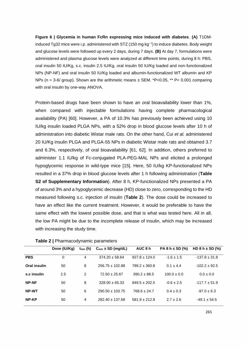
265
Figure 6 | Glycemia in human FcRn expressing mice induced with diabetes. (A) T1DM-
induced Tg32 mice were i.p. administered with STZ (150 mg kg−1) to induce diabetes. Body weight
and glucose levels were followed up every 2 days, during 7 days. (B) At day 7, formulations were
administered and plasma glucose levels were analyzed at different time points, during 8 h: PBS,
oral insulin 50 IU/Kg, s.c. insulin 2.5 IU/Kg, oral insulin 50 IU/Kg loaded and non-functionalized
NPs (NP-NF) and oral insulin 50 IU/Kg loaded and albumin-functionalized WT albumin and KP
NPs (n = 3-6/ group). Shown are the arithmetic means ± SEM. *P<0.05, ** P< 0.001 comparing
with oral insulin by one-way ANOVA.
Protein-based drugs have been shown to have an oral bioavailability lower than 1%,
when compared with injectable formulations having complete pharmacological
availability (PA) [60]. However, a PA of 10.3% has previously been achieved using 10
IU/kg insulin loaded PLGA NPs, with a 52% drop in blood glucose levels after 10 h of
administration into diabetic Wistar male rats. On the other hand, Cui et al. administered
20 IU/Kg insulin PLGA and PLGA-55 NPs in diabetic Wistar male rats and obtained 3.7
and 6.3%, respectively, of oral bioavailability [61, 62]. In addition, others preferred to
administer 1.1 IU/kg of Fc-conjugated PLA-PEG-MAL NPs and elicited a prolonged
hypoglycemic response in wild-type mice [15]. Here, 50 IU/kg KP-functionalized NPs
resulted in a 37% drop in blood glucose levels after 1 h following administration (Table
S2 of Supplementary Information). After 8 h, KP-functionalized NPs presented a PA
of around 3% and a hypoglycemic decrease (HD) close to zero, corresponding to the HD
measured following s.c. injection of insulin (Table 2). The dose could be increased to
have an effect like the current treatment. However, it would be preferable to have the
same effect with the lowest possible dose, and that is what was tested here. All in all,
the low PA might be due to the incomplete release of insulin, which may be increased
with increasing the study time.
Table 2 | Pharmacodynamic parameters
Dose (IU/Kg) tmin (h) Cmin ± SD (mg/dL) AUC 8 h PA 8 h ± SD (%) HD 8 h ± SD (%)
PBS 0 4 374.20 ± 58.64 927.8 ± 124.0 -1.6 ± 1.5 -137.8 ± 31.8
Oral insulin 50 8 256.75 ± 102.88 789.2 ± 360.8 0.1 ± 4.4 -102.2 ± 92.5
s.c insulin 2.5 2 72.50 ± 25.67 390.2 ± 88.5 100.0 ± 0.0 0.0 ± 0.0
NP-NF 50 8 328.00 ± 65.33 849.5 ± 202.5 -0.6 ± 2.5 -117.7 ± 51.9
NP-WT 50 6 290.50 ± 103.75 768.6 ± 24.7 0.4 ± 0.3 -97.0 ± 6.3
NP-KP 50 4 282.40 ± 137.68 581.9 ± 212.8 2.7 ± 2.6 -49.1 ± 54.5
266
AUC: area under the curve; PA: pharmacological availability; HD: hypoglycemic decrease; SD:
standard deviation; tmin: time with minimal concentration of glucose after administration of different
formulations.
Taken together, the in vivo glycemic decrease obtained using the KP variant is a clear
improvement of the therapeutic efficacy. In fact, the obtained in vivo results agree with
in vitro data, supporting that albumin decorated NPs increase the effect as a result of
improved FcRn-binding affinity. It is reported in the literature the use of FcRn knock out
mouse, confirming the FcRn-mediated transcytosis of FcRn-targeted NPs [15].
Interestingly, while the intestinal expression of FcRn in rodents has been demonstrated
to be developmentally down-regulated after weaning [63], this is not the case in humans
or in the human FcRn expressing mice, as the transgene is expressed under the human
promoter [64]. Thus, the transgenic mouse model mimics the tissue expression profile of
human FcRn, by not being down-regulated after weaning [27]. On the other hand, it has
been shown that the hFcRn transgenic mice have higher levels of FcRn expression in
the liver, lung, small intestine and heart, compared to Swiss Webster mice as well as the
human tissues [65]. Further investigation into the implication of these differences on the
PD/PK is required. In addition, human FcRn is globally expressed in many cells and
tissues including immune cells, vascular endothelial cells, epidermal keratinocytes, the
blood-brain-barrier, retina-blood barrier, lungs, kidneys, liver, genital tissues, mammary
glands as well as the placenta [10], which may pave the way for using FcRn-targeting
NPs for delivery to different body sites.
Either way, further studies are needed with higher number of animals to clarify the
statistical significance of the results as well as biodistribution and pharmacokinetic
studies. In addition, as previously mentioned, here it was used insulin as a model drug,
using diabetes as example. For this specific example, more studies should be considered
to address all the particularities of diabetes or even try to encapsulate as insulin glargine,
for the treatment of both diabetes types.
4. Conclusions
In this work, we report on a novel class of monodisperse 150 nm PLGA-PEG NPs, with
a drug loading over 10% and around 90% of conjugation efficiency. These NPs were
decorated with an engineered human albumin variant with improved human FcRn
binding, which enhanced transcytosis in 2-fold across polarized epithelia compared with
267
WT albumin decorated NPs. When tested for oral delivery in human FcRn transgenic
mice, induced with diabetes, such NPs were able to reduce glycemia up to around 40%
after 1 h and enhance pharmacologic availability. The novel concept described should
pave the way for further engineering of biodegradable albumin-decorated NP
formulations for tailored targeting of FcRn for enhanced intestinal delivery.
Author Contributions
C.A., J.N, J.T.A. and B.S. designed research; C.A., J.N., A.G. and R.N. performed
research; C.A., J.N., A.G. and R.N. analyzed data; C.A, J.T.A. and B.S. wrote the paper.
Conflict of Interest
J.T.A. is a co-inventor of pending patent applications related to albumin variants.
Acknowledgement
This project is a result of the project NORTE-01-0145-FEDER-000012, supported by
Norte Portugal Regional Operational Programme (NORTE 2020), under the PORTUGAL
2020 Partnership Agreement, through the European Regional Development Fund
(ERDF). This work was also financed by FEDER - Fundo Europeu de Desenvolvimento
Regional funds through the COMPETE 2020 - Operacional Programme for
Competitiveness and Internationalisation (POCI), Portugal 2020, and by Portuguese
funds through FCT - Fundação para a Ciência e a Tecnologia/ Ministério da Ciência,
Tecnologia e Ensino Superior in the framework of the project "Institute for Research and
Innovation in Health Sciences" (UID/BIM/04293/2019). Cláudia Azevedo would like to
thank to Fundação para a Ciência e a Tecnologia (FCT), Portugal for financial support
(SFRH/BD/117598/2016). The authors acknowledge the support of Abbot Laboratório,
Lda for the glucose strips as well as Flávia Sousa for JMP software guidance and the
following i3S Scientific Platforms: Biointerfaces and Nanotechnology (FCT -
UID/BIM/04293/2019), Histology and Electron Microscopy (PPBI-POCI-01-0145-
FEDER-022122) and Animal Facility. The authors thank Tore-Geir Iversen for assistance
with the Malvern Zetasizer Nano ZS instrument. This work was supported by the
Research Council of Norway (Grant no. 274993).
268
References
[1] P. Saeedi, I. Petersohn, P. Salpea, B. Malanda, S. Karuranga, N. Unwin, S. Colagiuri, L.
Guariguata, A.A. Motala, K. Ogurtsova, J.E. Shaw, D. Bright, R. Williams, Global and regional
diabetes prevalence estimates for 2019 and projections for 2030 and 2045: Results from the
International Diabetes Federation Diabetes Atlas, 9(th) edition, Diabetes Research and Clinical
Practice, 157 (2019) 107843.
[2] C. Bommer, V. Sagalova, E. Heesemann, J. Manne-Goehler, R. Atun, T. Barnighausen, J.
Davies, S. Vollmer, Global Economic Burden of Diabetes in Adults: Projections From 2015 to
2030, Diabetes Care, 41 (2018) 963-970.
[3] M.J. Davies, J.J. Gagliardino, L.J. Gray, K. Khunti, V. Mohan, R. Hughes, Real-world factors
affecting adherence to insulin therapy in patients with Type 1 or Type 2 diabetes mellitus: a
systematic review, Diabetic medicine: a Journal of the British Diabetic Association, 30 (2013) 512-
524.
[4] P. Fonte, F. Araujo, C. Silva, C. Pereira, S. Reis, H.A. Santos, B. Sarmento, Polymer-based
nanoparticles for oral insulin delivery: Revisited approaches, Biotechnology Advances, 33 (2015)
1342-1354.
[5] B. Sarmento, D. Ferreira, T. Vasconcelos Polymer-based delivery systems for oral delivery of
peptides and proteins, in Delivery Technologies for Biopharmaceuticals: Peptides, Proteins,
Nucleic Acids and Vaccines, (Eds: L. Jorgensen, H. M. Nielsen) John Wiley & Sons Ltd (2009)
pp. 207.
[6] F. Araujo, N. Shrestha, M.A. Shahbazi, P. Fonte, E.M. Makila, J.J. Salonen, J.T. Hirvonen,
P.L. Granja, H.A. Santos, B. Sarmento, The impact of nanoparticles on the mucosal translocation
and transport of GLP-1 across the intestinal epithelium, Biomaterials, 35 (2014) 9199-9207.
[7] C. Pereira, F. Araujo, P.L. Granja, H.A. Santos, B. Sarmento, Targeting membrane
transporters and receptors as a mean to optimize orally delivered biotechnological based drugs
through nanoparticle delivery systems, Current Pharmaceutical Biotechnology, 15 (2014) 650-
658.
[8] B. Moulari, A. Beduneau, Y. Pellequer, A. Lamprecht, Nanoparticle Targeting to Inflamed
Tissues of the Gastrointestinal Tract, Current Drug Delivery, 10 (2013) 9-17.
[9] M.R. Rekha, C.P. Sharma, Oral delivery of therapeutic protein/peptide for diabetes-future
perspectives, International Journal of Pharmaceutics, 440 (2013) 48-62.
[10] K.M.K. Sand, M. Bern, J. Nilsen, H.T. Noordzij, I. Sandlie, J.T. Andersen, Unraveling the
Interaction between FcRn and Albumin: Opportunities for Design of Albumin-Based Therapeutics,
Frontiers in Immunology, 5 (2015).
[11] P.J. Hornby, P.R. Cooper, C. Kliwinski, E. Ragwan, J.R. Mabus, B. Harman, S. Thompson,
A.L. Kauffman, Z. Yan, S.H. Tam, H. Dorai, G.D. Powers, J. Giles-Komar, Human and non-human
269
primate intestinal FcRn expression and immunoglobulin G transcytosis, Pharmaceutical
Research, 31 (2014) 908-922.
[12] B.L. Dickinson, K. Badizadegan, Z. Wu, J.C. Ahouse, X. Zhu, N.E. Simister, R.S. Blumberg,
W.I. Lencer, Bidirectional FcRn-dependent IgG transport in a polarized human intestinal epithelial
cell line, The Journal of Clinical Investigation, 104 (1999) 903-911.
[13] D.C. Roopenian, S. Akilesh, FcRn: the neonatal Fc receptor comes of age, Nature Reviews
Immunology, 7 (2007) 715.
[14] J.T. Andersen, B. Dalhus, J. Cameron, M.B. Daba, A. Plumridge, L. Evans, S.O. Brennan,
K.S. Gunnarsen, M. Bjørås, D. Sleep, I. Sandlie, Structure-based mutagenesis reveals the
albumin-binding site of the neonatal Fc receptor, Nature Communications, 3 (2012) 610.
[15] E.M. Pridgen, F. Alexis, T.T. Kuo, E. Levy-Nissenbaum, R. Karnik, R.S. Blumberg, R. Langer,
O.C. Farokhzad, Transepithelial transport of Fc-targeted nanoparticles by the neonatal fc receptor
for oral delivery, Science Translational Medicine, 5 (2013) 213ra167.
[16] Y. Shi, X. Sun, L. Zhang, K. Sun, K. Li, Y. Li, Q. Zhang, Fc-modified exenatide-loaded
nanoparticles for oral delivery to improve hypoglycemic effects in mice, Scientific Reports, 8
(2018) 726.
[17] A. Moreno-Aspitia, E.A. Perez, North Central Cancer Treatment Group N0531: Phase II Trial
of Weekly Albumin-Bound Paclitaxel (ABI-007; Abraxane®) in Combination with Gemcitabine in
Patients with Metastatic Breast Cancer, Clinical Breast Cancer, 6 (2005) 361-364.
[18] L. Hashem, M. Swedrowska, D. Vllasaliu, Intestinal uptake and transport of albumin
nanoparticles: potential for oral delivery, Nanomedicine, 13 (2018) 1255-1265.
[19] J.P. Martins, R. D'Auria, D. Liu, F. Fontana, M.P.A. Ferreira, A. Correia, M. Kemell, K.
Moslova, E. Makila, J. Salonen, L. Casettari, J. Hirvonen, B. Sarmento, H.A. Santos, Engineered
Multifunctional Albumin-Decorated Porous Silicon Nanoparticles for FcRn Translocation of
Insulin, Small, 14 (2018) e1800462.
[20] J. Nilsen, E. Trabjerg, A. Grevys, C. Azevedo, S.O. Brennan, M. Stensland, J. Wilson, K.M.K.
Sand, M. Bern, B. Dalhus, D.C. Roopenian, I. Sandlie, K.D. Rand, J.T. Andersen, An intact C-
terminal end of albumin is required for its long half-life in humans, Communications Biology, 3
(2020) 181.
[21] A. Grevys, J. Nilsen, K.M.K. Sand, M.B. Daba, I. Øynebråten, M. Bern, M.B. McAdam, S.
Foss, T. Schlothauer, T.E. Michaelsen, G.J. Christianson, D.C. Roopenian, R.S. Blumberg, I.
Sandlie, J.T. Andersen, A human endothelial cell-based recycling assay for screening of FcRn
targeted molecules, Nature Communications, 9 (2018) 621.
[22] P. Fonte, S. Soares, F. Sousa, A. Costa, V. Seabra, S. Reis, B. Sarmento, Stability study
perspective of the effect of freeze-drying using cryoprotectants on the structure of insulin loaded
into PLGA nanoparticles, Biomacromolecules, 15 (2014) 3753-3765.
270
[23] P. Fonte, F. Andrade, C. Azevedo, J. Pinto, V. Seabra, M. van de Weert, S. Reis, B.
Sarmento, Effect of the Freezing Step in the Stability and Bioactivity of Protein-Loaded PLGA
Nanoparticles Upon Lyophilization, Pharmaceutical Research, 33 (2016) 2777-2793.
[24] B. Sarmento, A. Ribeiro, F. Veiga, D. Ferreira, Development and validation of a rapid
reversed-phase HPLC method for the determination of insulin from nanoparticulate systems,
Biomedical Chromatography, 20 (2006) 898-903.
[25] F. Sousa, A. Cruz, P. Fonte, I.M. Pinto, M.T. Neves-Petersen, B. Sarmento, A new paradigm
for antiangiogenic therapy through controlled release of bevacizumab from PLGA nanoparticles,
Scientific Reports, 7 (2017) 3736.
[26] M.J. Maltesen, S. Bjerregaard, L. Hovgaard, S. Havelund, M. van de Weert, Analysis of
insulin allostery in solution and solid state with FTIR, Journal of Pharmaceutical Sciences, 98
(2009) 3265-3277.
[27] D.C. Roopenian, G.J. Christianson, T.J. Sproule, A.C. Brown, S. Akilesh, N. Jung, S.
Petkova, L. Avanessian, E.Y. Choi, D.J. Shaffer, P.A. Eden, C.L. Anderson, The MHC class I-like
IgG receptor controls perinatal IgG transport, IgG homeostasis, and fate of IgG-Fc-coupled drugs,
Journal of Immunology, 170 (2003) 3528-3533.
[28] B. Sarmento, A. Ribeiro, F. Veiga, D. Ferreira, R. Neufeld, Oral bioavailability of insulin
contained in polysaccharide nanoparticles, Biomacromolecules, 8 (2007) 3054-3060.
[29] J.T. Andersen, B. Dalhus, D. Viuff, B.T. Ravn, K.S. Gunnarsen, A. Plumridge, K. Bunting, F.
Antunes, R. Williamson, S. Athwal, E. Allan, L. Evans, M. Bjoras, S. Kjaerulff, D. Sleep, I. Sandlie,
J. Cameron, Extending serum half-life of albumin by engineering neonatal Fc receptor (FcRn)
binding, The Journal of Biological Chemistry, 289 (2014) 13492-13502.
[30] P.J. Kennedy, I. Perreira, D. Ferreira, M. Nestor, C. Oliveira, P.L. Granja, B. Sarmento,
Impact of surfactants on the target recognition of Fab-conjugated PLGA nanoparticles, European
Journal of Pharmaceutics and Biopharmaceutics, 127 (2018) 366-370.
[31] I. Pereira, F. Sousa, P. Kennedy, B. Sarmento, Carcinoembryonic antigen-targeted
nanoparticles potentiate the delivery of anticancer drugs to colorectal cancer cells, International
Journal of Pharmaceutics, 549 (2018) 397-403.
[32] J.S. Suk, Q. Xu, N. Kim, J. Hanes, L.M. Ensign, PEGylation as a strategy for improving
nanoparticle-based drug and gene delivery, Advanced drug delivery reviews, 99 (2016) 28-51.
[33] J.T. Huckaby, S.K. Lai, PEGylation for enhancing nanoparticle diffusion in mucus, Advanced
Drug Delivery Reviews, 124 (2018) 125-139.
[34] N. Bertrand, P. Grenier, M. Mahmoudi, E.M. Lima, E.A. Appel, F. Dormont, J.M. Lim, R.
Karnik, R. Langer, O.C. Farokhzad, Mechanistic understanding of in vivo protein corona formation
on polymeric nanoparticles and impact on pharmacokinetics, Nature Communications, 8 (2017)
777.

271
[35] K. Sempf, T. Arrey, S. Gelperina, T. Schorge, B. Meyer, M. Karas, J. Kreuter, Adsorption of
plasma proteins on uncoated PLGA nanoparticles, European Journal of Pharmaceutics and
Biopharmaceutics, 85 (2013) 53-60.
[36] D.J. Sobczynski, P. Charoenphol, M.J. Heslinga, P.J. Onyskiw, K. Namdee, A.J. Thompson,
O. Eniola-Adefeso, Plasma protein corona modulates the vascular wall interaction of drug carriers
in a material and donor specific manner, PloS one, 9 (2014) e107408.
[37] D. Charbonneau, M. Beauregard, H.A. Tajmir-Riahi, Structural analysis of human serum
albumin complexes with cationic lipids, The Journal of Physical Chemistry, 113 (2009) 1777-1784.
[38] J. Tang, F. Luan, X. Chen, Binding analysis of glycyrrhetinic acid to human serum albumin:
Fluorescence spectroscopy, FTIR, and molecular modeling, Bioorganic & Medicinal Chemistry,
14 (2006) 3210-3217.
[39] J. Fallingborg, Intraluminal pH of the human gastrointestinal tract, Danish Medical Bulletin,
46 (1999) 183-196.
[40] R. Dinarvand, N. Sepehri, S. Manoochehri, H. Rouhani, F. Atyabi, Polylactide-co-glycolide
nanoparticles for controlled delivery of anticancer agents, International Journal of Nanomedicine,
6 (2011) 877-895.
[41] E. Czuba, M. Diop, C. Mura, A. Schaschkow, A. Langlois, W. Bietiger, R. Neidl, A. Virciglio,
N. Auberval, D. Julien-David, E. Maillard, Y. Frere, E. Marchioni, M. Pinget, S. Sigrist, Oral insulin
delivery, the challenge to increase insulin bioavailability: Influence of surface charge in
nanoparticle system, International Journal of Pharmaceutics, 542 (2018) 47-55.
[42] D.J. McClements, Encapsulation, protection, and delivery of bioactive proteins and peptides
using nanoparticle and microparticle systems: A review, Advances in Colloid and Interface
Science, 253 (2018) 1-22.
[43] A.J. Thote, J.T. Chappell, R. Kumar, R.B. Gupta, Reduction in the Initial-Burst Release by
Surface Crosslinking of PLGA Microparticles Containing Hydrophilic or Hydrophobic Drugs, Drug
Development and Industrial Pharmacy, 31 (2005) 43-57.
[44] K. Langer, M.G. Anhorn, I. Steinhauser, S. Dreis, D. Celebi, N. Schrickel, S. Faust, V. Vogel,
Human serum albumin (HSA) nanoparticles: Reproducibility of preparation process and kinetics
of enzymatic degradation, International Journal of Pharmaceutics, 347 (2008) 109-117.
[45] M. Lopes, A. Derenne, C. Pereira, F. Veiga, R. Seica, B. Sarmento, A. Ribeiro, Impact of the
in vitro gastrointestinal passage of biopolymer-based nanoparticles on insulin absorption, RSC
Advances, 6 (2016) 20155-20165.
[46] P. Fonte, F. Araujo, V. Seabra, S. Reis, M. van de Weert, B. Sarmento, Co-encapsulation of
lyoprotectants improves the stability of protein-loaded PLGA nanoparticles upon lyophilization,
International Journal of Pharmaceutics, 496 (2015) 850-862.
[47] P. Fonte, P.R. Lino, V. Seabra, A.J. Almeida, S. Reis, B. Sarmento, Annealing as a tool for
the optimization of lyophilization and ensuring of the stability of protein-loaded PLGA
nanoparticles, International Journal of Pharmaceutics, 503 (2016) 163-173.
272
[48] P. Fonte, S. Soares, A. Costa, J.C. Andrade, V. Seabra, S. Reis, B. Sarmento, Effect of
cryoprotectants on the porosity and stability of insulin-loaded PLGA nanoparticles after freeze-
drying, Biomatter, 2 (2012) 329-339.
[49] H.R. Costantino, K. Griebenow, P. Mishra, R. Langer, A.M. Klibanov, Fourier-transform
infrared spectroscopic investigation of protein stability in the lyophilized form, Biochimica et
Biophysica Acta, 1253 (1995) 69-74.
[50] K.V. Abrosimova, O.V. Shulenina, S.V. Paston, FTIR study of secondary structure of bovine
serum albumin and ovalbumin, Journal of Physics: Conference Series, 769 (2016) 012016.
[51] C.B. Woitiski, R.A. Carvalho, A.J. Ribeiro, R.J. Neufeld, F. Veiga, Strategies toward the
improved oral delivery of insulin nanoparticles via gastrointestinal uptake and translocation,
BioDrugs: Clinical Immunotherapeutics, Biopharmaceuticals and Gene Therapy, 22 (2008) 223-
237.
[52] S. Attili-Qadri, N. Karra, A. Nemirovski, O. Schwob, Y. Talmon, T. Nassar, S. Benita, Oral
delivery system prolongs blood circulation of docetaxel nanocapsules via lymphatic absorption,
Proceedings of the National Academy of Sciences of the United States of America, 110 (2013)
17498-17503.
[53] M. Goldberg, I. Gomez-Orellana, Challenges for the oral delivery of macromolecules, Nature
Reviews Drug Discovery, 2 (2003) 289.
[54] J. Nilsen, M. Bern, K.M.K. Sand, A. Grevys, B. Dalhus, I. Sandlie, J.T. Andersen, Human and
mouse albumin bind their respective neonatal Fc receptors differently, Scientific Reports, 8 (2018)
14648.
[55] K.M. Sand, M. Bern, J. Nilsen, B. Dalhus, K.S. Gunnarsen, J. Cameron, A. Grevys, K.
Bunting, I. Sandlie, J.T. Andersen, Interaction with both domain I and III of albumin is required for
optimal pH-dependent binding to the neonatal Fc receptor (FcRn), The Journal of Biological
Chemistry, 289 (2014) 34583-34594.
[56] J.T. Andersen, M.B. Daba, G. Berntzen, T.E. Michaelsen, I. Sandlie, Cross-species binding
analyses of mouse and human neonatal Fc receptor show dramatic differences in immunoglobulin
G and albumin binding, The Journal of Biological Chemistry, 285 (2010) 4826-4836.
[57] J.E. Ayala, D.P. Bracy, O.P. McGuinness, D.H. Wasserman, Considerations in the design of
hyperinsulinemic-euglycemic clamps in the conscious mouse, Diabetes, 55 (2006) 390-397.
[58] J.E. Ayala, V.T. Samuel, G.J. Morton, S. Obici, C.M. Croniger, G.I. Shulman, D.H.
Wasserman, O.P. McGuinness, N.I.H.M.M.P.C. Consortium, Standard operating procedures for
describing and performing metabolic tests of glucose homeostasis in mice, Disease Models &
Mechanisms, 3 (2010) 525-534.
[59] K. Sonaje, K.J. Lin, S.P. Wey, C.K. Lin, T.H. Yeh, H.N. Nguyen, C.W. Hsu, T.C. Yen, J.H.
Juang, H.W. Sung, Biodistribution, pharmacodynamics and pharmacokinetics of insulin
analogues in a rat model: Oral delivery using pH-responsive nanoparticles vs. subcutaneous
injection, Biomaterials, 31 (2010) 6849-6858.
273
[60] J. Shaji, V. Patole, Protein and Peptide drug delivery: oral approaches, Indian journal of
Pharmaceutical Sciences, 70 (2008) 269-277.
[61] Y. Pan, H. Xu, H.Y. Zhao, G. Wei, J.M. Zheng, Study on preparation and oral efficacy of
insulin-loaded poly(lactic-co-glycolic acid) nanoparticles, Acta Pharmaceutica Sinica, 37 (2002)
374-377.
[62] F.D. Cui, A.J. Tao, D.M. Cun, L.Q. Zhang, K. Shi, Preparation of insulin loaded PLGA-Hp55
nanoparticles for oral delivery, Journal of Pharmaceutical Sciences, 96 (2007) 421-427.
[63] M.G. Martin, S.V. Wu, J.H. Walsh, Ontogenetic Development and Distribution of Antibody
Transport and Fc Receptor mRNA Expression in Rat Intestine, Digestive Diseases and Sciences,
42 (1997) 1062-1069.
[64] M. Pyzik, T. Rath, W.I. Lencer, K. Baker, R.S. Blumberg, FcRn: The Architect Behind the
Immune and Nonimmune Functions of IgG and Albumin, Journal of Immunology, 194 (2015)
4595-4603.
[65] T. Li, J.P. Balthasar, FcRn Expression in Wildtype Mice, Transgenic Mice, and in Human
Tissues, Biomolecules, 8 (2018).

274
Supplementary material
Engineered albumin-functionalized nanoparticles for improved FcRn
binding enhance oral delivery of insulin
Cláudia Azevedo, Jeannette Nilsen, Algirdas Grevys, Rute Nunes, Jan Terje Andersen, Bruno
Sarmento
Table S1 | Permeability percentage and apparent permeability coefficient calculated
after 4 h of incubation in MDCK-hFcRn cells.
Formulation Permeability ± SD (%) P app ± SD (x 10-6)
WT 11.44 ± 6.92 1.44 ± 0.87
KAHQ 5.20 ± 2.83 0.66 ± 0.36
KP 28.73 ± 4.90 3.63 ± 0.62
NPs-NF 0.31 ± 0.28 0.04 ± 0.04
NPs-WT 0.84 ± 0.36 0.11 ± 0.05
NPs-KAHQ 0.72 ± 0.57 0.09 ± 0.07
NPs- KP 1.86 ± 0.60 0.25 ± 0.08
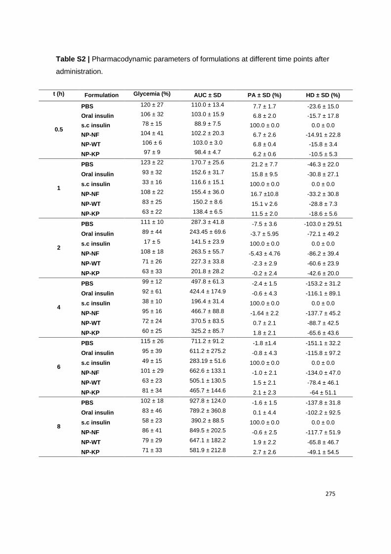
275
Table S2 | Pharmacodynamic parameters of formulations at different time points after
administration.
t (h) Formulation Glycemia (%) AUC ± SD PA ± SD (%) HD ± SD (%)
0.5
PBS 120 ± 27 110.0 ± 13.4 7.7 ± 1.7 -23.6 ± 15.0
Oral insulin 106 ± 32 103.0 ± 15.9 6.8 ± 2.0 -15.7 ± 17.8
s.c insulin 78 ± 15 88.9 ± 7.5 100.0 ± 0.0 0.0 ± 0.0
NP-NF 104 ± 41 102.2 ± 20.3 6.7 ± 2.6 -14.91 ± 22.8
NP-WT 106 ± 6 103.0 ± 3.0 6.8 ± 0.4 -15.8 ± 3.4
NP-KP 97 ± 9 98.4 ± 4.7 6.2 ± 0.6 -10.5 ± 5.3
1
PBS 123 ± 22 170.7 ± 25.6 21.2 ± 7.7 -46.3 ± 22.0
Oral insulin 93 ± 32 152.6 ± 31.7 15.8 ± 9.5 -30.8 ± 27.1
s.c insulin 33 ± 16 116.6 ± 15.1 100.0 ± 0.0 0.0 ± 0.0
NP-NF 108 ± 22 155.4 ± 36.0 16.7 ±10.8 -33.2 ± 30.8
NP-WT 83 ± 25 150.2 ± 8.6 15.1 v 2.6 -28.8 ± 7.3
NP-KP 63 ± 22 138.4 ± 6.5 11.5 ± 2.0 -18.6 ± 5.6
2
PBS 111 ± 10 287.3 ± 41.8 -7.5 ± 3.6 -103.0 ± 29.51
Oral insulin 89 ± 44 243.45 ± 69.6 -3.7 ± 5.95 -72.1 ± 49.2
s.c insulin 17 ± 5 141.5 ± 23.9 100.0 ± 0.0 0.0 ± 0.0
NP-NF 108 ± 18 263.5 ± 55.7 -5.43 ± 4.76 -86.2 ± 39.4
NP-WT 71 ± 26 227.3 ± 33.8 -2.3 ± 2.9 -60.6 ± 23.9
NP-KP 63 ± 33 201.8 ± 28.2 -0.2 ± 2.4 -42.6 ± 20.0
4
PBS 99 ± 12 497.8 ± 61.3 -2.4 ± 1.5 -153.2 ± 31.2
Oral insulin 92 ± 61 424.4 ± 174.9 -0.6 ± 4.3 -116.1 ± 89.1
s.c insulin 38 ± 10 196.4 ± 31.4 100.0 ± 0.0 0.0 ± 0.0
NP-NF 95 ± 16 466.7 ± 88.8 -1.64 ± 2.2 -137.7 ± 45.2
NP-WT 72 ± 24 370.5 ± 83.5 0.7 ± 2.1 -88.7 ± 42.5
NP-KP 60 ± 25 325.2 ± 85.7 1.8 ± 2.1 -65.6 ± 43.6
6
PBS 115 ± 26 711.2 ± 91.2 -1.8 ±1.4 -151.1 ± 32.2
Oral insulin 95 ± 39 611.2 ± 275.2 -0.8 ± 4.3 -115.8 ± 97.2
s.c insulin 49 ± 15 283.19 ± 51.6 100.0 ± 0.0 0.0 ± 0.0
NP-NF 101 ± 29 662.6 ± 133.1 -1.0 ± 2.1 -134.0 ± 47.0
NP-WT 63 ± 23 505.1 ± 130.5 1.5 ± 2.1 -78.4 ± 46.1
NP-KP 81 ± 34 465.7 ± 144.6 2.1 ± 2.3 -64 ± 51.1
8
PBS 102 ± 18 927.8 ± 124.0 -1.6 ± 1.5 -137.8 ± 31.8
Oral insulin 83 ± 46 789.2 ± 360.8 0.1 ± 4.4 -102.2 ± 92.5
s.c insulin 58 ± 23 390.2 ± 88.5 100.0 ± 0.0 0.0 ± 0.0
NP-NF 86 ± 41 849.5 ± 202.5 -0.6 ± 2.5 -117.7 ± 51.9
NP-WT 79 ± 29 647.1 ± 182.2 1.9 ± 2.2 -65.8 ± 46.7
NP-KP 71 ± 33 581.9 ± 212.8 2.7 ± 2.6 -49.1 ± 54.5

276

277
The potential of porcine ex vivo platform for intestinal permeability
screening of FcRn-targeted drugs
Cláudia Azevedo1,2,3, Jan Terje Andersen4,5, Bruno Sarmento1,2,6, Giovanni Traverso7,8,9*
1 Instituto de Investigação e Inovação em Saúde (i3S), University of Porto, Porto, Portugal;
2 Instituto de Engenharia Biomédica (INEB), University of Porto, Porto, Portugal;
3 Instituto Ciências Biomédicas Abel Salazar (ICBAS), University of Porto, Porto, Portugal;
4 Department of Immunology, Centre for Immune Regulation (CIR), Oslo University Hospital Rikshospitalet
and University of Oslo, Oslo, Norway.
5 Institute of Clinical Medicine and Department of Pharmacology, University of Oslo and Oslo University
Hospital, Oslo, Norway.
6 CESPU, Instituto de Investigação e Formação Avançada em Ciências e Tecnologias da Saúde & Instituto
Universitário de Ciências da Saúde, Gandra, Portugal
7 Department of Chemical Engineering and Koch Institute for Integrative Cancer Research, Massachusetts
Institute of Technology, Cambridge, MA, USA.
8 Division of Gastroenterology, Brigham and Women’s Hospital, Harvard Medical School, Boston, MA, USA
9 Department of Mechanical Engineering, Massachusetts Institute of Technology, Cambridge, MA, USA.
* Corresponding Author: Tel: (617) 417-8061; E-mail: [email protected]
278
Abstract
Conventionally, the intestinal permeability of drugs is evaluated using cell monolayers
models that lack morphological, physiological and architectural features, as well as,
realistic FcRn expression. In addition, is time-consuming, expensive and un-ethical to
use mice for large scale-screening of FcRn-targeted candidates. It is then critical to use
suitable models, for preclinical validation, that mimic the human intestine as the porcine
ex vivo model, due to its physiological and anatomical similarities. This study intended
to analyze the FcRn-potential in intestinal permeability analysis, using a porcine ex vivo
platform, able to analyze 96 samples at the same time. Besides, it allows the screening
of FcRn-targeting for transmucosal delivery, taken into consideration (cross-species)
receptor-ligand binding kinetics. After analyzing the porcine morphology tissue, the FcRn
expression across the gastrointestinal tract was verified. Studying the stomach,
duodenum and jejunum it was demonstrated that FcRn-expression is maintained for up
7 days. When evaluating the duodenum permeability of free engineered human albumin
variants, it was shown that KP is more efficiently transported. Taking this, the porcine ex
vivo platform revealed to be a potential model for the screening of FcRn-targeted oral
drug formulations.
Keywords: Engineered human albumin variants; FcRn expression; FcRn-mediated
transcytosis; Polymeric and mucodiffusive nanoparticles; Screening of FcR-targeted
drugs
Teaser: This study intended to analyze the FcRn-potential in intestinal permeability
analysis, using the porcine ex vivo platform.
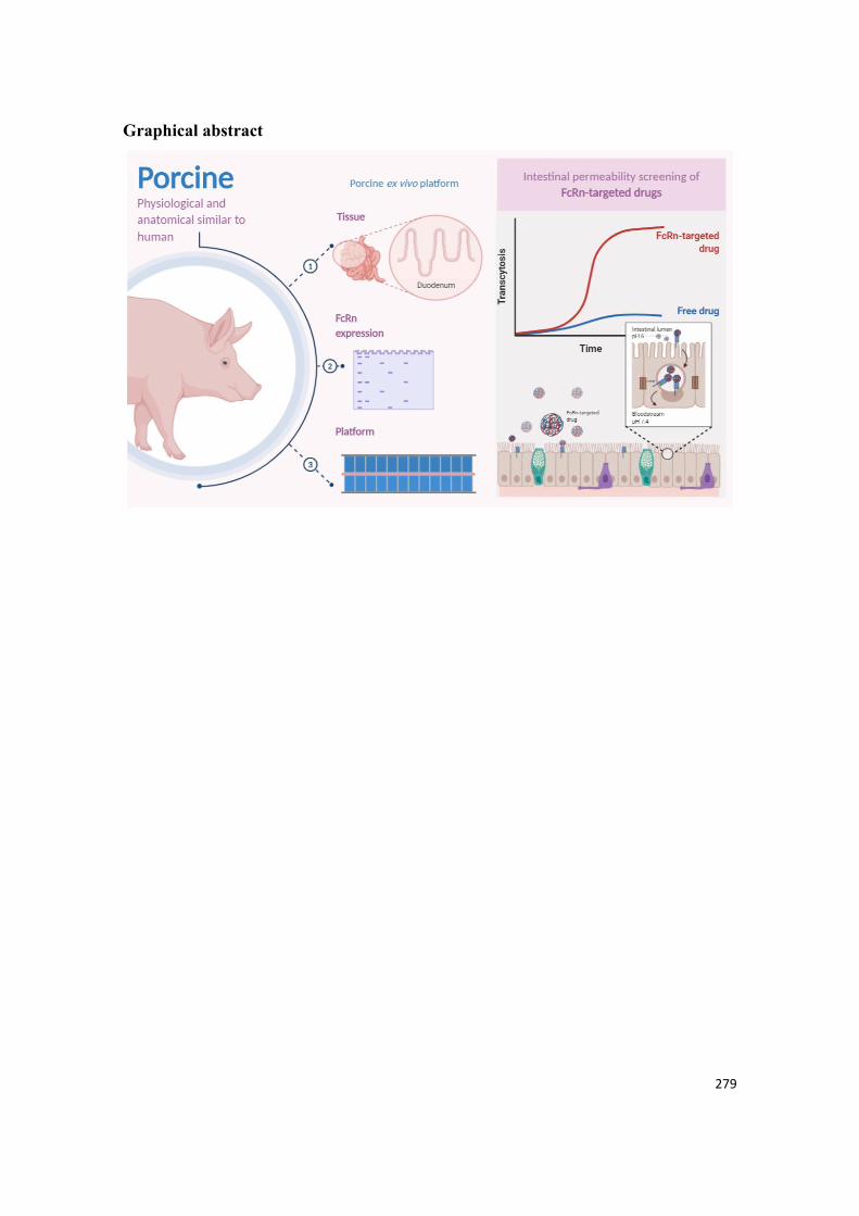
279
Graphical abstract
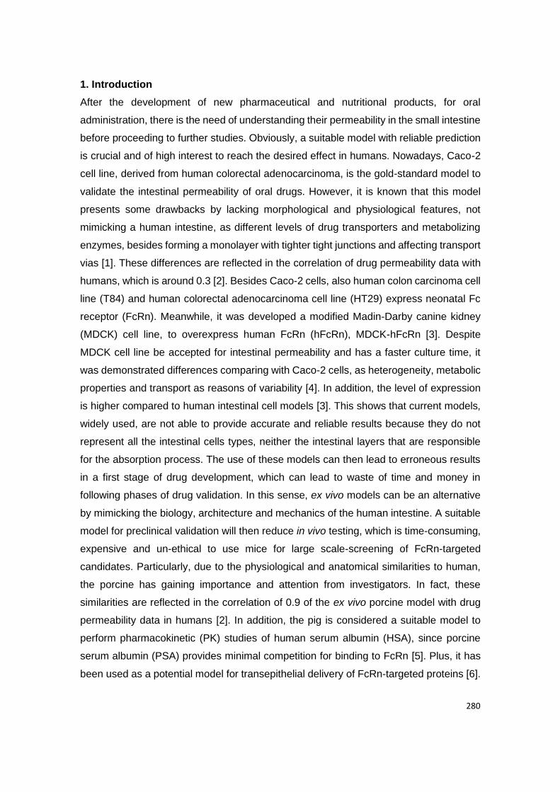
280
1. Introduction
After the development of new pharmaceutical and nutritional products, for oral
administration, there is the need of understanding their permeability in the small intestine
before proceeding to further studies. Obviously, a suitable model with reliable prediction
is crucial and of high interest to reach the desired effect in humans. Nowadays, Caco-2
cell line, derived from human colorectal adenocarcinoma, is the gold-standard model to
validate the intestinal permeability of oral drugs. However, it is known that this model
presents some drawbacks by lacking morphological and physiological features, not
mimicking a human intestine, as different levels of drug transporters and metabolizing
enzymes, besides forming a monolayer with tighter tight junctions and affecting transport
vias [1]. These differences are reflected in the correlation of drug permeability data with
humans, which is around 0.3 [2]. Besides Caco-2 cells, also human colon carcinoma cell
line (T84) and human colorectal adenocarcinoma cell line (HT29) express neonatal Fc
receptor (FcRn). Meanwhile, it was developed a modified Madin-Darby canine kidney
(MDCK) cell line, to overexpress human FcRn (hFcRn), MDCK-hFcRn [3]. Despite
MDCK cell line be accepted for intestinal permeability and has a faster culture time, it
was demonstrated differences comparing with Caco-2 cells, as heterogeneity, metabolic
properties and transport as reasons of variability [4]. In addition, the level of expression
is higher compared to human intestinal cell models [3]. This shows that current models,
widely used, are not able to provide accurate and reliable results because they do not
represent all the intestinal cells types, neither the intestinal layers that are responsible
for the absorption process. The use of these models can then lead to erroneous results
in a first stage of drug development, which can lead to waste of time and money in
following phases of drug validation. In this sense, ex vivo models can be an alternative
by mimicking the biology, architecture and mechanics of the human intestine. A suitable
model for preclinical validation will then reduce in vivo testing, which is time-consuming,
expensive and un-ethical to use mice for large scale-screening of FcRn-targeted
candidates. Particularly, due to the physiological and anatomical similarities to human,
the porcine has gaining importance and attention from investigators. In fact, these
similarities are reflected in the correlation of 0.9 of the ex vivo porcine model with drug
permeability data in humans [2]. In addition, the pig is considered a suitable model to
perform pharmacokinetic (PK) studies of human serum albumin (HSA), since porcine
serum albumin (PSA) provides minimal competition for binding to FcRn [5]. Plus, it has
been used as a potential model for transepithelial delivery of FcRn-targeted proteins [6].
281
In short, this work intends to evaluate an ex vivo platform, using porcine tissue, for
screening the intestinal permeability of FcRn-targeted drugs, taken into consideration
(cross-species) receptor-ligand binding kinetics.
2. Experimental section
2.1. Tissue dissection and cultivation
All animal procedures were conducted in accordance with protocols approved by the
Massachusetts Institute of Technology Committee on Animal Care in compliance with
federal, state, local and institutional regulations, including the Animal Welfare Act and
Public Health Service Policy. Small sections of intestinal tissue were isolated from freshly
procured intact GI tracts from pigs from selected local slaughterhouses. A section of the
tissue was cut out of the GI tract, dissected longitudinally and the outer muscle layer and
serosa were removed. For standard transport experiments, duodenum tissue was used.
The tissue was washed in a series of saline solutions under sterile conditions. The tissue
was then either mounted on the porcine ex vivo platform or maintained in culture. For
cultivation, the media advanced DMEM/F-12 (Life Technologies, Ref.: 12634028) was
used and supplemented with 10% fetal bovine serum (FBS) and 5% antibiotic–
antimycotic solution (Thermo Fisher Scientific, Ref.:15240062). Tissue was cultured in
this serum-free cell culture medium in a sealed polystyrene container that was then
stored in an incubator kept at 37°C (no supplement gas was provided). A set of
experiments was conducted with fixed time points as well as longer-term experiments
monitoring the tissue daily, with analyses performed at varying time points.
2.2. Immunohistochemistry
Sections of the porcine gastrointestinal (GI) tract were put into cassettes, fixed with 10%
(v/w) formalin for 24 h at 4ºC and then storage in 70% ethanol at 4ºC. Paraffin sections
(5-µm) on glass slides were dried at 70°C for 30 min, deparaffinized and heat induced
epitope retrieve (HIER) at 97ºC for 20 min in citrate buffer pH 6. These procedures were
completely automated by using a ThermoFisher IHC Autostainer 360. Briefly, for
immunohistochemistry (IHC), endogenous peroxidase blocking was done for 10 min
followed by the protein block for 30 min. Primary antibody (1:100 anti-FCGRT, abcam
ref.: ab193148) was incubated for 1 h and the secondary antibody (1:2000 goat anti-
rabbit IgG, abcam, ab205718) for 30 min. Both antibodies diluted with Tris Buffered
Saline with Tween 20 (TBST) and incubated at room temperature. Finally, an incubation
282
of 5 min with 3,3′-Diaminobenzidine (DAB) was done. The stained cells were then
mounted on a cover slide using ConsulMount (Thermo Fisher, 9990440). Representative
images using Koehler illumination were obtained with an Olympus Microfire digital
camera (M/N S97809) attached to an Olympus BX60 microscope.
2.3. RT–PCR analysis
The FcRn expression of ex vivo cultured porcine-derived intestinal tissue was
determined by reverse transcription polymerase chain reaction (RT–PCR) analysis and
detected by agarose electrophoresis. Initially, the tissue was removed from fresh GI,
frozen in liquid nitrogen and storage at -80ºC. Then, total RNA from each tissue sample
was extracted and purified with Quick-RNA plus (Zymo Research) followed by reverse
transcription into complementary DNA using a High-Capacity cDNA reverse transcription
kit (ThermoFisher Scientific). Target genes were amplified using the reported primers
(GAATTGGGCCCCGACAAT and CTGCTCACCCTTCCACTCC) [7] and Platinum™
SuperFi™ DNA Polymerase (ThermoFisher Scientific ref.: 12351010). The PCR
program was optimized and consisted of an enzyme activation step (98ºC, 10min), forty
cycles of denaturation (98ºC, 30 sec), annealing extension (62ºC, 1 min), extension
(72ºC, 45 sec), followed by an extension at 72ºC for 10 min. The expression level of
FcRn was measured with 1.5% agarose gel electrophoreses, with SYBR Safe DNA gel
stain (Thermofisher, S33102) and 100 bp DNA Ladder (New England BioLab, N3231S)
and detected using the Bio-Rad Image suite.
2.4. PLGA-PEG NPs production
Engineered albumin-conjugated and insulin encapsulated poly(lactic-co-glycolic)-
poly(ethylene glycol) (PLGA-PEG) nanoparticles (NPs) were produced by following the
procedures previously described [8]. Briefly, NPs were produced through a modified
solvent emulsification-evaporation method based on a w/o/w double emulsion technique.
It was used 100 mg of polymer, in which 95% was PLGA (PURASORB® PDLG 5004A,
in a 50/50 molar ratio of DL-lactide and Glycolide, kindly provided by Corbion) and 5%
PLGA-PEG-MAL (Akina, PolyScitech). To load the NPs, 75 mg/mL insulin solution
(Sigma Aldrich) in 0.1 M of HCl was added to the polymeric solution. Finally, the
conjugation was done through the covalent thioether bond between Cys34 from DI of
albumin and the maleimide group of PLGA-PEG-MAL on the surface of the NP, in a
molar ratio 1:0.1 of PLGA-PEG-MAL:Albumin [8]. Wild type (WT) albumin and
engineered albumin variants, namely K500A/K510Q (KAHQ) and K573P (KP), were
283
kindly provided by Jan Terje Andersen from Oslo University Hospital. PSA was obtained
from Sigma Aldrich.
2.5. Transport experiments using the ex vivo platform
For assembly of the ex vivo platform, freshly isolated intestinal tissue from the duodenum
was prepared according to the above-described tissue dissection procedure and
mounted on the manufactured interface design with a generic 96-well plate (96-well
plates, clear bottom, Corning), as previously described [2]. To the plate from the bottom,
the basolateral side, it was added 100 µL of Hanks' Balanced Salt solution (HBSS) pH
7.4. The duodenum was identified as the region of the small intestine approximately 1 m
after the stomach. Duodenum tissue was then carefully moved over the bottom magnetic
plate with the apical side facing upwards. Afterwards, the top magnetic plate was
mounted, and 90 µL of 189.83 µg/mL samples was added, at the apical side. Samples
were prepared at acidic pH, using HBSS buffered to pH 6.0 by addition of MES buffer
(Sigma Aldrich). After 4 h of incubation, samples were collected from the basolateral
side, with a syringe, at the bottom magnetic plate and quantified by ELISA. All
experiments, including sample incubation, were performed at room temperature. Results
were expressed in relative transport and apparent permeability (Papp), using the
following formulas:
Equation (1) Relative Transport = Amount of sample
Mean amount of control
Equation (2) 𝑃𝑎𝑝𝑝 = ∆Q
A ×C0 × ∆t∗ 100
where C0 is the initial concentration in the apical compartment (mg/mL), A is the surface
area of the insert (cm2), ∆t is the time during which the experiment occurred (seconds)
and ∆Q is the amount of compound detected in the basolateral side (mg).
2.6. Albumin quantification
The quantification was done using a two-way anti-albumin enzyme-linked
immunosorbent assay (ELISA). Briefly, 96 wells plates (Costar) were coated with 100 µL
of a polyclonal anti-albumin antibody produced in goat (Sigma Aldrich, A1151) diluted
1:2000 in PBS by incubation overnight at 4ºC. The wells were blocked with 200 µL
PBSTM solution, composed by PBS, 0.005% Tween 20 and 4% skimmed milk
(Acumedia), for 1 h at room temperature (RT) and then washed three times with 200 µL
PBST (PBS, 0.005% Tween 20). Samples were titrated 1:2 in PBSTM, and 100 µL of
284
each dilution was subsequently added in triplicates to wells and incubated for 1 h at RT.
The wells were washed as before. Alkaline phosphatase (AP)-conjugated polyclonal
anti-albumin from goat (Bethyl Laboratories, A80229AP) was diluted 1:4000 in PBSTM
and 100 µL was added per well and incubated for 1 h at RT. After washing as before,
visualization was performed by adding 100 µL of AP substrate (4-nitrophenyl phosphate
disodium salt hexahydrate substrate) (Sigma-Aldrich) diluted in diethanolamine buffer.
The absorbance was measured at 405 nm using the sunrise spectrophotometer
(TECAN).
2.7. Statistical analysis
Statistical analysis was performed using the GraphPad Prism Software vs. 6.0
(GraphPad Software Inc). Statistical significance of the difference between two groups
was evaluated by the Student’s t-test. Differences between groups were compared using
one-way analysis of variance (ANOVA) Dunnett’s multiple comparison test. Results are
expressed as mean ± standard deviation and geometric means with 95% confidence
intervals. The level of significance was set at probabilities of ∗P < 0.05, ∗∗P < 0.005, and
#P < 0.0001.
3. Results and discussion
3.1. FcRn expression across GI tract
Initially, the viability and morphology of the GI tract from the porcine ex vivo model, was
evaluated by hematoxylin and eosin (H&E) staining, to assure the maintenance of tissue
integrity (Figure S1). After this, FcRn expression was confirmed by RT-PCR, after
collecting the tissue, extract RNA and amplify cDNA (Figure 1). As expected, the porcine
receptor is broadly expressed throughout the GI tract, as previously confirmed [6, 7].
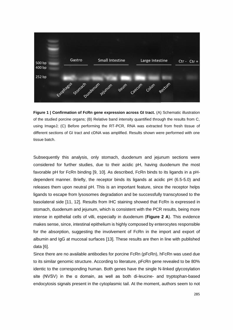
285
Figure 1 | Confirmation of FcRn gene expression across GI tract. (A) Schematic illustration
of the studied porcine organs; (B) Relative band intensity quantified through the results from C,
using ImageJ; (C) Before performing the RT-PCR, RNA was extracted from fresh tissue of
different sections of GI tract and cDNA was amplified. Results shown were performed with one
tissue batch.
Subsequently this analysis, only stomach, duodenum and jejunum sections were
considered for further studies, due to their acidic pH, having duodenum the most
favorable pH for FcRn binding [9, 10]. As described, FcRn binds to its ligands in a pH-
dependent manner. Briefly, the receptor binds its ligands at acidic pH (6.5-5.0) and
releases them upon neutral pH. This is an important feature, since the receptor helps
ligands to escape from lysosomes degradation and be successfully transcytosed to the
basolateral side [11, 12]. Results from IHC staining showed that FcRn is expressed in
stomach, duodenum and jejunum, which is consistent with the PCR results, being more
intense in epithelial cells of villi, especially in duodenum (Figure 2 A). This evidence
makes sense, since, intestinal epithelium is highly composed by enterocytes responsible
for the absorption, suggesting the involvement of FcRn in the import and export of
albumin and IgG at mucosal surfaces [13]. These results are then in line with published
data [6].
Since there are no available antibodies for porcine FcRn (pFcRn), hFcRn was used due
to its similar genomic structure. According to literature, pFcRn gene revealed to be 80%
identic to the corresponding human. Both genes have the single N-linked glycosylation
site (NVSV) in the α domain, as well as both di-leucine- and tryptophan-based
endocytosis signals present in the cytoplasmic tail. At the moment, authors seem to not
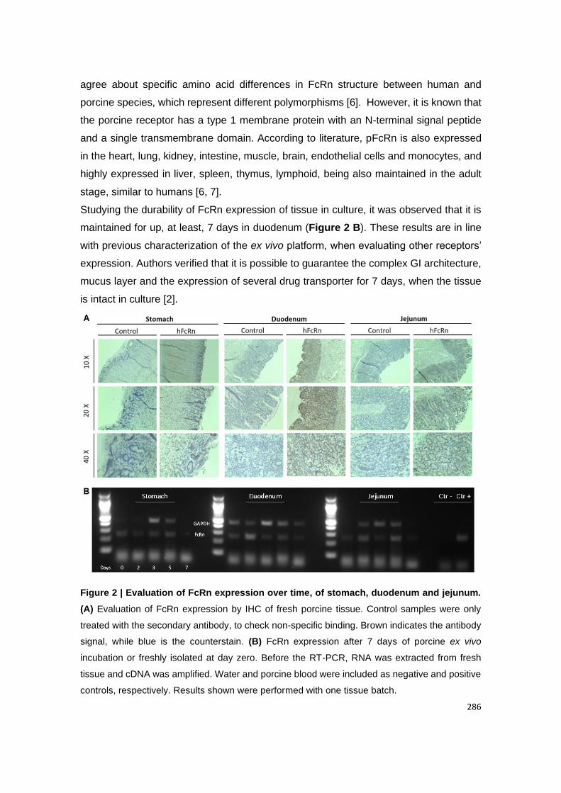
286
agree about specific amino acid differences in FcRn structure between human and
porcine species, which represent different polymorphisms [6]. However, it is known that
the porcine receptor has a type 1 membrane protein with an N-terminal signal peptide
and a single transmembrane domain. According to literature, pFcRn is also expressed
in the heart, lung, kidney, intestine, muscle, brain, endothelial cells and monocytes, and
highly expressed in liver, spleen, thymus, lymphoid, being also maintained in the adult
stage, similar to humans [6, 7].
Studying the durability of FcRn expression of tissue in culture, it was observed that it is
maintained for up, at least, 7 days in duodenum (Figure 2 B). These results are in line
with previous characterization of the ex vivo platform, when evaluating other receptors’
expression. Authors verified that it is possible to guarantee the complex GI architecture,
mucus layer and the expression of several drug transporter for 7 days, when the tissue
is intact in culture [2].
Figure 2 | Evaluation of FcRn expression over time, of stomach, duodenum and jejunum.
(A) Evaluation of FcRn expression by IHC of fresh porcine tissue. Control samples were only
treated with the secondary antibody, to check non-specific binding. Brown indicates the antibody
signal, while blue is the counterstain. (B) FcRn expression after 7 days of porcine ex vivo
incubation or freshly isolated at day zero. Before the RT-PCR, RNA was extracted from fresh
tissue and cDNA was amplified. Water and porcine blood were included as negative and positive
controls, respectively. Results shown were performed with one tissue batch.
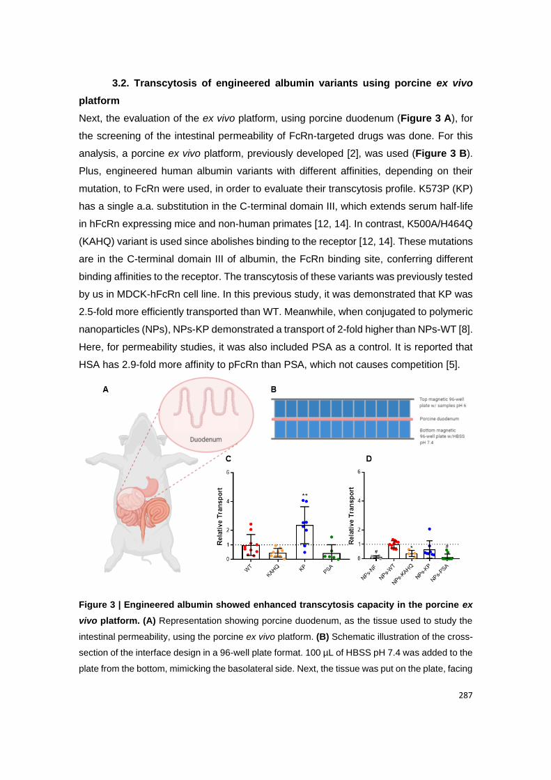
287
3.2. Transcytosis of engineered albumin variants using porcine ex vivo
platform
Next, the evaluation of the ex vivo platform, using porcine duodenum (Figure 3 A), for
the screening of the intestinal permeability of FcRn-targeted drugs was done. For this
analysis, a porcine ex vivo platform, previously developed [2], was used (Figure 3 B).
Plus, engineered human albumin variants with different affinities, depending on their
mutation, to FcRn were used, in order to evaluate their transcytosis profile. K573P (KP)
has a single a.a. substitution in the C-terminal domain III, which extends serum half-life
in hFcRn expressing mice and non-human primates [12, 14]. In contrast, K500A/H464Q
(KAHQ) variant is used since abolishes binding to the receptor [12, 14]. These mutations
are in the C-terminal domain III of albumin, the FcRn binding site, conferring different
binding affinities to the receptor. The transcytosis of these variants was previously tested
by us in MDCK-hFcRn cell line. In this previous study, it was demonstrated that KP was
2.5-fold more efficiently transported than WT. Meanwhile, when conjugated to polymeric
nanoparticles (NPs), NPs-KP demonstrated a transport of 2-fold higher than NPs-WT [8].
Here, for permeability studies, it was also included PSA as a control. It is reported that
HSA has 2.9-fold more affinity to pFcRn than PSA, which not causes competition [5].
Figure 3 | Engineered albumin showed enhanced transcytosis capacity in the porcine ex
vivo platform. (A) Representation showing porcine duodenum, as the tissue used to study the
intestinal permeability, using the porcine ex vivo platform. (B) Schematic illustration of the cross-
section of the interface design in a 96-well plate format. 100 µL of HBSS pH 7.4 was added to the
plate from the bottom, mimicking the basolateral side. Next, the tissue was put on the plate, facing
288
the apical side upwards. Finally, the magnetic plate from the top was added, pressing the tissue
to avoid leakage, and samples prepared in HBSS pH 6 were added. (C) Relative amounts of
naked albumin variants (WT, KAHQ, KP and PSA) detected in media collected at the basolateral
side, 4 h after adding the variants to the apical side in the porcine ex vivo platform (n=6-10); (D)
Relative amounts of NPs functionalized with albumin variants (n=4-10). The amounts were
quantified by two-way anti-albumin ELISA, where WT albumin was set to 1, as a control. These
results were obtained from 3 different pigs. Shown are the arithmetic means ± SD. ∗P < 0.05, ∗∗P
< 0.005, and #P < 0.0001, comparing with WT by one-way ANOVA Dunnett’s multiple comparison
test.
From the results from Figure 3 C, it is possible to verify that KP human albumin variant
maintains its pattern, of being more efficiently transported compared to WT, which is
translated in a ratio of 2.4-fold. These results confirm previous reported data related with
pFcRn binding affinity, which showed that kd (10-3/s) values for HSA-WT, HSA-KP and
PSA are around 15, 2.6 and 53, respectively [5]. kd is the constant of dissociation,
meaning that higher the affinity, lower the dissociation. The albumin quantification was
made by ELISA, using human albumin antibodies, which might not be the best way to
quantify PSA. However, it is known that PSA is 75.1% similar to HSA [5]. Despite this,
the results are in line with publish data, since PSA was less transported (Papp = 0.44 ±
0.53 x 10⁻⁶) by pFcRn, compared with WT (Table 1). Through the Papp obtained values
of free albumin variants, it was observed that these were 0.56-fold less transported in
this ex vivo platform, when correlating with results previously published using MDCK-
hFcRn in vitro model [8].
After this evaluation, albumin-functionalized NPs were also tested to further study the
potential of the porcine ex vivo platform (Figure 3 D). The produced NPs were previously
characterized, by being a monodisperse solution and having 150 nm in size, zeta
potential around -9 mV and 10% loaded with insulin [8]. The typical pattern behavior
between variants was observed, except for NPs-KP. Also, it was not expected that NPs,
as larger particles, presented higher Papp values, compared to free albumin.
Additionally, NPs-KAHQ and NPs-PSA were less transported as expected.
Nevertheless, non-functionalized NPs presented some signal, which is not supposed to
since they do not have HSA attached on the NPs’ surface. This might be due to the
presence of some possible interferents from the biologic fluids contained on the tissue.
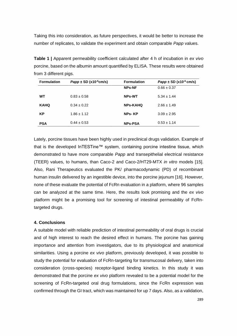
289
Taking this into consideration, as future perspectives, it would be better to increase the
number of replicates, to validate the experiment and obtain comparable Papp values.
Table 1 | Apparent permeability coefficient calculated after 4 h of incubation in ex vivo
porcine, based on the albumin amount quantified by ELISA. These results were obtained
from 3 different pigs.
Formulation Papp ± SD (x10-6cm/s) Formulation Papp ± SD (x10-6 cm/s)
NPs-NF 0.66 ± 0.37
WT 0.83 ± 0.58 NPs-WT 5.34 ± 1.44
KAHQ 0.34 ± 0.22 NPs-KAHQ 2.66 ± 1.49
KP 1.86 ± 1.12 NPs- KP 3.09 ± 2.95
PSA 0.44 ± 0.53 NPs-PSA 0.53 ± 1.14
Lately, porcine tissues have been highly used in preclinical drugs validation. Example of
that is the developed InTESTine™ system, containing porcine intestine tissue, which
demonstrated to have more comparable Papp and transepithelial electrical resistance
(TEER) values, to humans, than Caco-2 and Caco-2/HT29-MTX in vitro models [15].
Also, Rani Therapeutics evaluated the PK/ pharmacodynamic (PD) of recombinant
human insulin delivered by an ingestible device, into the porcine jejunum [16]. However,
none of these evaluate the potential of FcRn evaluation in a platform, where 96 samples
can be analyzed at the same time. Here, the results look promising and the ex vivo
platform might be a promising tool for screening of intestinal permeability of FcRn-
targeted drugs.
4. Conclusions
A suitable model with reliable prediction of intestinal permeability of oral drugs is crucial
and of high interest to reach the desired effect in humans. The porcine has gaining
importance and attention from investigators, due to its physiological and anatomical
similarities. Using a porcine ex vivo platform, previously developed, it was possible to
study the potential for evaluation of FcRn-targeting for transmucosal delivery, taken into
consideration (cross-species) receptor-ligand binding kinetics. In this study it was
demonstrated that the porcine ex vivo platform revealed to be a potential model for the
screening of FcRn-targeted oral drug formulations, since the FcRn expression was
confirmed through the GI tract, which was maintained for up 7 days. Also, as a validation,
290
through an intestinal permeability assay it was shown the characteristic pattern of
engineered human albumin variants. Therefore, this platform may have a positive and
significant impact on the development of preclinical FcRn-targeted drugs, saving some
time and money of the research by providing more reliable data.
Declaration of Competing Interest
G.T is a co-inventor of pending patent applications related to porcine ex vivo platform.
J.T.A. is a co-inventor of pending patent applications related to albumin variants.
Acknowledgements
This work was financed by Portuguese funds through FCT - Fundação para a Ciência e
a Tecnologia/ Ministério da Ciência, Tecnologia e Ensino Superior in the framework of
the project "Institute for Research and Innovation in Health Sciences"
UID/BIM/04293/2019. Cláudia Azevedo would like to thank to Fundação para a Ciência
e a Tecnologia (FCT, SFRH/BD/117598/2016), Portugal for financial support, as well as
recognition gave by Fulbright (PS00293218) and a grant from Fundação Luso Americana
para o Desenvolvimento (FLAD, Proj. 137/2019). The authors acknowledge the support
of Carneiro&Salgueirinho for some porcine tissue. This work was also supported by the
Koch Institute Support (core) Grant P30-CA14051 from the National Cancer Institute.
We thank the Hope Babette Tang Histology Facility at the Koch Institute at MIT for the
histology work and consultation.
Author Contributions
C.A., G.T and B.S. designed research; C.A. performed research; C.A. analyzed data;
C.A, G.T., J.T.A. and B.S. wrote the paper.
291
References
[1] A. Almeida, C. Azevedo, M.H. Macedo, B. Sarmento, 3D intestinal models towards a more
realistic permeability screening, in: Elsevier (Ed.) Nanotechnology for Oral Drug Delivery, 2020.
[2] T. von Erlach, S. Saxton, Y. Shi, D. Minahan, D. Reker, F. Javid, Y.-A.L. Lee, C.
Schoellhammer, T. Esfandiary, C. Cleveland, L. Booth, J. Lin, H. Levy, S. Blackburn, A. Hayward,
R. Langer, G. Traverso, Robotically handled whole-tissue culture system for the screening of oral
drug formulations, Nature Biomedical Engineering, (2020).
[3] B.L. Dickinson, K. Badizadegan, Z. Wu, J.C. Ahouse, X. Zhu, N.E. Simister, R.S. Blumberg,
W.I. Lencer, Bidirectional FcRn-dependent IgG transport in a polarized human intestinal epithelial
cell line, The Journal of clinical investigation, 104 (1999) 903-911.
[4] D.A. Volpe, Variability in Caco-2 and MDCK Cell-Based Intestinal Permeability Assays, Journal
of pharmaceutical sciences, 97 (2008) 712-725.
[5] D.S. Jason Cameron, Jan Terje Andersen, Inger Sandlie, Pharmacokinetic animal model in,
2018.
[6] C.M.A. Stirling, B. Charleston, H. Takamatsu, S. Claypool, W. Lencer, R.S. Blumberg, T.E.
Wileman, Characterization of the porcine neonatal Fc receptor--potential use for trans-epithelial
protein delivery, Immunology, 114 (2005) 542-553.
[7] L. Yang, W. Chonglong, L. Zhengzhu, X. Jingen, F. Weixuan, W. Wenwen, D. Xiangdong, L.
Jianfeng, Z. Qin, Tissues Expression, Polymorphisms Identification of FcRn Gene and Its
Relationship with Serum Classical Swine Fever Virus Antibody Level in Pigs, Asian-Australas J
Anim Sci, 25 (2012) 1089-1095.
[8] C. Azevedo, J. Nilsen, A. Grevys, R. Nunes, J.T. Andersen, B. Sarmento, Engineered albumin-
functionalized nanoparticles for improved FcRn binding enhances oral delivery of insulin, Journal
of Controlled Release, 327 (2020) 161-173.
[9] J. Fallingborg, Intraluminal pH of the human gastrointestinal tract, Danish medical bulletin, 46
(1999) 183-196.
[10] L. Ovesen, F. Bendtsen, U. Tage-Jensen, N.T. Pedersen, B.R. Gram, S.J. Rune, Intraluminal
pH in the stomach, duodenum, and proximal jejunum in normal subjects and patients with
exocrine pancreatic insufficiency, Gastroenterology, 90 (1986) 958-962.
[11] D.C. Roopenian, S. Akilesh, FcRn: the neonatal Fc receptor comes of age, Nature Reviews
Immunology, 7 (2007) 715.
[12] J.T. Andersen, B. Dalhus, J. Cameron, M.B. Daba, A. Plumridge, L. Evans, S.O. Brennan,
K.S. Gunnarsen, M. Bjørås, D. Sleep, I. Sandlie, Structure-based mutagenesis reveals the
albumin-binding site of the neonatal Fc receptor, Nature Communications, 3 (2012) 610.
[13] C. Azevedo, I. Pereira, B. Sarmento, Intestinal mucosal models to validate functionalized
nanosystems in: Wiley (Ed.) Characterization of Pharmaceutical Nano- and Microsystems, 2020.
[14] J.T. Andersen, B. Dalhus, D. Viuff, B.T. Ravn, K.S. Gunnarsen, A. Plumridge, K. Bunting, F.
Antunes, R. Williamson, S. Athwal, E. Allan, L. Evans, M. Bjoras, S. Kjaerulff, D. Sleep, I. Sandlie,
292
J. Cameron, Extending serum half-life of albumin by engineering neonatal Fc receptor (FcRn)
binding, The Journal of biological chemistry, 289 (2014) 13492-13502.
[15] J. Westerhout, E.v.d. Steeg, D. Grossouw, E.E. Zeijdner, C.A.M. Krul, M. Verwei, H.M.
Wortelboer, A new approach to predict human intestinal absorption using porcine intestinal tissue
and biorelevant matrices, European Journal of Pharmaceutical Sciences, 63 (2014) 167-177.
[16] M. Hashim, R. Korupolu, B. Syed, K. Horlen, S. Beraki, P. Karamchedu, A.K. Dhalla, R. Ruffy,
M. Imran, Jejunal wall delivery of insulin via an ingestible capsule in anesthetized swine-A
pharmacokinetic and pharmacodynamic study, Pharmacology research & perspectives, 7 (2019)
e00522.
[17] Kenhub, Histology of the lower digestive tract: want to learn more about it?, in,
https://www.kenhub.com/en/library/anatomy/histology-of-the-lower-digestive-tract, 2020.
[18] L.M. Gonzalez, A.J. Moeser, A.T. Blikslager, Porcine models of digestive disease: the future
of large animal translational research, Transl Res, 166 (2015) 12-27.
293
Supplementary material
The potential of porcine ex vivo platform for intestinal permeability
screening of FcRn-targeted drugs
Cláudia Azevedo, Jan Terje Andersen, Bruno Sarmento, Giovanni Traverso
1. Material and Methods
1.1. Hematoxylin and Eosin staining procedure
Sections of the porcine gastrointestinal (GI) tract were put into cassettes, fixed with 10%
(v/w) formalin for 24 h at 4ºC and then storage in 70% ethanol at 4ºC. Paraffin sections
(5-µm) on glass slides were dried at 70°C for 30 min, deparaffinized and heat induced
epitope retrieve (HIER) at 97ºC for 20 min in citrate buffer pH 6. These procedures were
completely automated by using a ThermoFisher IHC Autostainer 360. Briefly, for
hematoxylin and eosin (H&E) staining, paraffin sections were counterstained with
hematoxylin and eosin, according to standard protocols, using an automatic self-
contained slide stainer (ThermoShandon Varistain Gemini). The stained cells were then
mounted on a cover slide using ConsulMount (Thermo Fisher, #9990440).
Representative images using Koehler illumination were obtained with an Olympus
Microfire digital camera (M/N S97809) attached to an Olympus BX60 microscope.
2. Results
2.1. Tissue integrity of the porcine ex vivo model
Before studying the FcRn expression and albumin transcytosis, it was crucial to
understand how the porcine GI tract is organized. Initially, it was evaluated the viability
and morphology of the GI tract from the porcine ex vivo model, using H&E staining.
According to Figure S1 A it is possible to see morphologic differences throughout
intestinal sections. The porcine duodenum has a light-staining with a branched and
tubular structure with Brunner’s glands in its sub mucosa, which has a protective role
against the acidic chyme. Ileum is highlighted by the presence of lymphatic aggregates,
as Peyer’s patches, with phagocytic M-cells in the sub mucosa and lamina propria
regions. On the other hand, jejunum is marked by the lack of Brunner’s glands and
Peyer’s patches. In general, the absence of villi and Paneth cells and reduction of lymph
nodes aggregates distinguishes the large from the small intestine. However, crypts and
294
goblet cells are more abundant in the colon. By contrast, esophagus and stomach have
a different composition and cell types, since they are not responsible for the absorption
function. For instance, the mucosa of the stomach is in a columnar format and is covered
with a protective layer of alkaline mucus. In its epithelium it is possible to find gastric
glands, mucus neck cells to produce mucus, parietal cells to secrete acid, and chief cells
to release pepsinogen. These evidences are in line with what is described for humans
[17].
As it is shown, most of the GI tract presents mucosa, sub mucosa, mucularis externa
and serosa, which is particularly exemplified by a duodenum section (Figure S1 B). The
human intestine is described by having the same pattern and characteristics. The
mucosa has an importance role in absorption, since it is 90% composed by enterocytes
and microvilli (capillary branches as a door for the hepatic portal vein). Mucosa is divided
in epithelium, lamina propria (connective tissue), muscularis mucosae (smooth muscle)
and serosa (mesothelium). It is in epithelium where cells can proliferate, differentiate in
absorptive enterocytes or mucus secretory cells (goblet cells, enteroendocrine cells and
Paneth cells) and be apoptosed, by the presence of proliferative crypts. Particularly,
enterocytes, as polarized cells, are the ones responsible for absorbing nutrients and
transcytose them from the apical to the basolateral side. On the other hand, M cells
control the transepithelial transport of macromolecules or particles and microorganisms.
Sub mucosa is characterized by being a connective tissue with lymphatic and blood
vessels, ganglion and nerve cells. Finally, muscularis externa (smooth muscles) and a
serosa layer (Figure S1 C) [13].
However, it is reported that porcine and human intestine present some anatomical
differences, as the length of small intestine and shape of the colon, as described
elsewhere [18].
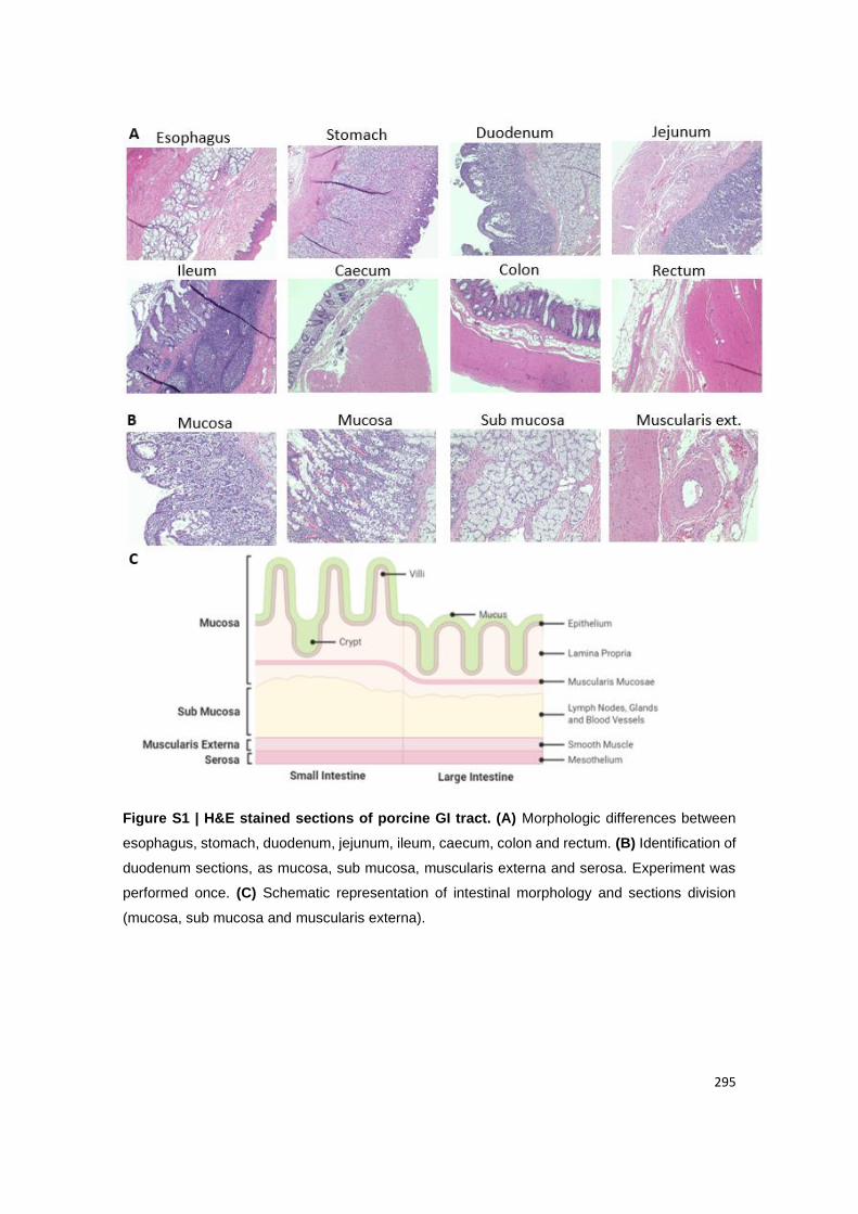
295
Figure S1 | H&E stained sections of porcine GI tract. (A) Morphologic differences between
esophagus, stomach, duodenum, jejunum, ileum, caecum, colon and rectum. (B) Identification of
duodenum sections, as mucosa, sub mucosa, muscularis externa and serosa. Experiment was
performed once. (C) Schematic representation of intestinal morphology and sections division
(mucosa, sub mucosa and muscularis externa).

296
297
The effect of hypergravity on intestinal permeability of nanoformulations
and molecules
Cláudia Azevedo1,2,3,*, Maria Helena Macedo1,2,3,*, Andreia Almeida1,2,3,*, Soraia Pinto1,2,3,*, Jack
J. W. A. van Loon4,5, Bruno Sarmento1,2,6,#
1 INEB - Instituto de Engenharia Biomédica, Universidade do Porto, Porto, Portugal
2 i3S – Instituto de Investigação e Inovação em Saúde, Universidade do Porto, Porto, Portugal
3 Instituto de Ciências Biomédicas Abel Salazar, Universidade do Porto, Porto, Portugal
4 Department of Oral and Maxillofacial Surgery/Oral Pathology, Amsterdam Movement Sciences &
Amsterdam Bone Center (ABC), Amsterdam University Medical Center Location VUmc, Amsterdam, The
Netherlands
5 European Space Agency (ESA), Technology Center (ESTEC), TEC-MMG LIS lab, Noordwijk, The
Netherlands
6 CESPU, Instituto de Investigação e Formação Avançada em Ciências e Tecnologias da Saúde, Gandra,
Portugal
*Equal contribution
#Corresponding author: Tel +351 226074949, [email protected]
298
Abstract
The oral administration of drugs remains a challenge due to rapid enzymatic degradation
and minimal absorption in the gastrointestinal tract. According to the literature,
mechanical forces, namely hypergravity can interfere with cellular integrity and drug
absorption, and there is no study describing its influence in the intestinal permeability. In
this study, the effect of hypergravity on intestinal Caco-2 cells and its influence in the
intestinal permeability of different nanoformulations and molecules was studied using the
large diameter centrifuge from the European Space Research and Technology Centre
of the European Space Agency. It was shown that the cellular metabolic activity and
integrity was maintained after exposure to different gravity levels. Expression of
important drug transporters and tight junctions’ proteins was evaluated and, interestingly,
most proteins demonstrated a switch of behavior in their expression. Furthermore,
paracellular transport of FITC-Dextran showed to significantly increase with hypergravity,
which agrees with the decrease of transepithelial electrical resistance and the increase
of claudin-2 at higher g-levels. The diffusion of camptothecin released from polymeric
micelles revealed a significant decrease, which agrees with the increased expression of
the P-gp observed with the increase in g-levels, responsible for pumping this drug out.
The neonatal Fc receptor-mediated transport of albumin-functionalized nanoparticles
loaded with insulin showed no significant changes when increasing the gravity levels. A
good correlation of cellular permeability studies in hypergravity is important when
developing new therapeutic nanoformulations. Thus, this study supports the potential of
simulated hypergravity to be used in oral delivery of nanoformulations and molecules.
Keywords: Caco-2 model; intestinal absorption; large diameter centrifuge;
nanoparticles; polymeric micelles.
Teaser: The current study shows that hypergravity influence the expression of certain
drug transporters and tight junctions’ proteins as well as the intestinal permeability of
nanoformulations and molecules.
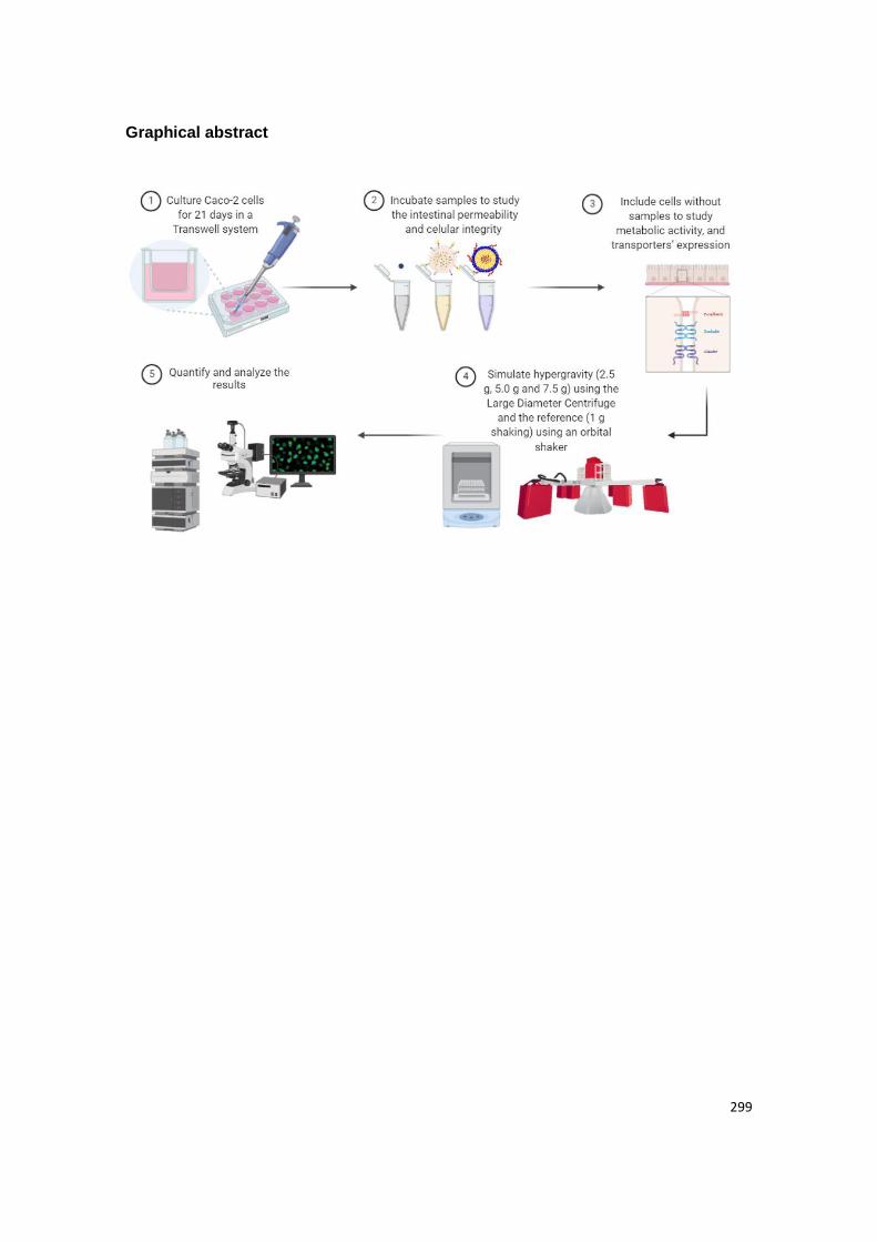
299
Graphical abstract
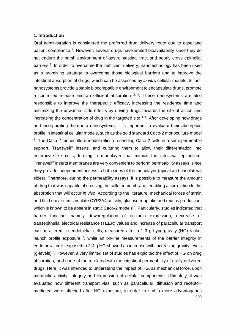
300
1. Introduction
Oral administration is considered the preferred drug delivery route due to ease and
patient compliance 1. However, several drugs have limited bioavailability since they do
not endure the harsh environment of gastrointestinal tract and poorly cross epithelial
barriers 2. In order to overcome the inefficient delivery, nanotechnology has been used
as a promising strategy to overcome those biological barriers and to improve the
intestinal absorption of drugs, which can be assessed by in vitro cellular models. In fact,
nanosystems provide a stable biocompatible environment to encapsulate drugs, promote
a controlled release and an efficient absorption 2, 3. These nanosystems are also
responsible to improve the therapeutic efficacy, increasing the residence time and
minimizing the unwanted side effects by driving drugs towards the site of action and
increasing the concentration of drug in the targeted site 1, 4. After developing new drugs
and incorporating them into nanosystems, it is important to evaluate their absorption
profile in intestinal cellular models, such as the gold standard Caco-2 monoculture model
5. The Caco-2 monoculture model relies on seeding Caco-2 cells in a semi-permeable
support, Transwell® inserts, and culturing them to allow their differentiation into
enterocyte-like cells, forming a monolayer that mimics the intestinal epithelium.
Transwell® inserts membranes are very convenient to perform permeability assays, since
they provide independent access to both sides of the monolayer (apical and basolateral
sides). Therefore, during the permeability assays, it is possible to measure the amount
of drug that was capable of crossing the cellular membrane, enabling a correlation to the
absorption that will occur in vivo. According to the literature, mechanical forces of strain
and fluid shear can stimulate CYP3A4 activity, glucose reuptake and mucus production,
which is known to be absent in static Caco-2 models 6. Particularly, studies indicated that
barrier function, namely downregulation of occludin expression, decrease of
transepithelial electrical resistance (TEER) values and increase of paracellular transport
can be altered, in endothelial cells, measured after a 1-3 g hypergravity (HG) rocket
launch profile exposure 7, while an on-line measurements of the barrier integrity in
endothelial cells exposed to 2-4 g HG showed an increase with increasing gravity levels
(g-levels) 8. However, a very limited set of studies has exploited the effect of HG on drug
absorption, and none of them related with the intestinal permeability of orally delivered
drugs. Here, it was intended to understand the impact of HG, as mechanical force, upon
metabolic activity, integrity and expression of cellular components. Ultimately, it was
evaluated how different transport vias, such as paracellular, diffusion and receptor-
mediated were affected after HG exposure, in order to find a more advantageous
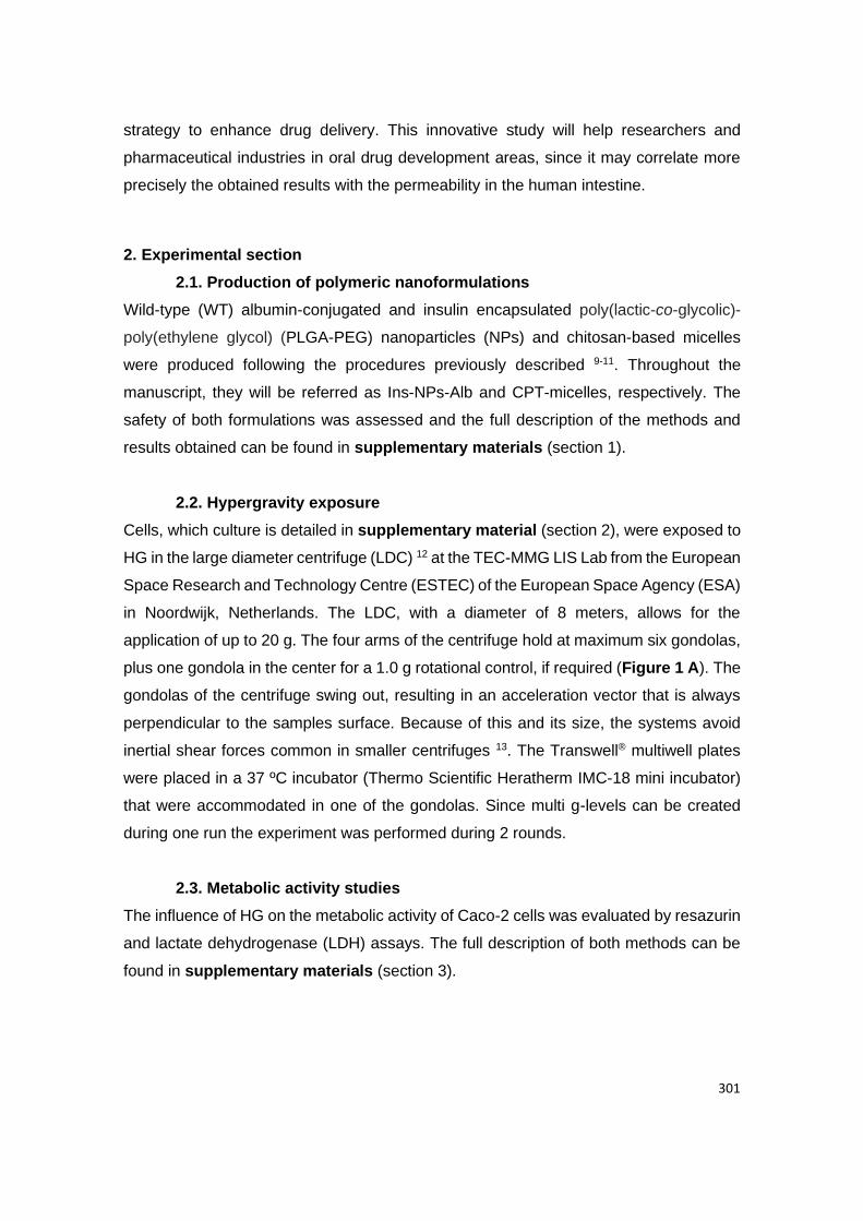
301
strategy to enhance drug delivery. This innovative study will help researchers and
pharmaceutical industries in oral drug development areas, since it may correlate more
precisely the obtained results with the permeability in the human intestine.
2. Experimental section
2.1. Production of polymeric nanoformulations
Wild-type (WT) albumin-conjugated and insulin encapsulated poly(lactic-co-glycolic)-
poly(ethylene glycol) (PLGA-PEG) nanoparticles (NPs) and chitosan-based micelles
were produced following the procedures previously described 9-11. Throughout the
manuscript, they will be referred as Ins-NPs-Alb and CPT-micelles, respectively. The
safety of both formulations was assessed and the full description of the methods and
results obtained can be found in supplementary materials (section 1).
2.2. Hypergravity exposure
Cells, which culture is detailed in supplementary material (section 2), were exposed to
HG in the large diameter centrifuge (LDC) 12 at the TEC-MMG LIS Lab from the European
Space Research and Technology Centre (ESTEC) of the European Space Agency (ESA)
in Noordwijk, Netherlands. The LDC, with a diameter of 8 meters, allows for the
application of up to 20 g. The four arms of the centrifuge hold at maximum six gondolas,
plus one gondola in the center for a 1.0 g rotational control, if required (Figure 1 A). The
gondolas of the centrifuge swing out, resulting in an acceleration vector that is always
perpendicular to the samples surface. Because of this and its size, the systems avoid
inertial shear forces common in smaller centrifuges 13. The Transwell® multiwell plates
were placed in a 37 ºC incubator (Thermo Scientific Heratherm IMC-18 mini incubator)
that were accommodated in one of the gondolas. Since multi g-levels can be created
during one run the experiment was performed during 2 rounds.
2.3. Metabolic activity studies
The influence of HG on the metabolic activity of Caco-2 cells was evaluated by resazurin
and lactate dehydrogenase (LDH) assays. The full description of both methods can be
found in supplementary materials (section 3).
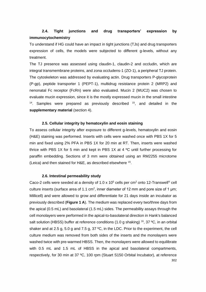
302
2.4. Tight junctions and drug transporters’ expression by
immunocytochemistry
To understand if HG could have an impact in tight junctions (TJs) and drug transporters
expression of cells, the models were subjected to different g-levels, without any
treatment.
The TJ presence was assessed using claudin-1, claudin-2 and occludin, which are
integral transmembrane proteins, and zona occludens-1 (ZO-1), a peripheral TJ protein.
The cytoskeleton was addressed by evaluating actin. Drug transporters P-glycoprotein
(P-gp), peptide transporter 1 (PEPT-1), multidrug resistance protein 2 (MRP2) and
nenonatal Fc receptor (FcRn) were also evaluated. Mucin 2 (MUC2) was chosen to
evaluate mucin expression, since it is the mostly expressed mucin in the small intestine
14. Samples were prepared as previously described 15, and detailed in the
supplementary material (section 4).
2.5. Cellular integrity by hematoxylin and eosin staining
To assess cellular integrity after exposure to different g-levels, hematoxylin and eosin
(H&E) staining was performed. Inserts with cells were washed once with PBS 1X for 5
min and fixed using 2% PFA in PBS 1X for 20 min at RT. Then, inserts were washed
thrice with PBS 1X for 5 min and kept in PBS 1X at 4 ºC until further processing for
paraffin embedding. Sections of 3 mm were obtained using an RM2255 microtome
(Leica) and then stained for H&E, as described elsewhere 15.
2.6. Intestinal permeability study
Caco-2 cells were seeded at a density of 1.0 x 10⁵ cells per cm2 onto 12-Transwell® cell
culture inserts (surface area of 1.1 cm2, inner diameter of 12 mm and pore size of 1 μm;
Millicell) and were allowed to grow and differentiate for 21 days inside an incubator as
previously described (Figure 1 A). The medium was replaced every two/three days from
the apical (0.5 mL) and basolateral (1.5 mL) sides. The permeability assays through the
cell monolayers were performed in the apical-to-basolateral direction in Hank's balanced
salt solution (HBSS) buffer at reference conditions (1.0 g shaking) 16, 37 ºC, in an orbital
shaker and at 2.5 g, 5.0 g and 7.5 g, 37 ºC, in the LDC. Prior to the experiment, the cell
culture medium was removed from both sides of the inserts and the monolayers were
washed twice with pre-warmed HBSS. Then, the monolayers were allowed to equilibrate
with 0.5 mL and 1.5 mL of HBSS in the apical and basolateral compartments,
respectively, for 30 min at 37 ºC, 100 rpm (Stuart S150 Orbital Incubator), at reference
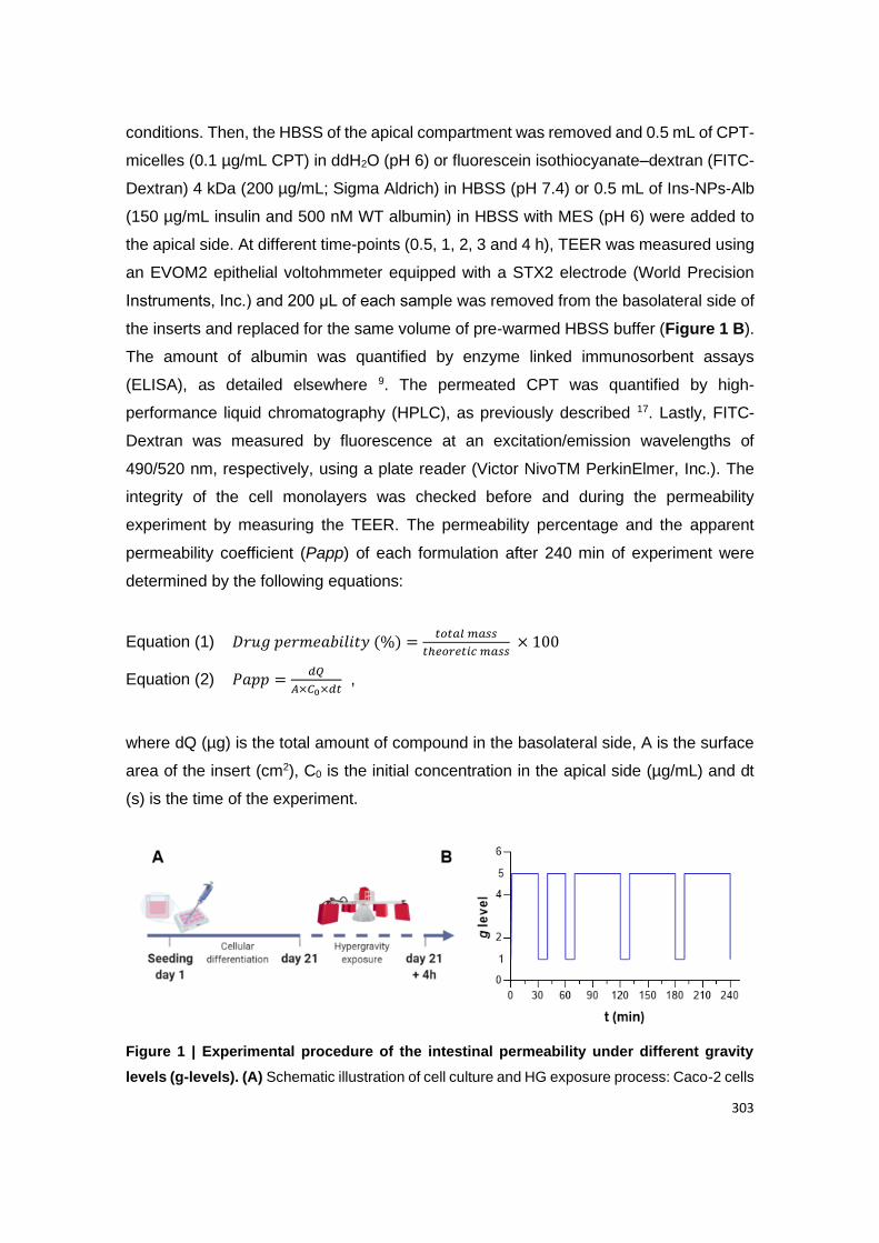
303
conditions. Then, the HBSS of the apical compartment was removed and 0.5 mL of CPT-
micelles (0.1 µg/mL CPT) in ddH2O (pH 6) or fluorescein isothiocyanate–dextran (FITC-
Dextran) 4 kDa (200 µg/mL; Sigma Aldrich) in HBSS (pH 7.4) or 0.5 mL of Ins-NPs-Alb
(150 µg/mL insulin and 500 nM WT albumin) in HBSS with MES (pH 6) were added to
the apical side. At different time-points (0.5, 1, 2, 3 and 4 h), TEER was measured using
an EVOM2 epithelial voltohmmeter equipped with a STX2 electrode (World Precision
Instruments, Inc.) and 200 μL of each sample was removed from the basolateral side of
the inserts and replaced for the same volume of pre-warmed HBSS buffer (Figure 1 B).
The amount of albumin was quantified by enzyme linked immunosorbent assays
(ELISA), as detailed elsewhere 9. The permeated CPT was quantified by high-
performance liquid chromatography (HPLC), as previously described 17. Lastly, FITC-
Dextran was measured by fluorescence at an excitation/emission wavelengths of
490/520 nm, respectively, using a plate reader (Victor NivoTM PerkinElmer, Inc.). The
integrity of the cell monolayers was checked before and during the permeability
experiment by measuring the TEER. The permeability percentage and the apparent
permeability coefficient (Papp) of each formulation after 240 min of experiment were
determined by the following equations:
Equation (1) 𝐷𝑟𝑢𝑔 𝑝𝑒𝑟𝑚𝑒𝑎𝑏𝑖𝑙𝑖𝑡𝑦 (%) =𝑡𝑜𝑡𝑎𝑙 𝑚𝑎𝑠𝑠
𝑡ℎ𝑒𝑜𝑟𝑒𝑡𝑖𝑐 𝑚𝑎𝑠𝑠 × 100
Equation (2) 𝑃𝑎𝑝𝑝 =𝑑𝑄
𝐴×𝐶0×𝑑𝑡 ,
where dQ (µg) is the total amount of compound in the basolateral side, A is the surface
area of the insert (cm2), C0 is the initial concentration in the apical side (µg/mL) and dt
(s) is the time of the experiment.
Figure 1 | Experimental procedure of the intestinal permeability under different gravity
levels (g-levels). (A) Schematic illustration of cell culture and HG exposure process: Caco-2 cells
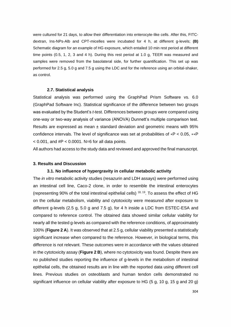
304
were cultured for 21 days, to allow their differentiation into enterocyte-like cells. After this, FITC-
dextran, Ins-NPs-Alb and CPT-micelles were incubated for 4 h, at different g-levels; (B)
Schematic diagram for an example of HG exposure, which entailed 10 min rest period at different
time points (0.5, 1, 2, 3 and 4 h). During this rest period at 1.0 g, TEER was measured and
samples were removed from the basolateral side, for further quantification. This set up was
performed for 2.5 g, 5.0 g and 7.5 g using the LDC and for the reference using an orbital-shaker,
as control.
2.7. Statistical analysis
Statistical analysis was performed using the GraphPad Prism Software vs. 6.0
(GraphPad Software Inc). Statistical significance of the difference between two groups
was evaluated by the Student’s t-test. Differences between groups were compared using
one-way or two-way analysis of variance (ANOVA) Dunnett’s multiple comparison test.
Results are expressed as mean ± standard deviation and geometric means with 95%
confidence intervals. The level of significance was set at probabilities of ∗P < 0.05, ∗∗P
< 0.001, and #P < 0.0001. N=6 for all data points.
All authors had access to the study data and reviewed and approved the final manuscript.
3. Results and Discussion
3.1. No influence of hypergravity in cellular metabolic activity
The in vitro metabolic activity studies (resazurin and LDH assays) were performed using
an intestinal cell line, Caco-2 clone, in order to resemble the intestinal enterocytes
(representing 90% of the total intestinal epithelial cells) 18, 19. To assess the effect of HG
on the cellular metabolism, viability and cytotoxicity were measured after exposure to
different g-levels (2.5 g, 5.0 g and 7.5 g), for 4 h inside a LDC from ESTEC-ESA and
compared to reference control. The obtained data showed similar cellular viability for
nearly all the tested g-levels as compared with the reference conditions, of approximately
100% (Figure 2 A). It was observed that at 2.5 g, cellular viability presented a statistically
significant increase when compared to the reference. However, in biological terms, this
difference is not relevant. These outcomes were in accordance with the values obtained
in the cytotoxicity assay (Figure 2 B), where no cytotoxicity was found. Despite there are
no published studies reporting the influence of g-levels in the metabolism of intestinal
epithelial cells, the obtained results are in line with the reported data using different cell
lines. Previous studies on osteoblasts and human tendon cells demonstrated no
significant influence on cellular viability after exposure to HG (5 g, 10 g, 15 g and 20 g)

305
during 3 and 4 h, respectively 20, 21. Additionally, the exposure of nasal epithelial cells to
10 g during three cycles of 20 min, with 20 min resting periods between each time, also
did not cause significant loss of cellular metabolic activity 22.
Figure 2 | Evaluation of metabolic activity of Caco-2 cells after HG exposure. (A) Evaluation
of cellular viability through resazurin assay; (B) Evaluation of cellular toxicity through LDH assay.
Shown are the arithmetic means ± SD. *P<0.05 comparing with the reference, by one-way
analysis of variance (ANOVA) Dunnett’s multiple comparison test. N=6.
3.2. Hypergravity might induce a tipping point on the expression of cellular
components
Before studying the effect of HG upon transport, it was crucial to understand how
transporters and TJs proteins might be affected (Figure 3 A).
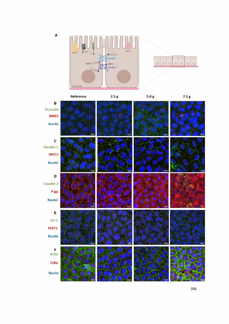
306
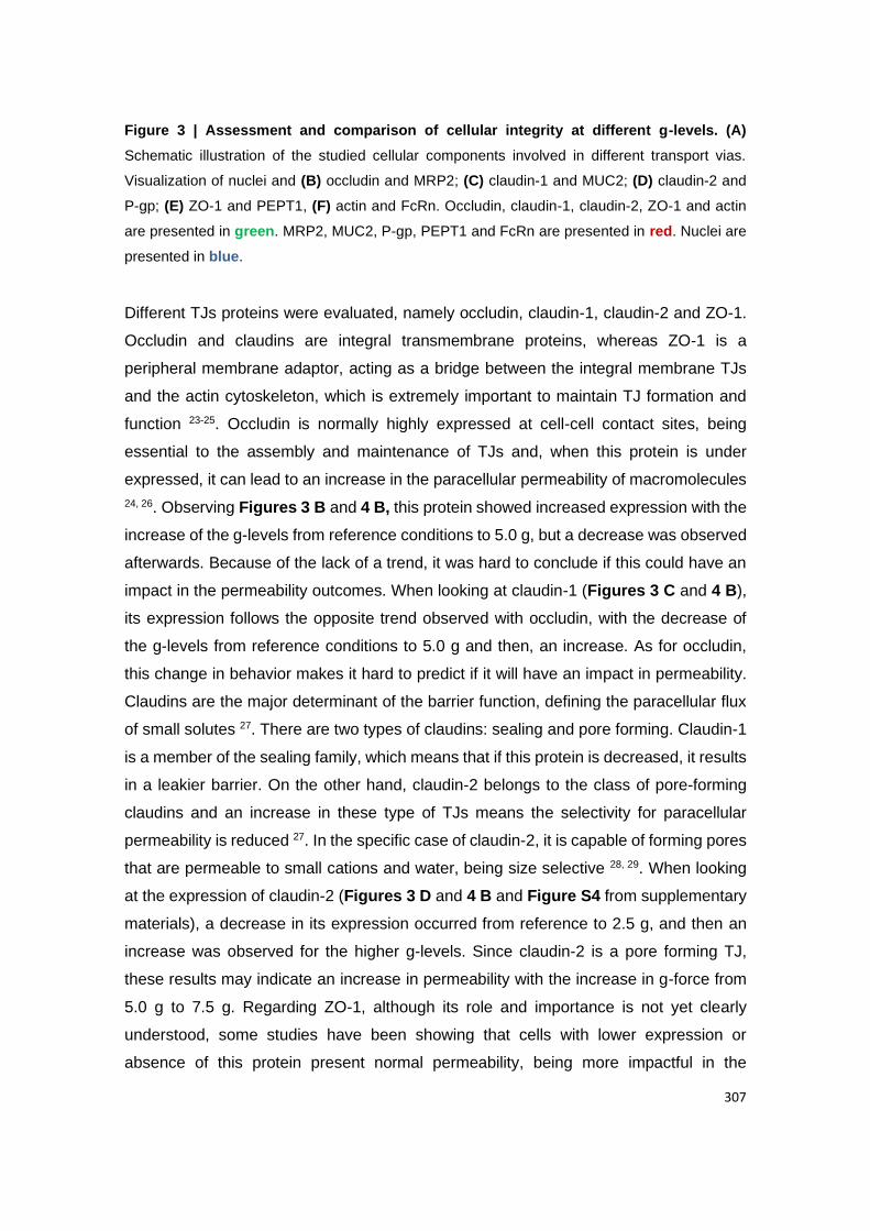
307
Figure 3 | Assessment and comparison of cellular integrity at different g-levels. (A)
Schematic illustration of the studied cellular components involved in different transport vias.
Visualization of nuclei and (B) occludin and MRP2; (C) claudin-1 and MUC2; (D) claudin-2 and
P-gp; (E) ZO-1 and PEPT1, (F) actin and FcRn. Occludin, claudin-1, claudin-2, ZO-1 and actin
are presented in green. MRP2, MUC2, P-gp, PEPT1 and FcRn are presented in red. Nuclei are
presented in blue.
Different TJs proteins were evaluated, namely occludin, claudin-1, claudin-2 and ZO-1.
Occludin and claudins are integral transmembrane proteins, whereas ZO-1 is a
peripheral membrane adaptor, acting as a bridge between the integral membrane TJs
and the actin cytoskeleton, which is extremely important to maintain TJ formation and
function 23-25. Occludin is normally highly expressed at cell-cell contact sites, being
essential to the assembly and maintenance of TJs and, when this protein is under
expressed, it can lead to an increase in the paracellular permeability of macromolecules
24, 26. Observing Figures 3 B and 4 B, this protein showed increased expression with the
increase of the g-levels from reference conditions to 5.0 g, but a decrease was observed
afterwards. Because of the lack of a trend, it was hard to conclude if this could have an
impact in the permeability outcomes. When looking at claudin-1 (Figures 3 C and 4 B),
its expression follows the opposite trend observed with occludin, with the decrease of
the g-levels from reference conditions to 5.0 g and then, an increase. As for occludin,
this change in behavior makes it hard to predict if it will have an impact in permeability.
Claudins are the major determinant of the barrier function, defining the paracellular flux
of small solutes 27. There are two types of claudins: sealing and pore forming. Claudin-1
is a member of the sealing family, which means that if this protein is decreased, it results
in a leakier barrier. On the other hand, claudin-2 belongs to the class of pore-forming
claudins and an increase in these type of TJs means the selectivity for paracellular
permeability is reduced 27. In the specific case of claudin-2, it is capable of forming pores
that are permeable to small cations and water, being size selective 28, 29. When looking
at the expression of claudin-2 (Figures 3 D and 4 B and Figure S4 from supplementary
materials), a decrease in its expression occurred from reference to 2.5 g, and then an
increase was observed for the higher g-levels. Since claudin-2 is a pore forming TJ,
these results may indicate an increase in permeability with the increase in g-force from
5.0 g to 7.5 g. Regarding ZO-1, although its role and importance is not yet clearly
understood, some studies have been showing that cells with lower expression or
absence of this protein present normal permeability, being more impactful in the
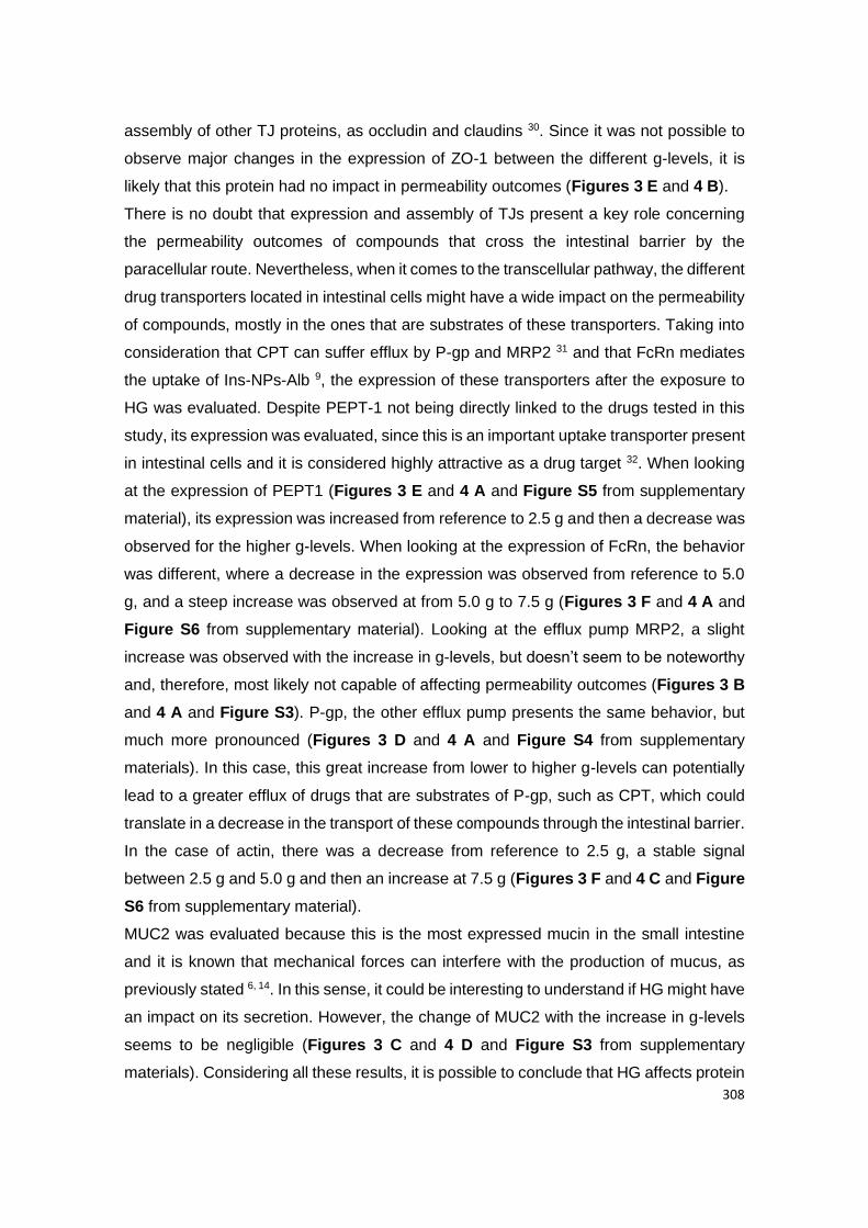
308
assembly of other TJ proteins, as occludin and claudins 30. Since it was not possible to
observe major changes in the expression of ZO-1 between the different g-levels, it is
likely that this protein had no impact in permeability outcomes (Figures 3 E and 4 B).
There is no doubt that expression and assembly of TJs present a key role concerning
the permeability outcomes of compounds that cross the intestinal barrier by the
paracellular route. Nevertheless, when it comes to the transcellular pathway, the different
drug transporters located in intestinal cells might have a wide impact on the permeability
of compounds, mostly in the ones that are substrates of these transporters. Taking into
consideration that CPT can suffer efflux by P-gp and MRP2 31 and that FcRn mediates
the uptake of Ins-NPs-Alb 9, the expression of these transporters after the exposure to
HG was evaluated. Despite PEPT-1 not being directly linked to the drugs tested in this
study, its expression was evaluated, since this is an important uptake transporter present
in intestinal cells and it is considered highly attractive as a drug target 32. When looking
at the expression of PEPT1 (Figures 3 E and 4 A and Figure S5 from supplementary
material), its expression was increased from reference to 2.5 g and then a decrease was
observed for the higher g-levels. When looking at the expression of FcRn, the behavior
was different, where a decrease in the expression was observed from reference to 5.0
g, and a steep increase was observed at from 5.0 g to 7.5 g (Figures 3 F and 4 A and
Figure S6 from supplementary material). Looking at the efflux pump MRP2, a slight
increase was observed with the increase in g-levels, but doesn’t seem to be noteworthy
and, therefore, most likely not capable of affecting permeability outcomes (Figures 3 B
and 4 A and Figure S3). P-gp, the other efflux pump presents the same behavior, but
much more pronounced (Figures 3 D and 4 A and Figure S4 from supplementary
materials). In this case, this great increase from lower to higher g-levels can potentially
lead to a greater efflux of drugs that are substrates of P-gp, such as CPT, which could
translate in a decrease in the transport of these compounds through the intestinal barrier.
In the case of actin, there was a decrease from reference to 2.5 g, a stable signal
between 2.5 g and 5.0 g and then an increase at 7.5 g (Figures 3 F and 4 C and Figure
S6 from supplementary material).
MUC2 was evaluated because this is the most expressed mucin in the small intestine
and it is known that mechanical forces can interfere with the production of mucus, as
previously stated 6, 14. In this sense, it could be interesting to understand if HG might have
an impact on its secretion. However, the change of MUC2 with the increase in g-levels
seems to be negligible (Figures 3 C and 4 D and Figure S3 from supplementary
materials). Considering all these results, it is possible to conclude that HG affects protein
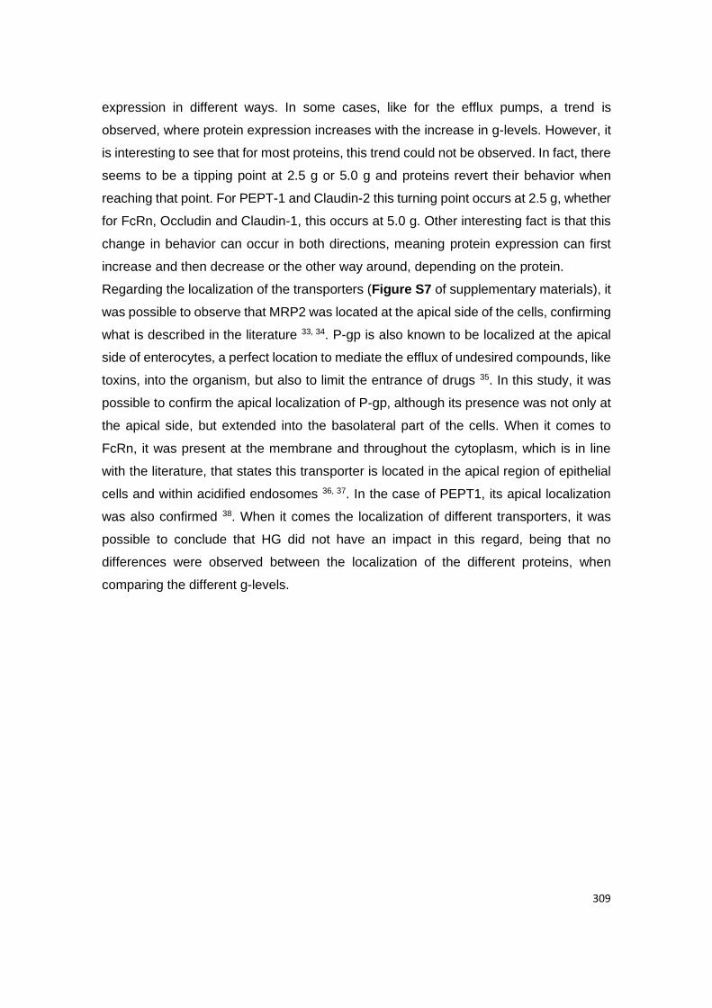
309
expression in different ways. In some cases, like for the efflux pumps, a trend is
observed, where protein expression increases with the increase in g-levels. However, it
is interesting to see that for most proteins, this trend could not be observed. In fact, there
seems to be a tipping point at 2.5 g or 5.0 g and proteins revert their behavior when
reaching that point. For PEPT-1 and Claudin-2 this turning point occurs at 2.5 g, whether
for FcRn, Occludin and Claudin-1, this occurs at 5.0 g. Other interesting fact is that this
change in behavior can occur in both directions, meaning protein expression can first
increase and then decrease or the other way around, depending on the protein.
Regarding the localization of the transporters (Figure S7 of supplementary materials), it
was possible to observe that MRP2 was located at the apical side of the cells, confirming
what is described in the literature 33, 34. P-gp is also known to be localized at the apical
side of enterocytes, a perfect location to mediate the efflux of undesired compounds, like
toxins, into the organism, but also to limit the entrance of drugs 35. In this study, it was
possible to confirm the apical localization of P-gp, although its presence was not only at
the apical side, but extended into the basolateral part of the cells. When it comes to
FcRn, it was present at the membrane and throughout the cytoplasm, which is in line
with the literature, that states this transporter is located in the apical region of epithelial
cells and within acidified endosomes 36, 37. In the case of PEPT1, its apical localization
was also confirmed 38. When it comes the localization of different transporters, it was
possible to conclude that HG did not have an impact in this regard, being that no
differences were observed between the localization of the different proteins, when
comparing the different g-levels.
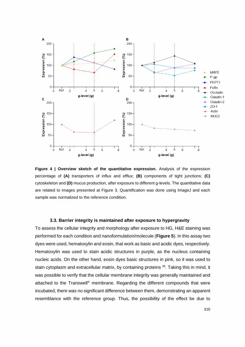
310
Figure 4 | Overview sketch of the quantitative expression. Analysis of the expression
percentage of (A) transporters of influx and efflux; (B) components of tight junctions; (C)
cytoskeleton and (D) mucus production, after exposure to different g-levels. The quantitative data
are related to images presented at Figure 3. Quantification was done using ImageJ and each
sample was normalized to the reference condition.
3.3. Barrier integrity is maintained after exposure to hypergravity
To assess the cellular integrity and morphology after exposure to HG, H&E staining was
performed for each condition and nanoformulation/molecule (Figure 5). In this assay two
dyes were used, hematoxylin and eosin, that work as basic and acidic dyes, respectively.
Hematoxylin was used to stain acidic structures in purple, as the nucleus containing
nucleic acids. On the other hand, eosin dyes basic structures in pink, so it was used to
stain cytoplasm and extracellular matrix, by containing proteins 39. Taking this in mind, it
was possible to verify that the cellular membrane integrity was generally maintained and
attached to the Transwell® membrane. Regarding the different compounds that were
incubated, there was no significant difference between them, demonstrating an apparent
resemblance with the reference group. Thus, the possibility of the effect be due to
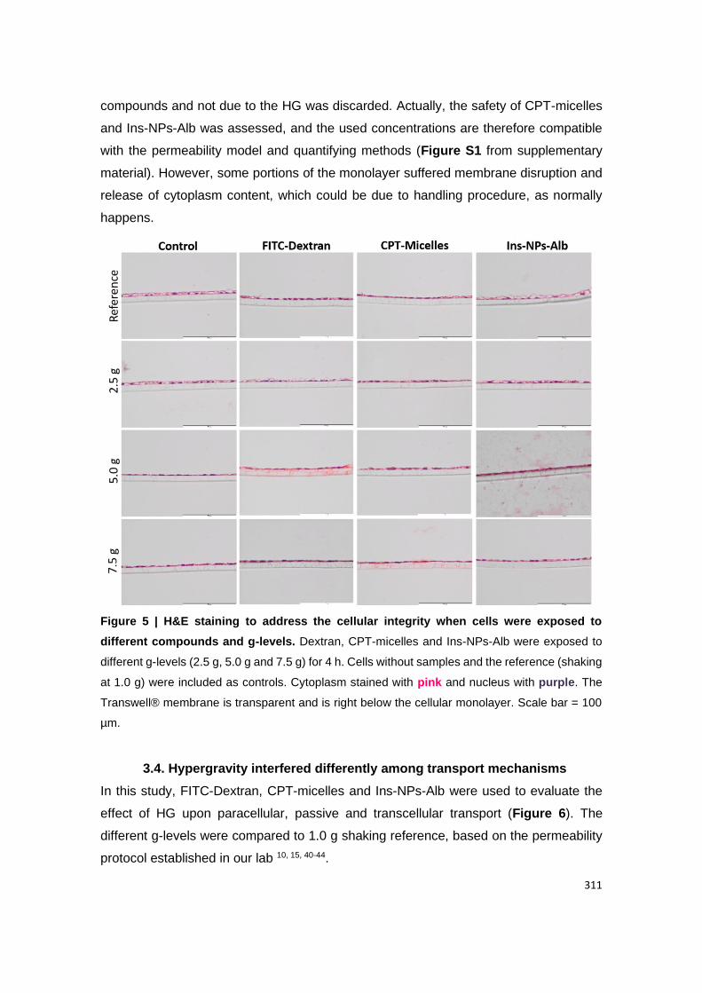
311
compounds and not due to the HG was discarded. Actually, the safety of CPT-micelles
and Ins-NPs-Alb was assessed, and the used concentrations are therefore compatible
with the permeability model and quantifying methods (Figure S1 from supplementary
material). However, some portions of the monolayer suffered membrane disruption and
release of cytoplasm content, which could be due to handling procedure, as normally
happens.
Figure 5 | H&E staining to address the cellular integrity when cells were exposed to
different compounds and g-levels. Dextran, CPT-micelles and Ins-NPs-Alb were exposed to
different g-levels (2.5 g, 5.0 g and 7.5 g) for 4 h. Cells without samples and the reference (shaking
at 1.0 g) were included as controls. Cytoplasm stained with pink and nucleus with purple. The
Transwell® membrane is transparent and is right below the cellular monolayer. Scale bar = 100
µm.
3.4. Hypergravity interfered differently among transport mechanisms
In this study, FITC-Dextran, CPT-micelles and Ins-NPs-Alb were used to evaluate the
effect of HG upon paracellular, passive and transcellular transport (Figure 6). The
different g-levels were compared to 1.0 g shaking reference, based on the permeability
protocol established in our lab 10, 15, 40-44.
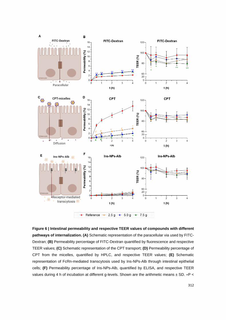
312
Figure 6 | Intestinal permeability and respective TEER values of compounds with different
pathways of internalization. (A) Schematic representation of the paracellular via used by FITC-
Dextran; (B) Permeability percentage of FITC-Dextran quantified by fluorescence and respective
TEER values; (C) Schematic representation of the CPT transport; (D) Permeability percentage of
CPT from the micelles, quantified by HPLC, and respective TEER values; (E) Schematic
representation of FcRn-mediated transcytosis used by Ins-NPs-Alb through intestinal epithelial
cells; (F) Permeability percentage of Ins-NPs-Alb, quantified by ELISA, and respective TEER
values during 4 h of incubation at different g-levels. Shown are the arithmetic means ± SD. ∗P <
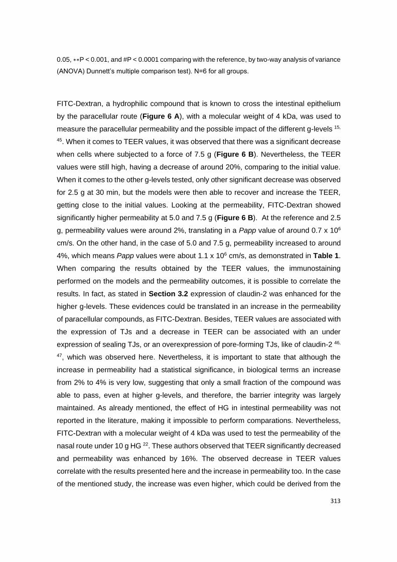
313
0.05, ∗∗P < 0.001, and #P < 0.0001 comparing with the reference, by two-way analysis of variance
(ANOVA) Dunnett’s multiple comparison test). N=6 for all groups.
FITC-Dextran, a hydrophilic compound that is known to cross the intestinal epithelium
by the paracellular route (Figure 6 A), with a molecular weight of 4 kDa, was used to
measure the paracellular permeability and the possible impact of the different g-levels 15,
45. When it comes to TEER values, it was observed that there was a significant decrease
when cells where subjected to a force of 7.5 g (Figure 6 B). Nevertheless, the TEER
values were still high, having a decrease of around 20%, comparing to the initial value.
When it comes to the other g-levels tested, only other significant decrease was observed
for 2.5 g at 30 min, but the models were then able to recover and increase the TEER,
getting close to the initial values. Looking at the permeability, FITC-Dextran showed
significantly higher permeability at 5.0 and 7.5 g (Figure 6 B). At the reference and 2.5
g, permeability values were around 2%, translating in a Papp value of around 0.7 x 10⁶
cm/s. On the other hand, in the case of 5.0 and 7.5 g, permeability increased to around
4%, which means Papp values were about 1.1 x 106 cm/s, as demonstrated in Table 1.
When comparing the results obtained by the TEER values, the immunostaining
performed on the models and the permeability outcomes, it is possible to correlate the
results. In fact, as stated in Section 3.2 expression of claudin-2 was enhanced for the
higher g-levels. These evidences could be translated in an increase in the permeability
of paracellular compounds, as FITC-Dextran. Besides, TEER values are associated with
the expression of TJs and a decrease in TEER can be associated with an under
expression of sealing TJs, or an overexpression of pore-forming TJs, like of claudin-2 46,
47, which was observed here. Nevertheless, it is important to state that although the
increase in permeability had a statistical significance, in biological terms an increase
from 2% to 4% is very low, suggesting that only a small fraction of the compound was
able to pass, even at higher g-levels, and therefore, the barrier integrity was largely
maintained. As already mentioned, the effect of HG in intestinal permeability was not
reported in the literature, making it impossible to perform comparations. Nevertheless,
FITC-Dextran with a molecular weight of 4 kDa was used to test the permeability of the
nasal route under 10 g HG 22. These authors observed that TEER significantly decreased
and permeability was enhanced by 16%. The observed decrease in TEER values
correlate with the results presented here and the increase in permeability too. In the case
of the mentioned study, the increase was even higher, which could be derived from the
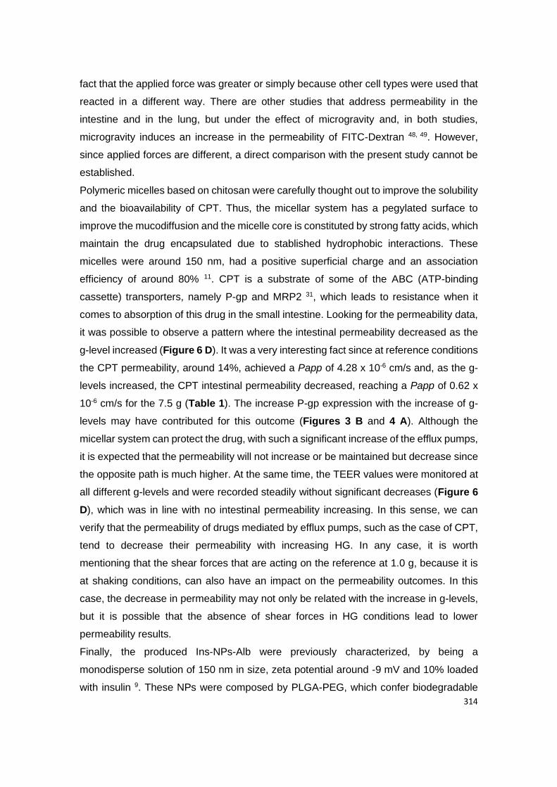
314
fact that the applied force was greater or simply because other cell types were used that
reacted in a different way. There are other studies that address permeability in the
intestine and in the lung, but under the effect of microgravity and, in both studies,
microgravity induces an increase in the permeability of FITC-Dextran 48, 49. However,
since applied forces are different, a direct comparison with the present study cannot be
established.
Polymeric micelles based on chitosan were carefully thought out to improve the solubility
and the bioavailability of CPT. Thus, the micellar system has a pegylated surface to
improve the mucodiffusion and the micelle core is constituted by strong fatty acids, which
maintain the drug encapsulated due to stablished hydrophobic interactions. These
micelles were around 150 nm, had a positive superficial charge and an association
efficiency of around 80% 11. CPT is a substrate of some of the ABC (ATP-binding
cassette) transporters, namely P-gp and MRP2 31, which leads to resistance when it
comes to absorption of this drug in the small intestine. Looking for the permeability data,
it was possible to observe a pattern where the intestinal permeability decreased as the
g-level increased (Figure 6 D). It was a very interesting fact since at reference conditions
the CPT permeability, around 14%, achieved a Papp of 4.28 x 10⁻⁶ cm/s and, as the g-
levels increased, the CPT intestinal permeability decreased, reaching a Papp of 0.62 x
10-6 cm/s for the 7.5 g (Table 1). The increase P-gp expression with the increase of g-
levels may have contributed for this outcome (Figures 3 B and 4 A). Although the
micellar system can protect the drug, with such a significant increase of the efflux pumps,
it is expected that the permeability will not increase or be maintained but decrease since
the opposite path is much higher. At the same time, the TEER values were monitored at
all different g-levels and were recorded steadily without significant decreases (Figure 6
D), which was in line with no intestinal permeability increasing. In this sense, we can
verify that the permeability of drugs mediated by efflux pumps, such as the case of CPT,
tend to decrease their permeability with increasing HG. In any case, it is worth
mentioning that the shear forces that are acting on the reference at 1.0 g, because it is
at shaking conditions, can also have an impact on the permeability outcomes. In this
case, the decrease in permeability may not only be related with the increase in g-levels,
but it is possible that the absence of shear forces in HG conditions lead to lower
permeability results.
Finally, the produced Ins-NPs-Alb were previously characterized, by being a
monodisperse solution of 150 nm in size, zeta potential around -9 mV and 10% loaded
with insulin 9. These NPs were composed by PLGA-PEG, which confer biodegradable
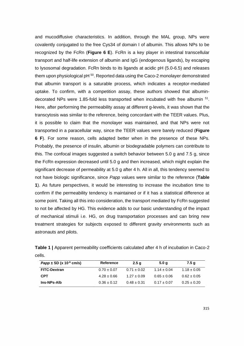
315
and mucodiffusive characteristics. In addition, through the MAL group, NPs were
covalently conjugated to the free Cys34 of domain I of albumin. This allows NPs to be
recognized by the FcRn (Figure 6 E). FcRn is a key player in intestinal transcellular
transport and half-life extension of albumin and IgG (endogenous ligands), by escaping
to lysosomal degradation. FcRn binds to its ligands at acidic pH (5.0-6.5) and releases
them upon physiological pH 50. Reported data using the Caco-2 monolayer demonstrated
that albumin transport is a saturable process, which indicates a receptor-mediated
uptake. To confirm, with a competition assay, these authors showed that albumin-
decorated NPs were 1.85-fold less transported when incubated with free albumin 51.
Here, after performing the permeability assay at different g-levels, it was shown that the
transcytosis was similar to the reference, being concordant with the TEER values. Plus,
it is possible to claim that the monolayer was maintained, and that NPs were not
transported in a paracellular way, since the TEER values were barely reduced (Figure
6 F). For some reason, cells adapted better when in the presence of these NPs.
Probably, the presence of insulin, albumin or biodegradable polymers can contribute to
this. The confocal images suggested a switch behavior between 5.0 g and 7.5 g, since
the FcRn expression decreased until 5.0 g and then increased, which might explain the
significant decrease of permeability at 5.0 g after 4 h. All in all, this tendency seemed to
not have biologic significance, since Papp values were similar to the reference (Table
1). As future perspectives, it would be interesting to increase the incubation time to
confirm if the permeability tendency is maintained or if it has a statistical difference at
some point. Taking all this into consideration, the transport mediated by FcRn suggested
to not be affected by HG. This evidence adds to our basic understanding of the impact
of mechanical stimuli i.e. HG, on drug transportation processes and can bring new
treatment strategies for subjects exposed to different gravity environments such as
astronauts and pilots.
Table 1 | Apparent permeability coefficients calculated after 4 h of incubation in Caco-2
cells.
Papp ± SD (x 10-6 cm/s) Reference 2.5 g 5.0 g 7.5 g
FITC-Dextran 0.70 ± 0.07 0.71 ± 0.02 1.14 ± 0.04 1.18 ± 0.05
CPT 4.28 ± 0.66 1.27 ± 0.09 0.65 ± 0.06 0.62 ± 0.05
Ins-NPs-Alb 0.36 ± 0.12 0.48 ± 0.31 0.17 ± 0.07 0.25 ± 0.20
316
4. Conclusions
In this study, the effect of different HG levels (2.5 g, 5.0 g and 7.5 g) in the intestinal
transport of nanoformulations and molecules for oral delivery, as well as the influence of
HG in the expression of TJs and drug transporters were evaluated. According to our
results, the exposure to HG did not affect the metabolic activity and integrity of intestinal
epithelial cells. Notwithstanding, the expression of TJs and transporters of Caco-2 cells,
as well as the intestinal transport of molecules and nanoformulations by paracellular,
passive and transcellular pathways were affected by the exposure to different g-levels.
Regarding the paracellular transport, FITC-Dextran increased with the increase of g-
levels. This can be also related with the increase of claudin-2 expression for higher g-
levels. On the other side, the passive transport of CPT from micelles by diffusion
decreased with the augment of g-levels, which is in line with the general increase of P-
gp expression, responsible for CPT efflux. Lastly, the receptor-mediated transport of Ins-
NPs-Alb by FcRn was maintained after exposure to HG. This pioneer study was
important to understand biologic mechanisms and can be crucial when developing new
formulations for oral delivery.
Acknowledgements
This project was supported by the Spin Your Thesis! 2020 programme, organized by
European Space Agency (ESA) Education Office. Additional funding included FCT -
Fundação para a Ciência e a Tecnologia/ Ministério da Ciência, Tecnologia e Ensino
Superior in the framework of the project “Institute for Research and Innovation in Health
Sciences” (UID/BIM/04293/2019). The authors are thankful for the ESA professional
Education Office, Nigel Savage, Emmanuelle Aubert and Alan Dowson for all the support
before, during and after the campaign. Cláudia Azevedo (SFRH/BD/117598/2016),
Maria Helena Macedo (SFRH/BD/131587/2017), Andreia Almeida
(SFRH/BD/118721/2016) and Soraia Pinto (SFRH/BD/144719/2019) would like to thank
to Fundação para a Ciência e a Tecnologia (FCT), Portugal for financial support. The
authors acknowledge the support of the following i3S Scientific Platforms: Biointerfaces
and Nanotechnology (FCT - UID/BIM/04293/2019), Bioimaging [member of the national
infrastructure PPBI – Portuguese Platform of Bioimaging (PPBI-POCI-01-0145-FEDER-
022122)] and Histology and Electron Microscopy (PPBI-POCI-01-0145-FEDER-
022122). The authors also are very thankful to Alamelu Sundaresan and Vivel Mann
(Texas Southern University, USA) for been mentors of this project and for giving useful
discussion and suggestions. Lastly, the authors would like to thank Merck for kindly
317
providing material for the campaign as well as for financially supporting the project, and
Thais Rizzi of Perkin-Elmer for giving technical support with Victor Nivo equipment.
Conflict of Interest
All the authors declare no conflict of interest.
Author Contributions
C.A., H.M., A.A., S.P. and J.W.A.L designed research; C.A., H.M., A.A. and S.P.
performed research; C.A., H.M., A.A., S.P. and J.W.A.L. analyzed data; C.A., H.M., A.A.,
S.P. and B.S. wrote the paper.

318
References
1. Sarmento B, Ferreira D, Vasconcelos T. Polymer-based delivery systems for oral delivery
of peptides and proteins. In: Jorgensen L, Nielsen HM, eds. Delivery Technologies for
Biopharmaceuticals: Peptides, Proteins, Nucleic Acids and Vaccines: John Wiley & Sons,
Ltd, 2009:207-227.
2. Araújo F, Shrestha N, Granja PL, et al. Antihyperglycemic potential of incretins orally
delivered via nano and microsystems and subsequent glucoregulatory effects. Curr
Pharm Biotechnol 2014;15:609-619.
3. Pereira C, Araújo F, Granja PL, et al. Targeting membrane transporters and receptors as
a mean to optimize orally delivered biotecnhological based drugs through nanoparticle
delivery systems. Curr Pharm Biotechnol 2014;15:650-658.
4. Moulari B, Beduneau A, Pellequer Y, et al. Nanoparticle targeting to inflamed tissues of
gastrointestinal tract. Curr Drug Deliv 2013;10:9-17.
5. Hidalgo IJ, Raub TJ, Borchardt RT. Characterization of the human colon carcinoma cell
line (Caco-2) as a model system for intestinal epithelial permeability. Gastroenterology
1989;96:736-749.
6. Kim HJ, Ingber DE. Gut-on-a-Chip microenvironment induces human intestinal cells to
undergo villus differentiation. Integr Biol 2013;5:1130-1140.
7. Sumanasekera WK, Sumanasekera GU, Mattingly KA, et al. Estradiol and
dihydrotestosterone regulate endothelial cell barrier function after hypergravity-induced
alterations in MAPK activity. Am J Physiol Cell Physiol 2007;293:566-573.
8. Szulcek R, van Bezu J, Boonstra J, et al. Transient intervals of hyper-gravity enhance
endothelial barrier integrity: Impact of mechanical and gravitational forces measured
electrically. PLoS One 2015;10:e0144269.
9. Azevedo C, Nilsen J, Grevys A, et al. Engineered albumin-functionalized nanoparticles
for improved FcRn binding enhances oral delivery of insulin. J Control Release
2020;327:161-173.
10. Almeida A, Silva D, Gonçalves V, et al. Synthesis and characterization of chitosan-
grafted-polycaprolactone micelles for modulate intestinal paclitaxel delivery. Drug Deliv
Transl Res 2018;8:387-397.
11. Almeida A, Araújo M, Novoa-Carballal R, et al. Novel amphiphilic chitosan micelles as
carriers for hydrophobic anticancer drugs. Mat Sci Eng C 2020;112:110920.
12. Loon JJWAv, Krausse J, Cunha H, et al. The large diameter centrifuge, LDC, for life and
physical sciences and technology. 2008.
13. van Loon JJWA, Folgering EHTE, Bouten CVC, et al. Inertial shear forces and the use of
centrifuges in gravity research. What is the proper control? J Biomech Eng 2003;125:342-
346.
319
14. Schneider H, Pelaseyed T, Svensson F, et al. Study of mucin turnover in the small
intestine by in vivo labeling. Sci Rep 2018;8:5760.
15. Macedo MH, Martinez E, Barrias C, et al. Development of an improved 3D in vitro
intestinal model to perform permeability studies of paracellular compounds. Front Bioeng
Biotechnol 2020;8.
16. Hubatsch I, Ragnarsson EGE, Artursson P. Determination of drug permeability and
prediction of drug absorption in Caco-2 monolayers. Nat Protoc 2007;2:2111-2119.
17. Martins SM, Wendling T, Gonçalves VM, et al. Development and validation of a simple
reversed-phase HPLC method for the determination of camptothecin in animal organs
following administration in solid lipid nanoparticles. J Chromatogr B 2012;880:100-107.
18. Araújo F, Sarmento B. Towards the characterization of an in vitro triple co-culture
intestine cell model for permeability studies. Int J Pharmaceut 2013;458:128-34.
19. Pereira C, Costa J, Sarmento B, et al. 3.3 - Cell-based in vitro models for intestinal
permeability studies. In: Sarmento B, ed. Concepts and Models for Drug Permeability
Studies: Woodhead Publishing, 2016:57-81.
20. Searby ND, Steele CR, Globus RK. Influence of increased mechanical loading by
hypergravity on the microtubule cytoskeleton and prostaglandin E2 release in primary
osteoblasts. Am J Physiol Cell Physiol 2005;289:148-158.
21. Costa-Almeida R, Carvalho DTO, Ferreira MJS, et al. Continuous exposure to simulated
hypergravity-induced changes in proliferation, morphology, and gene expression of
human tendon cells. Stem Cells Dev 2018;27:858-869.
22. Kim D, Kim YH, Kwon S. Enhanced nasal drug delivery efficiency by increasing
mechanical loading using hypergravity. Sci Rep 2018;8:168.
23. Gonzalez-Mariscal L, Betanzos A, Nava P, et al. Tight junction proteins. Prog Biophys
Mol Biol 2003;81:1-44.
24. Lee B, Moon KM, Kim CY. Tight junction in the intestinal epithelium: Its association with
diseases and regulation by phytochemicals. J Immunol Res 2018;2018:2645465.
25. Lee SH. Intestinal permeability regulation by tight junction: implication on inflammatory
bowel diseases. Intest Res 2015;13:11-8.
26. Al-Sadi R, Khatib K, Guo S, et al. Occludin regulates macromolecule flux across the
intestinal epithelial tight junction barrier. Am J Physiol Gastroint Liver Physiol
2011;300:1054-1064.
27. Gunzel D, Yu AS. Claudins and the modulation of tight junction permeability. Physiol Rev
2013;93:525-569.
28. Van Itallie CM, Fanning AS, Anderson JM. Reversal of charge selectivity in cation or
anion-selective epithelial lines by expression of different claudins. Am J Physiol Renal
Physiol 2003;285:1078-1084.
320
29. Rosenthal R, Milatz S, Krug SM, et al. Claudin-2, a component of the tight junction, forms
a paracellular water channel. J Cell Sci 2010;123:1913-1921.
30. Umeda K, Matsui T, Nakayama M, et al. Establishment and characterization of cultured
epithelial cells lacking expression of ZO-1. J Biol Chem 2004;279:44785-44794.
31. Lalloo AK, Luo FR, Guo A, et al. Membrane transport of camptothecin: facilitation by
human P-glycoprotein (ABCB1) and multidrug resistance protein 2 (ABCC2). BMC Med
2004;2:16.
32. Estudante M, Morais JG, Soveral G, et al. Intestinal drug transporters: An overview. Adv
Drug Deliv Rev 2013;65:1340-1356.
33. Liu YH, Di YM, Zhou ZW, et al. Multidrug resistance-associated proteins and implications
in drug development. Clin Exp Pharmacol Physiol 2010;37:115-120.
34. Yu XQ, Xue CC, Wang G, et al. Multidrug resistance associated proteins as determining
factors of pharmacokinetics and pharmacodynamics of drugs. Curr Drug Metab
2007;8:787-802.
35. Hediger MA, Romero MF, Peng JB, et al. The ABCs of solute carriers: physiological,
pathological and therapeutic implications of human membrane transport
proteinsIntroduction. Pflugers Arch 2004;447:465-468.
36. Ober RJ, Martinez C, Vaccaro C, et al. Visualizing the site and dynamics of IgG salvage
by the MHC class I-related receptor, FcRn. J Immunol 2004;172:2021-2029.
37. Pyzik M, Rath T, Kuo TT, et al. Hepatic FcRn regulates albumin homeostasis and
susceptibility to liver injury. Proc Natl Acad Sci USA 2017;114:2862-2871.
38. Shugarts S, Benet LZ. The role of transporters in the pharmacokinetics of orally
administered drugs. Pharm Res 2009;26:2039-2054.
39. Fischer AH, Jacobson KA, Rose J, et al. Hematoxylin and eosin staining of tissue and
cell sections. CSH Protoc 2008;4986.
40. Silva DS, Almeida A, Prezotti FG, et al. Self-aggregates of 3, 6-O, O’-dimyristoylchitosan
derivative are effective in enhancing the solubility and intestinal permeability of
camptothecin. Carbohydr Polym 2017;177:178-186.
41. Silva DS, Almeida A, Prezotti F, et al. Synthesis and characterization of 3, 6-O, O’-
dimyristoyl chitosan micelles for oral delivery of paclitaxel. Colloids Surf B Biointerfaces
2017;152:220-228.
42. Silva DS, M dos Santos D, Almeida A, et al. N-(2-hydroxy)-propyl-3-trimethylammonium,
o-mysristoyl chitosan enhances the solubility and intestinal permeability of anticancer
curcumin. Pharmaceutics 2018;10:245.
43. Sgorla D, Lechanteur A, Almeida A, et al. Development and characterization of lipid-
polymeric nanoparticles for oral insulin delivery. Expert Opin Drug Deliv 2018;15:213-
222.
321
44. Heep G, Almeida A, Marcano R, et al. Zein-casein-lysine multicomposite nanoparticles
are effective in modulate the intestinal permeability of ferulic acid. Int J Biol Macromol
2019;138:244-251.
45. Woting A, Blaut M. Small intestinal permeability and gut-transit time determined with low
and high molecular weight fluorescein isothiocyanate-dextrans in C3H mice. Nutrients
2018;10:685.
46. Li N, Wang D, Sui Z, et al. Development of an improved three-dimensional in vitro
intestinal mucosa model for drug absorption evaluation. Tissue Eng Part C Methods
2013;19:708-719.
47. Yi B, Shim KY, Ha SK, et al. Three-dimensional in vitro gut model on a villi-shaped
collagen scaffold. Biochip J 2017;11:219-232.
48. Alvarez R, Stork CA, Sayoc-Becerra A, et al. A Simulated microgravity environment
causes a sustained defect in epithelial barrier function. Sci Rep 2019;9:17531.
49. dos Santos MA, Bosquillon C, Russomano T, et al. Modelling the effects of microgravity
on the permeability of air interface respiratory epithelial cell layers. Adv Space Res
2010;46:712-718.
50. Roopenian DC, Akilesh S. FcRn: the neonatal Fc receptor comes of age. Nat Rev
Immunol 2007;7:715.
51. Hashem L, Swedrowska M, Vllasaliu D. Intestinal uptake and transport of albumin
nanoparticles: potential for oral delivery. Nanomedicine 2018;13:1255-1265.
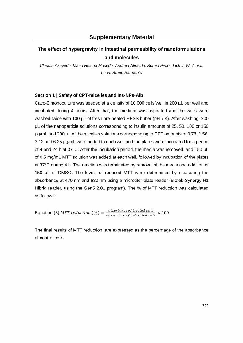
322
Supplementary Material
The effect of hypergravity in intestinal permeability of nanoformulations
and molecules
Cláudia Azevedo, Maria Helena Macedo, Andreia Almeida, Soraia Pinto, Jack J. W. A. van
Loon, Bruno Sarmento
Section 1 | Safety of CPT-micelles and Ins-NPs-Alb
Caco-2 monoculture was seeded at a density of 10 000 cells/well in 200 µL per well and
incubated during 4 hours. After that, the medium was aspirated and the wells were
washed twice with 100 μL of fresh pre-heated HBSS buffer (pH 7.4). After washing, 200
µL of the nanoparticle solutions corresponding to insulin amounts of 25, 50, 100 or 150
μg/mL and 200 µL of the micelles solutions corresponding to CPT amounts of 0.78, 1.56,
3.12 and 6.25 μg/mL were added to each well and the plates were incubated for a period
of 4 and 24 h at 37°C. After the incubation period, the media was removed, and 150 μL
of 0.5 mg/mL MTT solution was added at each well, followed by incubation of the plates
at 37°C during 4 h. The reaction was terminated by removal of the media and addition of
150 μL of DMSO. The levels of reduced MTT were determined by measuring the
absorbance at 470 nm and 630 nm using a microtiter plate reader (Biotek-Synergy H1
Hibrid reader, using the Gen5 2.01 program). The % of MTT reduction was calculated
as follows:
Equation (3) 𝑀𝑇𝑇 𝑟𝑒𝑑𝑢𝑐𝑡𝑖𝑜𝑛 (%) = 𝑎𝑏𝑠𝑜𝑟𝑏𝑎𝑛𝑐𝑒 𝑜𝑓 𝑡𝑟𝑒𝑎𝑡𝑒𝑑 𝑐𝑒𝑙𝑙𝑠
𝑎𝑏𝑠𝑜𝑟𝑏𝑎𝑛𝑐𝑒 𝑜𝑓 𝑢𝑛𝑡𝑟𝑒𝑎𝑡𝑒𝑑 𝑐𝑒𝑙𝑙𝑠 × 100
The final results of MTT reduction, are expressed as the percentage of the absorbance
of control cells.
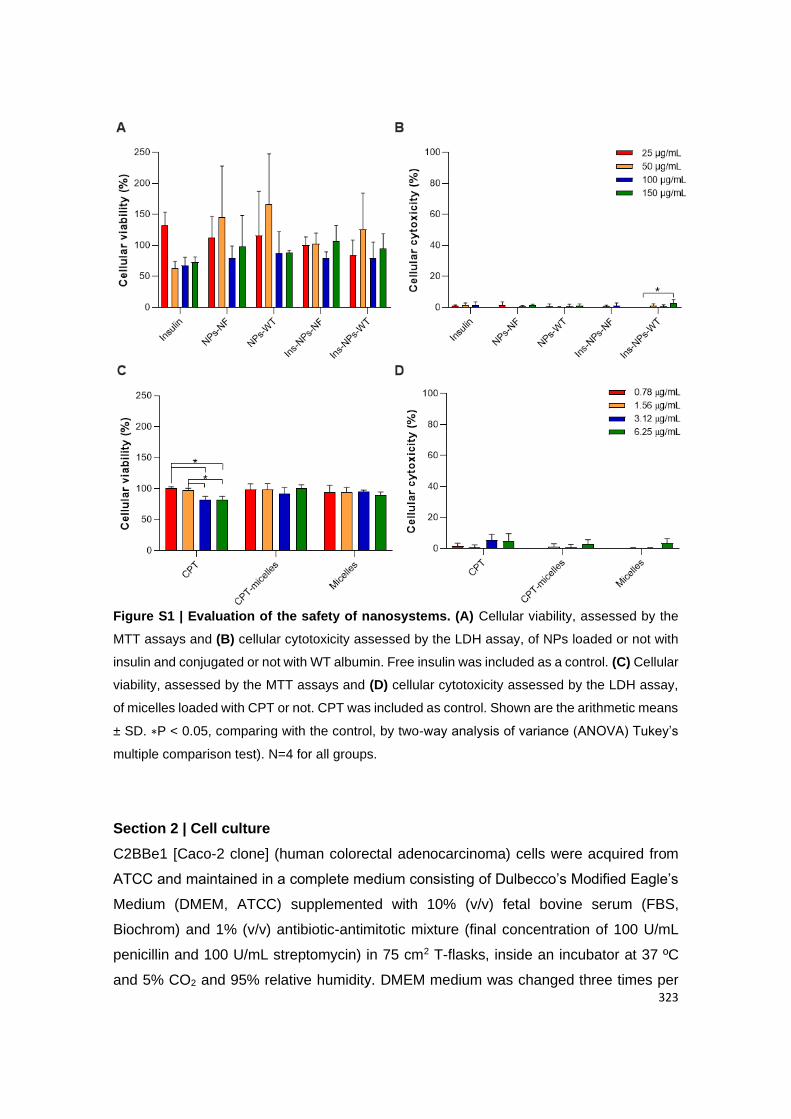
323
Figure S1 | Evaluation of the safety of nanosystems. (A) Cellular viability, assessed by the
MTT assays and (B) cellular cytotoxicity assessed by the LDH assay, of NPs loaded or not with
insulin and conjugated or not with WT albumin. Free insulin was included as a control. (C) Cellular
viability, assessed by the MTT assays and (D) cellular cytotoxicity assessed by the LDH assay,
of micelles loaded with CPT or not. CPT was included as control. Shown are the arithmetic means
± SD. ∗P < 0.05, comparing with the control, by two-way analysis of variance (ANOVA) Tukey’s
multiple comparison test). N=4 for all groups.
Section 2 | Cell culture
C2BBe1 [Caco-2 clone] (human colorectal adenocarcinoma) cells were acquired from
ATCC and maintained in a complete medium consisting of Dulbecco’s Modified Eagle’s
Medium (DMEM, ATCC) supplemented with 10% (v/v) fetal bovine serum (FBS,
Biochrom) and 1% (v/v) antibiotic-antimitotic mixture (final concentration of 100 U/mL
penicillin and 100 U/mL streptomycin) in 75 cm2 T-flasks, inside an incubator at 37 ºC
and 5% CO2 and 95% relative humidity. DMEM medium was changed three times per
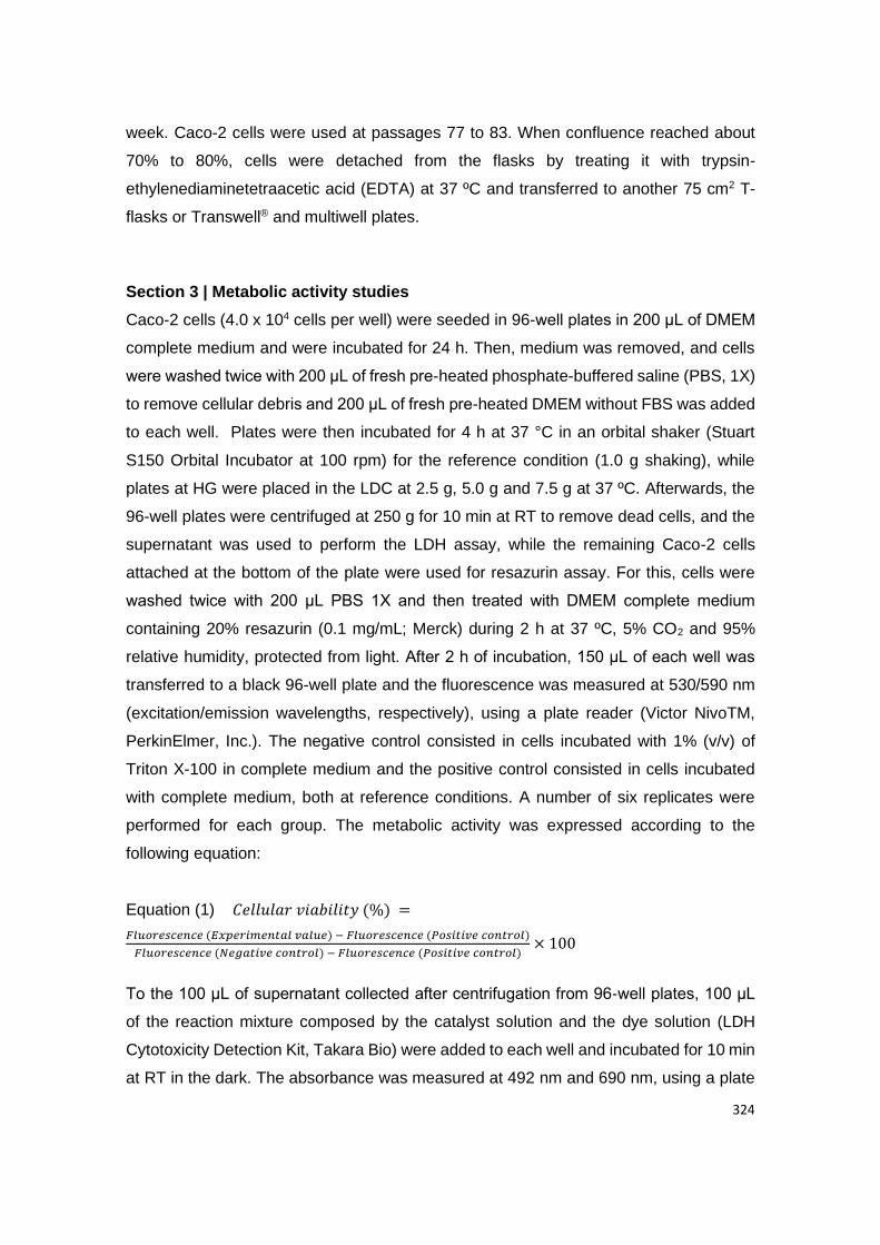
324
week. Caco-2 cells were used at passages 77 to 83. When confluence reached about
70% to 80%, cells were detached from the flasks by treating it with trypsin-
ethylenediaminetetraacetic acid (EDTA) at 37 ºC and transferred to another 75 cm2 T-
flasks or Transwell® and multiwell plates.
Section 3 | Metabolic activity studies
Caco-2 cells (4.0 x 10⁴ cells per well) were seeded in 96-well plates in 200 μL of DMEM
complete medium and were incubated for 24 h. Then, medium was removed, and cells
were washed twice with 200 μL of fresh pre-heated phosphate-buffered saline (PBS, 1X)
to remove cellular debris and 200 μL of fresh pre-heated DMEM without FBS was added
to each well. Plates were then incubated for 4 h at 37 °C in an orbital shaker (Stuart
S150 Orbital Incubator at 100 rpm) for the reference condition (1.0 g shaking), while
plates at HG were placed in the LDC at 2.5 g, 5.0 g and 7.5 g at 37 ºC. Afterwards, the
96-well plates were centrifuged at 250 g for 10 min at RT to remove dead cells, and the
supernatant was used to perform the LDH assay, while the remaining Caco-2 cells
attached at the bottom of the plate were used for resazurin assay. For this, cells were
washed twice with 200 μL PBS 1X and then treated with DMEM complete medium
containing 20% resazurin (0.1 mg/mL; Merck) during 2 h at 37 ºC, 5% CO2 and 95%
relative humidity, protected from light. After 2 h of incubation, 150 μL of each well was
transferred to a black 96-well plate and the fluorescence was measured at 530/590 nm
(excitation/emission wavelengths, respectively), using a plate reader (Victor NivoTM,
PerkinElmer, Inc.). The negative control consisted in cells incubated with 1% (v/v) of
Triton X-100 in complete medium and the positive control consisted in cells incubated
with complete medium, both at reference conditions. A number of six replicates were
performed for each group. The metabolic activity was expressed according to the
following equation:
Equation (1) 𝐶𝑒𝑙𝑙𝑢𝑙𝑎𝑟 𝑣𝑖𝑎𝑏𝑖𝑙𝑖𝑡𝑦 (%) =
𝐹𝑙𝑢𝑜𝑟𝑒𝑠𝑐𝑒𝑛𝑐𝑒 (𝐸𝑥𝑝𝑒𝑟𝑖𝑚𝑒𝑛𝑡𝑎𝑙 𝑣𝑎𝑙𝑢𝑒) − 𝐹𝑙𝑢𝑜𝑟𝑒𝑠𝑐𝑒𝑛𝑐𝑒 (𝑃𝑜𝑠𝑖𝑡𝑖𝑣𝑒 𝑐𝑜𝑛𝑡𝑟𝑜𝑙)
𝐹𝑙𝑢𝑜𝑟𝑒𝑠𝑐𝑒𝑛𝑐𝑒 (𝑁𝑒𝑔𝑎𝑡𝑖𝑣𝑒 𝑐𝑜𝑛𝑡𝑟𝑜𝑙) − 𝐹𝑙𝑢𝑜𝑟𝑒𝑠𝑐𝑒𝑛𝑐𝑒 (𝑃𝑜𝑠𝑖𝑡𝑖𝑣𝑒 𝑐𝑜𝑛𝑡𝑟𝑜𝑙)× 100
To the 100 μL of supernatant collected after centrifugation from 96-well plates, 100 μL
of the reaction mixture composed by the catalyst solution and the dye solution (LDH
Cytotoxicity Detection Kit, Takara Bio) were added to each well and incubated for 10 min
at RT in the dark. The absorbance was measured at 492 nm and 690 nm, using a plate
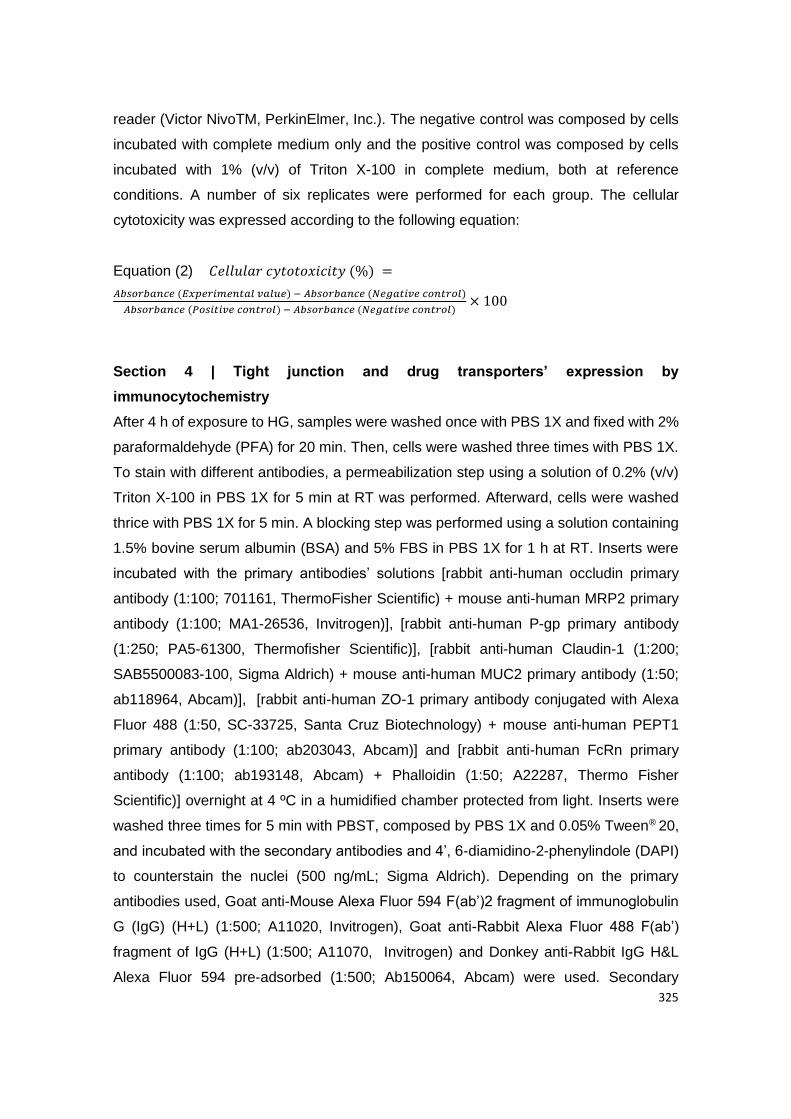
325
reader (Victor NivoTM, PerkinElmer, Inc.). The negative control was composed by cells
incubated with complete medium only and the positive control was composed by cells
incubated with 1% (v/v) of Triton X-100 in complete medium, both at reference
conditions. A number of six replicates were performed for each group. The cellular
cytotoxicity was expressed according to the following equation:
Equation (2) 𝐶𝑒𝑙𝑙𝑢𝑙𝑎𝑟 𝑐𝑦𝑡𝑜𝑡𝑜𝑥𝑖𝑐𝑖𝑡𝑦 (%) =
𝐴𝑏𝑠𝑜𝑟𝑏𝑎𝑛𝑐𝑒 (𝐸𝑥𝑝𝑒𝑟𝑖𝑚𝑒𝑛𝑡𝑎𝑙 𝑣𝑎𝑙𝑢𝑒) − 𝐴𝑏𝑠𝑜𝑟𝑏𝑎𝑛𝑐𝑒 (𝑁𝑒𝑔𝑎𝑡𝑖𝑣𝑒 𝑐𝑜𝑛𝑡𝑟𝑜𝑙)
𝐴𝑏𝑠𝑜𝑟𝑏𝑎𝑛𝑐𝑒 (𝑃𝑜𝑠𝑖𝑡𝑖𝑣𝑒 𝑐𝑜𝑛𝑡𝑟𝑜𝑙) − 𝐴𝑏𝑠𝑜𝑟𝑏𝑎𝑛𝑐𝑒 (𝑁𝑒𝑔𝑎𝑡𝑖𝑣𝑒 𝑐𝑜𝑛𝑡𝑟𝑜𝑙)× 100
Section 4 | Tight junction and drug transporters’ expression by
immunocytochemistry
After 4 h of exposure to HG, samples were washed once with PBS 1X and fixed with 2%
paraformaldehyde (PFA) for 20 min. Then, cells were washed three times with PBS 1X.
To stain with different antibodies, a permeabilization step using a solution of 0.2% (v/v)
Triton X-100 in PBS 1X for 5 min at RT was performed. Afterward, cells were washed
thrice with PBS 1X for 5 min. A blocking step was performed using a solution containing
1.5% bovine serum albumin (BSA) and 5% FBS in PBS 1X for 1 h at RT. Inserts were
incubated with the primary antibodies’ solutions [rabbit anti-human occludin primary
antibody (1:100; 701161, ThermoFisher Scientific) + mouse anti-human MRP2 primary
antibody (1:100; MA1-26536, Invitrogen)], [rabbit anti-human P-gp primary antibody
(1:250; PA5-61300, Thermofisher Scientific)], [rabbit anti-human Claudin-1 (1:200;
SAB5500083-100, Sigma Aldrich) + mouse anti-human MUC2 primary antibody (1:50;
ab118964, Abcam)], [rabbit anti-human ZO-1 primary antibody conjugated with Alexa
Fluor 488 (1:50, SC-33725, Santa Cruz Biotechnology) + mouse anti-human PEPT1
primary antibody (1:100; ab203043, Abcam)] and [rabbit anti-human FcRn primary
antibody (1:100; ab193148, Abcam) + Phalloidin (1:50; A22287, Thermo Fisher
Scientific)] overnight at 4 ºC in a humidified chamber protected from light. Inserts were
washed three times for 5 min with PBST, composed by PBS 1X and 0.05% Tween® 20,
and incubated with the secondary antibodies and 4’, 6-diamidino-2-phenylindole (DAPI)
to counterstain the nuclei (500 ng/mL; Sigma Aldrich). Depending on the primary
antibodies used, Goat anti-Mouse Alexa Fluor 594 F(ab’)2 fragment of immunoglobulin
G (IgG) (H+L) (1:500; A11020, Invitrogen), Goat anti-Rabbit Alexa Fluor 488 F(ab’)
fragment of IgG (H+L) (1:500; A11070, Invitrogen) and Donkey anti-Rabbit IgG H&L
Alexa Fluor 594 pre-adsorbed (1:500; Ab150064, Abcam) were used. Secondary

326
antibodies were added to samples for 2 h at 4 ºC in a humidified chamber protected from
light. Cells were then washed twice with PBST and once with PBS 1X, all for 5 min.
Inserts were mounted in slices using fluorescent mounting medium (Dako). Samples
were left to dry protected from light and were maintained at 4 ºC until visualization by a
spectral confocal laser scanning microscope TCS-SP5 AOBS (Leica). Since the aim was
to compare the amount and localization of tight and adherens junctions between samples
and also the expression of drug transporters, laser power and gain of each laser were
maintained for all samples regarding each antibody. The signal expression was
quantified using the ImageJ and samples were normalized according the following
expression:
Equation (4) 𝐸𝑥𝑝𝑟𝑒𝑠𝑠𝑖𝑜𝑛 (%) =𝑆𝑎𝑚𝑝𝑙𝑒𝑠 𝑖𝑛 𝐻𝐺
𝑅𝑒𝑓𝑒𝑟𝑒𝑛𝑐𝑒 𝑐𝑜𝑛𝑑𝑖𝑡𝑖𝑜𝑛× 100
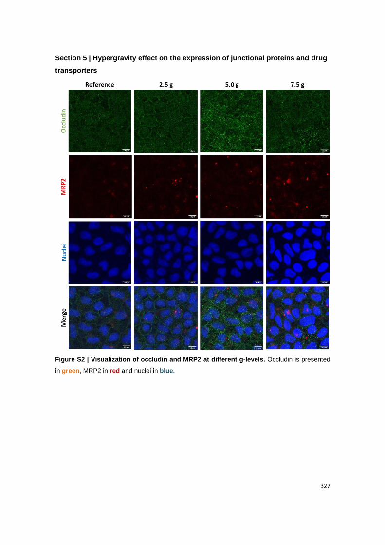
327
Section 5 | Hypergravity effect on the expression of junctional proteins and drug
transporters
Figure S2 | Visualization of occludin and MRP2 at different g-levels. Occludin is presented
in green, MRP2 in red and nuclei in blue.
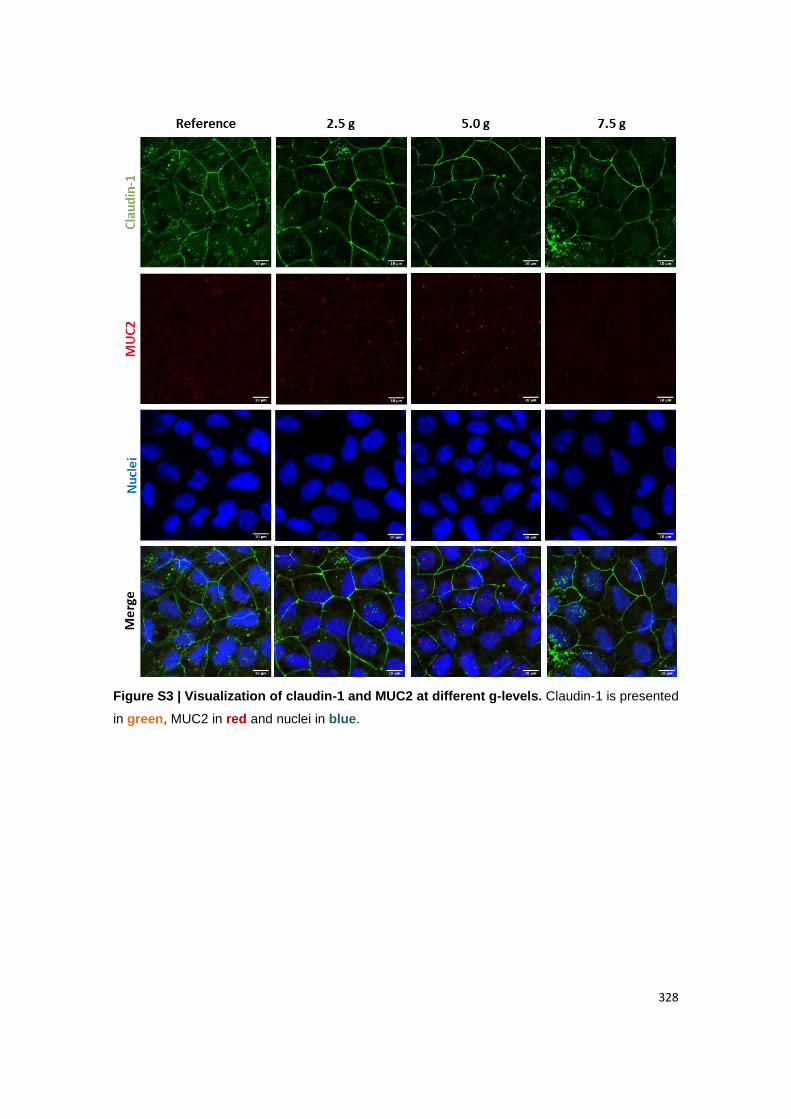
328
Figure S3 | Visualization of claudin-1 and MUC2 at different g-levels. Claudin-1 is presented
in green, MUC2 in red and nuclei in blue.
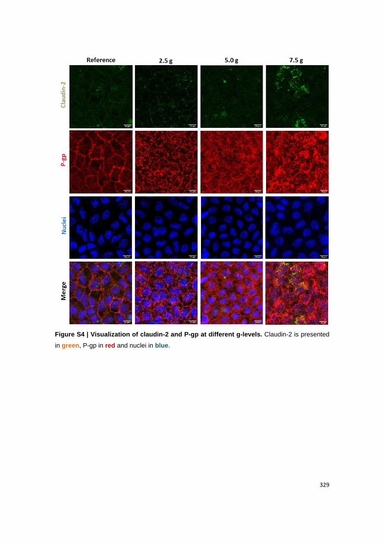
329
Figure S4 | Visualization of claudin-2 and P-gp at different g-levels. Claudin-2 is presented
in green, P-gp in red and nuclei in blue.
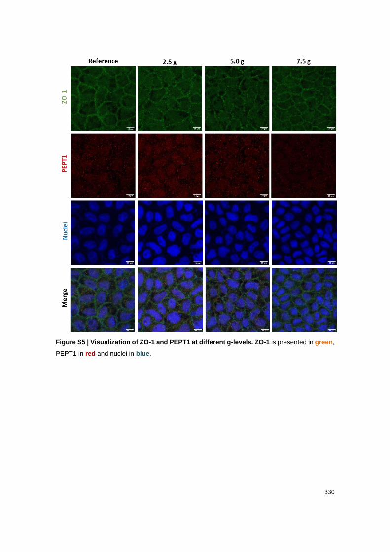
330
Figure S5 | Visualization of ZO-1 and PEPT1 at different g-levels. ZO-1 is presented in green,
PEPT1 in red and nuclei in blue.
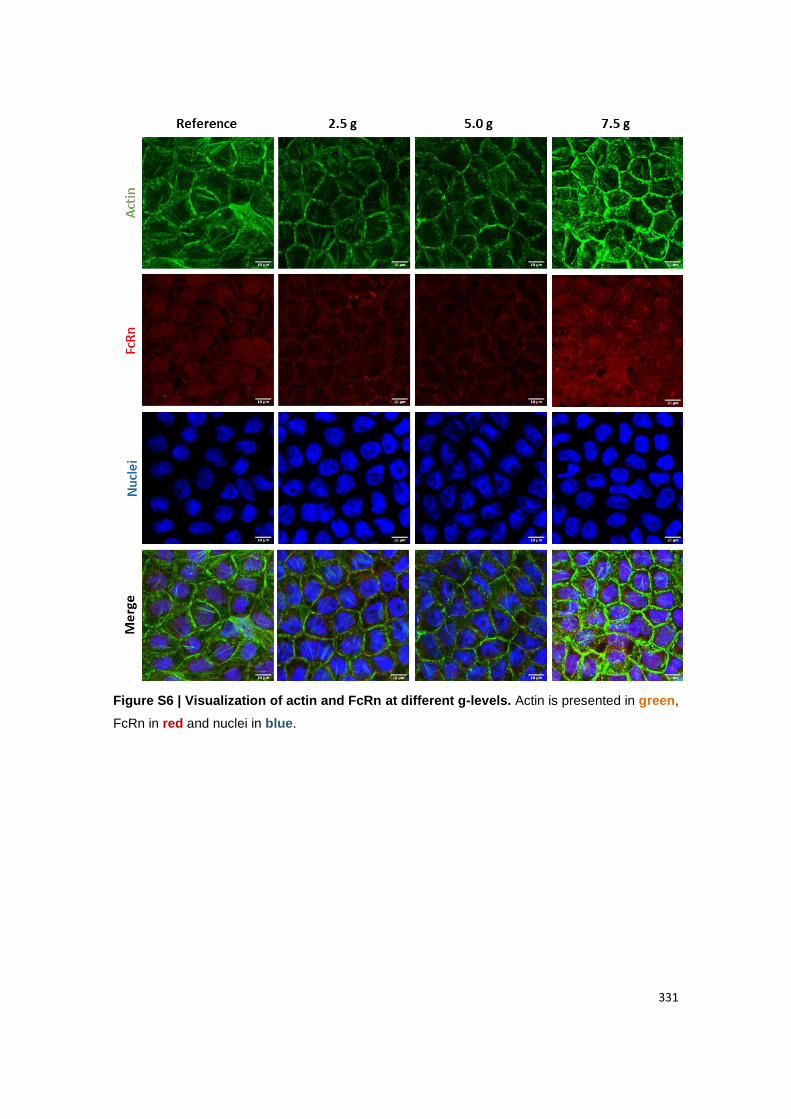
331
Figure S6 | Visualization of actin and FcRn at different g-levels. Actin is presented in green,
FcRn in red and nuclei in blue.
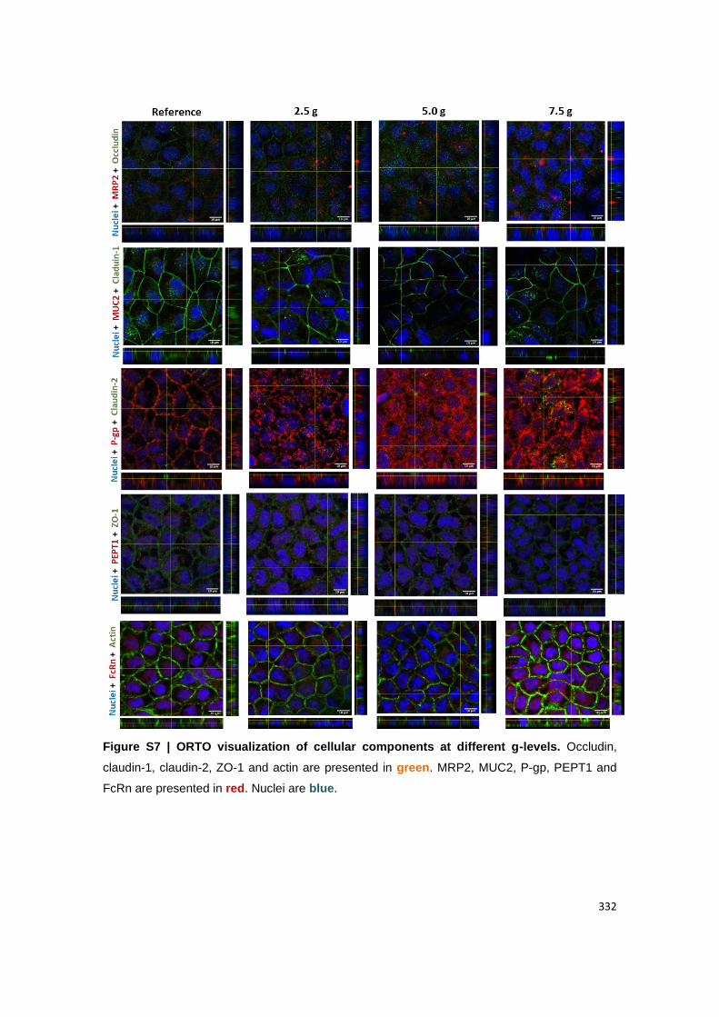
332
Figure S7 | ORTO visualization of cellular components at different g-levels. Occludin,
claudin-1, claudin-2, ZO-1 and actin are presented in green. MRP2, MUC2, P-gp, PEPT1 and
FcRn are presented in red. Nuclei are blue.
333
BIBLIOGRAPHIC NOTE
Cláudia Azevedo was born on July 26, 1990 in Trofa, Porto, Portugal. She obtained her
Bachelor in Science degree in Bioscience (2011) from Escola Superior de Biotecnologia
da Universidade Católica do Porto (ESB-UCP), Porto, Portugal. Further, she obtained
her Master in Science degree in Molecular Oncology, granted by Instituto de Ciências
Biomédicas de Abel Salazar (ICBAS), Porto, Portugal. Since 2017, she has been
developing her PhD thesis in Biomedical Sciences, PhD program from ICBAS, at Instituto
de Investigação e Inovação em Saúde (I3S) / Instituto de Engenharia Biomédica (INEB),
Porto, Portugal. Her research mainly focused on functionalized polymeric nanoparticles
for oral drug delivery. Her current interest is also focused in controlling delivery of drugs
across biological barriers, oral delivery of macromolecules, new materials for
nanoparticle drug delivery systems and in vivo/in vitro correlation of drug absorption
using cell-based co-culture models. She obtained several skills in the development and
characterization of nanosystems and proteins, as well as in performing molecular and
chemical techniques. She has also a broad training in cell culture techniques and cell-
based assays, as well as conducting ex vivo and in vivo experiments in porcine tissues
and mice models, respectively. Her research work resulted in 3 peer-reviewed original
articles (two published and two submitted for publication), 4 review articles (three
published and one submitted for publication), and 2 book chapters. Also, she actively
collaborated in other projects that resulted in peer reviewed publications.
During her doctorate she was also able to ameliorate her skills in innovation, strategy,
technology transfer and entrepreneurship, by participating in a startup acceleration
program with a business idea, to create a service to screen the intestinal permeability of
drugs, using in vitro models developed in the group. Additionally, she was always
involved in activities inside and outside the institute, as organizer (PhDay, Pint of
Science) or volunteer (performing diagnostic and serologic tests for COVID19, monitor
in primary schools, logistic of MIT European Career Fair). She also demonstrated
proactivity and networking by establishing collaborations with Massachusetts Institute of
Technology (MIT) and European Space Agency (ESA), diffusing the work of the group
and the institute through the world. Also, Cláudia showed initiative in attending some
courses related with biomolecular interactions and clinical trials. Fruit of all these skills,
was the recognition gave by Fulbright, a grant by FLAD, a prize of “Norte Empreendedor”
by Associação Empresarial de Portugal (AEP), an ITC conference grant by Cost and a
prize of the video competition by Controlled Release Society (CRS).

334