Embed Size (px)
Citation preview
Management Gastrointestinal Bleeding
Luke Gessel, MDAppreciation to: Trent Taylor MD and Sarita Gayle MD
Outline
Introduction
Upper GI Bleed (UGIB) Variceal Bleeding Peptic Ulcer
Disease
Lower GI Bleed (LGIB) Diverticular
Bleeding
Treatment General Measures Blood Products Pharmacologic
Treatments Endoscopic
Treatments Surgical Treatments Salvage Treatments
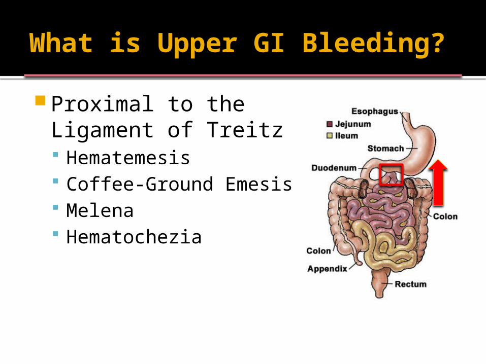
What is Upper GI Bleeding?
Proximal to the Ligament of Treitz Hematemesis Coffee-Ground Emesis Melena Hematochezia
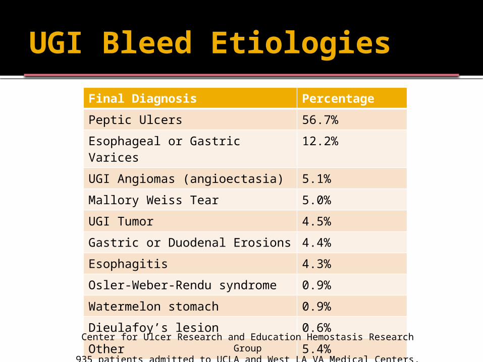
UGI Bleed Etiologies
Final Diagnosis Percentage
Peptic Ulcers 56.7%
Esophageal or Gastric Varices 12.2%
UGI Angiomas (angioectasia) 5.1%
Mallory Weiss Tear 5.0%
UGI Tumor 4.5%
Gastric or Duodenal Erosions 4.4%
Esophagitis 4.3%
Osler-Weber-Rendu syndrome 0.9%
Watermelon stomach 0.9%
Dieulafoy’s lesion 0.6%
Other 5.4%
Center for Ulcer Research and Education Hemostasis Research Group935 patients admitted to UCLA and West LA VA Medical Centers, 1996
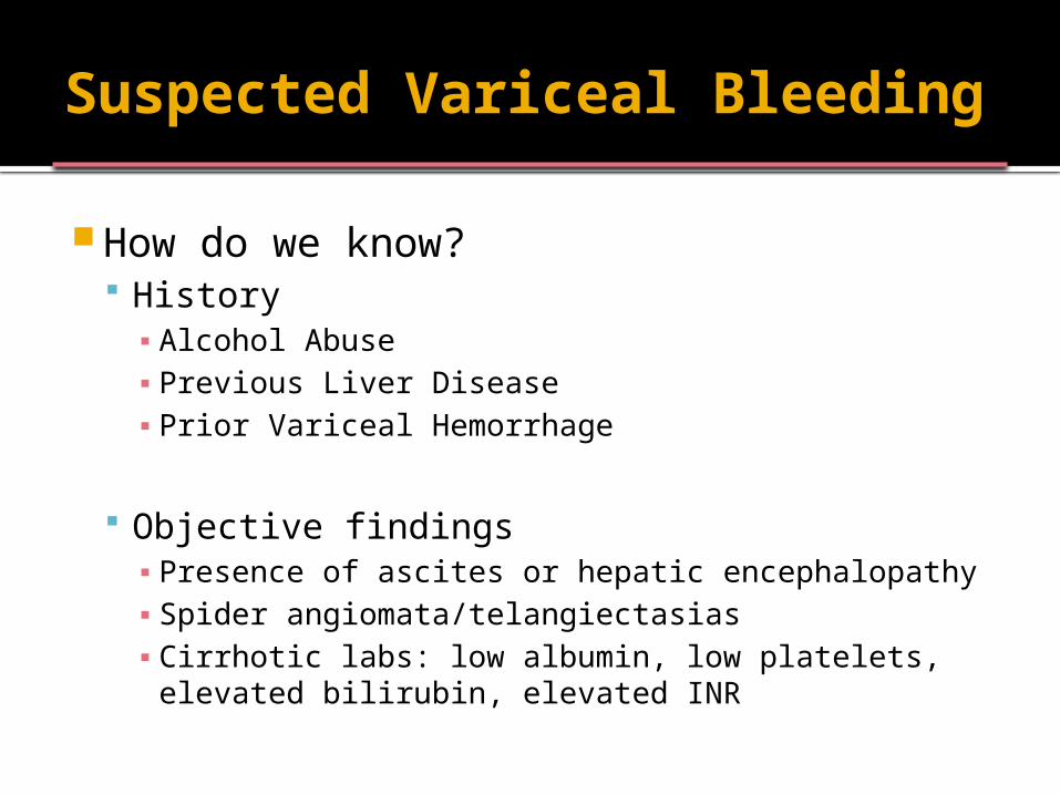
Suspected Variceal Bleeding
How do we know? History▪ Alcohol Abuse▪ Previous Liver Disease▪ Prior Variceal Hemorrhage
Objective findings▪ Presence of ascites or hepatic encephalopathy▪ Spider angiomata/telangiectasias▪ Cirrhotic labs: low albumin, low platelets,
elevated bilirubin, elevated INR
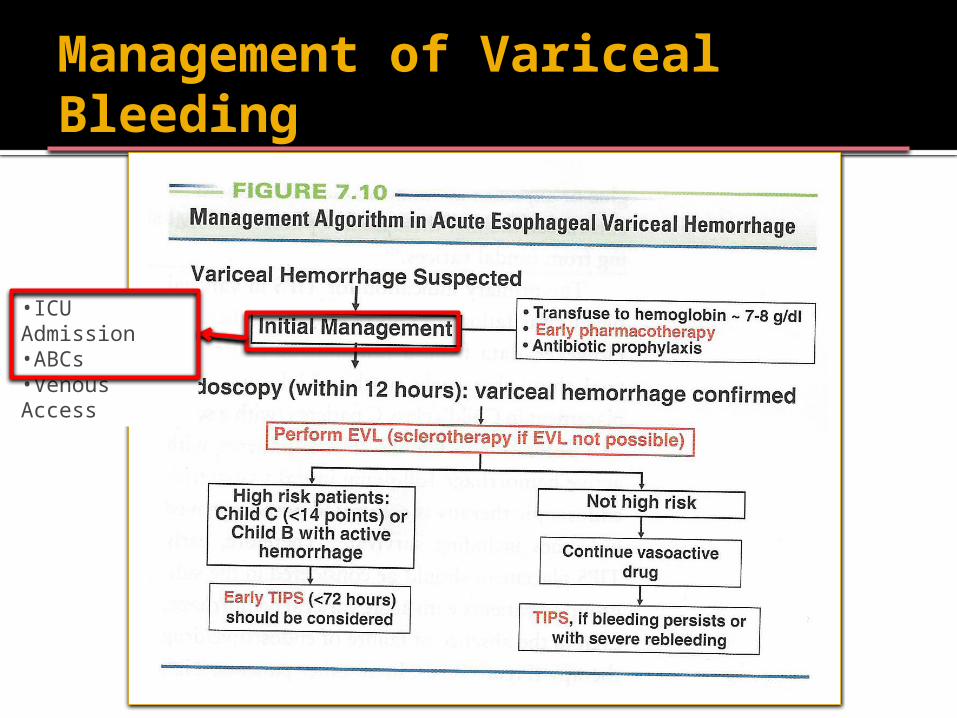
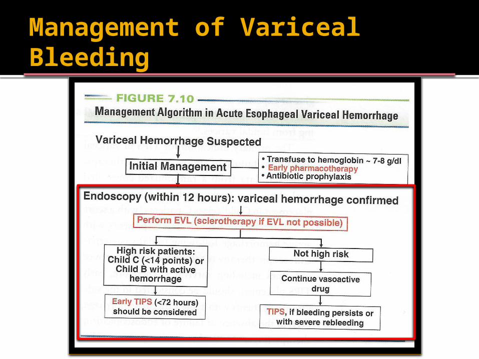
Management of Variceal Bleeding
•ICU Admission•ABCs•Venous Access
Variceal Bleeding
General measures Admit to ICU ABCs Venous Access
Initial resuscitation Hemodynamic Stability Goal Hgb 8 g/dL Correction of Coagulopathy
Appropriate Pharmacologic Therapy
Variceal Bleeding – ABC’s Airway/Breathing
Selectively consider intubation
“Patients with ongoing, significant hematemesis or those who may not be able to protect their airway for any reason and are at risk for aspiration should be considered for endotracheal intubation before undergoing endoscopy”
- ASGE Guideline GIE 2004
Variceal Bleeding – ABC’s
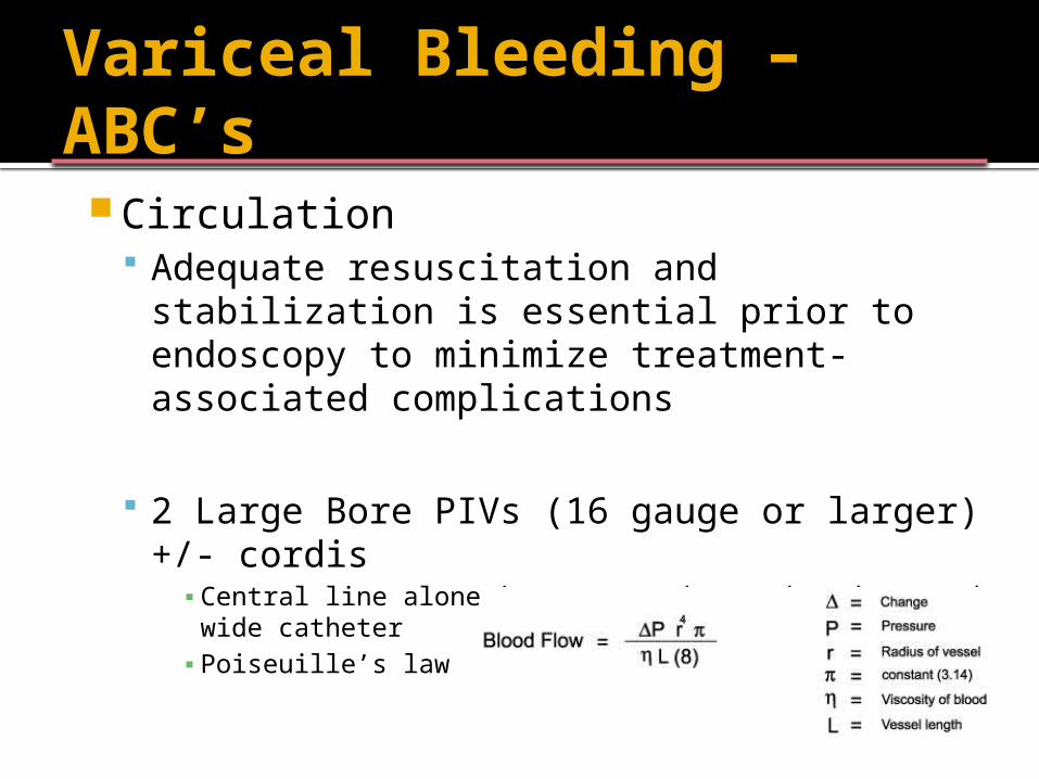
Circulation Adequate resuscitation and stabilization is
essential prior to endoscopy to minimize treatment-associated complications
2 Large Bore PIVs (16 gauge or larger) +/- cordis▪ Central line alone is not enough, need a short and wide
catheter▪ Poiseuille’s law
Hemodynamic stability
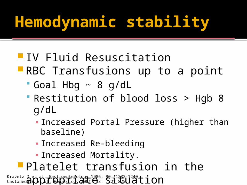
IV Fluid Resuscitation RBC Transfusions up to a point
Goal Hbg ~ 8 g/dL Restitution of blood loss > Hgb 8 g/dL ▪ Increased Portal Pressure (higher than
baseline) ▪ Increased Re-bleeding▪ Increased Mortality.
Platelet transfusion in the appropriate situation
Kravetz D et al. Gastroenterology 1986; 90: 1232-1240.Castaneda B et al. Hepatology 2001; 33: 821-825.
How much to transfuse?
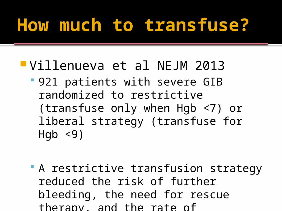
Villenueva et al NEJM 2013 921 patients with severe GIB
randomized to restrictive (transfuse only when Hgb <7) or liberal strategy (transfuse for Hgb <9)
A restrictive transfusion strategy reduced the risk of further bleeding, the need for rescue therapy, and the rate of complications and increased the rate of survival
Variceal Bleeding – ABC’s
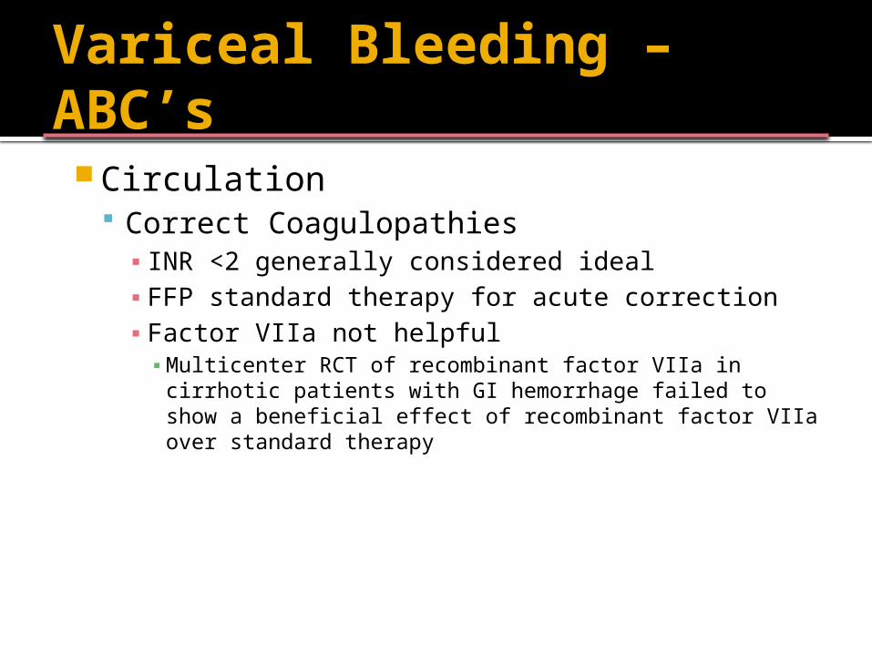
Circulation Correct Coagulopathies▪ INR <2 generally considered ideal▪ FFP standard therapy for acute correction▪ Factor VIIa not helpful▪ Multicenter RCT of recombinant factor VIIa in cirrhotic
patients with GI hemorrhage failed to show a beneficial effect of recombinant factor VIIa over standard therapy
Management of Variceal Bleeding
Variceal Bleeding - Octreotide
Early Pharmacologic Therapy Octreotide▪ Causes splanchnic vasoconstriction
decreased portal blood flow ▪ Inhibits release of vasodilatory peptides
(glucagon)▪ Local vasoconstrictive effects
50 mcg IV bolus followed by 50 mcg/h x 3-5 days
Variceal Bleeding - Octreotide
Octreotide efficacy controversial Corley et al, 2001 Meta analysis▪ Octreotide improved control of variceal hemorrhage compared
with:▪ All alternative therapies (other somatostatin analogues, sclerotherapy)
combined (RR 0.63; CI 0.51-0.77)▪ Vasopressin/terlipressin (RR 0.58; CI 0.42-0.81)▪ No additional intervention/placebo (RR 0.46; 0.32-0.67)
Gotzsche et al, 2005 Meta analysis▪ Somatostatin analogues showed generally negligible beneficial
effect ▪ 21 trials with 2588 patients▪ Did not reduce mortality significantly▪ Use saved ½ unit of blood per patient▪ Re-bleeding not significantly reduced in trials with low bias
Substantially reduced in other trials
Variceal Bleeding - Octreotide
AASLD Practice Guidelines, 2007 “Pharmacologic therapy (somatostatin or
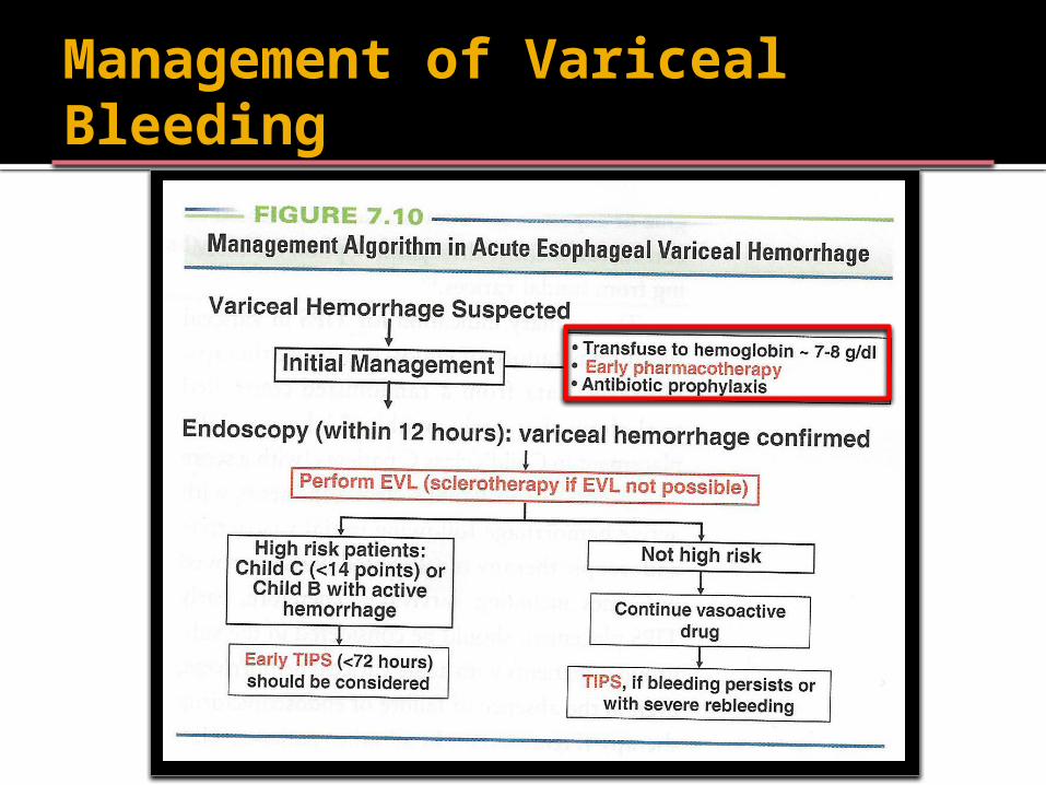
its analogues octreotide and vapreotide; terlipressin) should be initiated as soon as variceal hemorrhage is suspected and continued for 3-5 days after diagnosis is confirmed”
Class I, Level A
Variceal Bleeding - Antibiotics
AASLD Guidelines, 2007 Antibiotics should be given to cirrhotics in ANY type of GI
bleed (norfloxacin, ceftriaxone) for 7 days
Infections are present in ~20% of pts with cirrhosis who are hospitalized with GI bleeding; up to 50% develop an infection while hospitalized
A systematic review of eight placebo-controlled trials with a total of 864 patients found the antibiotics were associated with a significant reduction in mortality (RR 0.75, 95 percent CI 0.55 to 0.95)
Management of Variceal Bleeding
Variceal Bleeding - Endoscopy
AASLD Guidelines, 2007
“EGD, performed within 12 hours, should be used to make the diagnosis and to treat variceal hemorrhage, either with endoscopic variceal ligation (banding) or sclerotherapy”
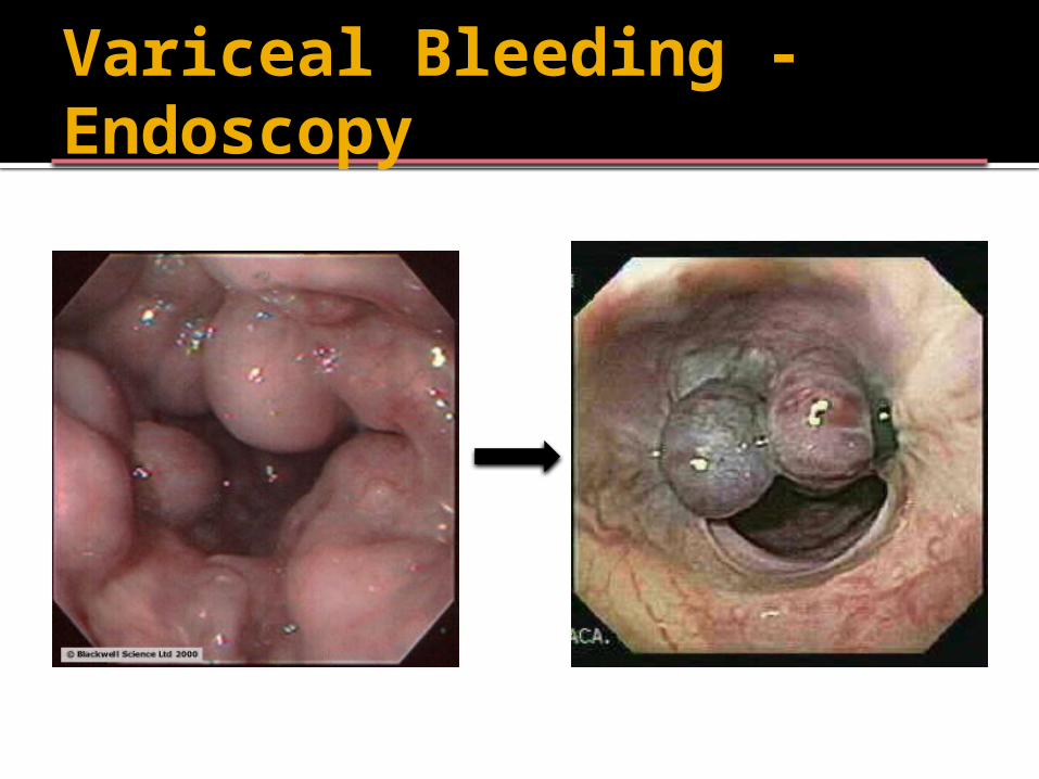
Variceal Bleeding - Endoscopy
Variceal Bleeding – Salvage Therapy
For variceal bleeding, 10-20% of patient’s bleeding cannot be controlled with endoscopic and/or pharmacologic therapy TIPS Balloon Tamponade ▪ Airway control▪ Bridging to more definitive therapy (TIPS)
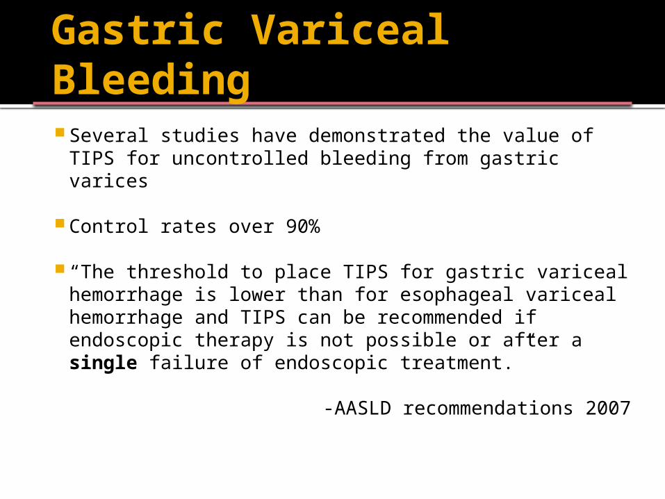
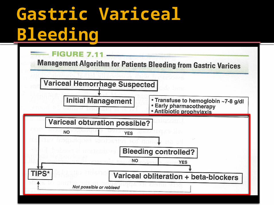
Gastric Variceal Bleeding Several studies have demonstrated the value of
TIPS for uncontrolled bleeding from gastric varices
Control rates over 90%
“The threshold to place TIPS for gastric variceal hemorrhage is lower than for esophageal variceal hemorrhage and TIPS can be recommended if endoscopic therapy is not possible or after a single failure of endoscopic treatment.”
-AASLD recommendations 2007
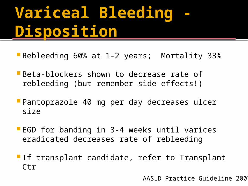
Variceal Bleeding - Disposition Rebleeding 60% at 1-2 years; Mortality 33%
Beta-blockers shown to decrease rate of rebleeding (but remember side effects!)
Pantoprazole 40 mg per day decreases ulcer size
EGD for banding in 3-4 weeks until varices eradicated decreases rate of rebleeding
If transplant candidate, refer to Transplant CtrAASLD Practice Guideline 2007
Gastric Variceal Bleeding
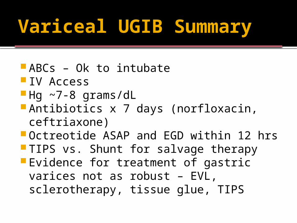
Variceal UGIB Summary
ABCs – Ok to intubate IV Access Hg ~7-8 grams/dL Antibiotics x 7 days (norfloxacin,
ceftriaxone) Octreotide ASAP and EGD within 12 hrs TIPS vs. Shunt for salvage therapy Evidence for treatment of gastric varices
not as robust – EVL, sclerotherapy, tissue glue, TIPS
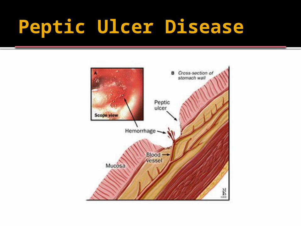
Peptic Ulcer Disease
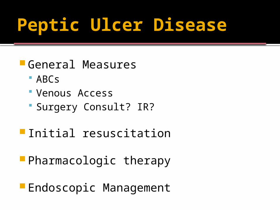
Peptic Ulcer Disease
General Measures ABCs Venous Access Surgery Consult? IR?
Initial resuscitation
Pharmacologic therapy
Endoscopic Management
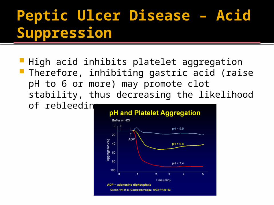
Peptic Ulcer Disease – Acid Suppression
High acid inhibits platelet aggregation Therefore, inhibiting gastric acid (raise pH to 6 or
more) may promote clot stability, thus decreasing the likelihood of rebleeding
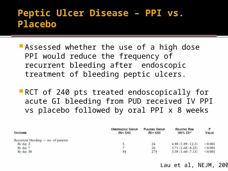
Peptic Ulcer Disease – PPI vs. Placebo
Assessed whether the use of a high dose PPI would reduce the frequency of recurrent bleeding after endoscopic treatment of bleeding peptic ulcers.
RCT of 240 pts treated endoscopically for acute GI bleeding from PUD received IV PPI vs placebo followed by oral PPI x 8 weeks
Lau et al, NEJM, 2000
Peptic Ulcer Disease – PPI vs. Placebo
Effect of preemptive infusion of omeprazole before endoscopy on the need for endoscopic therapy.
RCT of 638 with UGI bleeding randomized to receive omeprazole or placebo (each as an 80-mg intravenous bolus followed by an 8-mg infusion per hour) before endoscopy the next morning.
Lau et al, NEJM, 2007
Peptic Ulcer Disease – PPI vs. Placebo
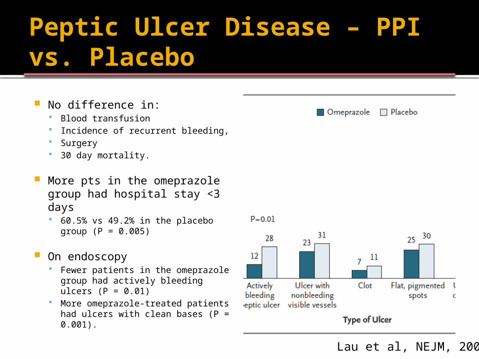
No difference in: Blood transfusion Incidence of recurrent bleeding, Surgery 30 day mortality.
More pts in the omeprazole group had hospital stay <3 days 60.5% vs 49.2% in the placebo
group (P = 0.005)
On endoscopy Fewer patients in the omeprazole
group had actively bleeding ulcers (P = 0.01)
More omeprazole-treated patients had ulcers with clean bases (P = 0.001).
Lau et al, NEJM, 2007
Peptic Ulcer Disease – PPI vs. Placebo
Conclusions: High-dose infusion of omeprazole at time of admission for 72 hours substantially reduces the
risk of recurrent bleeding.
Omeprazole 80 mg IV bolus, followed by 8 mg/hr x 72 hours
Discharge on omeprazole po for 8 weeks Lau, et al, NEJM, 2000Lau, et al, NEJM, 2007
Peptic Ulcer Disease – PPI
ASGE recommendation: “We recommend antisecretory therapy
with PPIs for patients with bleeding caused by peptic ulcers or in those with suspected peptic ulcer bleeding awaiting endoscopy.”
Peptic Ulcer Disease - Endoscopy
Endoscopy has been shown in randomized studies to lead to a: Reduction in blood transfusion requirements Shortened ICU and hospital stays Decreased need for surgery Lower mortality rate
Barkun A, et al. Ann Intern Med 2003.Spiegel BM, et al. Arch Intern Med 2001.
Peptic Ulcer Disease - Endoscopy
Endoscopic Options: Thermal coagulation Injection therapy Hemostatic clips Fibrin sealant (or glue) Argon plasma coagulation Combined Therapy
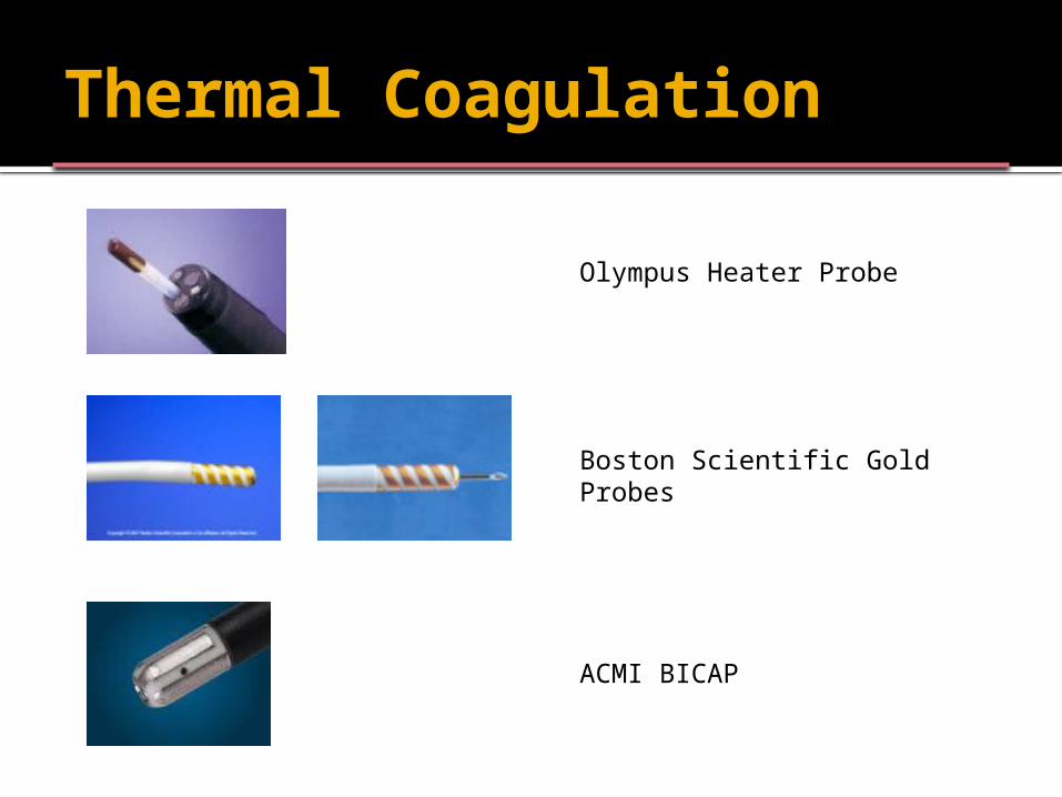
Thermal Coagulation
Olympus Heater Probe
Boston Scientific Gold Probes
ACMI BICAP
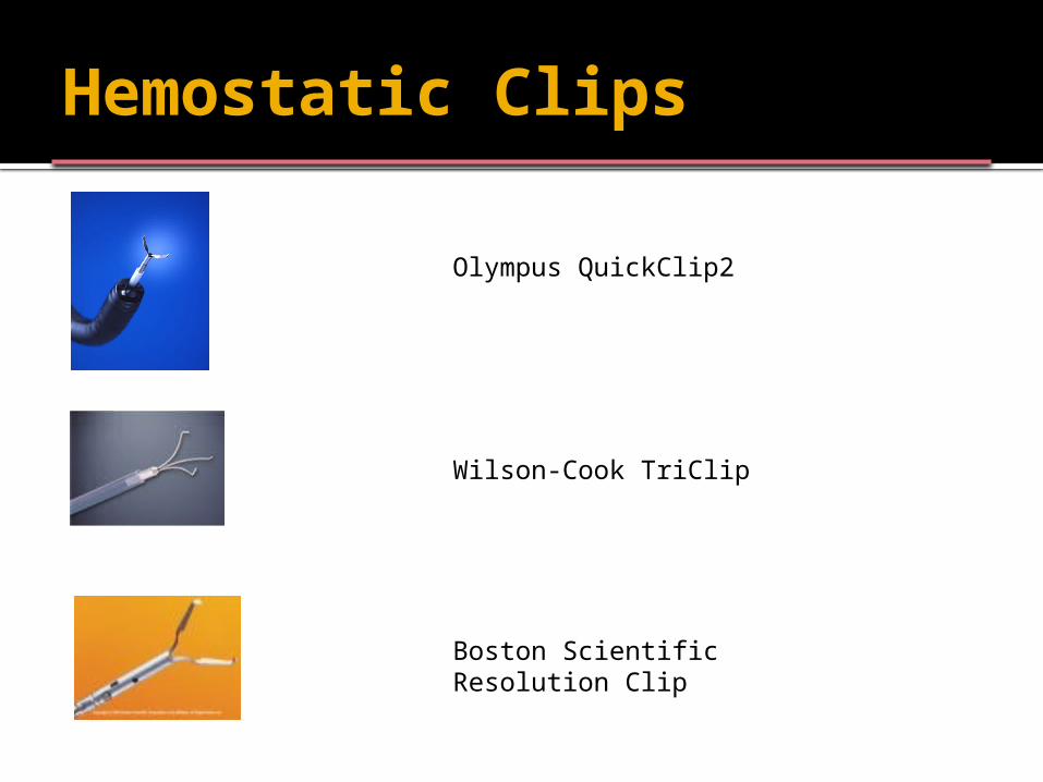
Hemostatic Clips
Olympus QuickClip2
Wilson-Cook TriClip
Boston Scientific Resolution Clip
0
10
20
30
40
50
60
Active bleeder NBVV Clot Dot Clean base
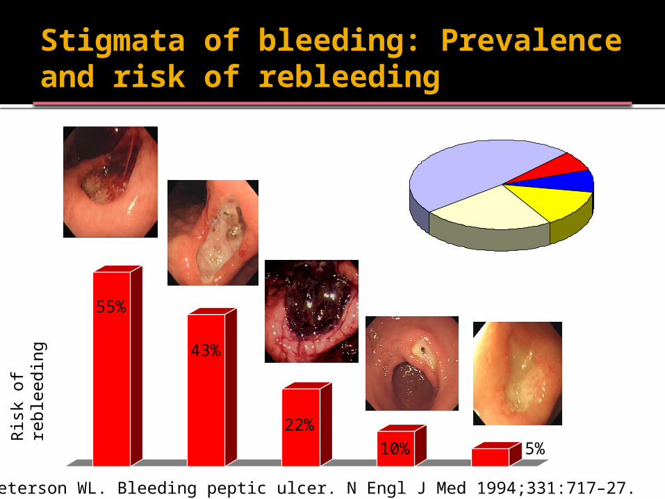
Stigmata of bleeding: Prevalence and risk of rebleeding
Dot23%
Clot13%
Bleeder7%NBVV8%
Clean base49%
Ris
k of
reble
edin
g
55%
43%
22% 10
%5%
Laine L, Peterson WL. Bleeding peptic ulcer. N Engl J Med 1994;331:717–27.
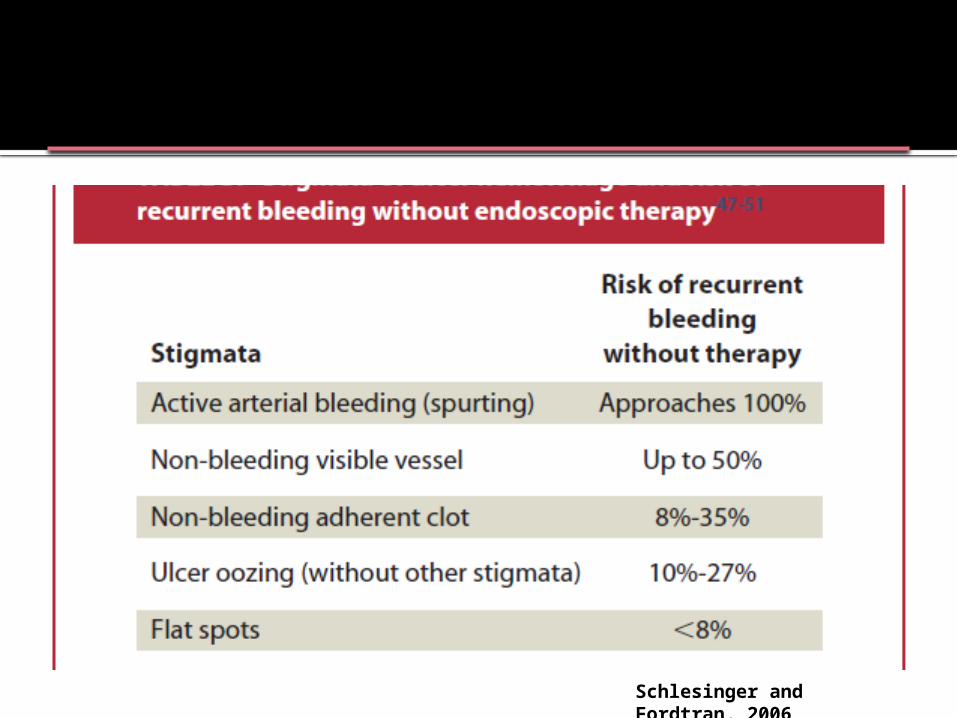
Schlesinger and Fordtran, 2006
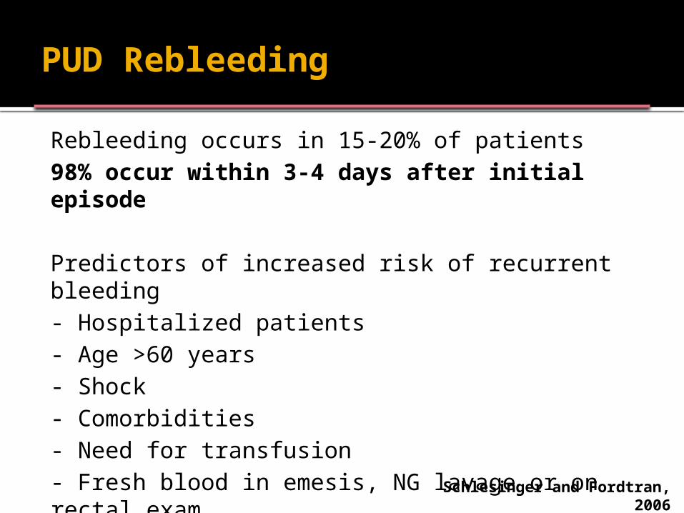
PUD Rebleeding
Rebleeding occurs in 15-20% of patients98% occur within 3-4 days after initial episode
Predictors of increased risk of recurrent bleeding - Hospitalized patients- Age >60 years- Shock- Comorbidities- Need for transfusion- Fresh blood in emesis, NG lavage or on rectal exam Schlesinger and Fordtran,
2006
PUD Rebleeding
Surgery versus Endoscopy for Rebleeding Endoscopic hemostasis achieved in 75% Mortality, duration of hospital stay,
duration of ICU stay, volume of transfusion similar
Pts who undergo surgery have more complications
PUD Rebleeding
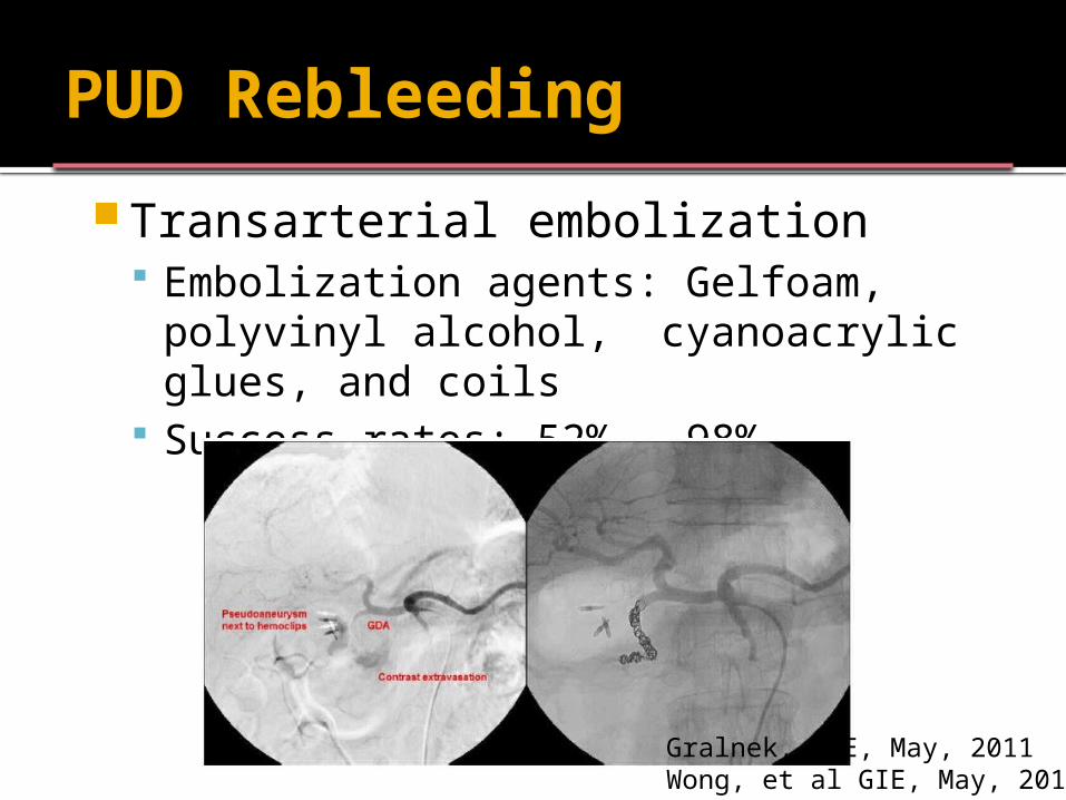
Transarterial embolization Embolization agents: Gelfoam, polyvinyl
alcohol, cyanoacrylic glues, and coils Success rates: 52% - 98%
Gralnek, GIE, May, 2011Wong, et al GIE, May, 2011
PUD Rebleeding
Author conclusion: TAE reduces the need for surgery without increasing overall mortality and is associated with fewer complications.
TAE should be considered, if not before, at least as an alternative to surgery in patients with PUD in whom endoscopic hemostasis fails
Gralnek, GIE, May, 2011Wong, et al GIE, May, 2011
Aspirin therapy after PUD bleeding
Should patients who take aspirin to prevent cardiovascular disease continue to take it after an acute UGI bleeding event?
Randomized, placebo-controlled trial of 156 pts with PUD in Hong Kong between 2003 to 2006
78 pts rec’d aspirin (80 mg/d) and 78 received placebo for 8 weeks immediately after endoscopy. All pts rec’d IV PPI followed by po PPI.
Sung, et al. Annals of Int Med, January, 2010.
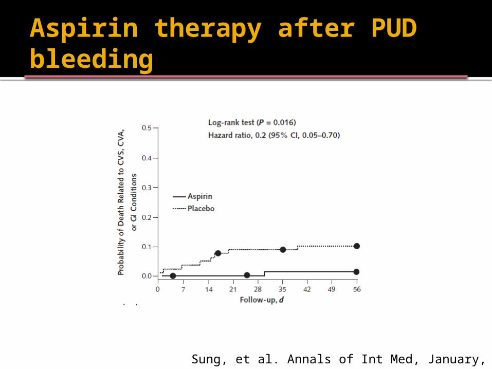
Aspirin therapy after PUD bleeding
Sung, et al. Annals of Int Med, January, 2010.
Aspirin therapy after PUD bleeding
Authors’ Conclusion: Among low-dose aspirin recipients who had peptic ulcer bleeding, continuous aspirin therapy may increase the risk for recurrent bleeding but potentially reduces mortality rates. Larger trials are needed to confirm these findings.
Sung, et al. Annals of Int Med, January, 2010.
PUD - Disposition
Disposition is according to clinical risk and endoscopic risk of rebleeding
All patients require outpatient therapy and close follow-up
Discharge on PPI, but NOT indefinitely
Don’t forget to check Helicobacter pylori and eradicate – AND VERIFY ERADICATION
Non-ulcer causes of UGIB Esophagitis
Rarely requires endoscopic therapy
Mallory-Weiss tear Usually self-limited, can require treatment if bleeding ongoing
Dieflafoy lesion
Aortoenteric fistula Endoscopy is diagnostic only CT imaging Prompt surgery consult
Tumors High rebleeding rate surgery often required for hemostasis
Peptic Ulcer Disease - Summary
ABC’s IV Access Resuscitation Surgery Consult? PPI before and after endoscopy Endoscopy
Within 24h for high-risk patients Remember to look for the etiology
(NSAIDs, H.Pylori, other)
ABIM Board Review Question
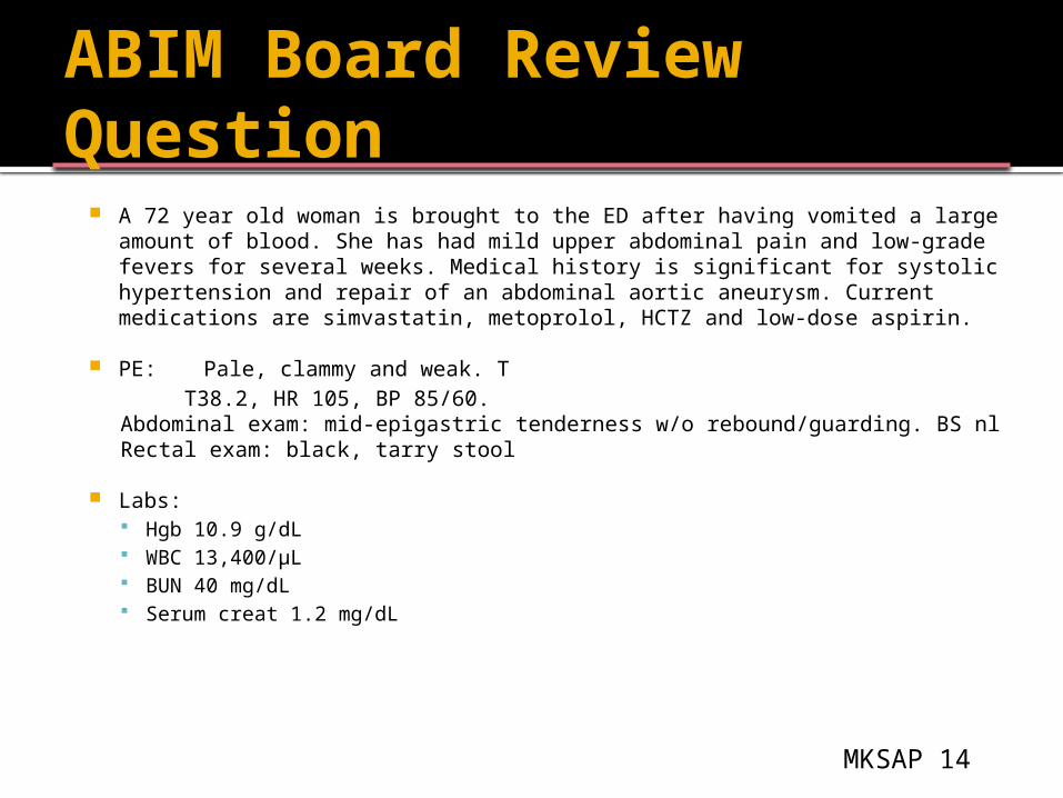
A 72 year old woman is brought to the ED after having vomited a large amount of blood. She has had mild upper abdominal pain and low-grade fevers for several weeks. Medical history is significant for systolic hypertension and repair of an abdominal aortic aneurysm. Current medications are simvastatin, metoprolol, HCTZ and low-dose aspirin.
PE: Pale, clammy and weak. TT38.2, HR 105, BP 85/60.
Abdominal exam: mid-epigastric tenderness w/o rebound/guarding. BS nl
Rectal exam: black, tarry stool
Labs: Hgb 10.9 g/dL WBC 13,400/µL BUN 40 mg/dL Serum creat 1.2 mg/dL
MKSAP 14
ABIM Board Review Question cont’d
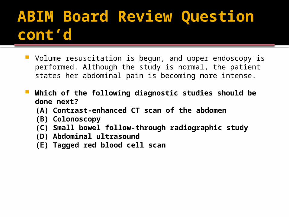
Volume resuscitation is begun, and upper endoscopy is performed. Although the study is normal, the patient states her abdominal pain is becoming more intense.
Which of the following diagnostic studies should be done next?
(A) Contrast-enhanced CT scan of the abdomen(B) Colonoscopy(C) Small bowel follow-through radiographic study(D) Abdominal ultrasound(E) Tagged red blood cell scan
ABIM Board Review Question
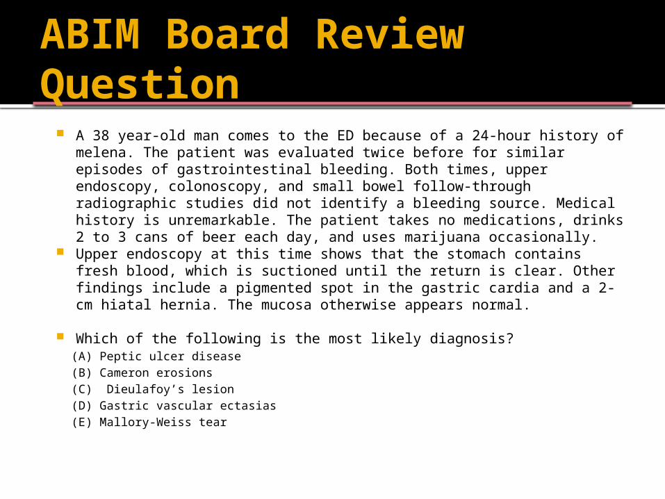
A 38 year-old man comes to the ED because of a 24-hour history of melena. The patient was evaluated twice before for similar episodes of gastrointestinal bleeding. Both times, upper endoscopy, colonoscopy, and small bowel follow-through radiographic studies did not identify a bleeding source. Medical history is unremarkable. The patient takes no medications, drinks 2 to 3 cans of beer each day, and uses marijuana occasionally.
Upper endoscopy at this time shows that the stomach contains fresh blood, which is suctioned until the return is clear. Other findings include a pigmented spot in the gastric cardia and a 2-cm hiatal hernia. The mucosa otherwise appears normal.
Which of the following is the most likely diagnosis?(A) Peptic ulcer disease(B) Cameron erosions(C) Dieulafoy’s lesion(D) Gastric vascular ectasias(E) Mallory-Weiss tear

Lower GI Bleeding
Blood loss distal to the ligament of Treitz
Lower GI Bleeding
Mortality low (~4%) – most common in older adults with intestinal ischemia and co-morbidities
Up to 13% with hematochezia have an UGIB
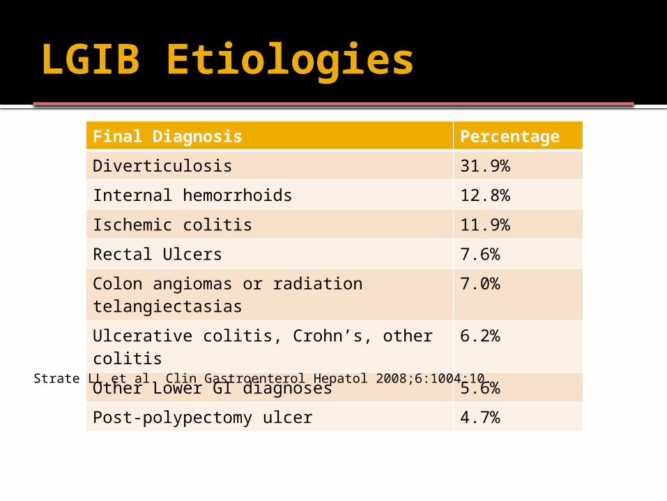
LGIB Etiologies
Final Diagnosis Percentage
Diverticulosis 31.9%
Internal hemorrhoids 12.8%
Ischemic colitis 11.9%
Rectal Ulcers 7.6%
Colon angiomas or radiation telangiectasias
7.0%
Ulcerative colitis, Crohn’s, other colitis 6.2%
Other Lower GI diagnoses 5.6%
Post-polypectomy ulcer 4.7%Strate LL et al. Clin Gastroenterol Hepatol 2008;6:1004:10.
LGIB
General Measures ABCs Venous Access Surgery Consult? IR?
Diagnostic Evaluation Endoscopy Other Diagnostic Evaluations
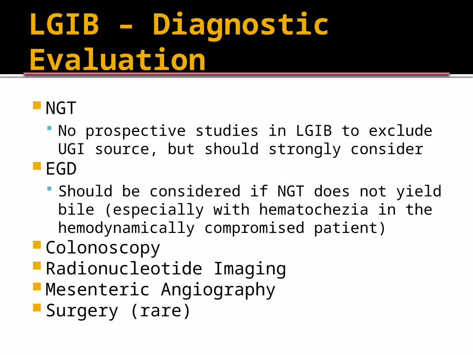
LGIB – Diagnostic Evaluation NGT
No prospective studies in LGIB to exclude UGI source, but should strongly consider
EGD Should be considered if NGT does not yield bile
(especially with hematochezia in the hemodynamically compromised patient)
Colonoscopy Radionucleotide Imaging Mesenteric Angiography Surgery (rare)
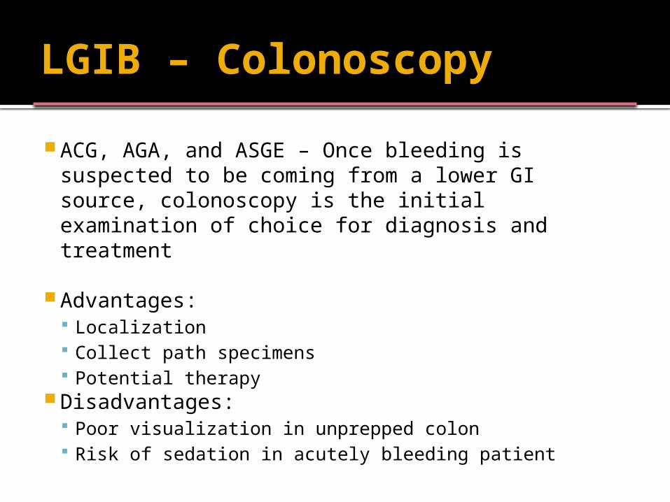
LGIB – Colonoscopy
ACG, AGA, and ASGE – Once bleeding is suspected to be coming from a lower GI source, colonoscopy is the initial examination of choice for diagnosis and treatment
Advantages: Localization Collect path specimens Potential therapy
Disadvantages: Poor visualization in unprepped colon Risk of sedation in acutely bleeding patient
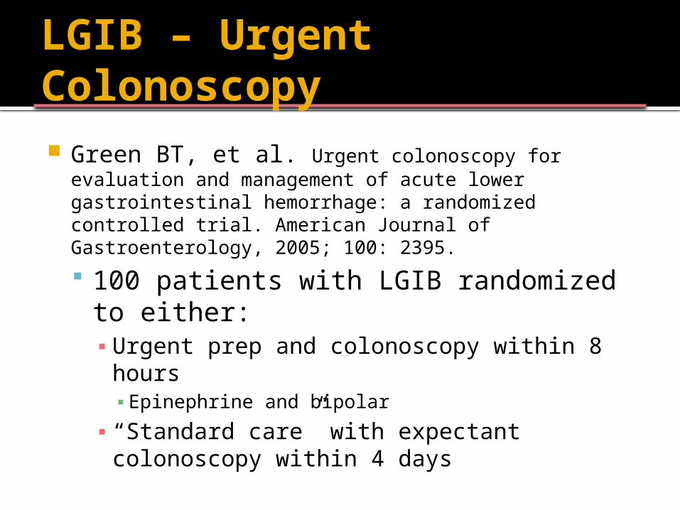
LGIB – Urgent Colonoscopy Green BT, et al. Urgent colonoscopy for evaluation and
management of acute lower gastrointestinal hemorrhage: a randomized controlled trial. American Journal of Gastroenterology, 2005; 100: 2395.
100 patients with LGIB randomized to either:▪ Urgent prep and colonoscopy within 8 hours▪ Epinephrine and bipolar
▪ “Standard care” with expectant colonoscopy within 4 days
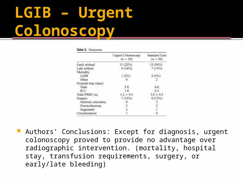
LGIB – Urgent Colonoscopy
Authors’ Conclusions: Except for diagnosis, urgent colonoscopy proved to provide no advantage over radiographic intervention. (mortality, hospital stay, transfusion requirements, surgery, or early/late bleeding)
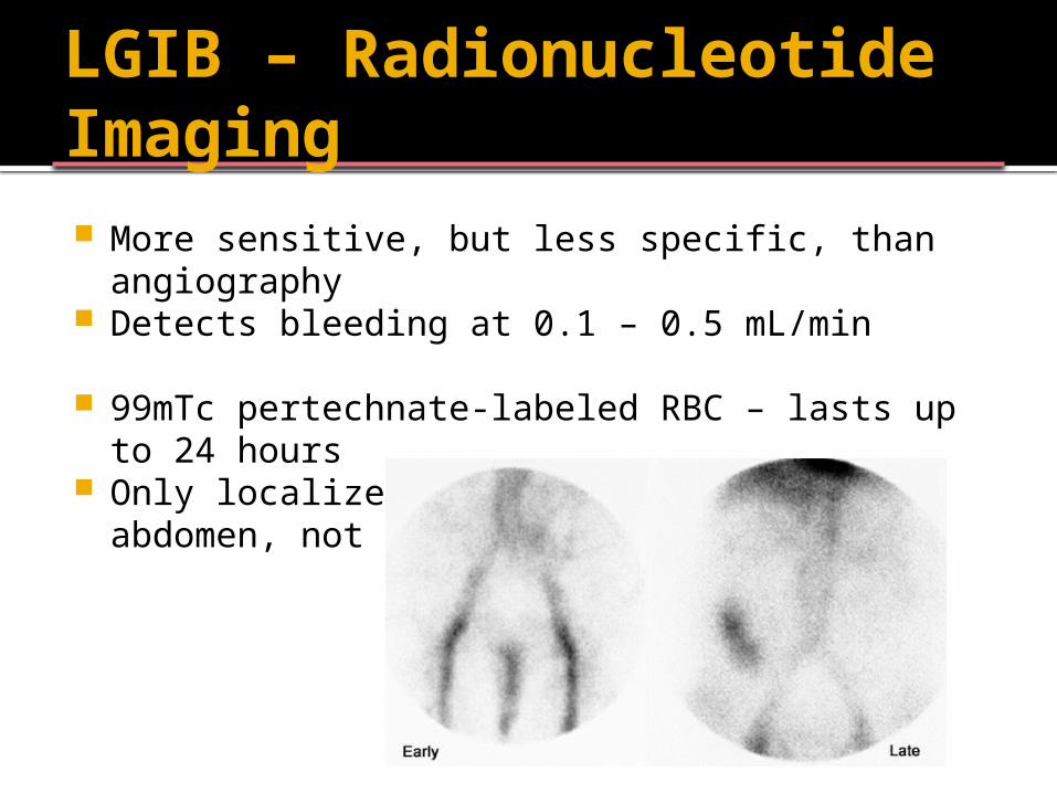
LGIB – Radionucleotide Imaging More sensitive, but less specific, than
angiography Detects bleeding at 0.1 – 0.5 mL/min
99mTc pertechnate-labeled RBC – lasts up to 24 hours
Only localizes to a general area of the abdomen, not a specific site
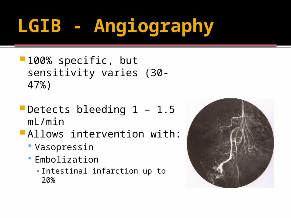
LGIB - Angiography
100% specific, but sensitivity varies (30-47%)
Detects bleeding 1 – 1.5 mL/min
Allows intervention with: Vasopressin Embolization▪ Intestinal infarction up to 20%
LGIB - Summary
ABC’s IV Access Resuscitation NGT/EGD? Colonoscopy as initial diagnostic choice
Urgent colonoscopy increases rate of diagnosis, but doesn’t change important outcomes
Radionucleotide Scan, Angiography Surgery Consult?