Embed Size (px)
Citation preview
Lumbar Spine Definitions and Diagnostic Criteria: Degeneration,
Herniation and Stenosis Thomas J. Gilbert MD, MPP
William J. Mullin MD Ronald S. Pobiel MD April 1, 2015 revision
Disc Degeneration: Spondylosis (Spondylosis Deformans) is a general term used for age-related changes to the disc.
This includes disc dessication, bulging and marginal osteophyte.
Disc degeneration (Intervertebral osteochondrosis) (Resnick) is characterized by disorganization
and dessication of the nucleus pulposis and by disc space narrowing. With loss of disc space
height there is annular bulging/laxity and mechanical failure of the disc (Herzog). It generally
represents the sequel of disc injury and may be symptomatic or asymptomatic (Fardon, Herzog).
We use disc space narrowing as our primary parameter to grade disc degeneration:
Mild - Desiccation with < 25% disc space narrowing
Moderate - Desiccation with 25-75% disc space narrowing
Severe - Desiccation with > 75% disc space narrowing
Some radiologists will use moderately severe for 75-90% disc space narrowing and severe for
complete collapse.
While disc desiccation is also a feature of disc degeneration, it is difficult to use this parameter
for grading. First, disc desiccation can be seen with normal aging. Second, the signal intensity
of a degenerated disc can vary with MRI field strength and pulse sequence selection. Finally,
this parameter does not facilitate comparison with CT findings.
2
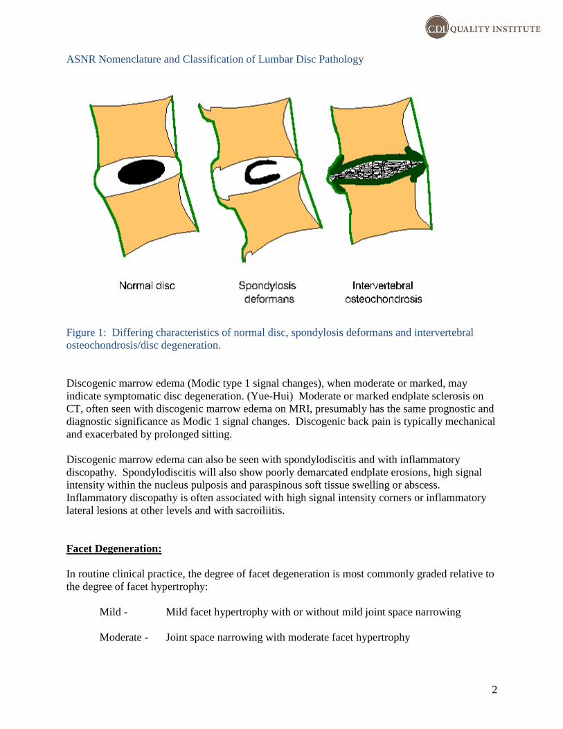
ASNR Nomenclature and Classification of Lumbar Disc Pathology
Figure 1: Differing characteristics of normal disc, spondylosis deformans and intervertebral
osteochondrosis/disc degeneration.
Discogenic marrow edema (Modic type 1 signal changes), when moderate or marked, may
indicate symptomatic disc degeneration. (Yue-Hui) Moderate or marked endplate sclerosis on
CT, often seen with discogenic marrow edema on MRI, presumably has the same prognostic and
diagnostic significance as Modic 1 signal changes. Discogenic back pain is typically mechanical
and exacerbated by prolonged sitting.
Discogenic marrow edema can also be seen with spondylodiscitis and with inflammatory
discopathy. Spondylodiscitis will also show poorly demarcated endplate erosions, high signal
intensity within the nucleus pulposis and paraspinous soft tissue swelling or abscess.
Inflammatory discopathy is often associated with high signal intensity corners or inflammatory
lateral lesions at other levels and with sacroiliitis.
Facet Degeneration:
In routine clinical practice, the degree of facet degeneration is most commonly graded relative to
the degree of facet hypertrophy:
Mild - Mild facet hypertrophy with or without mild joint space narrowing
Moderate - Joint space narrowing with moderate facet hypertrophy
3
Marked - Joint space narrowing with marked facet hypertrophy, marked irregularity
of subchondral bone and/or marked facet joint derangement
Facet hypertrophy may contribute directly to the degree of subarticular and foraminal stenosis.
Inflammatory facet arthropathy is characterized by subchondral marrow edema and periarticular
edema on STIR or fat saturation sequences. Moderate or marked subchondral marrow edema is
associated with symptomatic facet arthropathy. If the patient has tenderness over the facet joint
and has pain exacerbated by hyperextension, consideration might be given to a facet joint
injection in these patients. Inflammatory facet arthropathy can occur with degenerative arthritis
or with inflammatory spondyloarthropathy.
As with erosive osteoarthritis of the hands, inflammatory degenerative facet arthropathy is seen
in older patients and is more common in female patients. Facet autofusion is often seen at an
adjacent level, presumably representing the sequela of a previous inflammatory episode and
possibly foretelling the natural history of the disease. Facet inflammation with
spondyloarthropathy is typically seen with sacroiliitis, high signal corners, inflammatory
discopathy or lateral inflammatory lesions at additional levels.
Cystic facet arthropathy is characterized by extensive subchondral cyst formation and may also
indicate symptomatic facet arthropathy.
Erosive facet arthropathy is characterized by irregularity of subchondral bone and by widening
of the facet joint space. Facet joint diastasis when moderate or marked indicates segmental
hypermobility – usually in direct proportion to the degree of widening – and can be associated
with dynamic stenosis. Within the lumbar spine, this is most common at L4-5 and is more
common in female patients. Facet joint diastasis in association with retrolisthesis does not
indicate erosive disease.
Facet synovial cysts are frequently associated with facet degeneration. The presence, size,
location of a synovial cyst should be routinely reported. When synovial cysts project into the
subarticular recesses or neural foramina, they can result in compressive radiculopathy, and neural
compression should be highlighted if present. Synovial cysts associated with erosive changes
and facet diastasis can enlarge with axial loading and result in standing intolerance. This
possibility may need to be mentioned in the conclusion, as providers may not be aware of this
phenomenon.
High Signal Intensity Annular Fissures:
High signal intensity fissures are characterized by the presence of linear areas of high signal
intensity within the peripheral disc annulus on T2 FSE images. High signal intensity within the
fissure presumably reflects the ingrowth of angiogenic fibrosis. They should be noted on MRI
lumbar spine reports where they do show some association with discogenic pain. High signal
intensity annular fissures show a strong correlation with positive discography in patients with
discogenic back pain. (Schellhas)
Disc Herniation:
A disc herniation is defined as a focal displacement of disc material beyond the normal margins
4
of the intervertebral disc space resulting in a focal contour abnormality. (Fardon, Herzog,
Kreiner, Milette, Blaser, Dayo) The displaced disc material may contain nuclear, endplate
and/or annular fragments. Displacement of disc material most commonly occurs through a tear
or fissure in the disc annulus. It can also occur through a defect in the endplate apophysis (with
peripheral Schmorl node or posterior limbus-type deformities) or through an avulsion of the
endplate apophysis in juvenile or adolescent patients.
In the radiologic literature, the definition of a disc herniation has often been framed by
morphologic characteristics discernable on available imaging exams. Early radiologic literature
defined a herniation as a focal bulge of the disc annulus (double density sign) as this represented
the criteria for diagnosis on myelography. Subsequent definitions have been framed in terms of
CT findings. (Fardon, Costello)
Interventional radiologists have defined a disc herniation by the presence or absence of radicular
symptoms (Herkowitz). While acute radiculopathy with a positive straight leg raising sign has
significant positive predictive value for a disc herniation, it does not define the underlying
pathologic entity, and it is not entirely specific.
Disc protrusion and extrusions are subtypes of herniations. The subtype of herniation, if
apparent, should be classified according to the criteria below. (Masaryk, Herzog) If the subtype
of the herniation is not apparent, the general term herniation can be used. Fardon et al. states that
if the subtype is not apparent, “by reasons of simplicity and common usage, herniated disc is the
best general term to use.” (Fardon) In some markets the term herniation is not used because of
legal and compensation ramifications.
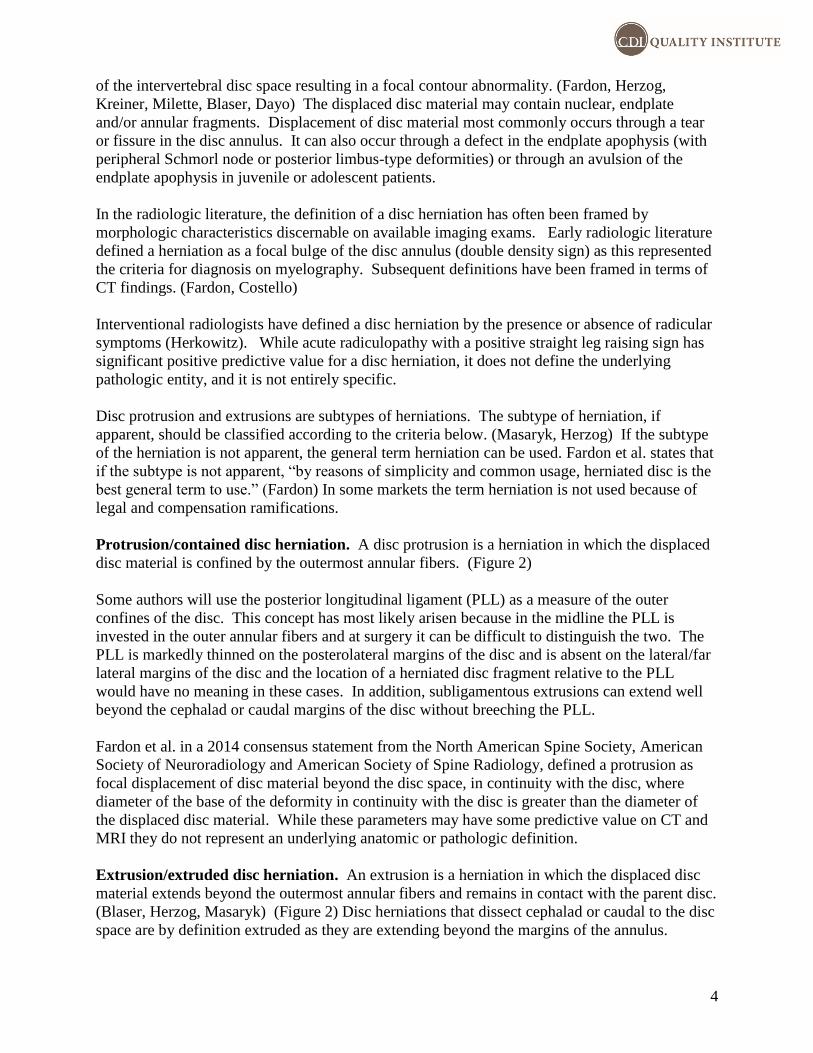
Protrusion/contained disc herniation. A disc protrusion is a herniation in which the displaced
disc material is confined by the outermost annular fibers. (Figure 2)
Some authors will use the posterior longitudinal ligament (PLL) as a measure of the outer
confines of the disc. This concept has most likely arisen because in the midline the PLL is
invested in the outer annular fibers and at surgery it can be difficult to distinguish the two. The
PLL is markedly thinned on the posterolateral margins of the disc and is absent on the lateral/far
lateral margins of the disc and the location of a herniated disc fragment relative to the PLL
would have no meaning in these cases. In addition, subligamentous extrusions can extend well
beyond the cephalad or caudal margins of the disc without breeching the PLL.
Fardon et al. in a 2014 consensus statement from the North American Spine Society, American
Society of Neuroradiology and American Society of Spine Radiology, defined a protrusion as
focal displacement of disc material beyond the disc space, in continuity with the disc, where
diameter of the base of the deformity in continuity with the disc is greater than the diameter of
the displaced disc material. While these parameters may have some predictive value on CT and
MRI they do not represent an underlying anatomic or pathologic definition.
Extrusion/extruded disc herniation. An extrusion is a herniation in which the displaced disc
material extends beyond the outermost annular fibers and remains in contact with the parent disc.
(Blaser, Herzog, Masaryk) (Figure 2) Disc herniations that dissect cephalad or caudal to the disc
space are by definition extruded as they are extending beyond the margins of the annulus.
5
Fardon et al. states that an extrusion is a disc contour abnormality where the diameter of the
displaced disc material is larger than the segment of disc maintaining continuity with the parent
disc. These parameters may have some predictive value on CT or MRI, however, do not
represent an underlying anatomic or pathologic definition.
Sequestration/sequestered disc fragment. If an extruded disc fragment separates and loses
contact with the parent disc, the fragment is referred to as a sequestration. (Fardon, Grenier,
Herzog)
Subligamentous Herniation. If an extruded or sequestered fragment dissects cephalad or
caudal to the parent disc deep to the posterior longitudinal spinal ligament it can be referred to as
a subligamentous herniation. (Figure 2)
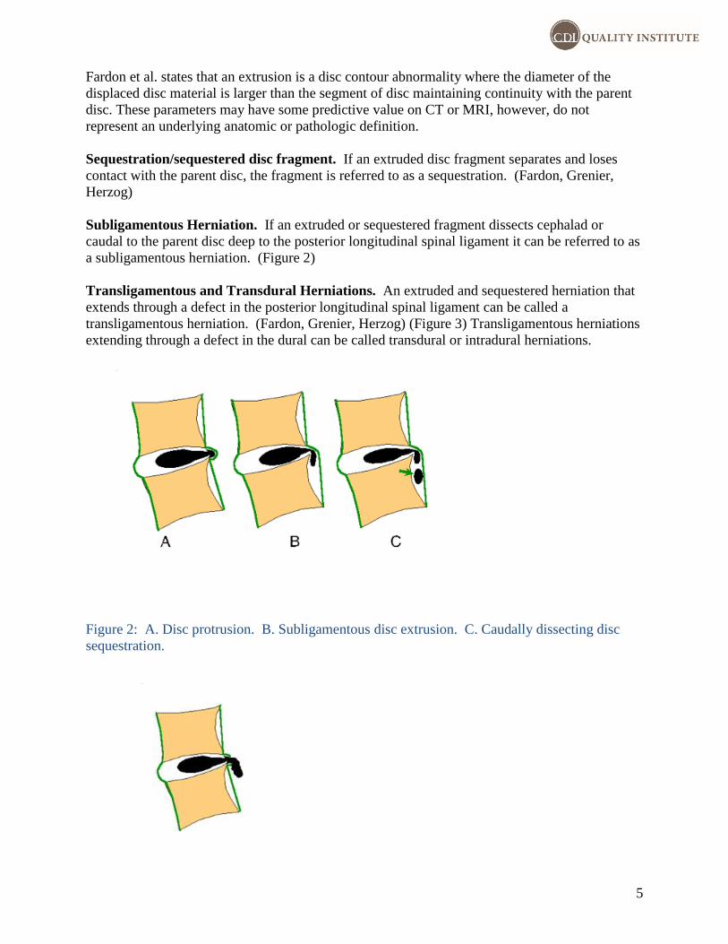
Transligamentous and Transdural Herniations. An extruded and sequestered herniation that
extends through a defect in the posterior longitudinal spinal ligament can be called a
transligamentous herniation. (Fardon, Grenier, Herzog) (Figure 3) Transligamentous herniations
extending through a defect in the dural can be called transdural or intradural herniations.
Figure 2: A. Disc protrusion. B. Subligamentous disc extrusion. C. Caudally dissecting disc
sequestration.
6
Figure 3: Transligamentous herniation (arrow) Modified rom the ASNR Nomenclature and
Classification of Lumbar Disc Pathology.
Herniation size. The maximum size in the AP direction may be reported relative to the
peripheral endplate, endplate osteophyte or bulging annulus. The transverse diameter of the
herniation can be measured or can be described using qualitative terms such as broad-based. The
degree of cephalad or caudal dissection of extruded fragments can be measured relative to the
endplate or reported relative to anatomic levels e.g. caudal extension to the level of the pedicle or
to the entry zone of the caudal neural foramen.
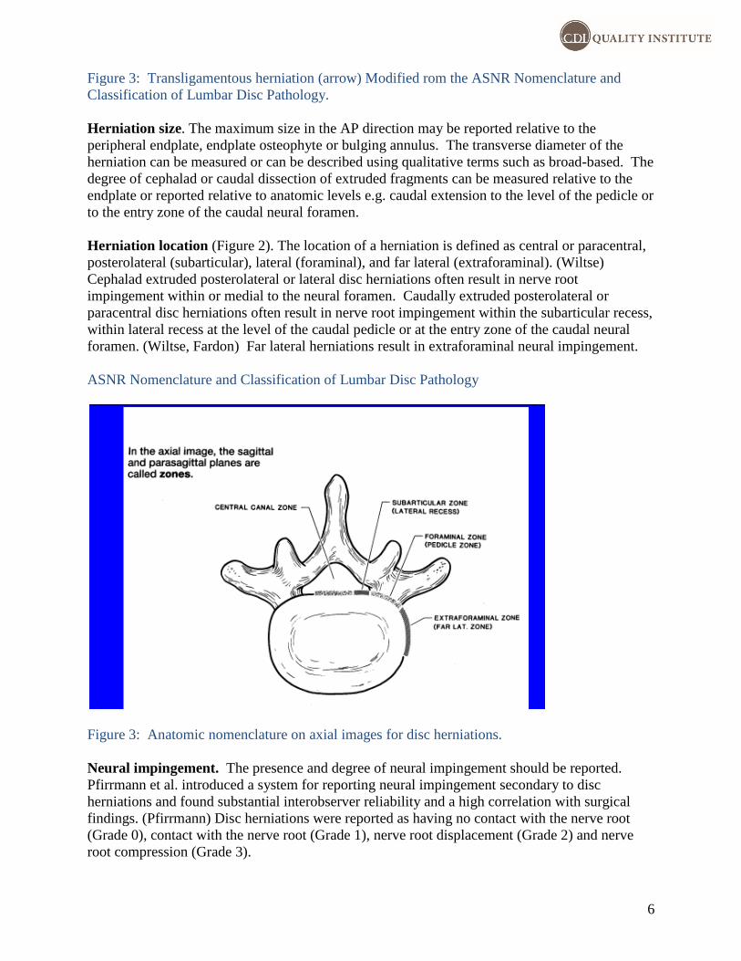
Herniation location (Figure 2). The location of a herniation is defined as central or paracentral,
posterolateral (subarticular), lateral (foraminal), and far lateral (extraforaminal). (Wiltse)
Cephalad extruded posterolateral or lateral disc herniations often result in nerve root
impingement within or medial to the neural foramen. Caudally extruded posterolateral or
paracentral disc herniations often result in nerve root impingement within the subarticular recess,
within lateral recess at the level of the caudal pedicle or at the entry zone of the caudal neural
foramen. (Wiltse, Fardon) Far lateral herniations result in extraforaminal neural impingement.
ASNR Nomenclature and Classification of Lumbar Disc Pathology
Figure 3: Anatomic nomenclature on axial images for disc herniations.
Neural impingement. The presence and degree of neural impingement should be reported.
Pfirrmann et al. introduced a system for reporting neural impingement secondary to disc
herniations and found substantial interobserver reliability and a high correlation with surgical
findings. (Pfirrmann) Disc herniations were reported as having no contact with the nerve root
(Grade 0), contact with the nerve root (Grade 1), nerve root displacement (Grade 2) and nerve
root compression (Grade 3).
7
Prognostic significance of herniation characteristics. The size and subtype of the herniations
have prognostic significance. Extruded, transligamentous and sequestered disc herniations are
exposed to epidural blood flow and have a greater tendency to resorb than do protrusions which
are contained by the outer annular fibers and may have limited exposure to the epidural space.
Resorption has been shown to occur through a process of peripheral neovascularization and
macrophage infiltration and shows a general correlation with a favorable natural history.
Large size and high signal intensity on T2 weighted images also shows a correlation with
resorption. Large high signal intensity extrusions often present with understated clinical
presentations and may resorb completely within 1-3 months. If symptoms persist in a patient
with a high signal intensity extrusion, repeat MRI is indicated prior to surgery to ensure that a
compressive lesion persists.
Stenosis:
Lumbar Central Stenosis (LSS). Dural sac area (DSA) is the most widely accepted measure for
central canal stenosis and correlates well with pain and function in many studies (NASS). The
DSA has been shown to correlate with clinical symptoms and function, and is more effective in
the assessment of central canal stenosis than is the AP diameter of the bony canal. (Ogikubo,
Haminashi, Bolender)
When grading the severity of LSS, moderate or severe disease should indicate, to the extent
possible, clinically significant disease in patients with appropriate clinical presentations. A grade
of moderate or severe should have a high positive predictive value of a good or excellent result
with surgical decompression at this level in a patient with neurogenic claudication (and no
confounding vascular disease). Since a dural sac area (DSA) of 75mm2 has been shown to result
in a measured increase in pressure on the cauda equina (Schönström), this has been set as the
threshold for moderate stenosis:
Mild - DSA 75-100 mm2
Moderate - DSA 50-75 mm2
Severe - DSA <50 mm2
The DSA can be estimated easily with the following formula: r1 x r2 x 3. Ogikubo et al. showed
a linear correlation of DSA with symptoms. (Ogikubo)
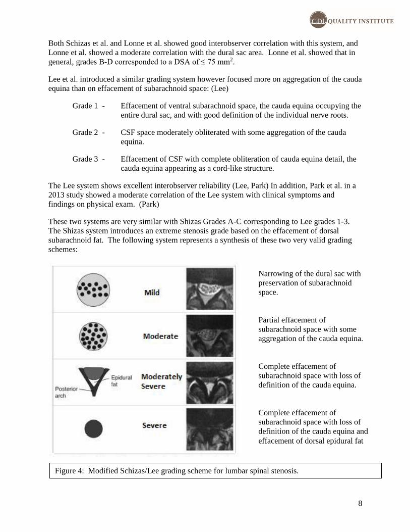
Shizas et al. has since introduced a more practical method for assessing central canal stenosis
which relies on effacement of subarachnoid space and dorsal epidural fat: (Shizas)
A - Narrowing of the dural sac with preservation of subarachnoid space.
B - Narrowing with rootlets occupying the whole dural sac, CSF persists with
decrease signal intensity, and nerve roots remain well defined.
C - Narrowing with loss of definition of the nerve roots and the CSF showing
a homogeneous gray signal. Dorsal epidural fat persists.
D - Same as C with effacement of dorsal epidural fat.
8
Both Schizas et al. and Lonne et al. showed good interobserver correlation with this system, and
Lonne et al. showed a moderate correlation with the dural sac area. Lonne et al. showed that in
general, grades B-D corresponded to a DSA of ≤ 75 mm2.
Lee et al. introduced a similar grading system however focused more on aggregation of the cauda
equina than on effacement of subarachnoid space: (Lee)
Grade 1 - Effacement of ventral subarachnoid space, the cauda equina occupying the
entire dural sac, and with good definition of the individual nerve roots.
Grade 2 - CSF space moderately obliterated with some aggregation of the cauda
equina.
Grade 3 - Effacement of CSF with complete obliteration of cauda equina detail, the
cauda equina appearing as a cord-like structure.
The Lee system shows excellent interobserver reliability (Lee, Park) In addition, Park et al. in a
2013 study showed a moderate correlation of the Lee system with clinical symptoms and
findings on physical exam. (Park)
These two systems are very similar with Shizas Grades A-C corresponding to Lee grades 1-3.
The Shizas system introduces an extreme stenosis grade based on the effacement of dorsal
subarachnoid fat. The following system represents a synthesis of these two very valid grading
schemes:
Figure 4: Modified Schizas/Lee grading scheme for lumbar spinal stenosis.
Narrowing of the dural sac with
preservation of subarachnoid
space.
Partial effacement of
subarachnoid space with some
aggregation of the cauda equina.
Complete effacement of
subarachnoid space with loss of
definition of the cauda equina.
Complete effacement of
subarachnoid space with loss of
definition of the cauda equina and
effacement of dorsal epidural fat
9
Subarticular recess stenosis. In academic settings, subarticular recess stenosis is frequently
defined as narrowing of the AP diameter to less than 3 mm. Practically, subarticular recess
stenosis is only significant if it results in nerve root impingement. Marked narrowing of the
subarticular recess can be present with no neural impingement.
The position of the traversing nerve root can vary markedly with respect to the subarticular
recess. With a high take-off, the nerve root at the level of the subarticular recess is extradural
and is susceptible to impingement. With a low take-off, the nerve root is intradural at the level
of the subarticular recess and is afforded much greater mobility.
Marked narrowing of the subarticular recess can be present with no neural impingement.
Displacement of the intradural nerve root does not correlate with symptomatic subarticular recess
stenosis to the extent that compression of an extradural nerve root does. Conjoined or partially
conjoined nerve roots are often tightly tethered within the subarticular recess and caudal neural
foramen and are very susceptible to impingement.
If subarticular recess stenosis is present, the presence or absence of root impingement should be
detailed. The severity of the stenosis itself can be graded qualitatively as mild, moderate or
severe. The presence of a conjoined nerve root should always be reported.
Foraminal stenosis. Grading of foraminal stenosis is relevant only to presence or absence of
nerve root or ganglionic impingement. The size of a normal foramen can vary markedly between
patients and marked narrowing of a developmentally large foramen is of no significance without
neural impingement.
Lee et al. in 2010 introduced a grading system for foraminal stenosis which focuses on the
presence or absence of perineual fat and/or neural compression. (Lee)
Mild - Narrowing of the neural foramen with partial effacement of
perineural fat on opposite borders (superior-inferior or
anterior-posterior) without neural deformity or
compression.
Moderate - Narrowing of the neural foramen with complete effacement
of perineural fat and no neural deformity or compression.
Severe - Narrowing of the neural foramen with complete effacement
of perineural fat and neural compression or deformity.
Park et al. in a subsequent study showed that this grading system has good interobserver
agreement and good clinical correlation.
The Lee grading system has some limitations. First, it does not accommodate cases in which
there is partial effacement of perineural fat and pincer-type impingement or up-down neural
compression. In these cases, there may not be complete effacement of perineural fat, however,
there is definite neural compression. Conversely, complete effacement of perineural fat within a
narrowed neural foramen can also be secondary to partial voluming of an intraforaminal disc
herniation.
10
The results of surgical decompression are less predictable in patients with lower grades of
impingement. (Weishaupt) Nerve root contact or effacement of perineural fat on one or two
borders is not typically associated with radicular symptoms and may be incidental to radicular
symptomatology (e.g. radiculitis seen in diabetic patients). Bowing of the L5 nerve root,
ganglion or post-ganglionic nerve on the far lateral margin of the L5-S1 disc is also seen
commonly and may not be associated with symptoms. Nerve root compression shows a more
significant correlation with symptoms and may predict symptom improvement with
decompression. In some cases, a selective nerve root block may be necessary to assess the
significance of foraminal stenosis relative to the patient’s symptoms.
If foraminal stenosis is present, the radiologist report should focus on effacement of perineual
fat, nerve root displacement and nerve root compression if present. Foraminal stenosis can be
graded qualitatively as mild, moderate or severe.
While the grading systems do show some correlation with symptoms, it is not uncommon to see
severe foraminal stenosis in an asymptomatic patient. As with all other spine findings,
correlation with clinical symptoms is the bedrock of clinical significance.
Far lateral foraminal stenosis. Far lateral foraminal stenosis can occur with foraminal stenosis
or can be isolated. The far lateral foramen is an anatomic entity unique to the L5-S1 level. The
far lateral foramen begins on the lateral margin of the pedicle and is delimited by the L5
transverse process superiorly and the S1 ala inferiorly.
Far lateral foraminal stenosis, far lateral disc herniations and far lateral neural impingement can
be difficult to identify on MRI. This problem may be mimimized with the use of new thin
section 3D MRI acquisitions and angled coronal/angled axial reformations. Thin section CT
with sagittal and axial oblique/coronal obilgue reformations can be very helpful and can be
definitive in patients in whom MRI findings are understated. Post-impingement swelling of the
L5 nerve is specific but not sensitive for symptomatic far lateral foraminal neural impingement.
11
References:
Blaser SI and Modic MT. Herniation of the intervertebral disc. Top Magn Reson Imag
1988;1(1):25-37.
Bolender NF, Schonstrom NS, Spengler DM. Role of computed
tomography and myelography in the diagnosis of central spinal
stenosis. J Bone Joint Surg Am. 1985;67(2):240-6.
Boos N, Rieder R, Schade V, et al. The diagnostic accuracy of magnetic resonance imaging,
work perception and psychosocial factors in identifying symptomatic disk herniations. Spine
1995;20:2613-2625.
Costello RF and Beall DP. Nomenclature and standard reporting terminology of intervertebral
disk herniation. Magn Reson Imaging Clin N Am 2007;15:167-174.
Deyo RA, Loeser JD, Bigos SJ. Herniated lumbar intervertebral disc. Ann Int Med
1990;112(8):589-603.
Fardon DF, Williams AL, Dohring EJ, et al. Lumbar spine nomenclature: version 2.0.
Recommendations from the combined task forces of the North American Spine Society, the
American Society of Spine Radiology and the American Society of Neuroradiology. The Spine
Journal 2014;14(11):2525-45.
Grenier N, Greselle J, Viral J et al. Normal and disrupted lumbar longitudinal ligaments:
Correlative MR and anatomic study. Radiology 1989;171(1):197-205.
Hamanishi C, Matukura N, Fujita M, Tomihara M, Tanaka S.
Cross-sectional area of the stenotic lumbar dural tube measured
from the transverse views of magnetic resonance imaging. J
Spinal Disord. 1994;7(5):388-93.
Herzog RJ. The Radiologic Assessment for a lumbar disc herniation. Spine 1996;21 (24S):19S-
38S.
Kreiner DS, Hwang SW, Easa JE, et al. An evidence-based clinical guideline for the diagnosis
and treatment of lumbar disc herniation with radiculopathy. Spine J 2014;14(1):180-91.
Lee GY, Lee JW, Choi HS, et al. A new grading system of lumbar central canal stenosis on
MRI: an easy and reliable method. Skeletal Radiol 2011;40:1033-9.
Lee S, Lee JW, Yeom JS et al. A practical MRI grading system for lumbar foraminal stenosis.
AJR 2010;194:1095-1098.
Lønne G, Ødegård B, Johnsen LG, et al. MRI evaluation of lumbar spinal stenosis: Is a rapid
visual assessment as good as area measurement? Eur Spine J 2014;23(6):1320-4.
Milette PC, Fontaine S, Lepanto L, et al. Differentiating lumbar disc protrusions, disc bulges,
and discs with normal contour but abnormal signal intensity. Spine 1999;24:44-53.
12
NASS Practice Guidelines. Diagnosis and Treatment of Degenerative Lumbar Spinal Stenosis
(Revised 2011).
https://www.spine.org/Documents/ResearchClinicalCare/Guidelines/LumbarStenosis.pdf
Ogikubo O, Forsberg L, Hansson T, et al. The relationship between the cross-sectional area of
the cauda equina and the preoperative symptoms in central lumbar spinal stenosis. Spine
2007;32:1423-1428.
Park HJ, Kim SS, Lee SY, et al. Clinical correlation of a New Imaging Method for Assessing
Lumbar Foraminal Stenosis. AJNR 2012;33:818-822.
Pfirrmann CW1, Dora C, Schmid MR, et al. MR image-based grading of lumbar nerve root
compromise due to disk herniation: reliability study with surgical correlation. Radiology. 2004
Feb;230(2):583-8.
Resnick and Kransdorf. Bone and Joint Imaging, 5th edition. Saunders.
Schönström N, Bolender NF, Spengler DM, Hansson TH. Pressure changes within the cauda
equina following constriction of the dural sac. An in vitro experimental study. Spine (Phila Pa
1976). 1984 Sep;9(6):604-7.
Schiza C, Theumann N, Burn A et al. Qualitative grading of severity of lumbar spinal stenosis
based on the morphology of the dural sac on magnetic resonance images. Spine 2010;35:1919-
24.
Ogikubo O, Forsberg L, Hansson T. The relationship between
the cross-sectional area of the cauda equina and the preoperative
symptoms in central lumbar spinal stenosis. Spine 2007;32(13):1423-8; discussion 1429.
Park HJ, Kim SS, Lee SY, et al. Clinical correlation of a new MR imaging method for assessing
lumbar spinal stenosis. AJNR 2012;33:818-822.
Schellhas KP, Pollei ST, Gundry CR, et al. Lumbar disc high-intensity zone. Correlation of
magnetic resonance imaging and discography. Spine 1996;21:79-86.
Weishaupt D, Zanetti M, Hodler J, Boos N. MR imaging of the lumbar spine: Prevalence of
intervertebral disk extrusion and sequestration, nerve root compression, end plate abnormalities,
and osteoarthritis of the facet joints in asymptomatic volunteers. Radiology 1998;209:661-666.
Wildermuth S, Zanetti M, Duewell S, et al. Lumbar spine: quantitative and qualitative
assessment of positional (upright flexion and extension) MR imaging and myelography.
Radiology 1998;207:391-98.
Wiltse LL, Berger PE, McCulloch JA. A system for reporting the size and location of lesions in
the spine. Spine 1997;22(13):1534-1537.
13
Yue-Hui Z, Chang-Qing Z, Lei-sheng J, et al. Modic changes: a systemic review of the
literature. Eur Spine J 2008;17:1289-99.
This is a guideline, not a policy. It is a summary and distillation of relevant literature and subspecialty guidelines. The
purpose of the CDI Quality Institute guidelines is to promote quality and continuity, where appropriate for medical practices
within the CDI/Insight enterprise, and to provide relevant and up to date background information to support the development
of policies within each individual practice. Guidelines should be adjusted for local standards of care, associated hospital or
network policies, hospital versus outpatient settings, different patient populations and your own risk tolerance. Guidelines
should also be modified to account for new information or publications that become available between revisions.