Embed Size (px)
Citation preview
Maurizio Galderisi
Lung Ultrasound
Head, Cardiovascular Emergencies and Onco-HaematologicalComplications
Laboratory of Standard and Advanced EchocardiographyFederico II University Hospital
Vice President, EACVICommunication Officer, ESC Council of Cardio-Oncology

“Speaker disclosure - I do not have an affiliation (financial or otherwise) with a pharmaceutical, medical device, or
communication and event planning company.”
OBJECTIVES
1.- To show the diagnostic capabilities of Lung UltraSound in Critical Care.2.- To illustrate the diagnostic modalities needed.3.- To highlight the fields of application. 4.- To encourage the use of standardized protocols and terminology.
Lung Approach in Critical Care
• Physical examination: insufficient for fine diagnosis
• Bedside chest radiography: limited accuracy
• Chest computed tomography: risk of transportation and limited availability
• Lung UltraSound (LUS): Easy available, Low cost
Lung Ultrasound (LUS) In Emergency
When LUS can be useful in ER:
• Pleural pathology• Pericardial pathology• Shortness of breath• Cianosis• Cough• Shock
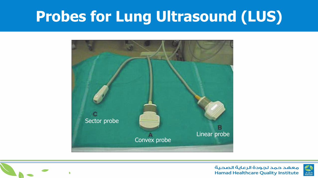
Probes for Lung Ultrasound (LUS)
Linear probeConvex probe
Sector probe
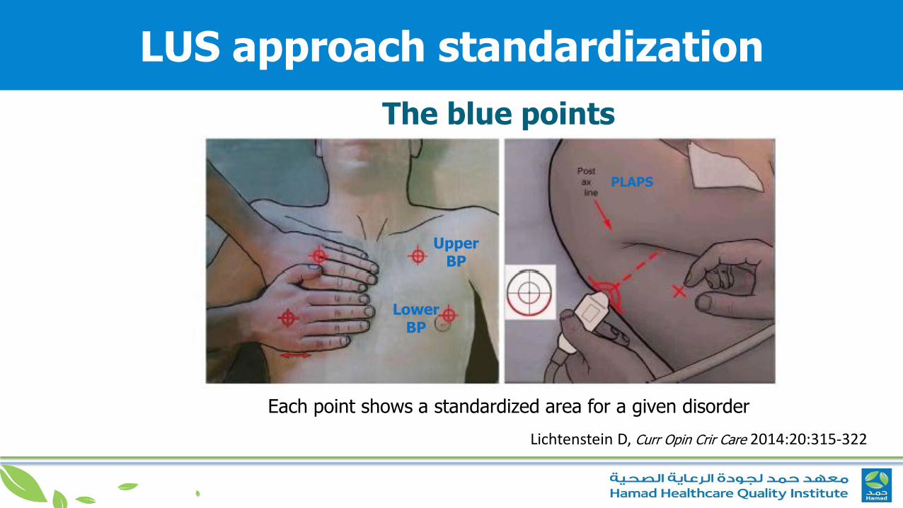
LUS approach standardization
The blue points
Each point shows a standardized area for a given disorder
Lichtenstein D, Curr Opin Crir Care 2014:20:315-322
PLAPS
Upper BP
Lower BP
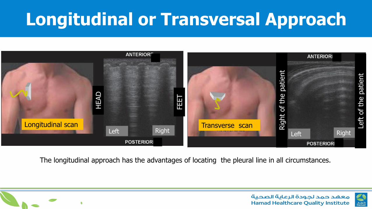
Longitudinal or Transversal Approach
The longitudinal approach has the advantages of locating the pleural line in all circumstances.
Longitudinal scan Transverse scan Left Left
Right Right
HEAD
FEET
Rig
ht
of
the p
atient
Left
of
the p
atient
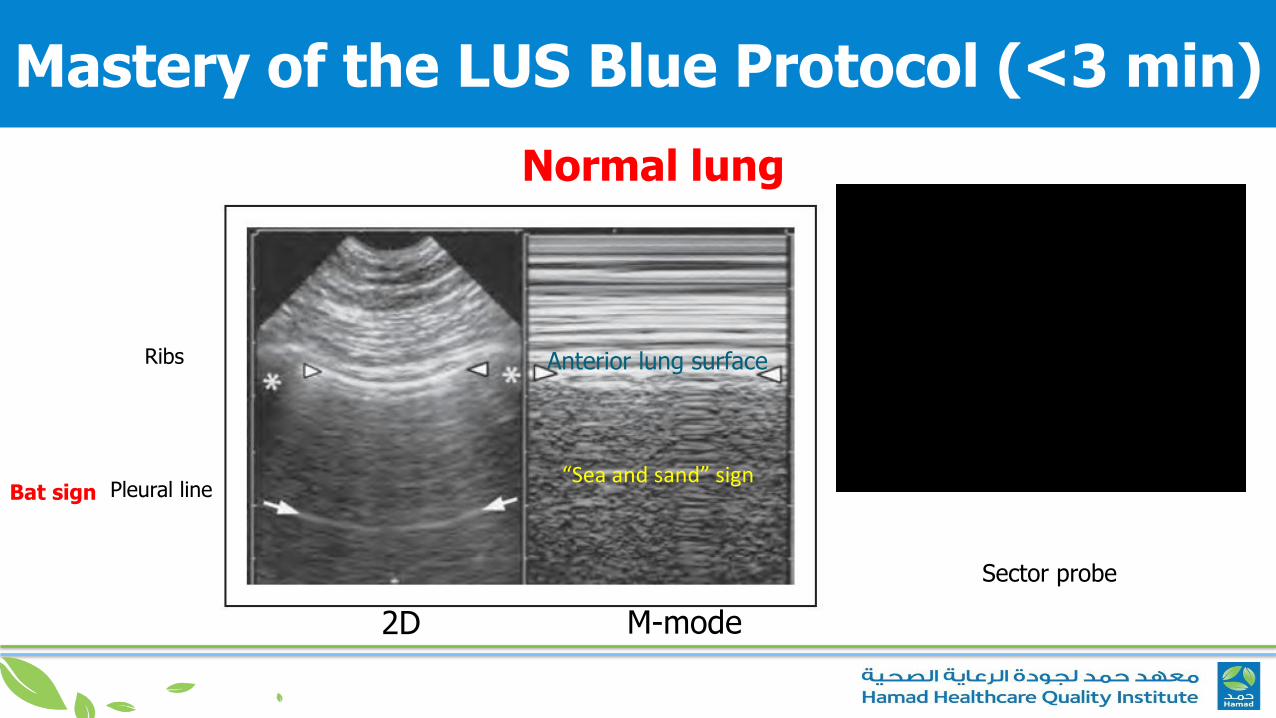
Mastery of the LUS Blue Protocol (<3 min)
Normal lung
2D M-mode
Ribs
Pleural line
Anterior lung surface
Bat sign“Sea and sand” sign
Sector probe
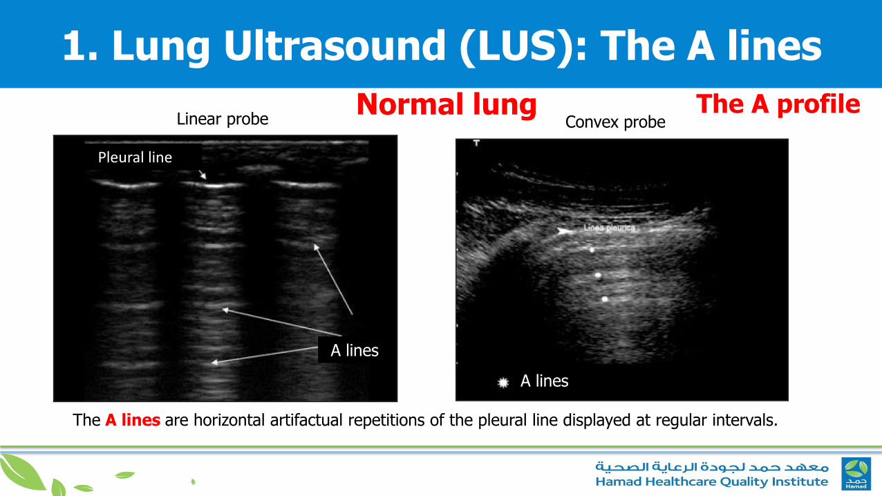
1. Lung Ultrasound (LUS): The A lines
Normal lung
Pleural line
A lines
A lines
Linear probe Convex probe
The A lines are horizontal artifactual repetitions of the pleural line displayed at regular intervals.
The A profile
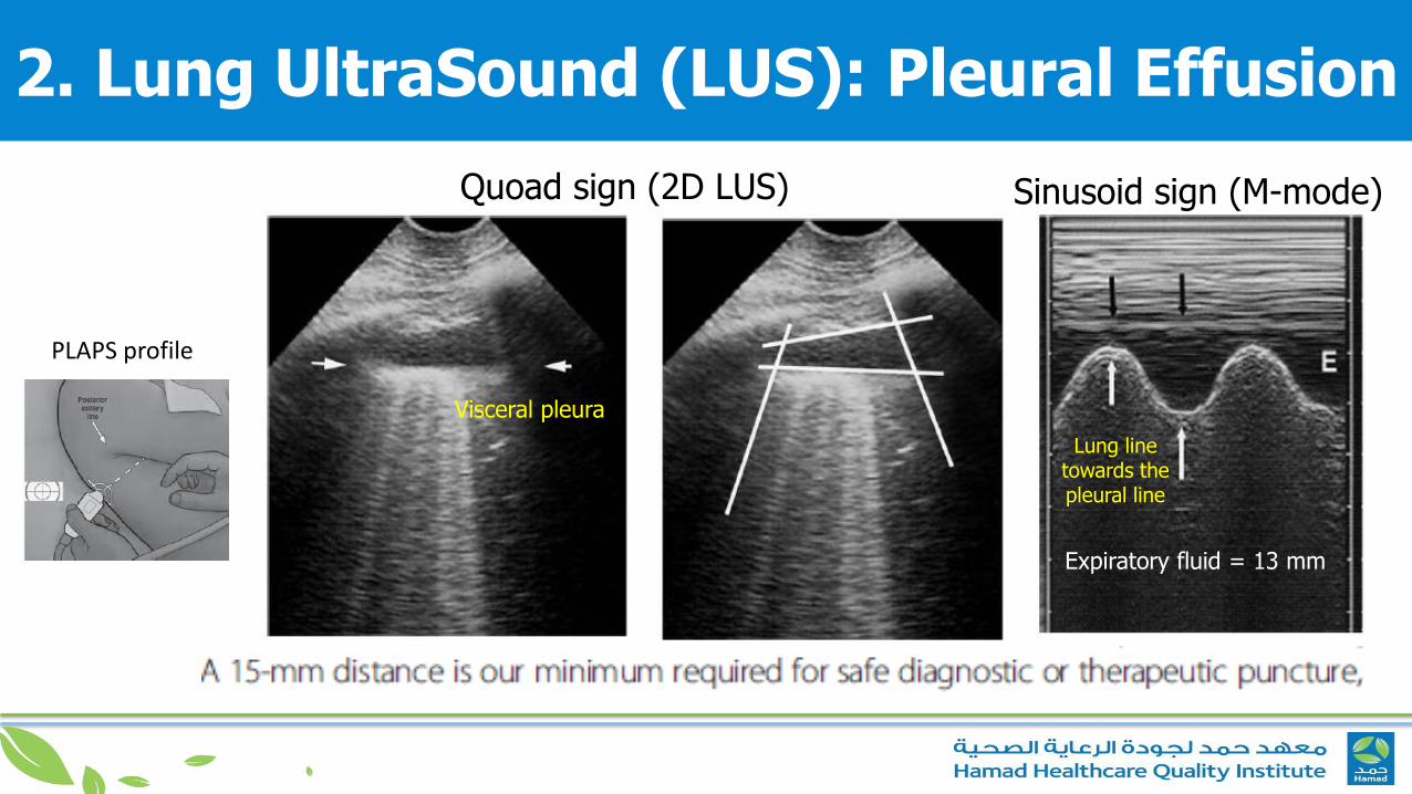
PLAPS profile
Visceral pleura
Quoad sign (2D LUS) Sinusoid sign (M-mode)
Lung line towards the pleural line
Expiratory fluid = 13 mm
2. Lung UltraSound (LUS): Pleural Effusion
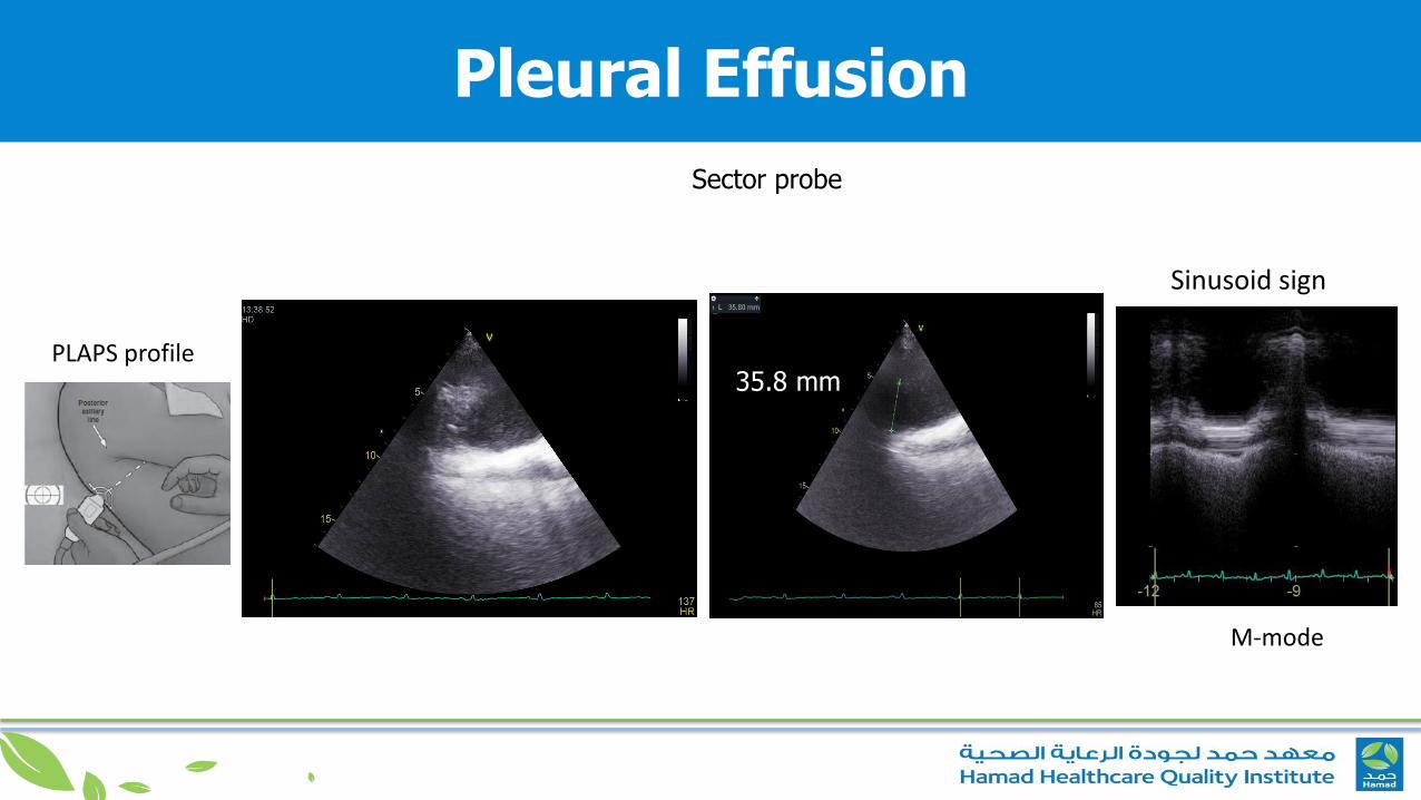
Pleural Effusion
PLAPS profile
Sector probe
35.8 mm
Sinusoid sign
M-mode
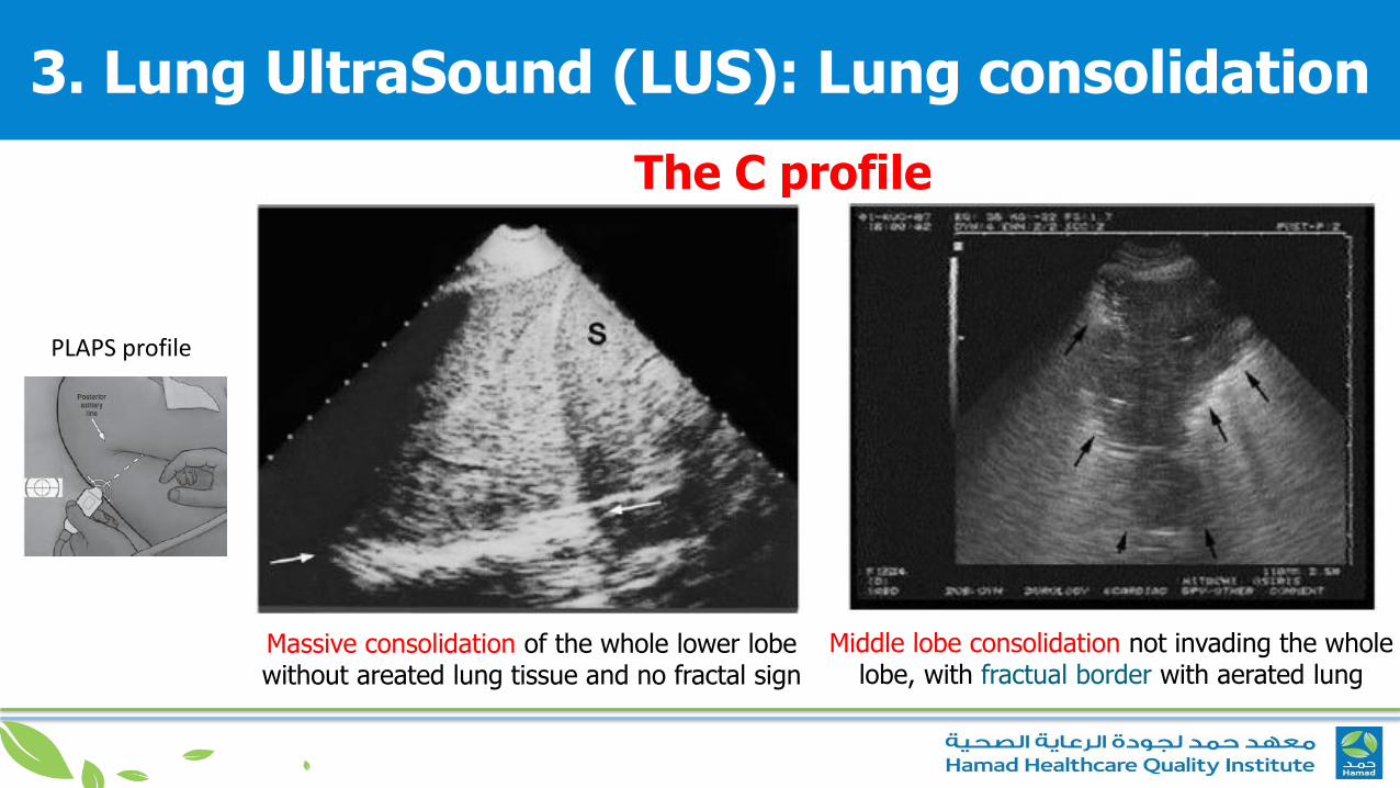
3. Lung UltraSound (LUS): Lung consolidation
PLAPS profile
The C profile
Massive consolidation of the whole lower lobewithout areated lung tissue and no fractal sign
Middle lobe consolidation not invading the wholelobe, with fractual border with aerated lung
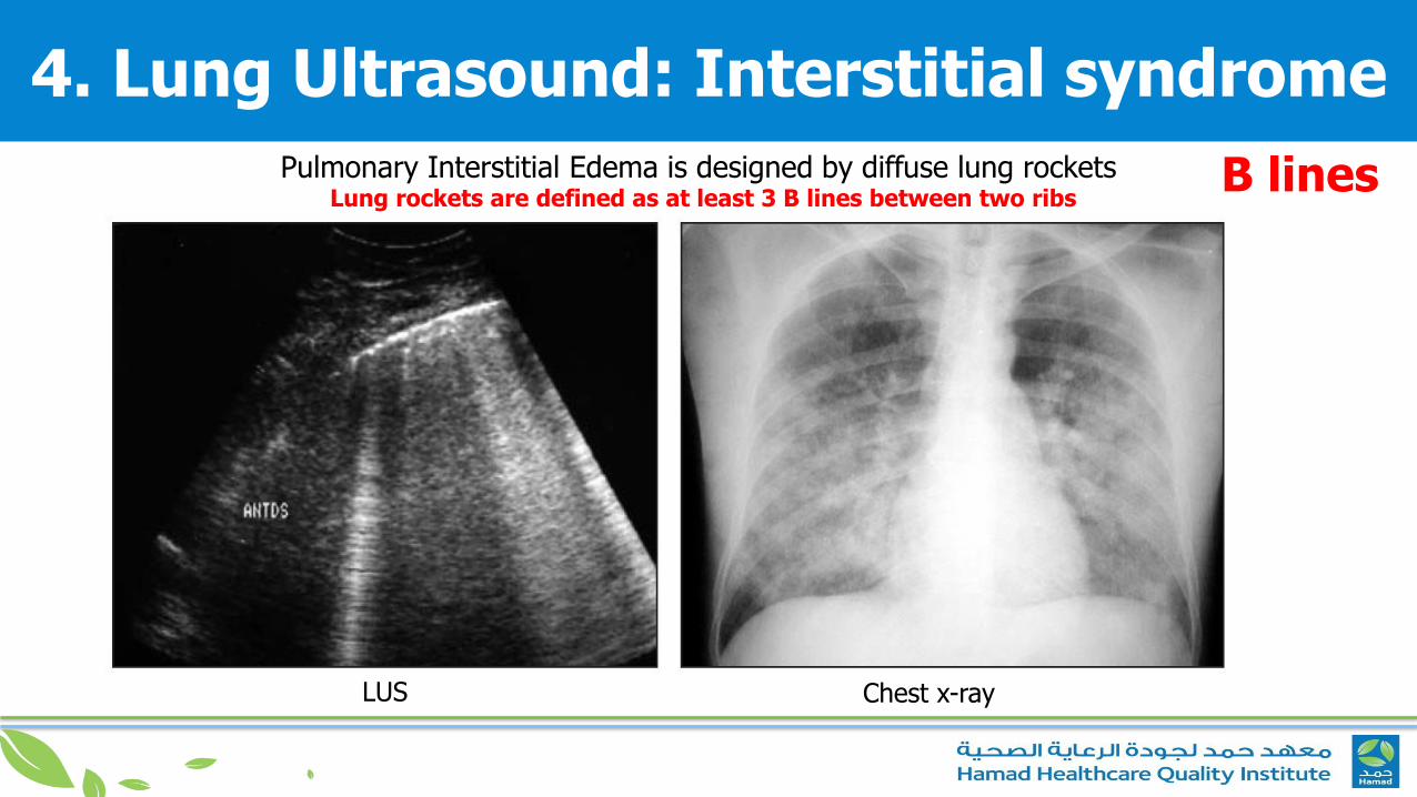
4. Lung Ultrasound: Interstitial syndrome
Pulmonary Interstitial Edema is designed by diffuse lung rocketsLung rockets are defined as at least 3 B lines between two ribs
LUS Chest x-ray
B lines
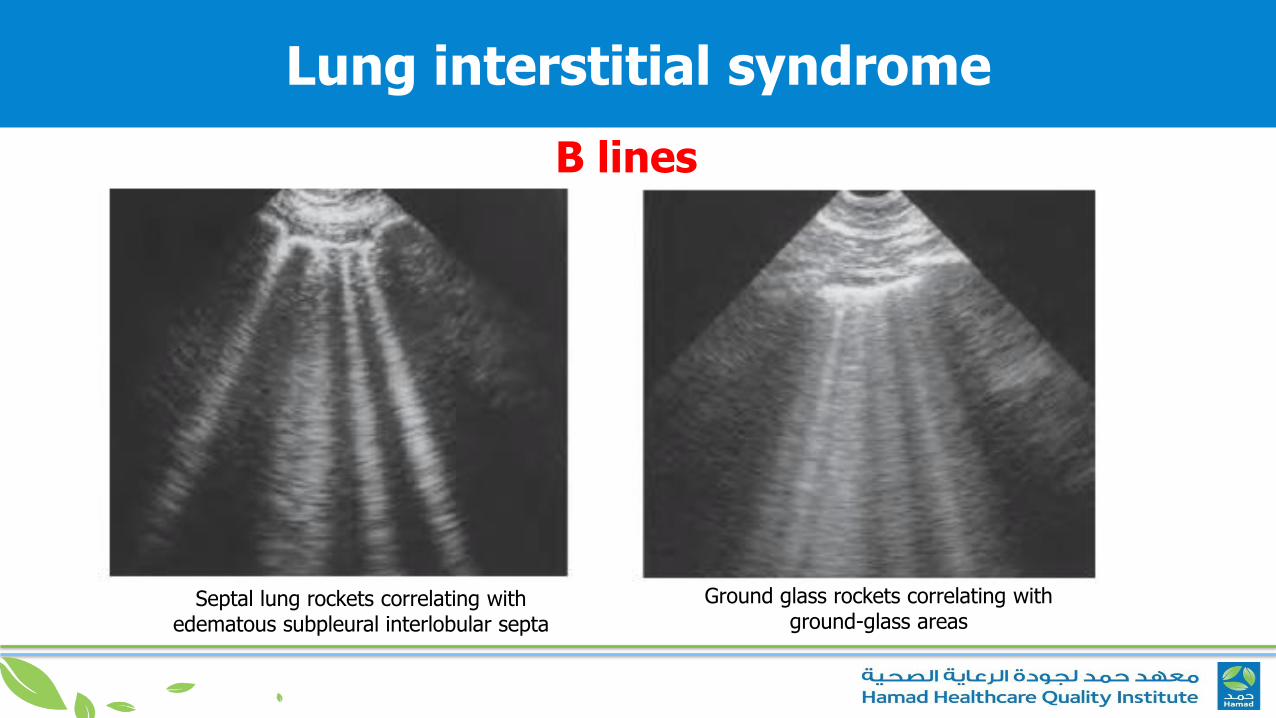
Ground glass rockets correlating with ground-glass areas
Septal lung rockets correlating with edematous subpleural interlobular septa
B lines
Lung interstitial syndrome
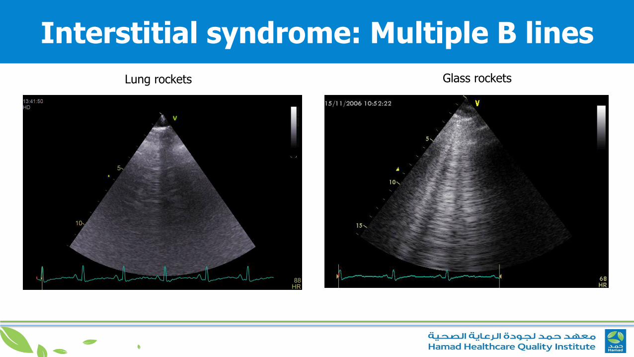
Interstitial syndrome: Multiple B lines
Lung rockets Glass rockets
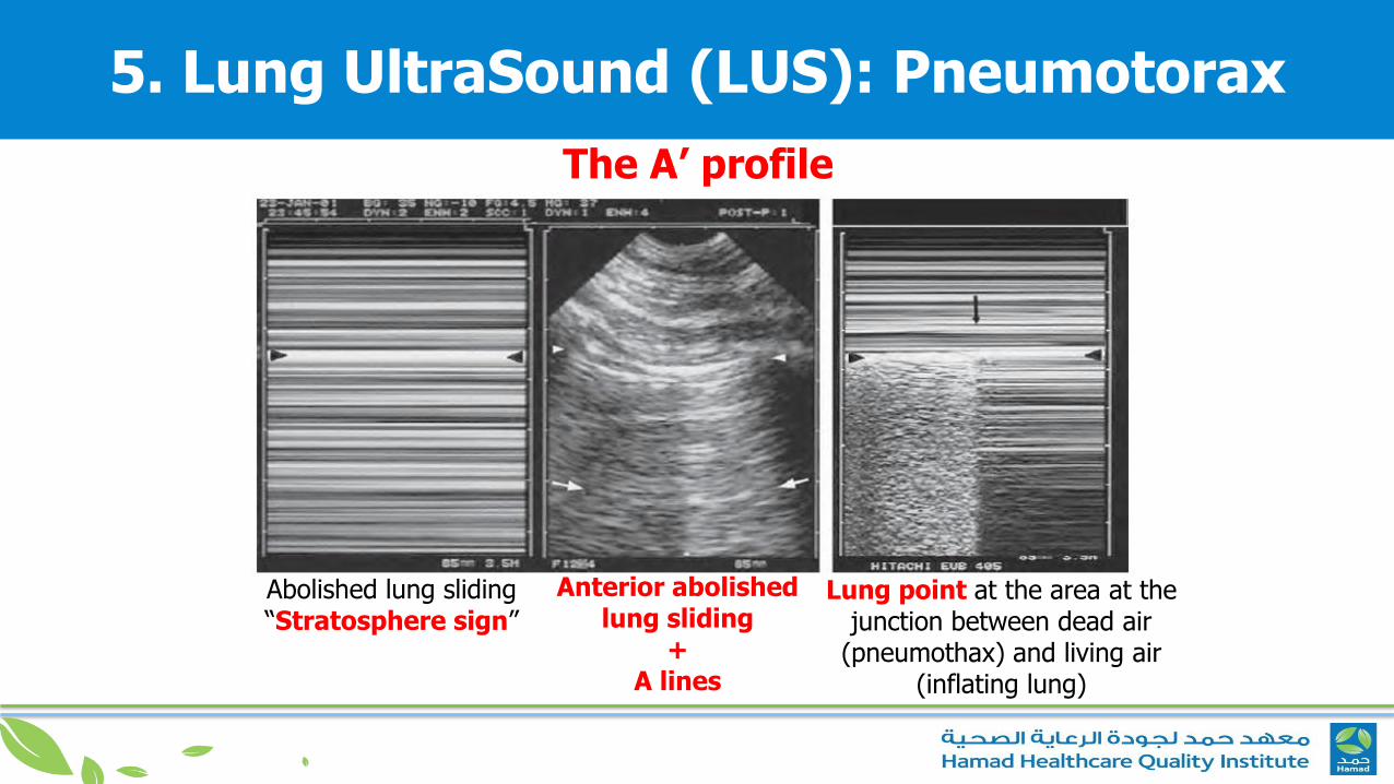
5. Lung UltraSound (LUS): Pneumotorax
Abolished lung sliding“Stratosphere sign”
Anterior abolishedlung sliding
+A lines
Lung point at the area at the junction between dead air
(pneumothax) and living air (inflating lung)
The A’ profile
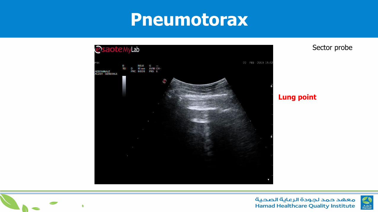
Pneumotorax
Lung point
Sector probe
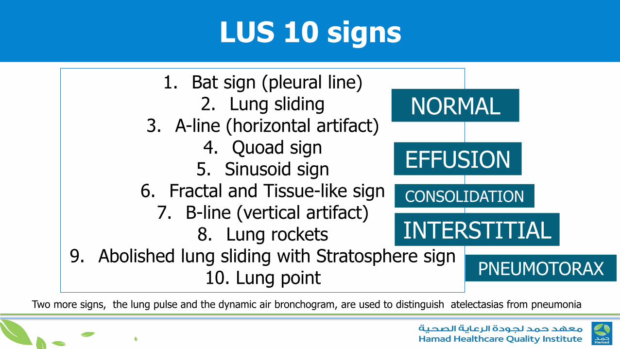
LUS 10 signs
1. Bat sign (pleural line) 2. Lung sliding
3. A-line (horizontal artifact)4. Quoad sign
5. Sinusoid sign6. Fractal and Tissue-like sign
7. B-line (vertical artifact) 8. Lung rockets
9. Abolished lung sliding with Stratosphere sign10. Lung point
Two more signs, the lung pulse and the dynamic air bronchogram, are used to distinguish atelectasias from pneumonia
NORMAL
EFFUSION
CONSOLIDATION
INTERSTITIAL
PNEUMOTORAX
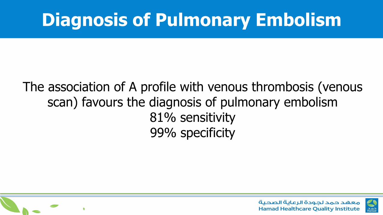
Diagnosis of Pulmonary Embolism
The association of A profile with venous thrombosis (venous scan) favours the diagnosis of pulmonary embolism
81% sensitivity 99% specificity
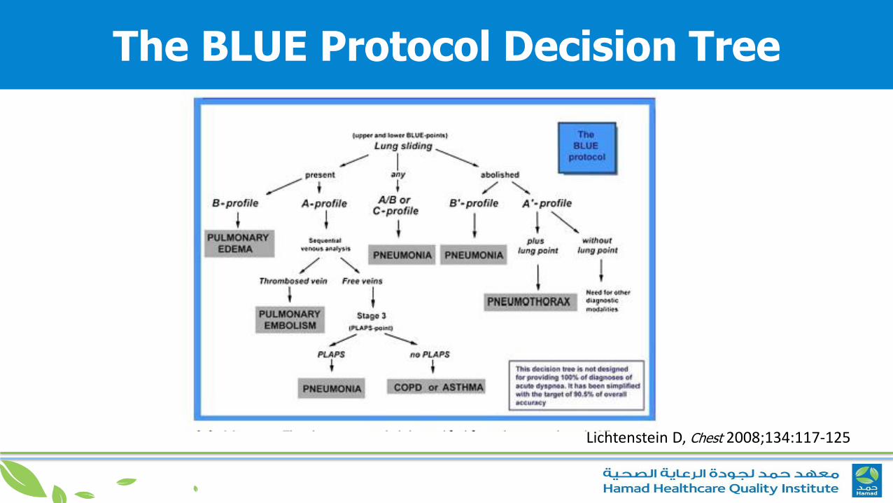
The BLUE Protocol Decision Tree
Lichtenstein D, Chest 2008;134:117-125
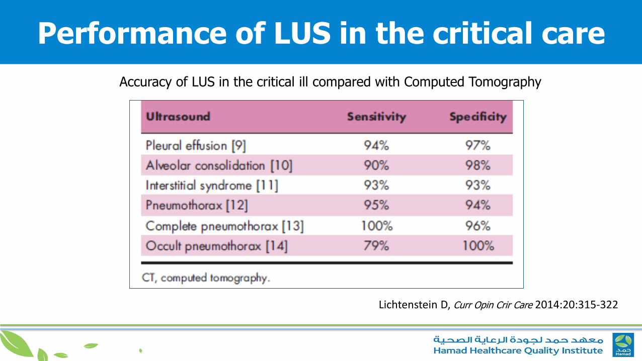
Performance of LUS in the critical care
Lichtenstein D, Curr Opin Crir Care 2014:20:315-322
Accuracy of LUS in the critical ill compared with Computed Tomography
The BLUE Protocol combined with simple Echo
The BLUE Protocol applies LUS and venous ultrasound for drawing profiles.
A simplified echocardiography without Doppler can be associated with the BLUE protocol.
The BLUE protocol can be adapted to multiple clinical settings:TraumaNeonate
Acure Respiratory Distress Syndrome (ARDS)
Fluid Administration limited by LUS Protocol
LUS can be used for answering two basic questions
1. Will the given patient benefit from fluid therapy ?
2. If administered, when stop fluid ?
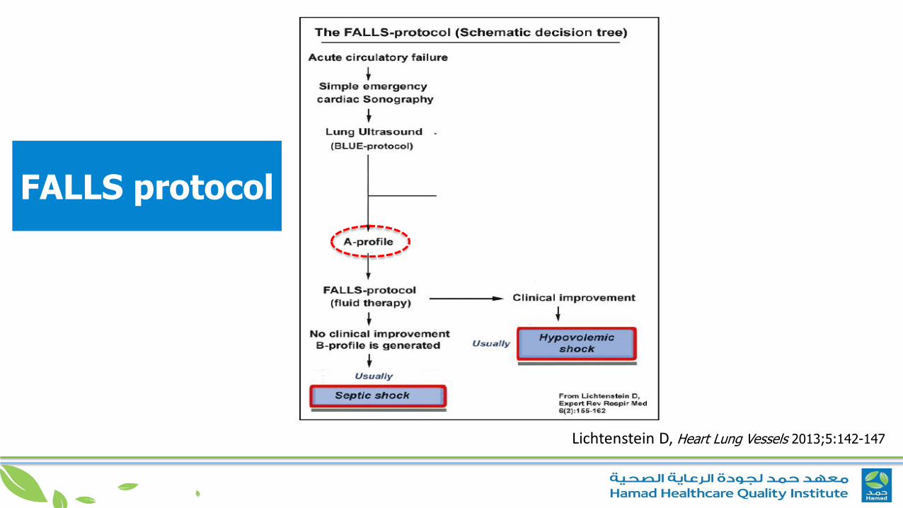
FALLS protocol
Lichtenstein D, Heart Lung Vessels 2013;5:142-147
Take home message
Lung UltraSound signs, either alone or combined to other point-of-care ultrasound techniques, are helpful in the diagnostic approach to patients with acute respiratory failure, circulatory shock or cardiac arrest.
Lung UltraSound monitoring can be performed at the bedside and used in mechanically ventilated patients, to assess the efficacy of treatments, to monitor the evolution of the respiratory disorder, and to help the weaning process.
Lung ultrasound can be used for early detection and management of respiratory complications under mechanical ventilation, such as pneumothorax, ventilator-associated pneumonia, atelectasis and pleural effusions.
Lung UltraSound is a useful diagnostic and monitoring tool that might become in the next future part of the basic knowledge of
physicians taking care of the critically ill patient.