Embed Size (px)
Citation preview
LUTEMBACHER'S SYNDROME: A RARE COMBINATION OF CONGENITAL AND ACQUIRED HEART DISEASE – A CASE REPORT AND REVIEW OF LITERATURE
Uguru SU, Raphael SC, Idoko PO, Onuh JA, Amusa GACardiology Unit, Department of Internal Medicine, Jos
University Teaching Hospital, P.M.B. 2076, Jos, Nigeria.
Correspondence: Samuel U. Uguru
E-mail: [email protected]
ABSTRACTLutembacher's syndrome is defined as the rare combination of congenital atrial septal defect and acquired mitral stenosis. The haemodynamic effects of this syndrome are a result of the interplay between the relative effects of the atrial septal defect and mitral stenosis. Mitral stenosis augments the left to right shunt through the atrial septal defect. The definition of Lutembacher's syndrome has undergone many changes. The earliest description in medical literature was found in a letter written by anatomist Johann Friedrich Meckel to Albrecht von Haller in 1750.In 1916, Lutembacher described his first case of this syndrome, involving a 61-year-old woman, and he attributed the mitral valvular lesion to congenital mitral stenosis. Because the mitral stenosis was, in fact, rheumatic in aetiology, the syndrome was defined eventually as a combination of congenital atrial septal defect and acquired, almost always rheumatic, mitral stenosis.
Keywords:Lutembacher's syndrome, congenital heart disease, valvular heart disease, atrial septal defect, mitral stenosis
35Jos Journal of Medicine, Volume 10 No. 1
CASE REPORTA 52-year-old woman presented to the
Cardiology Clinic with a 2-month history of progressive breathlessness, recurrent cough (productive of mucoid sputum) and bilateral leg swelling. She also had an associated history of orthopnoea and paroxysmal nocturnal dyspnoea. On examination, she had bilateral pitting pedal oedema up to the knees and bibasal crepitations. Pulse rate was 88 beats per minute (irregularly irregular), blood pressure was 100/74 mmHg and her jugular venous pressure was raised. Her apex beat was displaced to
ththe 6 left intercostal space, lateral to the anterior st ndaxillary line. Heart sounds were 1 and 2 only (with
the second heart sound having a loud pulmonary component). She also had grade 2/6 mitral and tricuspid regurgitation murmurs. Her liver was palpable 8 cm below the right costal margin and ascites was demonstrable by shifting dullness.
Electrocardiography (ECG) done showed atrial fibrillation with her chest x-ray showing cardiomegaly, upper lobe diversion and a full pulmonary conus. Echocardiography done showed sclerosis of the anterior mitral valve leaflet with hockey-stick appearance on B-mode. M-mode echocardiography revealed the classic Mexican hat appearance of the mitral valve consistent with mitral
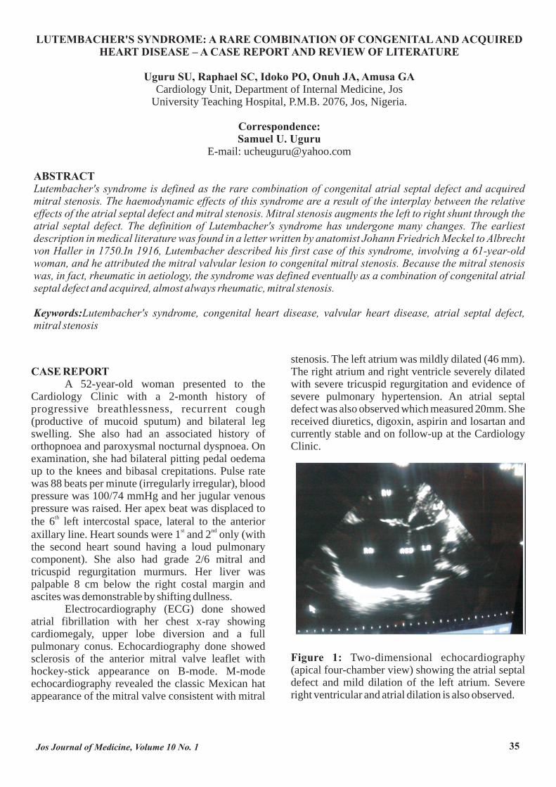
stenosis. The left atrium was mildly dilated (46 mm). The right atrium and right ventricle severely dilated with severe tricuspid regurgitation and evidence of severe pulmonary hypertension. An atrial septal defect was also observed which measured 20mm. She received diuretics, digoxin, aspirin and losartan and currently stable and on follow-up at the Cardiology Clinic.
Figure 1: Two-dimensional echocardiography (apical four-chamber view) showing the atrial septal defect and mild dilation of the left atrium. Severe right ventricular and atrial dilation is also observed.
37Jos Journal of Medicine, Volume 10 No. 1
38Jos Journal of Medicine, Volume 10 No. 1