Embed Size (px)
Citation preview

LV FUNCTION ASSESSMENT:WHAT IS BEYOND EJECTION FRACTION
Jamilah S AlRahimiAssistant Professor, KSU-HS
Consultant Noninvasive CardiologyKFCC, MNGHA-WR
Introduction
•LV function assessment in Heart Failure:• Abnormal myocardial excitation-contraction coupling (systolic failure)
• Abnormal relaxation and increased myocardial passive stiffness (diastolic failure) HFPEF

Doppler Echocardiography1. LV structure:
• Dilated, hypertrophied, or of normal size
2. LV function: • Systolic or Diastolic
3. Valvular heart disease4. Pericardial disease5. Right ventricular abnormalities
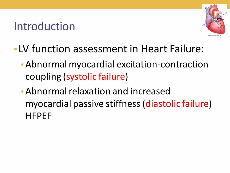
LV STRUCTURE

Measurement of LV Size
• I. Linear Internal Dimensions: • Commonly reported for end-diastole and
end-systole• Performed in the parasternal long-axis view• Perpendicular to the LV long axis and
measured at or immediately below the level of the mitral valve leaflet tips
• Obtained with (2D) echocardiography or 2D guided M-mode approach
• Chamber measurements should be reported indexed to BSA.

Measurement of LV Size
• II. Volumetric Measurements using 2D or 3D:• Volumetric measurements are usually based on tracings of the interface between
the compacted myocardium and the LV cavity in 4ch & 2 ch.
1) Apical foreshortened2) Endocardia dropout
1) Limited published data2) Heavily based on geometry
Volume = [5 (area)(length)]/6

LV Size By 3D Echo• 3D echo volume measurements do not rely on geometric assumptions. • Accurate and reproducible, should be used when available and feasible.• The issue of foreshortening is less relevant in 3D data sets
ASE201
5

LV Mass• Methods: M-mode echocardiography, 2DE, and 3DE

LV Systolic Function• LV Global Systolic Function
I. Fractional ShorteningII. EFIII. Global Longitudinal Strain
• LV Regional Functioni. Segmentation of the Left Ventricleii. Visual Assessmentiii. Regional Wall Motion during Infarction and Ischemiaiv. Regional Abnormalities in the Absence of Coronary Artery
Diseasev. Regional LV Deformation Measurements using Doppler & STE

LV GLOBAL SYSTOLIC FUNCTIONI. Fractional ShorteningII. EF

I. Fractional Shortening
• Represents a simple method for estimating ventricular function in the symmetrically contracting ventricle
% change of LV cavity dimension with systole• FS(%)= LVEDD- LVESD x 100
LVEDD• Derived from 2D-guided M-mode imaging or preferably from
2D images.• Normal range is 25-45% (95% CI)
Only measures the base of the heart
Apical motion abnormalities

II. Ejection Fraction• LV volume estimates may be derived from 2DE or 3DE• EF % =EDV - ESV/EDV x100• The biplane method of disks (modified Simpson’s rule) is the currently
recommended 2D method by consensus of the committee.
o Image poorly optimizedo Traced outside 2D sectoro Traced around the papillary
muscleo Far from MV annulus

LV Assessment Derived From 3DE

ASE201
5

LV REGIONAL FUNCTIONi. Segmentation of the Left Ventricleii. Visual Assessmentiii. Regional Abnormalities in the Absence of Coronary Artery Disease

I. Segmentation of the Left Ventricle• The ventricle is divided into segments reflect coronary perfusion territories.
17-segment model is commonly used. • The 16-segment model is recommended for routine assessing wall motion.
• Endocardial excursion and thickening of the tip of the apex are imperceptible

Coronary Perfusion Territories

II. Visual Assessment• Assessment & observation of segmental wall thickening and endocardial
motion, analyzed individually in multiple (thickening, shortening). • Watch for myocardial motion may be caused by adjacent segment tethering or
overall LV displacement.
• A semiquantitative wall motion score assessment: LV wall motion score index (average of the scores of all segments visualized). • (1) Normal or hyperkinetic• (2) Hypokinetic (reduced thickening)• (3) Akinetic (absent or negligible thickening, e.g., scar)• (4) Dyskinetic (systolic thinning or stretching, e.g., aneurysm).
• An aneurysm is focal dilatation and thinning (remodeling) with either akinetic or dyskinetic systolic deformation.
• The committee refrains from assigning a separate wall motion score for aneurysm.
ASE201
5

Use Of Contrast Agents
• Needed to improve endocardialdelineation
• If two or more contiguous LV endocardial segments are poorly visualized in apical views.
• Normal reference values for LV volumes with contrast enhancement are not well established.

III. Regional Abnormalities in the Absence of Coronary Artery Disease
Rationality noted in conditions as Myocarditis, sarcoidosis, and stress-induced
(takotsubo) cardiomyopathy.
Abnormal Septal motion patterns1. Postoperative status2. RV dysfunction caused by RV pressure
or volume overload. 3. Presence of conduction abnormalities
as LBBB, RV epicardial pacing…...

NEW MODALITIES• (2D) myocardial strain measurements by speckle- tracking
echocardiography (STE), especially global longitudinal strain (GLS)
• 3DE STE

Global Longitudinal Strain • Strain: change in length of an object within a certain
direction relative to its baseline length
• Lt is the length at time t, and L0 is the initial length at time 0.
• Assessed by speckle-tracking echo, in the three standard apical views and averaged
• Measurements should begin with the apical long-axis view to visualize aortic valve closure
• Peak GLS describes the relative length change of the LV myocardium between end-diastole and end-systole:
• ML is myocardial length at end-systole (MLs) and end-diastole (MLd). GLS is a negative number (Normal -20%).
• Limitation: intervendor and intersoftware variability
ASE201
5

Global Longitudinal Strain
• Several studies have demonstrated that abnormal global longitudinal strain develops prior to a fall in ejection fraction and precedes the appearance of clinically apparent heart failure. Detect subclinical decreases in cardiac function before the LVEF falls.
• Recent studies demonstrated good relation between global left ventricular longitudinal systolic strain and LVEF

Regional LV Deformation Measurements
• Echo quantification of regional myocardial function based on DTI or speckle-trackingtechniques. Longitudinal strain during LV systole for
global strain is the most commonly used.
• Despite promising data, quantitative assessment of the magnitude of regional LV deformation cannot be recommended yet because of lack of reference values, suboptimal reproducibility, and considerable intervendor measurement variability.
ASE201
5

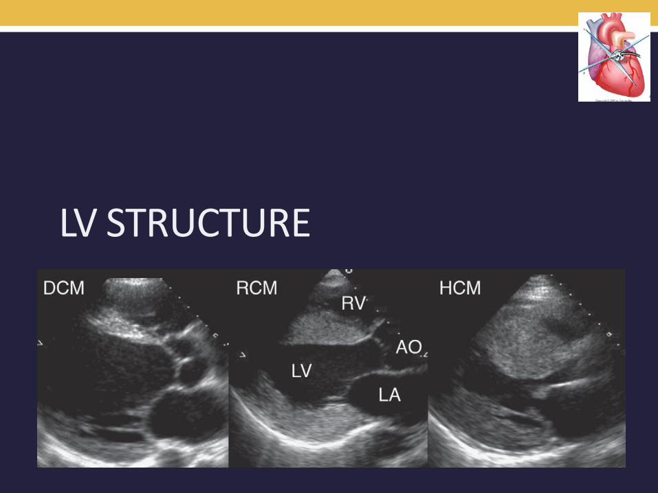
Color DTI Velocity Tracings
LONGITUDINAL STRAIN CURVES AND BULL’S-EYE PLOTS SHOWING SEGMENTAL PEAK SYSTOLIC LONGITUDINAL STRAIN
Examples

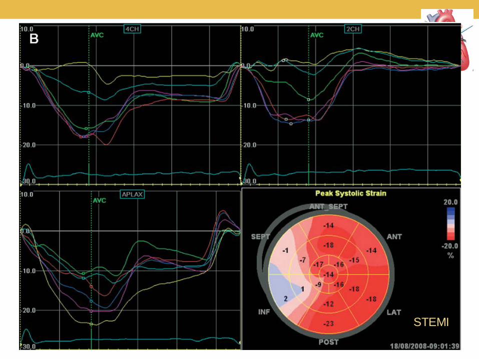
STEMI

chronic ischemic heart failure

Regional LV GLS Measurements
• Several studies demonstrated a good correlation between global left ventricular longitudinal strain measured with speckle tracking imaging and wall motion score index.

Cardiac Resynchronization Therapy in Chronic Heart Failure• An established therapy for patients with NYHA III/IV HR,
prolonged QRS duration (130 ms) and LV dyssynchrony.

Intraventricular Interventricular

New Techniques For Detection Of LV DyssynchronyResponse To CRT

New Techniques For Detection Of LV DyssynchronyResponse To CRT

3D Speckle-Tracking Echocardiographic Slices At End-systole

3D Regional And Slice Voxel Mechanics

DIASTOLIC LV FUNCTION

Echo Parameters To Evaluate The Diastolic Function
• 2-D Echocardiography and Morphology• LA Volume and function/LVH
• M-Mode Echocardiography• Color M-Mode Flow Propagation Velocity
• Pulsed Wave Doppler• Mitral Inflow/Pulmonary Veins
• Tissue Doppler• Mitral Annular velocities
• Other Modalities• Deformation Measurements• Left Ventricular Untwisting• Estimation of Left Ventricular Relaxation• Estimation of Left Ventricular Stiffness• Diastolic Stress Test JASE 2009; 22(2): 107-133

Normal Ranges & Severity For 2DE LA Volume
Male / Female
Normalrange
Mildly abnormal
Moderately abnormal
Severely abnormal
Max LA Volume/ BSA
(ml/m2)
16-34 35-41 42-48 >48
ASE201
5
Increase number of reviewed studies has resulted in a change in the recommended upper normal indexed LA volume
LA volume measurement is recommended by the disk summation algorithm


Septal e’ Lateral e’
LA volume
Septal e’ >8Lateral e’ >10 LA <34 ml/m2
Normal Function
Septal e’>8 Lateral e’ >10 LA >34 ml/m2
Normal function,
athlete's heart or constriction
Septal e’<8 Lateral e’ <10 LA >34ml/m2
E/A <0.8 DT>200 ms Av E/e’ <8 Ar-A<0ms
Val Δ E/A <0.5
Grade I
E/A 0.8 –1.5 DT 160 -200 ms
Av E/e’ 9-12 Ar-A>30ms
Val Δ E/A >0.5
Grade II
E/A >2 DT<160ms Av E/e’ >13 Ar-A>30ms
ValΔE/A >0.5
Grade III
Grading Diastolic DysfunctionJASE 2009; 22(2): 107-133


VALSALVA MANEUVER
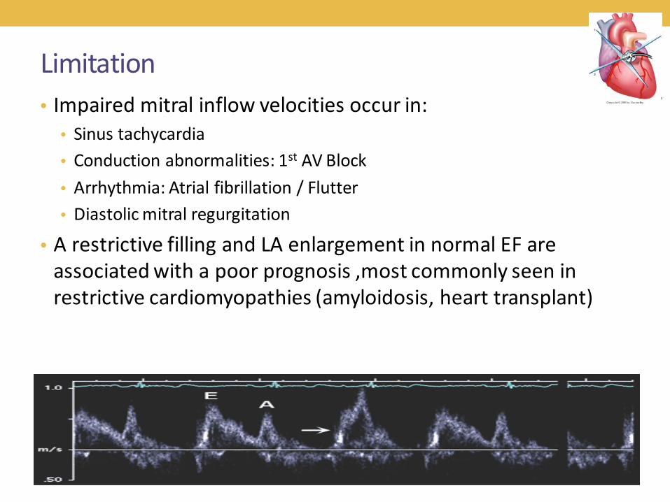
Limitation• Impaired mitral inflow velocities occur in:
• Sinus tachycardia• Conduction abnormalities: 1st AV Block• Arrhythmia: Atrial fibrillation / Flutter• Diastolic mitral regurgitation
• A restrictive filling and LA enlargement in normal EF are associated with a poor prognosis ,most commonly seen in restrictive cardiomyopathies (amyloidosis, heart transplant)

60 year-old with HF & Normal EF
JASE 2009; 22(2): 107-133

Patient with an anteroseptal MISeptal e’ 5cm/s, lateral e’ 10cm/s
JASE 2009; 22(2): 107-133

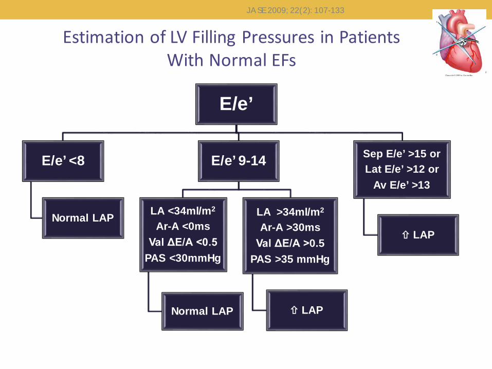
ESTIMATION OF LV FILLING PRESSURES
JASE 2009; 22(2): 107-133

JASE 2009; 22(2): 107-133
Estimation of LV Filling Pressures in Patients With Depressed EFs
Mitral E/A
E/A <1 &E <50 cm/s
Normal LAP
E/A >1-<2 or E/A <1 & E>50cm/s
E/e’<8, S/D >1
Ar-A<0 msValsalva ΔE/A <0.5
PAS <30mmHg
Normal LAP
E/e’ >15S/D <1
AR-A >30 msValsalva Δ E/A >0.5
PAS>35 mmHg
LAP
E/A>2, DT<150ms
LAP
E/e’
E/e’ <8
Normal LAP
E/e’ 9-14
LA <34ml/m2
Ar-A <0msVal ΔE/A <0.5
PAS <30mmHg
Normal LAP
LA >34ml/m2
Ar-A >30msVal ΔE/A >0.5
PAS >35 mmHg
LAP
Sep E/e’ >15 or Lat E/e’ >12 or
Av E/e’ >13
LAP
Estimation of LV Filling Pressures in Patients With Normal EFs
JASE 2009; 22(2): 107-133

LV SYSTOLIC FUNCTION
Normal
Septal e’<8 Lateral e’ <10 LA >34ml/m2
E/A
<0.8
DT>200 ms Grade I
>0.8
DT
160 -200 ms Grade II
<160ms Grade III
Abnormal
Filling Pressure (Av E/e’)
>=13
Inc LAP
<12
Normal
Diastolic Dysfunction
JASE 2009; 22(2): 107-133

Conclusion • The ASE guidelines currently recommend the use of 2D biplane
Simpson method• With ongoing technical advancement 3DE will likely replace
2DE as the routine method to assess LV volumes and EF• Although strain and SR are still largely of research interest,
once standardization of measurement is achieved and diagnostic accuracy is confirmed, strain and SR will become an integral part of clinical echocardiography for the evaluation of myocardial mechanics

GUIDELINES AND STANDARDSRECOMMENDATIONS FOR CARDIAC CHAMBERQUANTIFICATION BY ECHOCARDIOGRAPHY IN
ADULTS:AN UPDATE FROM THE AMERICAN SOCIETY
OF ECHOCARDIOGRAPHY AND THE EUROPEAN ASSOCIATION
OF CARDIOVASCULAR IMAGING
Journal of the American Society of EchocardiographyJanuary 2015
ASE201
5

RECOMMENDATIONS FOR THE EVALUATION OF LEFT
VENTRICULAR DIASTOLIC FUNCTION BY ECHOCARDIOGRAPHY
Journal of the American Society of EchocardiographyFebruary 2009

CURRENT AND EVOLVING ECHOCARDIOGRAPHIC TECHNIQUES
FOR THE QUANTITATIVE EVALUATION OF CARDIAC MECHANICS:
ASE/EAECONSENSUS STATEMENT ON METHODOLOGY
AND INDICATIONSENDORSED BY THE JAPANESE SOCIETY OF
ECHOCARDIOGRAPHY
Journal of the American Society of EchocardiographyMarch 2011

Questions !!