Embed Size (px)
Citation preview
www.bba-direct.com
Biochimica et Biophysica Acta 1642 (2003) 45–52
Lysosomal traffic of liganded endothelin B receptor
Natasha Foster, To Ha Loi, Robert Owe-Young, Keith K. Stanley*
Centre for Immunology, St. Vincent’s Hospital, University of NSW, Cnr West and Boundary Street, Darlinghurst NSW 2010, Sydney, Australia
Received 4 April 2003; received in revised form 17 June 2003; accepted 19 June 2003
Abstract
The endothelin B receptor (ETB) is an endothelial cell receptor found in caveolae. Studies with GFP-tagged ETB have suggested that the
protein is constitutively endocytosed and targeted to lysosomes where it is rapidly degraded. We report that iodinated endothelin-1 ligand
(ET-1) is taken up by cells transfected with ETB and remains undegraded for at least 17 h. Analysis of the intracellular traffic of endocytosed
ET-1 on isotonic Ficoll gradients shows that it is rapidly internalised to lysosomes by a chloroquine sensitive and cholesterol dependent
pathway. Low-temperature nonreducing SDS gels show that the ET-1 initially binds to full-length GFP-tagged ETB, which is rapidly clipped
at the amino-terminus and is then stable for at least 6 h. Analysis of GFP tagged ETB on reducing SDS gels shows that it is proteolytically
cleaved with a half time of approximately 3 h. However, nonreducing gels show that the receptor is virtually intact, suffering only a similar
cleavage to the liganded receptor. We conclude that the ETB receptor shows remarkable stability in lysosomes, held together by disulfide
bonds, and maintaining ligand binding for long periods of time.
D 2003 Elsevier B.V. All rights reserved.
Keywords: Chloroquine; Cyclodextrin; Degradation; Endothelin receptor; Lysosome; Traffic
1. Introduction
The endothelin receptors are seven transmembrane G
protein-coupled receptors (GPCRs) that control vascular
tone in response to endothelin. The endothelin A receptor
is located principally in smooth muscle cells and promotes a
long lasting vasoconstriction on binding endothelin [1]. The
endothelin B receptor (ETB) is found in endothelial cells and
produces a short lived vasodilatory response [2]. During
endocytosis, sequences in the cytoplasmic domain direct the
endothelin receptors into different trafficking pathways
within the cell. The ETA receptor cytoplasmic domain
contains signals that determine trafficking to recycling
endosomes, while the ETB receptor is targeted to lysosomes
for degradation [3,4]. This difference in trafficking could
account, at least in part, for the persistent signalling from the
ETA receptor compared to the ETB receptor.
An unusual property of the ETB receptor is its extremely
high affinity of substrate binding which has a half time of
dissociation in excess of 30 h [5]. Endothelin is not
dissociated from the receptor, even in the presence of SDS
or in the lowered pH of endosome/lysosome compartments
0167-4889/$ - see front matter D 2003 Elsevier B.V. All rights reserved.
doi:10.1016/S0167-4889(03)00097-1
* Corresponding author. Tel.: +612-8382-2833; fax: +612-8382-2391.
E-mail address: [email protected] (K.K. Stanley).
[6], allowing the trafficking of receptor–ligand complexes
to be followed using 125I labelled endothelin-1 ([125I]ET-1).
We have used this technique to compare the intracellular
traffic of ETB ligand–receptor complexes with that of green
fluorescent protein-tagged ETB (ETB–EGFP) which has
been used in a number of previous studies. Our data confirm
that the ETB receptor traffics to a lysosomal compartment,
but show that the receptor remains held together by disulfide
bonding, and ligand binding persists over a 17-h period.
2. Materials and methods
2.1. DNA construction
Several clones of ETB were obtained by amplifying a
full-length cDNA from human umbilical vein endothelial
cell cDNA using specific primers and cloning into pCR2.1.
The 3Vprimer used for the PCR inserted a FLAG epitope
before the stop codon. The orientation of the clones was
established using an EcoRV digestion, and clones with the
insert in the correct orientation were sequenced. One clone
encoding a full-length ETB was selected. The insert in this
clone was excised with NotI and BamHI and cloned in
pIRES digested with the same restriction enzymes. ETB–
N. Foster et al. / Biochimica et Biophysica Acta 1642 (2003) 45–5246
EGFP was constructed by PCR of the ETB open reading
frame using oligonucleotide primers allowing its insertion in
frame into pEGFP-N1. After cloning into this vector, the
nucleotide sequence of ETB was confirmed to be correct.
2.2. Transfection
MDCK cells were transfected with the ETB–FLAG
construct in pIRES and selected with 600 Ag/ml G418 in
complete high-glucose DMEM. G418-resistant clones were
picked and recloned after plating at low density. Although
detection of the ETB by immunofluorescence was difficult
in these cells, the presence of receptor was easily demon-
strated by binding of 125I [ET-1]. ETB–EGFP was transient-
ly transfected into COS cells using lipofectamine according
to the manufacturer’s instructions.
2.3. Cell culture
ETB-transfected MDCK cells were maintained in high-
glucose DMEM containing 10% FCS and tested for myco-
plasma contamination at 3-month intervals. Binding of
[125I]ET-1 (Amersham-Pharmacia) was performed in
DMEM containing 0.1% BSA (DMEM–BSA) at 37 jC. Asingle well of a six-well plate was sufficient for determining
cell-associated cpm; however, each Ficoll gradient used one
T75 flask of cells. Cholesterol depletion was carried out by
incubation for 1 h with 5 mM h-methyl cyclodextrin. This
treatment resulted in cholesterol lowerings of 25%.
2.4. Ficoll density gradient
ETB-transfected MDCK cells were cooled on ice, washed
twice with cold PBS containing 1 mM MgCl2 and 1 mM
CaCl2, and gently scraped into fresh PBS. Cells were
centrifuged at 390� g for 10 min at 4 jC and resuspended
in 2 ml of cold homogenisation buffer (0.25 M sucrose, 20
mM HEPES pH 7.4, 1 mM MgCl2 and protease inhibitor
cocktail (Roche Biochemicals)). Cells were disrupted with
20 to 25 strokes of a ball bearing homogeniser as described
[7] using a ball with a clearance of 10 Am. Cellular debris
and unbroken cells were removed by centrifugation at
100� g for 8 min at 4 jC. EDTA was added to supernatant
to give a final concentration of 1 mM.
Ficoll gradients, 1–22%, were prepared and run essen-
tially as described [8]. Each gradient consisted of 1.5 ml of
45% Nycodenz, overlayered with 8 ml of a 1% to 22%
linear Ficoll gradient prepared in 0.25 M sucrose, 10 mM
HEPES pH 7.4, 1 mM EDTA. The cell homogenate was
then laid on top and centrifuged in a Beckman VTi 65 rotor
at 65,000 rpm for 90 min at 4 jC.
2.5. b-Hexosaminidase assay
In a 96-well plate, 12.5 Al of each fraction was added to
12.5 Al of 0.6% Triton X-100 and mixed well. Samples were
incubated for 10 min at room temperature before 25 Al of 2mM 4-methylumbelliferyl-N-acetyl-h-D-glucosaminide in
0.2 M sodium citrate, pH 4.5, was added. The samples
were then placed at 37 jC for 1 h. The reaction was stopped
by the addition of 100 Al of 1 M sodium carbonate, pH 10.
The fluorescence was read at 460-nm emission, 355-nm
excitation using a Fluoroscan II spectrophotometer.
2.6. Western blots
Fractions were separated by SDS-PAGE and transferred
to a nitrocellulose membrane. For all blots, antibody incu-
bations were done in TBS (10 mM Tris pH 7.5, 100 mM
NaCl) with 0.1% Tween 20 and 1% BSA. Blocking of filters
used the same buffer containing 3% BSA. For EEA-1
detection, a rabbit antiserum was raised against the car-
boxy-terminal peptide CDACFNDLQG, and used at a
dilution 1:1000. Anti-GFP antibodies (Roche Biochemicals)
were used at a dilution of 1:5000. As secondary antibody,
HRP-conjugated anti-rabbit IgG (Amersham Life Sciences)
was diluted at 1:2000 to 1:10000. The blots were developed
using enhanced chemiluminescence (ECL) following man-
ufacturer’s instructions (NEN Life Sciences). The EEA1
antibody recognised a single protein of Mr = 80 kDa in
MDCK cells that was effectively competed by prior incu-
bation of the antibody in a solution of the peptide used to
raise the antibody.
2.7. Biotinylation of cell membrane proteins
Cells were biotinylated at 4 jC immediately prior to
homogenisation. After washing, cells were equilibrated in
cold 100 mM sodium bicarbonate, pH 8.0. A freshly pre-
pared solution of 0.5 mg/ml NHS-SS-biotin (Pierce) in the
same buffer was added and incubated for 8 min. This step
was repeated before quenching with 20 mM Tris pH 7.5, 120
mM NaCl. Biotinylated proteins were detected on Western
blots using HRP–streptavidin (1:5000) and ECL detection.
2.8. Low-temperature PAGE
Liganded ETB was detected by autoradiography of poly-
acrylamide gels run at 5 jC [8]. Cells incubated with
[125I]ET-1 were washed and collected in Laemmli sample
buffer containing 2% SDS at 4 jC. After shearing DNA by
multiple passages through a 21-gauge syringe needle, the
samples were directly loaded onto a 12% polyacrylamide
gel and electrophoresed with water cooling at 5 jC.
3. Results
3.1. Turnover of the ETB receptor
Previous studies have shown that the ETB receptor is
rapidly internalised and degraded in lysosomes [3,9]. These
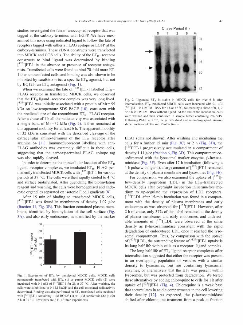
Fig. 2. Liganded ETB is stable in MDCK cells for over 6 h after
internalisation. ETB-transfected MDCK cells were incubated with 0.1 ACi[125I]ET-1 in DMEM–BSA for 1 h at 37 jC, followed by a chase of 0, 1, 2
or 6 h in DMEM–BSA without ligand. At the end of the incubation, cells
were washed and then solubilised in sample buffer containing 2% SDS.
Following PAGE at 5 jC, the gel was dried and autoradiographed. Arrows
show positions of 32- and 55-kDa forms.
N. Foster et al. / Biochimica et Biophysica Acta 1642 (2003) 45–52 47
studies investigated the fate of unoccupied receptor that was
tagged at the carboxy-terminus with EGFP. We have reex-
amined this issue using iodinated ligand to follow the fate of
receptors tagged with either a FLAG epitope or EGFP at the
carboxy-terminus. These cDNA constructs were transfected
into MDCK and COS cells. The ability of the ETB–receptor
constructs to bind ligand was determined by binding
[125I]ET-1 in the absence or presence of receptor antago-
nists. Transfected cells were found to bind 70-fold more ET-
1 than untransfected cells, and binding was also shown to be
inhibited by sarafotoxin 6c, a specific ETB agonist, but not
by BQ123, an ETA antagonist (Fig. 1).
When we examined the fate of [125I]ET-1 labelled ETB–
FLAG receptor in transfected MDCK cells, we observed
that the ETB ligand–receptor complex was very long lived.
[125I]ET-1 was initially associated with a protein of Mr = 55
kDa on low-temperature SDS PAGE [10], consistent with
the predicted size of the recombinant ETB–FLAG receptor.
After a chase of 1 h all the radioactivity was associated with
a single band of Mr = 32 kDa (Fig. 2). It then remained at
this apparent mobility for at least 6 h. The apparent mobility
of 32 kDa is consistent with the described cleavage of the
extracellular amino-terminus of the ETB receptor after
arginine 64 [11]. Immunofluorescent labelling with anti-
FLAG antibodies was extremely difficult in these cells,
suggesting that the carboxy-terminal FLAG epitope tag
was also rapidly cleaved.
In order to determine the intracellular location of the ETB
ligand–receptor complexes, we incubated ETB–FLAG per-
manently transfectedMDCK cells with [125I]ET-1 for various
periods at 37 jC. The cells were then rapidly cooled to 4 jCand surface biotinylated. After quenching the biotinylation
reagent and washing, the cells were homogenised and endo-
cytic organelles separated on isotonic Ficoll gradients [8].
After 15 min of binding to transfected MDCK cells,
[125I]ET-1 was found in membranes of density 1.07 g/cc
(fraction 11, Fig. 3B). This fraction contained plasma mem-
brane, identified by biotinylation of the cell surface (Fig.
3A), and also early endosomes, as identified by the marker
Fig. 1. Expression of ETB by transfected MDCK cells. MDCK cells
permanently transfected with ETB (1) or parent MDCK cells (2) were
incubated with 0.1 ACi of [125I]ET-1 for 2h at 37 jC. After washing, thecells were solubilised in 0.1 M NaOH and the cell associated radioactivity
determined. Binding was also performed on ETB transfected cells incubated
with [125I]ET-1 containing 1 AM BQ123 (3) or 1 AM sarafotoxin S6c (4) for
2 h at 37 jC. Error bars are S.E. of three experiments.
EEA1 (data not shown). After washing and incubating the
cells for a further 15 min (Fig. 3C) or 2 h (Fig. 3D), the
[125I]ET-1 progressively accumulated in a compartment of
density 1.11 g/cc (fraction 6, Fig. 3D). This compartment co-
sedimented with the lysosomal marker enzyme, h-hexosa-minidase (Fig. 3F). Even after 17-h incubation (following a
1-h pulse with ligand), a large amount of [125I]ET-1 remained
at the density of plasma membrane and lysosomes (Fig. 3E).
For comparison, we also examined the uptake of [125I]-
low-density lipoprotein (LDL) in the ETB-transfected
MDCK cells after overnight incubation in serum-free me-
dium to up-regulate the expression of LDL receptors.
[125I]LDL after 15-min incubation was found in a compart-
ment with the density of plasma membranes and early
endosomes as was observed for [125I]ET-1. However, after
2 h of chase, only 37% of this label remained at the density
of plasma membranes and early endosomes, and undetect-
able amounts of [125I]LDL were observed at the same
density as h-hexosaminidase consistent with the rapid
degradation of endocytosed LDL once it reached the lyso-
somal compartment. Thus, by comparison with the uptake
of [125I]LDL, the outstanding feature of [125I]ET-1 uptake is
its long half life within cells as a receptor–ligand complex.
The long half life of ETB ligand receptor complexes after
internalisation suggested that either the receptor was present
in an overlapping population of vesicles with a similar
density to lysosomes, but not containing lysosomal
enzymes, or alternatively that the ETB was present within
lysosomes, but was protected from degradation. We tested
these alternatives by adding chloroquine to cells for 1 h after
uptake of [125I]ET-1 (Fig. 4). Chloroquine is a weak base
that accumulates in acidic compartments in the cell lowering
their density [12]. As expected, the h-hexosaminidase
shifted after chloroquine treatment from a peak at fraction
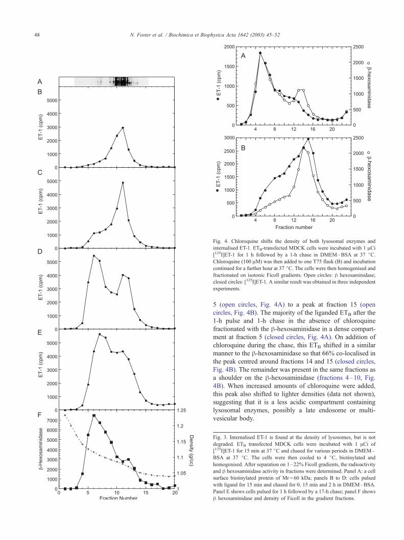
Fig. 4. Chloroquine shifts the density of both lysosomal enzymes and
internalised ET-1. ETB-transfected MDCK cells were incubated with 1 ACi[125I]ET-1 for 1 h followed by a 1-h chase in DMEM–BSA at 37 jC.Chloroquine (100 AM) was then added to one T75 flask (B) and incubation
continued for a further hour at 37 jC. The cells were then homogenised and
fractionated on isotonic Ficoll gradients. Open circles: h hexosaminidase;
closed circles: [125I]ET-1. A similar result was obtained in three independent
experiments.
N. Foster et al. / Biochimica et Biophysica Acta 1642 (2003) 45–5248
5 (open circles, Fig. 4A) to a peak at fraction 15 (open
circles, Fig. 4B). The majority of the liganded ETB after the
1-h pulse and 1-h chase in the absence of chloroquine
fractionated with the h-hexosaminidase in a dense compart-
ment at fraction 5 (closed circles, Fig. 4A). On addition of
chloroquine during the chase, this ETB shifted in a similar
manner to the h-hexosaminidase so that 66% co-localised in
the peak centred around fractions 14 and 15 (closed circles,
Fig. 4B). The remainder was present in the same fractions as
a shoulder on the h-hexosaminidase (fractions 4–10, Fig.
4B). When increased amounts of chloroquine were added,
this peak also shifted to lighter densities (data not shown),
suggesting that it is a less acidic compartment containing
lysosomal enzymes, possibly a late endosome or multi-
vesicular body.
Fig. 3. Internalised ET-1 is found at the density of lysosomes, but is not
degraded. ETB transfected MDCK cells were incubated with 1 ACi of
[125I]ET-1 for 15 min at 37 jC and chased for various periods in DMEM–
BSA at 37 jC. The cells were then cooled to 4 jC, biotinylated and
homogenised. After separation on 1–22% Ficoll gradients, the radioactivity
and h hexosaminidase activity in fractions were determined. Panel A: a cell
surface biotinylated protein of Mr = 60 kDa; panels B to D: cells pulsed
with ligand for 15 min and chased for 0, 15 min and 2 h in DMEM–BSA.
Panel E shows cells pulsed for 1 h followed by a 17-h chase; panel F shows
h hexosaminidase and density of Ficoll in the gradient fractions.
N. Foster et al. / Biochimica et Biophysica Acta 1642 (2003) 45–52 49
Since ETB has been reported to be located in caveolae
[13], we determined the effect of cholesterol depletion on
traffic to the lysosomal compartment. Fig. 5 shows an
experiment identical to that described in Fig. 4 except that
cells were pretreated with 5 mM methyl h-cylcodextrin for 1h which resulted in a 25% decrease in total cholesterol.
While the h-hexosaminidase shifts from fraction 5 (open
circles, Fig. 5A) to fraction 14 (open circles, Fig. 5B) similar
to Fig. 4, it can now be seen that virtually no [125I]ET-1 is
associated with this peak (closed circles, Fig. 5B). Thus,
although ETB is successfully internalised, since it does not
co-fractionate with either plasma membrane or early endo-
somal markers (see Western blot inset in Fig. 5), traffic to
lysosomes is effectively inhibited by cholesterol depletion.
Cholesterol depletion shifts the plasma membrane to a
denser fraction (fraction 10, Fig. 5B) as might be expected,
but the biotinylated plasma membrane bands do not overlap
with the peak of [125I]ET-1 in fractions 5 to 9.
Fig. 6. Pretreatment with chloroquine prevents traffic of ETB to lysosomes.
ETB-transfected MDCK cells were incubated in (A) DMEM–BSA or (B)
DMEM–BSA containing 100 AM chloroquine for 1 h at 37 jC. [125I]ET-1(1 ACi) was then added to both flasks and incubated for 1 h followed by a 1-h chase in DMEM containing 0.1% BSA and 100 AM chloroquine at 37 jC.The cells were then homogenised and fractionated on isotonic Ficoll
gradients. Open circles: h hexosaminidase; closed circles: [125I]ET-1. Inset
above each panel is a Western blot of EEA1. A similar result was obtained
in three independent experiments.
Fig. 5. Cholesterol depletion inhibits traffic of ETB to lysosomes. The
protocol of Fig. 6 was used except that the cells in panel Bwere incubated in 5
mMhmethyl cyclodextrin for 1 h at 37 jCbefore the experiment. Inset above
each panel is a Western Blot of EEA1 and Streptavidin–HRP detection of a
60-kDa surface biotinylated protein. A similar result was obtained in two
independent experiments.
When chloroquine was applied to cells for 1 h before
incubation with [125I]ET-1, internalisation of ligand was not
affected, as liganded receptors did not fractionate with early
endosomal (Fig. 6) or plasma membrane markers (data not
shown). However, under these conditions, [125I]ET-1 did not
reach the h-hexosaminidase containing compartment (Fig.
6B). Previous reports have suggested that traffic from
multivesicular bodies to lysosomes in rat liver can be
inhibited by chloroquine [14]. Thus, at least part of the
large peak of [125I]ET-1 in Fig. 6B is likely to represent
multivesicular bodies.
Since [125I]ET-1 bound to ETB–FLAG appeared to be
remarkably stable in a lysosome-like compartment, we went
back to examine the fate of ligand bound to EGFP-tagged
ETB receptors, which had been reported to be rapidly
degraded in transiently transfected COS cells [9]. We trans-
fected COS cells with an identical construct and incubated
with [125I]ET-1 for 15 min followed by a chase in unla-
belled medium for up to 4 h (Fig. 7A). The cells were
treated with cycloheximide for 1 h prior to addition of
[125I]ET-1 and kept cycloheximide in the chase medium so
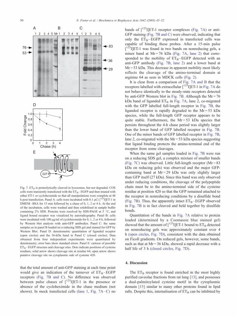
Fig. 7. ETB is proteolytically cleaved in lysosomes, but not degraded. COS
cells were transiently transfected with the ETB–EGFP and then treated with
either ET-1 or cycloheximide so that all manipulations were complete at 48
h post transfection. Panel A: cells were incubated with 0.1 ACi [125I]ET-1 inDMEM–BSA for 15 min followed by a chase of 0, 1, 2 or 4 h. At the end
of the incubation, cells were washed and then solubilised in sample buffer
containing 2% SDS. Proteins were resolved by SDS-PAGE at 5 jC, andligand bound receptor was visualized by autoradiography. Panel B: cells
were incubated with 100 Ag/ml of cycloheximide for 0, 1, 2 or 4 h, followed
by Western blot analysis with anti-GFP antibodies. Panel C: the same
samples as in panel B loaded on a reducing SDS gel and stained for GFP by
Western Blot. Panel D: densitometric quantitation of liganded receptor
(open circles) and the 36-kDa band in Panel C (closed circles). Data
obtained from four independent experiments were quantitated by
densitometry; error bars show standard errors. Panel E: cartoon of possible
ETB–EGFP structure and cleavage sites. Dots indicate positions of cysteine
residues; solid arrow shows cleavage site at residue 64; open arrow shows
putative cleavage site on cytoplasmic side of cysteine 420.
N. Foster et al. / Biochimica et Biophysica Acta 1642 (2003) 45–5250
that the total amount of anti-GFP staining at each time point
would give an indication of the turnover of ETB–EGFP
receptors (Fig. 7B and C). No difference was observed
between pulse chases of [125I]ET-1 in the presence or
absence of the cycloheximide in the chase medium (not
shown). In mock transfected cells (lane 1, Fig. 7A–C) no
bands of [125I]ET-1 receptor complexes (Fig. 7A) or anti-
GFP staining (Fig. 7B and C) were observed, indicating that
only the ETB–EGFP expressed in transfected cells was
capable of binding these probes. After a 15-min pulse
[125I]ET-1 was found in two bands on nonreducing gels, a
minor band at Mr = 76 kDa (Fig. 7A, lane 2) that corre-
sponded to the mobility of ETB–EGFP detected with an
anti-GFP antibody (Fig. 7B, lane 2) and a lower band at
Mr = 53 kDa. This decrease in apparent mobility most likely
reflects the cleavage of the amino-terminal domain at
arginine 64 as seen in MDCK cells (Fig. 2).
It is clear from a comparison of Fig. 7A and B that the
receptors labelled with extracellular [125I]ET-1 in Fig. 7A do
not behave identically to the steady-state receptors detected
by anti-GFP Western blot in Fig. 7B. Although the Mr = 76
kDa band of liganded ETB in Fig. 7A, lane 2, co-migrated
with the GFP labelled full-length receptor in Fig. 7B, the
liganded receptor is rapidly degraded to the Mr = 53 kDa
species, while the full-length GFP receptor appears to be
quite stable. Furthermore, the Mr = 53 kDa species that
persists throughout the 4-h chase period was slightly larger
than the lower band of GFP labelled receptor in Fig. 7B.
One of the minor bands of GFP labelled receptor in Fig. 7B,
lane 2, co-migrated with the Mr = 53 kDa species suggesting
that ligand binding protects the amino-terminal end of the
receptor from some cleavages.
When the same gel samples loaded in Fig. 7B were run
on a reducing SDS gel, a complex mixture of smaller bands
(Fig. 7C) was observed. Little full-length receptor (Mr = 83
kDa on reducing gels) was observed and the major GFP-
containing band at Mr = 29 kDa was only slightly larger
than GFP itself (27 kDa). Since this band was only observed
under reducing conditions, the cleavage of the polypeptide
chain must be to the amino-terminal side of the cysteine
residue at position 420 so that the GFP remained attached to
the receptor in nonreducing conditions by a disulfide bond
(Fig. 7B). Thus, the apparently intact ETB–EGFP observed
in Fig. 7B is in fact cleaved and held together by disulfide
bonds.
Quantitation of the bands in Fig. 7A relative to protein
loaded (determined by a Coomassie blue stained gel)
showed that the amount of [125I]ET-1 bound to ETB detected
on nonreducing gels was approximately constant over 4
h (open circles, Fig. 7D), consistent with the data obtained
on Ficoll gradients. On reduced gels, however, some bands,
such as that at Mr = 36 kDa, showed a rapid decrease with a
half life of 3 h (closed circles, Fig. 7D).
4. Discussion
The ETB receptor is found enriched in the most highly
purified caveolae fractions from rat lung [13], and possesses
a dual-palmitoylated cysteine motif in the cytoplasmic
domain [15] similar to many other proteins found in lipid
rafts. Despite this, internalisation of ETB can be inhibited by
N. Foster et al. / Biochimica et Biophysica Acta 1642 (2003) 45–52 51
hypertonic sucrose, suggesting that it occurs via clathrin
coat-mediated endocytosis [6]. Furthermore, the ETB recep-
tor is rapidly phosphorylated after ligand binding by a
GPCR kinase and desensitisation occurs within 4 min
[16]. Thus, the ETB receptor, like cholera toxin [17], appears
to be localised in caveolae, but internalised principally via a
clathrin-mediated pathway.
Once internalised, ETB accumulates at 18 jC in a
peripheral compartment co-localising with transferrin recep-
tor but sorts at 37 jC into a perinuclear compartment
labelled by di-I-LDL [6]. This is consistent with traffic
along the ‘classical’ endocytic pathway to a late endosome/
lysosome compartment. Using EGFP-tagged ETB, accumu-
lation in a LAMP-1 positive compartment has been ob-
served resulting in rapid degradation of the receptor [3,9].
These data have been interpreted as a constitutive traffic of
ETB to the lysosomal compartment.
We have studied the trafficking of the ETB receptor using
carboxy-terminal EGFP or FLAG to tag the receptor in an
unliganded state, and iodinated ET-1 ligand to follow the
fate of ligand–receptor complexes. In agreement with earlier
studies, we find that the ETB–EGFP receptor traffics to an
acidic compartment containing the majority of h-hexosa-minidase consistent with a late endosomal or lysosomal
compartment. The inhibition of traffic into this compartment
at a late step by chloroquine is consistent with it being a
lysosomal compartment [14]. However, our data demon-
strate two unusual properties of this traffic: first, that traffic
is inhibited by cholesterol depletion of the cells, and second,
that the receptor remains intact and retains bound ligand
over a long period of time (up to 17 h in our experiments).
The receptor was similarly resistant to degradation in
polarized (MDCK) and nonpolarised cells (COS) using both
transient and stable transfectants, suggesting that the results
are independent of cell type and level of expression.
When [125I]ET-1 is incubated with cells transfected with
ETB–EGFP, we initially detect the ligand receptor complex
at the expected size of ETB–EGFP. Within a 15-min pulse
of labeling, however, a large portion was also bound to a
smaller protein band. The apparent mobility of this band is
consistent with the cleavage previously reported at amino
acid 64 in human placenta [11] and occurs in both MDCK
and COS cells. The intact liganded ETB–EGFP chases into
the cleaved ETB–EGFP within the first 1 h of chase. After
binding to cells for 15 min, we know from Fig. 3 that the
ligand–receptor complexes are present at the cell surface or
in early endosomes, but not in lysosomes. Hence, this initial
cleavage of the receptor must occur in an early compartment
of the endocytic pathway.
The longevity of the full-length ETB–EGFP as detected
by Western blot, compared to the short-lived band of the
same size detected by ET-1 ligand binding, suggests that
ligand might induce a different traffic of the receptor, most
likely causing its rapid internalization and cleavage. The
cleavage site for removal of the 64-residue amino-terminus
(solid arrow, Fig. 7E) is on the extracellular surface of the
plasma membrane that would come into contact with pro-
teases in the lumen of endocytic organelles after endocytosis.
More difficult to explain is the GFP containing Mr = 29 kDa
band that is observed only when the samples are run on
reducing gels. Cleavage of ETB–EGFP in the last extrac-
ytoplasmic loop would result in a 36-kDa fragment that
contains the EGFP. A fragment of this size was observed and
determined to have a short half life, presumably because it
can be further cleaved to produce the 29-kDa EGFP-con-
taining fragment. Since the 29-kDa fragment is only ob-
served using reducing gels, the cleavage site must be on the
amino-terminal side of the cysteine at residue 420 (open
arrow, Fig. 7E) so that it remains tethered to the protein by a
disulfide bond under nonreducing conditions. Such a cyto-
plasmic cleavage is consistent with traffic of ETB–EGFP
into invaginated lysosomal membranes and subsequent
cleavage by lysosomal proteases. Thus, all of our data are
consistent with the passage of ETB–EGFP into lysosomes.
The resistance of ETB–EGFP to complete degradation
may be a consequence of the particular structure of the
receptor, or possibly its location in lipid rafts. It will be
interesting to determine if the ETA receptor, which has a
similar pattern of cysteine bonds to the ETB receptor, is
similarly stable, and if this contributes to the longevity of
ETA receptor signalling. With respect to the possible role of
lipid rafts, our studies using isotonic Ficoll gradients indi-
cated that traffic of ETB–EGFP to lysosomes was inhibited
after 25% depletion of total cellular cholesterol. It will be of
interest to determine if this is a general property shared by
other receptors, or if it is specific to ETB–EGFP. Since late
endosomes are known to be rich in cholesterol [18], it is
possible that receptors with an affinity for lipid rafts remain
associated with cholesterol during lysosomal traffic and that
traffic of these receptors is inhibited when this cholesterol is
depleted.
Acknowledgements
We thank the Scleroderma Association of NSW and
National Heart Foundation of Australia for financial support.
This project was partially funded by a grant from the New
South Wales Health Research and Development infra-
structure grant.
References
[1] R. Marsault, E. Feolde, C. Frelin, Receptor externalization determines
sustained contractile responses to endothelin-1 in the rat aorta, Am. J.
Physiol. 264 (1993) C687–C693.
[2] M. Yanagisawa, T. Masaki, Endothelin, a novel endothelium-derived
peptide. Pharmacological activities, regulation and possible roles in
cardiovascular control, Biochem. Pharmacol. 38 (1989) 1877–1883.
[3] Y. Abe, K. Nakayama, A. Yamanaka, T. Sakurai, K. Goto, Subtype-
specific trafficking of endothelin receptors, J. Biol. Chem. 275 (2000)
8664–8671.
N. Foster et al. / Biochimica et Biophysica Acta 1642 (2003) 45–5252
[4] J.D. Paasche, T. Attramadal, C. Sandberg, H.K. Johansen, H. Attra-
madal, Mechanisms of endothelin receptor subtype-specific targeting
to distinct intracellular trafficking pathways, J. Biol. Chem. 276
(2001) 34041–34050.
[5] W.G. Waggoner, S.L. Genova, V.A. Rash, Kinetic analyses demon-
strate that the equilibrium assumption does not apply to [125I]endo-
thelin-1 binding data, Life Sci. 51 (1992) 1869–1876.
[6] A. Oksche, G. Boese, A. Horstmeyer, J. Furkert, M. Beyermann, M.
Bienert, W. Rosenthal, Late endosomal/lysosomal targeting and lack
of recycling of the ligand-occupied endothelin B receptor, Mol. Phar-
macol. 57 (2000) 1104–1113.
[7] E.L. Mander, R.T. Dean, K.K. Stanley, W. Jessup, Apolipoprotein B
of oxidized LDL accumulates in the lysosomes of macrophages, Bio-
chim. Biophys. Acta 1212 (1994) 80–92.
[8] J.H. Perez, W.J. Branch, L. Smith, B.M. Mullock, J.P. Luzio, In-
vestigation of endosomal compartments involved in endocytosis
and transcytosis of polymeric immunoglobulin A by subcellular
fractionation of perfused isolated rat liver, Biochem. J. 251 (1988)
763–770.
[9] T. Bremnes, J.D. Paasche, A. Mehlum, C. Sandberg, B.
Bremnes, H. Attramadal, Regulation and intracellular trafficking
pathways of the endothelin receptors, J. Biol. Chem. 275 (2000)
17596–17604.
[10] T. Takasuka, T. Sakurai, K. Goto, Y. Furuichi, T. Watanabe, Human
endothelin receptor ETB. Amino acid sequence requirements for
super stable complex formation with its ligand, J. Biol. Chem. 269
(1994) 7509–7513.
[11] N. Akiyama, O. Hiraoka, Y. Fujii, H. Terashima, M. Satoh, K. Wada,
Y. Furuichi, Biotin derivatives of endothelin: utilization for affinity
purification of endothelin receptor, Protein Expr. Purif. 3 (1992)
427–433.
[12] R. Wattiaux, F. Gentinne, M. Jadot, F. Dubois, D.E. Wattiaux, S.
Coninck, Chloroquine allows to distinguish between hepatocyte ly-
sosomes and sinusoidal cell lysosomes, Biochem. Biophys. Res.
Commun. 190 (1993) 808–813.
[13] P. Oh, J.E. Schnitzer, Endothelin induced budding of caveolae: depend-
ence on G-protein activation and tyrosine phosphorylation, FASEB J.
15 (2001) A218.
[14] C.A. Hornick, A.L. Jones, G.H. Renaud, Effect of chloroquine on
low-density lipoprotein catabolic pathway in rat hepatocytes, Am. J.
Physiol. 246 (1984) G187–G194 (Chloroquine blocks multivesicular
body to lysosomes in rat liver).
[15] M. Roos, V. Soskic, S. Poznanovic, J. Godovac-Zimmermann, Post-
translational modifications of endothelin receptor B from bovine
lungs analyzed by mass spectrometry, J. Biol. Chem. 273 (1988)
924–931.
[16] N.J. Freedman, A.S. Ament, M. Oppermann, R.H. Stoffel, S.T. Exum,
R.J. Lefkowitz, Phosphorylation and desensitization of human endo-
thelin A and B receptors. Evidence for G protein-coupled receptor
kinase specificity, J. Biol. Chem. 272 (1997) 17734–17743.
[17] H. Shogomori, A.H. Futerman, Cholera toxin is found in detergent-
insoluble rafts/domains at the cell surface of hippocampal neurons but
is internalized via a raft-independent mechanism, J. Biol. Chem. 276
(2001) 9182–9188.
[18] T. Kobayashi, M.H. Beuchat, M. Lindsay, S. Frias, R.D. Palmiter, H.
Sakuraba, R.G. Parton, J. Gruenberg, Late endosomal membranes rich
in lysobisphosphatidic acid regulate cholesterol transport, Nat. Cell
Biol. 1 (1999) 113–118.