Embed Size (px)
Citation preview

Macular function by multifocal electroretinogramin diabetic macular edema after intravitrealtriamcinolone acetonide injection
M. KARACORLU1, H. OZDEMIR1, F. SENTURK1, S. ARF KARACORLU1, O. UYSAL2
1The Istanbul Retina Institute Inc.2University of Istanbul, Cerrahpasa School of Medicine, Department of Biostatistics, Istanbul - Turkey
INTRODUCTION
Diabetic retinopathy is a leading cause of visual impairmentin adults, and diabetic macular edema (DME) is the maincause of visual impairment in diabetic patients. The EarlyTreatment Diabetic Retinopathy Study (ETDRS) proved the
European Journal of Ophthalmology / Vol. 18 no. 4, 2008 / pp. 601-608
1120-6721/601-08$25.00/0© Wichtig Editore, 2008
PURPOSE. The purpose of this study was to assess macular function by multifocal electroretinography(mfERG) in eyes with diabetic macular edema (DME) after intravitreal triamcinolone ace-tonide (IVTA) injection.METHODS. Fifteen eyes of 15 patients with DME scheduled for 4 mg IVTA injection wereprospectively recruited. The response to treatment was monitored functionally by visualacuity (VA) measurement and mfERG and anatomically by foveal thickness measured by op-tical coherence tomography (OCT). The first-order kernel P1 mfERG responses from 0 to 7degrees (central) and 7 to 25 degrees (peripheral) were grouped and analyzed. Changes infunctional parameters (VAs and the P1 mfERG response amplitudes and peak latencies) andmorphometric parameters (OCT foveal thickness) in eyes with DME 1 and 3 months afterIVTA injection were compared with baseline values by Student t test.RESULTS. The mean baseline logMAR value for VAs of the patients before treatment was0.49±0.26. After treatment, it was 0.27±0.23 at 1 month and 0.26±0.18 at 3 months, anddifferences from pretreatment values were significant (for each, p<0.001). There were sta-tistically significant decreases in the mean foveal thickness at 1 and 3 months after treat-ment compared with pretreatment values (for each, p<0.001). There were also statisticallysignificant increases in the mean P1 response amplitude for both central and peripheralgroups at all examinations compared with pretreatment (for each, p<0.001). The mean P1peak latencies for both the central and peripheral groups were shortened, but not signifi-cantly.CONCLUSIONS. As well as the reduction in DME and improvement in VA, IVTA injection im-proves macular function as assessed by mfERG in diabetic patients. (Eur J Ophthalmol 2008;18: 601-8)
KEY WORDS. Diabetic macular edema, Intravitreal triamcinolone acetonide, Macular function,Multifocal electroretinogram
Accepted: January 30, 2008
value of focal photocoagulation in clinically significant ma-cular edema (CSME) due to focal diabetic edema associa-ted with leaking microaneurysms (1). In cases of CSME withdiffuse leakage, grid laser photocoagulation was recom-mended, but no large prospective trial has yet proved its va-lue. In roughly a quarter of these patients, visual acuity (VA)

Macular function by mfERG in diabetic macular edema after IVTA
602
declines by at least three ETDRS lines despite grid photo-coagulation (2).It was demonstrated in a prospective, randomized, control-led trial that a single 4-mg IVTA injection can reduce centralmacular thickness, as measured by optical coherence to-mography (OCT), in patients with CSME who have failedprevious focal photocoagulation (3). In another interventio-nal, prospective study by Jonas and Söfker (4), the effect ofIVTA on diffuse DME was evident as early as 1 week afterinjection and persisted for approximately 7 months. A groupled by Martidis showed in an interventional case series astatistically significant improvement in vision at months 1, 3,and 6, by 2.4, 2.4, and 1.3 Snellen lines, and a decrease inmacular thickness by 55%, 57.5%, and 38%, respectively(5). It was also shown that IVTA injection is effective in thetreatment of DME that has had no previous laser treatment(6). In all these studies, VA and morphologic features beforeand after IVTA injection were evaluated, but patients’ sub-jective appraisal of their visual function, which may differ,was not comprehensively discussed. The purpose of this study was to obtain a measure of ma-cular function before and after IVTA injection in patients withDME. To accomplish this, mfERG was performed on 15
eyes of 15 patients with DME before and after IVTA injecti-on, and the macular function was determined from the re-sult of mfERG.
METHODS
Fifteen eyes of 15 patients (10 men, 5 women) with non-in-sulin-treated diabetes mellitus and ME were included. Themean age of patients was 59.5 years (range 46–70 years),and the duration of diabetes mellitus ranged from 2 to 7years (mean 5.8 years). The eligibility criteria for this studyincluded 1) the presence of CSME due to diabetic retinopa-thy on fundus examination; 2) the presence of angiographi-cally confirmed DME; and 3) the presence of ME confirmedby OCT. Because several diseases may influence mfERGand VA, we excluded patients with moderate to dense lensopacity, corneal opacities, a history of refractive surgery,glaucoma or ocular hypertension, a history of intraocular in-flammation such as anterior or posterior uveitis, multifocalchoroiditis, a history of retinal detachment, a history of ocu-lar trauma, and optic neuropathy. In this consecutive series,no eyes had received previous laser photocoagulation. Eyes
TABLE I - CLINICAL CHARACTERISTICS OF PATIENTS WITH DIABETIC MACULAR EDEMA BEFORE AND AFTERINTRAVITREAL TRIAMCINOLONE ACETONIDE (IVTA) INJECTION
VA (logMAR) mfERG-c P1 mfERG-p P1 mfERG-c P1 mfERG-p P1 OCT mean foveal
amplitude amplitude peak time (ms) peak time (ms) thickness (µm)
(nV/deg2) (nV/deg2)
Pa- Age,
tient yr B 1 mo 3 mo B 1 mo 3 mo B 1 mo 3 mo B 1 mo 3 mo B 1 mo 3 mo B 1 mo 3 mo
1 65 0.7 0.4 0.4 13 15.5 21.5 10.3 11.4 16 40.2 39.8 37.7 35.7 39.8 40.8 330 296 210
2 55 0.3 0.2 0.2 31.9 47.7 30.2 16.6 23.9 15.5 41 36.7 35.7 41 36.7 35.7 589 216 238
3 60 0.2 0 0 24.2 32.2 29.6 14 19.4 17.8 42 36.7 35.7 42 36.7 35.7 532 224 230
4 64 0.5 0.3 0.4 16.2 17.6 14.1 8.9 11.7 7.6 42 40.8 40.8 41 38.7 40 648 312 455
5 61 0.7 0.2 0.2 7.5 10.9 13.8 3.7 6.2 7.1 45.9 41.8 42.8 43.8 40.8 40.8 412 238 245
6 61 0.2 0 0 15.6 18.1 22.3 10 8.9 9 37.7 37.7 39.8 36.7 36.7 36.6 386 214 255
7 53 0.7 0.3 0.3 8 10.6 13.2 9.4 10 11 41.8 37.7 37.7 38.7 37.7 37.7 439 204 280
8 61 0.4 0.3 0.2 13.5 14.8 29 6.3 10.9 14 43.8 38.7 40.8 37.7 35.7 36.7 423 214 210
9 66 1.2 1 0.7 6.9 16.9 17.6 7.2 11.2 12.7 35.7 40.8 39.8 36.7 34.7 36.7 538 316 330
10 67 0.4 0.2 0.2 11.2 21.1 19.5 6.9 9.7 13 37.7 38.7 40.8 39.8 37.7 37.7 496 350 270
11 65 0.3 0.2 0.2 30.6 35 34 17.7 28.3 28 36.7 35.7 35.7 35.7 35.7 34.7 510 269 240
12 66 0.7 0.4 0.5 8.6 13.3 9 6 6.9 6.6 34.7 33.6 33.6 37.7 36.7 37.7 519 217 397
13 55 0.4 0.2 0.2 10.4 17.7 17.3 7.2 11.8 11.8 36.7 38.7 38.7 37.7 37.7 37.7 418 298 290
14 58 0.4 0.2 0.2 11.7 21.5 19 7 10.5 12.5 37.7 38.7 38.7 39.8 37.7 40.8 510 245 234
15 67 0.3 0.2 0.2 11.2 20.1 17 6.5 7.7 6 36.7 36.7 37.7 37.7 37.7 38.7 455 236 310
VA = Visual acuity; mfERG-c = mfERG central; mfERG-p = mfERG peripheral; OCT = Optical coherence tomography; B = Baseline (pretreatment); 1 mo = 1 month after IVTA injection; 3mo = 3 months after IVTA injection
Karacorlu et al
603
with DME were compared with 15 eyes of 15 age-matchedcontrol subjects (mean age 61 years, range 47–69 years).Written informed consent was obtained from all subjects,and the study was conducted in accordance with the tenetsof the Declaration of Helsinki.The patients underwent complete ophthalmic examination,including corrected VA measurement (with ETDRS chart),slit lamp biomicroscopy, indirect ophthalmoscopy, colorfundus photography, fluorescein angiography, and OCT.Best-corrected VA, expressed as logMAR, was obtainedfrom a distance of 4 m. Fluorescein angiograms were per-formed on a Heidelberg scanning laser ophthalmoscope(Heidelberg Engineering, Heidelberg, Germany). OCT exami-nations were done using the OCT 3000 scanner (Carl ZeissOphthalmic System Inc., Humphrey Division, Dublin, CA). AllOCT examinations were done by the same operator and allscans had a scan length of 6 mm. The foveal thickness wasdefined as the distance between the vitreoretinal interfaceand the retinal pigment epithelium in the center of the fovea.For the injection of triamcinolone acetonide (Kenacort-A; 40
mg/mL, Bristol-Myers Squibb Co., Princeton, NJ) topicalproparacaine hydrochloride was applied to the ocular sur-face followed by preparation with 5% povidone iodine. Acotton-tipped applicator soaked in proparacaine hydrochlo-ride was then applied to the injection site 4 mm posterior tothe limbus. The injection consisted of 0.1 mL (4 mg) of acommercially available suspension of triamcinolone ace-tonide. Indirect ophthalmoscopy was used to confirm prop-er intravitreal localization of the suspension. Patients wereexamined on days 1 and 7 to detect any infection. The re-sponse to treatment was monitored functionally by VA andmfERG and anatomically by OCT foveal thickness after in-jection. Serial mfERG recording was performed in all subjects be-fore IVTA injection and 1 and 3 months after injection. ThemfERGs were recorded using the RETI-scan (Roland Con-sult, Weisbaden, Germany). Multiple retinal areas were stim-ulated with a stimulus array of 67 hexagons with a binary m-sequence. The overall stimulus pattern subtended an angleof approximately 30 degrees on either side of fixation. The
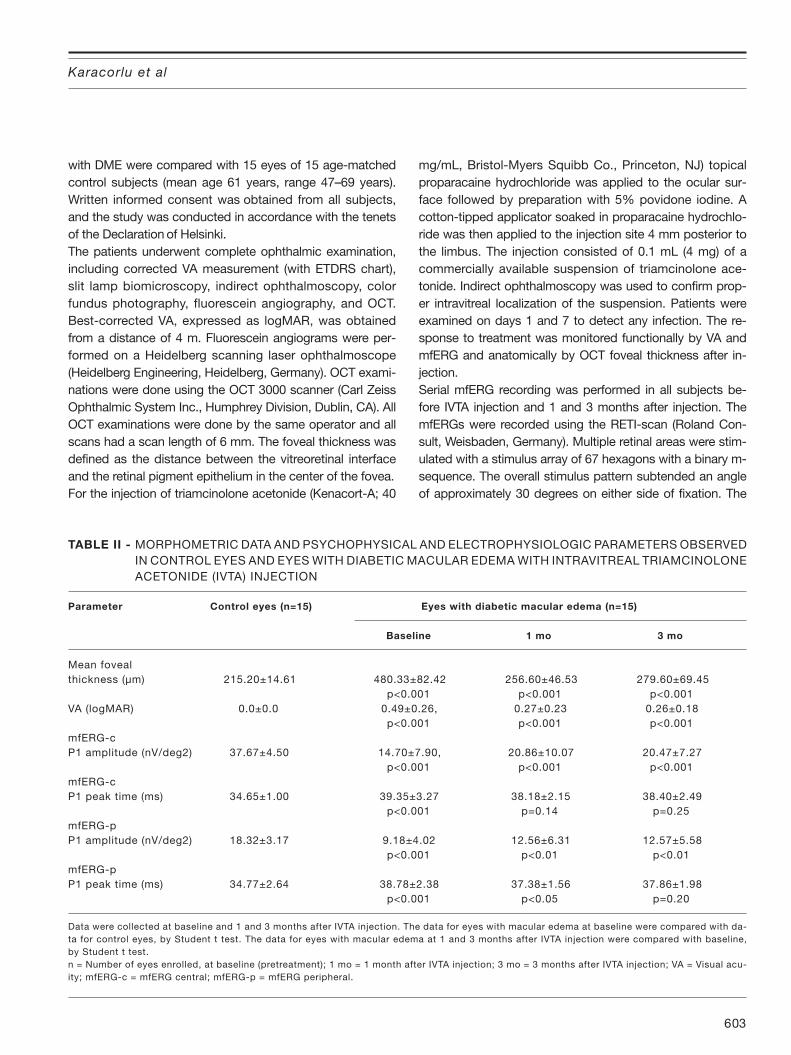
TABLE II - MORPHOMETRIC DATA AND PSYCHOPHYSICAL AND ELECTROPHYSIOLOGIC PARAMETERS OBSERVEDIN CONTROL EYES AND EYES WITH DIABETIC MACULAR EDEMA WITH INTRAVITREAL TRIAMCINOLONEACETONIDE (IVTA) INJECTION
Parameter Control eyes (n=15) Eyes with diabetic macular edema (n=15)
Baseline 1 mo 3 mo
Mean foveal thickness (µm) 215.20±14.61 480.33±82.42 256.60±46.53 279.60±69.45
p<0.001 p<0.001 p<0.001VA (logMAR) 0.0±0.0 0.49±0.26, 0.27±0.23 0.26±0.18
p<0.001 p<0.001 p<0.001mfERG-cP1 amplitude (nV/deg2) 37.67±4.50 14.70±7.90, 20.86±10.07 20.47±7.27
p<0.001 p<0.001 p<0.001mfERG-cP1 peak time (ms) 34.65±1.00 39.35±3.27 38.18±2.15 38.40±2.49
p<0.001 p=0.14 p=0.25mfERG-pP1 amplitude (nV/deg2) 18.32±3.17 9.18±4.02 12.56±6.31 12.57±5.58
p<0.001 p<0.01 p<0.01mfERG-pP1 peak time (ms) 34.77±2.64 38.78±2.38 37.38±1.56 37.86±1.98
p<0.001 p<0.05 p=0.20
Data were collected at baseline and 1 and 3 months after IVTA injection. The data for eyes with macular edema at baseline were compared with da-ta for control eyes, by Student t test. The data for eyes with macular edema at 1 and 3 months after IVTA injection were compared with baseline,by Student t test. n = Number of eyes enrolled, at baseline (pretreatment); 1 mo = 1 month after IVTA injection; 3 mo = 3 months after IVTA injection; VA = Visual acu-ity; mfERG-c = mfERG central; mfERG-p = mfERG peripheral.

Macular function by mfERG in diabetic macular edema after IVTA
604
luminance of the stimulus was 120 cd/m² for the brightflashes and 1 cd/m² for the dark flashes. The stimulus wasdisplayed on a black-and-white monochrome cathode raytube monitor.Before mfERG recording, patients’ pupils were fully dilatedwith eyedrops containing 0.5% tropicamide and 0.5%phenylephrine. All subjects were placed in ordinary roomlight for 15 minutes for light adaptation before testing. Goldfoil scleral electrodes were used for mfERG recording.Room lights were maintained throughout the recordings.
The mfERG recordings were divided into eight segments,and any segments with blinking, large eye movements, orlosses of fixation were discarded and recorded again. Reti-nal signals were filtered with low-frequency and high-fre-quency cutoffs of 10 Hz and 300 Hz, respectively, and am-plified with a gain of 100,000.The first-order kernel mfERG responses were analyzed. In-dividual mfERG responses for the hexagons were groupedinto the two concentric areas centered on the fovea, with acentral ring of 0 to 7 degrees (central group) and a peripher-
A B
C D
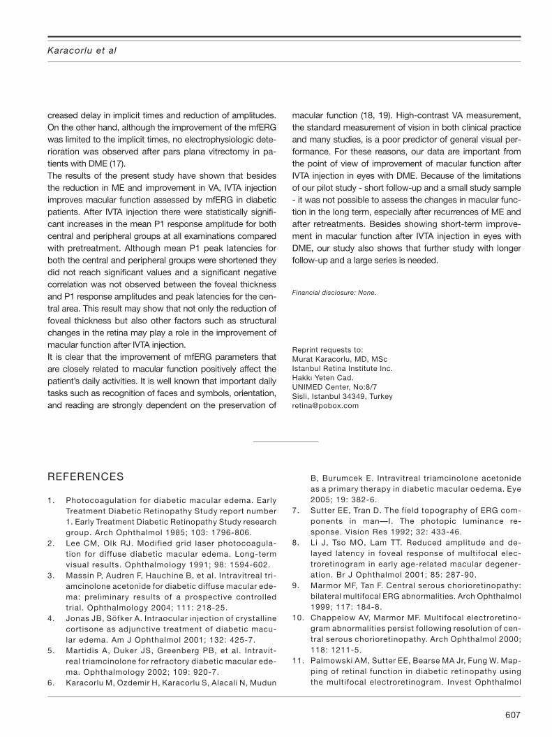
Fig. 1 - (A) Differences from baseline in the mean foveal thickness (µm) in eyes with diabetic macular edema (DME) 1 month after intravitreal tri-amcinolone acetonide (IVTA) injection, plotted against the differences in multifocal electroretinography (mfERG)-c (central) P1 amplitudes,y=–181.34–6.87x (r2=0.11, p=0.23). (B) Differences from baseline in the mean foveal thickness (µm) in eyes with DME 3 months after IVTA injec-tion, plotted against the difference in mfERG-c (central) P1 amplitudes, y=–218.03+2.99x (r2=0.03, p=0.50). (C) Differences from baseline in themean foveal thickness (µm) in eyes with DME 1 month after IVTA injection, plotted against the difference in mfERG-c (central) P1 peak times,y=–211.54+10.45x (r2=0.12, p>0.05). (D) Differences from baseline in the mean foveal thickness (µm) in eyes with DME 3 months after IVTAinjection, plotted against the difference in mfERG-c (central) P1 peak times, y=–192.82–8.30x (r2=0.13, p>0.05).

Karacorlu et al
605
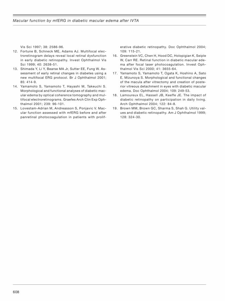
Fig. 3 - Multifocalelectroretinogra-phy of Patient 7at baseline (A, B)and 3 monthsafter treatment(C, D).
A B
C D
A B
DC
Fig. 2 - Fluorescein angiography and optical coherence tomographycross-sectional macular images of Patient 7 at baseline (A, B) and 3months after treatment (C, D).

Macular function by mfERG in diabetic macular edema after IVTA
606
al ring 7 to 25 degrees (peripheral group). The first positivepeak (P1) response amplitudes and P1 peak latencies forthe central and peripheral groups were then measured. Results from control eyes and eyes with DME at baselinewere compared by Student t test. Changes in functional pa-rameters (VAs and the P1 mfERG response amplitudes andpeak latencies) and morphometric parameters (OCT fovealthickness) in eyes with DME 1 and 3 months after IVTA in-jection were compared with baseline (pretreatment) valuesby Student t test. The P1 mfERG response amplitudes andpeak latencies in the central area and OCT foveal thicknesswere correlated with a linear correlation test.
RESULTS
Pretreatment and posttreatment clinical measurements arepresented in Table I. After 3 months follow-up, VA had im-proved in all eyes, with a mean of 2.5 and 2 lines at 1 and 3months after treatment, respectively. The mean baselinelogMAR value for VAs of the patients before treatment was0.49±0.26. After treatment it was significantly different fromthe pretreatment value (for each, p<0.001). The mean base-line foveal thickness was 480.33±82.42 µm. Thickness haddecreased at 1 and 3 months after treatment (for each,p<0.001) (Tab. II).There were statistically significant increases in the mean P1response amplitude for both central and peripheral groupsat all examinations compared with pretreatment (for each,p<0.001) (Tab. I). The mean P1 peak latencies were short-ened but not significantly in both the central and peripheralgroups at all examinations compared with pretreatment.During follow-up, no patient had recurrence of ME.Scatter plots of the foveal thickness as a function of the P1response amplitudes and peak latencies for central area areshown in Figure 1. The correlations between the fovealthickness and P1 response amplitudes and peak latenciesfor the central area were not significant. The OCT cross-sectional macular images, fluorescein angiography, andmfERGs of Patient 7 are shown in Figures 2 and 3.
DISCUSSION
DME is retinal thickening caused by accumulation of intrare-tinal fluid, primarily in the inner and outer plexiform layers,resulting from hyperpermeability of the retinal vasculature(1). ME affects approximately 29% of diabetic patients with
a disease duration of 20 years or more and is responsiblefor a significant degree of visual loss in this population (1).Various studies have shown the benefit of IVTA injection inpatients with DME (4-6). In these studies, VA and morpholo-gic features before and after treatment were evaluated,whereas patients’ subjective appraisal of visual function,which may differ, was not comprehensively discussed. Itwas acknowledged that DME affects visual function as partof the disease process and severely compromises the highlydeveloped functions of the macula, such as perception ofdetails, central fixation, color vision, and reading ability. Buthigh-contrast VA measurement is often a poor predictor ofgeneral visual performance. Important daily tasks such asrecognition of faces and symbols, orientation, and readingare strongly dependent on the preservation of the macularfunction. mfERG is a technique developed by Sutter and Tran (7) thatallows mapping of retinal function in the posterior polethrough simultaneous stimulation of different areas of theretina. In contrast to a conventional full-flash ERG, whichuses a global flash stimulus to stimulate the entire retina,mfERG utilizes an array of alternating flickers of hexagonalstimuli to stimulate individual retinal areas, and mfERG re-sponses are obtained using cross-correction between theraw recording and a pseudorandom m-sequence. ThemfERG recording can provide an objective assessment ofretinal function, and its sensitivity in detecting functional ab-normalities has been demonstrated in various macular dis-orders, including age-related macular degeneration andcentral serous chorioretinopathy (8-10). It is also well knownthat mfERG can assess retinal function in patients with dia-betic retinopathy (11-13). Yamamoto et al (14) showed thatthe mfERG from the macular area was a good objective in-dicator of macular function in patients with DME and wasstrongly correlated with morphologic changes in the macu-la. Using mfERG as an objective method of assessing retinalfunction may therefore be a useful tool in documenting thefunctional changes after treatment in patients with DME. Ithas been shown that in spite of unchanged values of retinalthickness and VA, panretinal photocoagulation seems tocause a functional impairment in the adjacent untreatedmacula, shown by reduced amplitudes measured by themfERG (15). It has also been shown that both the implicittimes and the amplitudes of the mfERGs are changed aftergrid laser photocoagulation for DME, although the results ofpsychophysical tests suggested little or no change in visualfunction (16). It was thought that damage to the outer retinacaused by photocoagulation could account for the in-
Karacorlu et al
607
creased delay in implicit times and reduction of amplitudes.On the other hand, although the improvement of the mfERGwas limited to the implicit times, no electrophysiologic dete-rioration was observed after pars plana vitrectomy in pa-tients with DME (17).The results of the present study have shown that besidesthe reduction in ME and improvement in VA, IVTA injectionimproves macular function assessed by mfERG in diabeticpatients. After IVTA injection there were statistically signifi-cant increases in the mean P1 response amplitude for bothcentral and peripheral groups at all examinations comparedwith pretreatment. Although mean P1 peak latencies forboth the central and peripheral groups were shortened theydid not reach significant values and a significant negativecorrelation was not observed between the foveal thicknessand P1 response amplitudes and peak latencies for the cen-tral area. This result may show that not only the reduction offoveal thickness but also other factors such as structuralchanges in the retina may play a role in the improvement ofmacular function after IVTA injection.It is clear that the improvement of mfERG parameters thatare closely related to macular function positively affect thepatient’s daily activities. It is well known that important dailytasks such as recognition of faces and symbols, orientation,and reading are strongly dependent on the preservation of
macular function (18, 19). High-contrast VA measurement,the standard measurement of vision in both clinical practiceand many studies, is a poor predictor of general visual per-formance. For these reasons, our data are important fromthe point of view of improvement of macular function afterIVTA injection in eyes with DME. Because of the limitationsof our pilot study - short follow-up and a small study sample- it was not possible to assess the changes in macular func-tion in the long term, especially after recurrences of ME andafter retreatments. Besides showing short-term improve-ment in macular function after IVTA injection in eyes withDME, our study also shows that further study with longerfollow-up and a large series is needed.
Financial disclosure: None.
Reprint requests to:Murat Karacorlu, MD, MScIstanbul Retina Institute Inc.Hakkı Yeten Cad.UNIMED Center, No:8/7 Sisli, Istanbul 34349, [email protected]
REFERENCES
1. Photocoagulation for diabetic macular edema. EarlyTreatment Diabetic Retinopathy Study report number1. Early Treatment Diabetic Retinopathy Study researchgroup. Arch Ophthalmol 1985; 103: 1796-806.
2. Lee CM, Olk RJ. Modified grid laser photocoagula-tion for diffuse diabetic macular edema. Long-termvisual results. Ophthalmology 1991; 98: 1594-602.
3. Massin P, Audren F, Hauchine B, et al. Intravitreal tri-amcinolone acetonide for diabetic diffuse macular ede-ma: preliminary results of a prospective controlledtrial. Ophthalmology 2004; 111: 218-25.
4. Jonas JB, Söfker A. Intraocular injection of crystallinecortisone as adjunctive treatment of diabetic macu-lar edema. Am J Ophthalmol 2001; 132: 425-7.
5. Martidis A, Duker JS, Greenberg PB, et al. Intravit-real triamcinolone for refractory diabetic macular ede-ma. Ophthalmology 2002; 109: 920-7.
6. Karacorlu M, Ozdemir H, Karacorlu S, Alacali N, Mudun
B, Burumcek E. Intravitreal triamcinolone acetonideas a primary therapy in diabetic macular oedema. Eye2005; 19: 382-6.
7. Sutter EE, Tran D. The field topography of ERG com-ponents in man—I. The photopic luminance re-sponse. Vision Res 1992; 32: 433-46.
8. Li J, Tso MO, Lam TT. Reduced amplitude and de-layed latency in foveal response of multifocal elec-troretinogram in early age-related macular degener-ation. Br J Ophthalmol 2001; 85: 287-90.
9. Marmor MF, Tan F. Central serous chorioretinopathy:bilateral multifocal ERG abnormalities. Arch Ophthalmol1999; 117: 184-8.
10. Chappelow AV, Marmor MF. Multifocal electroretino-gram abnormalities persist following resolution of cen-tral serous chorioretinopathy. Arch Ophthalmol 2000;118: 1211-5.
11. Palmowski AM, Sutter EE, Bearse MA Jr, Fung W. Map-ping of retinal function in diabetic retinopathy usingthe multifocal electroretinogram. Invest Ophthalmol
Macular function by mfERG in diabetic macular edema after IVTA
608
Vis Sci 1997; 38: 2586-96.12. Fortune B, Schneck ME, Adams AJ. Multifocal elec-
troretinogram delays reveal local retinal dysfunctionin early diabetic retinopathy. Invest Ophthalmol VisSci 1999; 40: 2638-51.
13. Shimada Y, Li Y, Bearse MA Jr, Sutter EE, Fung W. As-sessment of early retinal changes in diabetes using anew multifocal ERG protocol. Br J Ophthalmol 2001;85: 414-9.
14. Yamamoto S, Yamamoto T, Hayashi M, Takeuchi S.Morphological and functional analyses of diabetic mac-ular edema by optical coherence tomography and mul-tifocal electroretinograms. Graefes Arch Clin Exp Oph-thalmol 2001; 239: 96-101.
15. Lovestam-Adrian M, Andreasson S, Ponjavic V. Mac-ular function assessed with mfERG before and afterpanretinal photocoagulation in patients with prolif-
erative diabetic retinopathy. Doc Ophthalmol 2004;109: 115-21.
16. Greenstein VC, Chen H, Hood DC, Holopigian K, SeipleW, Carr RE. Retinal function in diabetic macular ede-ma after focal laser photocoagulation. Invest Oph-thalmol Vis Sci 2000; 41: 3655-64.
17. Yamamoto S, Yamamoto T, Ogata K, Hoshino A, SatoE, Mizunoya S. Morphological and functional changesof the macula after vitrectomy and creation of poste-rior vitreous detachment in eyes with diabetic macularedema. Doc Ophthalmol 2004; 109: 249-53.
18. Lamoureux EL, Hassell JB, Keeffe JE. The impact ofdiabetic retinopathy on participation in daily living.Arch Ophthalmol 2004; 122: 84-8.
19. Brown MM, Brown GC, Sharma S, Shah G. Utility val-ues and diabetic retinopathy. Am J Ophthalmol 1999;128: 324-30.