Embed Size (px)
Citation preview

COCIR SELF-REGULATORY INITIATIVE
FOR MEDICAL IMAGING EQUIPMENT
MAGNETIC RESONANCE EQUIPMENT MEASUREMENT OF ENERGY
CONSUMPTION 2011
REVISION : 1
DATE : June 2012
APPROVED : June 2012

Self-Regulatory Initiative for Medical Imaging Equipment
MRI – Measurement of energy consumption
MR Energy Measurement Procedure, Revision 1, June 2012 © COCIR
2
TABLE OF CONTENT
1. INTRODUCTION ............................................................................................................................ 3
2. SCOPE .......................................................................................................................................... 4
3. DEFINITIONS ................................................................................................................................. 4
4. SYSTEM POWER MODES ................................................................................................................ 5
5. USE MODES OVERVIEW ................................................................................................................. 5
OFF ............................................................................................................................................................ 5
LOW POWER ................................................................................................................................................ 5
SCAN AND READY-TO-SCAN ............................................................................................................................. 5
6. RESOURCES .................................................................................................................................. 7
7. UNIT UNDER TEST (UUT) ................................................................................................................ 7
7.1. POWER MEASUREMENT DEVICE ............................................................................................................ 7
8. COCIR MRI DATA COLLECTION SPREAD SHEET ................................................................................. 8
9. MEASURED RESULTS ..................................................................................................................... 8
10. INSTALLATION OF POWER MEASUREMENT DEVICE ....................................................................... 8
11. MEASUREMENT OF POWER AND ENERGY .................................................................................... 8
11.1. OFF MODE POWER MEASUREMENT ................................................................................................... 8
11.2. LOW POWER MODE POWER MEASUREMENT ..................................................................................... 8
11.3. READY-TO-SCAN MODE POWER MEASUREMENT ................................................................................... 9
11.4. SCAN MODE ENERGY MEASUREMENT ................................................................................................. 9
11.5. SEQUENCE DURATION DETERMINATION ............................................................................................. 10
12. HOW TO USE TEMPLATES ......................................................................................................... 11
Self-Regulatory Initiative for Medical Imaging Equipment
MRI – Measurement of energy consumption
MR Energy Measurement Procedure, Revision 1, June 2012 © COCIR
3
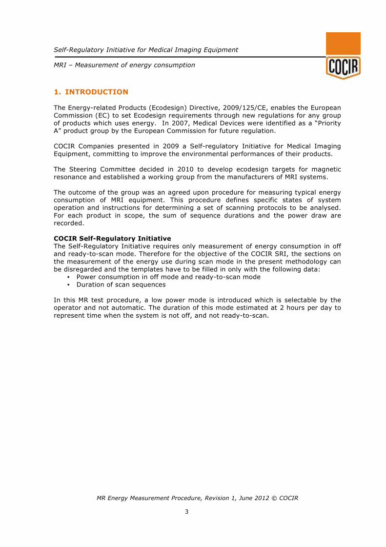
1. INTRODUCTION
The Energy-related Products (Ecodesign) Directive, 2009/125/CE, enables the European
Commission (EC) to set Ecodesign requirements through new regulations for any group
of products which uses energy. In 2007, Medical Devices were identified as a “Priority
A” product group by the European Commission for future regulation.
COCIR Companies presented in 2009 a Self-regulatory Initiative for Medical Imaging
Equipment, committing to improve the environmental performances of their products.
The Steering Committee decided in 2010 to develop ecodesign targets for magnetic
resonance and established a working group from the manufacturers of MRI systems.
The outcome of the group was an agreed upon procedure for measuring typical energy
consumption of MRI equipment. This procedure defines specific states of system
operation and instructions for determining a set of scanning protocols to be analysed.
For each product in scope, the sum of sequence durations and the power draw are
recorded.
COCIR Self-Regulatory Initiative The Self-Regulatory Initiative requires only measurement of energy consumption in off
and ready-to-scan mode. Therefore for the objective of the COCIR SRI, the sections on
the measurement of the energy use during scan mode in the present methodology can
be disregarded and the templates have to be filled in only with the following data:
• Power consumption in off mode and ready-to-scan mode
• Duration of scan sequences
In this MR test procedure, a low power mode is introduced which is selectable by the
operator and not automatic. The duration of this mode estimated at 2 hours per day to
represent time when the system is not off, and not ready-to-scan.
Self-Regulatory Initiative for Medical Imaging Equipment
MRI – Measurement of energy consumption
MR Energy Measurement Procedure, Revision 1, June 2012 © COCIR
4
2. SCOPE
This methodology can be used to measure all whole-body MRI systems. Equipment and
accessories beyond a basic MR product and not required for a basic scan, or customer-
provided equipment, such as optional MR coils, patient vital signs accessories, facility-
provided cooling water equipment and hardware for advanced medical applications, are
outside the scope of this procedure.
The use of the present methodology to measure permanent magnet open MRI has not
been evaluated.
The methodology is not suited for the measurement of technologies combining MRI with
other Imaging systems, such as MRI/CT or MRI/PET.
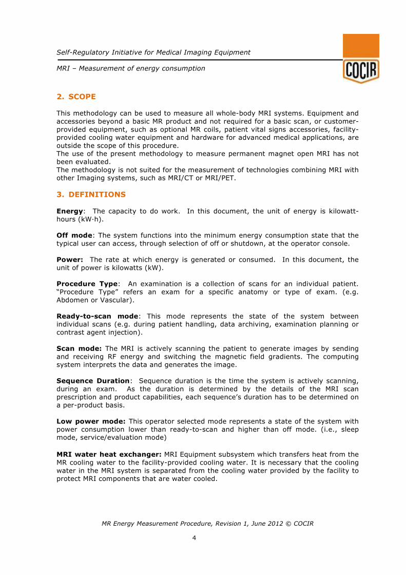
3. DEFINITIONS
Energy: The capacity to do work. In this document, the unit of energy is kilowatt-
hours (kW∙h).
Off mode: The system functions into the minimum energy consumption state that the
typical user can access, through selection of off or shutdown, at the operator console.
Power: The rate at which energy is generated or consumed. In this document, the
unit of power is kilowatts (kW).
Procedure Type: An examination is a collection of scans for an individual patient.
“Procedure Type” refers an exam for a specific anatomy or type of exam. (e.g.
Abdomen or Vascular).
Ready-to-scan mode: This mode represents the state of the system between
individual scans (e.g. during patient handling, data archiving, examination planning or
contrast agent injection).
Scan mode: The MRI is actively scanning the patient to generate images by sending
and receiving RF energy and switching the magnetic field gradients. The computing
system interprets the data and generates the image.
Sequence Duration: Sequence duration is the time the system is actively scanning,
during an exam. As the duration is determined by the details of the MRI scan
prescription and product capabilities, each sequence’s duration has to be determined on
a per-product basis.
Low power mode: This operator selected mode represents a state of the system with
power consumption lower than ready-to-scan and higher than off mode. (i.e., sleep
mode, service/evaluation mode)
MRI water heat exchanger: MRI Equipment subsystem which transfers heat from the
MR cooling water to the facility-provided cooling water. It is necessary that the cooling
water in the MRI system is separated from the cooling water provided by the facility to
protect MRI components that are water cooled.
Self-Regulatory Initiative for Medical Imaging Equipment
MRI – Measurement of energy consumption
MR Energy Measurement Procedure, Revision 1, June 2012 © COCIR
5
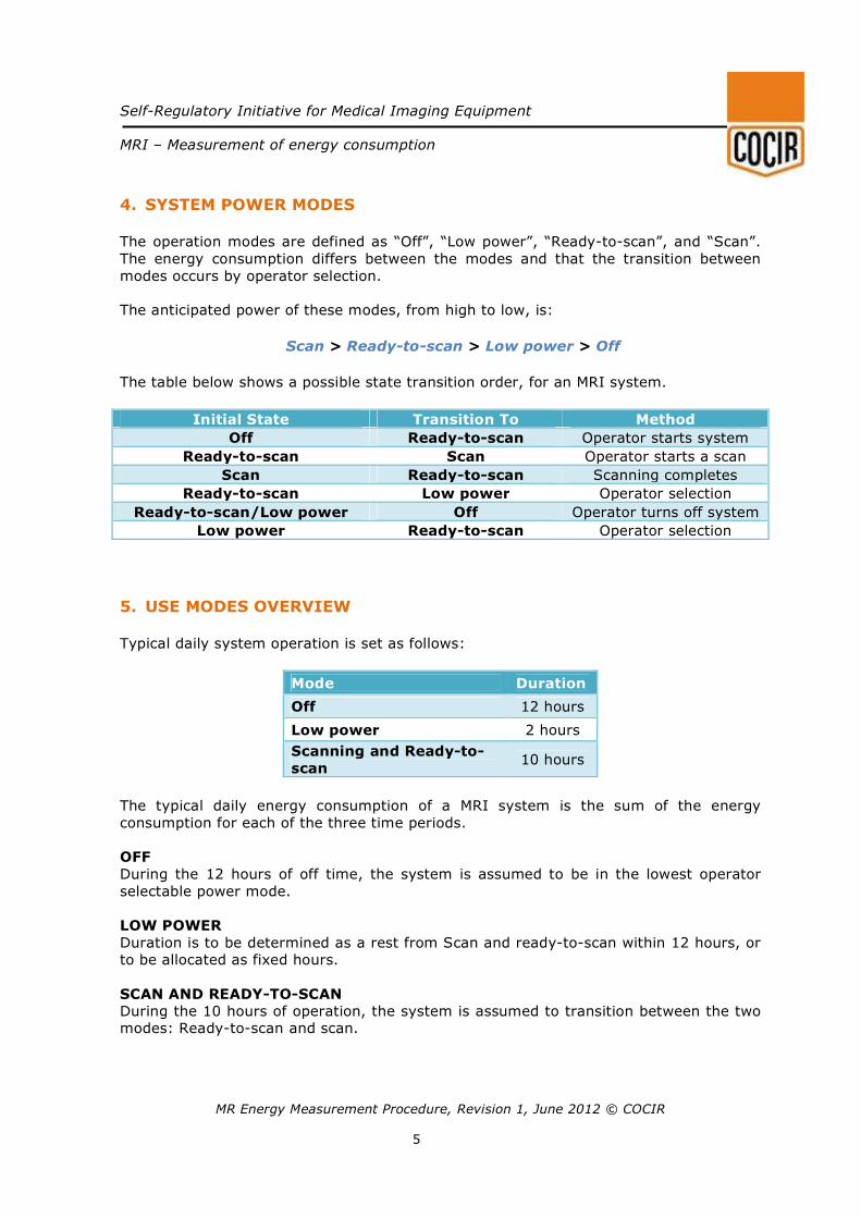
4. SYSTEM POWER MODES
The operation modes are defined as “Off”, “Low power”, “Ready-to-scan”, and “Scan”.
The energy consumption differs between the modes and that the transition between
modes occurs by operator selection.
The anticipated power of these modes, from high to low, is:
Scan > Ready-to-scan > Low power > Off
The table below shows a possible state transition order, for an MRI system.
Initial State Transition To Method
Off Ready-to-scan Operator starts system
Ready-to-scan Scan Operator starts a scan
Scan Ready-to-scan Scanning completes
Ready-to-scan Low power Operator selection
Ready-to-scan/Low power Off Operator turns off system
Low power Ready-to-scan Operator selection
5. USE MODES OVERVIEW
Typical daily system operation is set as follows:
Mode Duration
Off 12 hours
Low power 2 hours
Scanning and Ready-to-
scan 10 hours
The typical daily energy consumption of a MRI system is the sum of the energy
consumption for each of the three time periods.
OFF
During the 12 hours of off time, the system is assumed to be in the lowest operator
selectable power mode.
LOW POWER
Duration is to be determined as a rest from Scan and ready-to-scan within 12 hours, or
to be allocated as fixed hours.
SCAN AND READY-TO-SCAN During the 10 hours of operation, the system is assumed to transition between the two
modes: Ready-to-scan and scan.
Self-Regulatory Initiative for Medical Imaging Equipment
MRI – Measurement of energy consumption
MR Energy Measurement Procedure, Revision 1, June 2012 © COCIR
6
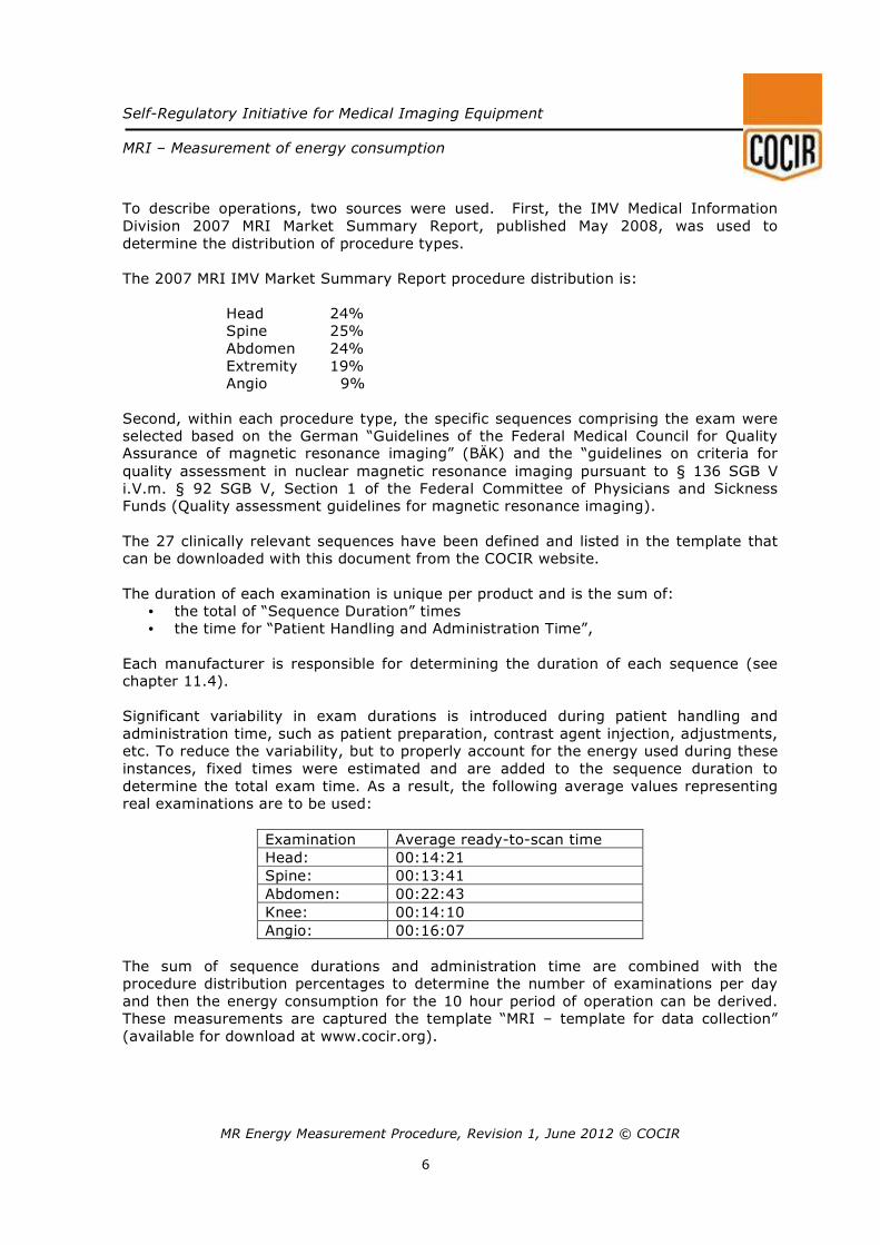
To describe operations, two sources were used. First, the IMV Medical Information
Division 2007 MRI Market Summary Report, published May 2008, was used to
determine the distribution of procedure types.
The 2007 MRI IMV Market Summary Report procedure distribution is:
Head 24%
Spine 25%
Abdomen 24%
Extremity 19%
Angio 9%
Second, within each procedure type, the specific sequences comprising the exam were
selected based on the German “Guidelines of the Federal Medical Council for Quality Assurance of magnetic resonance imaging” (BÄK) and the “guidelines on criteria for
quality assessment in nuclear magnetic resonance imaging pursuant to § 136 SGB V
i.V.m. § 92 SGB V, Section 1 of the Federal Committee of Physicians and Sickness
Funds (Quality assessment guidelines for magnetic resonance imaging).
The 27 clinically relevant sequences have been defined and listed in the template that
can be downloaded with this document from the COCIR website.
The duration of each examination is unique per product and is the sum of:
• the total of “Sequence Duration” times
• the time for “Patient Handling and Administration Time”,
Each manufacturer is responsible for determining the duration of each sequence (see
chapter 11.4).
Significant variability in exam durations is introduced during patient handling and
administration time, such as patient preparation, contrast agent injection, adjustments,
etc. To reduce the variability, but to properly account for the energy used during these
instances, fixed times were estimated and are added to the sequence duration to
determine the total exam time. As a result, the following average values representing
real examinations are to be used:
Examination Average ready-to-scan time
Head: 00:14:21
Spine: 00:13:41
Abdomen: 00:22:43
Knee: 00:14:10
Angio: 00:16:07
The sum of sequence durations and administration time are combined with the
procedure distribution percentages to determine the number of examinations per day
and then the energy consumption for the 10 hour period of operation can be derived.
These measurements are captured the template “MRI – template for data collection”
(available for download at www.cocir.org).
Self-Regulatory Initiative for Medical Imaging Equipment
MRI – Measurement of energy consumption
MR Energy Measurement Procedure, Revision 1, June 2012 © COCIR
7
6. RESOURCES
The following personnel are recommended:
• An engineer or technician familiar with the power distribution of the system and
power electronics safety.
• An engineer or applications specialist familiar with scanner operation and the
prescribing of clinical protocols.
7. UNIT UNDER TEST (UUT)
System Configuration: System configuration should be recorded and configured to
perform the set of specified procedures with appropriate RF receiving coils.
Installation: The system shall be installed and calibrated according to its specification,
including all system-critical items needed to perform a basic scan, e.g. gradient
amplifiers, RF unit, MR coils needed for the specific measurements, reconstruction
engine(s), required electronics such power supplies, controllers, console/computer,
cryogen compressor, water heat exchanger, patient table, magnet and helium-
conservation equipment.
Any equipment and accessories beyond basic product offering and not required for a
basic scan, or customer-provided equipment, e.g. optional MR coils, patient vital signs
accessories, facility-provided cooling water equipment and hardware for advanced
medical applications shall not be included in the measurement.
Environmental Conditions: The measurements are to be taken at a steady-state
operating temperature, and within manufacturer’s specified ambient temperature and
humidity limits.
Measurement: Prior to each mode’s measurement, the equipment shall remain in that
mode for sufficient time to allow temperature and other pertinent transient conditions
to stabilize.
Emulated System: For sequence duration determination, it is permissible to use a
device that emulates the hardware capabilities of the system, and uses the product
software, to ensure the same prescription restrictions as a full system.
7.1. POWER MEASUREMENT DEVICE
A device capable of measuring 3-phase voltage and current and calculating the integral
of power with respect to time (energy) or a power meter able to sample average power
ratings.
Self-Regulatory Initiative for Medical Imaging Equipment
MRI – Measurement of energy consumption
MR Energy Measurement Procedure, Revision 1, June 2012 © COCIR
8
8. COCIR MRI DATA COLLECTION SPREAD SHEET
The data obtained according to the present methodology have to be filled in the
appropriate template that can be downloaded from the COCIR website www.cocir.org.
9. MEASURED RESULTS
The measured values resulting from this procedure are:
• Power measured in Off mode
• Power measured in Ready-to-scan mode
• Power measured in Low Power mode
• Power measured in Scan mode (average value) for each sequence
• Duration of each scan sequence
• Energy consumption per examination
10. INSTALLATION OF POWER MEASUREMENT DEVICE
The power measurement device shall be installed onto the input to the main disconnect
panel of the system to ensure that all energy consumption of the MRI equipment is
captured, including the cryogen compressor and the MRI water heat exchanger.
11. MEASUREMENT OF POWER AND ENERGY
11.1. OFF MODE POWER MEASUREMENT
1) Ensure that the power meter is on and functioning.
2) Shutdown the system to the minimum energy consumption state that the user can
access.
3) Wait to ensure that all system elements have established low power operation.
4) Measure the average power draw (rate of energy consumption), for a period of at
least 10 minutes. If the system has a variable power usage in this mode, the
measurement duration shall be amended to at least one complete power usage
cycle, which shall be taken to be the cycle from minimum to maximum usage.
5) Record this value, in kilowatts.
11.2. LOW POWER MODE POWER MEASUREMENT
1) Ensure that the power meter is on and functioning.
2) Wait to ensure that all applicable system elements have adapted to this mode.
3) Measure the average power draw (rate of energy consumption), for a period of at
least 10 minutes. If the system has a variable power usage in this mode, the
measurement duration shall be amended to one complete power usage cycle, which
shall be taken to be the cycle from minimum to maximum usage.
4) Record this value, in kilowatts.
In case the MRI is not able to switch to a lower energy mode, the ready-to-scan power
rate has to be used.
Self-Regulatory Initiative for Medical Imaging Equipment
MRI – Measurement of energy consumption
MR Energy Measurement Procedure, Revision 1, June 2012 © COCIR
9
11.3. READY-TO-SCAN MODE POWER MEASUREMENT
1) Ensure that the power meter is on and functioning.
2) Prescribe a patient and execute any scan to ensure that the system is
functioning.
3) After the scan completes, record the average power draw (rate of energy
consumption), for a period of 12 minutes. Record this value, in kilowatts.
11.4. SCAN MODE ENERGY MEASUREMENT
Setting up Scan Programs
Prepare a scan program for each exam type according to the user manual using the
parameters defined in Appendix I. If it is not possible for the MRI system under test to
use a certain sequence specified in the Appendix I, use a sequence as close as possible
to the sequence specified given the same contrast and diagnostic results.
Store the scan programs for later usage on the same MRI system or MRI system type.
Measurement during scan with equipment actively scanning Procedure for Power Determination using exam type average:
1) Set the equipment to Ready-to-Scan mode (according to 11.3).
2) For each exam type et:
3) Take time “ts” and energy reading “Es” and start scan program
4) After completion of scan program: take time “te” and energy reading “Ee”
5) Calculate average power Pet = (Ee - Es) / (te - ts).
6) Transfer average power “Pet” to the evaluation sheet (row “sum scan time”, column
“Power / kW”).
Transfer sequence durations “ds” for all sequences used to the evaluation sheet
(column “Sequence duration”) 7) Consistency check: te - ts shall not deviate more than a few seconds from the sum of
sequences’ durations “ds”.
Procedure for Power Determination using power sampling:
1. Set the equipment to Ready-to-Scan mode (according to 11.3).
2. For each exam type et:
3. Start scan program and sample the average power consumption within short
intervals of time dt (e.g. every second). Pt is the sample at time t.
4. After completion of scan program: For each sequence s:
a. Calculate total energy consumption Es for the sequence:
ts: Start time of sequence; te: End time of sequence b. Calculate average power consumption P:
Ps = E /(te-ts) c. Consistency check: te - ts shall not deviate from the sequence duration ds.
5. Transfer sequence durations ds (column “Sequence duration”) and average power Ps (column “Power / kW”) for all sequences used to the evaluation sheet.
Self-Regulatory Initiative for Medical Imaging Equipment
MRI – Measurement of energy consumption
MR Energy Measurement Procedure, Revision 1, June 2012 © COCIR
10
11.5. SEQUENCE DURATION DETERMINATION
The exact prescription of each sequence is to be determined by the individual
manufacturers. Three criteria should be considered, when determining the prescription
parameters:
1) Parameters defined in Appendix I must be met.
2) SAR and dB/dt limits should not exceed IEC60601-2-33 First Control Mode
restrictions using a patient weight between 50 and 100kg.
3) The listed contrast type must be preserved (i.e. PD, T1, or T2-weighted)
4) Clinical considerations (i.e. reducing breath hold time for abdominal scans, or a
minor adjustment in default TR to obtain the minimum required number of slices
within one acquisition).
Record the duration of each sequence, as calculated by the system’s software and
displayed in the system’s user interface.
Self-Regulatory Initiative for Medical Imaging Equipment
MRI – Measurement of energy consumption
MR Energy Measurement Procedure, Revision 1, June 2012 © COCIR
11
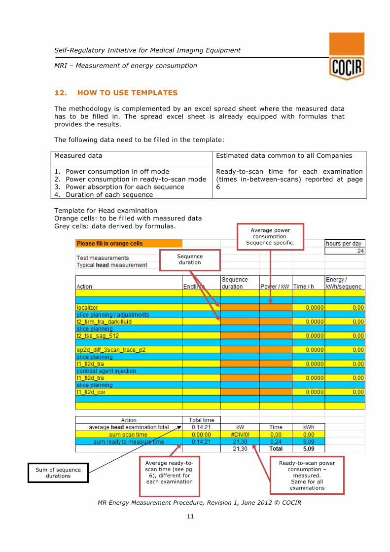
12. HOW TO USE TEMPLATES
The methodology is complemented by an excel spread sheet where the measured data
has to be filled in. The spread excel sheet is already equipped with formulas that
provides the results.
The following data need to be filled in the template:
Measured data
Estimated data common to all Companies
1. Power consumption in off mode
2. Power consumption in ready-to-scan mode
3. Power absorption for each sequence
4. Duration of each sequence
Ready-to-scan time for each examination
(times in-between-scans) reported at page
6
Template for Head examination
Orange cells: to be filled with measured data
Grey cells: data derived by formulas.
Average power consumption.
Sequence specific.
Sequence duration
Sum of sequence durations
Average ready-to-
scan time (see pg. 6), different for
each examination
Ready-to-scan power
consumption – measured.
Same for all examinations
Self-Regulatory Initiative for Medical Imaging Equipment
MRI – Measurement of energy consumption
MR Energy Measurement Procedure, Revision 1, June 2012 © COCIR
12
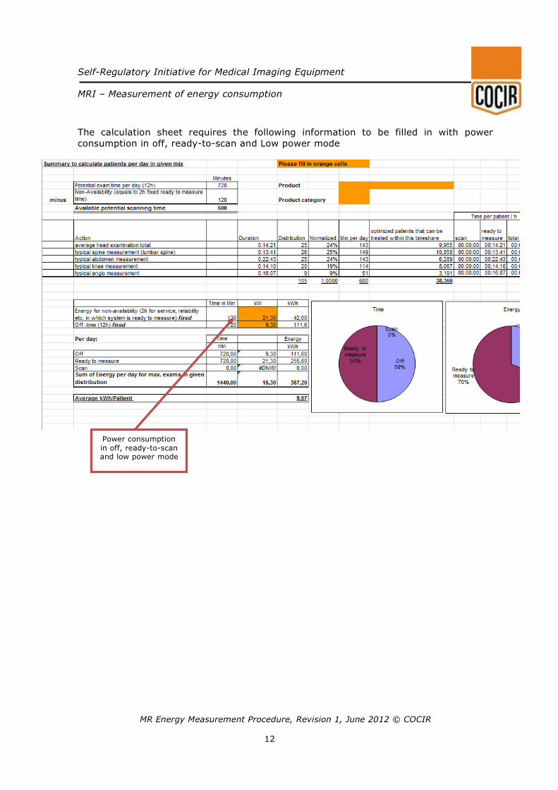
The calculation sheet requires the following information to be filled in with power
consumption in off, ready-to-scan and Low power mode
Power consumption
in off, ready-to-scan and low power mode
Self-Regulatory Initiative for Medical Imaging Equipment
MRI – Measurement of energy consumption
MR Energy Measurement Procedure, Revision 1, June 2012 © COCIR
13
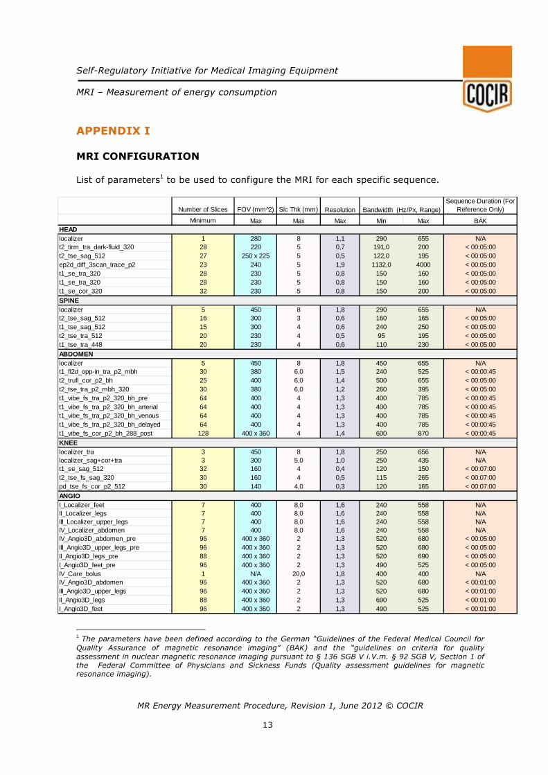
APPENDIX I MRI CONFIGURATION
List of parameters
1 to be used to configure the MRI for each specific sequence.
1 The parameters have been defined according to the German “Guidelines of the Federal Medical Council for
Quality Assurance of magnetic resonance imaging” (BAK) and the “guidelines on criteria for quality assessment in nuclear magnetic resonance imaging pursuant to § 136 SGB V i.V.m. § 92 SGB V, Section 1 of the Federal Committee of Physicians and Sickness Funds (Quality assessment guidelines for magnetic resonance imaging).
Number of Slices FOV (mm 2̂) Slc Thk (mm)Sequence Duration (For
Reference Only)
Minimum Max Max Max Min Max BÄK
localizer 1 280 8 1,1 290 655 N/At2_tirm_tra_dark-fluid_320 28 220 5 0,7 191,0 200 < 00:05:00t2_tse_sag_512 27 250 x 225 5 0,5 122,0 195 < 00:05:00ep2d_diff_3scan_trace_p2 23 240 5 1,9 1132,0 4000 < 00:05:00t1_se_tra_320 28 230 5 0,8 150 160 < 00:05:00t1_se_tra_320 28 230 5 0,8 150 160 < 00:05:00t1_se_cor_320 32 230 5 0,8 150 200 < 00:05:00
localizer 5 450 8 1,8 290 655 N/At2_tse_sag_512 16 300 3 0,6 160 165 < 00:05:00t1_tse_sag_512 15 300 4 0,6 240 250 < 00:05:00t2_tse_tra_512 20 230 4 0,5 95 195 < 00:05:00t1_tse_tra_448 20 230 4 0,6 110 230 < 00:05:00
localizer 5 450 8 1,8 450 655 N/At1_fl2d_opp-in_tra_p2_mbh 30 380 6,0 1,5 240 525 < 00:00:45t2_trufi_cor_p2_bh 25 400 6,0 1,4 500 655 < 00:05:00t2_tse_tra_p2_mbh_320 30 380 6,0 1,2 260 395 < 00:05:00t1_vibe_fs_tra_p2_320_bh_pre 64 400 4 1,3 400 785 < 00:00:45t1_vibe_fs_tra_p2_320_bh_arterial 64 400 4 1,3 400 785 < 00:00:45t1_vibe_fs_tra_p2_320_bh_venous 64 400 4 1,3 400 785 < 00:00:45t1_vibe_fs_tra_p2_320_bh_delayed 64 400 4 1,3 400 785 < 00:00:45t1_vibe_fs_cor_p2_bh_288_post 128 400 x 360 4 1,4 600 870 < 00:00:45
localizer_tra 3 450 8 1,8 250 656 N/Alocalizer_sag+cor+tra 3 300 5,0 1,0 250 435 N/At1_se_sag_512 32 160 4 0,4 120 150 < 00:07:00t2_tse_fs_sag_320 30 160 4 0,5 115 265 < 00:07:00pd_tse_fs_cor_p2_512 30 140 4,0 0,3 120 165 < 00:07:00
I_Localizer_feet 7 400 8,0 1,6 240 558 N/AII_Localizer_legs 7 400 8,0 1,6 240 558 N/AIII_Localizer_upper_legs 7 400 8,0 1,6 240 558 N/AIV_Localizer_abdomen 7 400 8,0 1,6 240 558 N/AIV_Angio3D_abdomen_pre 96 400 x 360 2 1,3 520 680 < 00:05:00III_Angio3D_upper_legs_pre 96 400 x 360 2 1,3 520 680 < 00:05:00II_Angio3D_legs_pre 88 400 x 360 2 1,3 520 690 < 00:05:00I_Angio3D_feet_pre 96 400 x 360 2 1,3 490 525 < 00:05:00IV_Care_bolus 1 N/A 20,0 1,8 400 400 N/AIV_Angio3D_abdomen 96 400 x 360 2 1,3 520 680 < 00:01:00III_Angio3D_upper_legs 96 400 x 360 2 1,3 520 680 < 00:01:00II_Angio3D_legs 88 400 x 360 2 1,3 690 525 < 00:01:00I_Angio3D_feet 96 400 x 360 2 1,3 490 525 < 00:01:00
ABDOMEN
KNEE
SPINE
HEAD
ANGIO
Resolution Bandwidth (Hz/Px, Range)


“Self-regulatory Initiative for medical imaging equipment”
Copyright © 2011 COCIR All Rights Reserved.
All material in this document is, unless otherwise stated, the property of COCIR. Copyright and other intellectual property laws protect these materials. Reproduction or retransmission of the materials, in whole or in part, in any manner, without the prior
written consent of the copyright holder, is an infringement of copyright law.