Embed Size (px)
Citation preview

From
Center for Surgical Sciences, Karolinska Institutet Division of Radiology
Huddinge University Hospital Stockholm, Sweden
MAGNETIC RESONANCE IMAGING IN
BREAST DIAGNOSIS
Maria KristoffersenWiberg
Stockholm 2002

All previously published papers were reproduced with permission from the publisher. Published and printed by Karolinska University Press Box 200, SE-171 77 Stockholm, Sweden © Maria KristoffersenWiberg, 2002 ISBN 91-7349-343-0
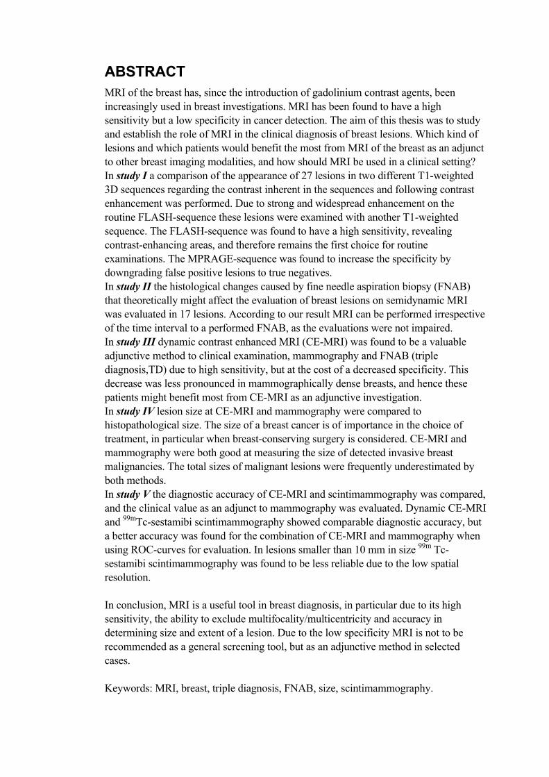
ABSTRACT MRI of the breast has, since the introduction of gadolinium contrast agents, been increasingly used in breast investigations. MRI has been found to have a high sensitivity but a low specificity in cancer detection. The aim of this thesis was to study and establish the role of MRI in the clinical diagnosis of breast lesions. Which kind of lesions and which patients would benefit the most from MRI of the breast as an adjunct to other breast imaging modalities, and how should MRI be used in a clinical setting? In study I a comparison of the appearance of 27 lesions in two different T1-weighted 3D sequences regarding the contrast inherent in the sequences and following contrast enhancement was performed. Due to strong and widespread enhancement on the routine FLASH-sequence these lesions were examined with another T1-weighted sequence. The FLASH-sequence was found to have a high sensitivity, revealing contrast-enhancing areas, and therefore remains the first choice for routine examinations. The MPRAGE-sequence was found to increase the specificity by downgrading false positive lesions to true negatives. In study II the histological changes caused by fine needle aspiration biopsy (FNAB) that theoretically might affect the evaluation of breast lesions on semidynamic MRI was evaluated in 17 lesions. According to our result MRI can be performed irrespective of the time interval to a performed FNAB, as the evaluations were not impaired. In study III dynamic contrast enhanced MRI (CE-MRI) was found to be a valuable adjunctive method to clinical examination, mammography and FNAB (triple diagnosis,TD) due to high sensitivity, but at the cost of a decreased specificity. This decrease was less pronounced in mammographically dense breasts, and hence these patients might benefit most from CE-MRI as an adjunctive investigation. In study IV lesion size at CE-MRI and mammography were compared to histopathological size. The size of a breast cancer is of importance in the choice of treatment, in particular when breast-conserving surgery is considered. CE-MRI and mammography were both good at measuring the size of detected invasive breast malignancies. The total sizes of malignant lesions were frequently underestimated by both methods. In study V the diagnostic accuracy of CE-MRI and scintimammography was compared, and the clinical value as an adjunct to mammography was evaluated. Dynamic CE-MRI and 99mTc-sestamibi scintimammography showed comparable diagnostic accuracy, but a better accuracy was found for the combination of CE-MRI and mammography when using ROC-curves for evaluation. In lesions smaller than 10 mm in size 99m Tc-sestamibi scintimammography was found to be less reliable due to the low spatial resolution. In conclusion, MRI is a useful tool in breast diagnosis, in particular due to its high sensitivity, the ability to exclude multifocality/multicentricity and accuracy in determining size and extent of a lesion. Due to the low specificity MRI is not to be recommended as a general screening tool, but as an adjunctive method in selected cases. Keywords: MRI, breast, triple diagnosis, FNAB, size, scintimammography.
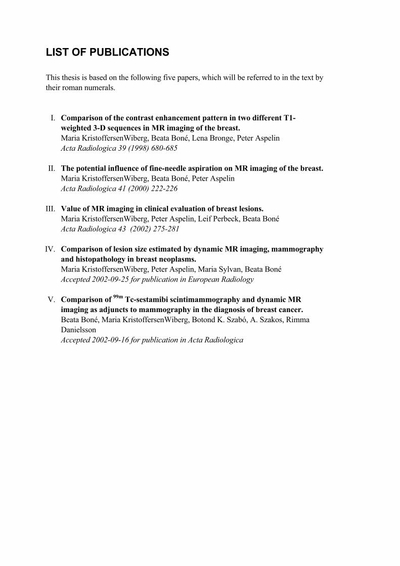
LIST OF PUBLICATIONS This thesis is based on the following five papers, which will be referred to in the text by their roman numerals.
I. Comparison of the contrast enhancement pattern in two different T1-weighted 3-D sequences in MR imaging of the breast. Maria KristoffersenWiberg, Beata Boné, Lena Bronge, Peter Aspelin Acta Radiologica 39 (1998) 680-685
II. The potential influence of fine-needle aspiration on MR imaging of the breast. Maria KristoffersenWiberg, Beata Boné, Peter Aspelin Acta Radiologica 41 (2000) 222-226
III. Value of MR imaging in clinical evaluation of breast lesions. Maria KristoffersenWiberg, Peter Aspelin, Leif Perbeck, Beata Boné Acta Radiologica 43 (2002) 275-281
IV. Comparison of lesion size estimated by dynamic MR imaging, mammography and histopathology in breast neoplasms. Maria KristoffersenWiberg, Peter Aspelin, Maria Sylvan, Beata Boné Accepted 2002-09-25 for publication in European Radiology
V. Comparison of 99m Tc-sestamibi scintimammography and dynamic MR imaging as adjuncts to mammography in the diagnosis of breast cancer. Beata Boné, Maria KristoffersenWiberg, Botond K. Szabó, A. Szakos, Rimma Danielsson Accepted 2002-09-16 for publication in Acta Radiologica
CONTENTS Introduction..........................................................................................................1
Background....................................................................................................1 Epidemiology ..........................................................................................1 Risk factors ..............................................................................................1
Breast diagnostic methods.............................................................................2 Clinical breast examination.....................................................................2 Mammography ........................................................................................3 Ultrasonography ......................................................................................3 Triple diagnosis .......................................................................................3 Computed tomography............................................................................4 Nuclear medicine imaging techniques....................................................4 Other methods .........................................................................................4
Magnetic resonance imaging.........................................................................5 History and background ..........................................................................5 Magnet types ...........................................................................................6 Basic principles of MRI ..........................................................................6 Contrast agents ........................................................................................7
Magnetic resonance imaging of the breasts ..................................................8 Surface coils ............................................................................................8 Artefacts...................................................................................................9 Spatial resolution...................................................................................11 Temporal resolution ..............................................................................12 The development of sequences .............................................................13 Non contrast enhanced breast MR imaging..........................................14 Contrast enhanced breast MR imaging.................................................15 Contrast agent injection.........................................................................18 Image evaluation ...................................................................................18 Current developments ...........................................................................25 MRI guided biopsies .............................................................................26
When to perform MRI of the breasts ..........................................................27 Aims ...................................................................................................................29
General aim..................................................................................................29 Specific aims................................................................................................29
I. .............................................................................................................29 II. ............................................................................................................29 III............................................................................................................29 IV. ..........................................................................................................29 V.............................................................................................................29
Material and methods.........................................................................................30 Subjects ........................................................................................................30
Study I....................................................................................................30 Study II ..................................................................................................30 Study III, IV and V................................................................................30
Methods........................................................................................................31 Study I....................................................................................................31
Study II .................................................................................................. 32 Study III, IV and V................................................................................ 32 Statistical methods ................................................................................ 33
Main results........................................................................................................ 35 Study I.......................................................................................................... 35 Study II ........................................................................................................ 35 Study III ....................................................................................................... 35 Study IV....................................................................................................... 35 Study V ........................................................................................................ 35
Results and methodological discussion ............................................................ 36 Study I.......................................................................................................... 36 Study II ........................................................................................................ 37 Study III ....................................................................................................... 37 Study IV....................................................................................................... 39 Study V ........................................................................................................ 42
Discussion.......................................................................................................... 45 Study I.......................................................................................................... 45 Study II ........................................................................................................ 46 Study III ....................................................................................................... 46 Study IV....................................................................................................... 48 Study V ........................................................................................................ 49
Conclusion ......................................................................................................... 51 Acknowledgements ........................................................................................... 52 References.......................................................................................................... 54
LIST OF ABBREVIATIONS AUC Area under the curve BCS Breast-conserving surgery BI-RADS Breast imaging reporting and data system BSE Breast self examination CBE Clinical breast examination CE Contrast enhanced CE-MRI Dynamic contrast enhanced magnetic resonance imaging CT Computer tomography DCIS Ductal cancer in situ EIC Extensive intraductal component EIS Electrical impedance scanning FA Flip angle FISP Fast imaging with steady-state precession FLASH Fast low angle shot FNA Fine needle aspiration FOV Field of view Gd-DTPA Gadopentetate dimeglumine HRT Hormone replacement therapy IDC Invasive ductal carcinoma ILC Invasive lobular carcinoma IR Inversion recovery ITC Invasive tubular carcinoma LCIS Lobular cancer in situ MPRAGE Magnetization prepared rapid gradient echo MRE Magnetic resonance elastography MRI Magnetic resonance imaging MRS Magnetic resonance spectroscopy MRT Magnetic resonance tomography NMR Nuclear magnetic resonance PD Proton density PET Positron emission tomography ROC Receiver-operating-characteristic RODEO Rotating delivery of excitation off-resonance ROI Region of interest SE Spin echo TA Time of acquisition TD Triple diagnosis TE Echo time TR Repetition time US Ultrasonography USPIO Ultrasmall superparamagnetic iron oxide particles

1
INTRODUCTION BACKGROUND
Epidemiology
Today breast cancer is the most common cancer among women, and the second most common of all cancers, lung-cancer being the most common overall. In the year 2000 almost 1 million new cases, 22% of all cancers in women were diagnosed worldwide. There were 375 000 deaths from breast cancer, of which more than half occurred in industrialized countries (123). In the United States it has been estimated for the year 2001 that 192 000 women will get the diagnosis breast cancer, 40 600 with in situ carcinoma, and 40 200 will die of breast cancer (17). Breast cancer is an uncommon malignancy in men, and represents less than 1% of all breast cancers. The 1-year survival rate in Europe in the year 2000 was 91% and at 5 years 65%, while in the USA the overall 5-year survival for localized disease was 96,8% but only 20,6% for patients with distant metastases (16, 123). In Sweden approximately 27 % of all female cancers are breast cancers, and each year 5800 new cases are diagnosed. The incidence, which increases with age, has since the 1970s increased by 2% per year until the 1990s when a decline was noted (124). During the same period the survival-rate has been unchanged or even increased. This is due to early detection of smaller and more frequent node-negative tumours, achieved by regular controls including mammographic screening and breast self examination (BSE), but also continuously improved treatment methods (89). Risk factors
One risk factor for developing breast cancer is genetic heritage, the genes BRCA1 and BRCA2, but also genes involved in oestrogen metabolism increases the risk. The most important factors influencing the risk of developing breast cancer are reproductive and hormonal factors with increased risk by early menarche, late age at first birth, low parity and late menopause. Oral contraceptives give a small rise in risk while taken, and the risk disappears 10 years after cessation of contraception. In the same way hormone replacement therapy (HRT) increases the risk in long-term use, with a greater risk if used in combination therapy with progestin. The Swedish Council on Technology Assessment in Health Care (SBU) in May 2002 presented a review on HRT, where they stated that the risk of developing breast cancer increases by years of HRT, and decreases after ceased treatment. The addition of gestagens does not decrease the cancer risk, but oestrogen-treatment-induced cancers seem to have a less malignant course than breast cancers not associated to HRT (146). High socio-economic status, increasing age up to 50 years of age, higher alcohol intake and obesity after menopause are other factors associated with increased breast cancer risk (93, 94, 123). In Sweden a correlation with breast cancer and socio-economic status has been shown in a study of 1462 women over a 24-year period. Higher socio-economic status
2
correlated to increased risk in all-site cancer mortality and elevated breast cancer morbidity, while the risk of cardiovascular disease was decreased (19). No preventive method for breast cancer has yet been defined. Special attention and control can so far only be planned among the known hereditary cancers. Therefore early detection of cancer is needed if an improved survival-rate or possible cure is to be achieved. Good diagnostic methods are necessary, not only for early detection but also for determining lesion type, size and assessment of spread to regional lymph nodes as well as distant metastases. In the choice of therapeutic approach and when breast-conserving surgery is considered this evaluation is particularly important. At present there is ongoing research to find out which factors cause the start and development of a breast cancer. There might be several factors involved considering that breast cancer is a complex and heterogeneous disease. Polyak (126) discusses the theory of the natural history of breast cancer as a progressive sequence through several defined clinical and histopathological stages. The series of events range from the normal cells via hyperplasia and atypical ductal hyperplasia progressing into in situ carcinoma and invasive carcinoma, to finally end up in a metastatic disease. A fully developed cancer is defined to exhibit sustained cell proliferation, disregard of growth and differentiation controlling signals, resistance to apoptosis, ability to invade surrounding tissue and induction of angiogenesis. All these factors can develop at different stages, maybe from different causes, and are present in pre-invasive states of cancer. A presently investigated hypothesis suggests that myoepithelial cells surrounding the mammary ducts can be responsible for limiting tumours to be invasive, as loss of this cell type only exists in invasive breast tumours thus permitting invasion and tumour progression. At present it is also considered that fibroblasts of mammary tissue, and possibly also macrophages, eosinophile granulocytes, endothelial cells, as well as mammary epithelial stem cells might be of importance in the cancer progression. A theory is presented that cancer is not just caused by the tumour cell itself and maybe not by a group of abnormally growing cells but represents an abnormal organ with multiple cell types communicating with each other. Therefore further research to find the causes pursuing cells to start a cancerous growth is necessary, also so that new treatments and effective prophylactic measurements could be defined (126). BREAST DIAGNOSTIC METHODS
Several methods have been used and studied in breast diagnostics, which will be shortly described below. Clinical breast examination
Clinical breast examination (CBE) or breast self examination (BSE) with palpation of breasts and regional lymph nodes is the basis for all evaluations. Breast lesions smaller than 1 cm are usually very difficult or impossible to find at palpation, in particular in large breasts, when lesions are located deep in the breast parenchyma and in younger women. Palpation is a subjective measurement of size of a lesion and there is a tendency to define the sizes in ranges not closer than 0,5-1 cm (153). Therefore BSE, although important, is of limited value in finding the small, early cancers. There is still a place for regular BSE for individual women, as a palpable mass will lead to further
3
investigation and clinical mammography, and a few percent of breast cancers (verified by cytology or surgery) will be detected solely by BSE as a palpable mass while having a normal mammogram (17). In the clinical evaluation a thorough palpation of the breasts and regional lymph node stations by an experienced surgeon or oncologist is of great importance. Mammography
Mammography is currently the only imaging modality suitable for screening as well as for an evaluation of patients with clinical symptoms. It is also a commonly available method with established criteria for the evaluation and performance of examinations. Mammography has a high sensitivity to detect malignancies in fatty breast tissue and also is the best method for detecting microcalcifications. However, in dense breast parenchyma it can be difficult to diagnose a malignant lesion, although microcalcifications are readily seen (77, 145). During many years experience has increased and a recommendation from the American college of radiology on how to interpret mammography has been used for many years, BI-RADS (Breast imaging reporting and data system) (3). Mammography is the most important method in breast diagnostics since many years. The now developed digital imaging gives more possibilities for mammography and will be of great diagnostic advantage, not only by decreasing the radiation doses. Ultrasonography
Ultrasonography (US) is frequently used as a complementary method for distinguishing cysts from solid lesions and as an aid at punctures (fine-needle or core biopsy), but it has not been useful as a screening method (66). With the development of small-part transducers US combined to mammography improve the detection rate of breast cancer, especially in younger women with dense parenchyma where mammography has known limitations. The use of colour Doppler US analysing the blood flow of a lesion is a new method, still being evaluated (9). The use of contrast media in US, which also is being evaluated, might improve the diagnosis and give new opportunities for localized treatment. Because of interpreter variability US is difficult to reproduce, and the evaluations are dependent on experience of the interpreter. During recent years studies have been performed to explore reliable characteristics of breast masses at US and to define signs typical of benign respective malignant lesions. So far US cannot be recommended for screening, not even in women with dense parenchyma, but is of adjunctive value in differentiation of diagnosed lesions (mammographic or palpable). However, biopsy of a lesion still has to be performed for best definition, and cannot be excluded in any work-up of breast masses (102, 128, 157). Triple diagnosis
A commonly used routine evaluation of breast lesions is called triple diagnosis (TD), which includes a combination of clinical breast examination, mammography and cytological examination of fine-needle-aspirations (47). This is at present the main routine investigation for a breast lesion in Sweden. In some cases where cytological
4
evaluation is difficult (for example in fibrotic lesions poor of cells) a core-biopsy is necessary to get a larger sample for a histopathological evaluation. Computed tomography
Chang tried Computed Tomography (CT) in breast examinations in 1977. He found the use of contrast media necessary and it has been confirmed that CT cannot be used as a screening tool for breast malignancies. However it can add useful information regarding recurrent malignancy, parasternal metastases or chest wall/skeletal engagement of breast malignancies. Clinically unsuspected lesions can thus be diagnosed and CT can be of value when choosing therapy and planning the area for radiation treatment (91, 105, 135). A disadvantage in using CT is the high radiation dose and CT should only be used in few selected patients. Nuclear medicine imaging techniques
Since the 1980s breast tumours have been evaluated using isotopes and many different scintigraphic techniques have been tried. During the last decade 99mTc-Sestamibi, initially used for myocardial evaluations, has gathered interest after showing accidental uptake in a breast tumour (72). Even though scintimammography shows uptake in tumours, the signal-to-noise ratio is not sufficient enough to reliably diagnose tumours smaller than 1 cm. Scintigraphy can therefore not be used for screening but can be used as a complementary examination in selected cases, as the specificity is high. A Sentinel-node method has been developed over recent years. Initially a dye was used to mark an area peritumourally or subdermally close to the tumour. Today lymphoscintigraphy or a combination of both methods is used for detection of the first lymph node to drain lymphatic from the tumour. With this method it is possible to exclude surgery of axillary lymph nodes if no metastases are diagnosed in the Sentinel node peri-operatively (37). Positron emission tomography (PET) is another method showing metabolic characteristics, most often using FDG (fluorodeoxyglucose), which also can be applied in breast tumours and metastasising disease (86). Although it is a sensitive method it is expensive and technically complex, available only in few centres. Because of the low availability and the low spatial resolution PET is at present used only in a few, selected cases (56, 132). Other methods
Thermography and transillumination are methods initially thought to be valuable in breast diagnostics, in particular as it uses no radiation. Initially there were findings supporting the use of thermography as a prognostic indicator (31, 64). Evaluations have shown thermography to have unacceptably high numbers of both false-positive and false-negative results and cannot be used as an independent prognostic indicator (108, 141, 142), thus not applicable in the diagnosis and detection of breast lesions. Transillumination was described during the 1920-30s, using red and infrared light to assess the reflection, scattering and absorption of the light when passed through breast tissue. During the past 50 years the method has improved by new techniques and
5
computer manipulation (2, 67, 68, 137), but is at present of no clinical use as its sensitivity is much lower than mammography, and since it is not able to identify small lesions and lesions situated deep in the breast parenchyma. The latest development is the use of electrical impedance scanning (EIS) using the quantifiable parameters conductance and capacitance, which has been studied at few sites. This method is thought to recognize the changed metabolism in the cancer cells, changes of cellular water content and amount of extracellular fluid as well as membrane properties, changed orientation of malignant cells, destruction of tight junctions and cell membranes. However the initial results are not promising and this method cannot in the present form be recommended for use in clinical breast diagnosing (95, 96, 100, 155). MAGNETIC RESONANCE IMAGING
History and background
Two scientists in the United States performed the first successful nuclear magnetic resonance experiment independently in 1946. Edward Purcell and Felix Bloch found that certain nuclei when placed in a strong magnetic field absorbed energy in the radio frequency range of the electromagnetic spectrum. This energy was re-emitted when the nuclei reverted to their original state in the magnetic field. Earlier Sir Joseph Larmor (Irish physicist, 1857-1942) had demonstrated that the strength of the magnetic field and the radio frequency matched each other, known as the Larmor relationship. This means that the angular frequency of precession of the nuclear spins is proportional to the strength of the magnetic field. Already in 1937 an American physicist, Dr Isidor Rabi, had come across the NMR-experiment when inventing the atomic and molecular beam magnetic resonance method of observing atomic spectra, for which he was awarded the Nobel Prize for Physics in 1944. At the time he disregarded the importance of the NMR experiment, as he thought it was an artefact. Bloch and Purcell termed the phenomenon NMR, Nuclear Magnetic Resonance. Their discovery started the use of NMR in spectroscopy of chemical compounds, which during the 1950s and 1960s became a widely used non-destructive technique for analyzing small samples. The Magnets used for these analyses at microscopic level were small, with a bore diameter of only a few centimetres. For their discovery Bloch and Purcell were awarded the Nobel Prize for Physics in 1952. An American MD, Raymond Damadian (22), in 1970-71demonstrated that tissue parameters (T1 and T2 relaxation times) could be measured in vitro by NMR, differing between tumours and normal tissues. This variation in relaxation times of neighbouring tissues in the MR-image is the basis for the exceptional tissue contrast using MRI compared to X-ray and CT. In 1973 Paul Lauterbur, Professor of Chemistry, published a new imaging technique, which he called zeugmatography (from Greek zeugmo meaning yoke or a joining together). He described the technique basic for spatial localization of objects by joining together a weak gradient magnetic field with the stronger main magnetic field. Dr R.R. Ernst (awarded the Nobel Prize in Chemistry in 1991) in 1974 described the application of Fourier transformation, which is a method of processing that converts
6
raw data that forms the MR-image into a viewable, understandable and interpretable image. Since then the development has been, and still is, continuously moving forward. It seems that everything previously thought impossible eventually becomes possible by using MRI, through new methods, new sequences, new contrast agents and new ways of processing achieved data. Magnet types
In medical imaging there are three different types of magnets used. All of them are producing homogenous magnetic fields. Permanent magnets: These types of magnets are based on the common bar magnet, requiring no electric current or cryogen, hence requiring very little maintenance. The magnetic field is vertically oriented within the bore of the magnet. The strength of these magnets usually is 0.1-0.4 T. Permanent magnets have a wider bore than superconductive, and hence can examine persons with claustrophobia or broad shoulders. Resistive magnets: The magnetic field is produced of a current sent through coils surrounding the bore. This requires constant power supply to keep the magnetic field, making these magnets more expensive to maintain, but no cryogens are required. These are the only type of MR scanners that can easily be switched on and off. Superconductive magnets: These are the most common magnets in medical use. Superconductive magnets have coils carrying a current in the same way as resistive magnets, but the superconductive coils are cooled by cryogens, so that the resistance in the coils will be close to zero, to prevent the current to decay. Thus, in this type of magnets there is a constant need for cryogens but no need for continuous power supply. The magnetic field is in line with the long axis of the bore direction, horizontally. Superconductive magnets operate clinically at the moment up to 3 T, but 7 T are developed for human examinations and are so far used for research. In research settings magnets with a field strength of more than 11 T are now used. Basic principles of MRI
The basis for the NMR phenomenon is the ability of the nucleus of an atom to absorb energy from radio waves when placed in a magnetic field. MRI for medical diagnostic purposes is based on the hydrogen nuclei that are the most abundant element in the body, accounting for about 70% of all the atoms in the body, including the hydrogen of water molecules. Magnetic resonance is sensitive to the relative concentration of hydrogen in different tissues and fluids as well as to their relation to surrounding nuclei and macromolecules. When placed in a strong magnetic field the hydrogen protons will align parallel or antiparallel to the main magnetic field vector, in contrast to the normally random orientation. In the strong magnetic field slightly more than 50% of the protons will align parallel to the magnetic field and thus create a net magnetization in that direction. This small net magnetization is making it possible to acquire MR images. The spin of the nucleus will align along the main magnetic field, called precession, which is dependent on the field strength and called the Larmor frequency. A radio-frequency
7
pulse (RF) is used to excite the protons with a frequency matching the protons of interest, causing a deflection of the net magnetization. The process when the nuclei return to their equilibrium state is called relaxation. There are two types of relaxation, longitudinal or transversal, called T1 and T2. Both parameters, T1 and T2 relaxation, are used to create the desired contrast in the images by choosing appropriate repetition times (TR) for the RF-pulses and echo times (TE) for collecting the emitted RF-pulse from the protons. Images can be T1-, T2- or proton density-weighted. A typical spin echo (SE) T1-image has a short TR and TE, and a typical SE T2-image has a long TR and TE. At present there are many different types of sequences used with different combinations of RF-pulses, TR, TE, flip angle and refocusing pulses to get as good coverage as possible in as short a time as possible with good T1- or T2-contrast. The gradients of the magnet are used in three directions to spatially localize each element of the examined part. One gradient is used for the choice of slice, where the RF-pulse excites the protons with the suiting frequency. The other two gradients are used for spatial encoding in the remaining two directions, using phase and frequency encoding. All these gradients are used in combination so that also oblique images in any desired direction can be acquired. Many slices can be acquired during the same TR, making the amount of slices possible to acquire dependent on the used TR as well as the TE. Instead of collecting data from single slices a whole volume can be excited by the RF-pulse, 3D-imaging, making it possible to reconstruct very thin slices and create reconstructions in any chosen plane in the postprocessing of the images. To make examination times shorter sampling of the acquired data into the k-space can be reduced in different ways, for example by using rectangular FOV, half-Fourier and keyhole. The spatial information is sampled at the periphery of the k-space, and the contrast of the images is sampled at the centre of k-space. Different sampling methods therefore can have an impact mainly on spatial resolution or contrast of the image. Contrast agents
Initially when MR imaging was developed no contrast medium was thought to be necessary as there were possibilities to create a great variety of sequences with different native contrast. In the early 1980s the first experiences of using paramagnetic compounds as contrast agents were made. Paramagnetic compounds have at least one unpaired electron, which has a magnetic movement much stronger than that of the proton. Iron particles are used as superparamagnetic substances to get as much T2∗ shortening as possible, as the interaction between several ferrum ions in the particles results in a much larger total magnetic moment than the individual atoms, causing a larger effect on T2∗ than on T1 compared to individual paramagnetic compounds. Therefore the relaxation times of the tissues will be shortened due to the effect on the local magnetic field at the atomic level. As the effects are indirect, the term contrast agent is used, and not contrast medium as for iodine in X-ray examinations. Both positive and negative contrast agents have been developed, positive mainly affecting the T1-value and negative mainly affecting the T2-value. During the 1990s many different gadolinium based contrast agents were developed, as well as some ferrum- and manganese-based. The most common used contrast agents are intravenously administered gadolinium in different complexes having a fast passage
8
through the vessels to the extracellular space. Gadolinium-based contrast agents are excreted via the kidneys, but have no deleterious effect on renal function in the concentrations presently used in routine examinations (10-20 ml of 0,5 mmol gadolinium/ml). Specific contrast agents are also administered intravenously. For example for selective uptake in Kupfer cells of the liver and spleen of iron based contrast agents as well as manganese-based contrast agent with uptake in liver cells and subsequent excretion into the biliary tracts. Contrast agents are also used for oral administration with either positive or negative effect, to fill the enteral tracts and facilitate the differentiation of bowl from surrounding tissues or tumours. (32, 112, 140). MAGNETIC RESONANCE IMAGING OF THE BREASTS
During the early 1970s reports by Damadian (22) of tissue characterization using NMR raised hopes that malignant tissues could be separated from normal tissues, also in the breast. In 1978 they published studies of breast specimens as well as lung and colon specimens, including malignancies, to create a normalised NMR malignancy index (41, 78). Mansfield et al in 1980-81 (6, 98) and Ross et al in 1982 (130) were among the first to obtain in vivo MR images of human breast. They used the body-coil which resulted in both poor spatial as well as temporal resolution and signal-to-noise ratio. Therefore it reached no clinical use until the developed local surface-coils were introduced by for example Axel (4) in 1984. Already in 1983 El Yousef et al (30) had tried a surface coil and 3D technique to improve breast MR imaging. The evaluation of data was focusing on the possibility to use the tissue parameters T1, T2 and proton density by calculating data or direct measurement of signal intensities in the images using appropriate chosen pulse sequences. During the early and mid 1980s the spatial resolution, signal-to-noise ratio and selection of appropriate sequences improved image quality significantly (52,53, 69). Although fibrosis and cysts could be diagnosed using these parameters, the inability to detect and diagnose malignancies at MRI still remained. The improved image quality during this period made it possible to examine breast implants, and diagnose implant failure with high accuracy, which still remains an important indication for breast MRI. The break-through for modern MR-mammography was the introduction of contrast enhanced imaging of the breasts in 1986 by Heywang et al (54), using the first paramagnetic contrast agent approved for clinical use, Gd-DTPA (gadopentetate dimeglumine). With this technique enhancing tissues were visualized as areas with signal increase at T1-images, due to the paramagnetic effect of Gadolinium. Since then many studies have been performed evaluating the methods, and currently a group of specialists in MRI of the breasts are elaborating a lexicon on how to interpret and perform gadolinium enhanced MR-mammography (62, 74, 114, 133). Surface coils
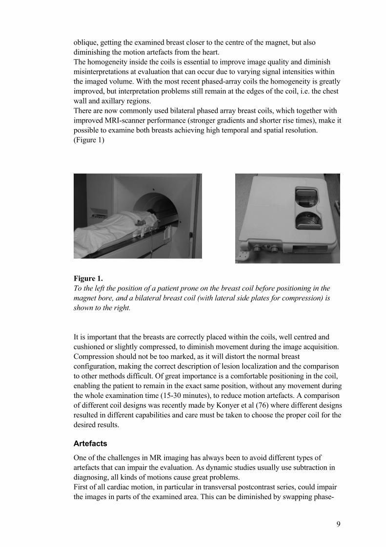
Ideally both breasts should be examined at the same time with high spatial and temporal resolution. The first built coils could only achieve a high resolution covering one breast at a time. The single breast coil had the advantage of positioning the patient
9
oblique, getting the examined breast closer to the centre of the magnet, but also diminishing the motion artefacts from the heart. The homogeneity inside the coils is essential to improve image quality and diminish misinterpretations at evaluation that can occur due to varying signal intensities within the imaged volume. With the most recent phased-array coils the homogeneity is greatly improved, but interpretation problems still remain at the edges of the coil, i.e. the chest wall and axillary regions. There are now commonly used bilateral phased array breast coils, which together with improved MRI-scanner performance (stronger gradients and shorter rise times), make it possible to examine both breasts achieving high temporal and spatial resolution. (Figure 1)
Figure 1. To the left the position of a patient prone on the breast coil before positioning in the magnet bore, and a bilateral breast coil (with lateral side plates for compression) is shown to the right. It is important that the breasts are correctly placed within the coils, well centred and cushioned or slightly compressed, to diminish movement during the image acquisition. Compression should not be too marked, as it will distort the normal breast configuration, making the correct description of lesion localization and the comparison to other methods difficult. Of great importance is a comfortable positioning in the coil, enabling the patient to remain in the exact same position, without any movement during the whole examination time (15-30 minutes), to reduce motion artefacts. A comparison of different coil designs was recently made by Konyer et al (76) where different designs resulted in different capabilities and care must be taken to choose the proper coil for the desired results. Artefacts
One of the challenges in MR imaging has always been to avoid different types of artefacts that can impair the evaluation. As dynamic studies usually use subtraction in diagnosing, all kinds of motions cause great problems. First of all cardiac motion, in particular in transversal postcontrast series, could impair the images in parts of the examined area. This can be diminished by swapping phase-
10
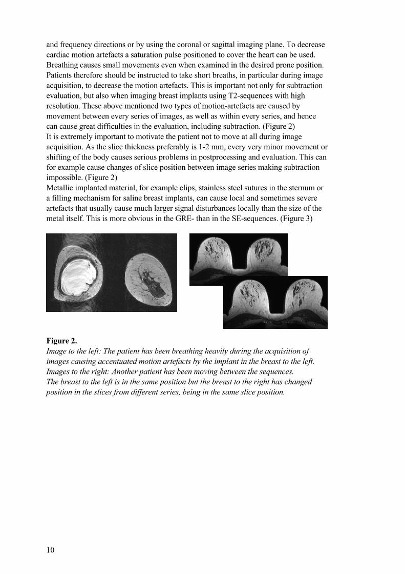
and frequency directions or by using the coronal or sagittal imaging plane. To decrease cardiac motion artefacts a saturation pulse positioned to cover the heart can be used. Breathing causes small movements even when examined in the desired prone position. Patients therefore should be instructed to take short breaths, in particular during image acquisition, to decrease the motion artefacts. This is important not only for subtraction evaluation, but also when imaging breast implants using T2-sequences with high resolution. These above mentioned two types of motion-artefacts are caused by movement between every series of images, as well as within every series, and hence can cause great difficulties in the evaluation, including subtraction. (Figure 2) It is extremely important to motivate the patient not to move at all during image acquisition. As the slice thickness preferably is 1-2 mm, every very minor movement or shifting of the body causes serious problems in postprocessing and evaluation. This can for example cause changes of slice position between image series making subtraction impossible. (Figure 2) Metallic implanted material, for example clips, stainless steel sutures in the sternum or a filling mechanism for saline breast implants, can cause local and sometimes severe artefacts that usually cause much larger signal disturbances locally than the size of the metal itself. This is more obvious in the GRE- than in the SE-sequences. (Figure 3)
Figure 2. Image to the left: The patient has been breathing heavily during the acquisition of images causing accentuated motion artefacts by the implant in the breast to the left. Images to the right: Another patient has been moving between the sequences. The breast to the left is in the same position but the breast to the right has changed position in the slices from different series, being in the same slice position.

11
Figure 3. Metallic artefact due to stainless steel suture in the sternum (transversal image to the left). A metallic artefact caused by the filling mechanism of an implant, placed laterally near the axilla (sagittal image to the right). Spatial resolution
The spatial resolution is critical for detecting small malignant lesions. A thin slice thickness is desired to get as accurate information as possible, but this will lead to a loss of signal, as the imaged voxel is smaller, thus giving away less signal. However, because of the different sizes, forms and extent of lesions it can be difficult to detect certain lesions due to the partial volume effect. For example in small lesions that are not completely covered by one separate slice (especially for malignancies growing in a duct-like pattern), or in a wide but thin invasive cancer in the plane of the slice. (Figure 4) In these cases the enhancement only contributes partially to the signal in the slice (and voxel), making it more difficult to discern a strongly enhancing lesion occupying only a part of the slice thickness. For example Furman-Haran et al (38) found that a reduction in spatial resolution causes an increase of false negatives and a decreased sensitivity. Therefore a compromise has to be made regarding slice thickness, choice of sequence and acceptable signal to noise ratio. A slice thickness of 2 mm is desired, and should not exceed 4 mm, to make it possible to detect malignant lesions of 4-5 mm sizes.
Figure 4. Partial volume effect: a small lesion occupying only a minor part of the total slice thickness, or ductal distribution of the lesion, occupying parts of the slice thickness (in some areas passing within the slice and sometimes crossing the slice).
12
The in plane resolution (the matrix) also decides the voxel size and is of great importance for the signal achieved per voxel. A matrix of 512 X 512 would be desired, but due to the longer acquisition times for higher matrixes and loss of signal in smaller voxels, 256 X 256 is often the clinically matrix used. When using a coronal or sagittal imaging plane a rectangular FOV (field of view) can be used, keeping the voxel size constant while decreasing the number of phase encodings, resulting in a decreased scan time. Using these imaging planes, not only can a higher matrix be used within the same aquisitiontime (TA), but also a decrease of cardiac artefacts is achieved. Ideally the in plane resolution should be 0,5-1 mm for the best evaluation possibilities, including not only enhancement characteristics but also the internal structure of lesions and evaluation of implant integrity. Recently Obenauer et al (116) reported a study of high in-plane resolution (matrix 420 x 512) compared to standard dynamic CE-MRI (matrix 210 x 256). In 51 hypervascularized lesions they found high in-plane resolution provided better visualization of morphologic patterns, but a diagnostic advantage compared to standard MRI was rare. Holland et al (57) reported the use of a very high resolution. For example an in-plane resolution of 117 µm has been used to identify both normal structures of the breast parenchyma and structures typical for pathologic changes in breast specimens. This high resolution provides images that correlate well to low- and high-magnification histological evaluation of the breast tissue. The visualization of malignancies will probably be even more pronounced by the use of contrast-enhancement (gadolinium-compounds) in vivo, but has not yet been proven. Very powerful gradients have to be used to achieve such high in-plane resolution. The at present modern magnets have possibilities to use gradients with strengths in each direction of 25mT/m and rise times of 300 µsec, which could enable pixel sizes on the order of 100 µm and 1 mm slice thickness. Temporal resolution
The temporal resolution is very important in the evaluation of enhancement kinetics. Typically a malignant lesion enhances strongly and early, combined with washout in the later phase. Using long imaging times per acquisition the possibility to discern a malignant from a benign lesion using dynamic parameters diminishes. The rim-enhancement and inhomogeneities in malignant lesions can also be overlooked, as they often are discernible in early post contrast images, but for this evaluation spatial resolution is the most crucial part (111). It is possible to use fast dynamic (1-30 seconds), dynamic (30 seconds – 3 minutes) and semidynamic (3-6 minutes) examinations, as well as using only one pre- and one post-contrast series of images. When deciding which temporal resolution is desired there always has to be made compromises concerning the imaging volume to be covered (i.e. one or two breasts or a part of one breast), which spatial resolution, which slice orientation and what type of sequence should be used. Using fast dynamic examinations result in a decreased coverage of the breasts and lower spatial resolution, in some cases only one slice with slice thickness more than 5 mm is possible if very high temporal resolution (a few seconds) is gained (12). Experiences at both our site (14, 79) and others (59) have shown that the sensitivity and specificity is not necessarily improved by using very fast dynamic sequences. The best
13
temporal resolution for clinical evaluations is at present a dynamic examination with 1-3 minutes acquisition time per sequence, but Buckley (18) showed that the temporal resolution did not seem to be as important as the spatial resolution. For a good evaluation of enhancement kinetics (using a dynamic examination) at least four, up to eight, sequences post contrast should be performed, allowing evaluation of the initial early contrast enhancement as well as the later phases often showing wash-out phenomenon in malignant lesions. Combinations of fast dynamic and dynamic/semidynamic examinations have been used to achieve high temporal resolution during the first minute(s) and high spatial resolution in the pre- and the late post-contrast series. (12, 131) The development of sequences
In the beginning of MRI in breast imaging the use of spin-echo sequences were the only choice. They revealed good tissue contrast enabling good measurements of T1-, T2- and proton density values, but the low spatial and temporal resolution was a drawback. In combination with the experience that tissue parameters alone did not allow differentiation of malignant from benign tissues many sites developed new sequences and methods for examination. El Yousef et al (29) in the early 1980s, using a 0,3 T superconductive magnet, tried to localize breast lesions by using a 3 D T1 spin echo (SE) sequence (TR(repetition time)/TE (echo time) 250/30 msec) with 4 mm slices covering one breast. At the site of the suspected lesion they positioned 8 mm slices using a spin echo sequence with TR/TE 1000/30 msec and 1000/120 msec, followed by an IR (inversion recovery) sequence with TR/TE/TI 1000/30/300 msec. The collected data were used to classify benign and malignant lesions using the morphological appearance, the T1- and T2-values and the signal intensity changes with respect to different sequences. Dash et al (24) used a spin echo sequence varying the TE and TR, so that four different sets of images were acquired (TR/TE 700/35, 700/70, 1600/35 and 1600/120). They found the best clinical use of images closest to T1- respective T2-weighting, and that the breast parenchymal pattern was easily recognised, as in mammography. The disadvantages were long scanning times, high costs, inability to identify microcalcifications and the smallest lesion size possible to evaluate was 5-8 mm. Martin and El Yousef (99) also studied the effect of hormonal changes during menstrual cycle and exogenous oestrogen-progesterone effects on T2-weighted imaging. Transverse relaxation was thought to be important in diagnosing breast malignancies by changes in the water exchange of malignancies. The hormonal effects leading to water retention in breast parenchyma did not prove to cause any significant changes in calculated T2-values, and hence not posing a problem for differentiation of carcinomas. Merchant et al in 1992 (103) reported the use of a mixed imaging sequence applied after lesion localisation by T1- and T2-weighted images. The mixed sequence used was a SE with TR of 680, an IR with TR of 2160 and TE:s 30,60,90 and 120 msec. The evaluation included calculation of pure proton-density, T1 and T2 images and reconstruction of multiecho SE, IR-modulus and IR-real images. They could not verify differentiation between malignant tumours and normal breast parenchyma by solely measuring T1 and T2 values, but elevated T1 and T2 values in benign tumours distinguishing them from normal parenchyma.
14
The difficulties to find a sequence with high sensitivity and specificity to detect malignancies of the breast still remained until the development of contrast enhanced MR imaging. Today the only remaining indication for non-enhanced MRI is the evaluation of implants. Non contrast enhanced breast MR imaging
At mammography implants are of high density and can obscure the surrounding tissues and breast parenchyma, even when using special techniques, making both diagnosis of implant rupture and exclusion of malignancy difficult. Ultrasound can detect lesions and implant ruptures, to some extent, but cannot reveal the dorsal areas behind the implant. MRI has many advantages in evaluation of breast implants, and has become even more important due to the increased use of cosmetic breast surgery using different kinds of implants, but also due to the increased use of reconstructive surgery with implants after mastectomy or breast conserving surgery. Different materials are used to fill the lumen of breast implants. Saline and silicone gel are more common in Sweden, but also oils and colloids have been used. The shell of a breast implant is made of polyurethane, and the surface can be smooth or textured. The implant is positioned subglandular or subpectoral/submuscular, and can contain a single or double lumen. The body forms a fibrous capsule surrounding the implant, and this capsule can in some women cause contractures. The latter can sometimes be caused by silicone bleeding into the surrounding tissues, causing granulomas. Ruptures of implants are mainly divided into intracapsular and extracapsular. The intracapsular implant rupture is the most common, and is characterized by a rupture of the implant shell but an intact fibrous capsule. An intracapsular rupture is not of clinical importance as long as the fibrous capsule is intact. A rupture of both the implant shell and the fibrous capsule characterize the extracapsular implant rupture, with silicone extruding into the surrounding tissues and sometimes migrating in the body, while extruded saline gets absorbed by the body. (Figure 5)
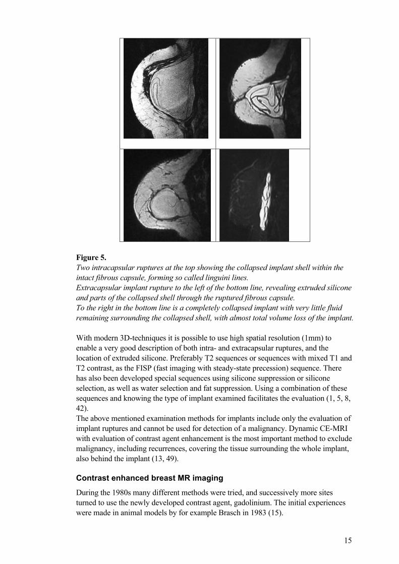
15
Figure 5. Two intracapsular ruptures at the top showing the collapsed implant shell within the intact fibrous capsule, forming so called linguini lines. Extracapsular implant rupture to the left of the bottom line, revealing extruded silicone and parts of the collapsed shell through the ruptured fibrous capsule. To the right in the bottom line is a completely collapsed implant with very little fluid remaining surrounding the collapsed shell, with almost total volume loss of the implant. With modern 3D-techniques it is possible to use high spatial resolution (1mm) to enable a very good description of both intra- and extracapsular ruptures, and the location of extruded silicone. Preferably T2 sequences or sequences with mixed T1 and T2 contrast, as the FISP (fast imaging with steady-state precession) sequence. There has also been developed special sequences using silicone suppression or silicone selection, as well as water selection and fat suppression. Using a combination of these sequences and knowing the type of implant examined facilitates the evaluation (1, 5, 8, 42). The above mentioned examination methods for implants include only the evaluation of implant ruptures and cannot be used for detection of a malignancy. Dynamic CE-MRI with evaluation of contrast agent enhancement is the most important method to exclude malignancy, including recurrences, covering the tissue surrounding the whole implant, also behind the implant (13, 49). Contrast enhanced breast MR imaging
During the 1980s many different methods were tried, and successively more sites turned to use the newly developed contrast agent, gadolinium. The initial experiences were made in animal models by for example Brasch in 1983 (15).
16
In breast MRI Heywang Köbrunner (54) made the initial experiences using gadolinium intravenously in 1986, and continued to develop the method during the coming years (55, 49, 48). Many sites soon used gadolinium as a contrast agent as this method was found to be superior to non-enhanced breast MRI in detecting malignancies. During the 1990s the examinations also developed from semidynamic to dynamic and/or fast dynamic. As the use of a contrast agent proved to more readily discern malignant from benign lesions the choice of sequence became important in another sense. A T1-weighted sequence with optimal sensitivity for gadolinium and ideally not as strong a signal for fat tissue was desired. The use of fat suppression or subtraction in the post processing was found necessary, as the fat signal is strong in T1-weighted sequences and might conceal a small contrast-enhancing lesion. Why are contrast enhancement techniques preferred?
At the initial use of gadolinium as an intravenous contrast agent any pronounced contrast enhancement was considered indicative of a pathologic finding. As experience grew, several factors were found important in the evaluation to differ benign and malignant lesions from the normally enhancing breast parenchyma, which often tended to have overlapping contrast enhancement patterns. First of all the dynamic sequence of enhancement was determined important, and recently the time-signal intensity curves have been found more specific than the enhancement rate in the early/first postcontrast measurement. The evaluations include peak time, presence of washout and the appearance of the curve. In relation to the increased spatial resolution and development of new sequences evaluation of the morphology of the lesions became possible. The internal architecture as well as the border delineation in the enhanced images has proved to be important in diagnosing malignancies, using the same principles as in mammographic evaluation of lesions (61). Which methods should be used?
Techniques facilitating detection of contrast enhancement
During the years many different techniques have been tried, often using fat suppression. One example is RODEO (rotating delivery of excitation off resonance), which used by Harms et al (45) has shown good accuracy, but has not reached widespread clinical use due to special technical requirements to construct the sequence. Pierce et al (125) tried a 3D-sequence with fat suppression and magnetization transfer contrast to improve lesion conspicuity for gadolinium-enhancing lesions, which only could be used as a complement because of long acquisition-times and only one pre- and post-contrast series was performed. Gilles et al (40) proved the use of subtraction, using an ordinary SE sequence for dynamic examination as well as for pre- and post-contrast series. The use of a SE sequence with 3 mm spatial resolution limited the examination to 10 slices to get a temporal resolution of 47 seconds, hence covering only a part of the breast. Therefore this has not been an alternative for routine examinations. The sensitivity for gadolinium as a contrast agent in different types of sequences has been studied. This was described by M. Deimling, Siemens, Erlangen, Germany showing curves at 1 T for 2D and 3D FLASH using different FA, and at 1,5 T for SE,

17
turboFLASH, MPRAGE and 3D FLASH at increasing tissue concentration of gadolinium contrast agent. Ideally the signal intensities of the tissue should increase linearly with tissue concentration of gadolinium contrast agent, otherwise there will be an increased risk of both false positive and false negative diagnoses. In both comparisons the 3D FLASH had the most linear curve with increasing concentration of Gd-DTPA. Usually the GRE sequences create intermediate signal intensity from fat, facilitating detection of enhancing lesions in the breast, making evaluation possible even without subtraction. Even though many sequence-types are used to facilitate delineation of enhancing lesions, there always must be a comparison between pre- and post-contrast series. Because of persisting problems of intermediate to high signal intensities from surrounding fat, the subtraction technique is frequently used in up to date evaluations. When using a subtraction technique the possible impairment of evaluation by motion artefacts must be considered, and when motion artefacts are present subtraction cannot be used in the evaluation. Techniques for improving temporal and spatial resolution
Initially the contrast enhancement was evaluated only in one pre- and one post-contrast series, which was possible with ordinary SE T1 sequences with long acquisition times. The findings stressing the dynamics of contrast enhancement, with varying wash-in and wash-out rates in malignant and benign lesions, demanded several post contrast series within the same period of time for improved evaluation. Hence the need for shorter acquisition times urged faster sequences, often gradient echo sequences, and the development and evaluation of dynamic examinations evolved. So-called keyhole techniques for two-dimensional sequences to achieve better temporal resolution have been tried, but at the cost of spatial resolution as well as an increased risk for mixing of signals from the previous slice. (21) The three-dimensional gradient echo sequences have so far proven to be the most useful in breast MRI, with possibilities to get both high spatial and temporal resolution. The sensitivity to gadolinium contrast agents is also very good, enabling the use of lower doses of contrast agent. This was studied by Kaiser et al (70) who showed that 0,1 mmol/kg was sufficient for diagnostic purposes in a 3D gradient echo sequence, instead of 0,2 mmol/kg that often was applied when using T1-weighted SE sequences. They stated that the higher dose resulted in “overinjection”, as they found a greater increase in signal intensity in enhancing areas in gradient echo images when using a 50% reduced contrast dose. This was confirmed by Oellinger et al (117), when they found MR (using a 3D sequence and the recommended standard dose of gadolinium intravenously) to be superior in detecting multicentricity in patients scheduled for surgery of a single diagnosed lesion in the breast. We found the same effect of different contrast doses when comparing our semidynamic and dynamic studies, see discussion study III. (79) An echo-planar sequence was used by Hulka et al (60) to achieve a temporal resolution of 6 seconds, but the slice thickness had to be 7 mm. This enabled good differentiation between benign and malignant lesions. The use of dynamic imaging has made evaluation of time-intensity curves possible. The importance of time-intensity curves profiles in the differentiation between malignant and benign lesions has been proven to be more specific than the use of fixed threshold
18
values in the first postcontrast series (80, 119). However the evaluation of morphology and internal architecture of the lesions still is important in the primary evaluation (7, 61, 114, 121) Combined techniques
Several sites have tried to use pre- and postcontrast images covering the whole breast combined with dynamic images covering the chosen lesion. For example Boetes et al (12) using a 3D MPRAGE (magnetization-prepared rapid gradient-echo) sequence in combination to turbo-FLASH (fast low angle shot), Mussurakis et al (111) using T1-series pre- and postcontrast combined with dynamic series, in difference to Stomper et al (143) and Stack et al (139) using spoiled gradient-recalled echo (SPGR or GRASS) for their dynamic evaluation combined with T1- and T2- series to localize the lesion centre. Recently Sardanelli et al (131) used 2D T1-weighted FLASH as pre- and post-contrast series, but also for a lesion-targeted dynamic sequence with 13-sec acquisition. Using these combined methods stresses the difficulties to find the exact location for placing the few and often thicker slices through the suspected lesion, to gain the possibility for high temporal resolution during the dynamic series. Boetes et al (12) also used the correlation to the contrast agent’s first pass through the aorta, for exact measuring of the time for contrast to pass to the lesion in the breast. The criterion for a lesion to be malignant when they used this technique was set to < 11,5 seconds after aortic enhancement. Kvistad et al (84) used a combination of a dynamic examination followed by a T2* weighted gradient echo sequence through the most enhancing area in the dynamic series. This T2* sequence made evaluation possible of the perfusion during 30 seconds in conjunction to a second contrast agent injection. They found a higher specificity using the T2* sequence and found the combination to have a potential to improve clinical utility of MRI of the breasts. Contrast agent injection
The intravenous injection should be performed as quickly as possible and as consistently as possible, ideally using a contrast injector, immediately followed by a saline injection (preferably 20 ml) to ensure the contrast bolus will be as tight as possible. The intravenous access needs to be in a vein large enough not to rupture at the injection. As the injection should be performed with the patient in position at the centre of the magnet a long line is needed to connect to the injector outside the magnet bore. A tight bolus of contrast agent is desired to get as good an evaluation of the initial contrast enhancement as possible, which is very important in the differentiation between benign and malignant lesions. This performance facilitates reproduction of imaging conditions between examinations, although the effect of varying heart rate, heart contractions as well as vessel resistance can make a difference in the contrast passage through the vessels. Image evaluation
All information acquired should be used and evaluated. For accurate evaluation of the MR-examination all previous investigations and results, including patient history and mammography, should be at hand.
19
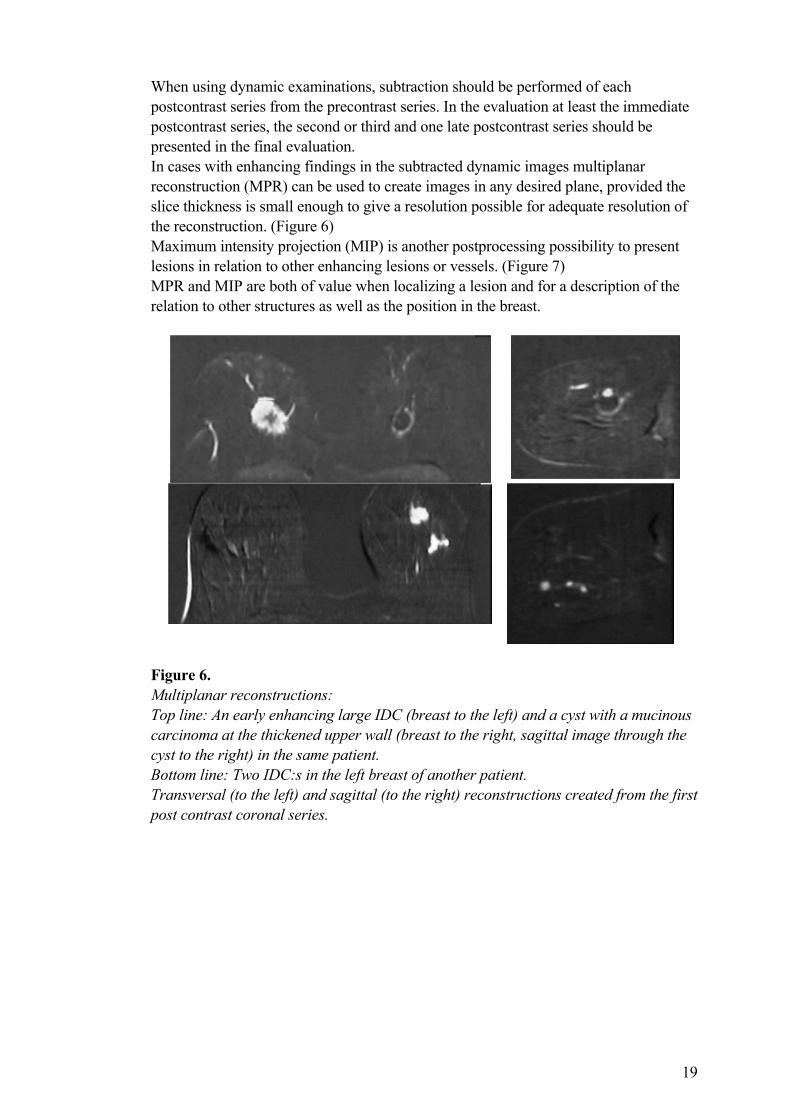
When using dynamic examinations, subtraction should be performed of each postcontrast series from the precontrast series. In the evaluation at least the immediate postcontrast series, the second or third and one late postcontrast series should be presented in the final evaluation. In cases with enhancing findings in the subtracted dynamic images multiplanar reconstruction (MPR) can be used to create images in any desired plane, provided the slice thickness is small enough to give a resolution possible for adequate resolution of the reconstruction. (Figure 6) Maximum intensity projection (MIP) is another postprocessing possibility to present lesions in relation to other enhancing lesions or vessels. (Figure 7) MPR and MIP are both of value when localizing a lesion and for a description of the relation to other structures as well as the position in the breast.
Figure 6. Multiplanar reconstructions: Top line: An early enhancing large IDC (breast to the left) and a cyst with a mucinous carcinoma at the thickened upper wall (breast to the right, sagittal image through the cyst to the right) in the same patient. Bottom line: Two IDC:s in the left breast of another patient. Transversal (to the left) and sagittal (to the right) reconstructions created from the first post contrast coronal series.
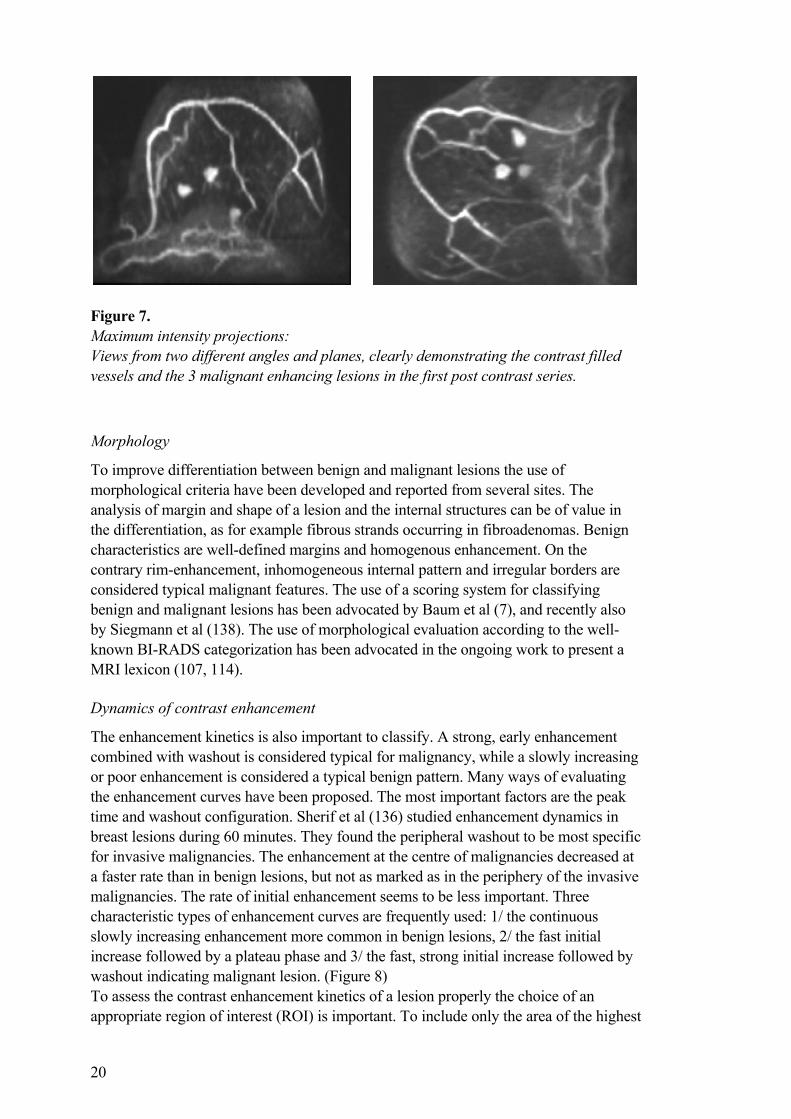
20
Figure 7. Maximum intensity projections: Views from two different angles and planes, clearly demonstrating the contrast filled vessels and the 3 malignant enhancing lesions in the first post contrast series.
Morphology
To improve differentiation between benign and malignant lesions the use of morphological criteria have been developed and reported from several sites. The analysis of margin and shape of a lesion and the internal structures can be of value in the differentiation, as for example fibrous strands occurring in fibroadenomas. Benign characteristics are well-defined margins and homogenous enhancement. On the contrary rim-enhancement, inhomogeneous internal pattern and irregular borders are considered typical malignant features. The use of a scoring system for classifying benign and malignant lesions has been advocated by Baum et al (7), and recently also by Siegmann et al (138). The use of morphological evaluation according to the well-known BI-RADS categorization has been advocated in the ongoing work to present a MRI lexicon (107, 114). Dynamics of contrast enhancement
The enhancement kinetics is also important to classify. A strong, early enhancement combined with washout is considered typical for malignancy, while a slowly increasing or poor enhancement is considered a typical benign pattern. Many ways of evaluating the enhancement curves have been proposed. The most important factors are the peak time and washout configuration. Sherif et al (136) studied enhancement dynamics in breast lesions during 60 minutes. They found the peripheral washout to be most specific for invasive malignancies. The enhancement at the centre of malignancies decreased at a faster rate than in benign lesions, but not as marked as in the periphery of the invasive malignancies. The rate of initial enhancement seems to be less important. Three characteristic types of enhancement curves are frequently used: 1/ the continuous slowly increasing enhancement more common in benign lesions, 2/ the fast initial increase followed by a plateau phase and 3/ the fast, strong initial increase followed by washout indicating malignant lesion. (Figure 8) To assess the contrast enhancement kinetics of a lesion properly the choice of an appropriate region of interest (ROI) is important. To include only the area of the highest

21
amount and speed of enhancement, the size and position of the ROI must be carefully selected. The ROI must not be too small or too large, and must not include vessels or tissues surrounding the lesion. The correct positioning of the ROI is therefore of importance in all three planes. The positioning at the periphery or at the centre of a lesion can also be of importance in the evaluation.
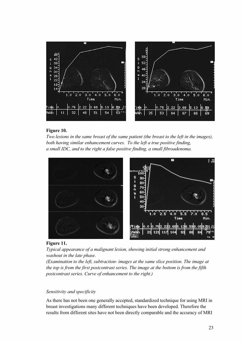
Figure 8. The three types of contrast enhancement curves: Continuously increasing (A), plateau (B) and washout (C) curve types. The different curve types can have a different initial phase: weak (D), medium (E) and strong (F) enhancement in the initial post contrast phase. Benign structures: The normal contrast agent enhancement in breast parenchyma is slow and continuously increasing, which also is the pattern for most benign changes. The enhancement rate of normal breast parenchyma varies during the menstrual cycle, showing stronger enhancement during the first and last week, and least enhancement during the second (and third) menstrual week (81, 129). (Figure 9) In the same way there is an increased enhancement in parenchyma during hormone replacement therapy (HRT). Therefore it is difficult to distinguish strongly enhancing benign changes from moderately enhancing malignancies during HRT and during the first or last weeks of the menstrual cycle, why examinations should ideally be scheduled during the second menstrual week. HRT should be ceased for at least three months before a new examination is scheduled. Even when using optimal examination conditions some benign lesions show a pattern of enhancement similar to malignancies, for example tissues with high proliferative activity or inflammation. This often causes proliferative fibroadenomas, hyperplasia and granulomatous changes to present as false positive findings (61, 79). (Figure 10)
1 2 3
A
B
C
D
E
F
1 2 3
ABCDEF
22
Malignancies: A strong and early enhancement within the first minutes followed by washout (decrease of signal) in the late phase, 5-10 minutes post contrast, is the typical malignant pattern (50, 80, 136). (Figure 11) There are many different patterns in between obvious benign and malignant lesions. The typical washout combined with the early and strong enhancement is not present in all malignancies. Therefore false negative findings also pose a problem in tumours with no typical malignant enhancement pattern, for example lobular and tubular carcinomas as well as some cancer in situ lesions.
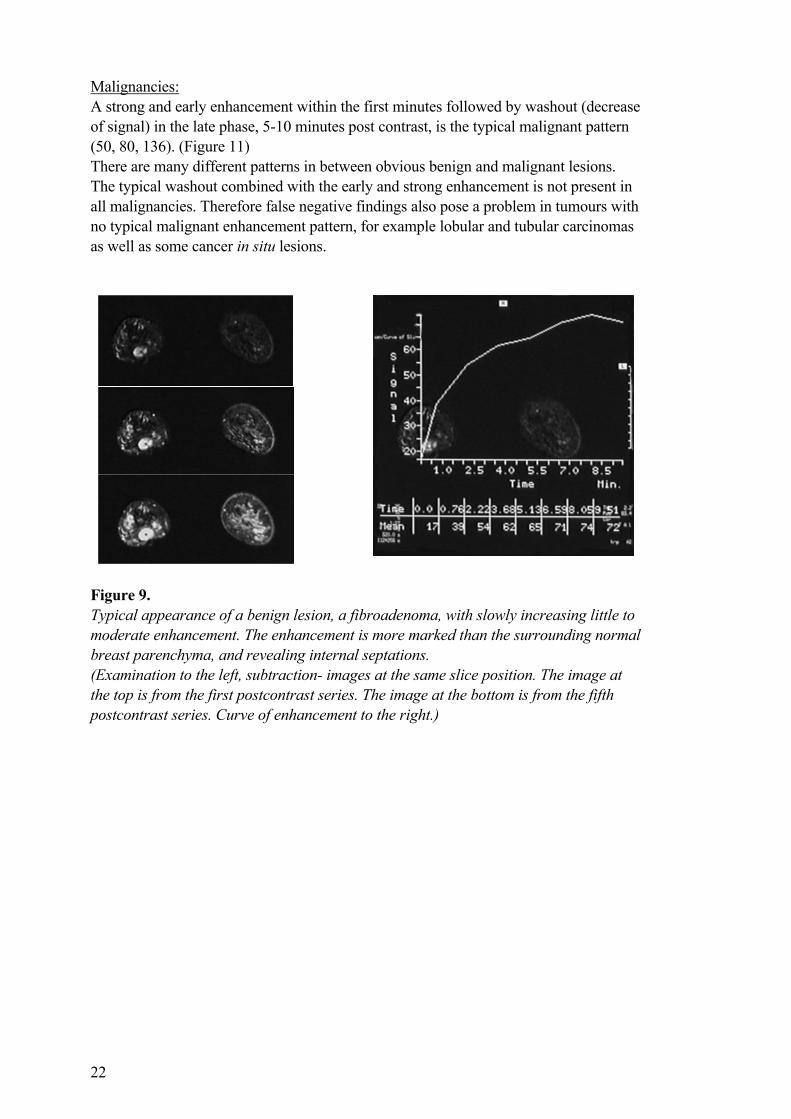
Figure 9. Typical appearance of a benign lesion, a fibroadenoma, with slowly increasing little to moderate enhancement. The enhancement is more marked than the surrounding normal breast parenchyma, and revealing internal septations. (Examination to the left, subtraction- images at the same slice position. The image at the top is from the first postcontrast series. The image at the bottom is from the fifth postcontrast series. Curve of enhancement to the right.)
23
Figure 10. Two lesions in the same breast of the same patient (the breast to the left in the images), both having similar enhancement curves. To the left a true positive finding, a small IDC, and to the right a false positive finding, a small fibroadenoma.
Figure 11. Typical appearance of a malignant lesion, showing initial strong enhancement and washout in the late phase. (Examination to the left, subtraction- images at the same slice position. The image at the top is from the first postcontrast series. The image at the bottom is from the fifth postcontrast series. Curve of enhancement to the right.) Sensitivity and specificity
As there has not been one generally accepted, standardized technique for using MRI in breast investigations many different techniques have been developed. Therefore the results from different sites have not been directly comparable and the accuracy of MRI
24
has been difficult to evaluate. The sensitivities and specificities vary a lot in different studies depending on the selection of patients and the technique used both for data acquisition and for the evaluation (59). In general the sensitivities in many studies have been high (85-100 %) but the specificities have been low (30-95%). Many factors influence the accuracy of breast MRI as mentioned before and summarized below. Different types of sequences with different temporal and spatial resolution as well as timing of the contrast agent injection relative to the data acquisition strongly influence the relative difference in enhancement between benign and malignant tissues. Different types of sequences with different temporal and spatial resolution have been tried. Different sequences have different inherent contrast and different abilities to reveal contrast enhancement. For example the enhancement of a malignant lesion can appear more pronounced in a 3D-sequence than in a 2D-sequence, or when using fat suppression sequences. The use of a contrast agent varies regarding the dose, but also the timing of the contrast agent injection relative to the data acquisition. These factors strongly influence the appreciation of a contrast agent enhancement and the relative difference in enhancement between benign and malignant tissues. The difference in magnet capability is also of importance, the magnet strength as well as the gradients and the type of surface-coil used. Heywang-Köbrunner et al (50) evaluated these differences in a multicenter study, discussing the effects of different magnet strengths in calculating sensitivity and specificity. To standardize mammographic evaluation and classification of lesions, the American College of Radiology (ACR) Breast imaging reporting and data system, BI-RADS, has been used for several years (3). Kim et al (73) used the descriptive terminology and final assessment categories of BI-RADS for mammography in their evaluation of breast cancers detected at MR examinations. They concluded that the lexicon might be applied also at MRI with some modifications. The use of a common evaluation standard for MRI of the breast, similar to BI-RADS, would greatly improve the possibilities to compare studies from different sites, as discussed in a review article by Kinkel and Hylton last year (74). In the same journal a group of radiologists with extensive breast MRI experience (62) presented their work to establish a lexicon for reporting breast MRI findings. They had found moderate interobserver agreement, where breast density and lesion type appeared to be reproducible. Other terms were found to need further refinement and testing. There is a need for development of a new terminology for assessing enhancement kinetics, multifocality and disease extent. Schnall and Ikeda (133) have presented the first recommendations from the Lesion Diagnosis Working Group, coming to a consensus that a report should include both an impression and a management recommendation. They suggested a division of the MR examination report in eight different groups with separate subgroup descriptions: patient history, comparison with previous studies, MRI technique and technical factors, findings, descriptors of focal enhancement, kinetic description, impression/summary of findings and finally recommendations. To facilitate the characterization and evaluation of breast MRI to achieve better specificity while keeping the high sensitivity Nunes et al (114) have developed an architectural interpretation model studying 454 patients. They found high negative predictive values for malignancy where there was absence of enhancing lesion, smooth
25
masses, lobulated masses with non-enhancing internal septations, lobulated masses with minimal or no enhancement and non-septated enhancing lobulated masses with low T2 signal intensity. Several other studies have established which morphologic factors that were of value in differentiation of benign and malignant lesions at MRI, for example by Baum et al, Liberman et al and Wedegärtner et al (7, 90, 154) during the last years, where Baum et al used a scoring system grading the degree of malignancy consisting of 5 different parameters, each graded 0-1 or 0-2, resulting in a maximum of 8 for a highly malignancy suspected lesion and 0-1 for a benign appearing lesion. The impact of staging of malignant breast lesions at MRI on the choice of treatment was discussed by Davis et al (26) in 1998. Fischer et al (33) in 1999 found MRI-findings to change the choice of therapy in almost 18% of 548 lesions (correctly changed in 14,3 %) due to unsuspected multifocality/multicentricity or contralateral malignancy. Recently Tillman et al (149) determined the impact of breast MRI on the clinical management of patients with early stage breast cancer, and in 20% of their 212 diagnosed breast cancers clinical management was changed based on the MRI findings. There was a strongly favourable effect on management in 8%, a somewhat favourable effect in 3 %, a somewhat unfavourable effect in 5 % and a strongly unfavourable effect in 1 %. They concluded that breast MRI appears to offer clinically useful information for determining optimal local treatment. In a recent review article Morris (106) discusses the importance of standardized and cautious evaluations, avoiding pitfalls, and a careful selection of patients that can benefit from MR examinations. Current developments
Among the more recent developments is the assessment of the diagnostic potential of ultrasmall superparamagnetic iron oxide particles (USPIO) in animal breast tumour models. The results indicate a possibility to estimate microvessel characteristics in tumours, which could be of diagnostic value as transendothelial permeability correlates with histologic tumour grade shown in a study by Turetschek et al (150). USPIO can cause artefacts at higher doses. Therefore a careful choice of sequence, related to the contrast dose, must be performed. Spectroscopy using the hydrogen proton, 1H MRS, can be applied for detection of choline-containing compounds in vivo, which increases with cellular proliferation and cause elevated composite choline levels in cancerous lesions. MRS-studies by Kvistad et al (85) found choline-containing compounds not to be specific for breast malignancy. Cecil et al (20) showed that MRS in combination with MRI could improve the specificity. Katz-Brull et al (71) recently published a review where they performed a pooled analysis of five published 1H MRS-studies from four different centers. They found both a high sensitivity and specificity, approaching 100% in a subgroup of women 40 years of age and younger. The factors limiting sensitivity were mainly technical which could be overcome. They concluded breast 1H MRS to be a robust method which is easy to interpret and will perform even better with the development of high-field MRS technologies for detection of the composite choline signal. Magnetic resonance elastography (MRE) is a phase-contrast-based MR imaging technique using mechanical waves transmitted into the tissue by means of an oscillator,
26
initially tried in tissue-simulating prototypes, for example by Manduca et al (97). This method is developed to evaluate the mechanical properties of tissues. Theoretically this could be of value in breast tissue, as palpation (being a physical examination assessing the stiffness of a tissue) for a long time has been well recognized in the detection of breast cancer. The elastic properties of the tissue will determine the local characteristics of the waves and could be displayed by using a motion-sensitive spin-echo MR sequence within the phase of the MR image. Recently Lorenzen et al and McKnight et al (92, 101) have described preliminary results of examining healthy volunteers and patients with malignant lesions, and in one of the studies also benign lesions. They both found MRE to be a new promising technique with high elasticity values in malignant lesions and lowest elasticity values in normal breast parenchyma and fatty tissue. Lorenzen et al (92) in a small number of patients noted an overlap in elasticity ranges of soft malignant lesions and stiff benign lesions, which might impair the evaluations. Further research in a larger number of patients is needed to evaluate the potential applications in breast diagnosis. MRI guided biopsies
Biopsies are an important part of the workup of breast lesions, commonly used as part of the so-called triple diagnosis mentioned earlier (47). When MRI became a diagnostic tool and some lesions were detected only at MRI the need for MRI-guided biopsies emerged (120). This posed technical problems, how to perform biopsies within the magnet bore and the necessity to use MR-compatible needles and biopsy-equipment. After initial use of “free-hand” techniques different methods and aids were developed at several sites. For example an add-on device for surface coils by Fisher et al (35) with the patient supine, which also was compared to a developed dedicated single breast biopsy coil (36) with the patient in prone position, where both systems were found suitable for biopsy and preoperative localization of lesions. These methods had some disadvantages; the thicker slice thicknesses used making it difficult to localize small lesions (less than 10 mm) and the risk of complications when performing biopsies close to the chest wall. Kuhl et al (82) evaluated another prototype using a patient support, a perforated breast compression device and an add-on stereotactic system with the patient in a semiprone position. This type of biopsy-aid also proved to be of great value in the MRI workup of breast lesions. Heywang-Köbrunner et al (51) developed a different prototype allowing both lateral and medial approach of the breast with the patient in prone position, using mediolateral compression by two independently movable plates, and having the possibility to use different needle angulations through flexible ribs of the plates. They also discussed the importance of non-magnetic needles as the metallic artefact can cause signal loss in an area larger than the lesion, making it difficult to establish correct needle position in the lesion. Warren and Kessar (152) presented a procedure of localization of breast lesions seen only at MRI. They placed a MR compatible cerebral embolisation device (an embolisation coil) at the site of the suspected lesion, in the same way as when placing a hook-wire at mammography in non-palpable lesions. They used the possibility of reconstructions in all planes using a 3D sequence to subsequently localize the suspected area in craniocaudal and lateral mammographies to insert the embolisation device, which position then could be verified at mammography. A second MR examination was performed to verify the position of the embolisation device before surgery. The

27
same method could also be performed using US instead of mammography. They found the only disadvantage of the procedure was that the patient then was scheduled for open surgical biopsy, rather than percutaneous biopsy of the lesion. Many different methods to perform MRI-guided biopsies of breast lesions have been developed and can be used, pointing out the importance of performing biopsies of lesions exclusively diagnosed at MRI as well as the need of MR-compatible equipment and preoperative marking material, combined with a careful choice of sequence. Helbich (46) recently discussed these issues, concluding that needle biopsy of lesions smaller than 10 mm cannot be recommended at present due to needle artefacts and tissue shift during the intervention. These localization techniques and devices can also be used for adequate guidance in monitoring the delivery of various thermal ablation techniques using temperature-sensitive sequences, for example if using interstitial laser therapy, focused ultrasound, radiofrequency-induced coagulation and cryotherapy (44). However, these methods are still developing and further clinical studies are needed to assess efficacy as well as safety. WHEN TO PERFORM MRI OF THE BREASTS
Experiences at different sites using different methods have so far come to these conclusions on when to use MRI of the breast in the clinical setting. MRI should only be used in selected cases with proper indications and as a complementary method to routine investigations with clinical evaluation, mammography, cytology and ultrasound. The evaluations should always comprise the findings from all methods used, including patient history and a discussion with the surgeon and/or oncologist planning the treatment of the patient. Indications for MRI of the breast:
• Diagnosis of implant failure in patients with symptoms (palpable finding or pain) or in differential diagnostic problem cases when there is a need to distinguish inflammatory from malignant tissue. (1, 8)
• In women with known genetic risk for breast cancer. (144, 151) • In patients with axillary lymph node metastases of unknown primary
malignancy. (118, 134) • Preoperative evaluation in order to exclude multifocality / multicentricity.
(33, 110) • Postoperative follow up for detecting recurrence in scars and fibrotic tissue,
with or without implants, when mammography and US are inconclusive. (13, 49)
• Monitoring the effect of chemotherapeutic treatment on the kinetics and size of contrast enhancing areas. (39)
NO indication for MRI of the breasts:
• Evaluation of inflammatory changes should be performed only in selected problem cases, and should not be used in general. For example it is impossible to distinguish mastitis from an inflammatory cancer.
• In patients with microcalcifications at mammography. • MRI cannot replace cytology / FNAB.
28
Future aspects of MRI of the breast: The evaluations and development of diagnostic criteria resulting in new recommendations and lexicons will continue, hopefully to increase the specificity of the evaluations even more. The implementation of new contrast agents is also hoped to increase specificity, as well as the addition of MRS, which might become more available in clinical diagnosis of breast lesions. Further research of contrast enhanced MRI of the breast is hoped to reveal prognostic information, which would then become another indication for breast MRI.
29
AIMS GENERAL AIM
The general aim of this thesis was to study and establish the role of MRI in the clinical diagnosis of breast lesions. How and when to use MRI in a clinical setting, including which kind of lesions and which kind of patients could benefit the most from MRI as an adjunct to other breast imaging modalities. SPECIFIC AIMS
The specific aims of the different papers were:
I. To compare the appearance of the same lesion in two different T1-weighted 3D sequences with regard to the contrast inherent in the sequences and following contrast injection. Furthermore, we wanted to evaluate whether a difference in the appearance of the lesions could influence the observer’s ability to make a correct diagnosis.
II. To evaluate whether any potential histological changes caused by fine needle aspiration biopsy (haematoma or oedema) would affect the contrast enhancement, thus influencing the evaluation of breast lesions at semidynamic MR imaging.
III. To determine the clinical value of dynamic MR imaging used as an adjunct to triple diagnosis (TD).
IV. To compare lesion-size at dynamic MR imaging and mammography with histopathology as “gold standard”, using a dynamic 3D-sequence.
V. To compare the diagnostic accuracy of dynamic MR imaging of the breasts to scintimammography on the basis of histopathologic findings, and to determine the clinical value of these methods as an adjunct to mammography.
30
MATERIAL AND METHODS The local ethical committee approved all the included studies, and all patients that took part in the studies gave their informed consent. SUBJECTS Study I
During 2 years, November 1993-November 1995, 20 patients with widespread and/or diffuse contrast enhancement on the routine FLASH sequence were selected from the 459 clinically MR examined women. These 20 patients with 27 lesions gave informed consent to a second MR examination with a different T1-weighted sequence, MPRAGE. The mean age of the women was 49 years (range 35-67 years). The time interval between the FLASH and MPRAGE examination was 3 days to 3,5 months with a mean of 23 days. Study II
During 3 years, 1993-1996, 15 patients with 17 lesions were examined with semidynamic MRI of the breast before and after fine-needle aspiration of the lesions. Their mean age was 54,5 (range 41-69) years and the time interval between the MR examinations was 3-13 days (mean 7,1 days). Study III, IV and V
From January 1996 to June 1997 (18 months) 97 consecutive patients scheduled for surgery of breast lesions, detected by palpation or mammography, were offered to take part in our prospective study. Four patients had to be excluded due to contra-indications for MRI and claustrophobia. (Figure 12) Study III: The remaining 93 women with 114 lesions, of which 64 were palpable, had a mean age of 54 years (range 33-81 years) and the mean interval between MR examination and surgery was 12 days (range 1-56 days). Study IV: From the 93 patients with 82 malignant lesions six were excluded in this study, as the maximum lesion size could not be measured at histopathology. This resulted in 76 malignant lesions in 65 patients. Their mean age was 56 years (range 33-81 years), and the mean interval between MR examination and surgery was 11 days (range 1-37 days). Study V: In three of the 93 patients scintimammography was not performed, hence 90 women with 111 lesions and a mean age of 54 years (range 33-81 years) were included in this study. Surgery was performed within two months after the examinations (mean 12 days).

31
REFERRAL OFPATIENT TO
BREAST SURGEON
PATIENT MEETSTHE BREAST
SURGEON
WEEKLYBREAST
CONFERENCE
Palpabel lumpScreening
mammographyClinical mammography
Referred from either ageneral practioner orspecialist practice,or our and otherhospital clinics
NO SURGERY
PATIENT ISSCHEDULED FOR
SURGERY
MRI OF THE BREASTS
Evaluation of MR-images by MRI-
radiologist, speciallytrained in MRI of thebreasts, but without
knowledge ofpreoperative diagnosis
If TD (triple diagnosis) isnot complete the
missing examinationsare performed, including
clinical examination,mammography andFNAC (fine needleaspiration cytology)
Breast surgeons ,mammographers and
cytologists meet to viewand discuss performedexaminations to plan
treatment, and if surgeryshould be performed.(No MRI-radiologist is
involved.)
REFERRAL TO MRI
Patient is offered apreoperative MRI of the
breasts.Acceptance of this will
lead to:
SURGERY
= Main flow of events for the patient
= Explanation of events
PATIENT SELECTION AND INVESTIGATION
Figure 12. Patient selection and investigation of study III, IV and V. Main flow of events and explanation of events. METHODS
All studies were performed on a superconductive 1.5 T Magnetom SP 63 (Siemens, Erlangen, Germany), using a dedicated receive-only bilateral breast coil and standard software. The patients were examined in the prone position at the isocenter of the magnet, with the breasts hanging into the breast coil. The contrast agent was injected via a previously inserted long line into a cubital vein, while the patient was in position in the magnet, and was immediately followed by 20 ml physiologic saline. The injection was performed manually, as a bolus dose, within 30 s. Study I
Two different T1-weighted 3-D-sequences were used: FLASH (fast low angle shot) with TR/TE (repetition time/echo time) 31/6 ms, flip angle (FA) 60°, 1 acquisition and acquisition time (TA) 6,23 min. One pre- and two post-contrast series were performed. MPRAGE (magnetization prepared rapid gradient echo) with TR/TE 10/4 ms, FA 10°, 1 acquisition and TA 3,02 min. One pre- and three post-contrast series were performed. In both sequences the field of view (FOV) was 350 mm with matrix 192 X 256 and the slices were in the transversal plane with a thickness of 2,2 mm and no interslice gap. Gadopentetate dimeglumine (Magnevist, Schering, Berlin, Germany) was injected intravenously at a dose of 0,1 mmol/kg b.w. The MR examinations were reviewed independently by 3 radiologists and the findings were divided into 5 categories: 1=normal, 2=benign, 3=probably benign, 4=probably malignant and 5=malignant. The evaluation and diagnosis at MRI was based on visual
32
evaluation of the contrast enhancement, which included the increase in signal intensity, the speed and pattern of enhancement but also the degree of irregularity of the border of the lesion. Study II
The MR examinations were semidynamic using a 3D gradient-echo, FLASH, with TR/TE 31/6 ms and FA 60° and TA 6,23 min. FOV was 350 mm, matrix 192 X 256 and slice thickness 2,2 mm with no gap. One pre- and 2 post-contrast series were performed in the transversal plane. Gadopentetate dimeglumine at a dose of 0,1 mmol/kg b.w. was injected intravenously. The FNAB (fine needle aspiration biopsy) was performed with a 21G (0.8 X 80 mm) needle. Signal intensities were measured in the pre- and two post-contrast series at the following sites in both the examinations before and after the FNAB: 1) within the lesion; 2) in the surrounding breast parenchyma; 3) in nearby muscle. The increase in signal intensity following contrast agent injection was expressed in percentage of the value of signal intensity before the contrast agent administration, in the same slice positions. Two percentage values were obtained, early and late contrast enhancement. Both were calculated in the examinations before and after the FNAB, and each individual patient served as its own control. Two radiologists experienced in breast MRI reviewed the examinations, but one radiologist performed all the measurements. Study III, IV and V
All patients were examined with TD (triple diagnosis) and dynamic contrast enhanced MR imaging (CE-MRI). Breast conserving surgery or mastectomy was performed and all lesions were histopathologically characterized. Mammographies were performed using the routine equipment at our site. Each breast was examined in three views, and when necessary suspected lesions were examined by means of additional views (spot filming or direct magnification by microfocus). At mammography the parenchyma was classified into 2 groups: predominantly dense or predominantly fatty, according to the BI-RADS classification. (3) Fine needle aspiration was performed by guidance of ultrasound (US) or stereotactic mammography, using a 0,7-mm needle mounted in a Coleco syringe holder. An experienced specialist in mammography performed all the US or mammography guided punctures, while an experienced cytologist performed punctures of palpable lesions and also immediately evaluated all samples. A dynamic MR examination with one pre- and seven post-contrast series in the coronal plane was performed, using T1-weighted 3D FLASH with TR/TE 12/5 ms, FA 25° and TA 87 s. The FOV was 320 mm with matrix 112 X 256 and slice thickness 2,2 mm with no interslice gap. Gadopentetate dimeglumine 0,2 mmol/kg b.w. was administered as a bolus dose. The postprocessing included subtraction, multiplanar reconstruction (MPR), calculation of time-intensity curves of enhancing regions and in some cases maximum intensity projection (MIP). One MRI radiologist specially trained in breast MRI made the evaluations of CE-MRI images, and a radiologist specially trained in scintigraphy evaluated the scintimammographies. Both were blinded to the results of other examination methods
33
(including mammography), clinical examination and cytology, only knowing the patients were scheduled for surgery of a lesion in one breast. CE-MRI image analysis was based on kinetic as well as morphological characteristics of the contrast enhancement. A strong, early enhancement and washout were considered malignant, while a slow, steady or poor enhancement was considered benign. Inhomogeneous or rim-enhancement and irregular borders of a lesion were classified as malignant, while homogeneous enhancement, well-defined or lobulated margins and internal septations were classified as benign. In Study III and V the lesions were graded in five diagnostic groups for each method used: 1=normal, 2=benign, 3=probably benign, 4=probably malignant and 5=malignant. When the results of the diagnostic techniques were discordant the final result was defined as the highest value, which was also used for the combination of methods in the ROC analysis in study V. Diagnostic groups 1-3 were defined as benign and diagnostic groups 4-5 as malignant in the calculations of sensitivity, specificity and diagnostic accuracy. In study IV two different radiologists measured the largest lesion diameters at mammography respective CE-MRI, and a pathologist measured the lesion diameters at histopathology. The sizes at CE-MRI were measured only in the first postcontrast series. Each of the measured sizes at CE-MRI and mammography were compared to histopathologic size. In study V scintimammography was performed using Tc-99m-Sestamibi where planar imaging was started 10 minutes after the injection of 700 MBq 99mTc-Sestamibi. Twelve patients received an intravenous injection in an antecubital vein on the contralateral side to the breast lesion. Seventy-eight patients were injected in a pedal vein to avoid false positive result in the axillary regions due to the perivascular infiltration and the potential uptake in the muscles. Two prone laterals and one supine anterior view were performed in each patient. Planar images with a matrix of 256 X 256 pixels were acquired for eight minutes for each projection. Data acquisition, reconstruction and postprocessing were performed on a Hermes-system, and images were classified based on visual interpretation. Sensitivity, specificity and diagnostic accuracy were calculated. Statistical methods
In study II T-test for paired samples was used for statistical evaluation. In study IV the differences in sizes were presented in box plots for the pure invasive, the pure in situ as well as for the mixed malignant lesions. Wilcoxon signed rank test and stratified Wilcoxon signed rank test were used for statistical evaluation of the differences in size. Graphic presentations of regression-lines present the differences between CE-MRI and mammographic sizes relative to the histopathologic size in the pure invasive and the mixed lesions. The regression-lines can be compared to the equality-line (where measurements of all methods are exactly the same in the same lesion).
34
In study V a comparison of sensitivities and specificities was performed by means of McNemar´s χ2 test (43). Receiver-operating-characteristic (ROC) curves were generated for each study by using the ROCKIT software package (version number: 0.9.1 for PC; C Metz, University of Chicago, Department of Radiology). The area under the ROC curve (Az) was also determined, which can be used to describe the overall diagnostic performance of a method. ROC analysis can also be used for comparison of diagnostic accuracy of two or more tests (104). Single ROC curves were drawn and the Az values were calculated, and complementary ROC comparison in pairs was performed between each single diagnostic method. Comparing ROC curves of mammography and the combined methods (mammography + CE-MRI respective scintimammography) also assessed the adjunctive use of CE-MRI and scintimammography. Univariate z-score test was used to determine a statistical significant difference between the Az values (statistical significance at a p-value <0,05).
35
MAIN RESULTS STUDY I
A MPRAGE sequence (due to lower sensitivity to contrast enhancement) can be used in a second examination in case of strong and wide-spread contrast enhancement using a 3D FLASH sequence (with high sensitivity to contrast enhancement), facilitating the down-grading of a false positive lesion to a true negative. STUDY II
Fine needle aspiration biopsy did not impair the evaluation of a breast lesion at subsequent semidynamic MR examinations. There were no differences in the appearance or in the contrast enhancement of the lesions, and no edema or hematoma was revealed. STUDY III
CE-MRI is of value as an additive method to triple diagnosis by increasing the sensitivity, but at the cost of decreasing specificity. Patients with mammographically dense breast parenchyma might benefit most, as the decrease in specificity in these patients is less pronounced. STUDY IV
CE-MRI and mammography are both good at measuring the size of detected invasive breast malignancies. The total size of a mixed lesion is frequently underestimated by both CE-MRI and mammography, although the invasive part can be equally well described and measured with both methods. The pure in situ lesions were too few to be evaluated, but are generally better described at mammography due to calcifications. STUDY V
CE-MRI and 99mTc-sestamibi showed individually comparable diagnostic accuracy. Better accuracy was found for the combination of CE-MRI and mammography when using ROC-curves and area under the curve for evaluation. In lesions smaller than 10 mm in size scintimammography is less reliable due to a low spatial resolution.
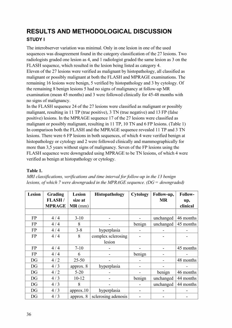
36
RESULTS AND METHODOLOGICAL DISCUSSION STUDY I
The interobserver variation was minimal. Only in one lesion in one of the used sequences was disagreement found in the category classification of the 27 lesions. Two radiologists graded one lesion as 4, and 1 radiologist graded the same lesion as 3 on the FLASH sequence, which resulted in the lesion being listed as category 4. Eleven of the 27 lesions were verified as malignant by histopathology, all classified as malignant or possibly malignant at both the FLASH and MPRAGE examinations. The remaining 16 lesions were benign, 5 verified by histopathology and 3 by cytology. Of the remaining 8 benign lesions 5 had no signs of malignancy at follow-up MR examination (mean 45 months) and 3 were followed clinically for 45-48 months with no signs of malignancy. In the FLASH sequence 24 of the 27 lesions were classified as malignant or possibly malignant, resulting in 11 TP (true positive), 3 TN (true negative) and 13 FP (false positive) lesions. In the MPRAGE sequence 17 of the 27 lesions were classified as malignant or possibly malignant, resulting in 11 TP, 10 TN and 6 FP lesions. (Table 1) In comparison both the FLASH and the MPRAGE sequence revealed 11 TP and 3 TN lesions. There were 6 FP lesions in both sequences, of which 4 were verified benign at histopathology or cytology and 2 were followed clinically and mammographically for more than 3,5 years without signs of malignancy. Seven of the FP lesions using the FLASH sequence were downgraded using MPRAGE to be TN lesions, of which 4 were verified as benign at histopathology or cytology. Table 1. MRI classifications, verifications and time interval for follow-up in the 13 benign lesions, of which 7 were downgraded in the MPRAGE sequence. (DG = downgraded) Lesion Grading
FLASH / MPRAGE
Lesion size at
MR (mm)
Histopathology Cytology Follow-up, MR
Follow-up,
clinical
FP 4 / 4 3-10 - - unchanged 46 monthsFP 4 / 4 8 - benign unchanged 45 monthsFP 4 / 4 3-8 hyperplasia - - - FP 4 / 4 8 complex sclerosing
lesion - - -
FP 4 / 4 7-10 - - - 45 monthsFP 4 / 4 6 - benign - - DG 4 / 2 25-50 - - - 48 monthsDG 4 / 3 approx. 8 hyperplasia - - - DG 4 / 2 5-20 - - benign 46 monthsDG 4 / 3 10-12 - benign unchanged 44 monthsDG 4 / 3 8 - - unchanged 44 monthsDG 4 / 3 approx.10 hyperplasia - - - DG 4 / 3 approx. 8 sclerosing adenosis - - -
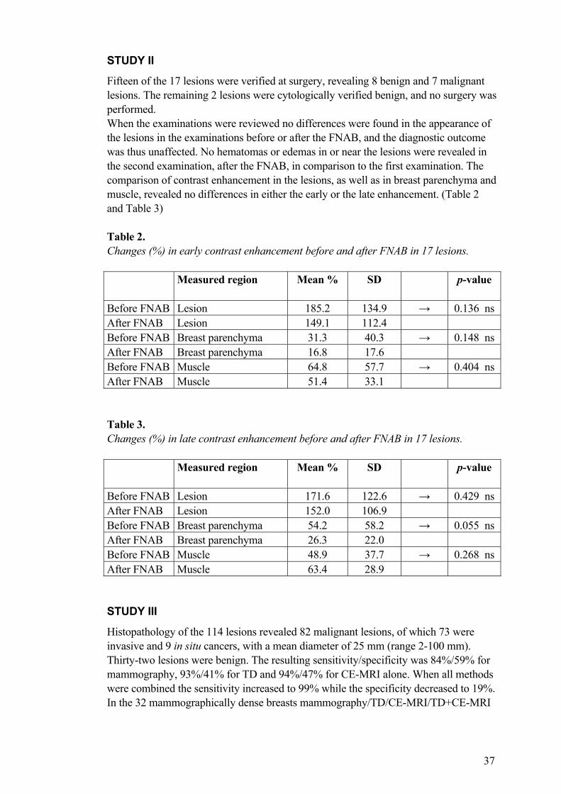
37
STUDY II
Fifteen of the 17 lesions were verified at surgery, revealing 8 benign and 7 malignant lesions. The remaining 2 lesions were cytologically verified benign, and no surgery was performed. When the examinations were reviewed no differences were found in the appearance of the lesions in the examinations before or after the FNAB, and the diagnostic outcome was thus unaffected. No hematomas or edemas in or near the lesions were revealed in the second examination, after the FNAB, in comparison to the first examination. The comparison of contrast enhancement in the lesions, as well as in breast parenchyma and muscle, revealed no differences in either the early or the late enhancement. (Table 2 and Table 3) Table 2. Changes (%) in early contrast enhancement before and after FNAB in 17 lesions. Measured region
Mean % SD p-value
Before FNAB Lesion 185.2 134.9 → 0.136 ns After FNAB Lesion 149.1 112.4 Before FNAB Breast parenchyma 31.3 40.3 → 0.148 ns After FNAB Breast parenchyma 16.8 17.6 Before FNAB Muscle 64.8 57.7 → 0.404 ns After FNAB Muscle 51.4 33.1 Table 3. Changes (%) in late contrast enhancement before and after FNAB in 17 lesions. Measured region
Mean % SD p-value
Before FNAB Lesion 171.6 122.6 → 0.429 ns After FNAB Lesion 152.0 106.9 Before FNAB Breast parenchyma 54.2 58.2 → 0.055 ns After FNAB Breast parenchyma 26.3 22.0 Before FNAB Muscle 48.9 37.7 → 0.268 ns After FNAB Muscle 63.4 28.9 STUDY III
Histopathology of the 114 lesions revealed 82 malignant lesions, of which 73 were invasive and 9 in situ cancers, with a mean diameter of 25 mm (range 2-100 mm). Thirty-two lesions were benign. The resulting sensitivity/specificity was 84%/59% for mammography, 93%/41% for TD and 94%/47% for CE-MRI alone. When all methods were combined the sensitivity increased to 99% while the specificity decreased to 19%. In the 32 mammographically dense breasts mammography/TD/CE-MRI/TD+CE-MRI
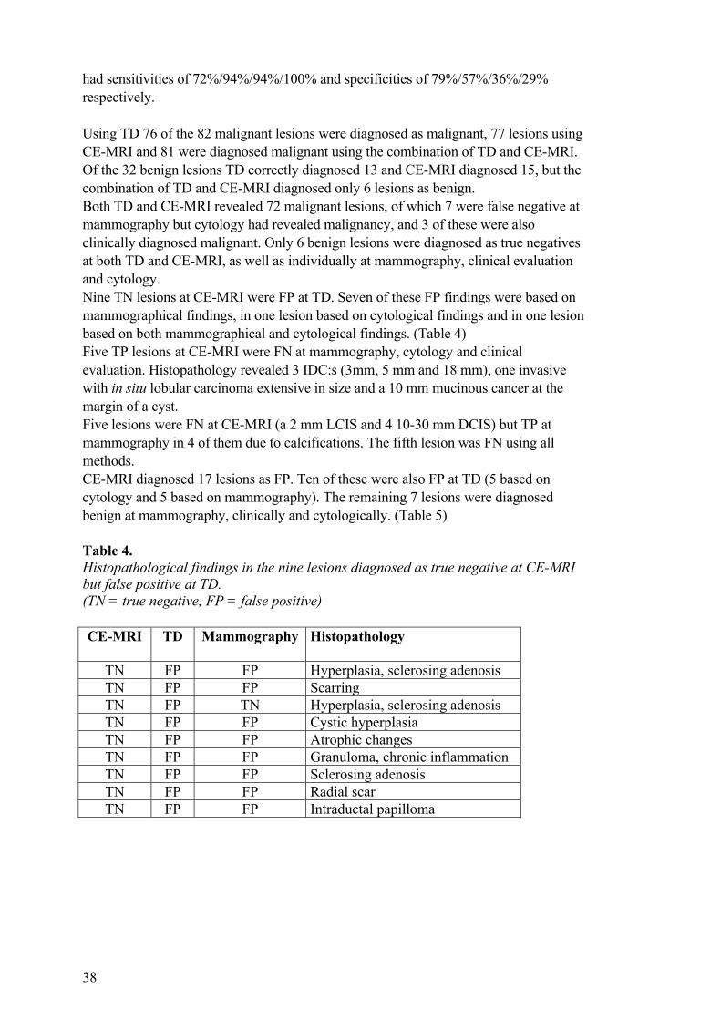
38
had sensitivities of 72%/94%/94%/100% and specificities of 79%/57%/36%/29% respectively. Using TD 76 of the 82 malignant lesions were diagnosed as malignant, 77 lesions using CE-MRI and 81 were diagnosed malignant using the combination of TD and CE-MRI. Of the 32 benign lesions TD correctly diagnosed 13 and CE-MRI diagnosed 15, but the combination of TD and CE-MRI diagnosed only 6 lesions as benign. Both TD and CE-MRI revealed 72 malignant lesions, of which 7 were false negative at mammography but cytology had revealed malignancy, and 3 of these were also clinically diagnosed malignant. Only 6 benign lesions were diagnosed as true negatives at both TD and CE-MRI, as well as individually at mammography, clinical evaluation and cytology. Nine TN lesions at CE-MRI were FP at TD. Seven of these FP findings were based on mammographical findings, in one lesion based on cytological findings and in one lesion based on both mammographical and cytological findings. (Table 4) Five TP lesions at CE-MRI were FN at mammography, cytology and clinical evaluation. Histopathology revealed 3 IDC:s (3mm, 5 mm and 18 mm), one invasive with in situ lobular carcinoma extensive in size and a 10 mm mucinous cancer at the margin of a cyst. Five lesions were FN at CE-MRI (a 2 mm LCIS and 4 10-30 mm DCIS) but TP at mammography in 4 of them due to calcifications. The fifth lesion was FN using all methods. CE-MRI diagnosed 17 lesions as FP. Ten of these were also FP at TD (5 based on cytology and 5 based on mammography). The remaining 7 lesions were diagnosed benign at mammography, clinically and cytologically. (Table 5) Table 4. Histopathological findings in the nine lesions diagnosed as true negative at CE-MRI but false positive at TD. (TN = true negative, FP = false positive) CE-MRI
TD Mammography Histopathology
TN FP FP Hyperplasia, sclerosing adenosis TN FP FP Scarring TN FP TN Hyperplasia, sclerosing adenosis TN FP FP Cystic hyperplasia TN FP FP Atrophic changes TN FP FP Granuloma, chronic inflammation TN FP FP Sclerosing adenosis TN FP FP Radial scar TN FP FP Intraductal papilloma
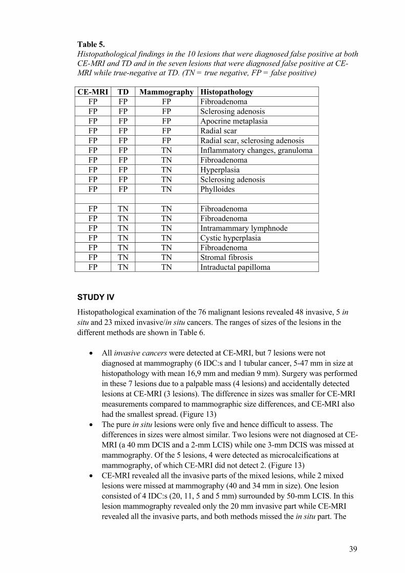
39
Table 5. Histopathological findings in the 10 lesions that were diagnosed false positive at both CE-MRI and TD and in the seven lesions that were diagnosed false positive at CE-MRI while true-negative at TD. (TN = true negative, FP = false positive) CE-MRI TD Mammography Histopathology
FP FP FP Fibroadenoma FP FP FP Sclerosing adenosis FP FP FP Apocrine metaplasia FP FP FP Radial scar FP FP FP Radial scar, sclerosing adenosis FP FP TN Inflammatory changes, granuloma FP FP TN Fibroadenoma FP FP TN Hyperplasia FP FP TN Sclerosing adenosis FP FP TN Phylloides
FP TN TN Fibroadenoma FP TN TN Fibroadenoma FP TN TN Intramammary lymphnode FP TN TN Cystic hyperplasia FP TN TN Fibroadenoma FP TN TN Stromal fibrosis FP TN TN Intraductal papilloma
STUDY IV
Histopathological examination of the 76 malignant lesions revealed 48 invasive, 5 in situ and 23 mixed invasive/in situ cancers. The ranges of sizes of the lesions in the different methods are shown in Table 6.
• All invasive cancers were detected at CE-MRI, but 7 lesions were not diagnosed at mammography (6 IDC:s and 1 tubular cancer, 5-47 mm in size at histopathology with mean 16,9 mm and median 9 mm). Surgery was performed in these 7 lesions due to a palpable mass (4 lesions) and accidentally detected lesions at CE-MRI (3 lesions). The difference in sizes was smaller for CE-MRI measurements compared to mammographic size differences, and CE-MRI also had the smallest spread. (Figure 13)
• The pure in situ lesions were only five and hence difficult to assess. The differences in sizes were almost similar. Two lesions were not diagnosed at CE-MRI (a 40 mm DCIS and a 2-mm LCIS) while one 3-mm DCIS was missed at mammography. Of the 5 lesions, 4 were detected as microcalcifications at mammography, of which CE-MRI did not detect 2. (Figure 13)
• CE-MRI revealed all the invasive parts of the mixed lesions, while 2 mixed lesions were missed at mammography (40 and 34 mm in size). One lesion consisted of 4 IDC:s (20, 11, 5 and 5 mm) surrounded by 50-mm LCIS. In this lesion mammography revealed only the 20 mm invasive part while CE-MRI revealed all the invasive parts, and both methods missed the in situ part. The
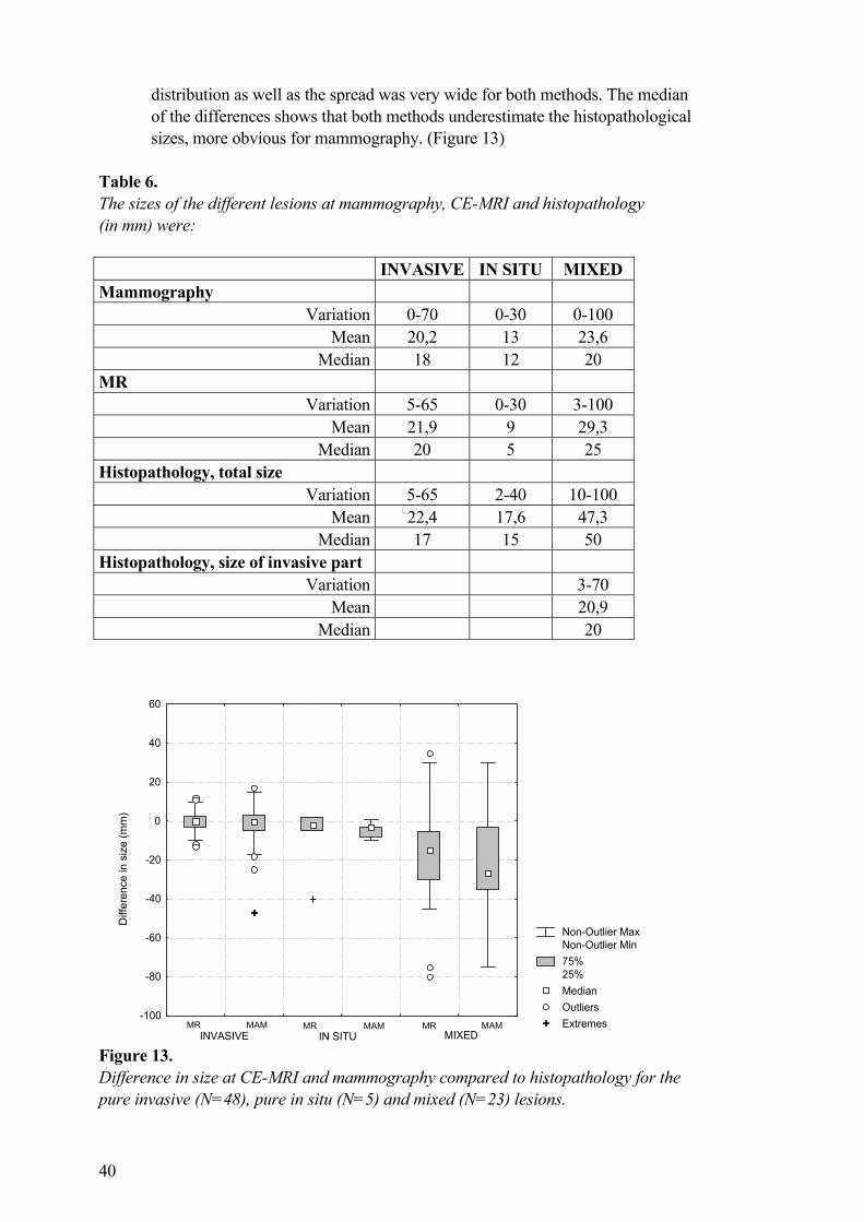
40
distribution as well as the spread was very wide for both methods. The median of the differences shows that both methods underestimate the histopathological sizes, more obvious for mammography. (Figure 13)
Table 6. The sizes of the different lesions at mammography, CE-MRI and histopathology (in mm) were:
INVASIVE IN SITU MIXED Mammography
Variation 0-70 0-30 0-100 Mean 20,2 13 23,6
Median 18 12 20 MR
Variation 5-65 0-30 3-100 Mean 21,9 9 29,3
Median 20 5 25 Histopathology, total size
Variation 5-65 2-40 10-100 Mean 22,4 17,6 47,3
Median 17 15 50 Histopathology, size of invasive part
Variation 3-70 Mean 20,9
Median 20
Figure 13. Difference in size at CE-MRI and mammography compared to histopathology for the pure invasive (N=48), pure in situ (N=5) and mixed (N=23) lesions.
Non-Outlier MaxNon-Outlier Min75%25%MedianOutliersExtremes
Diff
eren
ce in
siz
e (m
m)
-100
-80
-60
-40
-20
0
20
40
60
INVASIVE IN SITU MIXEDMR MAM MR MAM MR MAM
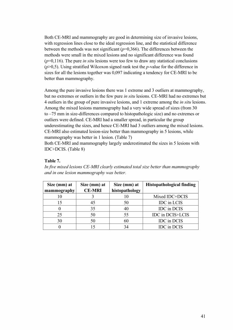
41
Both CE-MRI and mammography are good in determining size of invasive lesions, with regression lines close to the ideal regression line, and the statistical difference between the methods was not significant (p=0,366). The differences between the methods were small in the mixed lesions and no significant difference was found (p=0,116). The pure in situ lesions were too few to draw any statistical conclusions (p>0,5). Using stratified Wilcoxon signed rank test the p-value for the difference in sizes for all the lesions together was 0,097 indicating a tendency for CE-MRI to be better than mammography. Among the pure invasive lesions there was 1 extreme and 3 outliers at mammography, but no extremes or outliers in the few pure in situ lesions. CE-MRI had no extremes but 4 outliers in the group of pure invasive lesions, and 1 extreme among the in situ lesions. Among the mixed lesions mammography had a very wide spread of sizes (from 30 to –75 mm in size-differences compared to histopathologic size) and no extremes or outliers were defined. CE-MRI had a smaller spread, in particular the group underestimating the sizes, and hence CE-MRI had 3 outliers among the mixed lesions. CE-MRI also estimated lesion-size better than mammography in 5 lesions, while mammography was better in 1 lesion. (Table 7) Both CE-MRI and mammography largely underestimated the sizes in 5 lesions with IDC+DCIS. (Table 8) Table 7. In five mixed lesions CE-MRI clearly estimated total size better than mammography and in one lesion mammography was better.
Size (mm) at mammography
Size (mm) at CE-MRI
Size (mm) at histopathology
Histopathological finding
10 3 10 Mixed IDC+DCIS 15 45 50 IDC in LCIS 0 35 40 IDC in DCIS 25 50 55 IDC in DCIS+LCIS 30 50 60 IDC in DCIS 0 15 34 IDC in DCIS
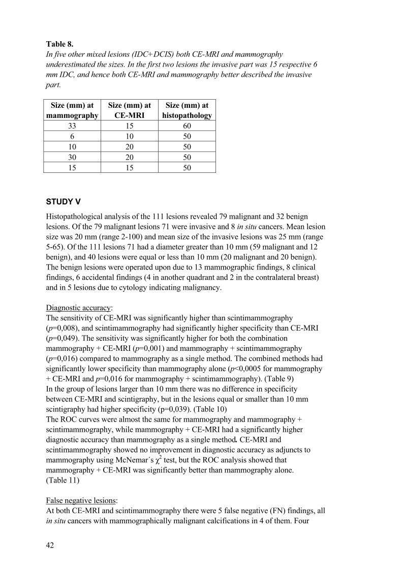
42
Table 8. In five other mixed lesions (IDC+DCIS) both CE-MRI and mammography underestimated the sizes. In the first two lesions the invasive part was 15 respective 6 mm IDC, and hence both CE-MRI and mammography better described the invasive part.
Size (mm) at mammography
Size (mm) at CE-MRI
Size (mm) at histopathology
33 15 60 6 10 50 10 20 50 30 20 50 15 15 50
STUDY V
Histopathological analysis of the 111 lesions revealed 79 malignant and 32 benign lesions. Of the 79 malignant lesions 71 were invasive and 8 in situ cancers. Mean lesion size was 20 mm (range 2-100) and mean size of the invasive lesions was 25 mm (range 5-65). Of the 111 lesions 71 had a diameter greater than 10 mm (59 malignant and 12 benign), and 40 lesions were equal or less than 10 mm (20 malignant and 20 benign). The benign lesions were operated upon due to 13 mammographic findings, 8 clinical findings, 6 accidental findings (4 in another quadrant and 2 in the contralateral breast) and in 5 lesions due to cytology indicating malignancy. Diagnostic accuracy: The sensitivity of CE-MRI was significantly higher than scintimammography (p=0,008), and scintimammography had significantly higher specificity than CE-MRI (p=0,049). The sensitivity was significantly higher for both the combination mammography + CE-MRI (p=0,001) and mammography + scintimammography (p=0,016) compared to mammography as a single method. The combined methods had significantly lower specificity than mammography alone (p<0,0005 for mammography + CE-MRI and p=0,016 for mammography + scintimammography). (Table 9) In the group of lesions larger than 10 mm there was no difference in specificity between CE-MRI and scintigraphy, but in the lesions equal or smaller than 10 mm scintigraphy had higher specificity (p=0,039). (Table 10) The ROC curves were almost the same for mammography and mammography + scintimammography, while mammography + CE-MRI had a significantly higher diagnostic accuracy than mammography as a single method. CE-MRI and scintimammography showed no improvement in diagnostic accuracy as adjuncts to mammography using McNemar´s χ2 test, but the ROC analysis showed that mammography + CE-MRI was significantly better than mammography alone. (Table 11) False negative lesions: At both CE-MRI and scintimammography there were 5 false negative (FN) findings, all in situ cancers with mammographically malignant calcifications in 4 of them. Four
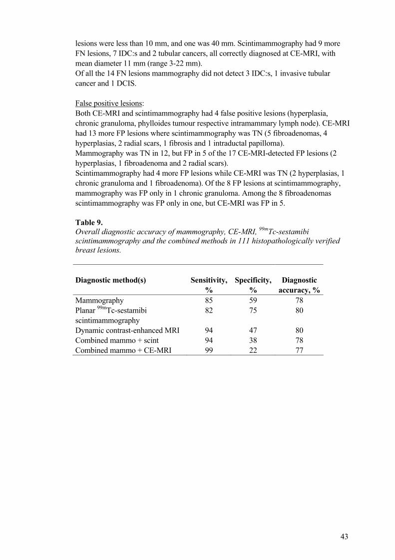
43
lesions were less than 10 mm, and one was 40 mm. Scintimammography had 9 more FN lesions, 7 IDC:s and 2 tubular cancers, all correctly diagnosed at CE-MRI, with mean diameter 11 mm (range 3-22 mm). Of all the 14 FN lesions mammography did not detect 3 IDC:s, 1 invasive tubular cancer and 1 DCIS. False positive lesions: Both CE-MRI and scintimammography had 4 false positive lesions (hyperplasia, chronic granuloma, phylloides tumour respective intramammary lymph node). CE-MRI had 13 more FP lesions where scintimammography was TN (5 fibroadenomas, 4 hyperplasias, 2 radial scars, 1 fibrosis and 1 intraductal papilloma). Mammography was TN in 12, but FP in 5 of the 17 CE-MRI-detected FP lesions (2 hyperplasias, 1 fibroadenoma and 2 radial scars). Scintimammography had 4 more FP lesions while CE-MRI was TN (2 hyperplasias, 1 chronic granuloma and 1 fibroadenoma). Of the 8 FP lesions at scintimammography, mammography was FP only in 1 chronic granuloma. Among the 8 fibroadenomas scintimammography was FP only in one, but CE-MRI was FP in 5. Table 9. Overall diagnostic accuracy of mammography, CE-MRI, 99mTc-sestamibi scintimammography and the combined methods in 111 histopathologically verified breast lesions. Diagnostic method(s)
Sensitivity,
%
Specificity,
%
Diagnostic
accuracy, % Mammography 85 59 78 Planar 99mTc-sestamibi scintimammography
82 75 80
Dynamic contrast-enhanced MRI 94 47 80 Combined mammo + scint 94 38 78 Combined mammo + CE-MRI 99 22 77
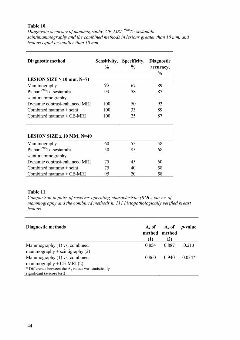
44
Table 10. Diagnostic accuracy of mammography, CE-MRI, 99mTc-sestamibi scintimammography and the combined methods in lesions greater than 10 mm, and lesions equal or smaller than 10 mm. Diagnostic method
Sensitivity,
%
Specificity,
%
Diagnostic accuracy,
% LESION SIZE > 10 mm, N=71 Mammography 93 67 89 Planar 99mTc-sestamibi scintimammography
93 58 87
Dynamic contrast-enhanced MRI 100 50 92 Combined mammo + scint 100 33 89 Combined mammo + CE-MRI
100 25 87
LESION SIZE ≤ 10 MM, N=40
Mammography 60 55 58 Planar 99mTc-sestamibi scintimammography
50 85 68
Dynamic contrast-enhanced MRI 75 45 60 Combined mammo + scint 75 40 58 Combined mammo + CE-MRI 95 20 58 Table 11. Comparison in pairs of receiver-operating-characteristic (ROC) curves of mammography and the combined methods in 111 histopathologically verified breast lesions
Diagnostic methods
Az of
method (1)
Az of
method (2)
p-value
Mammography (1) vs. combined mammography + scintigraphy (2)
0.854 0.887 0.213
Mammography (1) vs. combined mammography + CE-MRI (2)
0.860 0.940 0.034*
* Difference between the Az values was statistically significant (z-score test)
45
DISCUSSION STUDY I
MRI of the breast has, since the introduction of paramagnetic contrast medium (Gadolinium) in the examinations, evolved as an important complementary method to clinical evaluation, mammography and ultrasound. Many different types of sequences have been used in order to facilitate evaluation and reduce the number of false positive as well as false negative findings. For example Harms et al (45) using RODEO, Merchant et al (103) using a mixed imaging sequence and Pierce et al (125) using fat suppression and magnetization transfer contrast. Different enhancement patterns have been identified for benign and malignant lesions (14, 48, 50, 70, 139), but also the importance of fast sequences to enable dynamic examinations, often using subtraction techniques in the evaluation. (12, 40) There has been a large variation in both spatial and temporal resolution as well as in the interpretations of findings, affecting both sensitivity and specificity, which has made comparisons of different studies difficult as discussed by Hrung in 1999 (59). In recent years a standardized technique to facilitate evaluations and comparisons of results have been suggested and discussed by several authors (62, 74, 107, 114). In study I, which was performed before these discussions took place, we tried a different approach. In problem cases presenting a strong and widespread enhancement in the standard semidynamic examination we used a different type of T1 sequence as a second examination. When the study was performed we had no access to faster sequences, and hence both examinations were semidynamic with a rather low temporal resolution, but with a high spatial resolution. The routine FLASH sequence has a high sensitivity for contrast enhancement. As a second examination we introduced a MPRAGE sequence, which presented a less pronounced enhancement of normal breast parenchyma, facilitating the ability to distinguish malignant lesions from benign parenchyma and lesions. All malignant lesions of the study were equally well diagnosed in both sequences, but by using the less contrast-sensitive MPRAGE sequence we were able to downgrade 7 lesions that appeared malignant on the more contrast-sensitive FLASH sequence into the benign lesions they were. MPRAGE also facilitated the differentiation of malignant lesions from breast parenchyma with strong and spread enhancement on the FLASH sequence. The 20 patients studied were a selected group with examinations difficult to interpret, which might partly explain why 6 false positive lesions were found using both sequences. Hormonal influences have been proved to pose significant problems during some periods of the menstrual cycle (81), and the recommendation is to perform MRI during the second week. In this study we did not perform the examinations during any specific period of the menstrual cycle, but there were no differences between the groups (the downgraded and the remaining false positive findings) in age, cause of referral and time interval between the investigations. Even though the number of examined patients was too small for statistical evaluation we found half of the false positive lesions in the contrast-sensitive FLASH sequence could be downgraded to true negative lesions using the less contrast-sensitive MPRAGE sequence. These findings are not that important in clinical practice today, as the development of faster sequences with higher temporal resolution during the last years have facilitated the evaluations.
46
STUDY II
The use of fine-needle aspiration biopsy in diagnosis of breast lesions is a well-known method dating back many years (47), used to confirm or exclude malignancy. It also forms one of the important cornerstones of triple diagnosis (TD), together with clinical examination and mammography, often being complemented by ultrasound. Although a fine-needle is used and the procedure is quickly performed it may influence the histopathological diagnosis, for example by causing edema and hematoma. Klein et al (75) found that FNAB caused false positive findings at mammography during the first week after FNAB, but did not cause any false negative lesions. In a study be Lee et al (87) of histological changes after FNAB 17,4 % of the 184 examined cases revealed changes that were considered definite and probably caused by the FNAB, the most common being hematoma and hemosiderin deposition. Such changes could theoretically impair the MR investigation, not only because of hematomas causing signal changes in different degradation phases, but also by increased contrast enhancement caused by proliferation and reparative processes, thus causing an increase of false positive findings. In a retrospective study Fischer et al (34) studied the effect on the evaluation of MR examinations after performed fine needle and core biopsies. Their results cannot be directly compared to ours due to a thicker slice thickness of 4 mm. However, they found no disturbance of the evaluation of the MR examinations after performed FNAB, but core biopsies often caused hemorrhages that could impair the MR evaluation. In study II we performed a semidynamic MR examination with thin slices both before and after the FNAB was performed, measuring the signal intensities and also the contrast enhancement of the lesions in both examinations, but also of the nearby breast parenchyma and muscle. There were no changes at visual interpretation of the images and no change of the contrast enhancement in the lesion, the surrounding breast parenchyma or the muscle. In this study we had a short interval between the examinations of 3-13 days, hence including the period where mammographic evaluation is known to be problematic. This interval was to establish the FNAB had been diagnostic before performing the second MR examination. Our result showed that there was no impairment of the MR evaluation, even during the first post-puncture week, opposed to the difficulties described at mammography by Klein et al (75). In comparison to the study by Lee et al (87) we had fewer patients, and considering their result we should have expected changes in the tissues caused by the FNAB in at least 2 of the examined lesions. As we found no significant changes in any of the examined lesions we came to the conclusion that FNAB can be performed irrespective of when MRI is scheduled, although a larger study would be of value for better statistical evaluation. STUDY III
During the last 10-20 years there has been a marked evolution of breast imaging from a well defined use of mammography to the complementary use of many breast-imaging techniques, including ultrasound, MRI and scintigraphy. Although mammography has a high sensitivity (80-85%) and a relatively high specificity (50-70%) to detect malignancies it has some shortcomings, for example in dense parenchyma and scar tissue. Initially there was a reluctance to use MRI due to low spatial and temporal
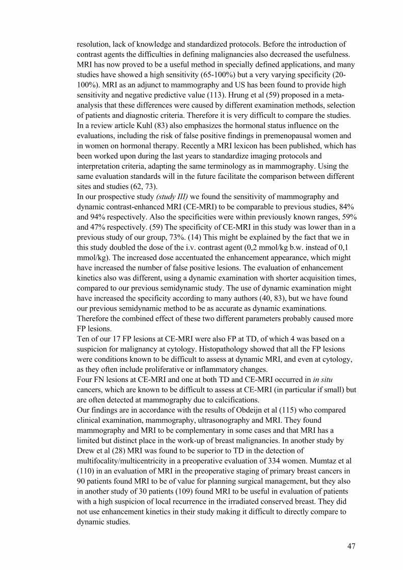
47
resolution, lack of knowledge and standardized protocols. Before the introduction of contrast agents the difficulties in defining malignancies also decreased the usefulness. MRI has now proved to be a useful method in specially defined applications, and many studies have showed a high sensitivity (65-100%) but a very varying specificity (20-100%). MRI as an adjunct to mammography and US has been found to provide high sensitivity and negative predictive value (113). Hrung et al (59) proposed in a meta-analysis that these differences were caused by different examination methods, selection of patients and diagnostic criteria. Therefore it is very difficult to compare the studies. In a review article Kuhl (83) also emphasizes the hormonal status influence on the evaluations, including the risk of false positive findings in premenopausal women and in women on hormonal therapy. Recently a MRI lexicon has been published, which has been worked upon during the last years to standardize imaging protocols and interpretation criteria, adapting the same terminology as in mammography. Using the same evaluation standards will in the future facilitate the comparison between different sites and studies (62, 73). In our prospective study (study III) we found the sensitivity of mammography and dynamic contrast-enhanced MRI (CE-MRI) to be comparable to previous studies, 84% and 94% respectively. Also the specificities were within previously known ranges, 59% and 47% respectively. (59) The specificity of CE-MRI in this study was lower than in a previous study of our group, 73%. (14) This might be explained by the fact that we in this study doubled the dose of the i.v. contrast agent (0,2 mmol/kg b.w. instead of 0,1 mmol/kg). The increased dose accentuated the enhancement appearance, which might have increased the number of false positive lesions. The evaluation of enhancement kinetics also was different, using a dynamic examination with shorter acquisition times, compared to our previous semidynamic study. The use of dynamic examination might have increased the specificity according to many authors (40, 83), but we have found our previous semidynamic method to be as accurate as dynamic examinations. Therefore the combined effect of these two different parameters probably caused more FP lesions. Ten of our 17 FP lesions at CE-MRI were also FP at TD, of which 4 was based on a suspicion for malignancy at cytology. Histopathology showed that all the FP lesions were conditions known to be difficult to assess at dynamic MRI, and even at cytology, as they often include proliferative or inflammatory changes. Four FN lesions at CE-MRI and one at both TD and CE-MRI occurred in in situ cancers, which are known to be difficult to assess at CE-MRI (in particular if small) but are often detected at mammography due to calcifications. Our findings are in accordance with the results of Obdeijn et al (115) who compared clinical examination, mammography, ultrasonography and MRI. They found mammography and MRI to be complementary in some cases and that MRI has a limited but distinct place in the work-up of breast malignancies. In another study by Drew et al (28) MRI was found to be superior to TD in the detection of multifocality/multicentricity in a preoperative evaluation of 334 women. Mumtaz et al (110) in an evaluation of MRI in the preoperative staging of primary breast cancers in 90 patients found MRI to be of value for planning surgical management, but they also in another study of 30 patients (109) found MRI to be useful in evaluation of patients with a high suspicion of local recurrence in the irradiated conserved breast. They did not use enhancement kinetics in their study making it difficult to directly compare to dynamic studies.
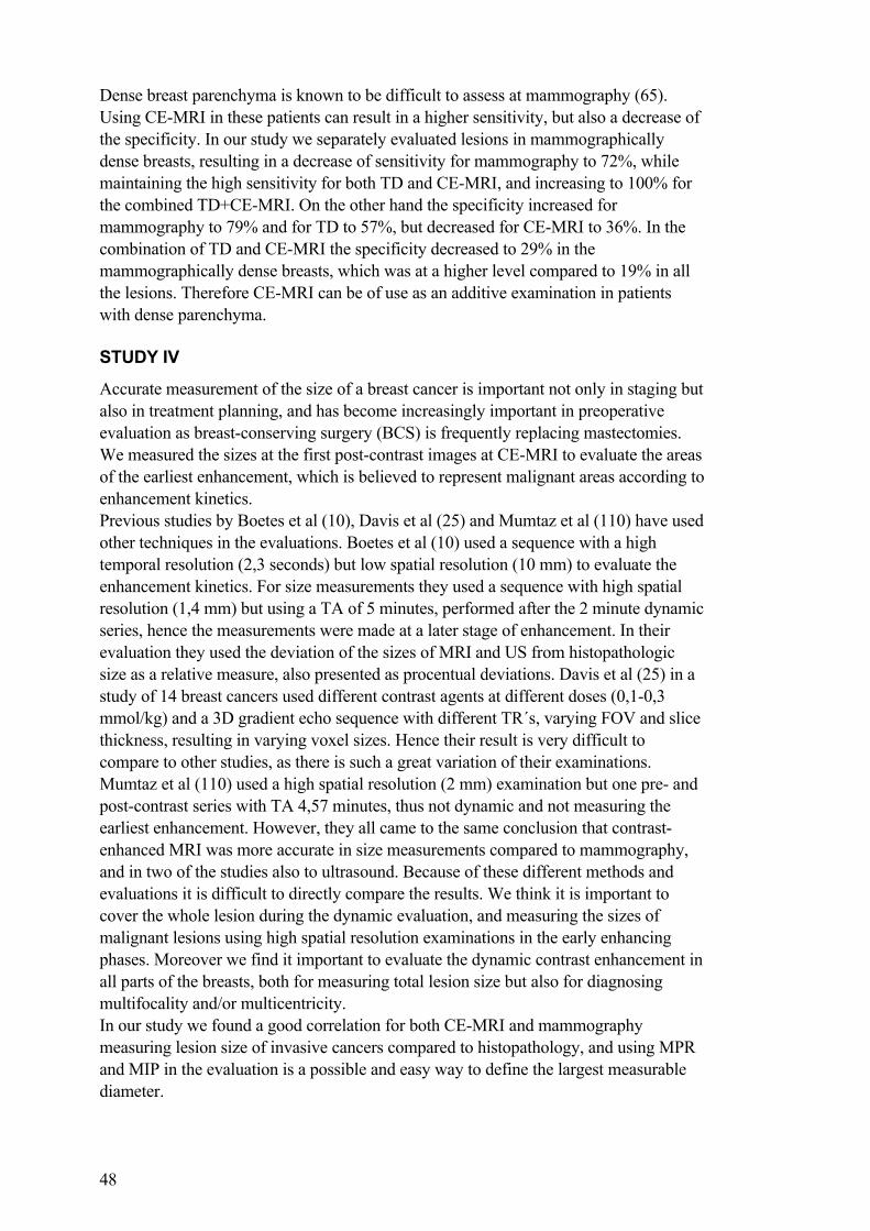
48
Dense breast parenchyma is known to be difficult to assess at mammography (65). Using CE-MRI in these patients can result in a higher sensitivity, but also a decrease of the specificity. In our study we separately evaluated lesions in mammographically dense breasts, resulting in a decrease of sensitivity for mammography to 72%, while maintaining the high sensitivity for both TD and CE-MRI, and increasing to 100% for the combined TD+CE-MRI. On the other hand the specificity increased for mammography to 79% and for TD to 57%, but decreased for CE-MRI to 36%. In the combination of TD and CE-MRI the specificity decreased to 29% in the mammographically dense breasts, which was at a higher level compared to 19% in all the lesions. Therefore CE-MRI can be of use as an additive examination in patients with dense parenchyma. STUDY IV
Accurate measurement of the size of a breast cancer is important not only in staging but also in treatment planning, and has become increasingly important in preoperative evaluation as breast-conserving surgery (BCS) is frequently replacing mastectomies. We measured the sizes at the first post-contrast images at CE-MRI to evaluate the areas of the earliest enhancement, which is believed to represent malignant areas according to enhancement kinetics. Previous studies by Boetes et al (10), Davis et al (25) and Mumtaz et al (110) have used other techniques in the evaluations. Boetes et al (10) used a sequence with a high temporal resolution (2,3 seconds) but low spatial resolution (10 mm) to evaluate the enhancement kinetics. For size measurements they used a sequence with high spatial resolution (1,4 mm) but using a TA of 5 minutes, performed after the 2 minute dynamic series, hence the measurements were made at a later stage of enhancement. In their evaluation they used the deviation of the sizes of MRI and US from histopathologic size as a relative measure, also presented as procentual deviations. Davis et al (25) in a study of 14 breast cancers used different contrast agents at different doses (0,1-0,3 mmol/kg) and a 3D gradient echo sequence with different TR´s, varying FOV and slice thickness, resulting in varying voxel sizes. Hence their result is very difficult to compare to other studies, as there is such a great variation of their examinations. Mumtaz et al (110) used a high spatial resolution (2 mm) examination but one pre- and post-contrast series with TA 4,57 minutes, thus not dynamic and not measuring the earliest enhancement. However, they all came to the same conclusion that contrast-enhanced MRI was more accurate in size measurements compared to mammography, and in two of the studies also to ultrasound. Because of these different methods and evaluations it is difficult to directly compare the results. We think it is important to cover the whole lesion during the dynamic evaluation, and measuring the sizes of malignant lesions using high spatial resolution examinations in the early enhancing phases. Moreover we find it important to evaluate the dynamic contrast enhancement in all parts of the breasts, both for measuring total lesion size but also for diagnosing multifocality and/or multicentricity. In our study we found a good correlation for both CE-MRI and mammography measuring lesion size of invasive cancers compared to histopathology, and using MPR and MIP in the evaluation is a possible and easy way to define the largest measurable diameter.
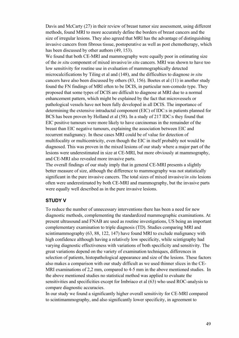
49
Davis and McCarty (27) in their review of breast tumor size assessment, using different methods, found MRI to more accurately define the borders of breast cancers and the size of irregular lesions. They also agreed that MRI has the advantage of distinguishing invasive cancers from fibrous tissue, postoperative as well as post chemotherapy, which has been discussed by other authors (49, 153). We found that both CE-MRI and mammography were equally poor in estimating size of the in situ component of mixed invasive/in situ cancers. MRI was shown to have too low sensitivity for routine use in evaluation of mammographically detected microcalcifications by Tiling et al and (148), and the difficulties to diagnose in situ cancers have also been discussed by others (83, 156). Boetes et al (11) in another study found the FN findings of MRI often to be DCIS, in particular non-comedo type. They proposed that some types of DCIS are difficult to diagnose at MRI due to a normal enhancement pattern, which might be explained by the fact that microvessels or pathological vessels have not been fully developed in all DCIS. The importance of determining the extensive intraductal component (EIC) of IDC:s in patients planned for BCS has been proven by Holland et al (58). In a study of 217 IDC:s they found that EIC positive tumours were more likely to have carcinomas in the remainder of the breast than EIC negative tumours, explaining the association between EIC and recurrent malignancy. In these cases MRI could be of value for detection of multifocality or multicentricity, even though the EIC in itself probably not would be diagnosed. This was proven in the mixed lesions of our study where a major part of the lesions were underestimated in size at CE-MRI, but more obviously at mammography, and CE-MRI also revealed more invasive parts. The overall findings of our study imply that in general CE-MRI presents a slightly better measure of size, although the difference to mammography was not statistically significant in the pure invasive cancers. The total sizes of mixed invasive/in situ lesions often were underestimated by both CE-MRI and mammography, but the invasive parts were equally well described as in the pure invasive lesions. STUDY V
To reduce the number of unnecessary interventions there has been a need for new diagnostic methods, complementing the standardized mammographic examinations. At present ultrasound and FNAB are used as routine investigations, US being an important complementary examination to triple diagnosis (TD). Studies comparing MRI and scintimammography (63, 88, 122, 147) have found MRI to exclude malignancy with high confidence although having a relatively low specificity, while scintigraphy had varying diagnostic effectiveness with variations of both specificity and sensitivity. The great variations depend on the variety of examination techniques, differences in selection of patients, histopathological appearance and size of the lesions. These factors also makes a comparison with our study difficult as we used thinner slices in the CE-MRI examinations of 2,2 mm, compared to 4-5 mm in the above mentioned studies. In the above mentioned studies no statistical method was applied to evaluate the sensitivities and specificities except for Imbriaco et al (63) who used ROC-analysis to compare diagnostic accuracies. In our study we found a significantly higher overall sensitivity for CE-MRI compared to scintimammography, and also significantly lower specificity, in agreement to
50
previous publications. The difference in sensitivity between the methods was larger than the difference in specificity. It is well known that MRI often fails in diagnosing in situ cancers, and all 5 FN lesions in our study were in situ lesions. A relatively high number of FP lesions, including hyperplastic changes and 5 of the 8 fibroadenomas showed early and strong enhancement, resulted in the low specificity. Scintimammography had almost as high sensitivity as CE-MRI and the specificity was higher, 75%. Previously for example Danielsson et al (23) and Prats et al (127) described that the sensitivity of scintimammography is strongly dependent on lesion size, and the low resolution of the method reduces the accuracy in lesions smaller than 10 mm. Among lesions larger than 10 mm in our study the two methods had comparable false positive rates, but among the lesions smaller than 10 mm the significant difference between CE-MRI and scintimammography remained. This might be explained by the size limitations of scintigraphy and the relatively higher specificity as all small benign lesions will not be detected, which will result in true negative findings and thus increase specificity. Therefore scintimammography has a doubtful role in evaluation of problem cases detected at mammography measuring less than10 mm. The overall diagnostic accuracy was equal when comparing mammography, CE-MRI and scintimammography of our study (78%, 80% and 80%) as independent methods. The combination of mammography and CE-MRI resulted in a high sensitivity of 99%, as most lesions missed by CE-MRI were diagnosed at mammography, but the specificity was also markedly decreased (22%) reducing the diagnostic value of this combination. When combining mammography and scintimammography the sensitivity was high, 94%, but the specificity also was low, 38%. In clinical use MRI and scintigraphy have proven their value in breast diagnosis, not only in inconclusive findings at mammography, but also for example in patients with dense parenchyma at mammography, axillary metastases of unknown primary (118, 134) and for exclusion of multifocality (33, 110). In our study we found CE-MRI and 99mTc-sestamibi scintimammography to show comparable diagnostic accuracy in differentiating malignant lesions from benign lesions and normal parenchyma. The diagnostic accuracy in lesions smaller than 10 mm is however vastly reduced in scintimammography due to the low resolution of the method.
51
CONCLUSION MRI of the breast has since the use of a Gadolinium contrast agent was introduced developed into a standardized method, and in recent years recommendations for a standardized interpretation of the examinations has been introduced. Many studies have been performed during the last 20 years using different methods, different selection criteria for included patients, different contrast agents and doses, different interpretation models and different evaluation criteria. In our studies we found that
• FLASH is a sequence with high sensitivity revealing contrast-enhancing areas, and therefore is the first choice in routine examinations. When atypical or spread increased enhancement was detected, the addition of the MPRAGE sequence improved the specificity of the MR investigation, and therefore can be used as a problem-solver when diffuse and marked contrast enhancement is found in the FLASH sequence.
• MRI evaluations were not impaired and could be performed with good accuracy after FNAB of a lesion, and thus MRI can be performed irrespective of the time interval to a performed fine-needle aspiration.
• CE-MRI of the breast is a valuable additive method to triple diagnosis (clinical examination, mammography and cytology) due to high sensitivity but at the cost of decreasing specificity, which was less pronounced in patients with mammographically dense breasts. Hence women with mammographically dense breasts might benefit most from CE-MRI as an additive method in differentiation of benign and malignant lesions.
• CE-MRI presents a slightly better measure of size of a lesion, although the difference to mammography was not statistically significant in the pure invasive lesions. Both CE-MRI and mammography often underestimated the total sizes of the mixed invasive/in situ lesions, although the invasive parts were equally well described in the mixed lesions as in the pure invasive lesions.
• If high sensitivity and spatial resolution is needed CE-MRI is to be preferred in clinical practice as an adjunct to mammography, rather than scintigraphy.
MRI is a useful tool in breast diagnosis in particular due to the high sensitivity, the possibility to exclude multifocality/multicentricity and good evaluations of size and extent of a lesion. The high sensitivity and high spatial resolution of CE-MRI is desired when evaluating problematic cases in conventional breast imaging methods. Due to the relatively low specificity it is not to be recommended as a screening tool, but as an additive method in selected cases.
52
ACKNOWLEDGEMENTS This work was carried out at the MRI department and in part at the Mammography department and Nuclear Medicine department of the Radiology department, the Surgical department and the Pathology department at Huddinge University Hospital. I wish to express sincere gratitude to those who in different ways have supported me in this work and have made this thesis a reality. My special thanks go to: Associate professor Beata Boné, my tutor, who has a genuine interest in breast imaging combined with a vast experience, and has managed to convey parts of that to me. Beata´s enthusiasm has followed this work through many years and discussions. Our first meeting was at the MRI department in 1992 when I was visiting for some months. Thank you Beata for many fruitful and interesting discussions covering many topics during the years when we shared an office, during the collection of the basic data and during the final work of this thesis. Professor Peter Aspelin, my co-tutor, who has always been there with encouragement, interest, support and time for discussions. Thank you for giving me the opportunity to start working at the Radiology Department, for the support to get started in research and also for maintained support with constructive criticism combined with thoughtful and often enjoyable remarks throughout these years. Associate professor Bo Persson, head of the Radiology department, who in the busy and demanding clinical activities of our department always manages to defend the important position of research linked to clinical issues. Thank you for your support during these years providing the time enabling me to finish this work. Associate professor Rimma Danielsson, my co-worker during parts of this work. Thank you for your support and encouragement during the collection of basic data, during parts of the evaluations and for sharing your knowledge in scintigraphy, scintimammography in particular. Associate professor Lena Bronge for many years of close work together at the MRI department and neuro-section, sharing many problems in common and being there for backup when needed. Thank you for your support and constructive comments on both research and daily clinical work at the department. Dr Botond K. Szabó, my co-worker during the last years, who has given new aspects to my work, shared the work of structuring background information and taken part in constructive discussions as well as statistical evaluations. Associate professors Leif Perbeck, Eva Reihnér, and Dr Fuat Celebioglu, colleagues at the Surgery department, for their support and interest in improving breast diagnostics. Statisticians Bo Nilsson and Lisa Jervall for expert statistical advice and many useful discussions during different phases of this work.
53
All the personnel at the MRI department who are always interested in new applications and developments as well as always trying to manage to examine even more patients. Special thanks to Mariana Ahlin who took great part in the organizing and performance of these MRI examinations, but also Sirkka Kihlman, Karin Lindberg and Kerstin Eriksson who all performed breast-MRI examinations being evaluated in this thesis. All the personnel at the Mammography department who were always willing to cope with all the data, images and paperwork needed, in particular during the data-collection years. Special thanks to Maj-Britt Hillborg and Monika Lindén who always knew where to find what was missing and Dr Christina Hedborg-Mellander for sharing her knowledge in breast diagnostics. Helena Forssell and Maj-Britt Stäring for expert secretarial help and support, but also for finding ways to solve many practical problems during the years. Associate professor Bengt Isberg for introducing me to research in the MRI-field and sharing his great knowledge in MRI, diagnostic examinations and assessments as well as the physics. Associate professor Susanne Müller for her enthusiasm, cheerfulness and humour always present to brighten up the days of clinical work. Thank you for taking care of the work at the MRI department during the busy times when I have been working with my thesis. All my colleagues and friends at the Radiology department, Huddinge University Hospital, who have had to share my part of the work during this period. Sarah H. Kristoffersen for expert assistance in the English language. Tony H.W. Nias (former professor of cancer research, the Oncology Department, St Thomas´s Hospital, London University) for introducing me to the world of medicine as well as the research world. Thank you for sharing many thoughts on work and life. My mother and father who taught me to care for even the smallest things, to always remain open-minded and never give up, to be curious, to think positive, but also to show respect and understanding. To my mother: Thank you for being my best friend throughout my life, always having time for discussions and practical help. Last but not least, special thanks to my husband and two children (John, Fredrik and Annika) for their support and understanding in particular during the last years, when I always have seemed to have too much on my mind and too much to do. Always standing by my side, no matter the situations, giving unconditional love and all sorts of assistance when needed. All the women participating in the examinations, making these studies possible. This work was in part supported by grants from the Petrus and Augusta Hedlund Foundation.
54
REFERENCES 1. Ahn CY, DeBruhl ND, Gorczyca DP, Shaw WW & Bassett LW.
Comparative silicone breast implant evaluation using mammography, sonography, and magnetic resonance imaging: experience with 59 implants. Plast Reconstr Surg 94 (1994) 620-627.
2.
Alveryd A, Andersson I, Aspegren K, Balldin G, Bjurstam N, Edström G, Fagerberg G, Glas U, Jarlman O, Larsson SA, Lidbrink E, Lingaas H, Löfgren M, Rudenstam C-M, Strender L, Samuelsson L, Tàbar L, Taube A, Wallberg H, Åkesson P & Hallberg D. Lightscanning versus mammography for the detection of breast cancer in screening and clinical practice. A Swedish multicenter study. Cancer 65 (1990) 1671-1677.
3. American College of Radiology. Breast imaging reporting and data system (BI-RADS), 3rd ed. American College of Radiology, Reston, Virginia, 1998.
4. Axel L. Surface coil magnetic resonance imaging. Journal of Computer Assisted Tomography 8 (1984) 381-384.
5. Azavedo E & Boné B. Imaging breasts with silicone implants. European Radiology 9 (1999) 349-355.
6. Bangert V, Mansfield P & Coupland RE. Whole-body tomographic imaging by NMR. British Journal of Radiology 54 (1981) 152-154.
7. Baum F, Fischer U, Vosshenrich R & Grabbe E. Classification of hypervascularized lesions in CE MR imaging of the breast. European Radiology 12 (2002) 1087-1092.
8. Berg WA, Caskey CI, Hamper UM, Kuhlman JE, Anderson ND, Chang BW, Sheth S & Zerhouni EA. Single- and double-lumen silicone breast implant integrity: Prospective evaluation of MR and US criteria. Radiology 197 (1995) 45-52.
9. Blohmer JU, Oellinger H, Schmidt C, Hufnagl P, Felix R & Lichtenegger W. Comparison of various imaging methods with particular evaluation of color doppler sonography for planning surgery for breast tumors. Arch. Gynecol. Obstet. 262 (1999) 159-171.
10. Boetes C, Mus RDM, Holland R, Barentsz JO, Strijk SP, Wobbes T, Hendriks JHCL & Ruys SHJ. Breast tumors: Comparative accuracy of MR imaging relative to mammography and US for demonstrating extent. Radiology 197 (1995) 743-747.
11. Boetes C, Strijk SP, Holland R, Barentsz JO, Van Der Sluis RF & Ruijs JHJ.
False-negative MR imaging of malignant breast tumors. European Radiology 7 (1997) 1231-1234.
55
12. Boetes C, Barentsz JO, Mus RD, van der Sluis RF, van Erning LJTO, Hendriks JHCL, Holland R & Ruys SHJ.
MR characterization of suspicious breast lesions with a Gadolinium-enhanced Turbo-FLASH subtraction technique. Radiology 193 (1994) 777-781.
13. Boné B, Aspelin P, Isberg B, Perbeck L & Veress B. Contrast-enhanced MR imaging of the breast in patients with breast implants after cancer surgery. Acta Radiologica 36 (1995) 111-116.
14. Boné B, Aspelin P, Bronge L, Isberg B, Perbeck L & Veress B. Sensitivity and specificity of MR mammography with histopathological correlation in 250 breasts. Acta Radiologica 37 (1996) 208-213.
15. Brasch RC, Weinmann H-J & Wesbey GE. Contrast-enhanced NMR imaging: Animal studies using gadolinium-DTPA complex. American Journal of Radiology 142 (1984) 625-630.
16. Bray F, Sankila R, Ferlay J & Parkin D.M. Estimates of cancer incidence and mortality in Europe in 1995. European Journal of Cancer 38 (2002) 99-166.
17. Brewster A & Helzlsouer K. Breast cancer epidemiology, prevention, and early detection. Current Opinion in Oncology 13 (2001) 420-425.
18. Buckley DL, Mussurakis S & Horsman A. Effect of temporal resolution on the diagnostic efficacy of contrast-enhanced MRI in the conservatively treated breast. Journal of Computer Assisted Tomography 22 (1998) 47-51.
19. Cabrera C, Helgesson Ö, Wedel H, Björkelund C, Bengtsson C & Lissner L. Socioeconomic status and mortality in Swedish women: opposing trends for cardiovascular disease and cancer. Epidemiology 12 (2001) 532-536.
20. Cecil KM, Schnall MD, Siegelman ES & Lenkinski RE. The evaluation of human breast lesions with magnetic resonance imaging and proton magnetic resonance spectroscopy. Breast Cancer Research and Treatment 68 (2001) 45-54.
21. Chenevert TL, Helvie MA, Aisen AM, Francis IR, Adler DD, Roubidoux MA & Londy FJ. Dynamic three-dimensional imaging with partial k-space sampling: initial application for gadolinium-enhanced rate characterization of breast lesions. Radiology 196 (1995) 135-142.
22. Damadian R. Tumor detection by nuclear magnetic resonance. Science Vol.171 (1971) 1151-1153
23. Danielsson R, Boné B, Gad A, Sylvan M & Aspelin P. Sensitivity and specificity of planar scintimammography with 99mTc-sestamibi. Acta Radiologica 40 (1999) 394-399.
56
24. Dash N, Lupetin AR, Daffner RH, Deeb ZL, Sefczek RJ & Schapiro RL. Magnetic resonance imaging in the diagnosis of breast disease. American Journal of Radiology 146 (1986) 119-125.
25. Davis PL, Staiger MJ, Harris KB, Ganott MA, Klementaviciene J, McCarty Jr KS & Tobon H. Breast cancer measurements with magnetic resonance imaging, ultrasonography, and mammography. Breast Cancer Research and Treatment 37 (1996) 1-9.
26. Davis PL & McCarty Jr KS. Magnetic resonance imaging in breast cancer staging. Topics in Magnetic Resonance Imaging 9 (1998) 60-75.
27. Davis PL & McCarty Jr KS. Technologic considerations for breast tumor size assessment. Breast Imaging 2 (1994) 623-631.
28. Drew PJ, Chatterjee S, Turnbull LW, Read J, Carleton PJ, Fox JN, Monson JRT & Kerin MJ. Dynamic contrast enhanced magnetic resonance imaging of the breast is superior to triple assessment for the pre-operative detection of multifocal breast cancer. Annals of Surgical Oncology 6 (1999) 599-603.
29. El Yousef SJ, O´Connell DM, Duchesneau RH, Smith MJ, Hubay CA & Guyton SP.
Benign and malignant breast disease: Magnetic Resonance and radiofrequency pulse sequences. American Journal of Radiology 145 (1985) 1-8.
30. El Yousef SJ, Alfidi RJ, Duchesneau RH, Hubay CA, Haaga JR, Bryan PJ, LiPuma JP & Ament AE. Initial experience with nuclear magnetic resonance (NMR) imaging of the human breast. Journal of Computer Assisted Tomography 7 (1983) 215-218.
31. Feig SA, Shaber GS, Schwartz GF, Patchefsky A, Lishitz HI, Edeiken., Nerlinger R, Curley RF & Wallace JD. Thermography, mammography, and clinical examination in breast cancer screening. Radiology 122 (1977) 123-127.
32. Felix R, Heshiki A, Hricak H, Chang K-H, Hosten N & Lemke A-J. Magnevist® Monograph, 4th revised edition. Blackwell Wissenschafts-Verlag, Berlin · Vienna 2001.
33. Fischer U, Kopka L & Grabbe E. Breast carcinoma: Effect of preoperative contrast-enhanced MR imaging on the therapeutic approach. Radiology 213 (1999) 881-888.
34. Fischer U, Vosshenrich R, Kopka L, Kahlen O & Grabbe E. Kontrastmittelgestützte dynamische MR-Mammographie nach diagnostischen und therapeutischen Eingriffen an der Mamma. Bildgebung 63 (1996) 94-100.
57
35. Fischer U, Vosshenrich R, Keating D, Bruhn H, Döler W, Oestmann JW & Grabbe E. MR-guided biopsy of suspect breast lesions with a simple stereotaxic add-on device for surface coils. Radiology 192 (1994) 272-273.
36. Fischer U, Vosshenrich R, Döler W, Hamadeh A, Oestmann JW & Grabbe E. MR imaging-guided breast intervention: Experience with two systems. Radiology 195 (1995) 533-538.
37. Frisell J, Bergqvist L, Liljegren G, Thorn M, Damm S, Rydman H & Danielsson R. Sentinel node in breast cancer – a Swedish pilot study of 75 cases. Europen Journal of Surgery 167:3 (2001) 179-183.
38. Furman-Haran E, Grobgeld D, Kelcz F & Degani H. Critical role of spatial resolution in dynamic contrast-enhanced breast MRI. Journal of Magnetic Resonance Imaging 13 (2001) 862-867.
39. Gilles R, Guinebretière J-M, Toussaint C, Spielman M, Rietjens M, Petit JY, Contesso G, Masselot J & Vanel D. Locally advanced breast cancer: contrast-enhanced subtraction MR imaging of response to preoperative chemotherapy. Radiology 191 (1994) 633-638.
40. Gilles R, Guinebretière J-M, Lucidarme O, Cluzel P, Janaud G, Finet J-F, Tardivon A, Masselot J & Vanel D. Nonpalpable breast tumors: Diagnosis with contrast-enhanced subtraction dynamic MR imaging. Radiology 191 (1994) 625-631.
41. Goldsmith M, Koutcher JA & Damadian R. NMR in cancer, XIII: application of the NMR malignancy index to human mammary tumours. British Journal of Cancer 38(4) (1978) 547-554.
42. Gorczyca DP, DeBruhl ND, Ahn CY, Hoyt A, Sayre JW, Nudell P, McCombs M, Shaw WW & Bassett LW. Silicone breast implant ruptures in an animal model: Comparison of mammography, MR imaging, US, and CT. Radiology 190 (1994) 227-232.
43. Gönen M, Panageas KS & Larson SM. Statistical issues in analysis of diagnostic imaging experiments with multiple observations per patient. Radiology 221 (2001) 763-767
44. Hall-Craggs MA. Interventional MRI of the breast: minimally invasive therapy. European Radiology 10 (2000) 59-62.
45. Harms SE, Flamig DP, Hesley KL, Meiches MD, Jensen RA, Evans WP, Savino DA & Wells RV. MR Imaging of the breast with rotating delivery of excitation off resonance: clinical experience with pathologic correlation. Radiology 187 (1993) 493-501.
58
46. Helbich TH. Localiztion and biopsy of breast lesions by magnetic resonance imaging guidance. Journal of Magnetic Resonance Imaging 13 (2001) 903-911.
47. Hermansen C, Skovgaard Poulsen H, Jensen J, Langfeldt B, Steenskov V, Frederiksen P & Jensen OM. Diagnostic reliability of combined physical examination, mammography, and fine-needle puncture (“triple-test”) in breast tumors. A prospective study. Cancer 60 (1987), 1866-1871.
48. Heywang-Köbrunner SH Contrast-enhanced magnetic resonance imaging of the breast. Investigative Radiology 29 (1994) 94-104.
49. Heywang SH, Hilbertz T, Beck R, Bauer WM, Eiermann W & Permanetter W.
Gd-DTPA enhanced MR imaging of the breast in patients with postoperative scarring and silicon implants. Journal of Computer Assisted Tomography 14 (1990) 348-356.
50. Heywang-Köbrunner SH, Bick U, Bradley WGJr., Boné B, Casselman J, Coulthard A, Fischer U, Müller-Schimpfle M, Oellinger H, Patt R, Teubner J, Friedrich M, Newstead G, Holland R, Schauer A, Sickles EA, Tabar L, Waisman J & Wernecke KD.
International investigation of breast MRI: results of a multicentre study (11 sites) concerning diagnostic parameters for contrast-enhanced MRI based on 519 histopathologically correlated lesions. European Radiology 11 (2001) 531-546.
51. Heywang-Köbrunner SH, Heinig A, Pickuth D, Alberich T & Spielmann RP. Interventional MRI of the breast: lesion localization and biopsy. European Radiology 10 (2000) 36-45.
52. Heywang SH, Fenzl G, Hahn D, Krischke I, Edmaier M, Eiermann W & Bassermann R. MR imaging of the breast: comparison with mammography and ultrasound. Journal of Computer Assissted Tomography 10 (1986) 615-620.
53. Heywang SH, Bassermann R, Fenzl G, Nathrath W, Hahn D, Beck R, Krischke I & Eiermann W. MRI of the breast—histopathologic correlation. European Journal of Radiology 7 (1987) 175-182.
54. Heywang SH, Hahn D, Schmidt H, Krischke I, Eiermann W, Bassermann R & Lissner J.
MR imaging of the breast using gadolinium-DTPA. Journal of Computer Assisted Tomography 10 (1986) 199-204.
55. Heywang SH, Wolf A, Pruss E, Hilbertz T, Eiermann W & Permanetter W. MR imaging of the breast with Gd-DTPA: Use and limitations. Radiology 171 (1989) 95-103.
56. Hoh CK & Schiepers C. 18-FDG imaging in breast cancer. Semin Nucl Med 29 (1999) 49-56.
59
57. Holland AE, Hendrick RE, Jin H, Russ PD, Barentsz JO & Holland R. Correlation of high-resolution breast MR imaging with histopathology; validation of a technique. Journal of Magnetic Resonance Imaging 11 (2000) 601-606.
58. Holland R, Connolly JL, Gelman R, Mravunac M, Hendriks JHCL, Verbeek ALM, Schnitt SJ, Silver B, Boyages J & Harris JR. The presence of an extensive intraductal component following a limited excision correlates with prominent residual disease in the remainder of the breast. Journal of Clinical Oncology 8 (1990) 113-118.
59. Hrung JM, Sonnad SS, Schwarts JS & Langlotz CP. Accuracy of MR imaging in the work-up of suspicious breast lesions: A diagnostic meta-analysis. Acad Radiol 6 (1999) 387-397.
60. Hulka CA, Smith BL, Sgroi DC, Tan L, Edminster WB, Semple JP, Campbell T, Kopans DB, Brady TJ & Weisskoff RM. Benign and malignant breast lesions: Differentiation with echo-planar MR imaging. Radiology 197 (1995) 33-38.
61. Ikeda O, Yamashita Y, Morishita S, Kido T, Kitajima M, Okamura K, Fukuda S & Takahashi M. Characterization of breast masses by dynamic enhanced MR imaging.
Acta Radiologica 40 (1999) 585-592. 62. Ikeda D.M, Hylton N.M, Kinkel K, Hochman MG, Kuhl CK, Kaiser WA,
Weinreb JC, Smazal SF, Degani H, Viehweg P, Barclay J & Schnall MD. Development, standardization and testing of a lexicon for reporting contrast-enhanced breast magnetic resonance imaging studies. Journal of Magnetic Resonance Imaging 13 (2001) 889-895.
63. Imbriaco M, Del Vecchio S, Riccardi A, Pace L, Di Salle F, Di Gennaro F, Salvatore M & Sodano A. Scintimammography with 99mTc-MIBI versus dynamic MRI for non-invasive characterization of breast masses. European Journal of Nuclear Medicine 28 (2001) 56-63.
64. Isard HJ, Sweitzer CJ & Edelstein GR. Breast thermography. A prognostic indicator for breast cancer survival. Cancer 62 (1988) 484-488.
65. Jackson V P, Hendrick R E, Feig S A & Kopans D B. Imaging of the radiographically dense breast. Radiology 188 (1993), 297-301.
66. Jackson VP, Reynolds HE & Hawes DR. Sonography of the breast. Seminars in Ultrasound, CT and MRI 17 (1996) 460-475.
67. Jarlman O, Andersson I, Balldin G & Larsson SA. Diagnostic accuracy of lightscanning and mammography in women with dense breasts. Acta Radiologica 33 (1992) 69-71.
60
68. Jarlman O, Balldin G, Andersson I, Löfgren M, Larsson AS & Linell F. Relation between lightscanning and the histologic and mammographic appearance of malignant breast tumors. Acta Radiologica 33 (1992) 63-68.
69. Kaiser WA. MRI of the female breast. First clinical results. Arch Int Physiol Biochim. 93 (1985) 67-76.
70. Kaiser WA. & Zeitler E. MR imaging of the breast: Fast imaging sequences with and without Gd-DTPA. Radiology 170 (1989) 681-686.
71. Katz-Brull R, Lavin PT & Lenkinski.E. Clinical utility of proton magnetic resonance spectroscopy in characterizing breast lesions. Journal of the National Cancer Institute 94 (2002) 1197-1203.
72. Khalkhali I, Mena I & Diggles L. Review of imaging techniques for the diagnosis of breast cancer: a new role of prone scintimammography using technetium-99m sestamibi. Euroepan Journal of Nuclear Medicine 21 (1994) 357-362.
73. Kim SJ, Morris EA, Liberman L, Ballon DJ, La Trenta LR, Hadar O, Abramson A & Dershaw DD. Observer variability and applicability of BI-RADS terminology for breast MR imaging: Invasive carcinomas as focal masses. American Journal of Radiology 177 (2001) 551-557.
74. Kinkel K & Hylton NM. Challenges to interpretation of breast MRI. Journal of Magnetic Resonance Imaging 13 (2001) 821-829.
75. Klein DL & Sickles EA. Effects of needle aspiration on the mammographic appearance of the breast. A guide to the proper timing of the mammography examination. Radiology 145 (1982) 44.
76. Konyer NB, Ramsay EA, Bronskill MJ & Plewes DB. Comparison of MR imaging breast coils. Radiology 222 (2002) 830-834.
77. Kopans DB. Breast Imaging. Second edition. Lippincott-Raven Publishers (1998)
78. Koutcher JA, Goldsmith M & Damadian R. NMR in cancer. X. A malignancy index to discriminate normal and cancerous tissue. Cancer 41 (1978) 174-182.
79. KristoffersenWiberg M, Aspelin P, Perbeck L & Boné B. Value of MR imaging in clinical evaluation of breast lesions. Acta Radiologica 43 (2002) 275-281.
61
80. Kuhl CK, Mielcareck P, Klaschik S, Leutner C, Wardelmann E, Gieseke J & Schild HH. Dynamic breast MR imaging: Are signal intensity time course data useful for differential diagnosis of enhancing lesions? Radiology 211 (1999) 101-110.
81. Kuhl CK, Seibert C, Sommer T, Kreft B, Gieseke J & Schild HH. Fokale und diffuse läsionen in der dynamischen MR-mammographie gesunder probandinnen. Fortschr. Röntgenstr. 163 (1995) 219-224.
82. Kuhl CK, Elevelt A, Leutner CC, Gieseke J, Pakos E & Schild H. Interventional breast MR imaging: Clinical use of a stereotactic localization and biopsy device. Radiology 204 (1997) 667-675.
83. Kuhl CK. MRI of breast tumors. European Radiology 10 (2000) 46-58.
84. Kvistad KA, Rydland J, Vainio J, Smethurst HB, Lundgren S, Fjøsne HE & Haraldseth O. Breast lesions: Evaluation with dynamic contrast-enhanced T1-weighted MR imaging and with T2*-weighted first-pass perfusion MR imaging. Radiology 216 (2000) 545-553.
85. Kvistad KA, Bakken IJ, Gribbestad IS, Ehrnholm B, Lundgren S, Fjøsne HE & Haraldseth O. Characterization of neoplastic and normal human breast tissues with in vivo 1H MR spectroscopy. Journal of magnetic resonance imaging 10 (1999) 159-164.
86. Ladjevardi S, Sörensen J, Hellman P, Juhlin C & Wärnberg F. FDG-PET vid ockult bröstcancer. Läkartidningen 99 (2002) 524-526.
87. Lee KC, Chan JKC & Ho LC. Histologic changes in the breast after fine-needle aspiration. American Journal of Surgical Pathology 18 (1994) 1039-1047.
88. Leinsinger GL, Friedl L, Tiling R, Scherr MK, Heiss DT, Kandizora C, Camerer B, Sommer H, Pfluger T & Hahn K. Comparison of dynamic MR imaging of the breast and sestamibi scintimammography for evaluation of indeterminate mammographic lesions. European Radiology 11 (2001) 2050-2057.
89. Levi F, Lucchini F, Negri E & La Vecchia C. The fall in breast cancer mortality in Europe. European Journal of Cancer 37 (2001) 1409-1412.
90. Liberman L, Morris EA, Lee ML-Y, Kaplan JB, LaTrenta LR, Menell JH, Abramson AF, Dashnaw SM, Ballon DJ & Dershaw DD. Breast lesions detected on MR imaging: Features and positive predictive value. American Journal of Radiology 179 (2002) 171-178.

62
91. Lindfors KK, Meyer JE Busse PM, Kopans DB, Munzenrider JE & Sawicka JM. CT evaluation of local and regional breast cancer recurrence. American Journal of radiology 145 (1985) 833-837.
92. Lorentzen J, Sinkus R, Lorenzen M, Dargatz M, Leussler C, Röschmann P & Adam G. MR elastography of the breast: preliminary clinical results. Fortschr Röntgenstr 174 (2002) 830-834.
93. Magnusson C, Baron J, Persson I, Wolk A, Bergström R, Trichopoulos D & Adami HO. Body size in different periods of life and breast cancer risk in post-menopausal women. Int .J. Cancer 76 (1998) 29-34.
94. Magnusson C, Baron JA, Correia N, Bergström R, Adami HO & Persson I. Breast-cancer risk following long-term oestrogen- and oestrogen-progestin-replacement therapy. Int. J. Cancer 81 (1999) 339-344.
95. Malich A, Böhm T, Facius M, Freessmeyer M, Fleck M, Anderson R & Kaiser WA. Additional value of electrical impedance scanning: experience of 240 histologically-proven breast lesions. European Journal of Cancer 37 (2001) 2324-2330.
96. Malich A, Boehm T, Facius M, Freesmeyer MG, Fleck M, Anderson R & Kaiser WA. Differentiation of mammographically suspicious lesions: Evaluation of breast ultrasound, MRI mammography and electrical impedance scanning as adjunctive technologies in breast cancer detection. Clinical Radiology 56 (2001) 278-283.
97. Manduca A, Oliphant TE, Dresner MA, Mahowald JL, Kruse SA, Amromin E, Felmlee JP, Greenleaf JF & Ehman RL. Magnetic resonance elastography: Non-invasive mapping of tissue elasticity. Medical Image Analysis 5 (2001) 237-254.
98. Mansfield P, Morris PG, Ordidge RJ, Pykett IL, Bangert V & Coupland RE. Human whole body imaging and detection of breast tumours by n.m.r. Philos Trans R Soc Lond B Biol Sci 289 (1980) 503-510.
99. Martin B & El Yousef SJ. Transverse relaxation time values in MR imaging of normal breast during menstrual cycle. Journal of Computer Assisted Tomography 10 (1986) 924-927.
100 Martín G, Martín R, Brieva MJ & Santamaría L. Electrical impedance scanning in breast cancer imaging: correlation with mammographic and histologic diagnosis. European Radiology 12 (2002) 1471-1478.
101 McKnight AL, Kugel JL, Rossman PJ, Manduca A, Hartmann LC & Ehman RL. MR elastography of breast cancer: Preliminary results. American Journal of Radiology 178 (2002) 1411-1417.
63
102 Mendelson EB & Tobin CE. Critical pathways in using breast US. RadioGraphics 15 (1995) 935-945.
103 Merchant TE, Thelissen GRP, de Graaf PW, Nieuwenhuizen CWEA, Kievit HCE & Den Otter W. Application of a mixed imaging sequence for MR imaging characterization of human breast disease. Acta Radiologica 34 (1993) 356-361.
104 Metz CE. ROC methodology in radiologic imaging. Invest Radiol 21 (1986) 720-733
105 Meyer JE & Munzenrider JE. Computed tomographic demonstration of internal mammary lymph-node metastasis in patients with locally recurrent breast carcinoma. Radiology 139 (1981) 661-663.
106 Morris E A. Breast cancer imaging with MRI. Radiologic Clinics of North America 40 (2002) 443-466.
107 Morris EA. Illustrated breast MR lexicon. Seminars in Roentgenology 36 (2001) 238-249.
108 Moskowitz M, Fox S, Brun del Re R, Milbrath J, Bassett L, Gold R & Shaffer K. The potential influence of liquid-crystal thermography in detecting significant mastopathy. Radiology 140 (1981) 659-66.
109 Mumtaz H, Davidson T, Hall-Craggs MA, Payley M, Walmsley K, Cowley G & Taylor I. Comparison of magnetic resonance imaging and conventional triple assessment in locally recurrent breast cancer. British Journal of Surgery 84 (1997) 1147-1151.
110 Mumtaz H, Hall-Craggs MA, Davidson T, Walmsley K, Thurell W, Kissin MW & Taylor I. Staging of symptomatic primary breast cancer with MR imaging. American Journal of Radiology 169 (1997) 417-424.
111 Mussurakis S, Gibbs P & Horsman A. Peripheral enhancement and spatial contrast uptake heterogeneity of primary breast tumours: Quantitative assessment with dynamic MRI. Journal of Computer Assisted tomography 22 (1998) 35-46.
112 Myhr G, Nordlid K, Bjørnerud A & Lihaug EG. Fokus på MRI og bruk av kontrastmidler. Nycomed Imaging AS, Oslo 1996.
113 Müller-Schimpfle M, Stoll P, Stern W, Kurz S, Dammann F & Claussen CD. Do mammography, sonography, and MR mammography have a diagnostic benefit compared with mammography and sonography? American Journal of Radiology 168 (1997) 1323-1329.
64
114 Nunes LW. Architectural-based interpretations of brest MR imaging. MRI Clinics of North America 9 (2001) 303-320.
115 Obdeijn I-MA, Kuijpers TJA, Van Dijk P, Wiggers T & Oudkerk M. MR lesion detection in a breast cancer population. Journal of Magnetic Resonance Imaging 6 (1996) 849 -854.
116 Obenauer S, Schorn C, Stelter B, Fischer U & Grabbe E. Contrast-enhanced high in-plane resolution dynamic MRI of the breast. Are there advantages in comparison to standard dynamic MRI? Journal of Clinical Imaging 26 (2002) 161-165.
117 Oellinger H, Heins S, Sander B, Schoenegg W, Flesch U, Meissner R & Felix R. Gd-DTPA enhanced MRI of the breast: the most sensitive method for detecting multicentric carcinomas in female breast? European Radiology 3 (1993) 223-226.
118 Orel SG, Weinstein SP, Schnall MD, Reynolds CA, Schuchter LM, Fraker DL & Solin LJ. Breast MR imaging in patients with axillary node metastases and unknown primary malignancy. Radiology 212 (1999) 543-549.
119 Orel SG. Differentiating benign from malignant enhancing lesions identified at MR imaging of the breast: Are time-signal intensity curves an accurate predictor? Radiology 211 (1999) 5-7.
120 Orel SG, Schnall MD, Powell CM, Hochman MG, Solin LJ, Fowble BL, Torosian MH & Rosato EF. Staging of suspected breast cancer: Effect of MR imaging and MR guided biopsy. Radiology 196 (1995) 115-122.
121 Orel SG, Schnall MD, LiVolsi VA & Troupin RH. Suspicious breast lesions: MR imaging with radiologic-pathologic correlation. Radiology 190 (1994) 485-493.
122 Palmedo H, Grünwald F, Bender H, Schomburg A, Mallmann P, Krebs D & Biersack HJ. Scintimammography with technetium-99m methoxyisobutylisonitrile: comparison with mammography and magnetic resonance imaging. European Journal of Nuclear Medicine 23 (1996) 940-946.
123 Parkin DM, Bray FI & Devesa SS. Cancer burden of the year 2000. The global picture. European Journal of Cancer 37 (2001) 4-66.
124 Persson I, Bergström R, Barlow L & Adami H-O. Recent trends in breast cancer incidence in Sweden. British Journal of Cancer 77 (1998) 167-169.
65
125 Pierce WB, Harms SE, Flamig DP, Griffey RH, Evans WP & Hagans JE. Three-dimensional gadolinium-enhanced MR imaging of the breast: Pulse sequence with fat suppression and magnetization transfer contrast. Radiology 181 (1991) 757-763.
126 Polyak K. On the birth of breast cancer. Review. Biochimica et Biophysica Acta 1552 (2001) 1-13.
127 Prats E, Aisa F, Abós MD, Villavieja L, García-López F, Asenjo MJ, Razola P & Banzo J. Mammography and 99mTc-MIBI scintimammography in suspected breast cancer. Journal of Nuclear Medicine 40 (1999) 296-301.
128 Rahbar G, Sie AC, Hansen GC, Prince JS, Melany ML, Reynolds HE, Jackson VP, Sayre JW & Bassett LW. Benign versus malignant solid breast masses: US differentiation. Radiology 213 (1999) 889-894.
129 Rieber A, Nüssle K, Merkle E, Kreienberg R, Tomczak R & Brambs H-J. MR mammography: influence of menstrual cycle on the dynamic contrast enhancement of fibrocystic disease. European Radiology 9 (1999) 1107-1112.
130 Ross RJ, Thompson JS, Kim K & Bailey RA. Nuclear magnetic resonance imaging and evaluation of human breast tissue: preliminary clinical trials. Radiology 143 (1982) 192-205.
131 Sardanelli F, Rescinito G, Giordano GD, Calabrese M & Parodi R. MR dynamic enhancement of breast lesions: high temporal resolution during the First-minute versus Eight-minute study. Journal of Computer Assisted Tomography 24 (2000) 724-731.
132 Schirrmeister H, Kuhn T, Guhlmann A, Santjohanser C, Horster T, Nussle K, Koretz K, Glatting G, Rieber A, Kreienberg R, Buck AC & Reske SN. Fluorine-18 2-deoxy-2-fluoro-D-glucose PET in the preoperative staging of breast cancer: comparison with the standard staging procedures. European Journal of Nuclear Medicine 28 (2001) 351-358.
133 Schnall MD & Ikeda DM. Lesion diagnosis working group report. Journal of Magnetic Resonance Imaging 10 (1999) 982-990.
134 Schorn C, Fischer U, Luftner-Nagel S, Westerhof JP & Grabbe E. MRI of the breast in patients with metastatic disease of unknown primary. European Radiology 9 (1999) 470-473.
135 Shea WJ, de Geer G & Webb WR. Chest wall after mastectomy. Part I: CT appearance of normal postoperative anatomy, postirradiation changes, and optimal scanning techniques. Part II: CT appearance of tumor recurrence. Radiology 162 (1987) 157-161 and 162-164.
66
136 Sherif H, Mahfouz A-E, Oellinger H, Hadijuana J, Blohmer J-U, Taupitz M, Felix R & Hamm B. Peripheral washout sign on contrast-enhanced MR images of the breast. Radiology 205 (1997) 209-213.
137 Sickles EA. Breast cancer detection with transillumination and mammography. American Journal of Radiology 142 (1984) 841-844.
138 Siegmann KC, Müller-Schimpfle M, Schick F, Remy CT, Fersis N, Ruck P, Gorriz C & Claussen CD. MR imaging-detected breast lesions: Histopathologic correlation of lesion characteristics and signal intensity data. American Journal of Radiology 178 (2002) 1403-1409.
139 Stack JP, Redmond OM, Codd MB, Dervan PA & Ennis JT. Breast Disease: Tissue characterization with Gd-DTPA enhancement profiles. Radiology 174 (1990) 491-494.
140 Stark DD & Bradley WG Jr. Magnetic Resonance Imaging. Third edition. Mosby (1999)
141 Sterns EE, Curtis AC, Miller S & Hancock JR. Thermography in breast diagnosis. Cancer 50 (1982) 323-325.
142 Sterns EE, Zee B, SenGupta S & Saunders FW. Thermography. Its relation to pathologic characteristics, vascularity, proliferation rate, and survival of patients with invasive ductal carcinoma of the breast. Cancer 77 (1996) 1324-1328.
143 Stomper PC, Herman S, Klippenstein DL, Winston JS, Edge SB, Arredondo MA, Mazurchuk RV & Blumenson LE. Suspect breast lesions: Findings at dynamic Gadolinium-enhanced MR imaging correlated with mammographic and pathologic features. Radiology 197 (1995) 387-395.
144 Stoutjesdijk MJ, Boetes C, Jager GJ, Beex L, Bult P, Hendriks JHCL, Laheij RJF, Massuger L, van Die LE, Wobbes T & Barentsz JO. Magnetic resonance imaging and mammography in women with a hereditary risk of breast cancer. Journal of the National Cancer Institute 93 (2001) 1095-1102.
145 Tabár, L & Dean PD. Teaching Atlas of Mammography. Second revised edition. Georg Thieme Verlag (1985)
146 The Swedish Council on Technology Assessment in Health Care. Behandling med östrogen. En evidensbaserad kunskapssammanställning. May 2002.
147 Tiling R, Sommer H, Pechmann M, Moser R, Kress K, Pfluger T, Tatsch K & Hahn K.
Comparison of Technetium-99m-Sestamibi scintimammography with contrast-enhanced MRI for diagnosis of breast lesions. Journal of Nuclear Medicine 38 (1997) 58-62.
67
148 Tiling R, Khalkhali I, Sommer H, Linke R, Moser R, Willemsen F, Pfluger T, Tatsch K & Hahn K. Limited value of scintimammography and contrast-enhanced MRI in the evaluation of microcalcification detected by mammography. Nuclear Medicine Communications 19 (1998) 55-62.
149 Tillman GF, Orel SG, Schnall MD, Schultz DJ, Tan JE & Solin LJ. Effect of breast magnetic resonance imaging on the clinical managment of women with early-stage breast carcinoma. Journal of Clinical Oncology 20 (2002) 3413-3423.
150 Turetschek K, Roberts TPL, Floyd E, Preda A, Novikov V, Shames DM, Carter WO & Brasch RC. Tumor microvascular characterization using ultrasmall superparamagnetic iron oxid particles (USPIO) in an experimental breast cancer model. Journal of Magnetic Resonance Imaging 13 (2001) 882-888.
151 Warner E, Plewes DB, Shumak RS, Catzavelos GC, Di Prospero LS, Yaffe MJ, Goel V, Ramsay E, Chart PL, Cole DEC, Taylor GA, Cutrara M, Samuels TH, Murphy JP, Murphy JM & Narod SA. Comparison of breast magnetic resonance imaging, mammography, and ultrasound for surveillance of women at high risk for hereditary breast cancer. Journal of Clinical Oncology 19 (2001) 3524-3531.
152 Warren R & Kessar P. A method of coil localization for breast lesions seen only on MRI. British Journal of Radiology 74 (2001) 548-551.
153 Weatherall PT, Evans GF, Metzger GJ, Saborrian MH & Leitch AM. MRI versus histologic measurement of breast cancer following chemotherapy: Comparison with X-ray mammography and palpation. Journal of Magnetic Resonance Imaging 13 (2001) 868-875.
154 Wedegärtner U, Bick U, Wörtler K, Rummeny E & Bongartz G. Differentiation between benign and malignant findings on MR-mammography: usefulness of morphological criteria. European Radiology 11 (2001) 1645-1650.
155 Wersebe A, Siegmann K, Krainick U, Fersis N, Vogel U, Claussen CD & Müller-Schimpfle M. Diagnostic potential of targeted electrical impedance scanning in classifying suspicious breast lesions. Investigative Radiology 37 (2002) 65-72.
156 Westerhof JP, Fischer U, Moritz JD & Oestmann JW. MR imaging of mammographically detected clustered microcalcifications: Is there any value? Radiology 207 (1998) 675-681
157 Zonderland HM. The role of ultrasound in the diagnosis of breast cancer. Seminars in Ultrasound, CT and MRI 21 (2000) 317-324.